


Simple Summary
The influence of the contact time of the serum with the blood clot and the stability of the separated serum in 24 cows at 25 °C and 4 °C during 0–24 h were analyzed. Concentrations of interleukin 1 (IL-1) and 6 (IL-6), tumor necrosis factor alpha (TNF-α), interferon gamma (IFN-γ), haptoglobin (Hp), serum amyloid A (SAA) and extracellular heat shock protein 70 (eHsp70) were determined by the ELISA method. The results showed that stability significantly depends on the preanalytical conditions: the shortest stability was recorded in the serum samples that remained in contact with the clot, while the separated serum showed a higher stability, especially at 4 °C. The conclusion indicates that temperature during transport and storage and rapid serum separation is essential for accurate assessment of inflammatory status in cattle.
Abstract
Assessment of the stability of inflammatory parameters in bovine blood samples is important because it enables evaluation of the impact of preanalytical factors on biomarker preservation, standardization of sample processing and storage conditions, as well as reliable and consistent interpretation of results in diagnostic and research studies. The aim of the work was to evaluate the stability of the most common inflammatory markers in the blood of cows in early lactation. The influence of the serum–clot contacts duration and the storage of the separated serum in 24 cows, at 25 °C and 4 °C, in intervals of 0–24 h, was examined. Concentrations of interleukin 1 and 6 (IL-1, IL-6), tumor necrosis factor α (TNF-α), interferon γ (IFN-γ), haptoglobin (Hp), serum amyloid A (SAA) and extracellular heat shock protein 70 (eHsp70) were determined by ELISA method, stability was assessed by ANOVA analysis and maximum permissible instability method. Stability was determined over a range of 2–15 h for IL-1, 8–24 h for IL-6, 9–13 h for TNF-α, 10–20 h for IFN-γ, 20–24 h for Hp, 15–24 h for SAA, and 9–12 h for eHsp70. The stability was strongly dependent on temperature and sample type, with storage at 4 °C providing the highest stability in most cases, while differences between serum and serum–clot samples were analyte-specific and less consistent compared to the effect of temperature.
1. Introduction
Inflammatory blood parameters such as cytokines, acute phase proteins, and chaperones represent interrelated components of the immune system that enable the organism to recognize, regulate, and respond to pathological and physiological challenges, and are essential for maintaining health and homeostasis [1,2].
Inflammatory biomarkers provide important information on the intensity and duration of immune activation and are widely used as diagnostic and prognostic tools in veterinary medicine [3,4]. However, their practical application depends not only on biological relevance but also on the reliability of laboratory measurement. A key limitation in interpreting these parameters is the influence of preanalytical factors, which include all steps from sample collection to laboratory analysis. Cytokines are particularly sensitive to these conditions due to their rapid degradation or possible in vitro production when samples are not processed promptly [5]. Moreover, extended time to centrifugation, transport conditions, storage temperature, hemolysis, and repeated freeze–thaw cycles can significantly alter measured concentrations [6]. In contrast, acute phase proteins demonstrate greater stability under variable external conditions, making them more suitable for routine field diagnostics [7]. The differences in stability among inflammatory mediators are largely explained by their distinct biological half-lives. Cytokines (e.g., IL-1, IL-6, TNF-α) have very short half-lives, typically ranging from minutes to hours, as they act as rapid signaling molecules that bind to receptors and are quickly degraded to allow tight regulation of the inflammatory response [8]. In contrast, acute phase proteins exhibit longer half-lives, generally ranging from one to several days, reflecting their role as stable plasma effectors involved in systemic inflammation and coagulation [9]. Circulating extracellular heat shock proteins such as eHSP70 as stress-associated immunomodulatory protein involved in inflammatory signaling, may be actively secreted or passively released during cellular stress or tissue injury, with half-lives ranging from minutes to days depending on the intensity of the stress response [10].
Previous studies have shown that proinflammatory cytokines exhibit pronounced variability during storage. Interleukin-1β (IL-1β) is highly unstable, with significantly reduced concentrations after 24 h at 4 °C and complete loss of detectability during long-term storage [11]. Interleukin-6 (IL-6) also shows variable behavior; in unseparated blood samples, its concentration may increase due to in vitro cytokine production at room temperature [12,13], whereas prolonged storage, particularly at 4 °C, leads to progressive degradation [11,14]. Findings for TNF-α and IFN-γ remain inconsistent, with reports of short-term stability at room and refrigerated temperatures [12,15,16], as well as studies showing concentration decreases under prolonged storage [11,14]. Additionally, delayed serum separation may artificially elevate cytokine levels due to leukocyte activation during coagulation and endotoxin-mediated stimulation [17,18,19]. In contrast, acute phase proteins such as haptoglobin (Hp) and serum amyloid A (SAA) are considerably more stable across different storage conditions, including repeated freeze–thaw cycles [20,21]. Data on extracellular heat shock proteins remain limited, although available evidence suggests that their stability depends on sample type and storage conditions [22,23]. Furthermore, differences between serum and plasma may additionally influence inflammatory biomarker concentrations [24,25], highlighting the need for further standardization of preanalytical procedures.
In cattle, the peripartum period and early lactation development is a critical physiological stage characterized by activation of the immune system, which frequently results in systemic inflammatory responses [26,27,28,29]. The issue of preanalytical variability is particularly relevant in cattle, as farms are often distant from laboratory facilities, resulting in prolonged and inconsistent sample transport conditions. To date, studies in dairy cows have not systematically evaluated the stability of inflammatory biomarkers under practical analytical conditions. This gap is important because understanding biomarker stability allows optimization of sampling, transport, and storage protocols under real field conditions. Therefore, the aim of this study is to determine the stability of IL-1, IL-6, TNF-α, IFN-γ, SAA, Hp, and eHsp70 in samples from early lactating cows subjected to different storage and transport conditions.
2. Materials and Methods
2.1. Organization of Sample and the Cow in the Trial
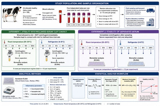
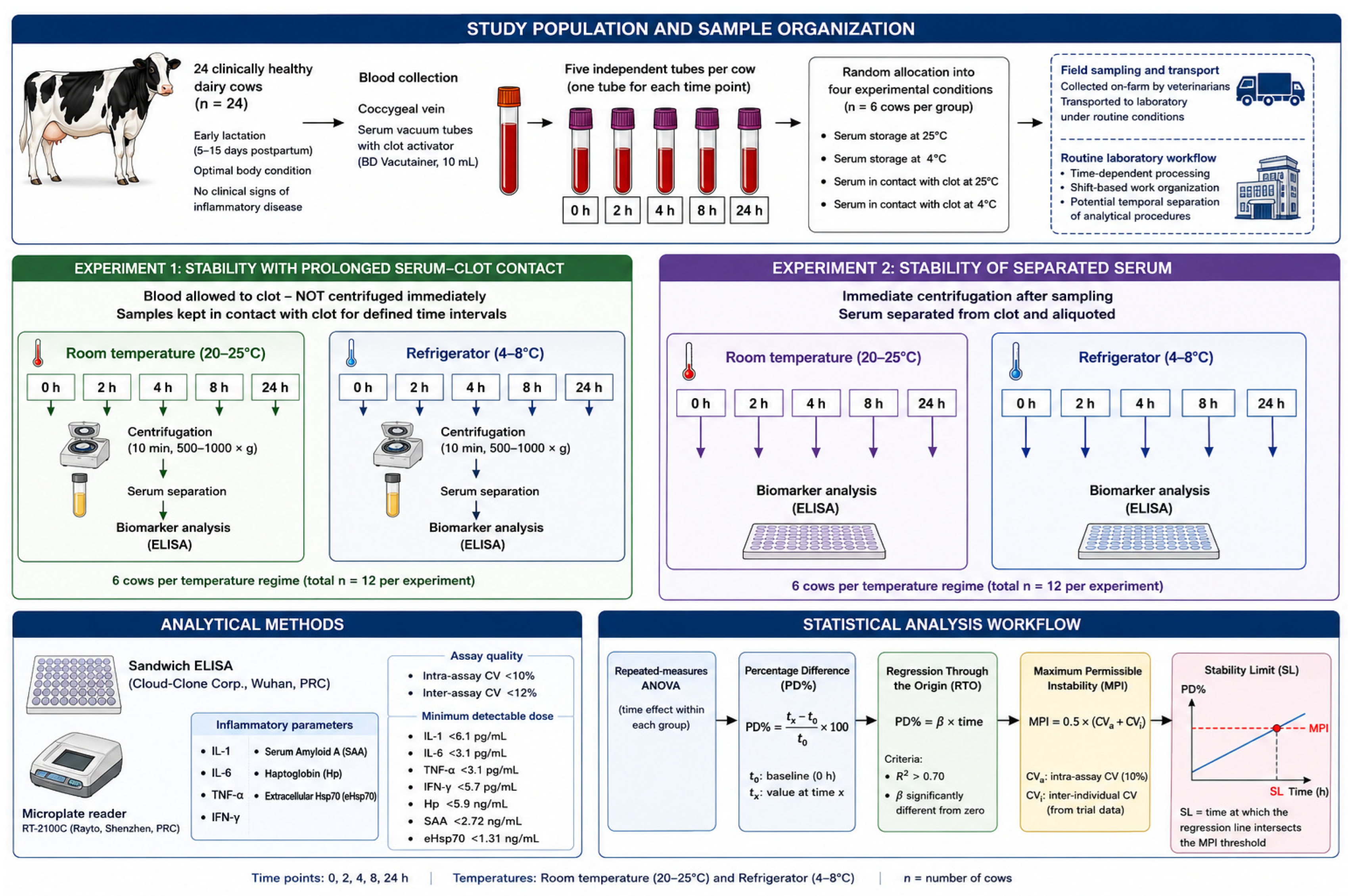
The total number of cows included in the study was 24 (n = 24). The cows were randomly allocated into four experimental conditions (n = 6 per group), corresponding to serum storage at 25 °C, serum storage at 4 °C, serum in contact with the clot at 25 °C, and serum in contact with the clot at 4 °C. All cows were in early lactation (5–15 days postpartum), showed no clinical signs of inflammatory disease, and were in optimal body condition. Five independent primary blood tubes were collected from each cow assigned to each experimental group. One tube was allocated to each predefined storage time (0, 2, 4, 8, and 24 h), so that each tube underwent only one storage condition and one analytical measurement. To avoid pseudoreplication, five independent primary blood tubes were collected from each cow at sampling, with each tube being assigned to a single predefined storage time (0, 2, 4, 8 or 24 h). Blood was collected by venipuncture of the coccygeal vein into 10 mL serum vacuum tubes with clot activator (BD Vacutainer, Franklin Lakes, NJ, USA). The flow diagram of the experiment is shown in Figure 1.
Figure 1.
The flow diagram of the experiment.
According to applicable standards for stability testing, the following should be included: at least three units, at least five sampling time points, and the number of stability conditions and study time periods defined in accordance with locally common procedures for sample collection, processing, transport, and storage [30,31]. In this study, cytokine stability was evaluated at 0, 2, 4, 8, and 24 h to reflect realistic preanalytical variability from field sampling to laboratory analysis. Blood samples were collected on-farm by veterinarians and transported to the laboratory under routine conditions, introducing potential variability in transport time and temperature exposure. Upon arrival, samples were processed in a time-dependent manner, taking into account routine laboratory workflows, including shift-based work organization and potential temporal separation of analytical procedures. In addition, variability arising from delayed serum separation and prolonged clot–serum contact prior to centrifugation was considered, as this may induce ongoing in vitro cellular activity and consequent changes in cytokine concentrations. Together, the selected time points were designed to simulate the full preanalytical chain and to assess the impact of real-world handling conditions on cytokine stability. The standard also states that, to avoid manipulation, the use of distinct primary tubes for each storage duration to be investigated is recommended; therefore, we opted for sampling five specimens from a single animal for each time point separately.
2.2. Stability of Inflammatory Parameters in Serum with Prolonged Contact with Clot
In the first experiment, the influence of the contact time of the serum with the blood clot on the concentration of inflammatory parameters was investigated. After coagulation, the blood samples were not centrifuged immediately but were kept in contact with the clot during defined time intervals: 0, 2, 4, 8 and 24 h. The samples were stored in two temperature regimes in parallel: at room temperature (20–25 °C) and at refrigerator temperature (4–8 °C). After each time interval, the samples were centrifuged (10 min at 500–1000× g), and serum was carefully separated and prepared for further analysis.
2.3. Stability of Inflammatory Parameters in Separated Serum
In the second experiment, the influence of the storage time of already separated serum on the concentration of inflammatory parameters was investigated. After an initial centrifugation immediately after sampling, the serum was separated from the clot and divided into aliquots. Serum samples were then stored for 0, 2, 4, 8 and 24 h, also in two temperature regimes: at room temperature (20–25 °C) and in a refrigerator (4–8 °C). After each time interval, the samples were analyzed.
2.4. Analytical Methods
Concentrations of inflammatory parameters (IL-1, IL-6, TNF-α, IFN-γ, SAA, Hp and eHsp70) were determined by validated immunological methods. Sandwich ELISAs (Cloud Clone Corp., Wuhan, China) were used, according to the manufacturer’s instructions. All samples were analyzed simultaneously and under the same laboratory conditions to minimize analytical variability. A microreader RT-2100C (Rayto, Shenzhen, China) was used for reading. The assays used had intra-assay CV < 10% and inter-assay CV < 12%. The minimum detectable dose is typically <6.1 pg/mL for IL-1, <3.1 pg/mL for IL-6, <3.1 pg/mL for TNF-α, <5.7 pg/mL for IFN-γ, <5.9 ng/mL for Hp, <2.72 ng/mL for SAA and <1.31 ng/mL for eHsp70.
2.5. Statistical Analysis
Statistical analyses were performed in accordance with the recommendations of official protocols for analyte stability assessment [30,31]. Data was analyzed using statistical methods for repeated measurements.
Data were analyzed using one-way repeated-measures ANOVA to evaluate the effect of storage time (0, 2, 4, 8, and 24 h) on the concentration of each inflammatory parameter. Analyses were performed separately for each storage condition (serum–clot contact and separated serum) and for each storage temperature (20–25 °C and 4–8 °C). The repeated-measures ANOVA can be represented by the following model:
where Yij is the measured concentration of the analyte for the j-th cow at the i-th storage time point, τi is the fixed effect of storage time, Sj represents individual cow (subject), and εij is the residual error. Separate models were fitted for each storage condition and temperature. The individual cow was considered the experimental unit, and storage time was treated as the within-subject factor. Since each cow contributed measurements at all storage times within its assigned experimental group, repeated measurements from the same animal were accounted for in the analysis. Dunnett’s multiple comparisons test was used to compare each storage time point with the baseline value (0 h). Results are presented as mean ± SD. Dunnett’s multiple comparisons test was used to compare each storage time point with baseline (0 h).
Results are presented as mean ± standard deviation (SD). For the purposes of these analyses, the mean value of the parameters and the standard deviations are shown.
The loss of analyte stability is defined as the difference between the baseline sample value (t0) and each successive test sample value (tx). In order to compare the difference observed with permissible error specifications and to facilitate comparison between quantities, the relative difference is expressed as percentage (PD%) according to the formula: PD% = (tx − t0)/t0) × 100. baseline sample; t0, average of replicate measurements) and its concentrations when stored for the maximum permissible storage time (test sample; tx average of replicate measurements).
Adjustment of the global instability equation may be performed using least-squares regression analysis. Since the experimental design assumes that the baseline sample exhibits no instability, the instability equation does not include an intercept term. Consequently, the regression model must be constrained to pass through the origin, a procedure commonly referred to as “Regression Through the Origin” (RTO). RTO fitting with a linear model, yields a first-degree equation, with a single coefficient (slope; β) and without intercept term, of the type: PD% = β × time (h). In addition to the regression equations, the coefficient of determination (R2) was calculated, and the regression parameter β was tested for statistical significance. According to the applicable standard, the R2 value must exceed 0.7, and the parameter β must be statistically significantly different from zero for the linear equation to be considered suitable for further evaluation of parameter instability in the sample. If these criteria are not satisfied, higher-order regression models should be applied until an adequate goodness-of-fit is achieved, defined as an R2 value greater than 0.7.
In the next step, the stability limit, which is defined as the time during which the change in the value of the inflammatory parameters does not exceed the maximum permissible instability (MPI), is determined [16]. The MPI was calculated as MPI = 0.5 × (CVa + CVi). Intra-assay variation according to the manufacturer’s prescription was taken for CVa values = 10%, while CVi was calculated based on data from individual cows from the trial.
In the graphical presentation, PD% values were plotted against storage time and linear regression analysis was applied for each analyte. The MPI threshold was included as a horizontal reference line defining the acceptable limit of stability. The results demonstrate that analyte stability is maintained while the regression line remains within the MPI boundary. Once the regression line reaches or exceeds the MPI threshold, a significant loss of stability is indicated. Accordingly, the time point at which the regression line intersects the MPI line is interpreted as the estimated stability limit (SL) for the respective analyte under the tested storage conditions.
3. Results
The initial values of the investigated parameters (0 h) represent the basic value for comparison with the other studied times (2, 4, 8 and 24 h). No statistically significant difference was found in the value of the parameters investigated in different temperature regimes in both experiments (column 0 h in Table 1).
Table 1.
Stability of inflammatory parameters in serum with prolonged clot contact and separated serum during experimental period.
The storage time of samples with nonseparated serum (clot–serum sample) before analysis significantly affects most of the examined markers, except acute phase proteins (Table 1). IL-1 showed a significant increase after 4 h at 4 °C and after 2 h at 25 °C, after which there was a statistically significant decrease in its value at 24 h in both temperature regimes. IL-6 was statistically significantly higher relative to baseline from 4 h onwards. TNF-α and IFN-γ showed a significant increase (serum–clot) or decrease (serum) after 24 h at 4 °C and already from 4 h at 25 °C. Hp and SAA did not show significant changes even after 24 h. eHsp70 showed a significant decrease at 4 °C and a significant increase at 25 °C from 8 h.
The storage time of free serum before analysis has a significant effect on most of the examined inflammatory markers, except for acute phase proteins (Table 1). IL-1 showed pronounced changes with a statistically significant decrease in concentrations after 4 h at 4 °C and 2 h at 25 °C. IL-6 started to decrease significantly from 8 h at 4 °C and 4 h at 25 °C, TNF-α showed a decrease after 24 h at 4 °C and from 8 h at 25 °C. IFN-γ decreased significantly after 24 h at 4 °C and after 8 h at 25 °C. eHsp70 showed a decrease after 8 h in both temperature regimes of serum storage.
Linear regression analysis demonstrated that changes in PD% values over time were dependent on both sample type and storage temperature for all investigated biomarkers (Table 2). The obtained coefficients of determination (R2) indicated good to excellent agreement between the experimental data and the regression models, with most models showing high R2 values (>0.75), confirming a pronounced time-dependent alteration in analyte stability.
Table 2.
Linear regression analysis of PD% changes during sample storage.
For IL-1, a negative trend in PD% values were observed under all storage conditions, indicating a progressive loss of stability over time. The most pronounced decrease was detected in serum samples stored at 25 °C (β = −3.867; p = 0.0033). In serum–clot samples stored at 25 °C, the linear model did not adequately describe the stability profile, and therefore a cubic regression model was applied, yielding an excellent fit (R2 = 0.99). All regression coefficients within the cubic model were statistically significant (p < 0.01).
IL-6 and TNF-α exhibited distinct behavior depending on the sample type. In serum–clot samples, a positive trend in PD% values were observed over time, suggesting an increase in analyte concentration or release during storage. In contrast, serum samples demonstrated a negative trend, indicating progressive loss of stability. All regression models for IL-6 and TNF-α were statistically significant (p < 0.05) and showed high coefficients of determination.
A similar pattern was observed for IFN-γ. Serum–clot samples displayed a positive trend in PD% values, particularly at 25 °C (β = 1.164; p = 0.007), whereas serum samples exhibited a negative trend associated with decreased stability over time. The regression models showed very good agreement with the experimental data (R2 = 0.85–0.97).
Hp and SAA demonstrated predominantly negative regression coefficients under all storage conditions, indicating a gradual decline in stability over time. These changes were more pronounced at 25 °C, especially for SAA in serum samples (β = −0.475; p = 0.0221). SAA exhibited very high coefficients of determination (R2 up to 0.98), confirming a strongly linear pattern of change.
Among all investigated biomarkers, eHsp70 showed the greatest dependence on sample type and storage temperature. Serum samples exhibited a negative trend in PD% values, whereas serum–clot samples stored at 25 °C showed a positive trend (β = 1.370; p = 0.0071). All regression models for eHsp70 were statistically significant (p < 0.05), with good coefficients of determination (R2 = 0.79–0.91).
Overall, the results indicate that storage time significantly affects the stability of all investigated biomarkers, with more pronounced effects observed at higher storage temperatures and depending on sample type. Positive regression coefficients indicate increasing PD% values over time, whereas negative coefficients reflect a progressive loss of analyte stability.
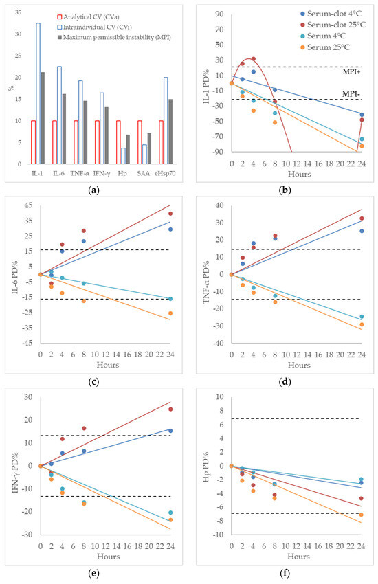
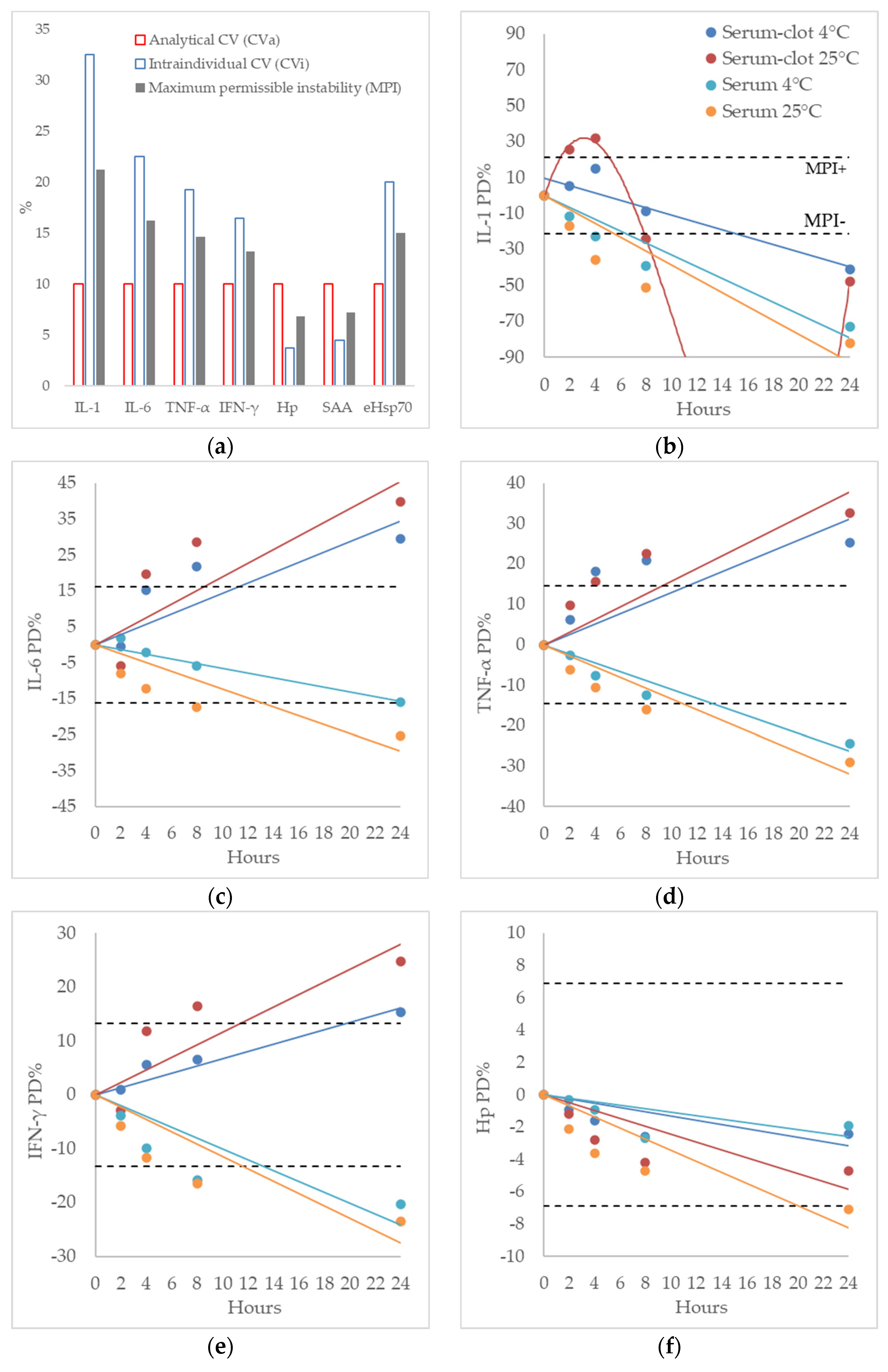
The MPI value was calculated based on analytical variability (CVa) and intraindividual variability (CVi). The CVi values were 32.5% for IL-1, 22.5% for IL-6, 19.25% for TNF-α, 16.5% for IFN-γ, 3.75% for Hp, 4.5% for SAA, and 20% for eHsp70. The arbitrary value based on the manufacturer’s maximal allowable level of variability was set at CVa = 10%. The calculated MPI for IL-1 was (21.25%), IL-6 (16.25%) and eHsp70 (15%) TNF-α (14.625%) and IFN-γ (13.25%), Hp (6.875%) and SAA (7.25%). The values of CVi, CVa and MPI are shown in Figure 2a.
Figure 2.
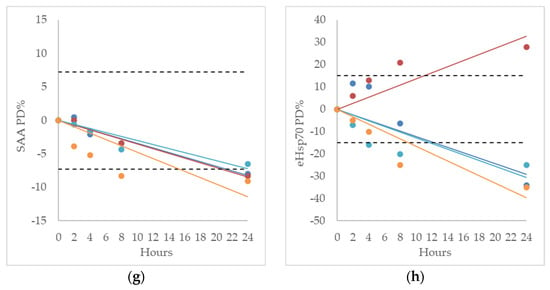
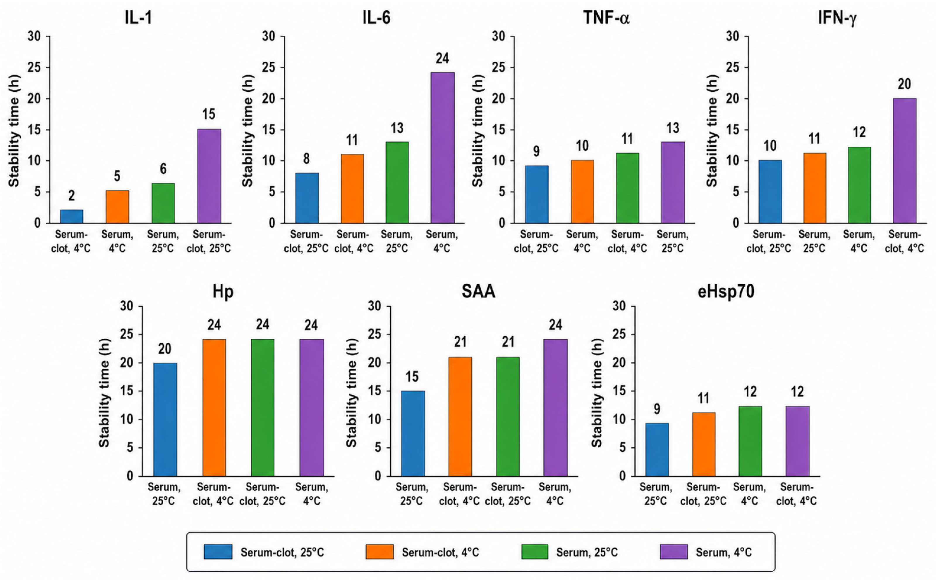
Blood serum inflammatory markers variability intraindividual (CVi) and analytical (CVa) coefficients of variations and maximum permissible instability (MPI) (a) and stability of IL-1 (b), stability of IL-6 (c), stability of TNF-α (d), stability of IFN-γ (e), stability of Haptoglobin-Hp (f), stability of Serum Amyloid A-SAA (g), stability of extracellular heat shock protein 70-eHsp70 (h). MPI plotted as a horizontal dashed line. Maintained stability of the analyte—the regression line remains within the MPI boundary. Loss of stability of the analyte regression line reaches or exceeds the MPI threshold. Time point at which the regression line intersects the MPI line is interpreted as the estimated stability limit.
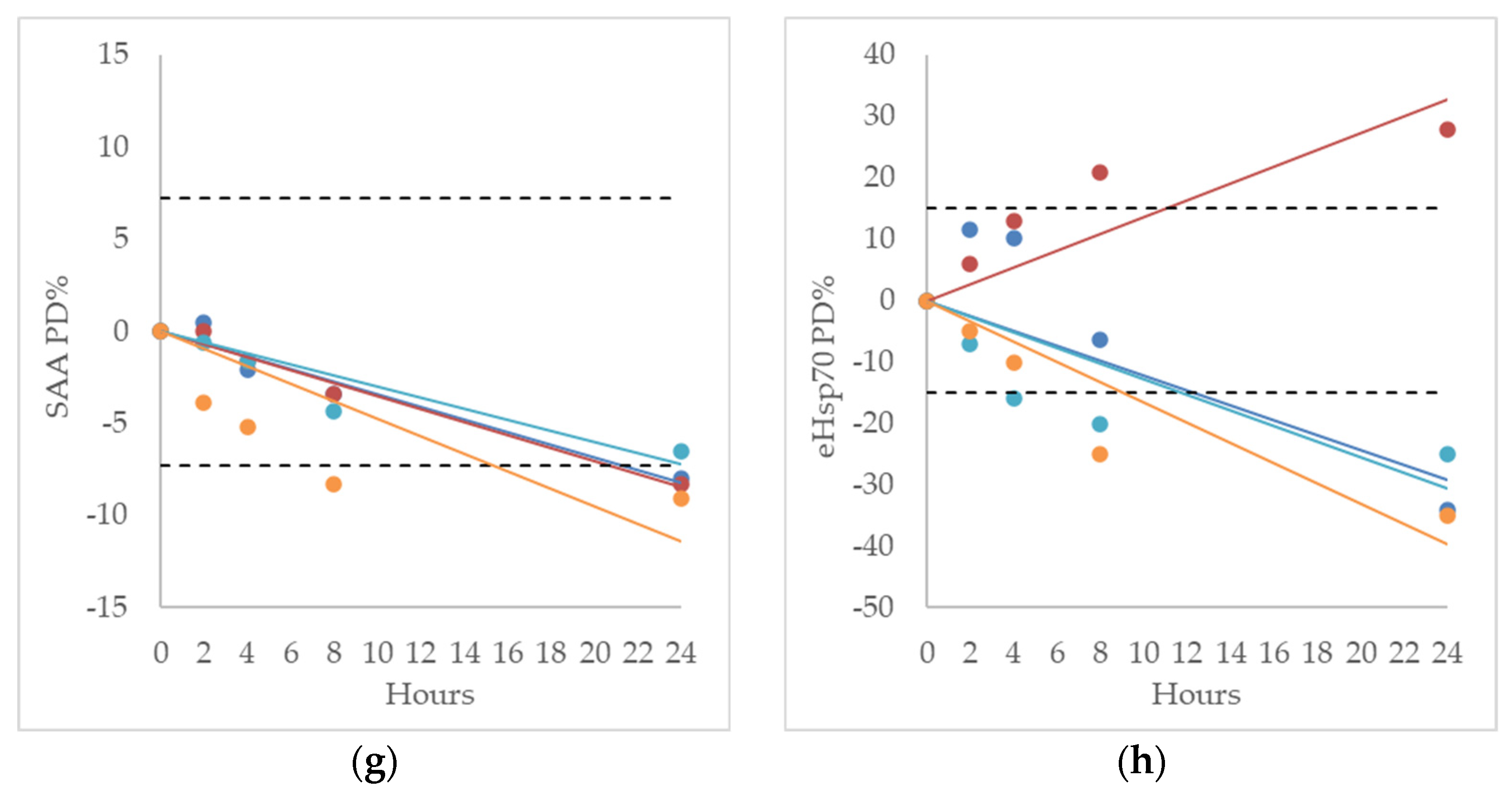
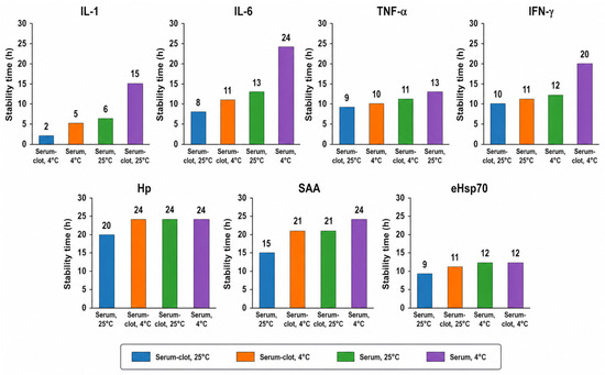
In the graphical presentation, PD% values were plotted against storage time and linear regression analysis was applied for each analyte. The MPI threshold was included as a horizontal reference line defining the acceptable limit of stability. The results demonstrate that analyte stability is maintained while the regression line remains within the MPI boundary. Once the regression line reaches or exceeds the MPI threshold, a significant loss of stability is indicated. Accordingly, the time point at which the regression line intersects the MPI line is interpreted as the estimated stability limit (SL) for the respective analyte under the tested storage conditions. IL-1 was stable for 2 h (serum–clot, 4 °C), 5 h (serum, 4 °C), 6 h (serum, 25 °C), and 15 h (serum–clot, 25 °C) (Figure 2b), whereas IL-6 was stable for 8 h (serum–clot, 25 °C), 11 h (serum–clot, 4 °C), 13 h (serum, 25 °C), and 24 h (serum, 4 °C) (Figure 2c). TNF-α was stable for 9 h (serum–clot, 25 °C), 10 h (serum, 4 °C), 11 h (serum–clot, 4 °C), and 13 h (serum, 25 °C) (Figure 2d), while IFN-γ was stable for 10 h (serum–clot, 25 °C), 11 h (serum, 25 °C), 12 h (serum, 4 °C), and 20 h (serum–clot, 4 °C) (Figure 2e). Hp was stable for 20 h (serum, 25 °C), 24 h (serum–clot, 4 °C), 24 h (serum–clot, 25 °C), and 24 h (serum, 4 °C) (Figure 2f), while SAA was stable for 15 h (serum, 25 °C), 21 h (serum–clot, 4 °C), 21 h (serum–clot, 25 °C), and 24 h (serum, 4 °C) (Figure 2g). Finally, eHsp70 was stable for 9 h (serum, 25 °C), 11 h (serum–clot, 25 °C), 12 h (serum, 4 °C), and 12 h (serum–clot, 4 °C) (Figure 2h). The stability time (Maximum Permissible Instability), obtained from the regression line presented in Figure 2b–h, is summarized in Figure 3.
Figure 3.
The stability time limit (Maximum Permissible Instability), obtained from the regression line (see Figure 2b–h) in the stability test.
The highest stability for most parameters was observed at 4 °C. This storage temperature yielded the maximum values for IL-6 (24 h in serum), IFN-γ (20 h in serum–clot), and SAA (24 h in serum), as well as maximum or equivalent maximum values for Hp (24 h across all conditions) and eHsp70 (12 h). Although exceptions were observed (e.g., IL-1 showing higher stability at 25 °C in serum–clot samples), the overall pattern clearly indicates that storage at 4 °C most frequently prolongs analyte stability.
The comparative analysis of serum–clot versus serum stability demonstrates clear but analyte-specific patterns without a universal advantage of either matrix. In the case of proinflammatory cytokines (IL-1, TNF-α, and to some extent IFN-γ), serum–clot samples frequently exhibited equal or higher stability compared to serum, particularly at 4 °C and, in certain instances, at 25 °C (e.g., IL-1 showed maximal stability under serum–clot conditions). This suggests that interactions with the clot may contribute to the preservation or maintenance of measured analyte levels, likely through binding processes or continuous release during the preanalytical phase. For IL-6 and SAA, however, the serum demonstrated a clear advantage, especially at 4 °C, where maximal stability values (24 h) were observed. This indicates that these proteins are more stable in a “cleaner” serum matrix following complete separation from the clot, where interactions with fibrin and cellular debris are reduced. For IFN-γ and TNF-α, differences were less pronounced and varied depending on temperature, indicating that the matrix effect is secondary to storage conditions. For Hp, stability was essentially equivalent between serum and serum–clot conditions (24 h across most combinations), suggesting a high intrinsic stability of this protein and minimal matrix-dependent influence. For eHsp70, serum and serum–clot samples showed very similar stability profiles, with a slight advantage of serum–clot at 25 °C but no meaningful differences at 4 °C.
4. Discussion
The obtained values of pro-inflammatory cytokines and proteins of the acute phase indicate a stable, but existing and moderate inflammatory status in the examined cows. IL-1β, as one of the primary cytokines of innate immunity, initiates the acute phase and inflammation signaling. Its stability in the measured ranges corresponds to the data that basic concentrations of IL-1β can be present without clinically pronounced disease, while a significant increase occurs only during intense inflammation [32,33,34]. IL-6 and TNF-α act as key triggers of the acute phase, synergistically activating the production of acute phase proteins, such as haptoglobin (Hp) and serum amyloid A (SAA). Their concentrations in this research are within the range of values recorded in healthy or mildly stressed animals, and a significant increase occurs only in clinically pronounced inflammatory conditions [33,35,36]. IFN-γ, associated with cellular immunity and NK and T-cell activation, is also stable in the measured ranges. This indicates the absence of pronounced immune stimulation, in accordance with the data that IFN-γ remains relatively stable in the absence of active infection, while elevations occur in more severe immune challenges [32,33,34]. Acute phase proteins Hp and SAA showed slightly elevated values in relation to reference values for healthy cows, suggesting a subclinical inflammatory response, while SAA remained in the upper physiological range without an indication of acute phase [35,36]. Extracellular Hsp70 (eHsp70), although less investigated in cattle, has been identified in recent studies as a potential stress marker and immunomodulator, as it can activate immune receptors and promote cytokine production through TLR-signaling. In present research, its values correspond to the range described in cows exposed to moderate stressors, such as metabolic or oxidative stress, having a role in cytoprotection and modulation of the immune response [6,37].
The intraindividual variability of the examined pro-inflammatory parameters largely corresponds to the results available in the EFLM Biological Variation Database [38]. Ranges of variability of inflammatory parameters have been determined as seasonal, between day, diurnal variation or variation after LPS challenge [39,40,41], but for now there are no extensive results with which our CVi and MPI values could be directly compared.
Based on the US FDA international guide, the validation of bioanalytical methods includes the following parameters: accuracy and precision, detectability, reproducibility, parallelism, short-term stability and freeze–thaw stability [42]. In this paper, the short-term stability of cytokines, acute phase proteins and extracellular chaperones in cows were investigated. The first results on the detection of circulating cytokines showed that the determination of these parameters is clinically relevant but methodologically burdened by various biological and non-biological preanalytical factors [43]. Knowing the factors mentioned are very important in the process of obtaining standardized and routine tests [44].
Data on IL-1 indicates marked instability during prolonged storage. In a study [11], it was shown that IL-1 levels were significantly reduced after 24 h at 4 °C, while after 30 days they were not detectable in serum samples. A significant decrease in IL-1 after 24 h was also shown in the present trial.
The effect of storage time and temperature on IL-6 concentrations shows significant variability depending on preanalytical conditions. Aziz et al. [12] showed that the concentration of IL-6 increases in a time-dependent pattern, whereby in unseparated heparinized plasma stored at room temperature it increases up to 30% per hour.
Wang et al. [13] noted a tenfold increase in plasma IL-6 after 2 h at room temperature, while this effect was attenuated by storage at 4 °C. In contrast, degradation occurs during longer storage, as confirmed in a study [11], where IL-6 levels decreased significantly after 24 h at 4 °C, and were undetectable after 30 days. Similarly, Thavasu et al. [14] showed a continuous decline in IL-6 over 12 h, particularly pronounced after 24 h at 4 °C, while Friebe and Volk [45] showed that IL-6 could remain stable for up to 8 h in all sample types. In this study, IL-6 was stable for 6 or 8 h in a clot–serum environment, or 24 h if the separated serum was stored at 4 °C.
Aziz et al. [12] found that TNF-α is unstable in untreated serum at room temperature, with an increase of 10% in serum and 26% in heparinized plasma. Yang et al. [15] showed that TNF-α remains relatively stable in the short term, with a slight increase only after 48 h at room temperature, while at 4 °C there are no significant changes. Verberk et al. [16] also confirmed the stability of TNF-α for up to 24 h in EDTA plasma, independent of temperature. However, Thavasu et al. [14] showed that TNF-α concentrations continuously decreased within 12 h. In another study [11], a significant decrease was recorded after 24 h and complete disappearance after 30 days. Also, TNF-α was found to be stable in EDTA plasma for up to 8 h but showed a slight increase in serum and heparinized plasma [45]. If the concentration of TNF-α is observed, it is stable in the first 24 h at refrigerator temperatures, after which it decreases. An increase in TNF-α was also found if serum was not separated from the clot for 4 h or longer. The maximum stability of the sampan based on intraindividual stability is lower, so depending on the temperature regime, the samples must be analyzed for 6–13 h.
IFN-γ showed a similar stability pattern as TNFα, but it is considered more stable than other cytokines in the literature. Thus, it was found that IFN-γ remains stable in untreated serum for up to 24 h, both at room and refrigerator temperatures [12]. These findings are confirmed by Yang et al. [15], who did not observe significant changes during all investigated time points. However, Verberk et al. [16] showed that after an initial stability of up to 3 h, there is a decrease of 16% after 24 h at room temperature. The drop in IFN-γ concentration during longer storage was also confirmed in the study [11]. Decreasing values were confirmed in this experiment with separated serum, both at room and refrigerator temperatures. In this trial, a biphasic response of cytokines in the serum that was not separated from the clot was determined, with an increase first, and then a decrease in cytokines. Valaperti et al. [17] showed that concentrations of IL-6, IFN-γ and TNF-α can be 10–30% higher within 3 to 24 h after sampling. Overall, the literature indicates that cytokines are relatively stable in a short period of time (up to several hours), while during the middle interval, there may be an artifactual increase due to in vitro production, while during longer storage, degradation and drop in concentrations dominate. The increase in TNF-α and IL-6 in untreated serum may be due to the activation of monocytes during blood clotting. A longer wait before sample processing increases the risk of artificial growth of these biomarkers, especially if the tubes contain small amounts of endotoxin [18,19].
Acute phase proteins Hp and SAA showed good stability in the samples. Depending on the type of storage, the maximum sample storage time in which no intraindividual variation will occur is within 16 to 24 h. The results of previous authors confirm the stability of these parameters, even during long-term storage and exposure of samples to repeated freezing–thawing cycles [20,21].
The stability of heat shock proteins in blood samples has not been sufficiently investigated, but available data show that it significantly depends on the type of sample and preanalytical conditions. In a study conducted by Zimmermann et al. [22] in vitro stability of HSP27 in serum and EDTA plasma was investigated. It was shown that HSP27 is stable in serum for at least 4–6 h at 4 °C, without significant changes in concentration, while in EDTA plasma there is a significant decrease (≈25–45%) during the same period. Similar time frames were obtained in this experiment for Hsp70. Also, one freeze–thaw cycle did not significantly affect the concentration in the serum, while in the plasma led to an artificial increase in the concentration (several times), which indicates potential interference or changes in the protein structure. Hsp are known to be exosomes secreted, showing stability in blood samples for 90 days [23], which may explain the bidirectional bias of Hsp70, since its value increased with prolonged clot–serum contact at room temperature.
Serum–clot and serum samples do not exhibit a consistent, universal difference in stability. Serum–clot conditions may favor the stability of certain cytokines (particularly IL-1 and, to some extent, IFN-γ), whereas serum provides better stability for IL-6 and SAA. Overall, the effect of sample type is analyte-specific and less pronounced compared to the dominant influence of storage temperature, with neither matrix demonstrating superiority across all investigated inflammatory parameters. During clot formation, leukocytes and platelets become activated and may release cytokines and other proteins; these molecules can subsequently diffuse from the clot into the liquid phase while the sample remains standing prior to separation [46]. In this way, the concentration of certain analytes in serum may be maintained or even increased, which can be misinterpreted as enhanced stability. This apparent or “false” stability reflects a dynamic equilibrium between analyte release from the clot and their potential degradation, rather than true chemical stability in the classical sense. Term “stability” should be interpreted with caution in the context of serum and serum–clot samples. A constant analyte concentration over time does not necessarily reflect true biochemical stability. In particular, in serum–clot systems, ongoing cellular activity, degranulation, and release of intracellular components may occur after blood collection. Therefore, the observed constancy of some biomarkers may represent a dynamic equilibrium between continued release from blood cells or clot components and concurrent degradation processes, rather than true analytical stability.
The limitation of the study is reflected in the fact that measurement was performed only in serum, although both separated serum and unseparated serum with clot were used. The coagulation process activates the cells and leads to the release of cytokines, so their value in sera is higher compared to plasma, but the values are generally correlated [24]. Since the direct correlation of inflammatory parameter values in plasma and serum is low [25], in future research it is necessary to perform stability tests in bovine plasma as well. A second limitation of the present study is that the experiments were performed exclusively in clinically healthy dairy cows during early lactation (5–15 days postpartum). This physiological stage was intentionally selected because circulating inflammatory biomarkers are more readily detectable during the transition period, allowing reliable evaluation of pre-analytical stability over time. However, the primary objective of this study was methodological rather than biological, namely to assess the effects of storage time, temperature, and sample handling on analyte stability. Therefore, the observed stability characteristics should be interpreted as being established under the tested experimental conditions. Although the pre-analytical mechanisms investigated are expected to be largely independent of physiological status, the present study was not designed to determine whether identical stability limits apply to cattle of different ages, production stages, breeds, or disease conditions. Further studies including additional animal populations are warranted to confirm the broader applicability of these findings.
5. Conclusions
The obtained results showed that the stability of inflammatory parameters in cows during early lactation significantly depends on the preanalytical conditions: the shortest stability was recorded in the serum samples that remained in contact with the clot, while the separated serum showed greater stability, especially at 4 °C. Cytokines are the least stable, followed by Hsp70, especially at higher temperatures and longer storage times, while acute phase proteins are significantly more stable. The obtained results are useful for planning laboratory protocols and determining the maximum time between sample separation and analysis, minimizing preanalytical errors and optimizing sample transport from the farm to the laboratory.
Author Contributions
Conceptualization, M.C. and N.M.; methodology, M.C., M.M.; software, J.S.; validation, N.M., M.C. and N.P.; formal analysis, M.M.; investigation, M.C., J.S.; resources, N.M.; data curation, M.C.; writing—original draft preparation, M.C.; writing—review and editing, N.M., J.S., N.P.; visualization, M.C., M.M.; supervision, N.P., J.S. All authors have read and agreed to the published version of the manuscript.
Funding
This research was funded by Provincial Secretariat for Higher Education and Scientific Research Activities (grant number 003785865 2025 09418 003 000 000 001) and Ministry of science of the Republic of Serbia (grant number 451-03-34/2026-03/200117).
Institutional Review Board Statement
The animal study protocol was approved by the Ethical Committee of the University of Novi Sad (04-77/37, 1 February 2025).
Informed Consent Statement
Not applicable.
Data Availability Statement
The original contributions presented in this study are included in the article. Further inquiries can be directed to the corresponding author.
Conflicts of Interest
The authors declare no conflicts of interest.
Abbreviations
The following abbreviations are used in this manuscript:
| IL-1 | Interleukin 1 |
| IL-6 | Interleukin 6 |
| TNF-α | Tumor Necrosis Factor Alpha |
| IFN-γ | Interferon gamma |
| Hp | Haptoglobin |
| SAA | Serum Amyloid A |
| eHsp70 | Extracellular Heat Shock Protein 70 |
References
- Gauldie, J.; Baumann, H. Cytokines and acute phase protein expression. In Cytokines and Inflammation; CRC Press: Boca Raton, FL, USA, 2024; pp. 275–305. [Google Scholar]
- Yaghoobpour, T.; Sheikhi, Z.; Nazifi, S. The impact of stress in domestic animals: Roles of heat shock proteins and acute-phase proteins. Vet. Res. Commun. 2025, 49, 258. [Google Scholar] [CrossRef] [Scilit] [PubMed]
- Guan, R.W.; Wang, D.M.; Wang, B.B.; Jiang, L.Y.; Liu, J.X. Prognostic potential of pre-partum blood biochemical and immune variables for postpartum mastitis risk in dairy cows. BMC Vet. Res. 2020, 16, 136. [Google Scholar] [CrossRef] [Scilit] [PubMed]
- Rodriguez-Jimenez, S.; Haerr, K.J.; Trevisi, E.; Loor, J.J.; Cardoso, F.C.; Osorio, J.S. Prepartal standing behavior as a parameter for early detection of postpartal subclinical ketosis associated with inflammation and liver function biomarkers in peripartal dairy cows. J. Dairy Sci. 2018, 101, 8224–8235. [Google Scholar] [CrossRef] [Scilit] [PubMed]
- Vincent, F.B.; Nim, H.T.; Lee, J.P.; Morand, E.F.; Harris, J. Effect of storage duration on cytokine stability in human serum and plasma. Cytokine 2019, 113, 453–457. [Google Scholar] [CrossRef] [Scilit] [PubMed]
- Keustermans, G.C.; Hoeks, S.B.; Meerding, J.M.; Prakken, B.J.; de Jager, W. Cytokine assays: An assessment of the preparation and treatment of blood and tissue samples. Methods 2013, 61, 10–17. [Google Scholar] [CrossRef] [Scilit] [PubMed]
- Toy, S.; Black, S.R.; Pastor, A.R.; Whiteside, D.P. Establishing Reference Intervals for Serum Inflammatory Markers and Investigating Their Storage Stability and Clinical Utility in Asian Wild Horses (Equus Ferus Przewalskii) Under Managed Care. J. Zoo Wildl. Med. 2025, 56, 54–61. [Google Scholar] [CrossRef] [Scilit] [PubMed]
- Liu, C.; Chu, D.; Kalantar-Zadeh, K.; George, J.; Young, H.A.; Liu, G. Cytokines: From Clinical Significance to Quantification. Adv. Sci. 2021, 8, e2004433. [Google Scholar] [CrossRef] [Scilit] [PubMed]
- Speelman, T.; Dale, L.; Louw, A.; Verhoog, N.J.D. The Association of Acute Phase Proteins in Stress and Inflammation-Induced T2D. Cells 2022, 11, 2163. [Google Scholar] [CrossRef] [Scilit] [PubMed]
- Guisasola, M.C.; Alonso, B.; Bravo, B.; Vaquero, J.; Chana, F. An overview of cytokines and heat shock response in polytraumatized patients. Cell Stress Chaperones 2018, 23, 483–489. [Google Scholar] [CrossRef] [Scilit] [PubMed]
- Kordulewska, N.K.; Topa, J.; Tańska, M.; Cieślińska, A.; Fiedorowicz, E.; Jarmołowska, B. Stability of interleukin-1β,-4,-6,-8,-10,-13, interferon-γ and tumor necrosis factor-α in human sera after repetitive freeze-thaw cycles and long storage. J. Pharm. Biomed. Anal. 2021, 196, 113900. [Google Scholar] [CrossRef] [Scilit] [PubMed]
- Aziz, N.; Detels, R.; Quint, J.J.; Li, Q.; Gjertson, D.; Butch, A.W. Stability of cytokines, chemokines and soluble activation markers in unprocessed blood stored under different conditions. Cytokine 2016, 84, 17–24. [Google Scholar] [CrossRef] [Scilit] [PubMed]
- Wang, Y.; Wang, B.; She, L.; Wang, J.; Chen, Y.; Huang, D.; Han, G.; Lu, M.; Wu, X.; Wan, Z.; et al. Effects of sample handling on the stability of interleukin-6 in patients with breast neoplasms. J. Lab. Med. 2022, 46, 115–119. [Google Scholar] [CrossRef] [Scilit]
- Thavasu, P.W.; Longhurst, S.; Joel, S.P.; Slevin, M.L.; Balkwill, F.R. Measuring cytokine levels in blood. Importance of anticoagulants, processing, and storage conditions. J. Immunol. Methods 1992, 153, 115–124. [Google Scholar] [CrossRef] [Scilit] [PubMed]
- Yang, X.; Arceo, T.; Fischer, S.K. Effect of delayed blood centrifugation on cytokine quantitation in serum and plasma. Bioanalysis 2025, 17, 913–921. [Google Scholar] [CrossRef] [Scilit] [PubMed]
- Verberk, I.M.W.; Nossent, E.J.; Bontkes, H.J.; Teunissen, C.E. Pre-analytical sample handling effects on blood cytokine levels: Quality control of a COVID-19 biobank. Biomark. Med. 2021, 15, 987–997. [Google Scholar] [CrossRef] [Scilit] [PubMed]
- Valaperti, A.; Bezel, P.; Vonow-Eisenring, M.; Franzen, D.; Steiner, U.C. Variability of cytokine concentration in whole blood serum and bronchoalveolar lavage over time. Cytokine 2019, 123, 154768. [Google Scholar] [CrossRef] [Scilit] [PubMed]
- Riches, P.; Gooding, R.; Millar, B.C.; Rowbottom, A.W. Influence of collection and separation of blood samples on plasma IL-1, IL-6, and TNF-α concentration. J. Immunol. Methods 1992, 153, 125–131. [Google Scholar] [CrossRef] [Scilit] [PubMed]
- Aziz, N.; Irwin, M.R.; Dickerson, S.S.; Butch, A.W. Spurious tumor necrosis factor alpha and interleukin-6 production by human monocytes from blood collected in endotoxin-contaminated vacutainer blood collection tubes. Clin. Chem. 2004, 50, 2215–2216. [Google Scholar] [CrossRef] [Scilit] [PubMed]
- Martins, P.G.M.A.; Moriel, P.; Arthington, J.D. Effects of storage temperature and repeated freeze–thaw cycles on stability of bovine plasma concentrations of haptoglobin and ceruloplasmin. J. Vet. Diagn. Investig. 2017, 29, 738–740. [Google Scholar] [CrossRef] [Scilit] [PubMed]
- Rhim, H.; Kwag, C.; Han, J.I. A preliminary study on effects of storage time and repeated freezing-thawing on the stability of avian serum amyloid A. Acta Vet. Scand. 2024, 66, 42. [Google Scholar] [CrossRef] [Scilit] [PubMed]
- Zimmermann, M.; Traxler, D.; Simader, E.; Bekos, C.; Dieplinger, B.; Lainscak, M.; Ankersmit, H.J.; Mueller, T. In vitro Stability of Heat Shock Protein 27 in Serum and Plasma Under Different Pre-analytical Conditions: Implications for Large-Scale Clinical Studies. Ann. Lab. Med. 2016, 36, 353–357. [Google Scholar] [CrossRef] [Scilit] [PubMed]
- Kalra, H.; Adda, C.G.; Liem, M.; Ang, C.-S.; Mechler, A.; Simpson, R.J.; Hulett, M.D.; Mathivanan, S. Comparative proteomics evaluation of plasma exosome isolation techniques and assessment of the stability of exosomes in normal human blood plasma. Proteomics 2013, 13, 3354–3364. [Google Scholar] [CrossRef] [Scilit] [PubMed]
- Rosenberg-Hasson, Y.; Hansmann, L.; Liedtke, M.; Herschmann, I.; Maecker, H.T. Effects of serum and plasma matrices on multiplex immunoassays. Immunol. Res. 2014, 58, 224–233. [Google Scholar] [CrossRef] [Scilit] [PubMed]
- Feng, X.; Zou, W.; Li, P.; Guo, K.; Ma, Y.; Hu, G.; Kang, J.; Yu, X.; Peng, M. Comparability evaluation of serum and plasma cytokine levels by multiplex bead-based flow cytometry. Clin. Chim. Acta 2025, 575, 120351. [Google Scholar] [CrossRef] [Scilit] [PubMed]
- Fritzen, A.; da Silva, A.S. Inflammation in the Transition Period of Dairy Cows: New Paradigms. Vet. Sci. 2025, 12, 1182. [Google Scholar] [CrossRef] [Scilit] [PubMed]
- Lassallette, E.; Baysse, A.P.; Gausseres, B.; Foucras, G.; Guerre, P. Targeted analysis of sphingolipids and cytokines in plasma of dairy cows after calving reveals distinct impacts of systemic inflammation, ketosis, and mastitis. J. Anim. Sci. Biotechnol. 2026, 17, 6. [Google Scholar] [CrossRef] [Scilit] [PubMed]
- Cincović, M.; Stojanović, D.; Djoković, R.; Majkić, M.; Starič, J.; Petrović, M.; Kovačević, Z. Relation Between Inflammatory Parameters and Insulin Resistance Indices in Cows During Early Lactation. Metabolites 2025, 15, 751. [Google Scholar] [CrossRef] [Scilit] [PubMed]
- Petrović, M.Ž.; Cincović, M.; Starič, J.; Djoković, R.; Belić, B.; Radinović, M.; Majkić, M.; Ilić, Z.Ž. The Correlation between Extracellular Heat Shock Protein 70 and Lipid Metabolism in a Ruminant Model. Metabolites 2022, 12, 19. [Google Scholar] [CrossRef] [Scilit] [PubMed]
- Gomez-Rioja, R.; Von Meyer, A.; Cornes, M.; Costelloe, S.; Vermeersch, P.; Simundic, A.M.; Nybo, M.; Baird, G.S.; Kristensen, G.B.B.; Cadamuro, J.; et al. Recommendation for the design of stability studies on clinical specimens. Clin. Chem. Lab. Med. 2023, 61, 1708–1718. [Google Scholar] [CrossRef] [Scilit] [PubMed]
- ISO 33405:2024; Reference Materials—Approaches for Characterization and Assessment of Homogeneity and Stability. ISO: Geneva, Switzerland, 2024.
- Lesta, A.; Marín-García, P.J.; Llobat, L. A Comprehensive Review of the Bovine Immune Response to Pathogens. Int. J. Mol. Sci. 2025, 26, 8461. [Google Scholar] [CrossRef] [Scilit] [PubMed]
- Chitko-McKown, C.G.; Engle, B.N.; Bennett, G.L.; Kuehn, L.A.; DeDonder, K.D.; Apley, M.D.; Harhay, G.P.; Clawson, M.L.; White, B.J.; Larson, R.L.; et al. Temporal profiling of haptoglobin and cytokines in feedlot cattle with bovine respiratory disease. Front. Vet. Sci. 2026, 12, 1713337. [Google Scholar] [CrossRef] [Scilit] [PubMed]
- Bochniarz, M.; Hahaj-Siembida, A.; Krajewska-Wędzina, M.; Osińska, M.; Tracz, A.; Trościańczyk, A.; Brodzki, P.; Krakowski, L.; Kosior-Korzecka, U.; Nowakiewicz, A. Cytokine inflammatory response in dairy cows with mastitis caused by Streptococcus agalactiae. J. Vet. Res. 2024, 68, 115–121. [Google Scholar] [CrossRef] [Scilit] [PubMed]
- Saco, Y.; Bassols, A. Acute phase proteins in cattle and swine: A review. Vet. Clin. Pathol. 2023, 52, 50–63. [Google Scholar] [CrossRef] [Scilit] [PubMed]
- Trefz, F.M.; Balmer, M.; Peters, L.M.; Bruckmaier, R.M.; Meylan, M. Association of results of the glutaraldehyde coagulation test with plasma acute phase protein concentrations and hematologic findings in hospitalized cows. Front. Vet. Sci. 2024, 11, 1404809. [Google Scholar] [CrossRef] [Scilit] [PubMed]
- Rakib, M.R.H.; Messina, V.; Gargiulo, J.I.; Lyons, N.A.; Garcia, S.C. Graduate Student Literature Review: Potential use of HSP70 as an indicator of heat stress in dairy cows—A review. J. Dairy Sci. 2024, 107, 11597–11610. [Google Scholar] [CrossRef] [Scilit] [PubMed]
- EFLM Biological Variation Database. Available online: https://biologicalvariation.eu/ (accessed on 6 April 2026).
- Grewal, S.; Aggarwal, A.; Alhussien, M.N. Seasonal alterations in the expression of inflammatory cytokines and cortisol concentrations in periparturient Sahiwal cows. Biol. Rhythm Res. 2021, 52, 1229–1239. [Google Scholar] [CrossRef] [Scilit]
- Rose, G.L.; Farley, M.J.; Flemming, N.B.; Skinner, T.L.; Schaumberg, M.A. Between-day reliability of cytokines and adipokines for application in research and practice. Front. Physiol. 2022, 13, 967169. [Google Scholar] [CrossRef] [Scilit] [PubMed]
- Jorda, A.; Eberl, S.; Nussbaumer-Pröll, A.; Sarhan, M.; Weber, M.; Tegrovsky, L.E.; Wahrmann, M.; al Jalali, V.; Bergmann, F.; Pracher, L.; et al. Reproducibility of LPS-Induced ex vivo cytokine response of healthy volunteers using a whole blood assay. J. Inflamm. Res. 2024, 17, 4781–4790. [Google Scholar] [CrossRef] [Scilit] [PubMed]
- US FDA. Bioanalytical Method Validation Guidance for Industry; Center for Drug Evaluation and Research: Silver Spring, MD, USA; Center for Veterinary Medicine: Rockville, MD, USA, 2018. [Google Scholar]
- Cannon, J.G.; Nrrad, J.L.; Poutsiaka, D.D.; Dinarello, C.D. Measuring circulating cytokines. J. Appl. Physiol. 1993, 75, 1897–1902. [Google Scholar] [CrossRef] [Scilit] [PubMed]
- Mire-Sluis, A.R. Cytokines-protein structure and biological activity: A complex relationship with implication for biological assays and standardization. Biologicals 1993, 21, 131–144. [Google Scholar] [CrossRef] [Scilit] [PubMed]
- Friebe, A.; Volk, H.D. Stability of tumor necrosis factor α, interleukin 6, and interleukin 8 in blood samples of patients with systemic immune activation. Arch. Pathol. Lab. Med. 2008, 132, 1802–1806. [Google Scholar] [CrossRef] [Scilit] [PubMed]
- Zhou, X.; Fragala, M.S.; McElhaney, J.E.; Kuchel, G.A. Conceptual and methodological issues relevant to cytokine and inflammatory marker measurements in clinical research. Curr. Opin. Clin. Nutr. Metab. Care 2010, 13, 541–547. [Google Scholar] [CrossRef] [Scilit] [PubMed]

Disclaimer/Publisher’s Note: The statements, opinions and data contained in all publications are solely those of the individual author(s) and contributor(s) and not of MDPI and/or the editor(s). MDPI and/or the editor(s) disclaim responsibility for any injury to people or property resulting from any ideas, methods, instructions or products referred to in the content. |
© 2026 by the authors. Licensee MDPI, Basel, Switzerland. This article is an open access article distributed under the terms and conditions of the Creative Commons Attribution (CC BY) license.