1. Introduction
Control of offspring sex is of substantial economic importance in cattle production systems, particularly in dairy herds where female calves are preferred for herd replacement and milk yield optimization [
1,
2]. Consequently, reliable pre-implantation sex determination has become an integral component of assisted reproductive technologies and embryo transfer programs, supporting genetic gain and production efficiency.
In mammals, male sex determination is initiated by Y-linked genetic factors, most notably the sex-determining region Y (SRY) gene, which triggers testis differentiation and male phenotypic development [
3,
4,
5]. Because Y-chromosome–specific sequences are absent from females, PCR-based detection of Y-linked markers has been widely applied for embryonic sex determination in cattle [
6,
7,
8,
9,
10]. Historically, SRY has been one of the most used targets, but other male-specific Y-linked markers have also been used for bovine embryo sexing.
Multiplex assays incorporating autosomal internal controls are commonly used to increase analytical reliability and reduce false-negative classification. Under optimized laboratory conditions, reported diagnostic accuracy is high. However, PCR performance is inherently dependent on DNA quantity and quality and is particularly sensitive to template limitation when only a small number of embryonic cells are available [
11]. Insufficient template DNA may result in low signal intensity and increased likelihood of allele drop-out and stochastic amplification effects, especially in low-input molecular workflows [
12]. Embryo sexing frequently relies on invasive sampling procedures, including blastomere biopsy at cleavage stage and blastocyst-stage trophectoderm biopsy, which reduces cellular input and may introduce variability in effective DNA template concentration [
11,
12].
Recent studies in preimplantation genetic testing have highlighted that blastocyst-stage trophectoderm biopsy sampling may not fully represent the genetic status of the entire embryo, as embryonic mosaicism and compartmental heterogeneity can lead to discordance between sampled and whole-embryo genotypes [
13,
14]. Low-template amplification is known to increase stochastic effects such as allelic drop-out and preferential locus amplification [
15,
16,
17], potentially altering detection reliability. While these phenomena are well described in genotyping contexts, their implications for PCR-based embryo sex determination within low-input, multiplex workflows remain underexplored. In addition, recent examples of sexing approaches demonstrate ongoing methodological evolution, including multiplexed marker panels and sequencing-based inference under constrained DNA input [
18]. Failure to distinguish biological sex ratio variation from sampling-induced technical bias may confound interpretation of embryo sexing outcomes, especially when evaluating the efficiency of X-sorted semen in applied breeding programs [
19,
20]. Recent rapid alternatives, including visual isothermal approaches, also highlight the continuing effort to shorten assay turnaround time and simplify embryo-sexing workflows under limited-input conditions [
21].
In the present assay, CDK1 was used as an autosomal internal amplification control rather than as a sex-specific marker. Successful CDK1 amplification therefore indicated the presence of amplifiable embryonic DNA and was used to define whether a reaction was technically evaluable, whereas MSY amplification was used for apparent Y-marker-based classification.
The aim of the present study was to characterize technical assay-output behavior in a low-input multiplex MSY/CDK1 PCR workflow across bovine embryo-derived sample categories under routine laboratory conditions. Specifically, we examined whether whole embryos, cleavage-stage blastomere biopsies, and blastocyst-stage trophectoderm biopsies differed in assay evaluability, defined by successful CDK1 amplification, and in assay-derived apparent MSY-based classification among technically evaluable samples. We hypothesized that, under low-input conditions, sample category may influence apparent Y-marker detection through differences in effective template input and amplification stability, thereby contributing to variation in both assay evaluability and apparent MSY detection across workflows. Because the sample categories were not assigned prospectively and were imbalanced with respect to semen type, sire, developmental stage, and PCR-date structure, the results were interpreted as exploratory assay-output observations rather than causal estimates of independent sample-category effects.
2. Materials and Methods
2.1. Origin of Embryos and In Vitro Production
Ovaries of Bos taurus were obtained from a regional commercial abattoir following routine slaughter. No experimental procedures were performed on live animals. The study protocol was reviewed and approved by the Institutional Animal Welfare Body of the Hungarian University of Agriculture and Life Sciences, Szent István Campus (MATE SZIC MÁB/1113-1/2024; approval date: 10 July 2024). According to the official certification, the study fell outside the scope of Directive 2010/63/EU and did not require authorization as an animal experiment. Individual donor-animal data (age, parity, reproductive history, and reproductive status) were not available because of the abattoir-based sourcing. Ovaries were collected immediately post mortem and transported to the laboratory within 1 h in phosphate-buffered saline (PBS) at room temperature (approximately 20–23 °C).
Cumulus–oocyte complexes (COCs) were aspirated from 2 to 8 mm follicles using standard follicular aspiration procedures [
22]. Selected COCs were subjected to in vitro maturation (IVM), fertilization (IVF), and embryo culture (IVC) using commercially available bovine embryo culture media (IVF Bioscience Ltd., Falmouth, Cornwall, UK) according to the manufacturer’s instructions and standardized laboratory procedures [
23]. For in vitro maturation, COCs were incubated at 38.8 °C in a humidified atmosphere containing 5.5–6.5% CO
2 for 21–24 h.
Fertilization was performed using frozen–thawed semen from two commercially available Holstein sires. Conventional semen was obtained from sire Reform, whereas X-sorted semen, flow-cytometrically enriched for X-bearing spermatozoa, was obtained from sire ICE Cube-S. Accordingly, semen type and sire were completely confounded in the present dataset and could not be disentangled analytically. Prior to fertilization, semen samples were processed by centrifugation (5 min, 328× g). No swim-up, float-up, or flotation-based sperm-selection procedure was applied. Sperm concentration was determined using a Makler counting chamber (Sefi Medical Instruments Ltd., Haifa, Israel) according to routine laboratory practice. Sperm motility was assessed subjectively by visual examination of sperm movement in the Makler chamber as part of routine semen evaluation before fertilization; however, computer-assisted sperm analysis was not performed, and individual motility values were not recorded quantitatively. The final sperm concentration used for fertilization was 2 × 10⁶ spermatozoa/mL.
COCs were not denuded prior to fertilization; fertilization was performed using cumulus-enclosed oocytes after in vitro maturation. Gametes were co-incubated for 16–19 h at 38.8 °C in a humidified atmosphere containing 5.5–6.5% CO2. Following gamete co-incubation, presumptive zygotes were denuded by vortexing and cultured in BO-IVC medium under sterile mineral oil overlay at 38.8 °C in a humidified atmosphere containing 5.5–6.5% O2, 5.5–6.5% CO2, and balanced N2.
Embryos evaluated at the blastocyst stage were morphologically assessed according to the guidelines of the International Embryo Technology Society (IETS), and only embryos classified as grade 1 or 2 were included in the whole-embryo and blastocyst-stage-biopsy categories [
24]. Because blastomere-biopsy samples were collected earlier, from cleavage-stage embryos, they were not subject to blastocyst-stage grading at the time of sampling.
Embryo sampling was performed across three embryo-derived sample categories within routine laboratory workflows. Whole-embryo samples consisted of intact embryos processed directly for molecular analysis and therefore represented the highest template-input category. In the blastomere-biopsy group, approximately two blastomeres were removed from cleavage-stage embryos (16–32-cell stage) for genetic testing. In the blastocyst-stage-biopsy group, approximately 5–6 trophectoderm cells were removed from day-7 blastocysts by laser-assisted micromanipulation under inverted microscopy, with care taken to minimize disturbance of the inner cell mass [
11]. The inner cell mass was not isolated in the present workflow; blastocyst-stage sampling was limited to trophectoderm biopsy. Biopsies were performed using an IX73 inverted microscope (Olympus, Tokyo, Japan) equipped with an Olympus U-HGLGPS illumination unit (Olympus, Tokyo, Japan), an XYRCOS DTS laser system (Hamilton Thorne, Beverly, MA, USA), Narishige micromanipulation equipment, including an MMO-4 three-axis hanging joystick oil hydraulic micromanipulator, an MM-94 motor-drive manipulator, and an IM-11-2 pneumatic microinjector (Narishige Scientific Instrument Lab., Tokyo, Japan). Commercially available biopsy and holding micropipettes were used for embryo manipulation, including a biopsy pipette (MBB-FP-L-30; CooperSurgical, Trumbull, CT, USA) and holding pipettes (MPH-MED-35 and MPH-XLG-30; CooperSurgical, Trumbull, CT, USA).
Representative microscopic images of the cleavage-stage blastomere biopsy and blastocyst-stage trophectoderm biopsy procedures are provided in
Supplementary Figure S1.
Because exact cell counts were not recorded for each sample, individual DNA input could not be quantified, and variation in sampled cell number may have contributed to heterogeneity in effective template input across biopsy-derived samples. For interpretation, sample type was therefore treated as an approximate ordinal input category rather than as an exact cell-number variable: whole embryo >> blastocyst-stage biopsy (approximately 5–6 trophectoderm cells) > blastomere biopsy (approximately two blastomeres). This ordinal weighting was used only to guide biological and technical interpretation and was not entered as a quantitative covariate in statistical models.
The biopsy collection/manipulation time was not prospectively recorded at the individual-sample level; all manipulations were performed according to routine laboratory practice and kept as short as technically feasible. Biopsied cells were collected in molecular biology-grade water and subsequently transferred to lysis buffer for molecular analysis. A schematic overview of the experimental workflow is provided in
Supplementary Figure S2.
The three sample categories arose within routine laboratory work and were not assigned by prospective experimental allocation. Rather, the different approaches were applied to different embryos according to practical and method-development considerations. Accordingly, the study should be regarded as observational and exploratory rather than as a controlled comparison of prospectively assigned sampling strategies.
Sample groups also arose partly across different routine phases over time. Within a given PCR run, however, all samples analyzed on the same day originated from the same laboratory program/workflow. A total of 141 embryo-derived samples were included in the study. The subgroup numbers in
Table 1 reflect the available routine laboratory material and practical workflow allocation rather than a prospectively balanced sampling design. Consequently, the distribution across sample category, semen type, and amplification outcome should not be interpreted as a planned factorial design or as reflecting predefined biological proportions. Samples were categorized according to amplification outcome as evaluable (CDK1 amplification present, regardless of MSY status), technical failure (complete absence of visible amplification bands), or control-missing, defined as detection of an MSY-sized band in the absence of the CDK1 internal control, precluding valid assay-based classification. Their distribution by sample category, semen type, and amplification outcome is presented in
Table 1.
2.2. Genomic DNA Extraction and Embryonic Sex Determination
Genomic DNA-containing lysates were prepared from individual embryo-derived samples using a QuickExtract™ DNA Extraction Solution-based low-input lysis protocol adapted from Nix et al. (2023) [
18]. Each embryo-derived sample was incubated in 5 µL QuickExtract™ DNA Extraction Solution (Lucigen, Middleton, WI, USA) at 65 °C for 15 min, followed by 98 °C for 2 min to ensure complete cell lysis. The resulting lysate was used directly as a PCR template without further purification. Direct quantification of DNA yield was not performed because lysates were used directly for downstream PCR.
Assay-derived apparent MSY-based classification was based on multiplex PCR detection of a Y-chromosome marker located within the male-specific region of the Y chromosome (MSY), together with an autosomal internal amplification control targeting cyclin-dependent kinase 1 (CDK1, chromosome 28). The primer sequences were adopted from Nix et al. (2023), who designed them based on the complete bovine Y-chromosome sequence [
18]. The MSY fragment (279 bp) was amplified using primers MSY_F (5′-AGGGTGAAGCAAATGGTCGT-3′) and MSY_R (5′-GGAGCAACAGTGTCCTGTGT-3′), whereas the autosomal CDK1 control fragment (590 bp) was amplified using primers CDK1_F (5′-GCCCAGACCCAGCATCATT-3′) and CDK1_R (5′-GGGAGTGCCCAAAGCTCTAAA-3′).
Although the primer sequences were adopted from Nix et al. [
18], the two-target MSY/CDK1 multiplex PCR setup, including reagent concentrations and cycling conditions, was optimized in-house for low-input embryo-derived lysates. PCR reactions were performed in a final volume of 20 µL and contained 2 µL template DNA, 4 µL 5× Phusion HF Buffer, 0.25 µL Phusion Hot Start II DNA Polymerase, 0.4 µL dNTP mix (10 mM each), 0.6 µL DMSO, 0.5 µL of each primer (10 µM; final concentration 0.25 µM per primer), and nuclease-free water to volume. Amplification was performed in a LifeEco TC-96 thermal cycler (Bioer Technology Co., Hangzhou, China) under the following in-house optimized conditions: initial denaturation at 98 °C for 5 min; 35 cycles of 98 °C for 10 s, 61.8 °C for 30 s, and 72 °C for 30 s; followed by a final extension at 72 °C for 5 min. The technical suitability of the optimized multiplex assay was assessed using male genomic DNA, female genomic DNA, and no-template controls in each PCR run.
PCR products were resolved on 1.5% agarose gels in 1× TBE buffer and visualized under UV illumination. Fragment sizes were verified using a GeneRuler 100 bp Plus DNA Ladder (Thermo Fisher Scientific, Waltham, MA, USA). A representative agarose gel image showing the expected MSY and CDK1 amplification products, together with the male genomic DNA control, female genomic DNA control, and no-template control, is provided in
Supplementary Figure S3. The same female genomic DNA control, male genomic DNA control, and no-template negative control were included in each PCR run. For genomic DNA controls, 2 µL DNA at a concentration of 30 ng/µL was used per reaction. The female control was expected to yield the CDK1 control band only, whereas the male control was expected to yield both the CDK1 and MSY bands. No amplification was observed in the no-template negative controls. Gel bands were scored visually according to expected fragment size relative to the molecular ladder. Samples were considered evaluable only when the 590 bp CDK1 control band was present. Reactions showing both the 279 bp MSY fragment and the 590 bp CDK1 fragment were classified as apparent MSY-positive, whereas reactions showing only the CDK1 fragment were classified as apparent MSY-negative. Reactions showing an MSY-sized band in the absence of the CDK1 control were classified as control-missing and considered non-evaluable for assay-based sex classification.
Because the assay detects the presence or absence of the Y-linked target conditional on successful internal control amplification, these results were interpreted as assay-derived apparent sex assignments rather than direct confirmation of true biological sex. To assess amplification robustness, a subset of samples was re-amplified from the same lysates under identical PCR conditions.
2.3. Statistical Analysis
All statistical analyses were performed in R (version 4.5.1; R Core Team, 2025) [
25]. Categorical variables are presented as counts and percentages with exact binomial 95% confidence intervals (Clopper–Pearson method) [
26]. The primary endpoints were assay evaluability, defined by successful amplification of the CDK1 internal control (CDK1-positive vs. non-evaluable), and apparent MSY-based classification among CDK1-positive samples (MSY-positive vs. MSY-negative).
Associations between sample category (whole embryo, blastomere biopsy, blastocyst-stage biopsy) and categorical outcomes were evaluated using Fisher’s exact test because of sparse contingency tables and small expected cell counts [
27,
28]. Differences in semen-type distribution across sample categories were also assessed using Fisher’s exact test. Fisher’s exact test and McNemar’s test were implemented using functions from the
stats package in R.
To summarize the observed pattern of apparent MSY-based classification while descriptively accounting for major dataset imbalance, multivariable logistic regression with binomial error distribution and logit link was performed. Penalized likelihood estimation with Firth correction was applied to reduce small-sample bias and mitigate potential separation [
29,
30]. Apparent MSY-based classification was modeled as a binary outcome (MSY-positive = 1, MSY-negative = 0), with sample category and semen type included as categorical predictors. Penalized logistic regression was implemented using the logistf package in R, which fits bias-reduced logistic regression models using Firth’s penalized likelihood approach and supports penalized profile-likelihood confidence intervals and penalized likelihood ratio tests. In practical terms, the model estimates odds ratios while applying a small penalty that stabilizes estimation when sample sizes are limited or contingency tables are sparse. Because of structural confounding between sample category, semen type, and sire, the regression model was used as a descriptive pattern-summary tool rather than as a basis for causal or independent effect inference.
Effect sizes are reported as odds ratios (OR) with 95% confidence intervals derived from penalized likelihood profiling. As descriptive model checks, convergence status, sparse-cell structure, and agreement between observed subgroup proportions and fitted probabilities were inspected. These checks were used only to support interpretation of the descriptive model and were not treated as inferential statistical tests. Because individual-level predictors beyond the observed categorical factors were not available, formal diagnostics for unmeasured confounding could not be performed. Model significance was assessed using penalized likelihood ratio tests. Because semen type and sire were completely confounded in the present dataset, the semen-type term was included only as a limited descriptive adjustment variable and was not interpreted as an isolated estimate of semen-type effect.
To assess same-lysate PCR repeatability, a random subset of samples with sufficient residual lysate was re-amplified under identical PCR conditions. Because repeat testing depended on lysate availability, selection bias cannot be excluded; however, samples were not selected based on initial amplification outcome or expected result. Concordance of assay evaluability and apparent MSY-based classification was summarized descriptively. As repeats were performed from the same lysates, this analysis reflects PCR repeatability rather than full workflow reproducibility, which would require independent repeat biopsy, extraction, and amplification.
All tests were two-sided. No formal adjustment for multiple comparisons was applied; therefore, p-values from subgroup, secondary, and sensitivity analyses were interpreted descriptively. For the predefined primary endpoints, statistical significance was set at p < 0.05. As an additional descriptive robustness check, the Firth logistic regression model was refitted after excluding each PCR date one at a time.
3. Results
3.1. Assay Evaluability According to Sampling Method
Assay evaluability differed according to sample category (Fisher’s exact test,
p = 0.0167). Evaluability was 90.6% (29/32) in blastocyst-stage biopsy samples, 82.9% (58/70) in whole embryos, and 64.1% (25/39) in blastomere-biopsy samples (
Table 2).
Overall, 112 of 141 samples were technically evaluable based on successful CDK1 amplification (79.4%; 95% CI: 71.8–85.8%), whereas 29 samples (20.6%) were non-evaluable and were excluded from subsequent analyses of apparent MSY-based classification among CDK1-positive reactions. Semen-type distribution differed significantly across sample categories (Fisher’s exact test, p = 2.69 × 10⁻¹³), although both conventional and X-sorted semen were represented in all three groups. Because sample groups also arose partly across different routine phases over time, samples were additionally indexed by PCR date for descriptive assessment of temporal structure.
Non-evaluable cases occurred across all sampling methods; amplification failure was most frequent in blastomere-biopsy samples.
3.2. Apparent MSY-Based Classification Among CDK1-Positive Embryos
Among CDK1-positive embryos (
n = 112), apparent MSY-positive classification differed according to sample category (Fisher’s exact test,
p = 1.481 × 10
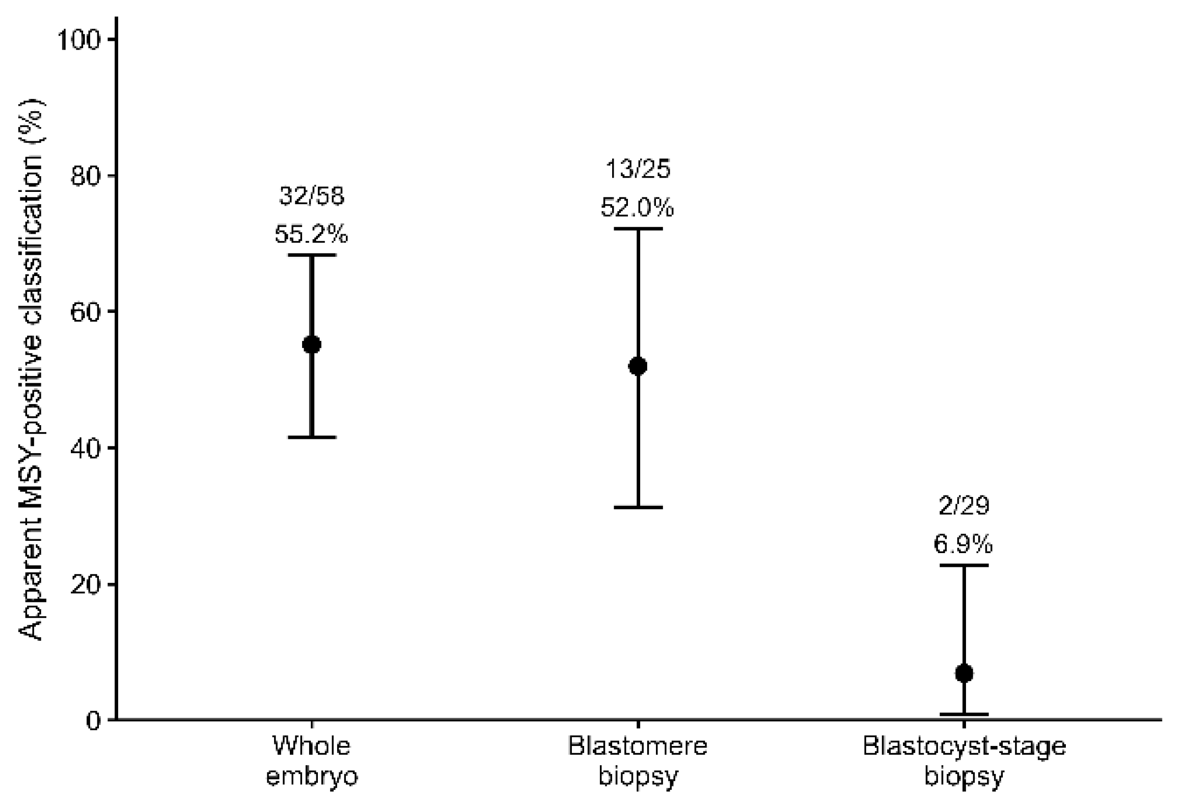
−5). The apparent MSY-positive classification rate was 55.2% (32/58; 95% CI: 41.5–68.3%) in whole embryos, 52.0% (13/25; 95% CI: 31.3–72.2%) in blastomere-biopsy samples, and 6.9% (2/29; 95% CI: 0.8–22.8%) in blastocyst-stage biopsy samples (
Figure 1).
As a descriptive sensitivity analysis, apparent MSY-based classification was examined separately within conventional- and X-sorted-semen subsets among CDK1-positive embryos (
Table 3). Because subgroup sizes were limited and uneven, these analyses were considered descriptive rather than confirmatory. Within the conventional-semen subset, apparent MSY-positive classification rates were 59.6% (31/52) in whole embryos, 70.0% (7/10) in blastomere-biopsy samples, and 14.3% (1/7) in blastocyst-stage biopsy samples (Fisher’s exact test,
p = 0.0625). Within the X-sorted-semen subset, the corresponding rates were 16.7% (1/6), 40.0% (6/15), and 4.5% (1/22), respectively (Fisher’s exact test,
p = 0.0239). These subgroup analyses were limited by small and imbalanced subgroup sizes, particularly in the whole-embryo X-sorted subgroup, and should therefore be interpreted as descriptive only.
An additional descriptive sensitivity analysis was performed across all embryos, using lower-bound and upper-bound definitions of MSY signal detection. Using a lower-bound definition in which only valid MSY-positive reactions were counted as positive, apparent MSY positivity across all embryos was 45.7% (32/70) in whole embryos, 33.3% (13/39) in blastomere-biopsy samples, and 6.2% (2/32) in blastocyst-stage biopsy samples (Fisher’s exact test, p = 0.0001663). Using an upper-bound definition that additionally included control-missing reactions as MSY-signal-detected, without treating them as valid male classifications, the corresponding proportions were 52.9% (37/70), 48.7% (19/39), and 9.4% (3/32), respectively (Fisher’s exact test, p = 4.084 × 10⁻⁵).
In a separate descriptive analysis, valid MSY-positive, valid MSY-negative, and control-missing reactions were retained as distinct outcome categories. The overall distribution of these outcome categories differed across sampling methods (Fisher’s exact test, p = 1.151 × 10⁻⁵). Control-missing reactions were observed in 5/70 whole embryos, 6/39 blastomere-biopsy samples, and 1/32 blastocyst-stage biopsy samples. Under the lower- and upper-bound definitions, the corresponding blastocyst-stage biopsy proportions were 6.2% and 9.4%, respectively.
Sample categories were not fully balanced across PCR dates and overlap between categories across dates was only partial. Accordingly, an exploratory sensitivity analysis was performed using a coarse early-versus-late PCR-date block. In this exploratory model, the PCR-date block term was not statistically significant (Firth logistic regression,
p = 0.623). The corresponding model estimates are provided in
Supplementary Table S2.
3.3. Multivariable Analysis of Apparent MSY-Based Classification
A multivariable penalized logistic regression model was fitted with apparent MSY-based classification as the outcome and sample category and semen type as categorical predictors. In this descriptive model, the overall likelihood-ratio test p-value was 2.97 × 10⁻⁶.
Compared with whole embryos, blastomere-biopsy samples had an OR of 1.80 (95% CI: 0.60–6.14;
p = 0.303), whereas blastocyst-stage-biopsy samples had an OR of 0.16 (95% CI: 0.029–0.631;
p = 0.0076) for apparent MSY-positive classification. The sample-category estimates from this penalized logistic regression model are presented in
Table 4. Given the exploratory nature of the study, and because semen type was markedly imbalanced across sample groups and completely confounded with sire, the semen-type coefficient was included only as part of the descriptive model and should not be interpreted as an independent semen-type effect. The corresponding semen-type estimate is provided in
Supplementary Table S2 for completeness. The odds-ratio estimates from the descriptive Firth penalized logistic regression model are additionally visualized in
Supplementary Figure S4.
Observed subgroup proportions and model-fitted probabilities are shown in
Supplementary Table S1. Fitted probabilities for sparse subgroups were interpreted cautiously because of small subgroup sizes and structural confounding.
As additional descriptive robustness checks, alternative Firth logistic regression models were fitted. The odds ratio for blastocyst-stage biopsy versus whole embryo was below 1 in the sample-category-only model, the model including semen type, and the model additionally including PCR-date block (
Supplementary Table S2). The PCR-date block term itself was not statistically significant. Leave-one-PCR-date sensitivity analyses showed that the odds ratio for blastocyst-stage biopsy versus whole embryo remained below 1 after exclusion of each individual PCR date, although precision decreased in sparse subsets and statistical significance was not retained after exclusion of one PCR date (
Supplementary Table S3).
3.4. Same-Lysate Repeat Amplification and Apparent MSY-Based Classification Stability
To assess same-lysate PCR repeatability, a randomly selected subset of samples with sufficient residual lysate available for repeat testing was re-amplified from the same lysates under identical PCR conditions. The repeat subset comprised 16 whole embryos, 12 blastomere-biopsy samples, and 12 blastocyst-stage biopsy samples.
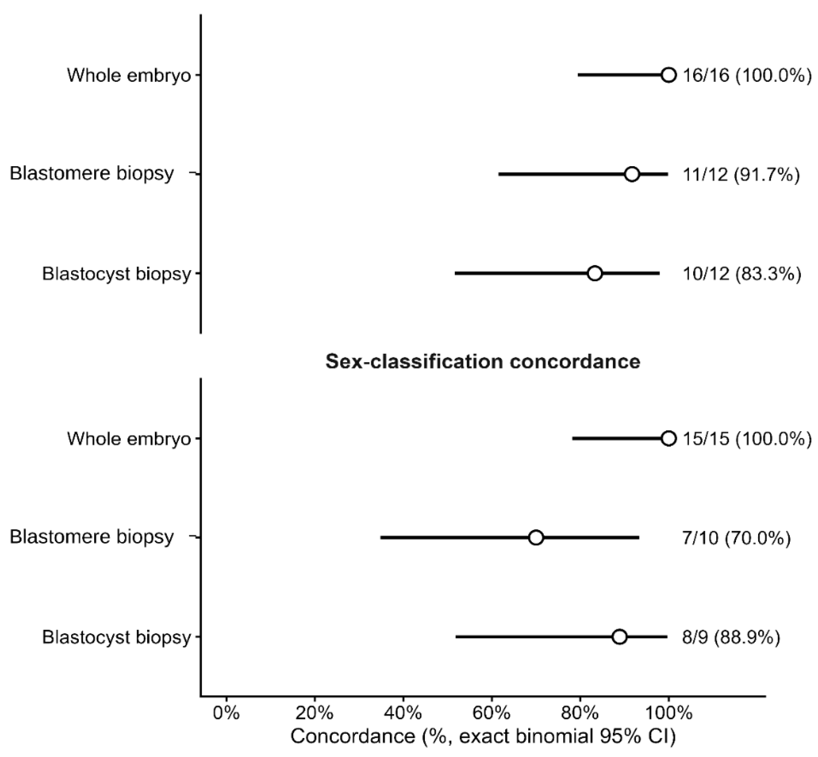
Evaluability concordance was 100.0% (16/16) in whole embryos, 91.7% (11/12) in blastomere-biopsy samples, and 83.3% (10/12) in blastocyst-stage biopsy samples. Among classification-evaluable repeat pairs, apparent MSY-based classification concordance was 100.0% (15/15), 70.0% (7/10), and 88.9% (8/9), respectively. Overall evaluability concordance across the repeat subset was 92.5% (37/40; 95% CI: 79.6–98.4%), and overall apparent MSY-based classification concordance was 88.2% (30/34; 95% CI: 72.5–96.7%) (
Figure 2).
Discordant evaluability results occurred in 0/16 whole embryos, 1/12 blastomere-biopsy samples, and 2/12 blastocyst-stage biopsy samples. Discordant apparent MSY-based classifications occurred in 0/15, 3/10, and 1/9 classification-evaluable repeat pairs, respectively.
Exact McNemar testing did not indicate a systematic directional bias between original and repeat amplification outcomes. For evaluability, no directional bias was detected despite the small number of discordant pairs (1 vs. 2; exact McNemar p = 1.00). Likewise, for apparent MSY-based classification among classification-evaluable repeat pairs, discordant calls were balanced in direction (2 vs. 2; exact McNemar p = 1.00).
4. Discussion
The present study was designed to characterize assay-output behavior in a low-input bovine embryo sexing workflow rather than to estimate biological sex ratios or validate diagnostic sexing accuracy. Three main observations emerged. First, assay evaluability differed across embryo-derived sample categories, with the lowest evaluability in blastomere-biopsy samples. Second, among technically evaluable reactions, apparent MSY-positive classification differed across sample categories and was lowest in blastocyst-stage biopsy samples. Third, in the repeat-amplification subset, classification concordance was complete among whole embryos and lower in biopsy-derived samples. Together, these findings show that assay evaluability and apparent MSY-positive classification varied across embryo-derived sample categories in this low-input PCR setting, which is relevant because biopsy-based bovine embryo sexing has long been used in experimental, field, and commercial contexts [
31,
32,
33,
34].
The particularly low apparent MSY-positive classification rate in blastocyst-stage trophectoderm biopsies deserves cautious interpretation. In the descriptive Firth model, the odds of apparent MSY-positive classification remained lower in this group than in whole embryos even after inclusion of semen type; however, this should not be interpreted as an independent biological effect of trophectoderm biopsy. Several non-mutually exclusive explanations may account for this pattern, including locus-specific dropout of the Y-linked target under low-template conditions, variation in cell recovery or lysis efficiency, trophectoderm–inner cell mass discordance, embryonic mosaicism, and the structural confounding of sample category with developmental stage, semen type, sire, and PCR-date structure. Because no independent reference method for true embryo sex was available, the finding supports the need for sample-type-specific validation rather than direct conclusions about biological sex ratio or diagnostic accuracy.
Previous bovine embryo-sexing studies primarily demonstrated the feasibility of PCR-based Y-marker detection using biopsied embryonic material and, in some cases, evaluated embryo survival or field applicability after transfer [
31,
32,
33,
35]. In contrast, the present study was not intended to validate embryo-sexing accuracy under optimized conditions, but to examine how different embryo-derived sample categories behaved within the same low-input MSY/CDK1 PCR workflow. This distinction is important because high reported accuracy under controlled or confirmatory conditions does not exclude sample-type-dependent differences in apparent Y-marker detection when template input is extremely limited [
12,
15,
36,
37,
38,
39,
40]. Therefore, the present findings complement earlier biopsy-based embryo-sexing studies by emphasizing assay evaluability, apparent MSY-positive classification, and same-lysate repeatability as assay-output measures rather than direct indicators of true embryo sex.
A plausible technical explanation is reduced and more variable effective template input in low-cell biopsy-derived samples. This was most evident for blastomere biopsies, which represented the lowest input category and had the lowest assay evaluability. Biopsy-based PCR sexing often relies on very small numbers of embryonic cells, including single-blastomere or other very low-cell-input approaches, making assay performance sensitive to template limitation [
31,
35,
41]. Low DNA input is known to increase stochastic amplification effects as template quantity approaches analytical limits [
12,
15,
36,
37,
38,
39,
40]. In blastomere-biopsy samples, CDK1 failure may reflect variable cell recovery during biopsy and transfer, handling-related damage, incomplete lysis, or stochastic PCR failure. Control-missing reactions, in which an MSY-sized band was observed without the CDK1 internal-control band, may reflect preferential amplification, multiplex imbalance, or other low-template PCR artifacts and were therefore conservatively classified as non-evaluable rather than as valid apparent MSY-positive classifications. In the present multiplex assay, the MSY assay targets a non-repeated Y-linked region, whereas the autosomal CDK1 internal control is present in two copies per diploid genome. This target-copy asymmetry may increase the probability that CDK1 remains detectable while the Y-linked target drops out, particularly when only a few template molecules enter the reaction and multiplex competition is present [
42,
43]. Fragment-length differences alone are unlikely to explain the pattern, because the shorter MSY amplicon would not by itself predict reduced detection [
44,
45].
Biological and sampling-representativeness mechanisms may also have contributed to the blastocyst-stage biopsy pattern. These samples were highly evaluable, indicating that amplification generally succeeded, yet apparent MSY-positive classification was selectively low among CDK1-positive reactions. Because blastocyst-stage biopsies differed from the other sample categories in both developmental stage and sampled compartment, trophectoderm non-representativeness, compartmental heterogeneity, or mosaicism cannot be excluded [
13,
14,
46]. Previous reports of sex-chromosomal mosaicism in bovine embryos, together with recent large-scale data on aneuploidy and mosaicism in genotyped cattle blastocysts, further support caution when interpreting sex-linked signals from highly limited or compartment-specific samples [
47,
48].
The observed patterns must also be interpreted in light of structural confounding. Sample category co-varied with developmental stage, semen-type composition, sire, and, to some extent, PCR-date structure. Because conventional semen was derived from sire Reform and X-sorted semen from sire ICE Cube-S, the independent effects of sample category, semen type, and sire are not statistically identifiable. Accordingly, the subgroup comparisons and the penalized multivariable model should not be interpreted causally. The Firth model is best viewed as a sparse-data descriptive summary that included semen type as a limited covariate, rather than as a model capable of separating independent sample-category, semen-type, and sire effects. Similarly, the semen-type term should not be interpreted as an isolated estimate of semen-type effect or as evidence for or against the established biological efficacy of sperm sex sorting [
19,
20,
49,
50,
51,
52,
53].
The small same-lysate repeat-amplification subset was consistent with the possibility of lower classification stability in biopsy-derived samples, but subgroup sizes were limited and repeats were not based on independent biopsy or extraction. Thus, this analysis should be interpreted as a preliminary indicator of PCR repeatability rather than full workflow reproducibility. Independent repeat biopsy, extraction, and amplification would be required to determine whether the same pattern persists across the full testing workflow.
Beyond their methodological implications, these findings are relevant to IVF/ET laboratories and cattle breeding programs, where biopsy-based embryo testing is increasingly incorporated into workflows for embryo sexing, genotyping, pre-transfer selection, and embryo genomic selection [
41,
54,
55,
56,
57,
58,
59]. Commercial embryo sexing has historically relied on biopsy-based PCR or related Y-marker detection, whereas sex-sorted semen remains the dominant upstream method for producing sex-biased embryo or calf crops [
31,
32,
33,
49,
51,
52,
53]. Rapid molecular approaches, including isothermal and visual detection methods, are also being developed to shorten turnaround time and simplify embryo-sexing workflows [
10,
18,
21]. Under applied conditions, sample-category-dependent differences in assay evaluability or apparent MSY-positive classification may bias comparisons between workflows and complicate interpretation when outputs from whole embryos and biopsy-derived samples are compared directly. These findings therefore support the need for sample-type-specific validation thresholds, explicit internal-control criteria, representative positive and negative controls, and repeat or confirmatory testing when template input is very low. Because many commercial embryo-testing platforms use proprietary amplification chemistries, marker panels, biopsy types, and scoring thresholds [
54,
55,
56,
57,
58,
59], the present study is not intended to compare named commercial tests. Instead, it emphasizes the need for platform- and biopsy-type-specific validation before results from one workflow or sample category are generalized to another.
Several limitations should be emphasized. The study was observational and exploratory rather than prospectively randomized; all biopsies were performed by a single operator; embryos originated from a single IVF workflow; exact cell counts, biopsy duration, and direct DNA quantification were not available for individual samples; sample categories were incompletely balanced across PCR dates; and no independent reference method for true embryo sex was available. Future studies should use prospectively balanced sample allocation, record exact biopsy cell number and manipulation time, include DNA-input quantification or pre-amplification metrics where feasible, separate semen type from sire, distribute sample categories across PCR runs, and validate PCR-based calls against an independent reference method such as whole-embryo genotyping, sequencing-based sex inference, or confirmed calf sex after transfer [
18,
48].
5. Conclusions
In this observational low-input MSY/CDK1 PCR workflow, embryo-derived sample categories showed differing assay evaluability and apparent MSY-positive classification patterns. Blastomere-biopsy samples had the lowest evaluability, whereas blastocyst-stage biopsy samples showed the lowest apparent MSY-positive rate among CDK1-positive reactions. Although low-input technical effects provide a plausible explanation for these patterns, biological and sampling-representativeness mechanisms, including trophectoderm non-representativeness, compartmental heterogeneity, or mosaicism, cannot be excluded. Because the sample category was structurally confounded with developmental stage, semen type, sire, and partly PCR-date structure, and because no independent reference method for true embryo sex was available, these findings should be interpreted as assay-output observations rather than diagnostic accuracy estimates. Future prospectively balanced studies with independent sex validation, recorded cell counts, biopsy-duration data, DNA-input assessment, and independent repeat extractions are needed before definitive diagnostic recommendations can be made.



 ,
,