


Abstract
Sex steroid hormones play a pivotal role in maintaining systemic homeostasis throughout life. Their age-related decline is closely associated with the onset of frailty, including sarcopenia and dementia. Here, this article provides a narrative review of the existing literature about the multifaceted roles of sex steroid hormones, particularly estrogens and androgens, in aging and age-related diseases. Sex steroid action is mediated by nuclear receptors such as estrogen receptor alpha (ERα) and androgen receptor (AR). Transcriptional activation through these receptors is orchestrated by epigenetic mechanisms, including histone modifications and chromatin remodeling. Beyond their reproductive functions, sex hormones also influence systemic physiology, metabolism, immune responses, and neuroplasticity. Clinical studies on hormone-deprivation therapies for prostate and breast cancers, as well as animal models, have revealed the key contributions of AR and ER activity to muscle integrity, bone density, and cognitive function. The sexual dimorphism in cognitive decline, especially in postmenopausal women, suggests the therapeutic potential of hormone supplementation and receptor-targeted strategies. Thus, AR- and ER-associated genes are considered promising targets for preventing frailty, sarcopenia, osteoporosis, and dementia. This review summarizes the current knowledge on sex hormone signaling in aging, with an emphasis on translational implications and future research directions.
1. Introduction
Sex steroid hormones, including estrogen and androgen, are secreted in large quantities during fetal development and puberty, playing essential roles in sexual differentiation, reproductive function, and growth [1,2,3]. However, their levels gradually decline with aging, particularly during menopause in women and late middle age in men [4,5,6]. This decline has been implicated in the onset of age-related conditions, such as dementia, osteoporosis, and sarcopenia, which collectively define frailty phenotypes. Frailty represents an intermediate and potentially reversible state between robust health and disability [5]. It is closely associated with sarcopenia, which is characterized by reduced skeletal muscle mass, diminished muscle strength, and impaired physical performance, as well as an increased risk of falls and fractures, making its prevention a critical clinical challenge [6]. Therefore, declines in sex hormone levels contribute to multimorbidity and represent a major factor that compromises healthy longevity in older adults.
Sex steroid hormones such as estrogen and androgen exert their biological effects by binding to specific intracellular receptors—the estrogen receptor (ER) [7] and androgen receptor (AR) [1], respectively. These receptors belong to the nuclear receptor superfamily and function as ligand-dependent transcription factors. Upon hormone binding, the receptor undergoes conformational changes, translocates to the nucleus, and binds to hormone response elements (HREs) within the genome. These interactions modulate the transcription of target genes involved in diverse physiological processes [1,7]. The transcriptional activities of ERs and ARs are further regulated by co-activators and co-repressors, which influence chromatin remodeling and epigenetic modifications [7].
Recent studies have highlighted the role of super-enhancers (SEs)—large clusters of enhancers with high transcriptional activity—in amplifying hormone-responsive gene expression [8]. Receptor activity is tightly orchestrated by co-regulators, including HATs, HDACs, methyltransferases, and chromatin-remodeling complexes, which collectively modulate nucleosome positioning and DNA accessibility. ERs and ARs preferentially occupy SEs, which induce high-level expression of key genes that define cell identity and function [9]. Therefore, SE dysregulation has emerged as a critical contributor to aging-related pathologies such as neurodegeneration and cancer [8]. Moreover, recent advances have revealed that multivalent protein–protein and protein–RNA interactions, which induces a physical phenomenon, liquid–liquid phase separation (LLPS) and drives the formation of nuclear condensates that spatially concentrate transcriptional machinery [10,11]. LLPS has emerged as a novel mechanism by which nuclear receptors and their co-regulators form dynamic, membrane-less compartments within the nucleus [9,10,11]. In addition to classical genomic actions, sex hormones also exert non-genomic effects through membrane-associated receptors and signaling cascades [12]. These rapid responses involve the activation of secondary messengers, such as cyclic AMP, calcium ions, and kinase pathways [13].
Collectively, these multifaceted mechanisms underscore the complexity of sex hormone signaling and its relevance to aging-related pathologies, such as dementia and frailty. This narrative review aims to provide a comprehensive and critical analysis of the latest research on the mechanistic insights into the action of sex hormone receptors for preventing aging and age-related diseases. First, we will summarize the recent advances in the molecular mechanisms of nuclear receptors which regulates epigenetic landscape. The important roles of sex hormones have been verified by epidemiological studies of prostate and breast cancer patients treated with hormone-deprivation therapy. Animal models have been used to demonstrate the physiological function of sex hormone receptors in mammals. Our goal is to investigate how the molecular mechanisms of ER or AR and their target genes are involved in various tissue function including prevention of osteoporosis, maintenance of muscle function, and preservation of neurocognitive function in order to gain a better understanding of the intricate relationship between biological and healthy aging. A deep understanding of such mechanisms is crucial for developing targeted interventions aimed at mitigating the effects of aging on frailty and age-related disease development. Furthermore, we aim to promote considerations about the future directions required to promote this critical field of research.
2. Methods
The article is structured as a narrative review, which contains a comprehensive and critical examination of the existing literature on the role of sex hormone action in preventing age-related diseases. This methodological approach allows for the integration of information from diverse experimental and clinical evidence. Unlike systematic reviews that aim to compile all relevant studies exhaustively, a narrative review emphasizes the interpretation and contextualization of the considered information.
A targeted search was conducted using PubMed and Google Scholar. This research was carried out in 2025 to identify relevant articles in this field. The search algorithm used for the different sources included the following terms: ‘Aging’; ‘frailty’; ‘androgen’; ‘estrogen’; ‘sex hormone’. Clinical evidence, particularly the epidemiological studies regarding hormone deprivation therapy for cancers, was searched by combining with the following terms: ‘prostate cancer’; ‘breast cancer’; ‘tamoxifen’; ‘androgen deprivation therapy’; ‘aromatase inhibitor’: ‘osteoporosis’; ‘cognitive decline’; ‘dementia’. Molecular mechanisms of nuclear receptors were searched combining with the following terms: ‘super-enhancers’; ‘epigenetics’; ‘phase separation’; ‘coactivator’; “transcription factor”. To find research associated with osteoporosis and sarcopenia, we additionally used keywords such as ‘knockout mouse’; ‘bone’; ‘muscle’. To find researches associated with dementia, we used keywords such as ‘brain’; ‘cognitive decline’; ‘dementia’; ‘Alzheimer’s disease’; ‘RNA binding protein’. Various types of studies were considered, including reviews, experimental studies, and clinical trials. Articles published in English between 2000 and 2025 were included. However, important studies before 2000 were also selected for providing the historical evidence to understand this research field. The author specializing in the field reviewed to determine their relevance and identify any methodological inconsistencies. Inclusion and exclusion criteria for the scientific papers were set for this review. The inclusion criteria included topics such as first or important reports of mouse models, analysis of target genes, sex-hormone receptor action by using biochemical and genome biological strategy, while the exclusion criteria included papers that were published prior to the year 2000. Particularly, timely and pertinent scientific literature showing the importance of exploring multiple dimensions in research concerning aging and sex hormones were included. Several hundred articles were retrieved, out of which approximately 150 papers met the inclusionary criteria and were reviewed. Moreover, this article employs a wide array of empirical sources, including clinical studies, basic research using cell lines, animal models, particularly knockout mouse model, genome biology and human organoids to reinforce its claims.
3. Association Between Sex Hormones and Dementia/Frailty
Numerous epidemiological and experimental studies have demonstrated that the decline in sex steroid hormones with aging is closely associated with the onset and progression of dementia [14,15]. In particular, estrogen deficiency following menopause has been linked to an increased risk of Alzheimer’s disease (AD), cognitive decline, and mood disorders [16,17]. Estrogen exerts neuroprotective effects through multiple mechanisms, including enhancement of synaptic plasticity, regulation of neurotransmitter systems, and reduction in oxidative stress and neuroinflammation [18,19,20]. Conversely, hormone replacement therapy (HRT) has been investigated as a potential intervention for preventing cognitive impairment, with mixed results depending on timing, dosage, and individual risk factors [20]. Consistently, hormone replacement therapy can produce divergent results if initiated during the perimenopausal period, compared with the postmenopausal ages in mice [21]. The concept of a “critical window” for HRT efficacy, in which early intervention may yield neuroprotective benefits, has gained attention in clinical research [22].
Age-related decline in testosterone levels in men has been implicated in sarcopenia, reduced physical performance and frailty [4,14,23]. Androgen signaling contributes to muscle protein synthesis, mitochondrial function, and neuromuscular connectivity [1,3,24,25]. Clinical studies have shown that low serum testosterone levels are associated with an increased incidence of frailty and decreased quality of life in elderly men [26,27]. In patients with prostate cancer, hormone deprivation therapy significantly reduces both muscle mass and density [28]. Anti-androgen treatments for prostate cancer led to declines in bone density [26]. Attenuation of sex hormone activity through hormone therapy also plays a role in cognitive impairment and the progression of AD [27,29]. An increased risk of dementia, particularly AD, has been demonstrated [29]. Breast cancer treatment involves estrogen inhibition using aromatase inhibitors for estrogen deprivation and tamoxifen, a selective estrogen receptor modulator (SERM) that functions as an antagonist in breast tissues to specifically block estrogen action [20]. The number of fractures is increased by aromatase inhibitor treatment in women; however, tamoxifen treatment does not [30]. In premenopausal women, however, tamoxifen treatment has also been associated with an increased incidence of fractures [31]. Therefore, the risk of osteoporosis and fractures in patients with breast cancer is enhanced by treatment with both tamoxifen and aromatase inhibitors, highlighting the importance of osteoporosis prevention in these patients. Regarding cognitive function, several reports have shown that tamoxifen treatment is associated with a decline in verbal memory and fluency [32]. Decline in verbal memory and executive functioning, particularly processing speed, has been reported with tamoxifen use in postmenopausal women [33]. Although anti-estrogen therapy represses some cognitive functions, some studies have shown a significant positive association with dementia [34]. In another report, treatment with aromatase inhibitors or tamoxifen in patients with breast cancer was associated with a reduced incidence of AD development [35]. Thus, whether anti-estrogen therapy increases the risk of dementia remains controversial. Understanding the complex interplay between sex hormones, receptor signaling, and aging-related pathologies is essential for developing targeted therapies to prevent or mitigate dementia and frailty.
4. Sex Differences in Age-Related Diseases
Sex differences in the prevalence, progression, and clinical presentation of geriatric diseases have long been recognized. For example, women are more likely to develop AD [15] and osteoporosis [36], whereas men are more prone to cardiovascular diseases and certain types of cancer [37,38]. Changes in bone structure contribute to sex-related differences in osteoporosis. In men, the loss of trabecular bone is primarily due to decreased bone formation, resulting in thinning of the trabecular bone, while the trabecular structure is relatively preserved. In contrast, the loss of trabecular bone in women is primarily caused by increased bone resorption, which leads to a decrease in trabecular number and structural integrity. Although both conditions result in decreased in bone mineral density, women are more likely to experience significant bone strength loss, resulting in a higher incidence of fractures [39,40]. AD is also an important geriatric disease, and its prevalence is higher in women, with a ratio of nearly 2:1 in many countries, with postmenopausal women accounting for more than 60% of cases [41]. Although women have a relatively long-life expectancy, reports indicate a higher age-adjusted risk of AD in women, regardless of survival rate. Furthermore, mild cognitive impairment, a precursor to AD dementia, progresses more rapidly to AD in women, regardless of age or education level [42]. Women carrying the apolipoprotein E (APOE)-4 genotype also exhibit higher amyloid-beta (Aβ) deposition, decreased brain connectivity, and greater cerebral hypometabolism [42]. These disparities are influenced not only by genetic and environmental factors but also by differences in sex hormone levels and receptor signaling pathways.
Estrogen has been shown to exert protective effects on the brain, bones, and vasculature, contributing to the lower incidence of cardiovascular disease in premenopausal women [43]. However, the abrupt decline in estrogen levels following menopause is associated with an increased risk of dementia, bone fractures, and metabolic disorders [20]. In contrast, testosterone levels in men decline more gradually, and low testosterone levels have been linked to sarcopenia, obesity, metabolic diseases, cardiovascular diseases, and cognitive impairment [4,13]. Understanding these sex-specific mechanisms is essential for developing personalized approaches to the prevention and treatment of age-related diseases. Tailoring interventions based on hormonal status, receptor expression, and genetic background may improve therapeutic outcomes and reduce health disparities in the elderly population.
5. Molecular Mechanisms for Epigenetic Regulation by Sex Hormone Receptors
Sex hormone receptors (ERs and ARs) function as ligand-dependent transcription factors that play a central role in epigenomic regulation [13]. Both ERα and ERβ are widely expressed in various human tissues, including reproductive organs, breast tissue, bone, and brain [44,45]. They also affect gene regulation in numerous cell types and primarily drive the development and maintenance of the male reproductive system by promoting cellular proliferation and differentiation [1,13]. In addition, they play physiological roles in maintaining the functions of the musculoskeletal, cardiovascular, immune, and central nervous systems [3]. Upon binding to their respective ligands, estrogen or androgen, these receptors undergo conformational changes that enable them to interact with specific DNA sequences known as HREs. This interaction initiates a cascade of transcriptional events that modulate gene expression in a tissue-specific and context-dependent manner [1,2,7]. Recent advances in chromatin biology have revealed that sex hormone receptors do not act alone but typically act as homo- or heterodimers with coactivators, such as histone acetyltransferases (HATs), including the steroid receptor coactivator (SRC) family and p300/CBP, as well as methyltransferases, histone deacetylases (HDACs), and chromatin remodeling factors [1,2,7]. These co-regulators dynamically alter the chromatin landscape, thereby influencing nucleosome positioning, histone modifications, and DNA accessibility. Through these mechanisms, sex hormone receptors orchestrate the activation or repression of target genes involved in cellular differentiation, metabolism, and stress responses (Figure 1A).
Figure 1.
Molecular mechanisms for sex hormone receptor transcriptional actions. (A) Nuclear receptors dimerize and bind to gene promoters or enhancers at hormone response elements (HREs). Co-activators and Co-repressors bind to the activation function-2 (AF-2) domain and recruit histone modification enzymes including histone deacetylases, acetyltransferases or protein lysine methyltransferases. Co-activators binding to the nuclear receptor complex promote with transcriptional activation, leading to the target genes to be expressed. T: testosterone, E2: estradiol. Ac: acetylation, CH3: methylation. (B) Transcription factors and transcriptional co-regulators form condensates through liquid–liquid phase separation (LLPS) by multivalent interactions mediated via intrinsically disordered regions (IDRs), thereby promoting the formation of super-enhancers. (C) Tissue-specific transcription factors called ‘pioneer factors’ such as forkhead box protein A1 (FOXA1) are important for enhancer activation and nuclear receptor recruitment.
One of the most striking discoveries in recent years has been the role of SEs [8], large clusters of enhancers with exceptionally high transcriptional activity in sex hormone signaling. ERs and ARs are known to bind preferentially to these regions, which are enriched with transcriptional co-activators, such as mediator complex subunit 1 (MED1) and Bromodomain Containing 4 (BRD4) [11,46]. SEs serve as regulatory hubs that drive robust expression of genes critical for cell identity and function [8]. They are occupied by cell-type-specific transcription factors, allowing the identification of candidate master transcription factors for many cell types, which may prove useful for further understanding the transcriptional control of cell state and for reprogramming cells [47].
Modulation of cellular SEs is essential for defining the characteristics of specific cell types [8]. The identification of these SE domains in various cell types could provide a valuable resource for further studies on cellular identity [9,47]. Moreover, their dysregulation has been implicated in age-related diseases, including neurodegeneration and cancer.
Furthermore, the concept of LLPS has emerged as a novel framework for understanding how sex hormone receptors and their associated proteins form dynamic, membrane-less compartments within the nucleus [9,11]. Multivalent interactions are mediated via the intrinsically disordered regions (IDRs) of proteins, thereby promoting the formation of SEs. These condensates concentrate transcriptional machinery at specific genomic loci, enhancing the efficiency and specificity of gene regulation (Figure 1B). LLPS is now recognized as a key mechanism in the spatial organization of nuclear processes, including hormone-responsive transcription [46,47,48]. Collectively, these findings demonstrate the complexity and precision of epigenomic regulation mediated by sex hormone receptors. Understanding these mechanisms provides valuable insights into how hormonal changes during aging contribute to the onset of dementia, frailty, and other age-related conditions.
6. Nuclear Regulatory Mechanisms Determining Tissue Specificity of Sex Hormone Receptors
Although sex hormones exert distinct functions across various tissues throughout the body, the molecular mechanisms underlying their tissue specificity have long remained unclear. Genome-wide identification of AR and ERα binding sites in prostate and breast cancer using chromatin immunoprecipitation followed by sequencing (ChIP-seq) revealed that Forkhead box protein A1 (FOXA1), a member of the Forkhead family, binds near these peak sequences (Figure 1C). FOXA1 plays a pivotal role in facilitating nuclear receptor binding to the genome and is referred to as a pioneer factor [49,50]. Most AR-binding sites are located in regions distal to the transcription start sites, known as enhancers, and regulate target genes from a distance. These SE-regulated genes, controlled by AR, FOXA1, and collaborating factors, are important for prostate cancer tumor growth and the acquisition of drug resistance [9,50,51]. Furthermore, AR interacts with cell type-specific SEs formed by master transcription factors, which are dependent on LLPS during prostate cancer progression [9]. Further analysis of tens of thousands of AR-binding sequences revealed motifs for transcription factors beyond FOX, such as ERG, NKX3-1, OCT1, and GATA2, which form complexes with AR. These transcription factors, termed collaborative factors, influence AR binding specificity and sensitivity [52,53]. ChIP-seq of these collaborative factors enables the identification of overlapping binding sites and elucidation of AR transcriptional networks. Nuclear receptors are thought to form large complexes on enhancers, referred to as MegaTrans complexes [46,54]. The tissue-specific signaling of sex hormones is governed by complex formation with collaborative factors. While classical estrogen signaling via estrogen response elements (EREs) is common across tissues, tissue-specific estrogen signaling depends on its interactions with other transcription factors [55]. For example, ETS family factors generate specific signals in the uterus, whereas FOXA1 functions as a pioneer factor in the mammary glands. In mouse kidneys and testes, AR signaling cooperates with AP1 transcription factor binding [53]. Changes in the composition of nuclear receptor complexes during disease progression have been linked to drug resistance in prostate cancer [9,51]. However, the analysis of nuclear receptor complexes in aging-related diseases, such as frailty, remains an important future challenge. Understanding the function of AR in a lineage-specific manner for developing SEs in target tissues, as well as the changes in AR activity with aging, would be important for preventing age-related disorders.
7. Roles of Sex Hormone Receptor in Skeletal Muscle Maintenance
Animal models have provided mechanistic insight into the influence of sex hormones on brain and muscle aging. Estrogen receptor knockout mice exhibit impaired spatial memory and increased Aβ accumulation [56], while AR-deficient mice show reduced muscle mass and strength [57]. These findings highlight the importance of receptor-mediated transcriptional regulation in maintaining tissue integrity during aging. Recent studies have also highlighted the role of sex hormones in the modulation of systemic homeostasis [3,13,43], both of which are key contributors to frailty.
Animal models deficient in sex hormone receptors have provided critical insights into the roles of estrogen and androgen signaling in skeletal muscle maintenance (Table 1).
Table 1.
Investigation of sex hormone receptor function by using animal models.
ER- and AR-knockout mice exhibit pronounced muscle atrophy, reduced muscle strength, and impaired regenerative capacity, underscoring the importance of receptor-mediated transcriptional regulation in muscle physiology [63,74,75,76]. As for estrogens, the decline by menopause appears to reduce muscle function and affect metabolic health in muscle [83]. In ERα-deficient mice, studies have shown decreased expression of genes involved in heat shock proteins (HSPs) [84]. Deletion of ERα expression leads to insulin sensitivity, mitochondrial biogenesis, oxidative phosphorylation, and muscle fiber integrity [58,59,60]. Thus, estrogen maintains muscle strength, suppresses apoptosis, and promotes metabolism [61,62]. The ERβ pathway has also been reported to be important for maintaining muscle mass [63]. These mice also displayed increased markers of inflammation and fibrosis, suggesting that estrogen signaling contributes to the preservation of muscle homeostasis through anti-inflammatory and metabolic pathways. Meanwhile, the ubiquitin ligases atrogin-1 (Fbxo32, MAFbx) and MuRF1 (TRIM63, MuRF2, IRF, and RNF28), which mediate muscle atrophy, are regulated by sex hormones. Regulation of the protein degradation system is also an important mechanism in maintaining muscle integrity [85,86].
Notably, androgen deprivation by orchiectomy has been shown to reduce muscle mass in the lower limbs [87]. Global AR knockout models demonstrate reduced muscle mass and strength, accompanied by the downregulation of anabolic signaling pathways, such as insulin-like growth factor 1 (IGF-1), and upregulation of catabolic factors, including myostatin and ubiquitin ligases [88,89,90,91]. To investigate the physiological role of AR in a tissue-specific manner, muscle-specific AR knockout mice were generated. The muscles of the lower limbs in these mice showed no significant changes in mass [75,92]. However, muscle force was reported to be reduced in muscle-specific AR knockout mice compared with controls. Thus, AR in muscles is supposed to control muscle quality rather than muscle mass [92].
The effects of androgens on metabolic machinery are important for muscle regulation. AR knockout alters glycolytic activity and accelerates the development of type 2 diabetes in males. Ammonia accumulation and oxidative stress affect mitochondrial function and can cause necrosis [93]. AR also regulates the expression of polyamine synthesis enzymes, such as ornithine decarboxylase 1 (ODC1) in mice [88]. Polyamines play an important role in preventing muscle atrophy. Another report on muscle-specific AR knockouts showed that factors regulating muscle strength are directly controlled by AR. Muscle force is controlled through the phosphorylation of proteins that interact with the muscle filaments. The phosphorylation system, including myosin light chain kinase 4 (MYLK4), is directly regulated by AR and dihydrotestosterone (DHT) [92]. Therefore, another important question is how muscle mass is regulated by AR. A recent study revealed that mesenchymal progenitor cells in muscle tissues produce and secrete IGF-1 via AR. Mesenchymal progenitor-specific AR knockout mice showed reduced lower limb muscle mass. Mechanistically, AR induces IGF-1 expression by binding to its promoter and enhancing IGF-1 secretion during muscle differentiation [94]. In addition, other studies have demonstrated that neuron-specific AR knockout mice indicate that the brain is important for the growth of lower limb muscle mass, suggesting a contribution of the central nervous system to androgen-induced muscle growth [95,96]. Collectively, AR activity controls muscle quality and mass via distinct pathways that promote skeletal muscle strength.
Thus, these results highlight the tissue-specific actions of sex hormone receptors and their downstream targets in maintaining skeletal muscle function. Importantly, the effects of receptor deficiency are often sex-dependent, with male and female animals exhibiting distinct phenotypes, reflecting differences in hormonal milieu and receptor expression patterns [57,97]. Recent studies using inducible and tissue-specific knockout models have further refined our understanding of receptor function during muscle aging. For example, muscle-specific deletion of ERα in adult mice leads to impaired glucose metabolism [58], while AR deletion in muscle satellite cells highlights the role of AR in filament-type selection and muscle force production [77]. These models provide valuable platforms for dissecting the molecular mechanisms by which sex hormones regulate muscle maintenance and for identifying potential therapeutic targets to counteract age-related muscle decline.
8. Roles of Sex Hormones in the Maintenance of Bone Mass
An increase in bone turnover with reduced bone formation after menopause leads to the decline of trabecular bone and overall bone density, and the risk for fracture is increased after menopause [83]. To analyze the role of sex steroid hormone receptors in the maintenance of bone mass, osteoblast-specific KO mice have been generated, demonstrating that ERα functions in both osteoblasts and osteoclasts [98,99,100]. In osteoclasts, the receptor activator of NF-κB (RANK) is activated by RANK ligand (RANKL), which is expressed by osteoblasts, thereby promoting osteoclast differentiation [101]. Conversely, osteoblasts secrete osteoprotegerin (OPG), which acts as a decoy receptor for RANKL and inhibits RANK activation [102]. Thus, the RANKL:OPG ratio is critical for osteoclastogenesis. Estrogen increases the transcription of OPG and suppresses RANKL expression, thereby reducing the RANKL:OPG ratio and inhibiting bone resorption [102]. Other reports showed that estrogen deficiency shifted bone homeostasis from formation to resorption by inducing the secretion of interleukin-6 and osteoclastogenesis [103,104]. Moreover, through its binding to enhancer regions, ERα activates the transcription of the apoptosis-inducing factor Fas ligand (FasL) in osteoblasts. ERα also activates matrix metalloproteinase 3 (MMP3), which cleaves FasL from the cell surface, enabling its secretion and inducing apoptosis in osteoclasts [105,106]. Analysis of AR knockout mice has also revealed that AR is essential for osteoblast-mediated mineralization, maintaining the structure of both trabecular and cortical bones [107,108]. AR has also been reported to suppress RANKL expression, suggesting that osteoblasts inhibit osteoclast differentiation via AR signaling. Thus, genes regulated by ER and AR are key regulators of the musculoskeletal system (Table 2).
Table 2.
Summary of ER and AR target genes in muscle and bone.
9. Sex Hormones and Alzheimer’s Disease
AD is characterized by progressive cognitive decline, accumulation of Aβ plaques, and neurofibrillary tangles composed of hyperphosphorylated tau protein [115,116]. Animal models have demonstrated that sex hormones can attenuate Aβ accumulation and improve cognitive performance, although the timing and duration of therapy are critical factors [14,15]. Androgen signaling plays a role in brain health, particularly in men, as declining testosterone levels are associated with cognitive deterioration and increased prevalence of AD. Longitudinal studies have shown that low free testosterone levels can be detected 5–10 years before AD diagnosis, suggesting that low testosterone is not merely a consequence but also a risk factor for AD [14]. Compared to age-matched controls, men with neuropathologically confirmed AD also exhibit reduced testosterone levels in the brain. Testosterone has been reported to reduce Aβ production and promote its clearance via activation of neprilysin (NEP) and other proteolytic enzymes [116,117] (Figure 2). Androgens affect not only reproductive brain regions but also non-reproductive areas such as the amygdala, preoptic area, medial hypothalamus, and hippocampus [2,24].
Figure 2.
Regulation of APP transcription and Amyloid β production by sex hormones (A). Mechanism of Amyloid β (Aβ) and αAPP production via proteolytic cleavage of APP. αAPP exhibits neuroprotective effects. (B). Proposed mechanism for androgen-mediated transcriptional regulation of APP expression. (C). Regulation of APP metabolism by sex hormones. ↑: Induced by sex hormones ↓: Suppressed by sex hormones.
Experimental studies have demonstrated that androgens promote neuroprotection, neuronal differentiation, survival, and central nervous system development, thereby influencing behavior, memory, and mood [23,24]. AR knockout models exhibit striking deficits in specific components of masculine sexual and territorial behaviors [118]. Androgen treatment improves memory deficits in female APOE-4 knockout mice [119]. AR overexpression inhibits fear memory in male mice [120], supporting the role of androgen signaling in maintaining neuronal integrity.
The regulation of Aβ processing and clearance by sex hormone has been investigated. Aβ precursor protein (APP) is processed by enzymes named secretases. There are two protein cleavage pathways for APP (Figure 2A). The secreted type of APP, called αAPP, is important for neuronal differentiation but is reduced in the AD brain [121]. In contrast, cleavage by β- and γ-secretases produces Aβ. We have previously reported that APP transcription is induced by AR [122]. Other groups have shown that Aβ production from APP is negatively regulated by AR [117], and thus αAPP is upregulated by androgen (Figure 2B). Therefore, androgen may regulate the ratio of αAPP to Aβ. In addition, Aβ clearance enzymes are also activated by androgen and contribute to Aβ deposition in the aged brain [116] (Figure 2C).
10. Estrogen Action to Preserve Cognitive Functions
A similar function for estrogen has also been reported. Women with AD have lower circulating and brain E2 levels than age-matched controls [15,16]. Previous research in rats has shown that old animals require greater E2 doses to elicit memory improvements that younger animals [83,123]. Old and young animals will show different signaling responses to estrogen treatment because they differ in receptor expression. In mice, the Aβ/αAPP ratio in the brain is increased by estrogen deprivation through ovariectomy and reduced by estrogen supplementation; however, this effect is primarily due to downregulation of αAPP [124]. In another report, brain estrogen concentrations remained high even after ovariectomy due to aromatase action [56]. Repressing aromatase expression dramatically decreases brain estrogen levels and induces Aβ accumulation. These results showed that estrogen deficiency reduces αAPP levels and accelerates Aβ plaque formation [56]. For Aβ clearance, ERα has been shown to upregulate the transcription of the Aβ-degrading enzyme NEP in human cellular models of AD [125]. Furthermore, ERα colocalizes with neurofibrillary tangles (NFTs) in the hippocampus of AD brains and physically interacts with tau protein, an interaction that is increased in AD brains [126]. In addition, tau overexpression suppresses ERα transcriptional activity, suggesting that tau inhibits beneficial ERα signaling and neuroprotection through its interaction with ERα in AD brains [127]. Similarly, ERβ also plays a beneficial role in Aβ clearance in AD [128]. Thus, ERs and ARs can influence the expression of genes involved in Aβ metabolism, tau phosphorylation, and neuroinflammation through interactions with chromatin remodeling complexes.
Moreover, E2 treatment activates Signal Transducer And Activator Of Transcription 3 (STAT3) and phosphorylates STAT5, inducing the expression of IGF-1 and phosphorylation of insulin receptor substrate I (IRSI) in the brain [2,129,130]. Consequently, activated Akt phosphorylates cAMP response element-binding protein (CREB), which in turn induces the expression of somatostatin (SRIF) and its receptor subtype, SST2. SRIF enhances expression of NEP and insulin-degrading enzyme (IDE) in the hippocampus, reducing Aβ levels and cell death. Akt phosphorylation also inactivates glycogen synthase kinase-3β (GSK3β) via serine phosphorylation, contributing to reduced cell death [2,131]. Furthermore, E2 promotes activation of the non-amyloidogenic pathway via mitogen-activated protein kinase/extracellular signal-regulated kinase (MAPK/ERK) signaling or EREs, suppressing Beta-Site Amyloid Precursor Protein Cleaving Enzyme 1 (BACE1) levels and Aβ production [131,132]. Maintenance of mitochondrial activity and inhibition of c-Jun N-terminal kinase (JNK) signaling are also considered critical mechanisms for neuroprotection [2].
Brain-derived neurotrophic factor (BDNF) is recognized as a key sex hormone signaling pathway in the brain [133,134]. BDNF is essential for brain development and promotes synapse formation and neural circuit establishment. It activates phosphorylation cascades via its receptor, tropomyosin-related kinase B (TrkB) [2]. Androgens and estrogens may act through BDNF or serve as upstream signals that regulate BDNF expression.
Estrogen reduces oxidative stress and neuroinflammation, both of which are implicated in the pathogenesis of AD. It enhances synaptic plasticity, promotes neuronal survival, and modulates neurotransmitter systems such as acetylcholine and serotonin [130]. E2 also enhances neurogenesis in various brain regions and supports region-specific learning and memory [20]. E2 rapidly increases dendritic spine density in the hippocampus, medial amygdala, and hypothalamus, thereby enhancing hippocampus-dependent memory. It promotes neuroplasticity and neurogenesis, helps maintain healthy cognitive function, and prevents cognitive decline in aging women [2,18,19]. Furthermore, cell type-specific conditional knockout mouse models revealed that estrogens mediated metabolic effects [83]. Estrogens modulate food intake via ERα signaling in the nucleus of the solitary tract and arcuate nucleus, modulate energy expenditure in the ventromedial nucleus of the hypothalamus [43,83].
Ribonucleoprotein (RNP) granules are diverse membrane-less RNA-protein assemblies in cells [135,136]. These granules are formed by multivalent assembly mechanisms, such as LLPS, to concentrate biological molecules. Paraspeckles are formed around non-coding RNA Nuclear Paraspeckle Assembly Transcript 1 (NEAT1), which facilitates their formation. Drosophila Behavior Human Splicing (DBHS) family proteins, Splicing Factor Proline And Glutamine Rich (PSF), Non-POU Domain Containing Octamer Binding (NONO), and Paraspeckle Component 1 (PSPC1), are core paraspeckle components [137,138]. PSF, also known as SFPQ, includes RNA- and DNA-binding domains. Physiologically, PSF is important for brain development. It also functions in epigenetic regulation and the regulation of pre-mRNA splicing in the nucleus. Enrichment of PSF expression in the nucleus has been observed, and nuclear condensates have been found. Thus, PSF is an important component of paraspeckles, and nuclear RNA granules are formed in the nucleus [138,139]. Another important RNA granule is stress granule (SG) [135,136]. SGs are formed in response to various cellular stresses and stop protein synthesis to protect cells. They include RNA and RNA-binding proteins (RBPs). A key factor, Ras-GTPase Activating Protein SH3 Domain-Binding Protein 2 (G3BP2), includes an RNA recognition motif and contributes to the formation of SGs [140]. We have previously shown that G3BP2 is a direct target gene of AR [1]. Transcripts associated with AD development are enriched among SG-associated RNAs, suggesting the involvement of SG in AD development [140]. SGs are also expected to promote tau phosphorylation. Several SG components are associated with phosphorylated tau in the brains of AD mouse models [141]. However, recent reports suggested that SG mitigates the development of neurodegeneration diseases [142,143]. G3BP2, which binds to tau, is thought to prevent its aggregation [142]. Although high expression levels of PSF and G3BP2 are observed in neuronal cells, especially in the hippocampus, their expression levels decline with aging. Both PSF and G3BP2 are targeted by ERα, suggesting that estrogen is involved in regulating the expression levels of G3BP2 and PSF [144]. Physical interactions and co-localization of PSF and G3BP2 in the nucleus and SGs have been observed in human neuronal cells. Nuclear interactions are important for regulating gene expression at the RNA level. AD-associated genes, such as Neuregulin 3 (NRG3) and Semaphorin 5B (SEMA5B), are cooperatively regulated by PSF and G3BP2. Clinically, reduced protein expression of PSF and G3BP 2 in AD tissues compared with controls has been demonstrated [144]. Thus, the nuclear interaction between G3BP2 and PSF enhances essential signals for neural activity and maintains cell viability. Sex hormones are also involved in the expression of PSF and G3BP2. Importantly, the expression levels of G3BP2 and PSF are reduced during AD development and aging [144]. Therefore, we propose that decreased function of this RNA-binding protein complex is critical for aging and AD development in the human brain.
11. Recent Advances in Sex Hormone Receptor Signaling in the Brain
While sex hormone signaling has been extensively studied in tissues such as breast cancer, other organs remain underexplored. Advances in next-generation sequencing have enabled comprehensive analysis of sex hormone signaling. The brain is one of the least understood target organs. Recent studies have combined CUT&RUN for ERα binding site identification, RNA-seq for estrogen signaling in ERα-expressing cells, and ATAC-seq for assessing chromatin accessibility [145]. These analyses revealed that 63% of ERα binding sites in the brain are unique and not found in other tissues, forming brain-specific signals enriched in genes related to synapse formation and ion channels—consistent with estrogen’s role in higher-order cognitive functions [145]. Single-cell RNA-seq has identified sex-specific signals in the brain, showing that ERα co-localizes with the neuron-specific transcription factor Nfix in distinct cell populations. ATAC-seq revealed that females predominantly exhibit ER signaling, whereas males show both AR- and ER-mediated sex-specific signals. These patterns are present even during fetal development, when androgen exposure in males activates estrogen signaling and male-specific brain programs [145,146].
Historically, sex hormone research has relied on rodent models [2,145,146]. However, recent studies have analyzed sex hormone signaling in human tissues. Surprisingly, human organoids exhibit different hormone responsiveness compared to mouse models. In human brain organoids, neural development and proliferation are primarily androgen-dependent, and estrogen effects are not observed, even in female-derived XX tissues. This study highlights the necessity of using human-derived cells and tissues for medical research [147].
12. Discussion
In this review, we summarized the mechanistic actions of hormone receptors as the machinery is important for considering therapeutic strategy for sex hormone actions. Their activity depends on co-regulators including HATs, HDACs, methyltransferases, and chromatin-remodeling complexes, which modify nucleosome positioning and DNA accessibility. Multivalent protein–protein or protein–RNA interactions promote LLPS, generating nuclear condensates that organize hormone-responsive transcription by concentrating transcriptional machinery [9,46]. ERs and ARs preferentially bind to SEs to drive high-level expression of cell-identity genes, and dysregulation of SEs contributes to aging-related diseases and cancer [46,47,148]. Nuclear receptors form large enhancer complexes, and alterations in these assemblies contribute to disease progression. Therefore, further investigation of ER or AR LLPS would be important to understand the complex features of nuclear receptor signaling. Another important issue of sex hormone receptors is tissue-specific action of these receptors. Tissue specificity in sex hormone signaling is further shaped by pioneer and collaborative transcription factors. FOXA1 primes chromatin for ER and AR binding in breast and prostate tissues [49], whereas other transcription factors collaborate with AR and establish AR-dependent regulatory networks in other tissues or disease settings [52]. The transcriptional logic by which hormone receptors exert tissue-specific actions in neurons, muscle, and bone remains insufficiently defined. Although knockout models have identified numerous ER and AR target genes in mice, deeper mechanistic insights will require integrative approaches such as ChIP-seq, ATAC-seq, and single-cell epigenomics to resolve tissue- and disease-specific regulatory landscapes.
These important roles of sex hormones were verified by two types of studies. First, clinical observations from hormone-deprivation therapies further underscore the physiological importance of sex hormones. Anti-androgen therapy for prostate cancer reduces muscle mass, bone density, and cognitive performance, and may elevate AD risk [29]. In breast cancer, estrogen-deprivation therapies such as aromatase inhibitors increase fracture risk, whereas tamoxifen exerts context-dependent skeletal effects [30,31]. However, cognitive outcomes remain inconsistent, with reports of both impairment and potential AD risk reduction, highlighting unresolved complexities in estrogen signaling in elderly women [32,33,34]. Secondly, complementary evidence from animal models demonstrates that loss of estrogen or androgen receptors accelerates aging phenotypes in the brain and muscle. ERα or ERβ deficiency induces mitochondrial dysfunction, inflammation, and compromised muscle integrity [58,63]. Tissue-specific AR deletions reveal distinct roles for AR in muscle force generation, metabolic homeostasis, and mesenchymal progenitor-derived IGF-1 production [74,75,76,77,92,93,94]. Neuron-specific AR deletion further implicates a brain–muscle axis in androgen-dependent growth. Together, results of these animal models highlight sex-dependent and tissue-specific mechanisms essential for preventing age-related functional decline.
In the latter part of this review, we focused on the mechanistic links between sex hormones and brain functions associated with frailty and AD pathogenesis. Of note, the role of epigenetic modification in cognitive health and the incidence of neurodegenerative diseases such as AD has attracted attention, particularly in relation to biological factors that can affect both epigenetic age and cognitive reserve [115,149]. Sex hormones are increasingly recognized as significant contributors to preserve epigenetic landscape, thus potentially lowering the risk of dementia [19,116]. Therefore, aging-related declines in sex steroid hormones critically influence the development of dementia and frailty. The higher prevalence of AD in postmenopausal women underscores the neuroprotective role of estrogen [20]. Estrogen deficiency reduces α-APP expression and enhances Aβ deposition, whereas estrogen replacement in animal models attenuates Aβ accumulation [56,124]. Androgen signaling similarly supports brain health by reducing Aβ production, enhancing clearance via neprilysin and related enzymes, and promoting neuronal integrity [116]. AR regulates APP transcription, shifts APP processing toward the neuroprotective α-secretase pathway, suppresses BACE1, and increases Aβ-degrading enzymes [116,122]. Low testosterone levels precede AD diagnosis, and mouse models exhibit impaired memory and reduced synaptic density by androgen deficiencies [14,29,119]. These findings collectively suggest that sex hormone receptors modulate Aβ metabolism, tau phosphorylation, and neuroinflammation through SE-mediated transcriptional programs and chromatin remodeling. Furthermore, our findings of sex-hormone-associated or -regulated RNA-binding proteins modulate gene expression to support the notion that decreased function of the RBP complex for regulating essential genes in the human brain at RNA level is also critical for aging and AD development [144]. ERα is enriched in hypothalamic nuclei governing reproduction and metabolism, and ERα depletion disrupts ERα–Melanocortin 4 receptor (MC4R) signaling [150], reducing physical activity and promoting obesity [43]. Activation of this pathway improves bone and metabolic health, suggesting a broader systemic role for estrogen signaling.
Despite these advances, major challenges remain in linking molecular mechanisms to aging-related diseases such as frailty and dementia. Integrative omics, including proteomics, transcriptomics, and single-cell technologies, combined with computational modeling will be essential for constructing comprehensive regulatory networks [145]. The emergence of organoid and tissue-engineered models provides experimental systems that more closely recapitulate human physiology [147]. By integrating these technologies with mechanistic biology, future research is poised to uncover deeper insights into disease mechanisms and accelerate clinical translation.
Based on these findings, therapeutic strategies targeting sex hormone signaling pathways hold substantial potential for mitigating age-related declines. Nevertheless, the benefit–risk profile of hormone therapy remains a subject of considerable debate. The dependence of breast and prostate cancers on estrogen and androgen signaling, respectively, restricts the use of sex hormone full activation in affected patients, and the extent to which hormone therapy influences cancer risk remains unresolved [151]. Findings from the Women’s Health Initiative demonstrated that oral conjugated estrogen combined with medroxyprogesterone acetate increases breast cancer risk, whereas estrogen-only therapy does not [152]. In addition, oral estrogen also alters hepatic coagulation factor synthesis, resulting in a two- to three-fold elevation in venous thromboembolism risk [20,153], indicating safer modulators of ER and AR activity is preferable. SERMs exhibit tissue-specific agonist or antagonist properties, enabling targeted therapeutic effects on bone, breast, lipids, and the central nervous system [20,154]. Agents such as raloxifene are effective for osteoporosis management [155]. SARMs provide anabolic benefits with reduced androgenic stimulation by selectively activating ARs in muscle while limiting prostate activation [156]. Both SERMs and SARMs induce ligand-specific receptor conformations that drive tissue-selective gene regulation, offering promising therapeutic avenues with improved safety profiles. Considering the risks of full activation of ER/AR, future studies should focus on identifying the key node of sex hormone-controlled crosstalk with specific tissue function and determine the essential network of ER/AR-regulated downstream programs. Moreover, analyzing the potential individualized methods to hormonal interventions by using anti-oxidants such as glutathione, polyphenol, dietary minerals, and vitamin E [157] would be promising. Such investigation of specific sex hormone axis may reveal new pharmacological target molecules for therapeutic strategy without toxicity.
13. Conclusions and Future Perspectives
This review has outlined the roles of sex hormones in skeletal muscle and brain function related to frailty and dementia. Frailty represents a multimorbid geriatric syndrome involving systemic organ dysfunction. Sex hormones may exert systemic effects through organ-to-organ communication, such as neural regulation of bone metabolism and muscle mass. Although not discussed in detail, sex hormone receptor functions in metabolic syndrome, cardiovascular health, and atherosclerosis are also clinically significant. The development of drugs targeting sex hormone pathways holds great promise. Animal models have elucidated organ- and cell-specific physiological roles, and next-generation sequencing has enabled genome-wide mapping of receptor binding sites and transcriptomic analysis. Long-term research has revealed that nuclear receptor function is modulated by physical phenomena such as LLPS, with MegaTrans complexes of RNA and proteins playing key roles. However, linking these mechanisms to age-related diseases, such as frailty and dementia, remains a challenge. Integrative omics approaches, including proteomics, RNA technologies, and computational modeling, may help address this gap. The emergence of organoid models has provided experimental systems that more closely mimic human pathology. Further elucidation of disease mechanisms and clinical translation is anticipated through the combination of new technologies and biological materials.
Funding
This work was supported by grants from the Japan Society for the Promotion of Science, Japan (20K07350 and 23K06408), Takeda Science Foundation, and Kobayashi Foundation.
Institutional Review Board Statement
Not applicable.
Informed Consent Statement
Not applicable.
Data Availability Statement
The data from this review are available to all researcher and should be requested to the corresponding author via email.
Correction Statement
This article has been published with a minor correction of the information included in the Institutional Review Board Statement and Informed Consent Statement, due to an editorial publication process error. This change does not affect the scientific content of the article.
Acknowledgments
I thank members of my institution involved in this study for helpful discussions.
Conflicts of Interest
The author declares no conflicts of interest.
Abbreviations
The following abbreviations are used in this manuscript:
| AMP | Adenosine monophosphate |
| ERG | ETS Transcription Factor ERG |
| NKX3-1 | NK3 Homeobox 1 |
| OCT1 | Octamer-Binding Transcription Factor 1 |
| GATA2 | GATA Binding Protein 2 |
| ETS | ETS Proto-Oncogene, Transcription Factor |
| AP1 | Activation protein 1 |
| Fbxo32 | F-Box Protein 32 |
| MAFbx | Muscle Atrophy F-box |
| MuRF1/2 | Muscle Ring Finger 1/2 |
| TRIM63 | Tripartite Motif Containing 63 |
| IRF | Interferon Regulatory Factor |
| RNF28 | Ring Finger Protein 28 |
| KO | knockout |
| CUT&RUN | Cleavage Under Target & Release Using Nuclease |
| ATAC | Assay for Transposase-Accessible Chromatin |
References
- Takayama, K. The biological and clinical advances of androgen receptor function in age-related diseases and cancer. Endocr. J. 2017, 64, 933–946. [Google Scholar] [CrossRef] [Scilit]
- Hasanpour, M.; Nourazarian, A.; Geranmayeh, M.H.; Nikanfar, M.; Khaki-Khatibi, F.; Rahbarghazi, R. The Dynamics of Neurosteroids and Sex-Related Hormones in the Pathogenesis of Alzheimer’s Disease. Neuromol. Med. 2018, 20, 215–224. [Google Scholar] [CrossRef] [Scilit] [PubMed]
- Chang, C.; Yeh, S.; Lee, S.O.; Chang, T.M. Androgen receptor (AR) pathophysiological roles in androgen-related diseases in skin, bone/muscle, metabolic syndrome and neuron/immune systems: Lessons learned from mice lacking AR in specific cells. Nucl. Recept. Signal. 2013, 11, e001. [Google Scholar] [CrossRef] [Scilit] [PubMed]
- Tajar, A.; Huhtaniemi, I.T.; O’Neill, T.W.; Finn, J.D.; Pye, S.R.; Lee, D.M.; Bartfai, G.; Boonen, S.; Casanueva, F.F.F.; Forti, G.; et al. Characteristics of androgen deficiency in late-onset hypogonadism: Results from the European Male Aging Study (EMAS). J. Clin. Endocrinol. Metab. 2012, 97, 1508–1516. [Google Scholar] [CrossRef] [Scilit]
- Joseph, C.; Kenny, A.M.; Taxel, P.; Lorenzo, J.A.; Duque, G.; Kuchel, G.A. Role of endocrine-immune dysregulation in osteoporosis, sarcopenia, frailty and fracture risk. Mol. Aspects Med. 2005, 26, 181–201. [Google Scholar] [CrossRef] [Scilit] [PubMed]
- Cauley, J.A. Estrogen and bone health in men and women. Steroids 2015, 99, 11–15. [Google Scholar] [CrossRef] [Scilit]
- Pauss, S.N.; Bates, E.A.; Martinez, G.J.; Bates, Z.T.; Kipp, Z.A.; Gipson, C.D.; Hinds, T.D., Jr. Steroid receptors and coregulators: Dissemination of sex differences and emerging technologies. J. Biol. Chem. 2025, 301, 108363. [Google Scholar] [CrossRef] [Scilit] [PubMed]
- Whyte, W.A.; Orlando, D.A.; Hnisz, D.; Abraham, B.J.; Lin, C.Y.; Kagey, M.H.; Rahl, P.B.; Lee, T.I.; Young, R.A. Master transcription factors and mediator establish super-enhancers at key cell identity genes. Cell 2013, 153, 307–319. [Google Scholar] [CrossRef] [Scilit]
- Takayama, K.; Kosaka, T.; Suzuki, T.; Hongo, H.; Oya, M.; Fujimura, T.; Suzuki, Y.; Inoue, S. Subtype-specific collaborative transcription factor networks are promoted by OCT4 in the progression of prostate cancer. Nat. Commun. 2021, 12, 3766. [Google Scholar] [CrossRef] [Scilit]
- Sabari, B.R.; Dall’Agnese, A.; Boija, A.; Klein, I.A.; Coffey, E.L.; Shrinivas, K.; Abraham, B.J.; Hannett, N.M.; Zamudio, A.V.; Manteiga, J.C.; et al. Coactivator condensation at super-enhancers links phase separation and gene control. Science 2018, 361, eaar3958. [Google Scholar] [CrossRef] [Scilit]
- Boija, A.; Klein, I.A.; Sabari, B.R.; Dall’Agnese, A.; Coffey, E.L.; Zamudio, A.V.; Li, C.H.; Shrinivas, K.; Manteiga, J.C.; Hannett, N.M.; et al. Transcription Factors Activate Genes through the Phase-Separation Capacity of Their Activation Domains. Cell 2018, 175, 1842–1855.e16. [Google Scholar] [CrossRef] [Scilit]
- Olde, B.; Leeb-Lundberg, L.M. GPR30/GPER1: Searching for a role in estrogen physiology. Trends Endocrinol. Metab. 2009, 20, 409–416. [Google Scholar] [CrossRef] [Scilit]
- Hoffmann, J.P.; Liu, J.A.; Seddu, K.; Klein, S.L. Sex hormone signaling and regulation of immune function. Immunity 2023, 56, 2472–2491. [Google Scholar] [CrossRef] [Scilit]
- Moffat, S.D.; Zonderman, A.B.; Metter, E.J.; Kawas, C.; Blackman, M.R.; Harman, S.M.; Resnick, S.M. Free testosterone and risk for Alzheimer disease in older men. Neurology 2004, 62, 188–193. [Google Scholar] [CrossRef] [Scilit] [PubMed]
- Vest, R.S.; Pike, C.J. Gender, sex steroid hormones, and Alzheimer’s disease. Horm. Behav. 2013, 63, 301–307. [Google Scholar] [CrossRef] [Scilit] [PubMed]
- Manly, J.J.; Merchant, C.A.; Jacobs, D.M.; Small, S.A.; Bell, K.; Ferin, M.; Mayeux, R. Endogenous estrogen levels and Alzheimer’s disease among postmenopausal women. Neurology 2000, 54, 833–837. [Google Scholar] [CrossRef] [Scilit]
- Lee, J.H.; Jiang, Y.; Han, D.H.; Shin, S.K.; Choi, W.H.; Lee, M.J. Targeting estrogen receptors for the treatment of Alzheimer’s disease. Mol. Neurobiol. 2014, 49, 39–49. [Google Scholar] [CrossRef] [Scilit] [PubMed]
- Tuscher, J.J.; Luine, V.; Frankfurt, M.; Frick, K.M. Estradiol-Mediated Spine Changes in the Dorsal Hippocampus and Medial Prefrontal Cortex of Ovariectomized Female Mice Depend on ERK and mTOR Activation in the Dorsal Hippocampus. J. Neurosci. 2016, 36, 1483–1489. [Google Scholar] [CrossRef] [Scilit]
- Engler-Chiurazzi, E.B.; Brown, C.M.; Povroznik, J.M.; Simpkins, J.W. Estrogens as neuroprotectants: Estrogenic actions in the context of cognitive aging and brain injury. Prog. Neurobiol. 2017, 157, 188–211. [Google Scholar] [CrossRef] [Scilit]
- Davis, S.R.; Pinkerton, J.; Santoro, N.; Simoncini, T. Menopause-Biology, consequences, supportive care, and therapeutic options. Cell 2023, 186, 4038–4058. [Google Scholar] [CrossRef] [Scilit]
- Mason, J.B.; Cargill, S.L.; Griffey, S.M.; Reader, J.R.; Anderson, G.B.; Carey, J.R. Transplantation of young ovaries restored cardioprotective influence in postreproductive-aged mice. Aging Cell 2011, 10, 448–456. [Google Scholar] [CrossRef] [Scilit]
- Rocca, W.A.; Grossardt, B.R.; Shuster, L.T. Oophorectomy, estrogen, and dementia: A 2014 update. Mol. Cell. Endocrinol. 2014, 389, 7–12. [Google Scholar] [CrossRef] [Scilit]
- Verdile, G.; Laws, S.M.; Henley, D.; Ames, D.; Bush, A.I.; Ellis, K.A.; Faux, N.G.; Gupta, V.B.; Li, Q.; Masters, C.L.; et al. Associations between gonadotropins, testosterone and β amyloid in men at risk of Alzheimer’s disease. Mol. Psychiatry 2014, 19, 69–75. [Google Scholar] [CrossRef] [Scilit]
- Muthu, S.J.; Seppan, P. Apoptosis in hippocampal tissue induced by oxidative stress in testosterone deprived male rats. Aging Male 2020, 23, 1598–1610. [Google Scholar] [CrossRef] [Scilit]
- Guo, G.; Kang, L.; Geng, D.; Han, S.; Li, S.; Du, J.; Wang, C.; Cui, H. Testosterone modulates structural synaptic plasticity of primary cultured hippocampal neurons through ERK-CREB signalling pathways. Mol. Cell. Endocrinol. 2020, 503, 110671. [Google Scholar] [CrossRef] [Scilit]
- Poon, Y.; Pechlivanoglou, P.; Alibhai, S.M.H.; Naimark, D.; Hoch, J.S.; Papadimitropoulos, E.; Hogan, M.E.; Krahn, M. Systematic review and network meta-analysis on the relative efficacy of osteoporotic medications: Men with prostate cancer on continuous androgen-deprivation therapy to reduce risk of fragility fractures. BJU Int. 2018, 121, 17–28. [Google Scholar] [CrossRef] [Scilit] [PubMed]
- Konaka, H.; Sugimoto, K.; Orikasa, H.; Iwamoto, T.; Takamura, T.; Takeda, Y.; Shigehara, K.; Iijima, M.; Koh, E.; Namiki, M.; et al. Effects of long-term androgen replacement therapy on the physical and mental statuses of aging males with late-onset hypogonadism: A multicenter randomized controlled trial in Japan (EARTH Study). Asian J. Androl. 2016, 18, 25–34. [Google Scholar] [CrossRef] [Scilit] [PubMed]
- Taaffe, D.R.; Newton, R.U.; Spry, N.; Joseph, D.J.; Galvão, D.A. Responsiveness to Resistance-Based Multimodal Exercise Among Men with Prostate Cancer Receiving Androgen Deprivation Therapy. J. Natl. Compr. Cancer Netw. 2019, 17, 1211–1220. [Google Scholar] [CrossRef] [Scilit]
- Zhu, S.; Ni, J.; Long, S.; Teng, Y.; Yao, Y.; Shi, J.; Tian, J. Androgen deprivation therapy and dementia risk: An updated and dose-response meta-analysis. J. Alzheimer’s Dis. 2025, 104, 547–562. [Google Scholar] [CrossRef] [Scilit] [PubMed]
- Schmidt, N.; Jacob, L.; Coleman, R.; Kostev, K.; Hadji, P. The impact of treatment compliance on fracture risk in women with breast cancer treated with aromatase inhibitors in the United Kingdom. Breast Cancer Res. Treat. 2016, 155, 151–157. [Google Scholar] [CrossRef] [Scilit]
- Kyvernitakis, I.; Kostev, K.; Hadji, P. The tamoxifen paradox-influence of adjuvant tamoxifen on fracture risk in pre- and postmenopausal women with breast cancer. Osteoporos. Int. 2018, 29, 2557–2564. [Google Scholar] [CrossRef] [Scilit]
- Boele, F.W.; Schilder, C.M.; de Roode, M.L.; Deijen, J.B.; Schagen, S.B. Cognitive functioning during long-term tamoxifen treatment in postmenopausal women with breast cancer. Menopause 2015, 22, 17–25. [Google Scholar] [CrossRef] [Scilit]
- Schilder, C.M.; Seynaeve, C.; Beex, L.V.; Boogerd, W.; Linn, S.C.; Gundy, C.M.; Huizenga, H.M.; Nortier, J.W.; van de Velde, C.J.; van Dam, F.S.; et al. Effects of tamoxifen and exemestane on cognitive functioning of postmenopausal patients with breast cancer: Results from the neuropsychological side study of the tamoxifen and exemestane adjuvant multinational trial. J. Clin. Oncol. 2010, 28, 1294–1300. [Google Scholar] [CrossRef] [Scilit]
- Liao, K.F.; Lin, C.L.; Lai, S.W. Nationwide Case-Control Study Examining the Association between Tamoxifen Use and Alzheimer’s Disease in Aged Women with Breast Cancer in Taiwan. Front. Pharmacol. 2017, 8, 612. [Google Scholar] [CrossRef] [Scilit]
- Branigan, G.L.; Soto, M.; Neumayer, L.; Rodgers, K.; Brinton, R.D. Association Between Hormone-Modulating Breast Cancer Therapies and Incidence of Neurodegenerative Outcomes for Women with Breast Cancer. JAMA Netw. Open 2020, 3, e201541. [Google Scholar] [CrossRef] [Scilit]
- Wade, S.W.; Strader, C.; Fitzpatrick, L.A.; Anthony, M.S.; O’Malley, C.D. Estimating prevalence of osteoporosis: Examples from industrialized countries. Arch. Osteoporos. 2014, 9, 182. [Google Scholar] [CrossRef] [Scilit] [PubMed]
- Saito, I.; Yamagishi, K.; Kokubo, Y.; Yatsuya, H.; Muraki, I.; Iso, H.; Inoue, M.; Tsugane, S.; Sawada, N. Lifetime Risk of Incident Coronary Heart Disease, Stroke, and Cardiovascular Disease: The Japan Public Health Center-Based Prospective Study. J. Atheroscler. Thromb. 2025, 32, 48–57. [Google Scholar] [CrossRef] [Scilit]
- Ihira, H.; Sawada, N.; Yamaji, T.; Goto, A.; Shimazu, T.; Kikuchi, H.; Inoue, S.; Inoue, M.; Iwasaki, M.; Tsugane, S.; et al. Occupational sitting time and subsequent risk of cancer: The Japan Public Health Center-based Prospective Study. Cancer Sci. 2020, 111, 974–984. [Google Scholar] [CrossRef] [Scilit] [PubMed]
- Vescini, F.; Chiodini, I.; Falchetti, A.; Palermo, A.; Salcuni, A.S.; Bonadonna, S.; De Geronimo, V.; Cesareo, R.; Giovanelli, L.; Brigo, M.; et al. Management of Osteoporosis in Men: A Narrative Review. Int. J. Mol. Sci. 2021, 22, 13640. [Google Scholar] [CrossRef] [Scilit] [PubMed]
- Greendale, G.A.; Huang, M.; Cauley, J.A.; Liao, D.; Harlow, S.; Finkelstein, J.S.; Hans, D.; Karlamangla, A.S. Trabecular Bone Score Declines During the Menopause Transition: The Study of Women’s Health Across the Nation (SWAN). J. Clin. Endocrinol. Metab. 2020, 105, e1872–e1882. [Google Scholar] [CrossRef] [Scilit]
- Alzheimer’s disease facts and figures. Alzheimer’s Dement. 2021, 17, 327–406. [CrossRef] [Scilit]
- Jett, S.; Malviya, N.; Schelbaum, E.; Jang, G.; Jahan, E.; Clancy, K.; Hristov, H.; Pahlajani, S.; Niotis, K.; Loeb-Zeitlin, S.; et al. Endogenous and Exogenous Estrogen Exposures: How Women’s Reproductive Health Can Drive Brain Aging and Inform Alzheimer’s Prevention. Front. Aging Neurosci. 2022, 14, 831807. [Google Scholar] [CrossRef] [Scilit] [PubMed]
- Mauvais-Jarvis, F.; Clegg, D.J.; Hevener, A.L. The role of estrogens in control of energy balance and glucose homeostasis. Endocr. Rev. 2013, 34, 309–338. [Google Scholar] [CrossRef] [Scilit] [PubMed]
- Bookout, A.L.; Jeong, Y.; Downes, M.; Yu, R.T.; Evans, R.M.; Mangelsdorf, D.J. Anatomical profiling of nuclear receptor expression reveals a hierarchical transcriptional network. Cell 2006, 126, 789–799. [Google Scholar] [CrossRef] [Scilit] [PubMed]
- Wilson, M.E.; Rosewell, K.L.; Kashon, M.L.; Shughrue, P.J.; Merchenthaler, I.; Wise, P.M. Age differentially influences estrogen receptor-alpha (ERalpha) and estrogen receptor-beta (ERbeta) gene expression in specific regions of the rat brain. Mech. Ageing Dev. 2002, 123, 593–601. [Google Scholar] [CrossRef] [Scilit]
- Nair, S.J.; Yang, L.; Meluzzi, D.; Oh, S.; Yang, F.; Friedman, M.J.; Wang, S.; Suter, T.; Alshareedah, I.; Gamliel, A.; et al. Phase separation of ligand-activated enhancers licenses cooperative chromosomal enhancer assembly. Nat. Struct. Mol. Biol. 2019, 26, 193–203. [Google Scholar] [CrossRef] [Scilit]
- Takayama, K.; Inoue, S. Targeting phase separation on enhancers induced by transcription factor complex formations as a new strategy for treating drug-resistant cancers. Front. Oncol. 2022, 12, 1024600. [Google Scholar] [CrossRef] [Scilit]
- Shin, Y.; Brangwynne, C.P. Liquid phase condensation in cell physiology and disease. Science 2017, 357, eaaf4382. [Google Scholar] [CrossRef] [Scilit]
- Lupien, M.; Eeckhoute, J.; Meyer, C.A.; Wang, Q.; Zhang, Y.; Li, W.; Carroll, J.S.; Liu, X.S.; Brown, M. FoxA1 translates epigenetic signatures into enhancer-driven lineage-specific transcription. Cell 2008, 132, 958–970. [Google Scholar] [CrossRef] [Scilit]
- Wang, Q.; Li, W.; Zhang, Y.; Yuan, X.; Xu, K.; Yu, J.; Chen, Z.; Beroukhim, R.; Wang, H.; Lupien, M.; et al. Androgen receptor regulates a distinct transcription program in androgen-independent prostate cancer. Cell 2009, 138, 245–256. [Google Scholar] [CrossRef] [Scilit]
- Sharma, N.L.; Massie, C.E.; Ramos-Montoya, A.; Zecchini, V.; Scott, H.E.; Lamb, A.D.; MacArthur, S.; Stark, R.; Warren, A.Y.; Mills, I.G.; et al. The androgen receptor induces a distinct transcriptional program in castration-resistant prostate cancer in man. Cancer Cell 2013, 23, 35–47. [Google Scholar] [CrossRef] [Scilit] [PubMed]
- Pihlajamaa, P.; Sahu, B.; Lyly, L.; Aittomäki, V.; Hautaniemi, S.; Jänne, O.A. Tissue-specific pioneer factors associate with androgen receptor cistromes and transcription programs. EMBO J. 2014, 33, 312–326. [Google Scholar] [CrossRef] [Scilit]
- Pihlajamaa, P.; Sahu, B.; Jänne, O.A. Determinants of Receptor- and Tissue-Specific Actions in Androgen Signaling. Endocr. Rev. 2015, 36, 357–384. [Google Scholar] [CrossRef] [Scilit]
- Liu, Z.; Merkurjev, D.; Yang, F.; Li, W.; Oh, S.; Friedman, M.J.; Song, X.; Zhang, F.; Ma, Q.; Ohgi, K.A.; et al. Enhancer activation requires trans-recruitment of a mega transcription factor complex. Cell 2014, 159, 358–373. [Google Scholar] [CrossRef] [Scilit]
- Gertz, J.; Savic, D.; Varley, K.E.; Partridge, E.C.; Safi, A.; Jain, P.; Cooper, G.M.; Reddy, T.E.; Crawford, G.E.; Myers, R.M. Distinct properties of cell-type-specific and shared transcription factor binding sites. Mol. Cell 2013, 52, 25–36. [Google Scholar] [CrossRef] [Scilit] [PubMed]
- Yue, X.; Lu, M.; Lancaster, T.; Cao, P.; Honda, S.; Staufenbiel, M.; Harada, N.; Zhong, Z.; Shen, Y.; Li, R. Brain estrogen deficiency accelerates Abeta plaque formation in an Alzheimer’s disease animal model. Proc. Natl. Acad. Sci. USA 2005, 102, 19198–19203. [Google Scholar] [CrossRef] [Scilit]
- Yeh, S.; Tsai, M.Y.; Xu, Q.; Mu, X.M.; Lardy, H.; Huang, K.E.; Lin, H.; Yeh, S.D.; Altuwaijri, S.; Zhou, X.; et al. Generation and characterization of androgen receptor knockout (ARKO) mice: An in vivo model for the study of androgen functions in selective tissues. Proc. Natl. Acad. Sci. USA 2002, 99, 13498–13503. [Google Scholar] [CrossRef] [Scilit] [PubMed]
- Ribas, V.; Drew, B.G.; Zhou, Z.; Phun, J.; Kalajian, N.Y.; Soleymani, T.; Daraei, P.; Widjaja, K.; Wanagat, J.; de Aguiar Vallim, T.Q.; et al. Skeletal muscle action of estrogen receptor α is critical for the maintenance of mitochondrial function and metabolic homeostasis in females. Sci. Transl. Med. 2016, 8, 334ra54. [Google Scholar] [CrossRef] [Scilit]
- Ribas, V.; Nguyen, M.T.; Henstridge, D.C.; Nguyen, A.K.; Beaven, S.W.; Watt, M.J.; Hevener, A.L. Impaired oxidative metabolism and inflammation are associated with insulin resistance in ERalpha-deficient mice. Am. J. Physiol. Endocrinol. Metab. 2010, 298, E304–E319. [Google Scholar] [CrossRef] [Scilit]
- Hevener, A.L.; Ribas, V.; Moore, T.M.; Zhou, Z. The Impact of Skeletal Muscle ERα on Mitochondrial Function and Metabolic Health. Endocrinology 2020, 161, bqz017. [Google Scholar] [CrossRef] [Scilit] [PubMed] [PubMed Central]
- Torres, M.J.; Kew, K.A.; Ryan, T.E.; Pennington, E.R.; Lin, C.T.; Buddo, K.A.; Fix, A.M.; Smith, C.A.; Gilliam, L.A.; Karvinen, S.; et al. 17β-Estradiol Directly Lowers Mitochondrial Membrane Microviscosity and Improves Bioenergetic Function in Skeletal Muscle. Cell Metab. 2018, 27, 167–179.e7. [Google Scholar] [CrossRef] [Scilit]
- Capllonch-Amer, G.; Sbert-Roig, M.; Galmés-Pascual, B.M.; Proenza, A.M.; Lladó, I.; Gianotti, M.; García-Palmer, F.J. Estradiol stimulates mitochondrial biogenesis and adiponectin expression in skeletal muscle. J. Endocrinol. 2014, 221, 391–403. [Google Scholar] [CrossRef] [Scilit]
- Seko, D.; Fujita, R.; Kitajima, Y.; Nakamura, K.; Imai, Y.; Ono, Y. Estrogen Receptor β Controls Muscle Growth and Regeneration in Young Female Mice. Stem Cell Rep. 2020, 15, 577–586. [Google Scholar] [CrossRef] [Scilit]
- Krum, S.A.; Brown, M. Unraveling estrogen action in osteoporosis. Cell Cycle 2008, 7, 1348–1352. [Google Scholar] [CrossRef] [Scilit]
- Sims, N.A.; Dupont, S.; Krust, A.; Clement-Lacroix, P.; Minet, D.; Resche-Rigon, M.; Gaillard-Kelly, M.; Baron, R. Deletion of estrogen receptors reveals a regulatory role for estrogen receptors-beta in bone remodeling in females but not in males. Bone 2002, 30, 18–25. [Google Scholar] [CrossRef] [Scilit] [PubMed]
- Saxon, L.K.; Galea, G.; Meakin, L.; Price, J.; Lanyon, L.E. Estrogen receptors α and β have different gender-dependent effects on the adaptive responses to load bearing in cancellous and cortical bone. Endocrinology 2012, 153, 2254–2266. [Google Scholar] [CrossRef] [Scilit]
- Galea, G.L.; Meakin, L.B.; Sugiyama, T.; Zebda, N.; Sunters, A.; Taipaleenmaki, H.; Stein, G.S.; van Wijnen, A.J.; Lanyon, L.E.; Price, J.S. Estrogen receptor α mediates proliferation of osteoblastic cells stimulated by estrogen and mechanical strain, but their acute down-regulation of the Wnt antagonist Sost is mediated by estrogen receptor β. J. Biol. Chem. 2013, 288, 9035–9048. [Google Scholar] [CrossRef] [Scilit]
- Lee, K.C.; Jessop, H.; Suswillo, R.; Zaman, G.; Lanyon, L.E. The adaptive response of bone to mechanical loading in female transgenic mice is deficient in the absence of oestrogen receptor-alpha and -beta. J. Endocrinol. 2004, 182, 193–201. [Google Scholar] [CrossRef] [Scilit]
- Almeida, M.; Iyer, S.; Martin-Millan, M.; Bartell, S.M.; Han, L.; Ambrogini, E.; Onal, M.; Xiong, J.; Weinstein, R.S.; Jilka, R.L.; et al. Estrogen receptor-α signaling in osteoblast progenitors stimulates cortical bone accrual. J. Clin. Investig. 2013, 123, 394–404. [Google Scholar] [CrossRef] [Scilit] [PubMed]
- Määttä, J.A.; Büki, K.G.; Gu, G.; Alanne, M.H.; Vääräniemi, J.; Liljenbäck, H.; Poutanen, M.; Härkönen, P.; Väänänen, K. Inactivation of estrogen receptor α in bone-forming cells induces bone loss in female mice. FASEB J. 2013, 27, 478–488. [Google Scholar] [CrossRef] [Scilit] [PubMed]
- Melville, K.M.; Kelly, N.H.; Khan, S.A.; Schimenti, J.C.; Ross, F.P.; Main, R.P.; van der Meulen, M.C. Female mice lacking estrogen receptor-alpha in osteoblasts have compromised bone mass and strength. J. Bone Miner. Res. 2014, 29, 370–379. [Google Scholar] [CrossRef] [Scilit] [PubMed]
- Windahl, S.H.; Börjesson, A.E.; Farman, H.H.; Engdahl, C.; Movérare-Skrtic, S.; Sjögren, K.; Lagerquist, M.K.; Kindblom, J.M.; Koskela, A.; Tuukkanen, J.; et al. Estrogen receptor-α in osteocytes is important for trabecular bone formation in male mice. Proc. Natl. Acad. Sci. USA 2013, 110, 2294–2299. [Google Scholar] [CrossRef] [Scilit] [PubMed]
- Martin-Millan, M.; Almeida, M.; Ambrogini, E.; Han, L.; Zhao, H.; Weinstein, R.S.; Jilka, R.L.; O’Brien, C.A.; Manolagas, S.C. The estrogen receptor-alpha in osteoclasts mediates the protective effects of estrogens on cancellous but not cortical bone. Mol. Endocrinol. 2010, 24, 323–334. [Google Scholar] [CrossRef] [Scilit] [PubMed]
- Ophoff, J.; Van Proeyen, K.; Callewaert, F.; De Gendt, K.; De Bock, K.; Vanden Bosch, A.; Verhoeven, G.; Hespel, P.; Vanderschueren, D. Androgen signaling in myocytes contributes to the maintenance of muscle mass and fiber type regulation but not to muscle strength or fatigue. Endocrinology 2009, 150, 3558–3566. [Google Scholar] [CrossRef] [Scilit]
- Chambon, C.; Duteil, D.; Vignaud, A.; Ferry, A.; Messaddeq, N.; Malivindi, R.; Kato, S.; Chambon, P.; Metzger, D. Myocytic androgen receptor controls the strength but not the mass of limb muscles. Proc. Natl. Acad. Sci. USA 2010, 107, 14327–14332. [Google Scholar] [CrossRef] [Scilit]
- Hosoi, T.; Yakabe, M.; Sasakawa, H.; Sasako, T.; Ueki, K.; Kato, S.; Tokuoka, S.M.; Oda, Y.; Abe, M.; Matsumoto, T.; et al. Sarcopenia phenotype and impaired muscle function in male mice with fast-twitch muscle-specific knockout of the androgen receptor. Proc. Natl. Acad. Sci. USA 2023, 120, e2218032120. [Google Scholar] [CrossRef] [Scilit]
- Dubois, V.; Laurent, M.R.; Sinnesael, M.; Cielen, N.; Helsen, C.; Clinckemalie, L.; Spans, L.; Gayan-Ramirez, G.; Deldicque, L.; Hespel, P.; et al. A satellite cell-specific knockout of the androgen receptor reveals myostatin as a direct androgen target in skeletal muscle. FASEB J. 2014, 28, 2979–2994. [Google Scholar] [CrossRef] [Scilit]
- Russell, P.K.; Clarke, M.V.; Cheong, K.; Anderson, P.H.; Morris, H.A.; Wiren, K.M.; Zajac, J.D.; Davey, R.A. Androgen receptor action in osteoblasts in male mice is dependent on their stage of maturation. J. Bone Miner. Res. 2015, 30, 809–823. [Google Scholar] [CrossRef] [Scilit]
- Notini, A.J.; McManus, J.F.; Moore, A.; Bouxsein, M.; Jimenez, M.; Chiu, W.S.; Glatt, V.; Kream, B.E.; Handelsman, D.J.; Morris, H.A.; et al. Osteoblast deletion of exon 3 of the androgen receptor gene results in trabecular bone loss in adult male mice. J. Bone Miner. Res. 2007, 22, 347–356. [Google Scholar] [CrossRef] [Scilit]
- Chiang, C.; Chiu, M.; Moore, A.J.; Anderson, P.H.; Ghasem-Zadeh, A.; McManus, J.F.; Ma, C.; Seeman, E.; Clemens, T.L.; Morris, H.A.; et al. Mineralization and bone resorption are regulated by the androgen receptor in male mice. J. Bone Miner. Res. 2009, 24, 621–631. [Google Scholar] [CrossRef] [Scilit]
- Sinnesael, M.; Jardi, F.; Deboel, L.; Laurent, M.R.; Dubois, V.; Zajac, J.D.; Davey, R.A.; Carmeliet, G.; Claessens, F.; Vanderschueren, D. The androgen receptor has no direct antiresorptive actions in mouse osteoclasts. Mol. Cell. Endocrinol. 2015, 411, 198–206. [Google Scholar] [CrossRef] [Scilit]
- Sinnesael, M.; Claessens, F.; Laurent, M.; Dubois, V.; Boonen, S.; Deboel, L.; Vanderschueren, D. Androgen receptor (AR) in osteocytes is important for the maintenance of male skeletal integrity: Evidence from targeted AR disruption in mouse osteocytes. J. Bone Miner. Res. 2012, 27, 2535–2543. [Google Scholar] [CrossRef] [Scilit]
- Camon, C.; Garratt, M.; Correa, S.M. Exploring the effects of estrogen deficiency and aging on organismal homeostasis during menopause. Nat. Aging 2024, 4, 1731–1744. [Google Scholar] [CrossRef] [Scilit] [PubMed]
- Vasconsuelo, A.; Milanesi, L.; Boland, R. Participation of HSP27 in the antiapoptotic action of 17beta-estradiol in skeletal muscle cells. Cell Stress Chaperones 2010, 15, 183–192. [Google Scholar] [CrossRef] [Scilit] [PubMed]
- Edström, E.; Altun, M.; Hägglund, M.; Ulfhake, B. Atrogin-1/MAFbx and MuRF1 are downregulated in aging-related loss of skeletal muscle. J. Gerontol. Ser. A Biol. Sci. Med. Sci. 2006, 61, 663–674. [Google Scholar] [CrossRef] [Scilit] [PubMed]
- Clavel, S.; Coldefy, A.S.; Kurkdjian, E.; Salles, J.; Margaritis, I.; Derijard, B. Atrophy-related ubiquitin ligases, atrogin-1 and MuRF1 are up-regulated in aged rat Tibialis Anterior muscle. Mech. Ageing Dev. 2006, 127, 794–801. [Google Scholar] [CrossRef] [Scilit]
- Davidyan, A.; Pathak, S.; Baar, K.; Bodine, S.C. Maintenance of muscle mass in adult male mice is independent of testosterone. PLoS ONE 2021, 16, e0240278. [Google Scholar] [CrossRef] [Scilit]
- Lee, N.K.; Skinner, J.P.; Zajac, J.D.; MacLean, H.E. Ornithine decarboxylase is upregulated by the androgen receptor in skeletal muscle and regulates myoblast proliferation. Am. J. Physiol. Endocrinol. Metab. 2011, 301, E172–E179. [Google Scholar] [CrossRef] [Scilit]
- Rana, K.; Lee, N.K.; Zajac, J.D.; MacLean, H.E. Expression of androgen receptor target genes in skeletal muscle. Asian J. Androl. 2014, 16, 675–683. [Google Scholar] [CrossRef] [Scilit]
- Wyce, A.; Bai, Y.; Nagpal, S.; Thompson, C.C. Research Resource: The androgen receptor modulates expression of genes with critical roles in muscle development and function. Mol. Endocrinol. 2010, 24, 1665–1674. [Google Scholar] [CrossRef] [Scilit]
- Rana, K.; Chiu, M.W.; Russell, P.K.; Skinner, J.P.; Lee, N.K.; Fam, B.C.; Zajac, J.D.; MacLean, H.E. Muscle-specific androgen receptor deletion shows limited actions in myoblasts but not in myofibers in different muscles in vivo. J. Mol. Endocrinol. 2016, 57, 125–138. [Google Scholar] [CrossRef] [Scilit]
- Sakakibara, I.; Yanagihara, Y.; Himori, K.; Yamada, T.; Sakai, H.; Sawada, Y.; Takahashi, H.; Saeki, N.; Hirakawa, H.; Yokoyama, A.; et al. Myofiber androgen receptor increases muscle strength mediated by a skeletal muscle splicing variant of Mylk4. iScience 2021, 24, 102303. [Google Scholar] [CrossRef] [Scilit]
- Ghaibour, K.; Schuh, M.; Souali-Crespo, S.; Chambon, C.; Charlot, A.; Rizk, J.; Rovito, D.; Rerra, A.I.; Cai, Q.; Messaddeq, N.; et al. Androgen receptor coordinates muscle metabolic and contractile functions. J. Cachexia Sarcopenia Muscle 2023, 14, 1707–1720. [Google Scholar] [CrossRef] [Scilit] [PubMed]
- Sakai, H.; Uno, H.; Yamakawa, H.; Tanaka, K.; Ikedo, A.; Uezumi, A.; Ohkawa, Y.; Imai, Y. The androgen receptor in mesenchymal progenitors regulates skeletal muscle mass via Igf1 expression in male mice. Proc. Natl. Acad. Sci. USA 2024, 121, e2407768121. [Google Scholar] [CrossRef] [Scilit]
- Clarke, M.V.; Russell, P.K.; Zajac, J.D.; Davey, R.A. The androgen receptor in the hypothalamus positively regulates hind-limb muscle mass and voluntary physical activity in adult male mice. J. Steroid Biochem. Mol. Biol. 2019, 189, 187–194. [Google Scholar] [CrossRef] [Scilit] [PubMed]
- Davey, R.A.; Clarke, M.V.; Russell, P.K.; Rana, K.; Seto, J.; Roeszler, K.N.; How, J.M.Y.; Chia, L.Y.; North, K.; Zajac, J.D. Androgen Action via the Androgen Receptor in Neurons Within the Brain Positively Regulates Muscle Mass in Male Mice. Endocrinology 2017, 158, 3684–3695. [Google Scholar] [CrossRef] [Scilit]
- Kawano, H.; Sato, T.; Yamada, T.; Matsumoto, T.; Sekine, K.; Watanabe, T.; Nakamura, T.; Fukuda, T.; Yoshimura, K.; Yoshizawa, T.; et al. Suppressive function of androgen receptor in bone resorption. Proc. Natl. Acad. Sci. USA 2003, 100, 9416–9421. [Google Scholar] [CrossRef] [Scilit]
- Roforth, M.M.; Atkinson, E.J.; Levin, E.R.; Khosla, S.; Monroe, D.G. Dissection of estrogen receptor alpha signaling pathways in osteoblasts using RNA-sequencing. PLoS ONE 2014, 9, e95987. [Google Scholar] [CrossRef] [Scilit]
- Monroe, D.G.; Getz, B.J.; Johnsen, S.A.; Riggs, B.L.; Khosla, S.; Spelsberg, T.C. Estrogen receptor isoform-specific regulation of endogenous gene expression in human osteoblastic cell lines expressing either ERalpha or ERbeta. J. Cell. Biochem. 2003, 90, 315–326. [Google Scholar] [CrossRef] [Scilit]
- Krum, S.A.; Miranda-Carboni, G.A.; Lupien, M.; Eeckhoute, J.; Carroll, J.S.; Brown, M. Unique ERalpha cistromes control cell type-specific gene regulation. Mol. Endocrinol. 2008, 22, 2393–2406. [Google Scholar] [CrossRef] [Scilit]
- Nakamura, T.; Imai, Y.; Matsumoto, T.; Sato, S.; Takeuchi, K.; Igarashi, K.; Harada, Y.; Azuma, Y.; Krust, A.; Yamamoto, Y.; et al. Estrogen prevents bone loss via estrogen receptor alpha and induction of Fas ligand in osteoclasts. Cell 2007, 130, 811–823. [Google Scholar] [CrossRef] [Scilit]
- Bord, S.; Ireland, D.C.; Beavan, S.R.; Compston, J.E. The effects of estrogen on osteoprotegerin, RANKL, and estrogen receptor expression in human osteoblasts. Bone 2003, 32, 136–141. [Google Scholar] [CrossRef] [Scilit]
- Jilka, R.L.; Hangoc, G.; Girasole, G.; Passeri, G.; Williams, D.C.; Abrams, J.S.; Boyce, B.; Broxmeyer, H.; Manolagas, S.C. Increased osteoclast development after estrogen loss: Mediation by interleukin-6. Science 1992, 257, 88–91. [Google Scholar] [CrossRef] [Scilit]
- Weitzmann, M.N.; Roggia, C.; Toraldo, G.; Weitzmann, L.; Pacifici, R. Increased production of IL-7 uncouples bone formation from bone resorption during estrogen deficiency. J. Clin. Investig. 2002, 110, 1643–1650. [Google Scholar] [CrossRef]
- Garcia, A.J.; Tom, C.; Guemes, M.; Polanco, G.; Mayorga, M.E.; Wend, K.; Miranda-Carboni, G.A.; Krum, S.A. ERα signaling regulates MMP3 expression to induce FasL cleavage and osteoclast apoptosis. J. Bone Miner. Res. 2013, 28, 283–290. [Google Scholar] [CrossRef] [Scilit]
- Krum, S.A.; Miranda-Carboni, G.A.; Hauschka, P.V.; Carroll, J.S.; Lane, T.F.; Freedman, L.P.; Brown, M. Estrogen protects bone by inducing Fas ligand in osteoblasts to regulate osteoclast survival. EMBO J. 2008, 27, 535–545. [Google Scholar] [CrossRef] [Scilit] [PubMed]
- Kang, H.Y.; Shyr, C.R.; Huang, C.K.; Tsai, M.Y.; Orimo, H.; Lin, P.C.; Chang, C.; Huang, K.E. Altered TNSALP expression and phosphate regulation contribute to reduced mineralization in mice lacking androgen receptor. Mol. Cell. Biol. 2008, 28, 7354–7367. [Google Scholar] [CrossRef] [Scilit]
- Russell, P.K.; Clarke, M.V.; Skinner, J.P.; Pang, T.P.; Zajac, J.D.; Davey, R.A. Identification of gene pathways altered by deletion of the androgen receptor specifically in mineralizing osteoblasts and osteocytes in mice. J. Mol. Endocrinol. 2012, 49, 1–10. [Google Scholar] [CrossRef] [Scilit]
- Ogawa, M.; Yamaji, R.; Higashimura, Y.; Harada, N.; Ashida, H.; Nakano, Y.; Inui, H. 17β-estradiol represses myogenic differentiation by increasing ubiquitin-specific peptidase 19 through estrogen receptor α. J. Biol. Chem. 2011, 286, 41455–41465. [Google Scholar] [CrossRef] [Scilit] [PubMed]
- Ogawa, M.; Kitakaze, T.; Harada, N.; Yamaji, R. Female-specific regulation of skeletal muscle mass by USP19 in young mice. J. Endocrinol. 2015, 225, 135–145. [Google Scholar] [CrossRef] [Scilit] [PubMed]
- Kousteni, S.; Bellido, T.; Plotkin, L.I.; O’Brien, C.A.; Bodenner, D.L.; Han, L.; Han, K.; DiGregorio, G.B.; Katzenellenbogen, J.A.; Katzenellenbogen, B.S.; et al. Nongenotropic, sex-nonspecific signaling through the estrogen or androgen receptors: Dissociation from transcriptional activity. Cell 2001, 104, 719–730. [Google Scholar] [CrossRef] [Scilit]
- Pantschenko, A.G.; Zhang, W.; Nahounou, M.; McCarthy, M.B.; Stover, M.L.; Lichtler, A.C.; Clark, S.H.; Gronowicz, G.A. Effect of osteoblast-targeted expression of bcl-2 in bone: Differential response in male and female mice. J. Bone Miner. Res. 2005, 20, 1414–1429. [Google Scholar] [CrossRef] [Scilit]
- Krum, S.A.; Chang, J.; Miranda-Carboni, G.; Wang, C.Y. Novel functions for NFκB: Inhibition of bone formation. Nat. Rev. Rheumatol. 2010, 6, 607–611. [Google Scholar] [CrossRef] [Scilit]
- Mödder, U.I.; Hoey, K.A.; Amin, S.; McCready, L.K.; Achenbach, S.J.; Riggs, B.L.; Melton, L.J., 3rd; Khosla, S. Relation of age, gender, and bone mass to circulating sclerostin levels in women and men. J. Bone Miner. Res. 2011, 26, 373–379. [Google Scholar] [CrossRef] [Scilit]
- Serrano-Pozo, A.; Frosch, M.P.; Masliah, E.; Hyman, B.T. Neuropathological alterations in Alzheimer disease. Cold Spring Harb. Perspect. Med. 2011, 1, a006189. [Google Scholar] [CrossRef] [Scilit] [PubMed]
- Lei, Y.; Renyuan, Z. Effects of Androgens on the Amyloid-β Protein in Alzheimer’s Disease. Endocrinology 2018, 159, 3885–3894. [Google Scholar] [CrossRef] [Scilit]
- Gouras, G.K.; Xu, H.; Gross, R.S.; Greenfield, J.P.; Hai, B.; Wang, R.; Greengard, P. Testosterone reduces neuronal secretion of Alzheimer’s beta-amyloid peptides. Proc. Natl. Acad. Sci. USA 2000, 97, 1202–1205. [Google Scholar] [CrossRef] [Scilit] [PubMed]
- Juntti, S.A.; Tollkuhn, J.; Wu, M.V.; Fraser, E.J.; Soderborg, T.; Tan, S.; Honda, S.; Harada, N.; Shah, N.M. The androgen receptor governs the execution, but not programming, of male sexual and territorial behaviors. Neuron 2010, 66, 260–272. [Google Scholar] [CrossRef] [Scilit]
- Raber, J.; Bongers, G.; LeFevour, A.; Buttini, M.; Mucke, L. Androgens protect against apolipoprotein E4-induced cognitive deficits. J. Neurosci. 2002, 22, 5204–5209. [Google Scholar] [CrossRef] [Scilit]
- Ramzan, F.; Baumbach, J.; Monks, A.D.; Zovkic, I.B. Histone H2A.Z is required for androgen receptor-mediated effects on fear memory. Neurobiol. Learn. Mem. 2020, 175, 107311. [Google Scholar] [CrossRef] [Scilit]
- Milward, E.A.; Papadopoulos, R.; Fuller, S.J.; Moir, R.D.; Small, D.; Beyreuther, K.; Masters, C.L. The amyloid protein precursor of Alzheimer’s disease is a mediator of the effects of nerve growth factor on neurite outgrowth. Neuron 1992, 9, 129–137. [Google Scholar] [CrossRef] [Scilit]
- Takayama, K.; Fujiwara, K.; Inoue, S. Amyloid precursor protein, an androgen-regulated gene, is targeted by RNA-binding protein PSF/SFPQ in neuronal cells. Genes Cells 2019, 24, 719–730. [Google Scholar] [CrossRef] [Scilit]
- Foster, T.C.; Sharrow, K.M.; Kumar, A.; Masse, J. Interaction of age and chronic estradiol replacement on memory and markers of brain aging. Neurobiol. Aging 2003, 24, 839–852. [Google Scholar] [CrossRef] [Scilit]
- Levin-Allerhand, J.A.; Lominska, C.E.; Wang, J.; Smith, J.D. 17Alpha-estradiol and 17beta-estradiol treatments are effective in lowering cerebral amyloid-beta levels in AbetaPPSWE transgenic mice. J. Alzheimer’s Dis. 2002, 4, 449–457. [Google Scholar] [CrossRef] [Scilit] [PubMed]
- Liang, K.; Yang, L.; Yin, C.; Xiao, Z.; Zhang, J.; Liu, Y.; Huang, J. Estrogen stimulates degradation of beta-amyloid peptide by up-regulating neprilysin. J. Biol. Chem. 2010, 285, 935–942. [Google Scholar] [CrossRef] [Scilit] [PubMed]
- Wang, C.; Zhang, F.; Jiang, S.; Siedlak, S.L.; Shen, L.; Perry, G.; Wang, X.; Tang, B.; Zhu, X. Estrogen receptor-α is localized to neurofibrillary tangles in Alzheimer’s disease. Sci. Rep. 2016, 6, 20352. [Google Scholar] [CrossRef] [Scilit]
- Long, J.M.; Holtzman, D.M. Alzheimer Disease: An Update on Pathobiology and Treatment Strategies. Cell 2019, 179, 312–339. [Google Scholar] [CrossRef] [Scilit]
- Perianes-Cachero, A.; Canelles, S.; Aguado-Llera, D.; Frago, L.M.; Toledo-Lobo, M.V.; Carrera, I.; Cacabelos, R.; Chowen, J.A.; Argente, J.; Arilla-Ferreiro, E.; et al. Reduction in Aβ-induced cell death in the hippocampus of 17β-estradiol-treated female rats is associated with an increase in IGF-I signaling and somatostatinergic tone. J. Neurochem. 2015, 135, 1257–1271. [Google Scholar] [CrossRef] [Scilit]
- Tundo, G.; Ciaccio, C.; Sbardella, D.; Boraso, M.; Viviani, B.; Coletta, M.; Marini, S. Somatostatin modulates insulin-degrading-enzyme metabolism: Implications for the regulation of microglia activity in AD. PLoS ONE 2012, 7, e34376. [Google Scholar] [CrossRef] [Scilit]
- Abdel-Aleem, G.A.; Khaleel, E.F.; Mostafa, D.G.; Elberier, L.K. Neuroprotective effect of resveratrol against brain ischemia reperfusion injury in rats entails reduction of DJ-1 protein expression and activation of PI3K/Akt/GSK3b survival pathway. Arch. Physiol. Biochem. 2016, 122, 200–213. [Google Scholar] [CrossRef] [Scilit] [PubMed]
- Cui, J.; Ait-Ghezala, G.; Sambamurti, K.; Gao, F.; Shen, Y.; Li, R. Sex-Specific Regulation of β-Secretase: A Novel Estrogen Response Element (ERE)-Dependent Mechanism in Alzheimer’s Disease. J. Neurosci. 2022, 42, 1154–1165. [Google Scholar] [CrossRef] [Scilit]
- Li, R.; He, P.; Cui, J.; Staufenbiel, M.; Harada, N.; Shen, Y. Brain endogenous estrogen levels determine responses to estrogen replacement therapy via regulation of BACE1 and NEP in female Alzheimer’s transgenic mice. Mol. Neurobiol. 2013, 47, 857–867. [Google Scholar] [CrossRef] [Scilit]
- Sato, K.; Takayama, K.; Inoue, S. Expression and function of estrogen receptors and estrogen-related receptors in the brain and their association with Alzheimer’s disease. Front. Endocrinol. 2023, 14, 1220150. [Google Scholar] [CrossRef] [Scilit]
- Harte-Hargrove, L.C.; Maclusky, N.J.; Scharfman, H.E. Brain-derived neurotrophic factor-estrogen interactions in the hippocampal mossy fiber pathway: Implications for normal brain function and disease. Neuroscience 2013, 239, 46–66. [Google Scholar] [CrossRef] [Scilit]
- Zhang, K.; Daigle, J.G.; Cunningham, K.M.; Coyne, A.N.; Ruan, K.; Grima, J.C.; Bowen, K.E.; Wadhwa, H.; Yang, P.; Rigo, F.; et al. Stress Granule Assembly Disrupts Nucleocytoplasmic Transport. Cell 2018, 173, 958–971.e17. [Google Scholar] [CrossRef] [Scilit] [PubMed]
- Becker, L.A.; Huang, B.; Bieri, G.; Ma, R.; Knowles, D.A.; Jafar-Nejad, P.; Messing, J.; Kim, H.J.; Soriano, A.; Auburger, G.; et al. Therapeutic reduction of ataxin-2 extends lifespan and reduces pathology in TDP-43 mice. Nature 2017, 544, 367–371. [Google Scholar] [CrossRef] [Scilit] [PubMed]
- Ripin, N.; Parker, R. Formation, function, and pathology of RNP granules. Cell 2023, 186, 4737–4756. [Google Scholar] [CrossRef] [Scilit]
- Godet, A.C.; Roussel, E.; David, F.; Hantelys, F.; Morfoisse, F.; Alves, J.; Pujol, F.; Ader, I.; Bertrand, E.; Burlet-Schiltz, O.; et al. Long non-coding RNA Neat1 and paraspeckle components are translational regulators in hypoxia. Elife 2022, 11, e69162. [Google Scholar] [CrossRef] [Scilit] [PubMed]
- Takeuchi, A.; Iida, K.; Tsubota, T.; Hosokawa, M.; Denawa, M.; Brown, J.B.; Ninomiya, K.; Ito, M.; Kimura, H.; Abe, T.; et al. Loss of Sfpq Causes Long-Gene Transcriptopathy in the Brain. Cell Rep. 2018, 23, 1326–1341. [Google Scholar] [CrossRef] [Scilit]
- Sato, K.; Takayama, K.; Inoue, S. Stress granules sequester Alzheimer’s disease-associated gene transcripts and regulate disease-related neuronal proteostasis. Aging 2023, 15, 3984–4011. [Google Scholar] [CrossRef] [Scilit]
- Maziuk, B.F.; Apicco, D.J.; Cruz, A.L.; Jiang, L.; Ash, P.E.A.; da Rocha, E.L.; Zhang, C.; Yu, W.H.; Leszyk, J.; Abisambra, J.F.; et al. RNA binding proteins co-localize with small tau inclusions in tauopathy. Acta Neuropathol. Commun. 2018, 6, 71. [Google Scholar] [CrossRef] [Scilit] [PubMed] [PubMed Central]
- Wang, C.; Terrigno, M.; Li, J.; Distler, T.; Pandya, N.J.; Ebeling, M.; Tyanova, S.; Hoozemans, J.J.M.; Dijkstra, A.A.; Fuchs, L.; et al. Increased G3BP2-Tau interaction in tauopathies is a natural defense against Tau aggregation. Neuron 2023, 111, 2660–2674.e9. [Google Scholar] [CrossRef] [Scilit] [PubMed]
- Glineburg, M.R.; Yildirim, E.; Gomez, N.; Rodriguez, G.; Pak, J.; Li, X.; Altheim, C.; Waksmacki, J.; McInerney, G.M.; Barmada, S.J.; et al. Stress granule formation helps to mitigate neurodegeneration. Nucleic Acids Res. 2024, 52, 9745–9759. [Google Scholar] [CrossRef] [Scilit] [PubMed]
- Takayama, K.; Suzuki, T.; Sato, K.; Saito, Y.; Inoue, S. Cooperative nuclear action of RNA-binding proteins PSF and G3BP2 to sustain neuronal cell viability is decreased in aging and dementia. Aging Cell 2024, 23, e14316. [Google Scholar] [CrossRef] [Scilit] [PubMed]
- Gegenhuber, B.; Wu, M.V.; Bronstein, R.; Tollkuhn, J. Gene regulation by gonadal hormone receptors underlies brain sex differences. Nature 2022, 606, 153–159. [Google Scholar] [CrossRef] [Scilit]
- Wu, M.V.; Manoli, D.S.; Fraser, E.J.; Coats, J.K.; Tollkuhn, J.; Honda, S.; Harada, N.; Shah, N.M. Estrogen masculinizes neural pathways and sex-specific behaviors. Cell 2009, 139, 61–72. [Google Scholar] [CrossRef] [Scilit]
- Kelava, I.; Chiaradia, I.; Pellegrini, L.; Kalinka, A.T.; Lancaster, M.A. Androgens increase excitatory neurogenic potential in human brain organoids. Nature 2022, 602, 112–116. [Google Scholar] [CrossRef] [Scilit]
- Zheng, Z.Z.; Xia, L.; Hu, G.S.; Liu, J.Y.; Hu, Y.H.; Chen, Y.J.; Peng, J.Y.; Zhang, W.J.; Liu, W. Super-enhancer-controlled positive feedback loop BRD4/ERα-RET-ERα promotes ERα-positive breast cancer. Nucleic Acids Res. 2022, 50, 10230–10248. [Google Scholar] [CrossRef] [Scilit]
- Liu, Z.; Zhang, S.; James, B.T.; Galani, K.; Mangan, R.J.; Fass, S.B.; Liang, C.; Wagle, M.M.; Boix, C.A.; Tanigawa, Y.; et al. Single-cell multiregion epigenomic rewiring in Alzheimer’s disease progression and cognitive resilience. Cell 2025, 188, 4980–5002.e29. [Google Scholar] [CrossRef] [Scilit]
- Krause, W.C.; Rodriguez, R.; Gegenhuber, B.; Matharu, N.; Rodriguez, A.N.; Padilla-Roger, A.M.; Toma, K.; Herber, C.B.; Correa, S.M.; Duan, X.; et al. Oestrogen engages brain MC4R signalling to drive physical activity in female mice. Nature 2021, 599, 131–135. [Google Scholar] [CrossRef] [Scilit]
- Vinogradova, Y.; Coupland, C.; Hippisley-Cox, J. Use of hormone replacement therapy and risk of breast cancer: Nested case-control studies using the QResearch and CPRD databases. BMJ 2020, 371, m3873. [Google Scholar] [CrossRef] [Scilit]
- Rossouw, J.E.; Anderson, G.L.; Prentice, R.L.; LaCroix, A.Z.; Kooperberg, C.; Stefanick, M.L.; Jackson, R.D.; Beresford, S.A.; Howard, B.V.; Johnson, K.C.; et al. Risks and benefits of estrogen plus progestin in healthy postmenopausal women: Principal results from the Women’s Health Initiative randomized controlled trial. JAMA 2002, 288, 321–333. [Google Scholar]
- Hulley, S.; Grady, D.; Bush, T.; Furberg, C.; Herrington, D.; Riggs, B.; Vittinghoff, E.; Heart and Estrogen/progestin Replacement Study (HERS) Research Group. Randomized Trial of Estrogen Plus Progestin for Secondary Prevention of Coronary Heart Disease in Postmenopausal Women. JAMA 1998, 280, 605–613. [Google Scholar] [CrossRef] [Scilit] [PubMed]
- Liu, J.H. Selective estrogen receptor modulators (SERMS): Keys to understanding their function. Menopause 2020, 27, 1171–1176. [Google Scholar] [CrossRef] [Scilit]
- Ettinger, B.; Black, D.M.; Mitlak, B.H.; Knickerbocker, R.K.; Nickelsen, T.; Genant, H.K.; Christiansen, C.; Delmas, P.D.; Zanchetta, J.R.; Stakkestad, J.; et al. Reduction of vertebral fracture risk in postmenopausal women with osteoporosis treated with raloxifene: Results from a 3-year randomized clinical trial. Multiple Outcomes of Raloxifene Evaluation (MORE) Investigators. JAMA 1999, 282, 637–645. [Google Scholar] [CrossRef] [Scilit]
- Wen, J.; Syed, B.; Leapart, J.; Shehabat, M.; Ansari, U.; Akhtar, M.; Razick, D.; Pai, D. Selective Androgen Receptor Modulators (SARMs) Effects on Physical Performance: A Systematic Review of Randomized Control Trials. Clin. Endocrinol. 2025, 102, 3–27. [Google Scholar] [CrossRef] [Scilit] [PubMed]
- Tan, B.L.; Norhaizan, M.E.; Liew, W.P.; Sulaiman Rahman, H. Antioxidant and Oxidative Stress: A Mutual Interplay in Age-Related Diseases. Front. Pharmacol. 2018, 9, 1162. [Google Scholar] [CrossRef] [Scilit] [PubMed]
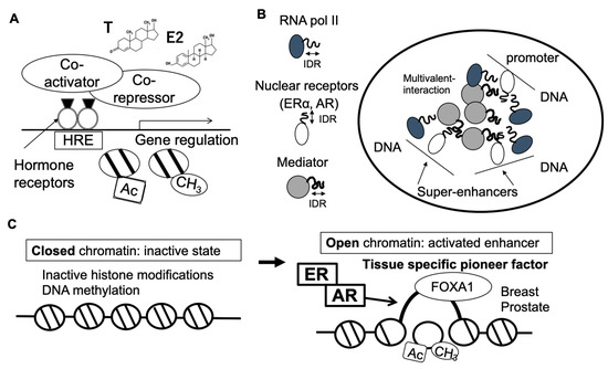
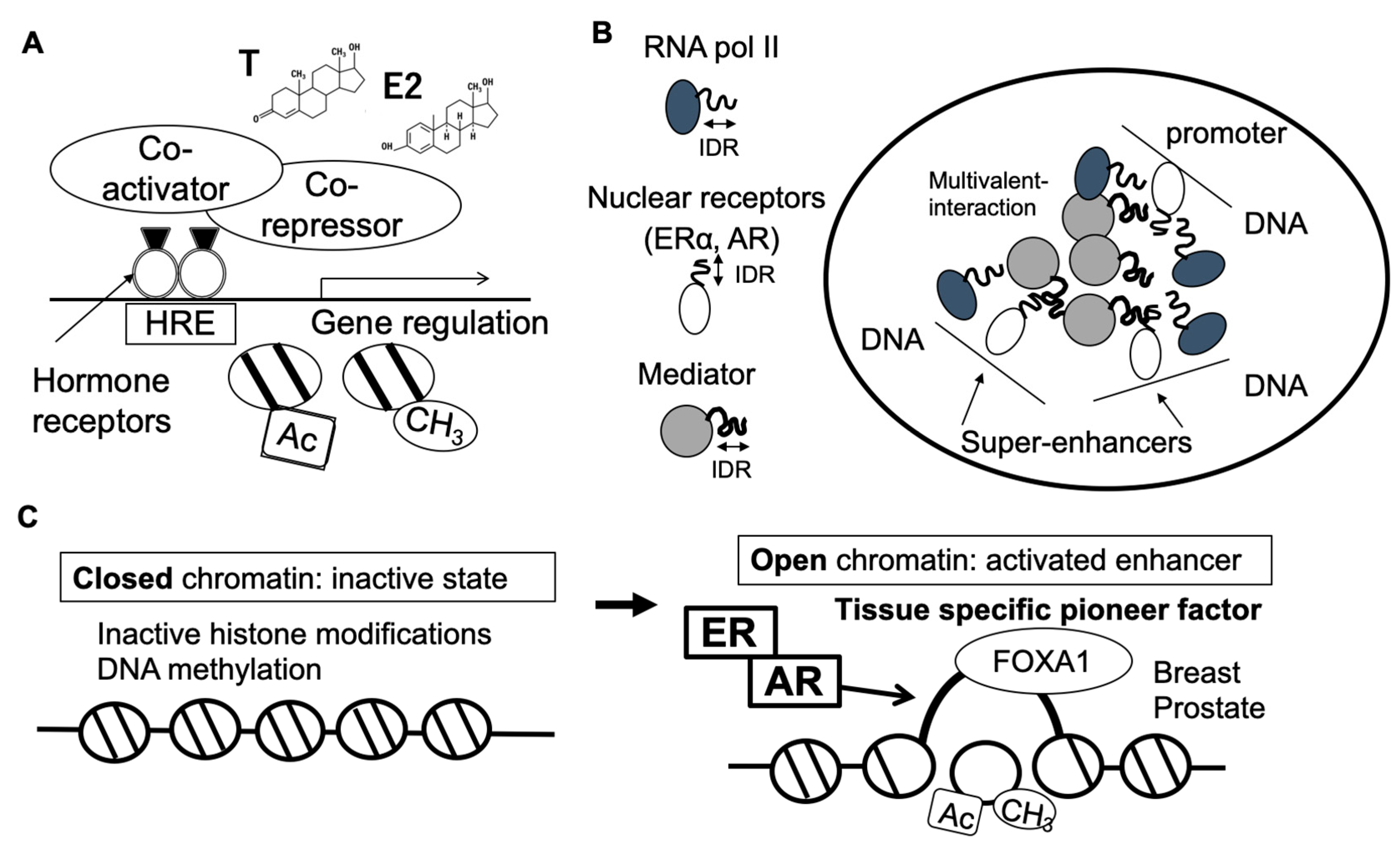
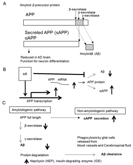
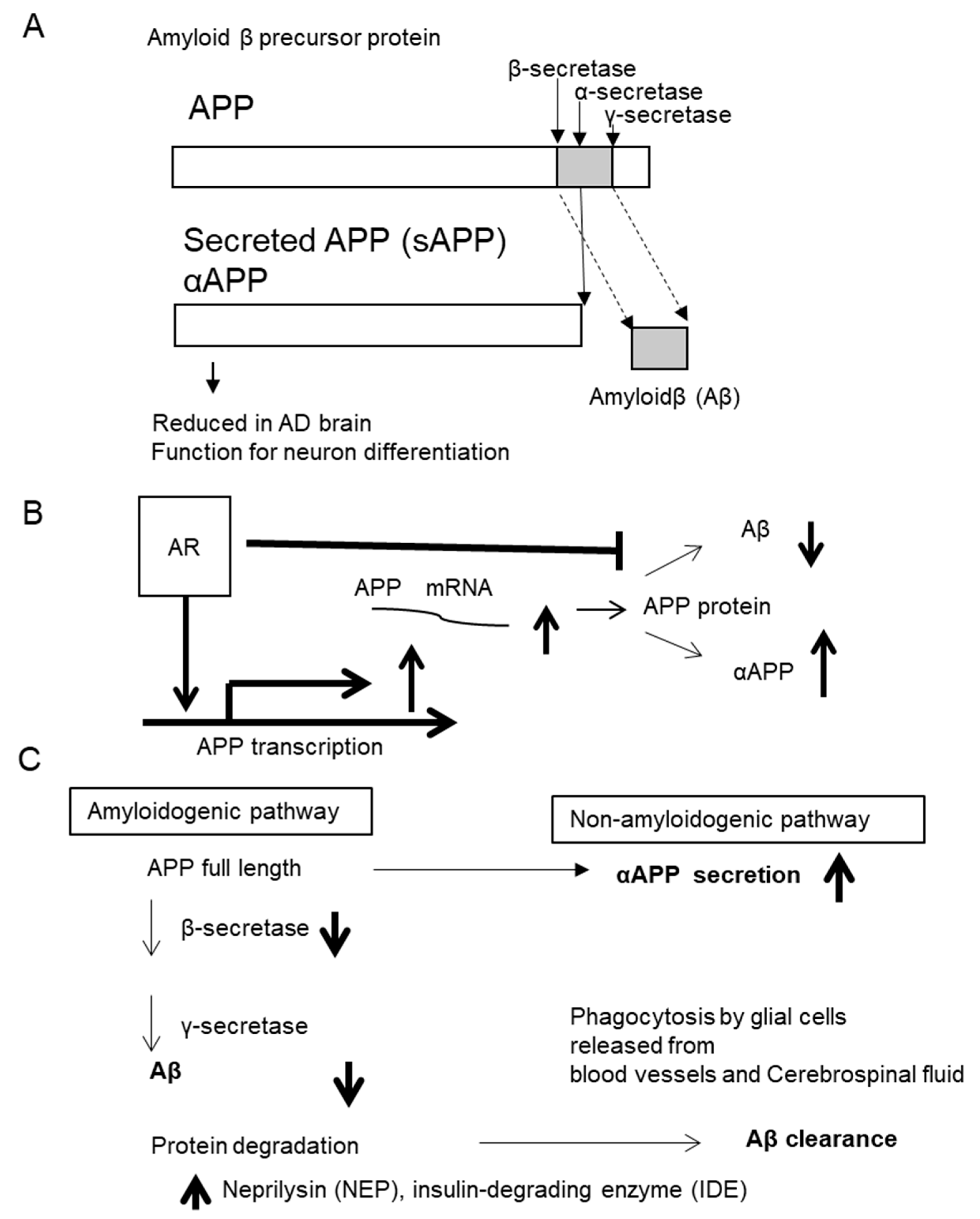

Disclaimer/Publisher’s Note: The statements, opinions and data contained in all publications are solely those of the individual author(s) and contributor(s) and not of MDPI and/or the editor(s). MDPI and/or the editor(s) disclaim responsibility for any injury to people or property resulting from any ideas, methods, instructions or products referred to in the content. |
© 2026 by the author. Licensee MDPI, Basel, Switzerland. This article is an open access article distributed under the terms and conditions of the Creative Commons Attribution (CC BY) license.