


Abstract
Background: Acute myeloid leukemia (AML) is an aggressive hematologic malignancy in which relapse and treatment resistance remain major clinical challenges. Ferroptosis, an iron-dependent form of regulated cell death driven by lipid peroxidation, has emerged as a potential therapeutic vulnerability in AML. Objectives: This study aimed to systematically evaluate ferroptosis-related mechanisms, therapeutic targeting strategies, and their translational relevance in AML. Methods: This systematic review followed PRISMA 2020 and was prospectively registered in PROSPERO. Eligible studies were primary experimental or translational investigations using human AML cell lines and/or adult patient-derived or ex vivo AML material. In vitro components were appraised descriptively using QUIN domains, whereas patient-derived evidence was assessed using structured methodological domains. Ferroptosis-specific confirmation and translational relevance were evaluated separately. Results: Eighteen studies met the eligibility criteria. Recurrent mechanisms involved the SLC7A11/GSH/GPX4 axis, NRF2-mediated redox adaptation, iron handling and ferritinophagy, lipid remodeling, ceramide metabolism, and nanoparticle-mediated redox disruption. Eight studies met the review-defined criteria for confirmed ferroptosis, seven were strongly supportive, and three were suggestive or ferroptosis-associated. Several approaches reduced AML cell viability or enhanced sensitivity to established therapies. No clinical intervention studies were identified. Conclusions: Ferroptosis-related pathways represent biologically relevant and potentially exploitable preclinical vulnerabilities in AML. However, evidence remains heterogeneous and predominantly preclinical, warranting standardized ferroptosis-specific validation and larger patient-derived studies before clinical translation.
1. Introduction
Acute myeloid leukemia (AML) is an aggressive and biologically heterogeneous hematologic malignancy characterized by the clonal expansion of immature myeloid progenitor cells (“blasts”) in the bone marrow and/or peripheral blood. Accumulation of leukemic blasts suppresses normal hematopoiesis, resulting in anemia, neutropenia, and thrombocytopenia [1]. Despite substantial advances in intensive chemotherapy, targeted therapies, and supportive care, long-term survival remains limited, particularly among older patients and those with adverse-risk genetic features [1,2]. Allogeneic hematopoietic stem cell transplantation remains an important potentially curative strategy for selected patients with high-risk disease; nevertheless, relapse continues to represent a major cause of treatment failure [1,3].
From a biological perspective, AML blasts frequently exhibit elevated levels of reactive oxygen species (ROS), which can promote DNA damage, genomic instability, and activation of oncogenic signaling pathways, including PI3K/AKT and MAPK. Concurrently, leukemic cells rely on antioxidant systems, particularly glutathione-dependent defenses, to tolerate this increased oxidative burden. This redox adaptation supports leukemic cell survival while creating a potentially exploitable metabolic vulnerability [4].
This redox adaptation provides a mechanistic link between AML biology and ferroptosis. Ferroptosis is an iron-dependent form of regulated cell death driven by lipid peroxidation and is therefore closely connected to both iron handling and oxidative stress. The elevated basal oxidative state and metabolic reprogramming observed in AML may render AML cells more susceptible to interventions. Accordingly, integrating redox biology with ferroptosis-related mechanisms may reveal therapeutically relevant vulnerabilities, particularly in relapsed or treatment-resistant AML [4,5].
1.1. Acute Myeloid Leukemia (AML)
1.1.1. Definition, Classification, Prognosis and Risk Factors
Acute myeloid leukemia has conventionally been defined by the presence of ≥20% myeloid blasts in the bone marrow or peripheral blood, although exceptions have long existed for selected recurrent genetic abnormalities. Contemporary World Health Organization (WHO) and International Consensus Classification (ICC) criteria place greater emphasis on genetically defined disease entities and permit the diagnosis of AML below the traditional 20% blast threshold in several settings. However, the two classification systems differ in their specific blast-count requirements [3,6]. Genomic heterogeneity is a defining feature of AML, necessitating cytogenetic and molecular characterization for accurate disease classification.
Both the WHO 2022 and ICC 2022 classifications place substantial emphasis on recurrent genetic abnormalities while retaining morphologic and clinical features as complementary diagnostic criteria [3,6]. Core-binding-factor rearrangements and NPM1 mutations define specific AML entities in both classification systems, whereas the criteria for CEBPA-mutated AML differ between the WHO and ICC. The WHO recognizes AML with CEBPA mutation, including biallelic CEBPA mutations and selected single bZIP-region mutations, and retains a ≥20% blast requirement for this entity. In contrast, the ICC specifically recognizes AML with in-frame bZIP CEBPA mutations, generally using a ≥10% blast threshold. Blast-count requirements therefore vary among individual genetically defined entities and between classification systems [3,6].
In addition, the 2022 ELN classification stratifies AML into favorable-, intermediate, and adverse-risk groups according to cytogenetic and molecular abnormalities [1]. Favorable-risk lesions include core-binding-factor AML, NPM1-mutated AML without FLT3-ITD in the absence of adverse-risk cytogenetic abnormalities, and in-frame bZIP CEBPA mutations. FLT3-ITD AML without adverse-risk genetic lesions and t(9;11)/MLLT3::KMT2A are classified as intermediate risk, whereas other KMT2A rearrangements, adverse cytogenetic abnormalities, TP53 mutations, and mutations in myelodysplasia-related genes are assigned to the adverse-risk group [1]. AML may arise de novo, following a pre-existing myeloid neoplasm, or after prior cytotoxic therapy. Environmental exposures, including tobacco smoke, ionizing radiation, and benzene, have also been associated with an increased risk of AML [1].
The marked molecular heterogeneity and redox-associated metabolic complexity of AML underscore the need for therapeutic strategies that exploit disease-specific metabolic and oxidative vulnerabilities. A more precise understanding of redox dysregulation, iron handling, and ferroptosis susceptibility may therefore facilitate the identification of therapeutically actionable targets, particularly in high-risk, relapsed, or treatment-resistant AML.
1.1.2. Reactive Oxygen Species (ROS) and Acute Myeloid Leukemia (AML) Pathogenesis
Reactive oxygen species (ROS) comprise reactive oxygen-derived radicals and non-radical species generated during cellular metabolism. Major ROS include superoxide (O2•−), hydrogen peroxide (H2O2), hydroxyl radicals (•OH), and singlet oxygen (1O2), whereas reactive nitrogen species including nitric oxide (NO•) and peroxynitrite (ONOO−) constitute a related group of reactive molecules that also contribute to cellular redox biology. At low-to-moderate concentrations, reactive species participate in physiological signaling. However, when their production exceeds cellular antioxidant capacity, oxidative stress develops [7,8,9,10,11]. Thus, under physiological conditions, ROS act as essential signaling mediators that have a role in the regulation of gene expression, proliferation, differentiation and adaptive stress responses. This controlled signaling role is referred to as “oxidative eustress” in contrast with “oxidative distress”, in which the uncontrolled accumulation of ROS results in molecular damage, disrupting cellular homeostasis by causing membrane disruption, enzyme dysfunction and DNA mutations [7,12].
This pathological imbalance is particularly relevant in the context of AML pathogenesis, as it plays a dual role in disease initiation and progression. Elevated ROS levels in hematopoietic stem and progenitor cells can induce DNA damage, mutations, and chromosomal instability, thereby contributing to leukemogenesis. In an oxidative microenvironment, the aberrant signaling pathways enhance the survival and proliferation of leukemic blasts while also impairing normal differentiation processes [8]. AML cells maintain elevated ROS levels that support oncogenic signaling while relying on antioxidant systems to prevent lethal oxidative damage. At the same time, this redox vulnerability lays the foundation for a therapeutic approach in which additional ROS-inducing agents or antioxidant inhibitors can push AML cells beyond a tolerable oxidative threshold. Overall, ROS biology contributes to AML pathogenesis while also providing potential therapeutic vulnerabilities [9].
1.2. Ferroptosis: Definition and Correlation with AML
Ferroptosis is an iron-dependent form of regulated cell death driven by the accumulation of phospholipid hydroperoxides and failure of lipid-peroxide detoxification [5,13]. A central protective mechanism is the cystine/glutamate antiporter–glutathione–GPX4 axis. GPX4 uses glutathione to reduce phospholipid hydroperoxides, thereby limiting oxidative membrane damage; consequently, loss of GPX4 activity or depletion of glutathione can promote ferroptotic cell death [12,13].
Iron contributes to ferroptosis by facilitating radical-generating reactions that promote lipid peroxidation. Morphologically, ferroptotic cells exhibit mitochondrial shrinkage, increased membrane density and reduced or absent mitochondrial cristae, while nuclear integrity is relatively maintained, a trait that distinguishes ferroptosis from other forms of regulated cell death [13,14,15]. Given the dependence of ferroptosis on redox homeostasis, lipid metabolism, and iron availability, pharmacological manipulation of ferroptosis-regulatory pathways has attracted increasing interest as a potential therapeutic strategy in hematologic malignancies [4,10,14,16,17].
Ferroptosis can be pharmacologically modulated through agents that alter cystine uptake, GPX4 activity, lipid-peroxide detoxification, or iron-dependent redox balance. Erastin inhibits system Xc- (SLC7A11/SLC3A2), thereby reducing cystine uptake, depleting intracellular glutathione, and indirectly impairing GPX4 activity [18]. RSL3 disrupts GPX4-dependent ferroptosis defense and promotes the accumulation of phospholipid hydroperoxides [19]. FIN56 promotes ferroptosis through GPX4 degradation and perturbation of the mevalonate pathway, including activation of squalene synthase, thereby increasing susceptibility to lipid peroxidation [5]. Ferroptosis-targeting strategies have also been investigated in other hematologic malignancies, supporting broader interest in this form of regulated cell death [20]. Ferrostatin-1 and liproxstatin-1 are commonly used as ferroptosis-directed rescue agents, whereas iron chelators such as deferoxamine provide complementary mechanistic support by limiting iron-dependent oxidative injury. Functional rescue is most informative when interpreted together with cell-death and lipid-peroxidation evidence [12,13].
AML cells may be particularly susceptible to ferroptosis because of altered iron metabolism, elevated basal ROS levels, and dependence on antioxidant defense systems [21]. Evidence suggests that leukemic blasts and leukemic stem cells depend on antioxidant homeostasis to survive within the oxidative bone marrow microenvironment, a trait that may increase their vulnerability to disruptions in ferroptosis-regulating pathways. Inhibition of GPX4 or blockade of system Xc- induces selective ferroptotic death in AML cells, while, at the same time, under certain conditions, normal hematopoietic populations are spared. Furthermore, increased expression of ferroptosis-associated regulators has been associated with poor prognosis and chemoresistance in AML patients, supporting the biological and potential prognostic relevance of ferroptosis-regulatory pathways in AML [10,17,22,23]. Consequently, the emerging evidence supporting the involvement of ferroptosis in AML pathobiology highlights its potential clinical significance and underscores the need for further systematic investigation into ferroptosis-related mechanisms and therapeutic interventions [24,25].
1.3. Aim and Rationale of the Review
This systematic review aimed to synthesize contemporary evidence on ferroptosis-related signaling pathways, pharmacological modulators, antioxidant defense systems, and iron-dependent metabolic vulnerabilities in AML, with particular emphasis on their therapeutic and translational implications for high-risk, relapsed, or refractory disease.
2. Materials and Methods
2.1. Protocol and Registration
This systematic review was conducted in accordance with the Preferred Reporting Items for Systematic Reviews and Meta-Analyses (PRISMA) guidelines [26] and the updated PRISMA 2020 statement. The complete PRISMA checklist is provided as Supplementary Table S1. Prior to the initiation of the review process, a detailed protocol was developed and registered in the International Prospective Register of Systematic Reviews (PROSPERO; registration number CRD420261373662). Any deviations from the original protocol were clearly described and justified in the present manuscript.
2.2. Research Question and Objectives
The primary objective of this systematic review was to evaluate the therapeutic effects of ferroptosis induction and ferroptosis modulation in acute myeloid leukemia (AML), with particular emphasis on evidence derived from established human AML cell lines and adult patient-derived or ex vivo AML material.
The secondary objectives were to: (i) investigate the role of major ferroptosis-associated regulators, including GPX4, SLC7A11, ACSL4, NRF2, and proteins involved in iron metabolism, AML cell survival, therapeutic response, and treatment resistance; (ii) assess the effects of ferroptosis-modulating agents, including erastin, FIN56, RSL3, sorafenib, sulfasalazine, ferrostatin-1, liproxstatin-1, and deferoxamine, on relevant biological and therapeutic outcomes; (iii) evaluate whether ferroptosis-targeting strategies enhance the efficacy of conventional chemotherapy, targeted therapies, or other anti-leukemic treatments; (iv) synthesize the molecular and metabolic mechanisms associated with ferroptosis sensitivity and resistance in AML, including glutathione metabolism, oxidative stress, iron homeostasis, mitochondrial dysfunction, lipid peroxidation, and antioxidant defense; and (v) summarize the translational and therapeutic potential of ferroptosis modulation in AML to inform future preclinical and clinically relevant investigations.
2.3. Eligibility Criteria
The eligibility criteria for the current review were defined a priori using the PICOSE (Population, Intervention, Comparator, Outcome, Study Design, Exclusion Criteria) framework (Table 1). Studies were considered eligible for inclusion if they were primary experimental or translational investigations reporting original data on ferroptosis-related mechanisms or therapeutic targeting strategies in Acute Myeloid Leukemia (AML). Eligible studies were required to use established human AML cell lines and/or adult patient-derived or ex vivo AML material. Eligible designs included studies investigating ferroptosis-associated pathways or molecular regulators and studies evaluating pharmacological modulation of ferroptosis-related targets.
Table 1.
Eligibility criteria according to the PICOSE framework.
Studies were excluded if they were secondary publications or did not report original experimental data. Excluded publication types included systematic reviews; meta-analyses; umbrella reviews; narrative reviews; editorials; scoping reviews; commentaries; expert opinions; posters; protocols; database-mining studies; clinical guidelines; book chapters; letters without original data; case reports; small descriptive case series and conference abstracts lacking sufficient extractable information; purely bioinformatic, in silico, proteomic, genomic or machine-learning studies without experimental validation in human-derived AML models; and clinical studies with secondary reference to ferroptosis. Furthermore, animal-only studies without human-derived AML material were excluded. The age criterion was also applied; therefore, studies restricted exclusively to pediatric AML populations were excluded. Finally, studies without AML-specific findings were also excluded from the review.
To ensure a comprehensive and methodologically rigorous study selection process, manual screening of the reference lists of the included studies and relevant review articles was conducted to identify additional eligible publications. Forward and backward citation tracking procedures were also performed to ensure that any potentially relevant study that was not retrieved through the initial database search was identified and assessed for eligibility. All eligibility criteria and study selection procedures were predefined and implemented in accordance with the protocol registered in PROSPERO.
2.4. Search Strategy
A comprehensive literature search was conducted in MEDLINE (via PubMed), Scopus, the Cochrane Library (CENTRAL), and the Science Citation Index Expanded. ClinicalTrials.gov and the World Health Organization International Clinical Trials Registry Platform (ICTRP) were additionally searched to identify potentially relevant ongoing or unpublished clinical studies.
The search strategy combined controlled vocabulary and free-text terms using Boolean operators and truncation where appropriate. The formal eligibility window extended from 1 January 2020 to 30 May 2026. The lower date limit was prespecified to focus the systematic synthesis on contemporary experimental and translational evidence reflecting recent developments in AML molecular characterization and ferroptosis-related therapeutic research. Studies published before 2020 were therefore excluded from the formal synthesis, although seminal earlier studies could be considered as contextual evidence where relevant. Detailed search strategies are provided in Supplementary Table S2.
The search strategy was developed by the review team and internally reviewed for methodological consistency, with reference to PRISMA and PRESS guidance. Reference lists of included studies and relevant reviews were screened manually, and backward and forward citation tracking was performed to maximize retrieval and reduce the risk of missing eligible evidence.
Only studies published in languages accessible to the review team (Greek, English, German, Italian, French, Turkish) were included in the analysis. Studies were not excluded solely on the basis of initial full-text inaccessibility; reasonable efforts were made to retrieve potentially eligible articles through available institutional and open-access resources.
2.5. Screening and Data Management
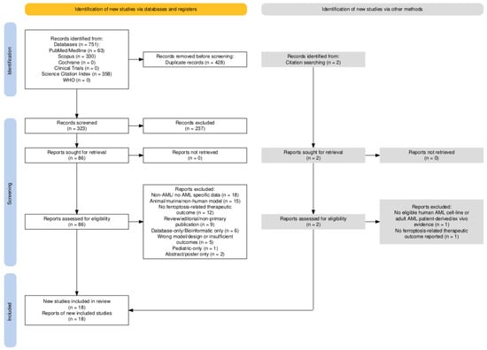
All retrieved records were imported into Rayyan (web-based platform; Rayyan Systems Inc., Cambridge, MA, USA) and deduplicated before screening. Two reviewers independently screened titles and abstracts against the predefined eligibility criteria, followed by independent full-text assessment of potentially eligible reports. Screening decisions remained concealed between reviewers during the independent assessment stage. Disagreements were resolved by consensus, with consultation of a senior third reviewer when necessary. Reasons for exclusion at full-text assessment were recorded systematically. The study-selection process is summarized in the PRISMA 2020 flow diagram (Figure 1).
Figure 1.
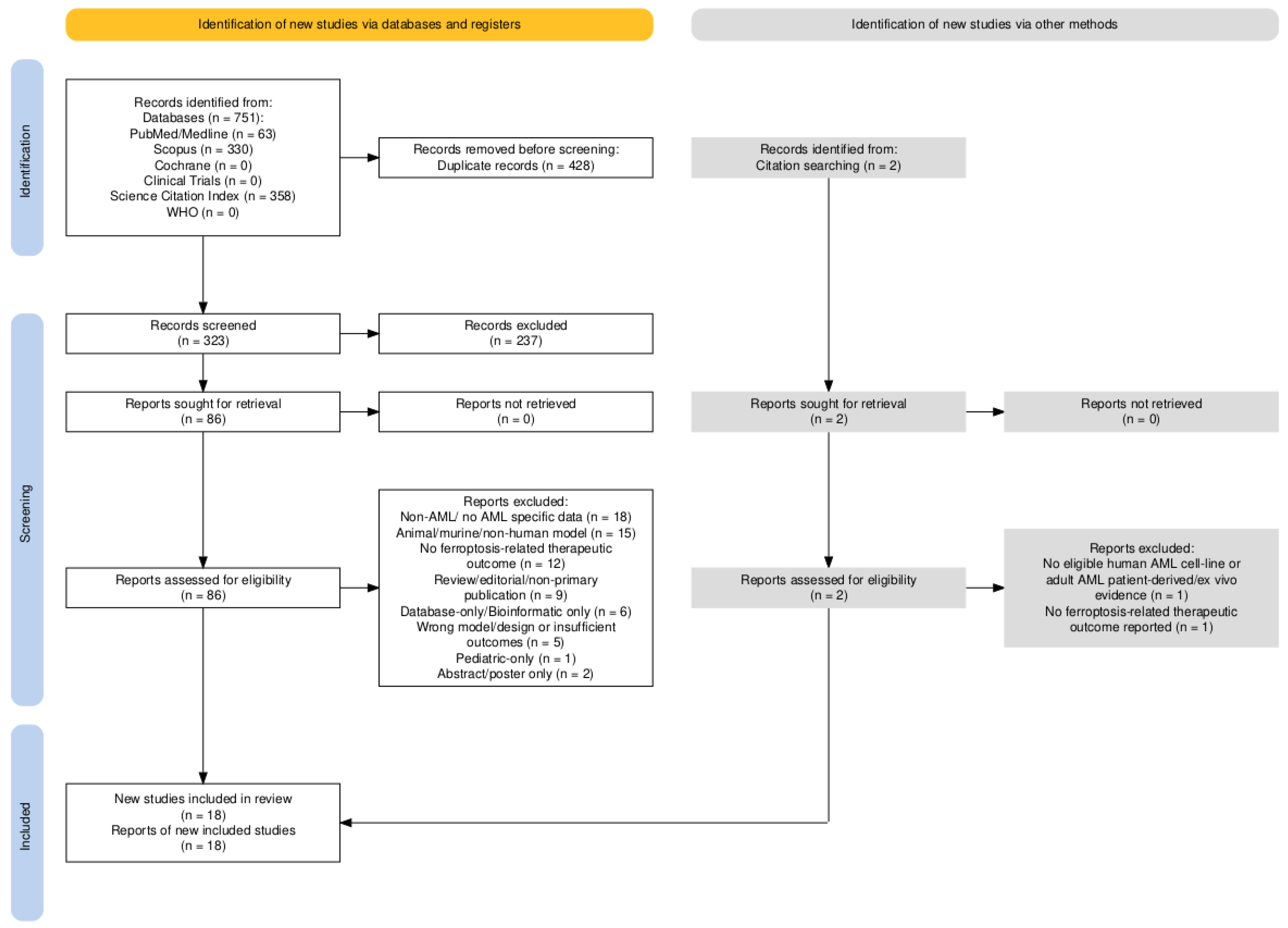
Search strategy flow diagram. Flow diagram developed according to the PRISMA 2020 guidelines for systematic reviews, illustrating the identification, screening, eligibility assessment, and inclusion of records retrieved from databases, registers, and additional sources. The flow diagram was created using Google Docs (web-based application; Google LLC., Mountain View, CA, USA).
2.6. Data Extraction
Following study selection, relevant data were systematically extracted in accordance with the review objectives. Extracted information included study identification, AML model type, eligible cell lines and/or patient-derived samples, disease context, intervention or exposure, comparator/control conditions, ferroptosis-related outcomes, therapeutic outcomes, and main findings.
Data were organized into structured tables, provided below, where human AML cell-line evidence was separated from human-derived or patient-derived AML evidence. Public datasets, such as TCGA, GEO/GSE series, UALCAN and BeatAML, when reported in the included studies, were recorded only as supportive evidence. Accordingly, murine cell lines, animal-derived models, xenografts/PDX-only data, normal comparator-only data, and database-only findings were not included in the main synthesis. Data extraction was performed at the evidence-component level. Therefore, when included studies contained both eligible and non-eligible data components, only data pertaining to human AML cell lines and/or adult AML patient-derived or ex vivo samples were incorporated into the main synthesis. Lastly, data extraction was performed independently by two reviewers, while any disagreements were resolved through discussion and consensus.
2.7. Methodological Appraisal
The methodological quality of the eligible experimental evidence was assessed independently from ferroptosis-specific confirmation and translational relevance. Given the predominantly mechanistic in vitro and ex vivo nature of the included studies, methodological quality was evaluated at the evidence-component level rather than by generating an overall numerical score or percentage-based quality category.
For human AML cell-line experiments, the QUIN framework [27] was used as a structured item-level appraisal framework. Although QUIN was originally developed and validated for the appraisal of in vitro studies in dentistry, it was applied in the present review solely as a structured item-level methodological framework. Given the absence of a validated AML-specific appraisal tool appropriate for the heterogeneous mechanistic in vitro designs included in this review, individual QUIN domains were interpreted separately and were not combined into an aggregate numerical score, percentage-based score, or overall risk-of-bias category. The appraisal considered study objectives, experimental-model selection and characterization, comparator and control groups, methodological reproducibility, outcome measurement, statistical analysis, completeness of reporting, sample-size justification, and procedures intended to reduce experimental bias, including randomization, blinding, and independent outcome assessment, where applicable.
Patient-derived and ex vivo AML evidence was appraised using structured methodological domains appropriate to experimental human-derived material. These included patient and sample characterization, number of independent biological samples, description and consistency of sample handling, definition of the experimental intervention and comparator conditions, appropriateness of outcome assessment, biological replication, statistical analysis, and completeness of reporting. Particular attention was given to distinguishing direct ex vivo therapeutic testing of primary AML samples from the use of patient-derived material solely for molecular, expression, prognostic, or supportive analyses.
Individual methodological domains were classified as adequately reported (A), partially or inadequately reported (P), not reported (NR), or not applicable (NA). These judgments were interpreted at the individual-domain level and were not combined into an overall numerical score, percentage-based score, or global low/moderate/high risk-of-bias category. The same domain-level approach was applied to patient-derived and ex vivo evidence, with assessment adapted to the characteristics of human-derived experimental material. These review-specific domains were used descriptively and were not treated as a separately validated risk-of-bias instrument.
2.8. Evidence Strength and Characterization
The strength of the experimental evidence was evaluated separately from methodological quality. Given that the included literature consisted predominantly of preclinical mechanistic studies using AML cell lines and patient-derived or ex vivo material, no formal GRADE certainty-of-evidence assessment was performed and no overall high, moderate, low, or very low certainty ratings were assigned.
Instead, the evidence was characterized according to three independent dimensions: (i) the degree of ferroptosis-specific experimental confirmation, (ii) reproducibility across independent AML models, and (iii) translational relevance of the experimental system.
Ferroptosis-specific confirmation was evaluated according to the extent to which the reported cell-death phenotype was supported by functional and mechanistic evidence specific to ferroptosis. Particular consideration was given to the demonstration of lipid peroxidation, functional cell-death or viability effects, rescue with established ferroptosis inhibitors such as ferrostatin-1 or liproxstatin-1, iron-chelation rescue, and genetic manipulation of established ferroptosis regulators. Changes in reactive oxygen species, malondialdehyde, intracellular iron, glutathione, GPX4, SLC7A11, or related molecular markers alone were not considered sufficient to establish ferroptotic cell death.
Reproducibility was evaluated according to whether the main findings were demonstrated across multiple independent AML cell lines, complementary experimental models, or primary AML samples rather than being restricted to a single experimental model. Translational relevance was assessed by distinguishing studies limited to established AML cell lines from those incorporating supportive patient-derived molecular or clinical data and those performing direct ex vivo therapeutic testing in primary AML samples.
These dimensions were evaluated independently and were not combined into an overall numerical score or global certainty rating. Accordingly, methodological quality, ferroptosis-specific confirmation, reproducibility, and translational relevance were considered complementary but conceptually distinct features of the evidence base.
For the purposes of evidence synthesis, ferroptosis-specific evidence was further classified into three categories. “Confirmed ferroptosis” required a functional cell-death or loss-of-viability phenotype accompanied by evidence of lipid peroxidation and functional reversal by a recognized ferroptosis-directed rescue intervention, particularly ferrostatin-1 or liproxstatin-1. Rescue by iron chelation, lipophilic radical-trapping antioxidants, or genetic manipulation of established ferroptosis regulators was considered additional mechanistic support.
Evidence was classified as “strongly supportive of ferroptosis” when a functional anti-leukemic effect was accompanied by multiple mechanistically consistent ferroptosis-related findings, such as lipid peroxidation, iron dysregulation, glutathione depletion, GPX4 or SLC7A11 modulation, or genetic manipulation of ferroptosis-regulatory pathways, but definitive ferroptosis-specific pharmacological rescue was not demonstrated. Study-level classification was based on the evidence supporting the principal therapeutic or mechanistic claim of each study; the use of a ferroptosis inhibitor in ancillary or combination experiments was not, by itself, considered sufficient for a study-level classification as confirmed ferroptosis.
Evidence was classified as “suggestive or ferroptosis-associated” when the proposed mechanism relied predominantly on changes in reactive oxygen species, malondialdehyde, intracellular iron, glutathione, GPX4, SLC7A11, or related molecular markers without sufficient functional ferroptosis-specific rescue. Such biomarker changes alone were not considered sufficient to establish ferroptotic cell death. These categories were developed solely to structure the narrative synthesis and should not be interpreted as validated certainty-of-evidence grades. Translational relevance was characterized separately as direct ex vivo patient-derived evidence, supportive human-derived evidence, or evidence restricted to experimental cell-line/preclinical models. Direct ex vivo evidence referred to studies in which primary AML cells or blasts underwent experimental therapeutic or mechanistic manipulation, whereas supportive human-derived evidence referred to the use of patient samples or clinical datasets primarily for molecular, expression, prognostic, or resistance-related analyses.
2.9. Use of Generative AI Tools
Generative AI tools (GPT-5.5; OpenAI, San Francisco, CA, USA) were used to assist with the initial drafting of graphical abstracts and schematic figures. All AI-generated text and visual elements were critically reviewed, modified, and validated against the original scientific sources by the authors. The final content, interpretation of findings, study selection, methodological appraisal, and conclusions were determined exclusively by the authors, who assume full responsibility for the manuscript.
3. Results
3.1. Eligible Studies—PRISMA Flow Diagram
Initial records were identified through database and register searches, according to the sources listed in the PROSPERO Protocol. A total of 751 records were identified from databases, including PubMed/Medline (n = 63), Scopus (n = 330) and the Science Citation Index (n = 358); no records were identified from registers. The remaining databases yielded no result, so no records were identified from Cochrane, ClinicalTrials.gov or WHO registries. After deduplication, 428 duplicate records were removed.
After duplicate removal, 323 records were screened by title and abstract. Of these, 237 records were excluded based on the predefined criteria listed in the PROSPERO protocol. The remaining 86 reports were sought for retrieval, and all of them were successfully retrieved. Therefore, 86 reports were assessed for eligibility.
Following final eligibility screening, which was performed by two independent reviewers, 68 reports were excluded. The main reasons for exclusion at full-text assessment were non-AML disease or absence of extractable AML-specific data (n = 18); animal-only, murine-cell-line-only, in vivo only, or other non-human models without eligible human AML component (n = 15); absence of ferroptosis-related therapeutic mechanism or outcome (n = 12); review, editorial, commentary, protocol, or other non-primary publication type (n = 9); database-only or bioinformatic-only design without eligible experimental validation (n = 6); wrong model or study design, or insufficient overall outcome data (n = 5); pediatric-only or non-adult AML population (n = 1); and, lastly, abstract/poster-only format with insufficient data (n = 2). Additionally, 2 reports identified through citation searching were excluded after the full-text assessment, as the first one did not include an eligible human AML cell-line or adult AML patient-derived or ex vivo population (n = 1), while the other did not report a ferroptosis-related therapeutic outcome (n = 1).
3.2. Methodological Quality and Characterization of the Evidence
The methodological appraisal of the in vitro experimental components is summarized in Table 2, whereas the appraisal and translational characterization of patient-derived evidence are presented in Table 3. Ferroptosis-specific experimental validation and translational relevance are summarized separately in Table 4 according to the criteria defined in Section 2.8. Across the included studies, study objectives, outcome-measurement methods, statistical analyses, and results were generally adequately reported. Formal sample-size calculations were generally not reported, while the reporting of model-selection or sampling procedures and the adequacy of comparator groups varied between studies. Several QUIN items related to operator details, randomization, outcome-assessor information, and blinding were recorded as not applicable to the respective experimental designs.
Table 2.
Item-level methodological appraisal of eligible in vitro AML experimental components using the QUIN framework.
Table 3.
Domain-level methodological appraisal and translational characterization of patient-derived AML evidence.
Table 4.
Ferroptosis-targeting strategies, experimental validation, and translational relevance in AML.
Methodological quality was evaluated independently from the degree of ferroptosis-specific confirmation. Considerable variation was observed in the experimental evidence used to support ferroptotic cell death. The strongest evidence was provided by studies combining a functional loss-of-viability or cell-death phenotype with assessment of lipid peroxidation and functional rescue using established ferroptosis-directed interventions, particularly ferrostatin-1 or liproxstatin-1. Iron-chelation rescue, antioxidant rescue, and genetic manipulation of established ferroptosis regulators were considered additional mechanistic support.
Other studies demonstrated functional anti-leukemic effects together with multiple ferroptosis-associated molecular or metabolic alterations, including changes in lipid peroxidation, intracellular iron, glutathione metabolism, GPX4, SLC7A11, or related regulatory pathways, but did not provide definitive ferroptosis-specific pharmacological rescue. These studies were therefore interpreted as strongly supportive of ferroptosis rather than confirmatory. Studies relying predominantly on changes in reactive oxygen species, malondialdehyde, intracellular iron, glutathione, GPX4, SLC7A11, or related biomarkers without adequate functional rescue were interpreted as suggestive or ferroptosis-associated evidence rather than definitive demonstration of ferroptotic cell death.
Translational relevance was also considered separately from methodological quality and ferroptosis-specific confirmation. Studies performing direct ex vivo therapeutic testing in primary AML samples were distinguished from studies using patient-derived material or clinical datasets only for molecular, expression, prognostic, or resistance-related analyses, as well as from studies restricted to established AML cell lines. This distinction was retained throughout the evidence synthesis to avoid interpreting all human-derived evidence as equivalent to direct patient-derived therapeutic validation.
Among the studies incorporating patient-derived material, eight included direct ex vivo therapeutic or mechanistic testing of primary AML samples, whereas four used patient-derived material primarily for molecular, expression, prognostic, or other supportive analyses. Reporting of patient/sample origin and the number of independent biological samples was generally adequate, although the level of detail regarding sample processing and clinical characterization varied across studies. Direct ex vivo evidence was therefore distinguished from supportive human-derived evidence throughout the synthesis and was not considered equivalent in translational relevance.
3.3. Main Results of the Included Studies
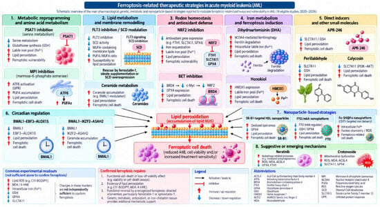
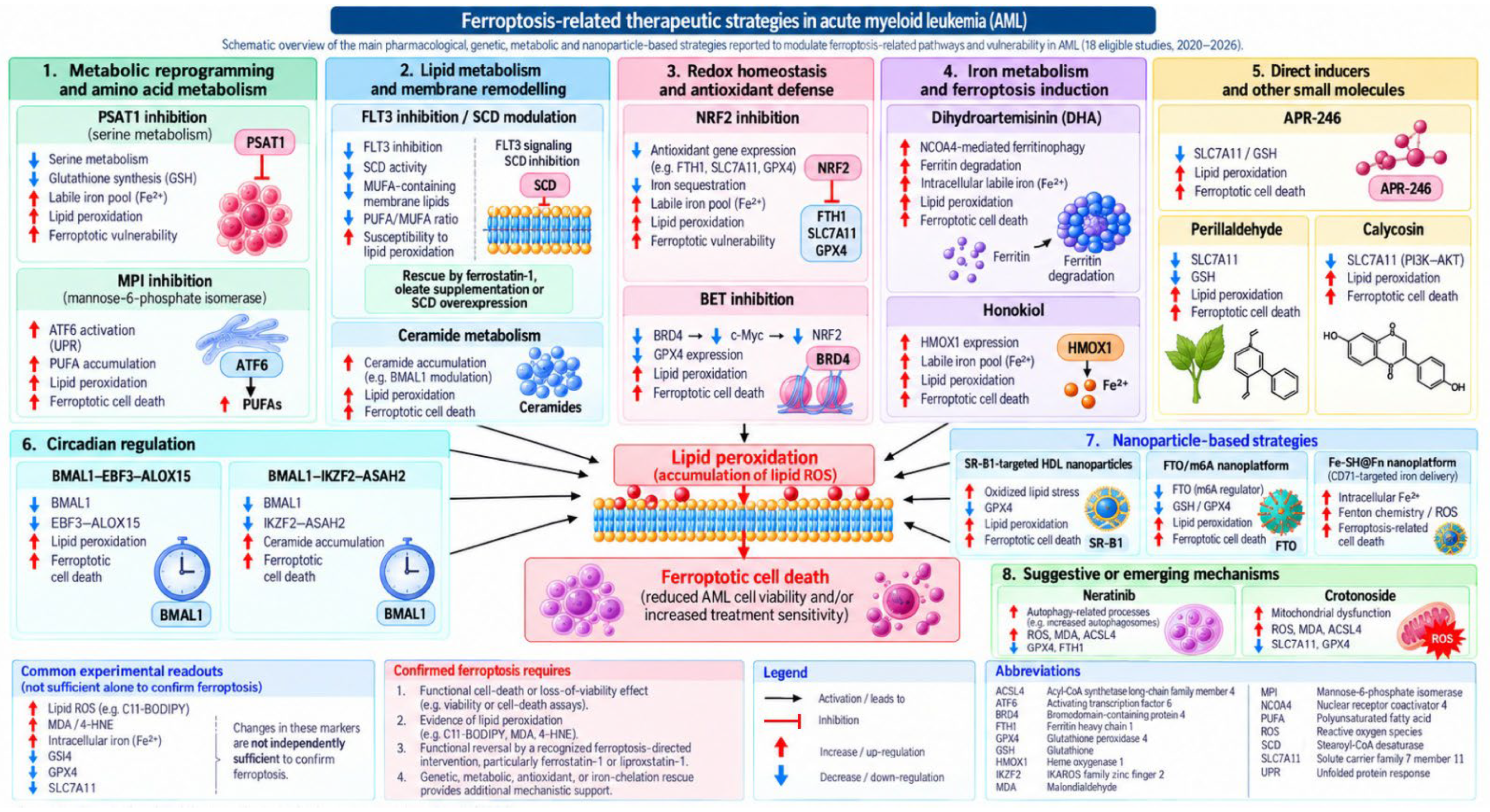
The main findings of the included studies are summarized in Table 4 according to experimental model, intervention, ferroptosis-specific validation, evidence category, and translational relevance. The approaches targeting ferroptosis are summarized in Figure 2. Additional details on the experimental models and human-derived components, principal pathways and interventions, controls/comparators, assays and ferroptosis-related validation, therapeutic findings, evidence categories, and analytical interpretation are provided in Supplementary Table S4.
Figure 2.
Ferroptosis-related therapeutic strategies in acute myeloid leukemia. Schematic summary of the principal pharmacological, metabolic, genetic, and nanoparticle-based mechanisms that modulate ferroptosis-related vulnerability in AML.
4. Discussion
The present systematic review indicates that ferroptosis-related vulnerabilities in acute myeloid leukemia converge on a relatively limited number of interconnected metabolic and redox pathways rather than on a single therapeutic mechanism. Across the included studies, the most consistently implicated processes involved the SLC7A11/GSH/GPX4 antioxidant axis, NRF2-mediated redox adaptation, iron handling and ferritinophagy, lipid remodeling, and metabolic mechanisms associated with treatment resistance. Collectively, these findings support the biological relevance of ferroptosis-related pathways in AML while also demonstrating substantial heterogeneity in the experimental strategies used to exploit them.
A central finding of the review was the recurrent dependence of AML cells on antioxidant systems that limit lipid-peroxide accumulation. Disruption of cystine uptake, glutathione availability, GPX4 activity, or upstream regulatory pathways repeatedly increased susceptibility to oxidative and lipid-peroxidative stress across different AML models [28,29,30,31,32,33,34]. However, the revised evidence assessment indicates that convergence on the same pathway should not be interpreted as equivalent evidence of ferroptotic cell death. Studies such as those by Birsen et al. [28], Catanzaro et al. [29], and Liu et al. [31] incorporated functional cell-death outcomes together with lipid-peroxidation-related changes and rescue approaches, providing comparatively stronger ferroptosis-specific confirmation. In contrast, other studies demonstrated biologically consistent alterations in iron, reactive oxygen species, glutathione, GPX4, SLC7A11, or related regulators without equally definitive ferroptosis-specific functional rescue. These latter findings remain mechanistically informative, but their interpretation requires greater caution.
This distinction is particularly important because biochemical changes commonly associated with ferroptosis are not independently sufficient to establish ferroptotic cell death. Increased reactive oxygen species, malondialdehyde, or intracellular iron or changes in glutathione, GPX4, and SLC7A11 may accompany ferroptosis, but they may also occur during broader oxidative or metabolic stress. Accordingly, the strongest evidence in this review was derived from studies that combined functional loss of viability or cell death with lipid-peroxidation assessment and reversal by ferroptosis-directed rescue interventions. Using the revised evidence framework, eight of the eighteen included studies met the review-defined criteria for confirmed ferroptosis, seven were classified as strongly supportive, and three as suggestive or ferroptosis-associated. This stratification provides a more conservative interpretation of the literature and avoids treating mechanistically heterogeneous studies as providing the same level of evidence.
Within the antioxidant-defense network, NRF2 emerged as an important mediator of ferroptosis resistance and therapeutic adaptation. Yu et al. [30] showed that inhibition of NRF2 increased venetoclax-associated iron and lipid-peroxidative stress, whereas Liu et al. [31] provided stronger functional evidence by combining NRF2 and GPX4 targeting with ferroptosis-specific rescue in AML cell-line models. Together with the PSAT1-mediated resistance mechanism described by Zhu et al. [34], these findings suggest that resistance to apoptosis-directed therapy may coexist with, or potentially generate, dependence on ferroptosis-protective metabolic pathways. This observation is therapeutically relevant because ferroptosis modulation may therefore be most valuable not as an isolated cytotoxic strategy, but as a mechanism for sensitizing metabolically adapted or treatment-resistant AML cells to established therapies.
Lipid metabolism represented a second major area of mechanistic consistency. Sabatier et al. [39] linked FLT3-mutant AML to dependence on fatty-acid anabolic and desaturation pathways that protect against lipid oxidative stress, whereas Woodley et al. [41] demonstrated that disruption of mannose metabolism increased polyunsaturated fatty-acid accumulation, lipid peroxidation, and ferroptotic susceptibility. Zheng et al. [40] further connected circadian regulation and ceramide metabolism to GPX4 degradation and sorafenib sensitivity, while Wang et al. [38] identified a BMAL1/EBF3/ALOX15-related mechanism influencing lipid oxidation. Although these studies examined different upstream regulators, they converge on a common biological principle: the lipid composition and antioxidant capacity of AML cells substantially influence their susceptibility to ferroptosis-related stress. This consistency across mechanistically distinct interventions strengthens the interpretation that lipid metabolic adaptation is an important determinant of ferroptosis sensitivity in AML.
Iron metabolism and iron mobilization formed another recurrent mechanistic theme. Grignano et al. [36] showed that dihydroartemisinin promoted ferritinophagy and increased intracellular labile iron, accompanied by C11-BODIPY-detected lipid peroxidation. DHA-mediated cell death was strongly inhibited by ferrostatin-1 and the iron chelator deferasirox, thereby meeting the review-defined criteria for confirmed ferroptosis. The study further implicated MT2A/MT1M-dependent antioxidant adaptation, whereas experiments in primary AML cells provided direct ex vivo mechanistic support; however, the strongest ferroptosis-specific rescue evidence was obtained in AML cell-line models. Lai et al. [35] demonstrated HMOX1-dependent lipid peroxidation with partial rescue by ferrostatin-1 and HMOX1 inhibition. Together, these findings indicate that ferroptosis-related vulnerability in AML may arise not only through direct impairment of GPX4-dependent lipid-peroxide detoxification but also through increased availability of redox-active iron. By contrast, the neratinib study by Ma et al. [25] and the crotonoside study by Ma et al. [43] primarily demonstrated ferroptosis-associated biochemical and molecular changes without equally definitive ferroptosis-specific rescue. These studies therefore support a possible mechanistic connection between autophagy, mitochondrial dysfunction, iron handling, and ferroptosis, but provide less definitive evidence than studies incorporating functional rescue.
An important antecedent to this contemporary literature is the 2019 study by Trujillo-Alonso et al. [44], which falls outside the prespecified publication window of the present systematic synthesis. That work demonstrated that low ferroportin expression increased susceptibility of leukemia cells to ferumoxytol-mediated intracellular iron accumulation, oxidative stress, and cell death. Although the study was not included among the formally eligible studies because it preceded the January 2020 cutoff, it provides relevant mechanistic context for the subsequent development of iron-dependent therapeutic strategies in AML. Importantly, its findings are better interpreted as foundational evidence of iron-driven oxidative vulnerability than as definitive ferroptosis according to the stricter ferroptosis-specific validation criteria applied in the present review. This contextual distinction preserves the predefined eligibility framework while acknowledging the contribution of earlier mechanistic work.
The translational relevance of the evidence was similarly heterogeneous. Several studies incorporated direct ex vivo experimental testing of primary AML cells, including Birsen et al. [28], Catanzaro et al. [29], Liu et al. [31], Zhong et al. [32], Grignano et al. [36], Lin et al. [24], Sabatier et al. [39], and Woodley et al. [41]. These studies provide stronger translational support than investigations in which patient-derived material was used mainly for expression, prognostic, or resistance-associated analyses. Nevertheless, even direct ex vivo evidence was generally based on relatively small patient samples and short-term experimental outcomes. Therefore, the presence of human-derived material should not be interpreted uniformly as evidence of clinical efficacy. Rather, the included literature spans a continuum from mechanistic cell-line studies to direct patient-derived experimental validation, with no clinical intervention studies demonstrating therapeutic benefit in patients.
The most clinically relevant findings may therefore lie in combination strategies rather than in ferroptosis induction as monotherapy. Across the included studies, ferroptosis-related modulation enhanced or restored sensitivity to venetoclax, cytarabine, FLT3 inhibitors, sorafenib, and BET inhibition [30,31,32,39,40,41]. This pattern suggests that ferroptosis-targeting approaches may be particularly useful in AML subtypes characterized by metabolic adaptation or acquired treatment resistance. The recurrence of treatment-sensitization effects across distinct pathways also supports the concept that ferroptosis may function as a complementary therapeutic vulnerability rather than as a uniform standalone death mechanism.
Nanoparticle-based approaches further extend this concept by attempting to improve intracellular delivery, selectivity, and redox disruption. The HDL-based strategy evaluated by Lin et al. [24] is particularly relevant because it combined direct activity in patient-derived AML material with relative sparing of normal CD34-positive cells. Yang et al. [37] and Cao et al. [42] also demonstrated that nanoparticle-mediated modulation of iron handling or glutathione/GPX4-related defenses can produce substantial anti-leukemic effects. However, these approaches remain at an early preclinical stage, and the extent to which their activity is specifically attributable to ferroptosis varies between studies. Their future translational value will therefore depend not only on ferroptosis-specific validation but also on pharmacokinetics, selectivity for leukemic over normal hematopoietic cells, reproducibility, and feasibility of clinical delivery.
Taken together, the evidence supports ferroptosis-related pathways as biologically meaningful therapeutic vulnerabilities in AML, but the strength of evidence is not uniform across the literature. The most convincing studies combine functional cell-death outcomes, lipid-peroxidation assessment, mechanistic perturbation, ferroptosis-directed rescue, and direct testing in patient-derived material. Other studies provide important mechanistic support but remain less definitive because they rely primarily on ferroptosis-associated biomarkers or cell-line models. Future research should therefore prioritize standardized ferroptosis-specific validation, larger and well-characterized adult AML patient-derived cohorts, resistant and relapse-associated models, and clinically relevant combination strategies. Particular attention should also be given to selectivity against normal hematopoietic cells and to the identification of biomarkers capable of defining ferroptosis-sensitive AML subgroups. Such work will be necessary to determine whether the substantial preclinical promise of ferroptosis modulation can be translated into clinically meaningful therapeutic benefit.
5. Conclusions
The available evidence supports ferroptosis-related pathways as biologically relevant and potentially exploitable therapeutic vulnerabilities in acute myeloid leukemia. Across the included studies, recurrent mechanisms involved disruption of the SLC7A11/GSH/GPX4 antioxidant axis, NRF2-mediated redox adaptation, iron handling, lipid peroxidation, fatty-acid remodeling, and other metabolic pathways associated with treatment resistance.
However, the strength of ferroptosis-specific evidence varied substantially between studies. Only a subset combined functional cell-death outcomes, lipid-peroxidation assessment, and ferroptosis-directed rescue, whereas others relied predominantly on molecular or biochemical changes consistent with ferroptosis. Accordingly, the available literature should not be interpreted as providing uniform confirmation of ferroptotic cell death across all investigated interventions.
Translational evidence also remains limited. Although several studies incorporated direct ex vivo testing of primary AML material, much of the evidence was derived from established AML cell lines, and other patient-derived analyses were primarily molecular, prognostic, or supportive rather than therapeutic. No clinical intervention studies have yet established the efficacy or safety of ferroptosis-targeting strategies in patients with AML.
Overall, ferroptosis modulation should therefore be considered a promising preclinical and hypothesis-generating therapeutic strategy, rather than an established clinical approach. Future studies should prioritize standardized ferroptosis-specific assays, rigorous functional rescue experiments, larger and well-characterized patient-derived AML models, assessment of selectivity against normal hematopoietic cells, and clinically relevant combination strategies. These studies will be necessary to determine whether ferroptosis-targeting approaches can ultimately provide meaningful therapeutic benefit in AML.
6. Limitations
Several limitations should be considered when interpreting the findings of this review. Most included studies were preclinical, and much of the evidence was derived from established AML cell-line models. Although several investigations incorporated primary AML cells, patient-derived samples, or direct ex vivo experimental testing, the methodological depth and translational relevance of the human-derived evidence were heterogeneous. Studies also differed substantially in the AML models examined, ferroptosis-modulating interventions, treatment concentrations, exposure durations, comparator conditions, and outcome measures.
Ferroptosis was likewise not evaluated uniformly across studies. Whereas some investigations combined functional cell-death or viability outcomes with lipid-peroxidation assessment and ferroptosis-directed rescue experiments, others relied on a narrower set of biochemical or molecular markers, including ROS, MDA, intracellular iron, GPX4, or SLC7A11. This methodological heterogeneity limits direct comparisons across studies and complicates evidence synthesis.
No eligible clinical intervention studies evaluating ferroptosis-targeting strategies in AML were identified. Furthermore, the prespecified restriction to studies published from January 2020 onward may have excluded earlier mechanistically relevant evidence; seminal pre-2020 studies were therefore considered contextually but were not incorporated into the formal systematic synthesis. Restriction to languages accessible to the review team may also have introduced language bias.
Accordingly, the available evidence supports ferroptosis modulation as a preclinical therapeutic vulnerability but does not yet establish clinical efficacy. Further studies employing standardized ferroptosis-specific assays, rigorous functional rescue experiments, well-characterized patient-derived AML models, and clinically relevant combination strategies are required before these approaches can be meaningfully translated into clinical investigation.
Supplementary Materials
The following supporting information can be downloaded at: https://www.mdpi.com/article/10.3390/ijtm6040046/s1, Table S1: PRISMA 2020 Checklist for the reporting of this systematic review [26]; Table S2: Search Strategy for Each Database: Detailed search strategies used across the selected bibliographic databases and registries, including search terms, Boolean operators, and search restrictions; Table S3: Questions and Criteria for Risk-of-Bias Tools [27]; Table S4: Detailed experimental characteristics, therapeutic interventions, ferroptosis-specific validation, and translational relevance of the 18 included studies in acute myeloid leukemia (AML) [24,25,28,29,30,31,32,33,34,35,36,37,38,39,40,41,42,43].
Author Contributions
Conceptualization, F.-M.M. and E.G. (Elisavet Georgiou); data curation, F.-M.M., E.P., M.-L.T.A., E.G. (Efthymios Giraleas) and E.G. (Elisavet Georgiou); formal analysis, F.-M.M., E.P., M.-L.T.A., E.G. (Efthymios Giraleas) and E.G. (Elisavet Georgiou); investigation, F.-M.M., E.P., M.-L.T.A., E.G. (Efthymios Giraleas) and E.G. (Elisavet Georgiou); methodology, F.-M.M., E.P., M.-L.T.A., E.G. (Efthymios Giraleas) and E.G. (Elisavet Georgiou); project administration, F.-M.M. and E.G. (Elisavet Georgiou); resources, F.-M.M. and E.G. (Elisavet Georgiou); software, F.-M.M.; supervision, F.-M.M. and E.G. (Elisavet Georgiou); validation, F.-M.M. and E.G. (Elisavet Georgiou); visualization, F.-M.M.; writing—original draft, F.-M.M., E.P., M.-L.T.A., E.G. (Efthymios Giraleas) and E.G. (Elisavet Georgiou); writing—review and editing, F.-M.M., M.-L.T.A., E.G. (Efthymios Giraleas) and E.G. (Elisavet Georgiou). All authors have read and agreed to the published version of the manuscript.
Funding
This research received no external funding.
Data Availability Statement
All data extracted and analyzed in this systematic review are presented in the manuscript and Supplementary Materials. No analytic code was generated or used. Additional data extraction materials are available from the corresponding authors upon reasonable request.
Conflicts of Interest
The authors declare no competing interests.
Abbreviations
| ACSL4 | Acyl-CoA synthetase long-chain family member 4 |
| AKT | Protein kinase B |
| AML | Acute myeloid leukemia |
| BET | Bromodomain and extraterminal domain |
| BMMCs | Bone marrow mononuclear cells |
| ELN | European LeukemiaNet |
| FLT3 | Fms-like tyrosine kinase 3 |
| FLT3-ITD | Fms-like tyrosine kinase 3 internal tandem duplication |
| FSP1 | Ferroptosis suppressor protein 1 |
| GPX4 | Glutathione peroxidase 4 |
| GSH | Glutathione |
| HMOX1 | Heme oxygenase 1 |
| ICC | International Consensus Classification |
| MAPK | Mitogen-activated protein kinase |
| MDA | Malondialdehyde |
| NRF2 | Nuclear factor erythroid 2-related factor 2 |
| PBMCs | Peripheral blood mononuclear cells |
| PDX | Patient-derived xenograft |
| PI3K | Phosphoinositide 3-kinase |
| QUIN | Quality Assessment Tool for In Vitro Studies |
| ROS | Reactive oxygen species |
| SLC7A11 | Solute carrier family 7 member 11 |
| WHO | World Health Organization |
References
- Döhner, H.; Wei, A.H.; Appelbaum, F.R.; Craddock, C.; DiNardo, C.D.; Dombret, H.; Ebert, B.L.; Fenaux, P.; Godley, L.A.; Hasserjian, R.P.; et al. Diagnosis and management of AML in adults: 2022 recommendations from an international expert panel on behalf of the ELN. Blood 2022, 140, 1345–1377. [Google Scholar] [CrossRef] [Scilit]
- Jianati, R.; Chen, H.; Yang, X.; Yan, L.; Guo, Y.; Fan, C.; Hao, X.; Zhu, G.; Shi, Z. Targeting cell death pathways in acute myeloid leukemia: Molecular mechanisms and clinical implications. Oncol. Rep. 2025, 54, 172. [Google Scholar] [CrossRef] [Scilit]
- Arber, D.A.; Orazi, A.; Hasserjian, R.P.; Borowitz, M.J.; Calvo, K.R.; Kvasnicka, H.-M.; Wang, S.A.; Bagg, A.; Barbui, T.; Branford, S.; et al. International Consensus Classification of myeloid neoplasms and acute leukemias: Integrating morphologic, clinical, and genomic data. Blood 2022, 140, 1200–1228. [Google Scholar] [CrossRef] [Scilit]
- Tavakoli, F.; Araskhan, M.A.; Karimpour, F.; Soleimani Samarkhazan, H.; Hasheminasab, H.; Roshandel, E.; Vazifeh Shiran, N.; Dehghanifard, A.; Mohammadi, M.H. Ferroptosis in AML: Nanoparticles, biomarkers, and immune rewiring for therapeutic breakthroughs. Discov. Oncol. 2025, 16, 1937. [Google Scholar] [CrossRef] [Scilit]
- Stockwell, B.R. Ferroptosis turns 10: Emerging mechanisms, physiological functions, and therapeutic applications. Cell 2022, 185, 2401–2421. [Google Scholar] [CrossRef] [Scilit]
- Khoury, J.D.; Solary, E.; Abla, O.; Akkari, Y.; Alaggio, R.; Apperley, J.F.; Bejar, R.; Berti, E.; Busque, L.; Chan, J.K.C.; et al. The 5th edition of the World Health Organization Classification of Haematolymphoid Tumours: Myeloid and Histiocytic/Dendritic Neoplasms. Leukemia 2022, 36, 1703–1719. [Google Scholar] [CrossRef] [Scilit]
- Fan, C.; Yang, X.; Yan, L.; Shi, Z. Oxidative stress is two-sided in the treatment of acute myeloid leukemia. Cancer Med. 2024, 13, e6806. [Google Scholar] [CrossRef] [Scilit]
- Sies, H.; Jones, D.P. Reactive oxygen species (ROS) as pleiotropic physiological signalling agents. Nat. Rev. Mol. Cell Biol. 2020, 21, 363–383. [Google Scholar] [CrossRef] [Scilit]
- O’Brien, C.; Jones, C.L. Unraveling lipid metabolism for acute myeloid leukemia therapy. Curr. Opin. Hematol. 2025, 32, 77–86. [Google Scholar] [CrossRef] [Scilit]
- Favale, G.; Capone, V.; Carannante, D.; Verrilli, G.; Beato, A.; Fayyaz, F.; Benedetti, R.; Altucci, L.; Carafa, V. Role of ferroptosis in AML pathophysiology and therapeutic strategies. In Cell Death Regulation in Pathology; Carafa, V., Ed.; IntechOpen: London, UK, 2025. [Google Scholar] [CrossRef] [Scilit]
- Zhang, W.; Wen, W.; Tan, R.; Zhang, M.; Zhong, T.; Wang, J.; Chen, H.; Fang, X. Ferroptosis: Potential therapeutic targets and prognostic predictions for acute myeloid leukemia. Oncol. Lett. 2024, 28, 574. [Google Scholar] [CrossRef] [Scilit]
- Akiyama, H.; Zhao, R.; Ostermann, L.B.; Li, Z.; Tcheng, M.; Yazdani, S.J.; Moayed, A.; Pryor, M.L., II; Slngh, S.; Baran, N.; et al. Mitochondrial regulation of GPX4 inhibition-mediated ferroptosis in acute myeloid leukemia. Leukemia 2024, 38, 729–740, Correction in Leukemia 2024, 38, 926. https://doi.org/10.1038/s41375-024-02202-0.. [Google Scholar] [CrossRef] [Scilit]
- Jiang, X.; Stockwell, B.R.; Conrad, M. Ferroptosis: Mechanisms, biology and role in disease. Nat. Rev. Mol. Cell Biol. 2021, 22, 266–282. [Google Scholar] [CrossRef] [Scilit]
- Ashoub, M.H.; Razavi, R.; Heydaryan, K.; Salavati-Niasari, M.; Amiri, M. Targeting ferroptosis for leukemia therapy: Exploring novel strategies from its mechanisms and role in leukemia based on nanotechnology. Eur. J. Med. Res. 2024, 29, 224. [Google Scholar] [CrossRef] [Scilit]
- Reikvam, H.; Rolfsnes, M.G.; Rolsdorph, L.; Sandnes, M.; Selheim, F.; Hernandez-Valladares, M.; Bruserud, Ø. Ferritin in acute myeloid leukemia: Not only a marker of inflammation and iron overload, but also a regulator of cellular iron metabolism, signaling and communication. Int. J. Mol. Sci. 2025, 26, 5744. [Google Scholar] [CrossRef] [Scilit]
- Ma, Z.; Ye, W.; Huang, X.; Li, X.; Li, F.; Lin, X.; Hu, C.; Wang, J.; Jin, J.; Zhu, B.; et al. The ferroptosis landscape in acute myeloid leukemia. Aging 2023, 15, 13486–13503. [Google Scholar] [CrossRef] [Scilit]
- Cui, Z.; Fu, Y.; Yang, Z.; Gao, Z.; Feng, H.; Zhou, M.; Zhang, L.; Chen, C. Comprehensive analysis of a ferroptosis pattern and associated prognostic signature in acute myeloid leukemia. Front. Pharmacol. 2022, 13, 866325. [Google Scholar] [CrossRef] [Scilit]
- Wang, B.; Wang, H.-L.; Ye, Y.-F.; Song, J.-Z.; Li, J. Ferroptosis regulation and acute leukemia: Mechanisms and therapeutic perspectives. Biochem. Biophys. Res. Commun. 2026, 800, 153284. [Google Scholar] [CrossRef] [Scilit]
- Zhou, Q.; Meng, Y.; Li, D.; Yao, L.; Le, J.; Liu, Y.; Sun, Y.; Zeng, F.; Chen, X.; Deng, G. Ferroptosis in cancer: From molecular mechanisms to therapeutic strategies. Signal Transduct. Target. Ther. 2024, 9, 55. [Google Scholar] [CrossRef] [Scilit]
- Shi, J.; Lu, Y.; Wei, W.; Ma, G.; Li, C.; Li, L.; Wang, Y.; Wang, Y.; Xu, R.; Cui, S. Ferroptosis: A novel pharmacological mechanism against multiple myeloma. Front. Pharmacol. 2025, 16, 1606804. [Google Scholar] [CrossRef] [Scilit]
- Wang, Z.; Yang, Q.; Yang, Y.; Liu, M.; Zeng, J.; Gan, L.; Zhou, Q.; Xue, L.; Yang, Z.; Yang, Z.; et al. Regulating ferroptosis in leukemic stem cells: From stemness preservation to targeted differentiation strategies. Stem Cell Rev. Rep. 2026, 22, 296–323. [Google Scholar] [CrossRef] [Scilit]
- Zhu, J.; Shen, P.; Xu, Y.; Zhang, X.; Chen, Q.; Gu, K.; Ji, S.; Yang, B.; Zhao, Y. Ferroptosis: A new mechanism of traditional Chinese medicine for cancer treatment. Front. Pharmacol. 2024, 15, 1290120. [Google Scholar] [CrossRef] [Scilit]
- Zhang, H.; Sun, C.; Sun, Q.; Li, Y.; Zhou, C.; Sun, C. Susceptibility of acute myeloid leukemia cells to ferroptosis and evasion strategies. Front. Mol. Biosci. 2023, 10, 1275774. [Google Scholar] [CrossRef] [Scilit]
- Lin, A.Y.; Rink, J.S.; Yang, E.; Small, S.; Gerber, J.J.; Zak, T.J.; Altman, J.; Abaza, Y.; Platanias, L.C.; Gordon, L.I.; et al. Targeting scavenger receptor class B type 1 with a bioinspired ligand induces apoptosis or ferroptosis in AML. Blood Neoplasia 2025, 2, 100122. [Google Scholar] [CrossRef] [Scilit]
- Ma, H.; Liu, Y.; Miao, Z.; Cheng, S.; Zhu, Y.; Wu, Y.; Fan, X.; Yang, J.; Li, X.; Guo, L. Neratinib inhibits proliferation and promotes apoptosis of acute myeloid leukemia cells by activating autophagy-dependent ferroptosis. Drug Dev. Res. 2022, 83, 1641–1653. [Google Scholar] [CrossRef] [Scilit]
- Page, M.J.; McKenzie, J.E.; Bossuyt, P.M.; Boutron, I.; Hoffmann, T.C.; Mulrow, C.D.; Shamseer, L.; Tetzlaff, J.M.; Akl, E.A.; Brennan, S.E.; et al. The PRISMA 2020 statement: An updated guideline for reporting systematic reviews. BMJ 2021, 372, n71. [Google Scholar] [CrossRef] [Scilit]
- Sheth, V.H.; Shah, N.P.; Jain, R.; Bhanushali, N.; Bhatnagar, V. Development and validation of a risk-of-bias tool for assessing in vitro studies conducted in dentistry: The QUIN. J. Prosthet. Dent. 2024, 131, 1038–1042. [Google Scholar] [CrossRef] [Scilit]
- Birsen, R.; Larrue, C.; Decroocq, J.; Johnson, N.; Guiraud, N.; Gotanegre, M.; Cantero-Aguilar, L.; Grignano, E.; Huynh, T.; Fontenay, M.; et al. APR-246 induces early cell death by ferroptosis in acute myeloid leukemia. Haematologica 2022, 107, 403–416. [Google Scholar] [CrossRef] [Scilit]
- Catanzaro, E.; Turrini, E.; Kerre, T.; Sioen, S.; Baeyens, A.; Guerrini, A.; Abdi Bellau, M.L.; Sacchetti, G.; Paganetto, G.; Krysko, D.V.; et al. Perillaldehyde is a new ferroptosis inducer with a relevant clinical potential for acute myeloid leukemia therapy. Biomed. Pharmacother. 2022, 154, 113662. [Google Scholar] [CrossRef] [Scilit]
- Yu, X.; Wang, Y.; Tan, J.; Li, Y.; Yang, P.; Liu, X.; Lai, J.; Zhang, Y.; Cai, L.; Gu, Y.; et al. Inhibition of NRF2 enhances the acute myeloid leukemia cell death induced by venetoclax via the ferroptosis pathway. Cell Death Discov. 2024, 10, 35. [Google Scholar] [CrossRef] [Scilit]
- Liu, X.; Zhong, S.; Qiu, K.; Chen, X.; Wu, W.; Zheng, J.; Liu, Y.; Wu, H.; Fan, S.; Nie, D.; et al. Targeting NRF2 uncovered an intrinsic susceptibility of acute myeloid leukemia cells to ferroptosis. Exp. Hematol. Oncol. 2023, 12, 47. [Google Scholar] [CrossRef] [Scilit]
- Zhong, M.; Zhong, S.; Qiu, K.; Peng, X.; Liu, X.; Sui, S.; Dai, Z.; Wang, X.; Nie, D.; Yu, Z.; et al. Synergistic effects of BET inhibitors and ferroptosis inducers via targeted inhibition of the BRD4/c-Myc/NRF2 pathway in AML. Eur. J. Pharmacol. 2025, 998, 177652. [Google Scholar] [CrossRef] [Scilit]
- Xu, C.; Chang, W.; Jiang, H.; Liu, X.; Liu, S.; Wang, D.; Xiao, Y. Calycosin induces ferroptosis by SLC7A11 through the PI3K/Akt pathway in acute myelocytic leukemia. Rev. Bras. Farmacogn. 2024, 34, 776–784. [Google Scholar] [CrossRef] [Scilit]
- Zhu, X.; Huang, W.; Zhang, M.; Tao, Y.; Zhang, H. The ATF4/PSAT1/JNK signaling axis suppresses ferroptosis to drive venetoclax resistance in AML. Exp. Cell Res. 2026, 460, 115045. [Google Scholar] [CrossRef] [Scilit]
- Lai, X.; Sun, Y.; Zhang, X.; Wang, D.; Wang, J.; Wang, H.; Zhao, Y.; Liu, X.; Xu, X.; Song, H.; et al. Honokiol induces ferroptosis by upregulating HMOX1 in acute myeloid leukemia cells. Front. Pharmacol. 2022, 13, 897791. [Google Scholar] [CrossRef] [Scilit]
- Grignano, E.; Cantero-Aguilar, L.; Tuerdi, Z.; Chabane, T.; Vazquez, R.; Johnson, N.; Zerbit, J.; Decroocq, J.; Birsen, R.; Fontenay, M.; et al. Dihydroartemisinin-induced ferroptosis in acute myeloid leukemia: Links to iron metabolism and metallothionein. Cell Death Discov. 2023, 9, 97. [Google Scholar] [CrossRef] [Scilit]
- Yang, S.; Wang, J.; Tu, K.; Huang, X.; Lv, L.; Peng, M.; Xu, Q.; Liu, H.; Sun, Q.; Cai, L. AML-targeted metal–polyphenol nanoplatform induces ferroptosis–ICD cascade for antitumor immunity boosting. Adv. Sci. 2026, 13, e20544. [Google Scholar] [CrossRef] [Scilit]
- Wang, D.; Wang, F.; Zhang, H.; Chen, P.; Yang, M. Circadian clock protein BMAL1 accelerates acute myeloid leukemia by inhibiting ferroptosis through the EBF3/ALOX15 axis. Cancer Sci. 2023, 114, 3446–3460. [Google Scholar] [CrossRef] [Scilit]
- Sabatier, M.; Birsen, R.; Lauture, L.; Mouche, S.; Angelino, P.; Dehairs, J.; Goupille, L.; Boussaid, I.; Heiblig, M.; Boet, E.; et al. C/EBPα confers dependence to fatty acid anabolic pathways and vulnerability to lipid oxidative stress-induced ferroptosis in FLT3-mutant leukemia. Cancer Discov. 2023, 13, 1720–1747, Correction in Cancer Discov. 2023, 13, 2106. https://doi.org/10.1158/2159-8290.cd-23-0768.. [Google Scholar] [CrossRef] [Scilit]
- Zheng, H.; Lin, Z.; Wang, D.; Zhang, J.; Zeng, T.; Shen, J.; Li, J.-D.; Yang, M. BMAL1 depletion remodels ceramide metabolism to regulate ferroptosis and sorafenib chemosensitivity in acute myeloid leukemia. iScience 2025, 28, 112054. [Google Scholar] [CrossRef] [Scilit]
- Woodley, K.; Dillingh, L.S.; Giotopoulos, G.; Madrigal, P.; Rattigan, K.M.; Philippe, C.; Dembitz, V.; Magee, A.M.S.; Asby, R.; van de Lagemaat, L.N.; et al. Mannose metabolism inhibition sensitizes acute myeloid leukaemia cells to therapy by driving ferroptotic cell death. Nat. Commun. 2023, 14, 2132. [Google Scholar] [CrossRef] [Scilit]
- Cao, K.; Du, Y.; Bao, X.; Han, M.; Su, R.; Pang, J.; Liu, S.; Shi, Z.; Yan, F.; Feng, S. Glutathione-bioimprinted nanoparticles targeting of N6-methyladenosine FTO demethylase as a strategy against leukemic stem cells. Small 2022, 18, e2106558. [Google Scholar] [CrossRef] [Scilit]
- Ma, C.; Wang, J.; Cui, S.; Xu, R. Crotonoside induces ferroptosis and mitochondrial dysfunction in AML. Eur. J. Pharmacol. 2025, 1002, 177859. [Google Scholar] [CrossRef] [Scilit]
- Trujillo-Alonso, V.; Pratt, E.C.; Zong, H.; Lara-Martinez, A.; Kaittanis, C.; Rabie, M.O.; Longo, V.; Becker, M.W.; Roboz, G.J.; Grimm, J.; et al. FDA-approved ferumoxytol displays anti-leukaemia efficacy against cells with low ferroportin levels. Nat. Nanotechnol. 2019, 14, 616–622. [Google Scholar] [CrossRef] [Scilit]

Disclaimer/Publisher’s Note: The statements, opinions and data contained in all publications are solely those of the individual author(s) and contributor(s) and not of MDPI and/or the editor(s). MDPI and/or the editor(s) disclaim responsibility for any injury to people or property resulting from any ideas, methods, instructions or products referred to in the content. |
© 2026 by the authors. Licensee MDPI, Basel, Switzerland. This article is an open access article distributed under the terms and conditions of the Creative Commons Attribution (CC BY) license.