


Abstract
Background/Objectives: Low-dose phthalate exposure acts as an adjuvant for immunoglobulin production and exacerbates atopic dermatitis (AD)-like lesions; however, research on orally administered di-(2-ethylhexyl) phthalate (DEHP)-mediated AD aggravation remains limited. This study evaluated the protective effects of crude saponins from red ginseng (CS) against DEHP-aggravated AD using a 2,4-dinitrochlorobenzene (DNCB)-induced male BALB/c mouse model. Methods: AD was induced by repeated topical DNCB application, followed by oral DEHP exposure with concurrent CS application at three doses (5, 15, and 50 mg/kg). Clinical severity scores, skin histopathology, organ indices, and serum allergic mediators were assessed. Results: DEHP exposure significantly increased erythema, edema, excoriation, epidermal/dermal thickening, and tissue eosinophil and mast cell infiltration. CS treatment markedly reduced clinical scores, eosinophil infiltration, and plasma histamine, serum immunoglobulin E (IgE), interleukin-5 (IL-5), and thymic stromal lymphopoietin (TSLP), suppressing Th2-biased systemic inflammation. Conclusions: These findings indicate that CS attenuates DEHP-exacerbated AD-like inflammation in a mouse model, potentially through modulation of Th2-associated allergic responses. Although further validation in human-relevant models is required, these results provide proof-of-concept evidence supporting the investigation of standardized red ginseng saponin preparations as potential adjunctive approaches for environmentally aggravated AD.
1. Introduction
Di-(2-ethylhexyl) phthalate (DEHP), one of the most ubiquitous phthalates detected in the environment, is widely used as a plasticizer in polyvinyl chloride (PVC)-based consumer products, including food packaging materials, medical devices, toys, and cosmetics [1,2,3,4]. Because DEHP is not chemically bound to plastic matrices, it can readily migrate into surrounding environments, particularly under conditions of heat or contact with fatty substances, making oral ingestion a major route of human exposure [4]. Infants and young children are considered especially vulnerable populations because of frequent hand-to-mouth behaviors and repeated contact with food packaging materials and consumer products containing phthalates [3].
Recent epidemiological evidence has highlighted the potential role of DEHP exposure in allergic diseases. A 4-year follow-up study of the Taiwan Maternal and Infant Cohort demonstrated that DEHP exposure may exaggerate allergic responses in boys with atopic dermatitis (AD), suggesting a clinically relevant association between phthalate exposure and disease progression [1]. Consistent with these findings, analysis of the third Korean National Environmental Health Survey revealed significant associations between phthalate exposure and allergic diseases among Korean children [5]. These observations are particularly concerning given that the CHECK cohort study detected urinary DEHP metabolites in virtually all Korean infants examined, with exposure levels increasing throughout the first 15 months of life [6]. Furthermore, the 5th Korean National Environmental Health Survey (2021–2023), conducted among 6608 participants aged ≥3 years in the Republic of Korea, confirmed continuous DEHP exposure across all age groups, including preschool children [7].
In addition to its well-established endocrine-disrupting properties, DEHP has been increasingly recognized as an immunomodulatory chemical capable of enhancing allergic inflammation. Previous studies have demonstrated that DEHP acts as an adjuvant during allergen sensitization, promoting T helper type 2 (Th2)-skewed immune responses and immunoglobulin production [8]. Mechanistically, DEHP has been reported to activate peroxisome proliferator-activated receptor gamma (PPARγ), thereby facilitating Th2 differentiation and cytokine production [9]. Moreover, DEHP-induced upregulation of thymic stromal lymphopoietin (TSLP) has been implicated in the amplification of allergic inflammation through dendritic cell-mediated immune activation [10]. Importantly, experimental evidence has shown that DEHP exposure directly exacerbates AD-like skin lesions in mice, supporting its role as a potential aggravating factor in allergic skin diseases [8].
AD is a chronic relapsing inflammatory skin disorder characterized by epidermal barrier dysfunction, mast cell activation, and Th2-dominant immune dysregulation [11]. Affecting approximately 15–20% of children and 1–3% of adults worldwide, AD imposes a substantial burden on patients, caregivers, and healthcare systems. Although topical corticosteroids and topical calcineurin inhibitors remain the cornerstone of treatment for mild-to-moderate AD [12], prolonged use of these therapies is associated with adverse effects such as skin atrophy, irritation, and concerns regarding long-term safety [13]. Consequently, there is growing interest in identifying safe and effective natural compounds that can provide long-term immunomodulatory benefits in chronic allergic diseases.
Red ginseng, a processed form of Panax ginseng produced through repeated steaming and drying, has long been used as a traditional medicinal agent with diverse pharmacological properties, including antioxidant, anti-inflammatory, and immunomodulatory activities [14,15]. Among its bioactive constituents, ginsenosides have been reported to suppress excessive Th2 immune responses and restore immune homeostasis [16]. Experimental studies further suggest that ginseng-derived saponins ameliorate AD-like skin inflammation through suppression of TSLP-mediated Th2 differentiation, reduction in serum immunoglobulin E (IgE) production, and attenuation of inflammatory cytokine responses [16,17]. These findings support the potential therapeutic application of red ginseng in allergic skin diseases characterized by Th2-dominant inflammation.
Despite growing evidence supporting both the pathogenic role of DEHP in allergic diseases and the anti-inflammatory effects of red ginseng, their interaction has received little attention. Whether red ginseng crude saponins can attenuate AD exacerbation induced by chronic oral DEHP exposure remains unknown. Although previous studies have independently investigated DEHP-mediated allergic responses and the therapeutic effects of ginseng-derived compounds, few studies have evaluated their relationship within an environmentally relevant model of allergic disease aggravation. To our knowledge, no previous study has specifically examined the protective effects of red ginseng crude saponins against DEHP-exacerbated AD.
The 2,4-dinitrochlorobenzene (DNCB)-induced BALB/c mouse model is widely used in immunological research because it reliably induces Th2-dominant immune responses and reproduces key features of human atopic dermatitis. Compared with spontaneous AD models such as NC/Nga mice, it provides greater experimental control over environmental influences and high reproducibility, making it suitable for evaluating therapeutic interventions.
We hypothesized that red ginseng crude saponins would alleviate atopic dermatitis exacerbated by DEHP exposure by suppressing Th2-dominant inflammatory responses and allergic mediator production. The present study therefore aimed to investigate the therapeutic potential of red ginseng crude saponins in a murine model of AD exacerbated by chronic oral exposure to DEHP. We evaluated clinical symptoms, histopathological alterations, serum immunological markers and Th2-associated inflammatory responses to determine whether red ginseng crude saponins can mitigate environmentally exacerbated allergic skin inflammation. From a translational perspective, environmentally aggravated AD may be particularly relevant to individuals with persistent phthalate exposure, including children and other susceptible populations with pre-existing allergic skin disease. Therefore, understanding whether an intervention can attenuate allergic inflammation under continued environmental chemical exposure may provide a clinically relevant bridge between conventional experimental AD models and real-world disease aggravation. In this context, the present study was designed as a proof-of-concept investigation into whether red ginseng crude saponins retain anti-inflammatory activity in conditions exacerbated by DEHP.
2. Materials and Methods
2.1. Test Materials
Crude saponin (CS) was provided by the Central Research Institute of Korea Ginseng Corporation (Gwacheon, Gyeonggi-do, Republic of Korea). To obtain crude saponin from red ginseng, 10 kg of fresh ginseng was sliced and extracted in 20 L of 70% ethanol at room temperature for 7 days. The ethanol extract was concentrated, and the organic solvent was removed. Saponins were then adsorbed using 10 L of Diaion HP-20 (Mitsubishi Kasei Corp., Tokyo, Japan) column chromatography. The adsorbed saponins were eluted and concentrated with 40 L of 100% ethanol, and 100 g of the extract was obtained and used in the experiments. The compositional analysis of the extracted saponin is presented in Table 1.
Table 1.
Component Analysis of Red Ginseng Crude Saponin Prepared Using HP-20 Column Chromatography (KGCLPF-16-02(0)).
2.2. Animals
A total of 54 six-week-old specific pathogen-free (SPF) BALB/c male mice (body weight 20.7 ± 0.9 g) were purchased from Raon Bio (Gyeonggi-do, Republic of Korea) and acclimatized for one week. Mice were housed in standard cages with conventional bedding and were provided with a commercial diet (Purina Rat Chow, Cargill Agri Purina, inc., Seongnam, Republic of Korea) and water ad libitum. The animal facility was maintained at a temperature of 23 ± 2 °C and humidity of 50 ± 5%, with a 12 h light/dark cycle (07:00–19:00) and an illuminance of 150–300 lux. No additional environmental enrichment was provided during the study. All experimental procedures were approved by the Institutional Animal Care and Use Committee (IACUC) of Daejeon University (Approval No. DJUARB2025-016/Date: 28 May 2025). Animals were monitored throughout the study, with body weight, food and water intake assessed every 3 days. Humane endpoints were predefined as >20% body weight loss or severe clinical deterioration. No animals reached these criteria or required early euthanasia. No a priori inclusion or exclusion criteria were established.
2.3. Study Protocol
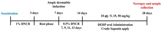
After the acclimatization period, mice were weighed and randomly assigned to ensure comparable baseline body weights among the groups. Mice were divided into six experimental groups (n = 9 per group), with one group designated as the untreated Normal Control (NC). No formal a priori sample size calculation was performed. The sample size of nine mice per group was selected based on previous studies using the DNCB-induced atopic dermatitis mouse model, which have consistently demonstrated that this sample size is sufficient to detect biologically meaningful treatment effects [18,19,20]. A total of 54 mice were used in the study, and all animals completed the experimental protocol without any dropouts. Histological evaluations of skin tissues were performed using all nine mice per group (n = 9). However, due to volume limitations inherent to blood collection in mice, serum and whole blood analyses were conducted on a subset of seven randomly selected mice per group (n = 7). All treatments and measurements were performed by the same investigator using a consistent experimental procedure. Treatment was administered in the same order throughout the study. Histological evaluation and image analysis were performed without blinding. All animals underwent dorsal skin depilation using an animal clipper and an electric shaver. The remaining groups received an initial sensitization with 1% DNCB prepared in a 3:1 mixture of acetone (Sigma-Aldrich, St. Louis, MO, USA; Cat. No. 179124) and olive oil (Sigma-Aldrich, St. Louis, MO, USA; Cat. No. O1514), which was topically applied to the dorsal skin at a volume of 200 µL once daily for 3 consecutive days, followed by a 4-day rest period. AD-like skin lesions were subsequently induced by applying 0.5% DNCB to the dorsal skin every other day for 7 days. In the allergen-combined treatment group, DEHP dissolved in olive oil was administered orally at a concentration of 10 µg/mL (0.2 mL per dose) for 2 weeks. Concurrently, CS dissolved in distilled water (DW) was applied topically to the dorsal skin at doses of 5, 15, or 50 mg/kg (200 µL per dose) for 2 weeks in the test substance treatment groups. To maintain AD symptoms throughout the treatment period, 0.5% DNCB was applied every other day to all groups except the NC group. Clinical skin scores were assessed on days 0, 7, 14, and 28 after AD induction, evaluating erythema, edema/hematoma, pruritus, excoriation, and scaling/dryness on a scale of 0 to 3 (0: none, 1: mild, 2: moderate, 3: severe), with the total score calculated as the sum of all individual item scores. On day 28, mice were anesthetized by inhalation of isoflurane (ISOTROY; Troikaa Pharmaceuticals Ltd., Thol, India) and blood was collected via the abdominal aorta, followed by harvesting of the spleen, liver, and skin tissue (Figure 1).
Figure 1.
Experimental schedule for DNCB-induced atopic dermatitis and treatment protocol. Mice were sensitized by topical application of 1% DNCB for 3 consecutive days, followed by a 4-day rest period. AD-like skin lesions were subsequently induced by topical application of 0.5% DNCB every other day for 7 days. Thereafter, DEHP was administered orally, and CS was applied topically at doses of 5, 15, and 50 mg/kg for 2 weeks, while 0.5% DNCB application was continued every other day to maintain AD-like skin lesions. Mice were sacrificed on day 28 for sample collection. The experimental groups were defined as follows: DNCB, DNCB-induced AD group; DEHP, DNCB-induced AD with DEHP administration; CS-L, DNCB-induced AD with DEHP and low-dose CS (5 mg/kg); CS-M, DNCB-induced AD with DEHP and medium-dose CS (15 mg/kg); and CS-H, DNCB-induced AD with DEHP and high-dose CS (50 mg/kg). Abbreviations: AD, atopic dermatitis; DNCB, 2,4-dinitrochlorobenzene; DEHP, di-(2-ethylhexyl) phthalate; CS, crude saponin.
2.4. Hematological Analysis
Blood collected from the abdominal aorta was transferred into ethylenediaminetetraacetic acid (EDTA) tubes (Becton, Dickinson and Company, Franklin Lakes, NJ, USA, 2025) and mixed by gently inverting the tubes 6–8 times. Whole blood was used for analysis, and measurements were performed using an automated hematology analyzer (ADVIA 2120i, Siemens Healthcare Diagnostics Inc., Tarrytown, NY, USA). The parameters analyzed included white blood cell (WBC) count, eosinophil count, neutrophil count, and lymphocyte count.
2.5. Histological Evaluation
Dorsal skin tissue (1.5 × 1.5 cm2) was excised from each mouse and pinned to a cork board to prevent tissue shrinkage, then fixed in 10% neutral buffered formalin and processed into paraffin blocks. Sections of 4 µm thickness were prepared and stained with hematoxylin and eosin (H&E) or toluidine blue (pH 4.0). Histological analysis was performed using a BX43 light microscope (Olympus Corporation, Tokyo, Japan). The thickness of the dermis and epidermis was measured in each specimen, and the degree of eosinophil infiltration within the dermis was evaluated. Quantitative analysis was conducted using Image J software, version 1.54k (Wayne Rasband, National Institutes of Health, Bethesda, MD, USA), with measurements taken at 10 different sites per skin section and averaged. Mast cell infiltration was assessed by counting the number of mast cells in the dermis across 10 fields of view at 400× magnification, and mast cell degranulation was evaluated at 1000× magnification. The degree of degranulation was classified as normal (degranulation in less than 1/3 of cells), moderate (less than 2/3), or severe (less than 3/3), and the degranulation index was calculated as follows:
Degranulation index (%) = [(Normal × 0) + (Moderate × 50) + (Severe × 100)]/Total number of mast cells
2.6. ELISA Measurement
Blood collected via abdominal aortic puncture was processed separately for serum and plasma analyses. For serum analyses, blood was transferred into serum separator tubes (Becton, Dickinson and Company, Franklin Lakes, NJ, USA) and allowed to clot at room temperature for 30 min, followed by centrifugation at 3400 rpm for 15 min. Serum TSLP, IL-4, IL-5, and IgE levels were measured using ELISA kits. TSLP, IL-4, and IL-5 were measured using kits from Cusabio (Wuhan, China; Cat. Nos. CSB-EL025141MO, CSB-E04634m, CSB-E04637m and CSB-E07983m respectively). For histamine analysis, plasma was separated from blood collected in EDTA-containing tubes (Becton, Dickinson and Company, Franklin Lakes, NJ, USA) by centrifugation (3400 rpm for 15 min) and stored until analysis. Plasma histamine levels were measured using an ELISA kit from LS Bio (Seattle, WA, USA; Cat. No. LS-F28398). All assays were performed according to the respective manufacturers’ instructions. Absorbance was measured at 450 nm using a microplate reader (Multiskan GO, Thermo Fisher Scientific, Vantaa, Finland).
2.7. Statistical Analysis
Histological image data were quantified using Image J software (Wayne Rasband, National Institutes of Health, Bethesda, MD, USA). Results are expressed as mean, standard error. Statistical analyses were performed using SPSS version 20 (IBM SPSS Statistics, Armonk, NY, USA), with the level of significance set at p < 0.05. All measured parameters were first analyzed by one-way ANOVA, followed by Duncan’s multiple range test to assess differences between individual groups. All data obtained from the evaluated samples (n = 9 per group for histological analyses; n = 7 per group for blood and serum analyses) were included in the statistical analysis without exclusion.
3. Results
3.1. Crude Saponin Ameliorates Skin and Systemic Manifestations of DEHP-Exacerbated Atopic Dermatitis
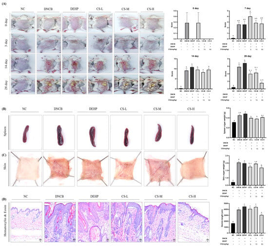
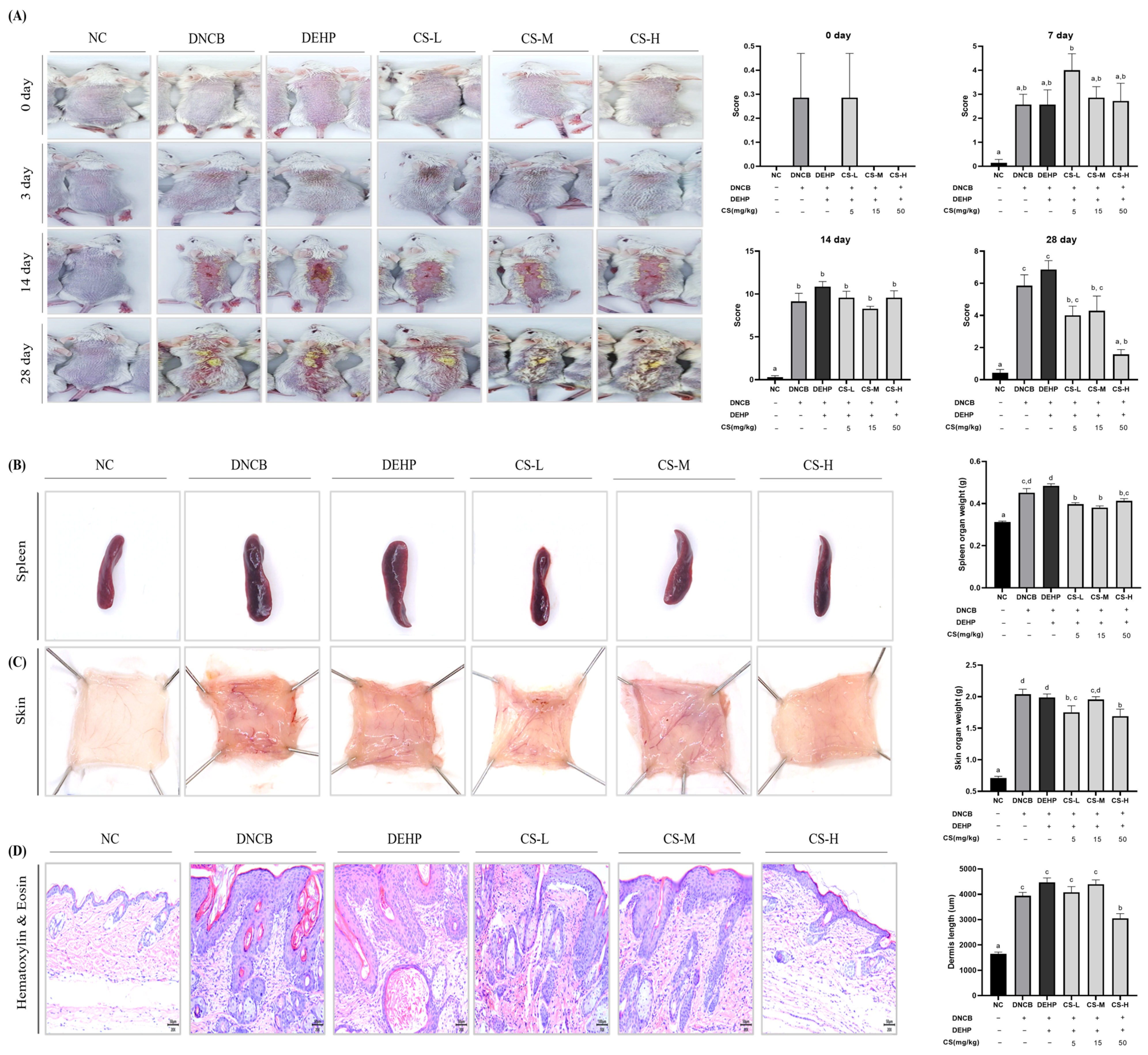
Clinical skin lesion scores were monitored throughout the experimental period. At day 14, all AD-induced groups showed significantly higher lesion scores than the NC group (0.43 ± 0.53), with the DEHP co-administration group exhibiting the highest score (10.86 ± 1.57). Following two weeks of CS treatment, lesion scores were markedly reduced by day 28, with the CS-H group showing a significant reduction to 1.57 ± 1.79 compared with the DEHP co-administration group (Figure 2A).
Figure 2.
Crude Saponin Ameliorates Skin and Systemic Manifestations of DEHP-Exacerbated Atopic Dermatitis. (A) Representative dorsal skin photographs and clinical skin lesion scores at 0, 7, 14, and 28 days after DNCB sensitization. Skin lesion severity was evaluated based on erythema, edema/hematoma, pruritus, excoriation, and scaling/dryness (score 0–3 per criterion; maximum 15). (B) Representative spleen photographs and spleen weights at sacrifice. (C) Representative dorsal skin specimens and dorsal skin weights at sacrifice. (D) Representative hematoxylin and eosin (H&E)-stained skin sections and dermal thickness measurements. Scale bar = 100 μm. The treatment matrices beneath the quantitative graphs indicate DNCB and DEHP exposure and the respective CS doses for each experimental group; the same group assignments apply to the corresponding representative images. Data are expressed as mean ± SE (n = 7 per group for (A–C) and n = 9 per group for (D)). Different letters (a–c) above the bars indicate statistically significant differences among groups as determined by Duncan’s multiple range test (p < 0.05). Abbreviations and group definitions are as described in Figure 1.
Spleen weight, assessed as an indicator of systemic immune activation, was highest in the DEHP co-administration group (0.48 ± 0.05 g) compared with the NC group (0.31 ± 0.01 g) and was significantly reduced following CS treatment (Figure 2B).
At sacrifice, dorsal skin weight was assessed as an indicator of skin thickening associated with AD-like lesions. All AD-induced groups showed significantly greater skin weights than the NC group (0.71 ± 0.20 g), whereas the CS-H group showed a significant reduction to 1.69 ± 0.29 g compared with the DEHP co-administration group (Figure 2C).
Histopathological analysis further demonstrated a significant reduction in dermal thickness in the CS-H group compared with the DEHP co-administration group, supporting the ameliorative effect of CS on DEHP-exacerbated skin lesions (Figure 2D).
3.2. Crude Saponin Restores Leukocyte Subset Balance and Suppresses TSLP Activation in DEHP-Exacerbated Atopic Dermatitis
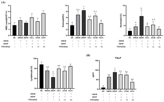
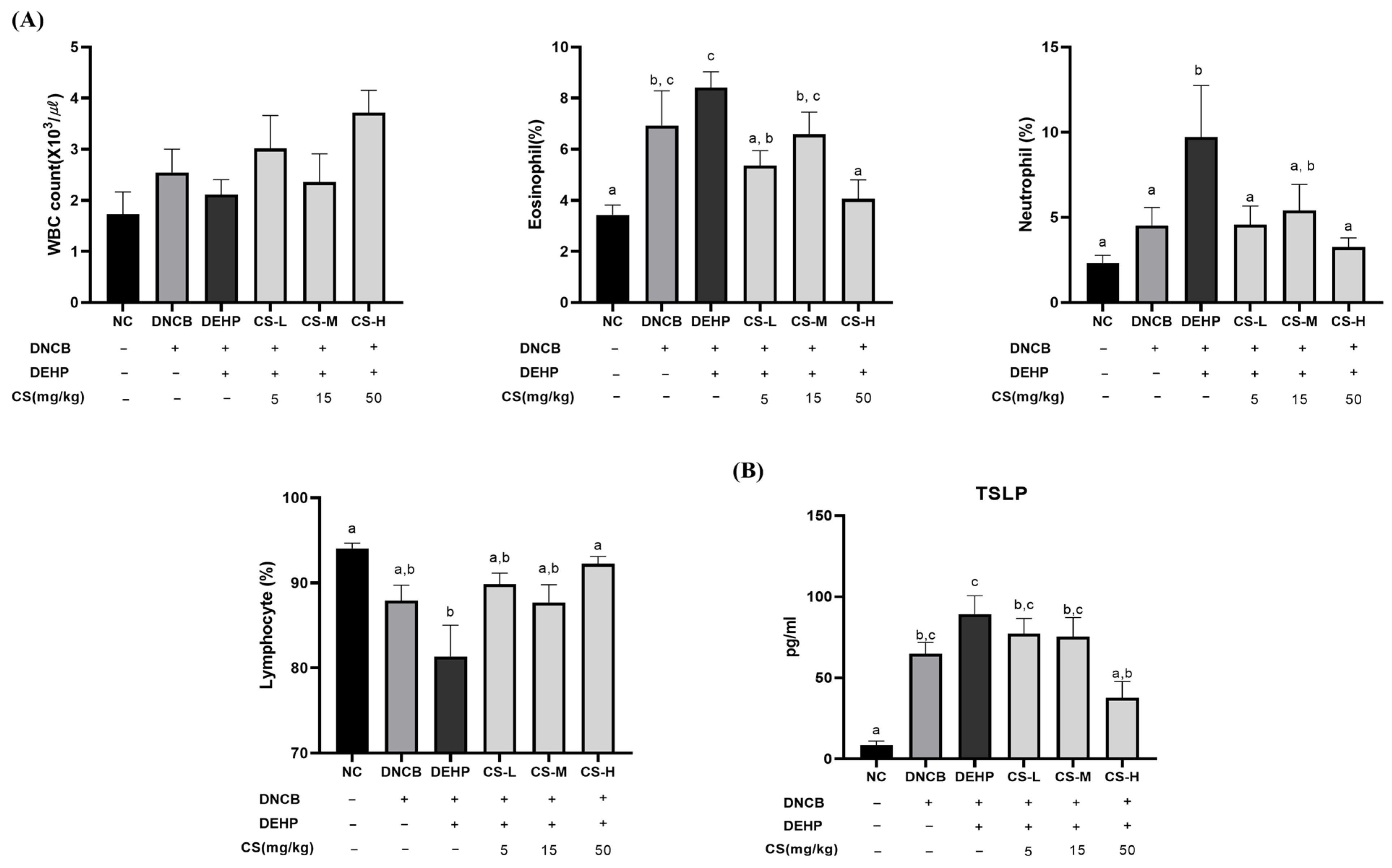
To assess changes in peripheral immune cell profiles following AD induction and CS treatment, total white blood cell (WBC) counts and differential leukocyte proportions were analyzed. Total WBC counts tended to increase in the AD-induced groups compared with the NC group, with the highest value observed in the CS-H group; however, no statistically significant differences were observed among the groups. The lymphocyte proportion was significantly decreased in the DEHP co-administration group compared with the NC group, whereas the CS-H group showed a significant recovery to 92.26 ± 2.21%. In contrast, the neutrophil proportion was significantly elevated in the DEHP co-administration group (9.71 ± 8.01%) compared with the NC group (2.30 ± 1.25%) and was significantly reduced to 3.26 ± 1.42% in the CS-H group. Similarly, the eosinophil proportion was highest in the DEHP co-administration group (8.41 ± 1.63%) and was significantly reduced in the CS-H group (Figure 3A).
Figure 3.
Crude Saponin Restores Leukocyte Subset Balance and Suppresses TSLP Activation in DEHP-Exacerbated Atopic Dermatitis. Peripheral blood leukocyte analysis and serum TSLP levels. (A) Complete blood count (CBC) analysis showing total white blood cell (WBC) counts and differential proportions of lymphocytes, neutrophils, and eosinophils. (B) Serum TSLP concentrations were measured by ELISA. The treatment matrices beneath the graphs indicate DNCB and DEHP exposure and the respective CS doses for each experimental group. Data are expressed as mean ± SE (n = 7 per group). Different letters above the bars indicate statistically significant differences among groups as determined by Duncan’s multiple range test (p < 0.05). Abbreviations and group definitions are as described in Figure 1.
To further assess changes in an upstream regulator of allergic inflammation, serum TSLP concentrations were measured. TSLP levels increased following AD induction, with the highest concentration observed in the DEHP co-administration group (77.26 ± 24.9 pg/mL). CS treatment reduced TSLP levels, with the CS-H group showing a significantly lower concentration (37.63 ± 26.8 pg/mL) than the DEHP co-administration group (Figure 3B).
3.3. Crude Saponin Attenuates Eosinophil and Mast Cell Infiltration in DEHP-Exacerbated Atopic Dermatitis
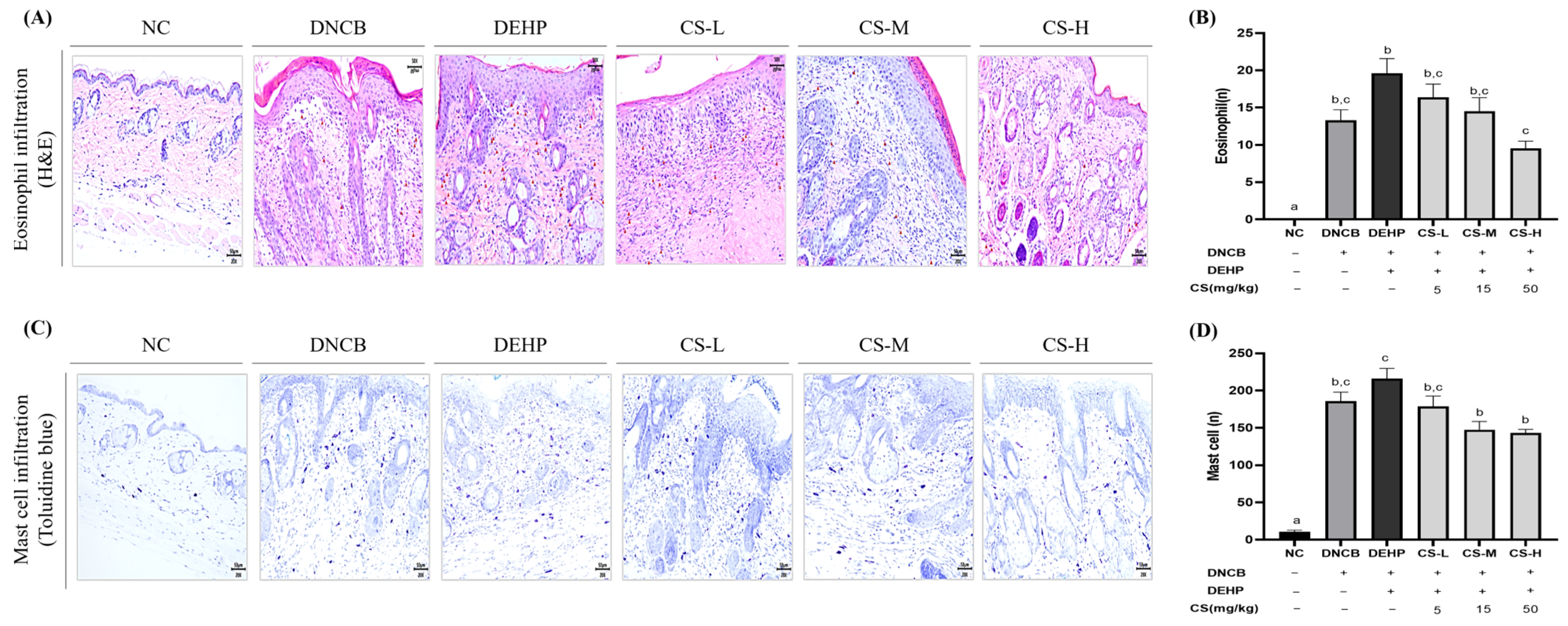
To evaluate eosinophil infiltration in the dermal layer following AD induction, eosinophil and mast cell infiltration was assessed histologically. Eosinophil infiltration was significantly greater in all AD-induced groups than in the NC group (0.08 ± 0.1 cells/HPF) (Figure 4A,B). Notably, eosinophil infiltration was significantly higher in the DEHP co-treated group (19.61 ± 5.9 cells/HPF) than in the DNCB group (13.30 ± 4.2 cells/HPF), indicating that DEHP further exacerbated eosinophilic inflammation within the pre-existing AD environment. In contrast, the CS-treated groups showed a dose-dependent reduction in eosinophil infiltration, with the CS-H group exhibiting a significant decrease to 9.54 ± 2.9 cells/HPF compared with the DEHP group.
Figure 4.
Crude Saponin Attenuates Eosinophil and Mast Cell Infiltration in DEHP-Exacerbated Atopic Dermatitis. Histological assessment of inflammatory cell infiltration in dorsal skin tissue. (A) Representative H&E-stained dorsal skin sections with eosinophils indicated by red arrowheads. (B) Quantification of infiltrated eosinophil counts per high-power field. (C) Representative toluidine blue-stained dorsal skin sections showing mast cell distribution. (D) Quantification of mast cell counts per high-power field. Scale bars = 50 μm. The treatment matrices beneath the quantitative graphs indicate DNCB and DEHP exposure and the respective CS doses for each experimental group; the same group assignments apply to the corresponding representative histological images. Data are expressed as mean ± SE (n = 9 per group). Different letters above the bars indicate statistically significant differences among groups as determined by Duncan’s multiple range test (p < 0.05). Abbreviations and group definitions are as described in Figure 1.
Dermal mast cell infiltration showed a similar pattern (Figure 4C,D). Mast cell counts were significantly elevated in the DEHP co-treated group (224.88 ± 33.6 cells/HPF) compared with the NC group (10.75 ± 5.3 cells/HPF), whereas the CS-H group showed a significant reduction to 143.13 ± 13.51 cells/HPF. Collectively, these findings indicate that CS treatment attenuated inflammatory cell infiltration in the skin under DEHP-exacerbated AD conditions.
3.4. Effects of Crude Saponin on Allergic Inflammatory Mediators in DEHP-Exacerbated Atopic Dermatitis
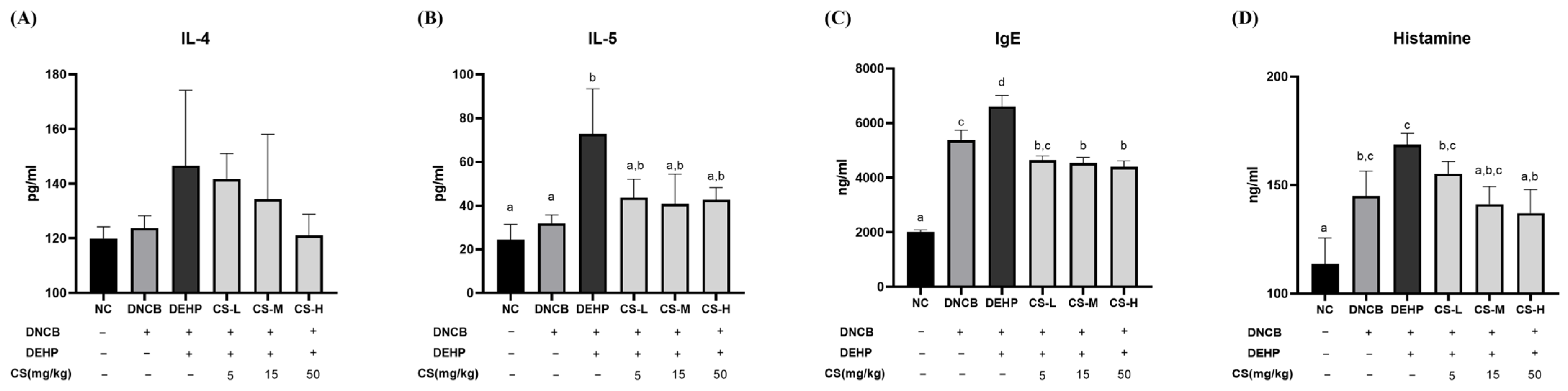
To evaluate changes in allergic inflammatory mediators following AD induction and CS treatment, serum IL-4 and IL-5 concentrations were assessed. Serum IL-4 levels showed an increasing trend from the NC group to the DNCB and DEHP co-administration groups, followed by a gradual decrease with increasing CS dose; however, no statistically significant differences were observed among the groups (Figure 5A). In contrast, serum IL-5 levels were significantly elevated in the DEHP co-administration group compared with the NC and DNCB groups. Although IL-5 levels decreased in all CS-treated groups, these reductions did not reach statistical significance compared with the DEHP co-treated group (Figure 5B).
Figure 5.
Effects of Crude Saponin on Allergic Inflammatory Mediators in DEHP-Exacerbated Atopic Dermatitis. Concentrations of (A) serum IL-4, (B) serum IL-5, (C) serum IgE, and (D) plasma histamine measured by ELISA. The treatment matrices beneath the graphs indicate DNCB and DEHP exposure and the respective CS doses for each experimental group. Data are expressed as mean ± SE (n = 7 per group). Different letters above the bars indicate statistically significant differences among groups as determined by Duncan’s multiple range test (p < 0.05). Abbreviations and group definitions are as described in Figure 1.
Serum IgE levels were significantly elevated in all AD-induced groups compared with the NC group, with the highest concentration observed in the DEHP co-treated group (6605.5 ± 1139.5 ng/mL). In contrast, the CS-H group showed a significant reduction to 4398.0 ± 625.8 ng/mL compared with the DEHP co-treated group (Figure 5C).
Plasma histamine levels increased following AD induction, with the DEHP co-administration group showing the highest concentration (168.69 ± 14.6 ng/mL), which was higher than that in the DNCB group (144.99 ± 32.5 ng/mL). CS treatment resulted in a dose-dependent reduction in histamine levels, with the CS-H group showing a significant decrease to 137.10 ± 30.69 ng/mL compared with the DEHP group (Figure 5D).
4. Discussion
Several studies have previously reported an association between phthalate exposure and the aggravation of AD [21]. DEHP has been shown to induce Th2-skewed immune responses via peroxisome proliferator-activated receptor gamma (PPARγ) activation and to amplify allergic inflammation through an adjuvant effect that promotes immunoglobulin production in the presence of allergens [8,9]. The present study demonstrates that oral DEHP exposure exacerbates allergic skin lesions in DNCB-sensitized mice. The aggravation of AD induced by DEHP exposure was associated with increased excoriation, scaling, and dryness of dorsal skin tissue, along with worsened eosinophil and mast cell infiltration and an increase in mast cell numbers. Furthermore, DEHP co-administration was associated with elevated serum total IgE and IL-5 levels, whereas IL-4 showed an increasing trend without reaching statistical significance. These findings suggest that DEHP is involved in the pathophysiology of AD not by independently inducing the condition, but rather by deepening immunological imbalance within a pre-existing allergic inflammatory environment. Collectively, these results support the hypothesis that DEHP acts as an adjuvant aggravating factor in the AD response.
Beyond these effects, DEHP appears to dysregulate TSLP expression and leukocyte subpopulations, both of which serve as upstream regulators of allergic immune responses. Phthalate compounds have been reported to induce TSLP expression through reactive oxygen species (ROS) generation and NF-κB (p65) activation in allergic skin and epithelial models, thereby amplifying Th2-type inflammatory responses [22]. TSLP is further recognized as a factor that promotes dendritic cell activation and facilitates T cell migration to the skin and lymph nodes, thereby inducing lymphocyte redistribution [23]. The failure of DEHP co-administration to produce a statistically significant increase in TSLP expression compared to the DNCB-alone group in the present study may be attributable to the predominance of downstream signaling mediator-driven immune activation over TSLP-mediated pathways within the already-activated Th2 inflammatory milieu [24]. Additionally, DEHP has been reported to reduce total leukocyte and lymphocyte counts in immunotoxicity models [25], a finding consistent with the hematological observations of the present study.
IgE is recognized as the terminal effector molecule of AD pathology, arising from the complex interplay of T cell activation, B cell class switching, and lymphoid organ cross-talk; it is characterized by a short half-life and continuous secretion from plasma cells [26]. Notably, IgE binds to the high-affinity receptor FcεRI on the surface of mast cells to establish an allergen-specific sensitized state, and subsequent cross-linking of FcεRI by allergens triggers mast cell degranulation and histamine release, playing a pivotal role in pruritus and acute exacerbation of AD [27]. As previously described, TSLP-activated dendritic cells promote the Th2 differentiation of naïve T cells and drive increased secretion of IL-4 and IL-5, the resultant elevation of IL-5 accelerates eosinophil influx into lesional skin through enhanced hematopoiesis, anti-apoptotic effects, and amplified secretion of IL-4- and IL-13-dependent eotaxin (CCL11) [28,29]. The observation that the DEHP co-administration group exhibited the highest levels of serum total IgE and IL-5, accompanied by increased eosinophil counts in blood and tissue, is consistent with enhanced Th2-associated eosinophilic inflammation under DEHP co-exposure. Because eotaxin expression and dendritic-cell signaling were not directly measured, involvement of the IL-5–eotaxin and TSLP–dendritic cell pathways remain a literature-supported mechanistic possibility rather than a mechanism demonstrated in the present study.
These results indicate that DEHP exposure acts as a complex aggravating factor that broadly amplifies the Th2 immune response in AD from upstream signaling to effector-stage responses, underscoring the clinical importance of managing environmental DEHP exposure levels. Most previous studies investigating the allergic effects of DEHP have primarily focused on downstream inflammatory markers, including Th2 cytokines and serum IgE. By evaluating TSLP, an upstream epithelial-derived initiator of allergic inflammation, the present study provides a more integrated assessment of DEHP-induced immune dysregulation across multiple stages of the allergic cascade.
In response to this DEHP-mediated immune dysregulation, the red ginseng CS applied in the present study demonstrated significant ameliorative effects across histological lesions, cytokine profiles, and blood cell subpopulations. Ginsenoside Rh2, a constituent of CS, has been reported to suppress TSLP expression in keratinocytes by blocking the nuclear translocation of NF-κB (p65) [16]. In the present study, CS treatment was associated with lower TSLP levels and a partial normalization of the proportions of peripheral leukocyte subsets. While previous studies have reported that individual ginsenosides can modulate NF-κB–TSLP signaling and pathways related to ILC2 activation, these molecular pathways were not directly assessed in the present study [16,30]. Therefore, the observed changes should be interpreted as consistent with these previously proposed mechanisms, rather than as evidence of them. Furthermore, CS treatment was associated with a concentration-dependent decreasing trend in IL-4 levels, although the differences did not reach statistical significance, along with reductions in eosinophil and mast cell infiltration. These findings may be related to the reported capacity of ginsenoside constituents to suppress Th2 differentiation through inhibition of GATA3 expression and to regulate the IL-5–eotaxin–eosinophil axis by reducing NF-κB-dependent IL-5 and eotaxin (CCL11) secretion [31,32]. However, GATA3, eotaxin, and NF-κB activities were not directly measured in the present study, and their involvement remains to be experimentally verified. Notably, even under the aggravated inflammatory conditions involving DEHP co-administration, high-dose CS application reduced eosinophil infiltration, mast cell infiltration, and dermal thickness by approximately 51%, 37%, and 35%, respectively—a magnitude of inhibition comparable to or consistent with that reported in previous studies of red ginseng extracts and saponins evaluated in the absence of environmental immunotoxicants [31]. These findings suggest that CS retains anti-inflammatory activity even under inflammatory conditions exacerbated by immunotoxicants such as DEHP. Previous studies have largely focused on the anti-atopic effects of red ginseng extracts or individual ginsenosides in conventional allergen-induced AD models. However, whether these protective effects are preserved under conditions of environmental immunotoxicant exposure has remained largely unexplored. The present findings demonstrate that CS retains substantial immunomodulatory activity even in the presence of DEHP-mediated immune amplification. Furthermore, the IgE–mast cell–histamine axis, which was markedly elevated under DEHP-exacerbated AD conditions, was attenuated following CS treatment. Previous studies have reported that ginsenosides such as Rg3 and Rh2 can modulate IgE-dependent mast cell responses through pathways involving Ca2+ influx and MAPK/NF-κB signalling [33,34]. While the reductions in mast cell infiltration and histamine observed in the present study are consistent with these previously reported effects, the signalling pathways were not directly evaluated and therefore require experimental verification. Overall, the coordinated changes in TSLP, Th2-associated cytokines, IgE, eosinophils, mast cells and histamine suggest that CS treatment can reduce multiple components of the allergic inflammatory response in DEHP-induced AD, although the specific signalling mechanisms underlying these effects require further investigation.
Despite these findings, several aspects should be considered when interpreting the present results. First, although the DNCB-induced model reproduces key features of Th2-dominant allergic skin inflammation, it represents an experimentally induced acute/subacute inflammatory condition and does not fully recapitulate the heterogeneous, chronic, and relapsing nature of human atopic dermatitis. In addition, only male BALB/c mice were used in the present study, precluding assessment of potential sex-dependent differences in immune and endocrine responses. Future studies incorporating both sexes and complementary experimental models would therefore help determine the reproducibility and broader biological relevance of the observed effects. Second, DEHP was administered by oral gavage to ensure controlled and reproducible dosing, whereas human exposure generally occurs at lower levels over prolonged periods and through multiple routes, including dietary intake, inhalation, and dermal contact. Thus, exposure paradigms that more closely reflect environmentally relevant patterns are warranted to further evaluate the relationship between DEHP exposure and allergic skin inflammation. Third, the CS preparation used in this study contained multiple ginsenosides, and the observed effects therefore cannot be attributed to a single constituent. The relative contributions of individual ginsenosides, as well as possible additive or synergistic interactions among them, remain to be clarified through fractionation and studies using purified compounds. Finally, although the coordinated changes in TSLP, Th2-associated cytokines, IgE, eosinophils, mast cells, and histamine support modulation of the allergic inflammatory cascade, the present findings do not establish direct causal inhibition of specific signaling pathways. Additional studies examining pathway-specific molecular targets and temporal changes in skin tissue will be necessary to clarify the mechanisms by which red ginseng CS attenuates DEHP-exacerbated allergic inflammation.
To our knowledge, this study is among the first to demonstrate the protective effects of red ginseng CS against AD exacerbated by chronic exposure to an environmental immunotoxicant. While previous studies have primarily investigated ginseng-derived compounds in conventional AD models, the present findings extend these observations by showing that CS remains effective under conditions of environmentally aggravated allergic inflammation. These results provide experimental evidence that red ginseng CS can attenuate allergic inflammation under DEHP-exacerbated conditions in a mouse model and support further mechanistic and translational investigation.
5. Conclusions
This study provides proof-of-concept evidence that red ginseng crude saponin reduces DEHP-exacerbated AD-like inflammation in a DNCB-induced mouse model, as indicated by improvements in clinical skin lesions, inflammatory cell infiltration and allergic mediators. These results imply that the modulation of environmentally aggravated allergic inflammation could be a significant direction for future AD research. However, the present results are preclinical and do not demonstrate clinical efficacy or the direct inhibition of molecular pathways proposed in the previous literature. Further studies are required to standardize the saponin preparation, define active constituents and clinically relevant dosing, and evaluate formulation, dermal exposure, safety and efficacy in human-relevant models before clinical investigation can be considered.
Author Contributions
H.-w.S.: Conceptualization, Methodology, Data curation, Writing—original draft. J.-H.O.: Writing—review & editing, Data curation. S.-H.B.: Investigation, Formal analysis. T.-H.K.: Investigation, Formal analysis. S.-Y.H.: Supervision, Project administration. All authors have read and agreed to the published version of the manuscript.
Funding
This research was supported by the Daejeon University Research Grants (2025), grant number 20250183.
Institutional Review Board Statement
The animal study protocol was approved by the Institutional Animal Care and Use Committee (IACUC) of Daejeon University (protocol code: DJUARB2025-016; approval date: 28 May 2025).
Informed Consent Statement
Not applicable.
Data Availability Statement
The datasets generated or analyzed during the current study are available from the corresponding author upon reasonable request.
Acknowledgments
This paper is based on the master’s thesis of the first author, submitted to the Graduate School of Daejeon University in February 2026. The authors would like to thank all laboratory members of the Daejeon University Environmental Health Center for their technical assistance and support throughout this study.
Conflicts of Interest
The authors declare no conflicts of interest.
Abbreviations
The following abbreviations are used in this manuscript:
| CS | Crude saponins from red ginseng |
| DEHP | Di-(2-ethylhexyl) phthalate |
| DNCB | 2,4-dinitrochlorobenzene |
| PVC | Polyvinyl chloride |
| AD | Atopic dermatitis |
| Th2 | T helper type 2 |
| PPAR | Proliferator-activated receptor gamma |
| TSLP | Thymic stromal lymphopoietin |
| NC | Normal control |
| DW | Distilled water |
| EDTA | Ethylenediaminetetraacetic acid |
| WBC | White blood cell |
| H&E | Hematoxylin and eosin |
| ELISA | Enzyme-linked immunosorbent assay |
| IL-4 | Interleukin-4 |
| IgE | Immunoglobulin E |
| IL-5 | Interleukin-5 |
| SST | Serum separator tubes |
| ROS | Reactive oxygen species |
| PPARγ | Peroxisome proliferator-activated receptor gamma |
References
- Lu, C.L.; Wen, H.J.; Wang, C.S.; Sun, C.W.; Wu, M.T.; Chen, M.L.; Hsieh, C.J.; Wang, S.L.; TMICS Study Group. Di-2-ethylhexyl phthalate may exaggerate allergic response in boys with atopic dermatitis: 4-year follow-up of the Taiwan Maternal and Infant Cohort Study. Sci. Total Environ. 2025, 1006, 180844. [Google Scholar] [CrossRef] [Scilit]
- Khalifa, M.; Fayed, R.H.; Sedik, A.A.; Khalil, H.M.A. Dose-dependent toxic effects of di-(2-ethylhexyl) phthalate in male rats: Focus on behavioral alterations and inducing TLR4/NF-κB signaling pathway. Toxicol. Appl. Pharmacol. 2023, 468, 116515. [Google Scholar] [CrossRef] [Scilit]
- Braun, J.M.; Sathyanarayana, S.; Hauser, R. Phthalate exposure and children’s health. Curr. Opin. Pediatr. 2013, 25, 247–254. [Google Scholar] [CrossRef] [Scilit]
- Yang, J.; Song, W.; Wang, X.; Li, Y.; Sun, J.; Gong, W.; Sun, C. Migration of phthalates from plastic packages to convenience foods and its cumulative health risk assessments. Food Addit. Contam. Part B Surveill. 2019, 12, 151–158. [Google Scholar] [CrossRef] [Scilit]
- Lee, J.Y.; Lee, J.; Huh, D.A.; Moon, K.W. Association between environmental exposure to phthalates and allergic disorders in Korean children: Korean National Environmental Health Survey (KoNEHS) 2015–2017. Int. J. Hyg. Environ. Health 2021, 238, 113857. [Google Scholar] [CrossRef] [Scilit]
- Kim, S.; Lee, J.; Park, J.; Kim, H.J.; Cho, G.J.; Kim, G.H.; Eun, S.H.; Lee, J.J.; Choi, G.; Suh, E.; et al. Urinary phthalate metabolites over the first 15 months of life and risk assessment: CHECK cohort study. Sci. Total Environ. 2017, 607, 881–887. [Google Scholar] [CrossRef] [Scilit]
- National Institute of Environmental Research. Release of the Results of the 5th Korean National Environmental Health Survey (2021–2023). 2024. Available online: https://www.mcee.go.kr/home/web/board/read.do?boardCategoryId&boardId=1716930&boardMasterId=1&decorator&maxIndexPages=10&maxPageItems=10&menuId&orgCd&pagerOffset=1140&searchKey&searchValue&utm_source=chatgpt.com (accessed on 1 August 2026).
- Takano, H.; Yanagisawa, R.; Inoue, K.; Ichinose, T.; Sadakane, K.; Yoshikawa, T. Di-(2-ethylhexyl) phthalate enhances atopic dermatitis-like skin lesions in mice. Environ. Health Perspect. 2006, 114, 1266–1269. [Google Scholar] [CrossRef] [Scilit]
- Stark, J.M.; Coquet, J.M.; Tibbitt, C.A. The role of PPAR-γ in allergic disease. Curr. Allergy Asthma Rep. 2021, 21, 45. [Google Scholar] [CrossRef] [Scilit]
- You, H.; Li, R.; Wei, C.; Chen, S.; Mao, L.; Zhang, Z.; Yang, X. Thymic stromal lymphopoietin neutralization inhibits the immune adjuvant effect of di-(2-ethylhexyl) phthalate in a BALB/c mouse asthma model. PLoS ONE 2016, 11, e0159479. [Google Scholar] [CrossRef] [Scilit]
- Szymański, U.; Cios, A.; Ciepielak, M.; Stankiewicz, W. Cytokines and apoptosis in atopic dermatitis. Postep. Dermatol. Alergol. 2021, 38, 1–13. [Google Scholar] [CrossRef] [Scilit]
- Shin, N.Y. Pharmacotherapy for Atopic Dermatitis Treatment. Korea Pharmaceutical Information Center. 2022. Available online: https://health.kr/Menu.PharmReview/View.asp?PharmReview_IDX=8324 (accessed on 1 August 2026).
- Choi, B.C. Atopic Dermatitis (1). Korea Pharmaceutical Information Center. 2018. Available online: https://common.health.kr/shared/healthkr/pharmreview/%EC%95%84%ED%86%A0%ED%94%BC%ED%94%BC%EB%B6%80%EC%97%BC%28Atopic%20Dermatitis%29%20%281%29_JS.pdf (accessed on 1 August 2026).
- Ryu, G.H. Present status of red ginseng products and its manufacturing process. Food Ind. Nutr. 2003, 8, 38–42. [Google Scholar]
- Lee, K.S.; Choi, K.J.; Ko, S.R.; Jang, J.G.; Yang, C.B. Maillard browning reaction and antioxidant activity of red ginseng stored for long periods. Korean J. Ginseng Sci. 1988, 12, 121–127. [Google Scholar]
- Ko, E.S.; Park, S.J.; Lee, J.H.; Cui, C.-H.; Hou, J.; Kim, M.-H.; Kim, S.C. Ginsenoside Rh2 ameliorates atopic dermatitis in NC/Nga mice by suppressing NF-κB-mediated thymic stromal lymphopoietin expression and T helper type 2 differentiation. Int. J. Mol. Sci. 2019, 20, 6111. [Google Scholar] [CrossRef] [Scilit]
- Lorz, L.R.; Kim, M.Y.; Cho, J.Y. Medicinal potential of Panax ginseng and its ginsenosides in atopic dermatitis treatment. J. Ginseng Res. 2020, 44, 8–13. [Google Scholar] [CrossRef] [Scilit]
- Yun, H.R.; Ahn, S.W.; Seol, B.; Vasileva, E.A.; Mishchenko, N.P.; Fedoreyev, S.A.; Stonik, V.A.; Han, J.; Ko, K.S.; Rhee, B.D.; et al. Echinochrome A treatment alleviates atopic dermatitis-like skin lesions in NC/Nga mice via IL-4 and IL-13 suppression. Mar. Drugs 2021, 19, 622. [Google Scholar] [CrossRef] [Scilit]
- Oh, J.S.; Lee, S.J.; Choung, S.Y. Lithospermum erythrorhizon alleviates atopic dermatitis-like skin lesions by restoring immune balance and skin barrier function in 2,4-dinitrochlorobenzene-induced NC/Nga mice. Nutrients 2021, 13, 3209. [Google Scholar] [CrossRef] [Scilit]
- Zhou, J.R.; Kanda, Y.; Tanaka, A.; Manabe, H.; Nohara, T.; Yokomizo, K. Anti-hyaluronidase activity in vitro and amelioration of mouse experimental dermatitis by tomato saponin, esculeoside A. J. Agric. Food Chem. 2016, 64, 403–408. [Google Scholar] [CrossRef] [Scilit]
- Kang, J.; Ding, Y.; Li, B.; Liu, H.; Yang, X.; Chen, M. TRPA1-mediated aggravation of allergic contact dermatitis induced by DINP and regulated by NF-κB activation. Sci. Rep. 2017, 7, 43586. [Google Scholar] [CrossRef] [Scilit]
- Kang, J.; Song, J.; Shen, S.; Li, B.; Yang, X.; Chen, M. Diisononyl phthalate aggravates allergic dermatitis by activation of NF-κB. Oncotarget 2016, 7, 85472–85482. [Google Scholar] [CrossRef] [Scilit]
- Wallmeyer, L.; Dietert, K.; Sochorova, M.; Gruber, A.D.; Kleuser, B.; Vávrová, K.; Hedtrich, S. TSLP is a direct trigger for T cell migration in filaggrin-deficient skin equivalents. Sci. Rep. 2017, 7, 774. [Google Scholar] [CrossRef] [Scilit]
- Jin, J.; Sunusi, S.; Lu, H. Group 2 innate lymphoid cells (ILC2s) are important in typical type 2 immune-mediated diseases and an essential therapeutic target. J. Int. Med. Res. 2022, 50, 03000605211053156. [Google Scholar] [CrossRef] [Scilit]
- Linghu, D.; Zhu, Z.; Zhang, D.; Luo, Y.; Ma, J.; Li, T.; Sun, Z.; Xie, Z.; Sun, J.; Cao, C. Diethylhexyl phthalate induces immune dysregulation and is an environmental immune disruptor. J. Hazard. Mater. 2024, 480, 136244. [Google Scholar] [CrossRef] [Scilit]
- Miranda-Waldetario, M.C.G.; Fernandes-Braga, W.; de Lafaille, M.A.C. Type 2 IgG Memory B Cells and Long-Lived IgE Plasma Cells in the Persistence of Allergy. Immunol. Rev. 2026, 337, e70089. [Google Scholar] [CrossRef] [Scilit]
- Ahn, K.M. Role of mast cells in allergic inflammation and innate immunity. Korean J. Pediatr. 2004, 47, 1137–1141. [Google Scholar]
- Kang, K.Y.; Han, J.K.; Kim, Y.H. The effects of Prunus Armeniaca Linne Var fractions on Th2 cytokine expression and atopic dermatitis of NC/Nga mouse. J. Korean Med. Pediatr. 2016, 30, 29–59. [Google Scholar] [CrossRef] [Scilit][Green Version]
- Chung, I.Y. Hypereosinophilia-associated diseases and the therapeutic agents in development. Hanyang Med. Rev. 2013, 33, 65–74. [Google Scholar] [CrossRef] [Scilit]
- Lee, H.S.; Park, H.W. Ginsenoside Rg1 attenuates asthma features in mice co-exposed to house dust mite allergen and diesel exhaust particle by modulating epithelial ILC2 interactions. J. Ginseng Res. 2026, 50, 100919. [Google Scholar] [CrossRef] [Scilit]
- Kim, H.S.; Lee, D.H.; Kim, B.K.; Yoon, S.K.; Kim, M.H.; Lee, J.Y.; Kim, H.O.; Park, Y.M. Effects of topically applied Korean red ginseng and its genuine constituents on atopic dermatitis-like skin lesions in NC/Nga mice. Int. Immunopharmacol. 2011, 11, 406–413. [Google Scholar] [CrossRef] [Scilit]
- Lee, I.S.; Uh, I.J.; Kim, K.S.; Kim, K.H.; Park, J.; Kim, Y.; Jung, J.H.; Jung, H.J.; Jang, H.J. Anti-inflammatory effects of ginsenoside Rg3 via NF-κB pathway in A549 cells and human asthmatic lung tissue. J. Immunol. Res. 2016, 2016, 7521601. [Google Scholar] [CrossRef] [Scilit]
- Ke, Y.; Huang, L.; Song, Y.; Liu, Z.; Liang, L.; Wang, L.; Wang, T. Preparation and pharmacological effects of minor ginsenoside nanoparticles: A review. Front. Pharmacol. 2022, 13, 974274. [Google Scholar] [CrossRef] [Scilit]
- Xiong, S.; Jia, Y.; Liu, C. IgE-expressing long-lived plasma cells in persistent sensitization. Front. Immunol. 2022, 13, 979012. [Google Scholar]


Disclaimer/Publisher’s Note: The statements, opinions and data contained in all publications are solely those of the individual author(s) and contributor(s) and not of MDPI and/or the editor(s). MDPI and/or the editor(s) disclaim responsibility for any injury to people or property resulting from any ideas, methods, instructions or products referred to in the content. |
© 2026 by the authors. Licensee MDPI, Basel, Switzerland. This article is an open access article distributed under the terms and conditions of the Creative Commons Attribution (CC BY) license.