


Abstract
Matrix metalloproteinases (MMPs) have long been recognized as key mediators of tumor invasion and metastasis due to their capacity to degrade extracellular matrix components. However, clinical failure of broad-spectrum MMP inhibitors has revealed a more complex and context-dependent role for these proteases in cancer. Among them, macrophage metalloelastase, MMP-12, has emerged as a particularly intriguing enzyme with both tumor-promoting and tumor-suppressive functions. Predominantly expressed by tumor-associated macrophages, MMP-12 occupies a unique position at the interface of proteolysis, inflammation, and immune regulation within the tumor microenvironment. Accumulating evidence from experimental models and clinical studies demonstrates that MMP-12 can exert potent anti-tumorigenic effects, primarily through inhibition of angiogenesis. Mechanistically, MMP-12 generates angiostatin and other anti-angiogenic fragments, suppresses vascular endothelial growth factor signaling, and reduces tumor vascularization, thereby limiting tumor growth and metastatic expansion. In several cancer types, including lung, colorectal, and hepatocellular carcinoma, elevated MMP-12 expression has been associated with reduced tumor progression and improved patient outcomes. Conversely, MMP-12 can also promote tumor progression through extracellular matrix remodeling, facilitation of invasion, and modulation of inflammatory pathways, particularly in environments characterized by chronic inflammation or immunosuppressive macrophage phenotypes. These seemingly contradictory roles are governed by multiple context-dependent factors, including macrophage polarization, tumor type, disease stage, and microenvironmental cues such as hypoxia and cytokine signaling. In this review, we comprehensively examine the molecular regulation, functional mechanisms, and clinical relevance of MMP-12 in cancer. We highlight the dualistic nature of MMP-12 activity and discuss its implications for therapeutic strategies, emphasizing the need for selective and context-aware targeting approaches rather than broad inhibition of MMP activity.
1. Background
Matrix metalloproteinases (MMPs) are a family of zinc-dependent endopeptidases that regulate extracellular matrix (ECM) turnover, tissue remodeling, wound repair, embryonic development, and overall tissue homeostasis [1]. Their catalytic activity depends on a Zn2+ ion within the active site, which enables them to cleave a broad range of ECM components, including fibrillar and non-fibrillar collagens, fibronectin, laminin, vitronectin, elastin, and basement membrane-associated glycoproteins [2]. Because of this ECM-degrading capacity, MMPs have long been considered important regulators of cancer invasion and metastasis [3]. By disrupting the structural barriers that normally restrict tumor expansion, MMPs can facilitate tumor cell migration, local tissue invasion, intravasation, and dissemination to distant organs [4,5]. This concept initially led to the development of broad-spectrum MMP inhibitors, many of which were designed to block the catalytic Zn2+ ion in the conserved active site [6]. Although these inhibitors showed promising anti-tumor effects in preclinical models, their clinical translation was largely unsuccessful. The major limitations included poor selectivity among MMP family members, inadequate bioavailability, limited therapeutic efficacy, off-target interactions, and dose-limiting toxicities, particularly musculoskeletal adverse effects [7,8]. These failures prompted a major re-evaluation of MMP biology and revealed that MMPs are not simply tumor-promoting enzymes [9]. Instead, individual MMPs can exert either pro-tumorigenic or anti-tumorigenic effects depending on tumor type, disease stage, cellular source, substrate availability, and the biochemical and immunological context of the tumor microenvironment [10,11].
Among MMP family members, matrix metalloproteinase-12, also known as MMP-12 or macrophage metalloelastase, has emerged as a particularly complex and biologically intriguing protease [12]. MMP-12 is distinct from many other MMPs because it is predominantly produced by macrophages, including tumor-associated macrophages, rather than being expressed mainly by tumor cells or stromal fibroblasts [13]. This cellular origin places MMP-12 at a critical intersection between matrix remodeling, inflammation, immune regulation, and tumor biology [14]. Structurally, MMP-12 contains the conserved features of secreted MMPs, including a signal peptide, a pro-domain that maintains the enzyme in a latent form, and a catalytic domain containing the Zn2+-binding motif required for proteolytic activity (Figure 1) [15]. The structural organization of MMP-12, especially its catalytic domain and substrate-binding regions, contributes to its strong elastolytic activity and its ability to process a diverse set of ECM and non-ECM substrates [16].
Figure 1.
Domain architectures of human matrix metalloproteinases. Schematic representation of the major domain organizations across the human MMP family. Most MMPs contain a signal peptide, propeptide, catalytic domain, hinge region, and hemopexin-like domain, whereas matrilysins lack the hemopexin domain, gelatinases contain fibronectin type II repeats within the catalytic domain, membrane-type MMPs contain either transmembrane/cytoplasmic domains or a GPI anchor, and MMP-23 has a unique structure containing transmembrane, cysteine-array, and immunoglobulin-like domains. Created using Microsoft Copilot 365 and reviewed by the authors.
The substrate profile of MMP-12 is broad and includes elastin, fibronectin, laminin, vitronectin, type IV collagen, and heparan sulfate proteoglycans. This enzymatic versatility allows MMP-12 to influence tissue architecture, inflammatory responses, and the generation of bioactive fragments within the tumor microenvironment [17]. Its ability to degrade elastin is particularly relevant in tissues rich in elastic fibers, such as the lung and vascular wall, and has been extensively studied in pulmonary diseases, including emphysema, chronic obstructive pulmonary disease, asthma, and allergic airway inflammation [18]. Beyond pulmonary pathology, MMP-12 has also been implicated in cardiovascular disease, neurological disorders, inflammatory conditions, bone-related diseases, and multiple cancers, including lung, colorectal, breast, liver, prostate, and skin cancers [19]. In cancer, MMP-12 can promote tumor progression in a context-dependent manner by degrading ECM and basement membrane components, remodeling the tumor microenvironment to facilitate invasion and metastasis, and modulating inflammatory and immune signals that support tumor growth, angiogenesis, immune evasion, and poor clinical outcome [20]. On the other hand, MMP-12 can also function as an anti-tumorigenic protease. One of the best-described mechanisms is its ability to generate angiostatin-like fragments from plasminogen. These fragments inhibit endothelial cell proliferation and suppress tumor angiogenesis, thereby limiting the blood supply required for tumor growth and expansion. In some experimental cancer models, particularly lung carcinoma models, higher MMP-12 expression has been associated with reduced tumor vascularization and slower tumor growth. MMP-12 may also contribute to anti-tumor immunity in settings where macrophages adopt a pro-inflammatory, M1-like phenotype. In this context, MMP-12-producing macrophages may participate in tumor suppression rather than tumor promotion [10].
The apparently contradictory roles of MMP-12 highlight the importance of biological context. The outcome of MMP-12 activity appears to depend on several interconnected factors, including the phenotype of macrophages within the tumor microenvironment, the type and stage of cancer, local cytokine signaling, oxygen tension, substrate availability, and the balance between ECM degradation and generation of anti-angiogenic fragments [20]. For example, MMP-12 produced by pro-inflammatory macrophages may contribute to anti-tumor immunity and angiogenesis inhibition [21], whereas MMP-12 produced in a chronically inflamed or immunosuppressive tumor microenvironment may support matrix degradation, tumor invasion, and metastasis [22]. Therefore, MMP-12 cannot be classified as uniformly beneficial or harmful in cancer. Rather, it should be viewed as a dynamic regulator whose biological effect is shaped by the surrounding cellular and molecular environment.
The structural features of MMP-12 are especially important for understanding its biological behavior and therapeutic targeting. Like other MMPs, MMP-12 shares a conserved catalytic Zn2+-binding site, which explains why broad-spectrum active-site inhibitors often lack selectivity. However, MMP-12 also contains structural elements around the catalytic cleft and substrate-binding pockets that may be exploited to design more selective inhibitors. Available crystallographic studies of MMP-12 have provided valuable insight into its catalytic domain architecture, substrate recognition, inhibitor-binding modes, and structural determinants of selectivity. These data are essential for the rational design of next-generation MMP-12-targeted agents that avoid the limitations of earlier broad-spectrum MMP inhibitors. Several pharmaceutical efforts have attempted to develop MMP-12 inhibitors, either as selective agents or as dual or multi-target inhibitors against related MMPs such as MMP-9, MMP-13, or MMP-2 [23]. These include compounds developed for inflammatory lung diseases, cystic fibrosis, asthma, sarcoidosis, inflammatory bowel disease, and cancer-associated indications. However, as with many MMP-targeted strategies, clinical progress has been limited by challenges related to selectivity, safety, and disease-context dependency [24,25]. These limitations emphasize the need for more refined approaches that consider not only enzymatic inhibition but also the biological setting in which MMP-12 is active.
This review focuses on the structural biology of MMP-12 and its dual roles in cancer. It first summarizes the general biology of MMPs, and it then examines the structural and regulatory features of MMP-12, including its catalytic domain, Zn2+-dependent activity and substrate specificity. Next, the review evaluates evidence supporting the tumorigenic functions of MMP-12, particularly its roles in extracellular matrix degradation, tumor invasion, inflammation, and metastasis. It also discusses the anti-tumorigenic activities of MMP-12, including angiostatin generation, suppression of angiogenesis, and macrophage-mediated anti-tumor responses. Finally, the review considers how tumor type, macrophage polarization, disease stage, and the tumor microenvironment determines whether MMP-12 functions as a tumor promoter or tumor suppressor. By integrating structural, biological, and translational perspectives, this review aims to provide a balanced framework for understanding MMP-12 as both a therapeutic target and a context-dependent regulator of cancer progression.
2. Structural Features of MMP-12
MMP family members are commonly grouped into six major classes based on their structural organization, substrate preference, and cellular localization. These include collagenases, gelatinases, stromelysins, matrilysins, membrane-type MMPs, and other MMPs, such as MMP-12, MMP-19, MMP-20, MMP-21, and MMP-23 [26,27]. Functionally, these groups also differ in their predominant substrate preferences. Gelatinases (MMP-2 and MMP-9) efficiently process gelatin and basement-membrane components, including type IV collagen, as well as elastin, fibronectin, and laminin. Collagenases (MMP-1, MMP-8, and MMP-13) are distinguished by their ability to efficiently cleave native fibrillar collagens, whereas stromelysins (MMP-3, MMP-10, and MMP-11) act on a broad range of non-fibrillar ECM substrates but do not efficiently cleave intact fibrillar collagen. Matrilysins (MMP-7 and MMP-26) have broad substrate repertoires despite their relatively simple domain architecture, while membrane-type MMPs mediate localized pericellular proteolysis and can process ECM components as well as other pro-MMPs [27,28]. The remaining MMPs show more heterogeneous substrate preferences; notably, MMP-12 displays strong elastolytic activity while also processing gelatin, fibronectin, laminin, vitronectin, and several collagen types [29].
Although these groups share the conserved catalytic machinery characteristic of MMPs, they differ in domain composition, substrate-binding regions, membrane association, and biological function [14]. Collagenases are distinguished by their ability to cleave the native collagen triple helix into characteristic one-quarter and three-quarter fragments, a function that allows them to remodel fibrillar collagen-rich tissues [30]. In addition to collagen degradation, collagenases can process several ECM and non-ECM substrates, including protease-activated receptor-1, IL-8, TNF-α, and insulin-like growth factor-binding proteins [29,31,32,33,34]. Gelatinases are structurally unique because they contain fibronectin-like repeats within their catalytic domain, which enhance their ability to bind gelatin and denatured collagens [35,36]. This structural feature enables gelatinases to participate extensively in collagenous ECM remodeling through the degradation of collagens, fibronectin, aggrecan, vitronectin, laminin, non-ECM substrates, and pro-TNF-α [33,37]. Stromelysins have a general domain organization similar to collagenases but do not efficiently cleave native fibrillar collagens. Instead, they degrade a broad range of other ECM components and contribute to protease activation cascades by removing propeptide domains from latent MMPs, including pro-MMP-9 [38,39,40]. Matrilysins, particularly MMP-7/matrilysin-1, have a compact domain architecture because they lack the hinge and hemopexin-like domains present in many other MMPs [41,42]. MMP-7 participates in ECM degradation and tissue remodeling, including embryo implantation and postpartum uterine involution, and is implicated in cancer progression through cleavage of ECM, basement membrane, non-ECM, and cell-surface substrates such as laminin, type IV collagen, fibronectin, elastin, proteoglycans, entactin, E-cadherin, β4-integrin, TNF-α, and Fas ligand [43,44,45,46]. Membrane-type MMPs comprise six membrane-anchored subtypes: MT1-, MT2-, MT3-, and MT5-MMP contain a transmembrane domain with a short cytoplasmic tail, whereas MT4- and MT6-MMP are glycosylphosphatidylinositol-anchored, enabling localized pericellular proteolysis that supports cell migration, invasion, angiogenesis, and experimental metastasis in cancer models [47,48,49,50].
Among the less conventional MMPs, MMP-12 is the most extensively investigated and is often referred to as macrophage metalloelastase because of its strong association with macrophage biology [51,52,53]. MMP-12 is predominantly associated with macrophages but can also be expressed by lung structural cells, including airway epithelial and smooth muscle cells, under inflammatory conditions. In macrophages, MMP-12 is markedly regulated by activation state: classical stimulation with LPS/IFN-γ up-regulates MMP-12, while alternative activation with IL-4 also increases its expression [54,55]. Other members of this subgroup are less well characterized. MMP-19 is expressed by macrophages, fibroblasts, monocytes, and endothelial cells through pathways involving ERK1/2 and p38 signaling [56]. MMP-20, also known as enamelysin, was initially cloned from porcine enamel organ and subsequently identified as the human homolog from odontoblastic cells; although mainly associated with tooth development, altered MMP-20 expression has also been reported in malignancies such as pancreatic ductal adenocarcinoma and laryngeal squamous cell carcinoma [57,58]. MMP-21 was cloned from human placental cDNA, whereas MMP-23 was cloned from an ovarian cDNA library; however, compared with many other MMP family members, relatively fewer studies have investigated their biological and pathological functions [59,60].
Structurally, MMP-12 shares the conserved architecture typical of secreted MMPs and belongs to the metzincin superfamily. Members of this superfamily contain a conserved zinc-binding catalytic sequence, commonly represented as HExxHxxGxxH/D, and an invariant methionine-containing 1,4-β-turn known as the Met-turn [61,62]. Full-length pro-MMP-12 is secreted as an approximately 54-kDa inactive zymogen, which is processed to active forms of approximately 45 kDa and, after further removal of the hemopexin domain, approximately 22 kDa. Its N-terminal propeptide contains the conserved cysteine-switch motif, PRCGXPD, in which the cysteine thiol coordinates the catalytic Zn2+ ion and maintains the enzyme in a latent state [37,63]. Activation of pro-MMP-12 requires disruption of the cysteine–zinc interaction within the propeptide, allowing the catalytic site to become accessible. The active catalytic domain is approximately 22 kDa and contains the conserved HEXXHXXGXXH zinc-binding motif required for proteolytic activity. The C-terminal hemopexin-like domain is approximately 23 kDa and is structurally related to hemopexin/vitronectin-like domains [64,65]. This domain contributes to substrate recognition, macromolecular interactions, and functional specificity. Based on structural similarity, MMP-1 and MMP-13 are among the MMPs most closely related to MMP-12, with approximately 49% structural similarity reported among these enzymes [16,66]. The three-dimensional organization of MMP-12 and the principal structural elements of its catalytic domain are illustrated in Figure 2.
Figure 2.
Structural organization and key features of human MMP-12. (A) Three-dimensional structure of MMP-12 showing the catalytic (CAT, cyan) and hemopexin-like (PEX, magenta) domains connected by a flexible linker (LINK). (B) Detailed view of the catalytic domain highlighting major structural and substrate-recognition elements, including the S-loop, S1′ loop, βIV and βV strands, αB and αC helices, the catalytic zinc ion (Cat. Zn), and the conserved zinc-binding motif. Metal ions are shown as spheres. Zinc ions are shown as gray spheres, and calcium ions are shown as green spheres. Molecular structures were visualized and rendered using PyMOL version 3.1.3.
Although MMP-12 and other MMPs share a broadly conserved three-dimensional fold, important structural differences exist within the active-site region, particularly in the S1′ pocket [67]. The size and shape of this pocket are major determinants of substrate recognition and inhibitor binding, and these differences are largely governed by sequence variation, the length and conformation of the S1′ loop, and the position of key amino acid residues surrounding the catalytic cleft [68]. MMP-1 and MMP-7 contain larger amino acid side chains, such as arginine and tyrosine, which restrict the S1′ pocket and create a smaller, more closed binding cavity [69]. In contrast, MMP-2, MMP-8, MMP-9, and MMP-12 contain leucine in corresponding regions, resulting in a more open and larger S1′ pocket [70]. The S1′ loop of MMP-12 resembles that of MMP-8, as both enzymes contain a similar helical turn around residues Asp244–Phe248. However, unlike MMP-8, whose S1′ subsite is partly closed by Arg214, the MMP-12 S1′ pocket is more open and extends as a channel toward the protein surface. More broadly, MMP-12, MMP-8, and MMP-14 share a relatively long S1′ specificity loop, whereas MMP-2 and MMP-9 have shorter S1′ loops and narrower/intermediate S1′ pocket architectures [15,71].
3. Expression Patterns in Normal and Tumor Tissues
MMP-12 expression is relatively restricted under physiological conditions and is most prominently observed in macrophages, particularly in response to inflammatory stimuli [72]. In normal tissues, MMP-12 is mainly associated with macrophage-driven wound healing, tissue remodeling, and host-defense responses, whereas in pathological settings, including cancer, its expression is often upregulated in the tumor microenvironment, where it is produced predominantly by tumor-associated macrophages, although expression by tumor cells and other stromal components has also been reported in a context-dependent manner [73]. Beyond cancer, MMP-12 expression is particularly prominent in the lung during infectious and inflammatory responses, where alveolar and recruited macrophages represent major cellular sources [74]. During bacterial infection, macrophage-derived MMP-12 can be mobilized to phagolysosomes and contribute directly to intracellular bacterial killing, and MMP-12 deficiency has been associated with impaired clearance of both Gram-positive and Gram-negative bacteria in experimental lung and peritoneal infection models [75]. MMP-12 expression is also increased during pulmonary infection with pathogens such as Pneumocystis and Pseudomonas aeruginosa, with MMP-12-expressing macrophage populations particularly evident during Pneumocystis infection and contributing to infection-associated inflammatory and tissue-remodeling responses [74,76]. MMP-12 regulation has additionally been reported during systemic inflammatory conditions relevant to sepsis, including experimental endotoxemia, where LPS exposure can markedly induce MMP-12 expression and activity [77]. These findings further illustrate that MMP-12 expression is highly dependent on tissue location, inflammatory state, and the phenotype of the responding macrophage population.
The level and functional consequences of MMP-12 expression vary substantially across cancer types; in lung cancer, some studies have linked macrophage-derived MMP-12 to reduced tumor vascularization, generation of angiostatin, and improved patient prognosis, supporting a context-dependent anti-tumorigenic role, although other reports associate high MMP-12 expression with recurrence, metastasis, or poorer survival [21]. In contrast, in other malignancies, elevated MMP-12 levels have been linked to enhanced invasion and tumor progression [20]. The cellular source and activation state of MMP-12-producing macrophages likely determine its function, because MMP-12 expression can reflect distinct TAM polarization programs; pro-inflammatory M1-like macrophages are generally associated with anti-tumor immunity, whereas alternatively activated M2-like TAMs more often promote immunosuppression, angiogenesis, matrix remodeling, invasion, and tumor progression [78,79]. This highlights the importance of considering not only expression levels but also cellular context when interpreting the role of MMP-12 in cancer.
4. Regulation of MMP-12 Expression and Activity
MMP-12 is secreted as an approximately 54-kDa inactive zymogen in which the conserved cysteine residue of the propeptide coordinates the catalytic Zn2+ ion and prevents substrate access to the active site. Activation requires disruption of this cysteine-switch interaction and removal of the N-terminal propeptide, generating an approximately 45-kDa catalytically active enzyme [80,81,82]. A distinctive feature of MMP-12 is its rapid additional C-terminal processing, including autocatalytic cleavage, which removes the hemopexin-like domain and generates a mature approximately 22-kDa catalytic-domain form [83,84]. Thus, MMP-12 maturation involves sequential N-terminal and C-terminal processing, with activation of the catalytic domain preceding further proteolytic removal of the C-terminal region [80,85,86]. The 22-kDa catalytic form retains strong proteolytic activity, including elastolytic activity, indicating that the catalytic domain alone is sufficient for substantial substrate degradation [87]. Although autolytic processing is an important mechanism, the precise pathways governing MMP-12 maturation under different physiological and pathological conditions remain incompletely defined [88].
MMP-12 expression and activity are tightly controlled by multilayered regulatory mechanisms acting at transcriptional, post-transcriptional, and post-translational levels, reflecting the need to precisely regulate this protease because its biological effects can be either tumor-promoting or tumor-suppressive depending on the cellular and tissue context [89]. In cancer, these regulatory mechanisms are profoundly influenced by the tumor microenvironment (TME), particularly by inflammatory signals, hypoxia, and cellular crosstalk between tumor and stromal compartments [90]. Inflammatory regulation of MMP-12 is highly dependent on both the nature of the stimulus and the activation state of the responding cell. Cytokines and growth factors can regulate MMP-12 through distinct transcriptional pathways rather than simply increasing its expression uniformly [55,91,92]. For example, GM-CSF induces MMP-12 transcription in monocytic cells through increased AP-1 binding to a proximal AP-1 site within the Mmp12 promoter [93]. IL-4 and IL-13 can also promote macrophage MMP-12 expression through IL-4Rα-dependent STAT6 activation, providing a mechanistic link between cytokine-driven macrophage activation and MMP-12 production [94]. Conversely, the effects of inflammatory stimuli vary with cellular and temporal context, and MMP-12 is additionally regulated beyond transcription. In LPS/IFN-γ-stimulated macrophages, Mmp12 translation is repressed during early inflammation despite relatively modest transcriptional changes, whereas MMP-12 production recovers during the resolution phase [95,96]. Thus, transcriptional and translational mechanisms cooperate to determine the magnitude and timing of MMP-12 production during inflammatory responses.
Macrophage polarization is another important determinant of MMP-12 expression, as MMP-12 is strongly linked to activated macrophage states and may be elevated in pro-inflammatory/M1-like macrophages, while alternatively activated/M2-like TAMs can show distinct, context-dependent patterns of MMP-12 expression that may either diminish or contribute to tumor-promoting remodeling depending on the tumor microenvironment [78,89,97]. This polarization-dependent regulation underscores the importance of cellular origin in determining the functional consequences of MMP-12 activity within the TME. Hypoxia adds an additional layer of regulation because macrophages preferentially accumulate within poorly vascularized and oxygen-deprived regions of many solid tumors, where HIF-1α and HIF-2α reshape macrophage transcriptional and functional programs [98,99]. Importantly, recent evidence provides a more direct mechanistic connection between hypoxia and MMP-12 expression. Exposure of bone marrow-derived macrophages to 1% O2 increased Mmp12 expression, while stabilization of HIF-2α enhanced transcriptional regulation of Mmp12; ChIP-qPCR further demonstrated increased HIF-2α occupancy at the Mmp12 promoter. These findings indicate that hypoxia can directly influence MMP-12 expression through HIF-2α-dependent mechanisms in addition to indirectly altering macrophage phenotype and cytokine signaling. Within tumors, such regulation is likely to occur alongside hypoxia-induced changes in angiogenesis, immune function, metabolism, and extracellular-matrix remodeling, thereby further contributing to the context-dependent biological effects of MMP-12 [100,101,102].
It is important to note that the conventional M1/M2 macrophage classification represents a simplified framework and does not fully capture the phenotypic diversity of macrophages within tumors. Tumor-associated macrophages exist across a dynamic spectrum of activation states shaped by cytokines, metabolites, hypoxia, extracellular-matrix signals, interactions with tumor and stromal cells, and therapeutic pressure [103]. Individual macrophages may therefore simultaneously display features traditionally attributed to both M1- and M2-like states or adopt transcriptional and functional programs that do not conform to either category. Accordingly, the terms M1-like and M2-like are used here as descriptive reference points rather than as discrete macrophage populations [104]. MMP-12 expression and function should therefore be considered within this broader continuum of macrophage states, in which local environmental cues, cellular localization, substrate availability, and associated proteolytic networks collectively determine whether MMP-12 activity favors tumor suppression or tumor progression [105].
The context-dependent functions of MMP-12 are unlikely to arise from intrinsic differences in the MMP-12 molecule itself, but rather from differences in the molecular environment in which the enzyme is produced and activated. Macrophage polarization determines not only the amount of MMP-12 released, but also the accompanying cytokines, proteases, endogenous inhibitors, extracellular-matrix composition, and available substrates that surround the enzyme [106]. In an M1-like, pro-inflammatory setting, MMP-12 activity may occur together with anti-tumor immune responses and preferential processing of substrates such as plasminogen, favoring angiostatin generation, suppression of vascularization, and restriction of tumor growth. In contrast, M2-like or immunosuppressive tumor-associated macrophages are embedded within a microenvironment enriched in matrix-remodeling signals, growth factors, regulatory immune cells, and altered protease/inhibitor networks [21,107]. Under these conditions, the same MMP-12 catalytic activity may preferentially contribute to degradation of extracellular-matrix and basement-membrane components, processing of cytokines and chemokines, and establishment of an invasion-permissive and immunosuppressive niche. Importantly, these differences can occur even within the same tumor because macrophages occupy spatially distinct microenvironmental niches with different substrate availability, oxygen tension, cytokine exposure, and local TIMP concentrations [108,109]. Thus, the biological consequence of MMP-12 activity is determined not simply by whether the enzyme is expressed, but by the polarization state and spatial localization of its cellular source together with the local repertoire of accessible substrates and interacting proteolytic regulators.
Multiple intracellular pathways integrate inflammatory and stress-related cues to regulate MMP-12 expression, with MAPK cascades, particularly ERK, JNK, and p38, enhancing cytokine-driven transcriptional responses, while PI3K/AKT signaling may also contribute in selected cellular contexts, including IL-1β-induced MMP-12 expression in airway smooth muscle cells and macrophage activation programs [63,110,111]. Crosstalk among MAPK, PI3K/AKT, NF-κB, and related inflammatory pathways can further amplify or modulate MMP-12 transcription in a context-dependent manner, while post-transcriptional mechanisms, including mRNA stability, RNA-binding proteins, microRNAs, and translational control, likely provide additional layers of regulation, although these mechanisms remain less well characterized for MMP-12 than for inflammatory gene regulation more broadly [112]. These processes enable rapid adaptation to environmental changes within the tumor microenvironment, allowing dynamic regulation of MMP-12 expression in response to fluctuating inflammatory and metabolic conditions. At the post-translational level, MMP-12 activity is mainly controlled by proteolytic activation of latent pro-MMP-12 and by endogenous inhibitors such as TIMPs and α2-macroglobulin; like other MMPs, pro-MMP-12 requires propeptide removal to expose the catalytic site, a process that can occur through autocatalytic processing or protease-dependent cascades in the extracellular/tumor microenvironment [87,113]. Once activated, MMP-12 is regulated by endogenous tissue inhibitors of metalloproteinases (TIMPs), which bind MMP catalytic domains in a 1:1 stoichiometric complex and inhibit proteolysis by coordinating the catalytic Zn2+ ion [114]. Although all four mammalian TIMPs (TIMP-1, TIMP-2, TIMP-3, and TIMP-4) can inhibit multiple MMP family members, their expression, localization, and affinities for individual MMPs differ, making the local TIMP repertoire an important determinant of net proteolytic activity [115]. For MMP-12, direct evidence supports regulation by TIMP-1 and TIMP-2. TIMP-1 can physically associate with MMP-12 in vivo; during experimental liver fibrosis, increased MMP-12–TIMP-1 complex formation was observed in advanced disease, indicating that changes in TIMP-1 availability can restrict MMP-12-dependent elastin degradation [116]. TIMP-2 also directly inhibits MMP-12 catalytic activity, including cell-associated MMP-12, although TIMP-2-bound MMP-12 can remain associated with cellular membranes, indicating that inhibition of catalysis does not necessarily abolish spatial localization of the enzyme [117]. TIMP-3 differs from the other TIMPs because of its strong association with the extracellular matrix and broad metalloproteinase inhibitory profile; thus, its localization may provide spatial control of extracellular MMP activity, although direct quantitative characterization of TIMP-3 inhibition of MMP-12 remains comparatively limited. TIMP-4 is less extensively characterized in relation to MMP-12, and current evidence is insufficient to define a specific MMP-12–TIMP-4 regulatory interaction. Therefore, MMP-12 activity in tissues is determined not simply by its expression and activation, but by the abundance, localization, and availability of individual TIMPs within the surrounding microenvironment [118].
Spatial and temporal regulation adds further complexity to MMP-12 function in the tumor microenvironment, as localized secretion by macrophages, compartmentalization within distinct tissue niches, and interactions with extracellular-matrix components can concentrate MMP-12 activity in specific regions where it exerts context-dependent effects on matrix remodeling, inflammation, angiogenesis, invasion, or tumor suppression [21,65]. Collectively, these regulatory mechanisms highlight that MMP-12 is not constitutively active but is instead dynamically controlled by a network of inflammatory, hypoxic, and signaling inputs. This tight regulation underlies its context-dependent functions in cancer, providing a mechanistic basis for its dual roles in tumor suppression and tumor progression.
5. Anti-Tumorigenic Role of MMP-12 in Cancer
MMP-12 is one of the clearest examples of why MMPs should not be regarded as uniformly tumor-promoting enzymes. Although many MMPs contribute to extracellular matrix degradation, invasion, and metastatic dissemination, substantial experimental and clinical evidence indicates that MMP-12 can also exert tumor-restraining functions in specific cancer contexts (Figure 3). These protective effects are most strongly associated with inhibition of tumor angiogenesis, suppression of tumor growth, limitation of metastatic outgrowth, and improved clinical outcomes in selected malignancies (Table 1). Therefore, MMP-12 represents a context-dependent protease whose biological impact depends on the tumor type, cellular source, substrate availability, and the angiogenic or immune balance within the tumor microenvironment.
Figure 3.
Dual context-dependent roles of MMP-12 in cancer. MMP-12, also known as macrophage metalloelastase, is mainly produced by tumor-associated macrophages and can exert either anti-tumorigenic or tumorigenic functions depending on the biological context. In anti-tumorigenic settings, MMP-12 promotes plasminogen cleavage and angiostatin generation, suppresses VEGF signaling, reduces endothelial proliferation and angiogenesis, decreases microvessel density, and limits tumor growth or metastatic outgrowth. In tumor-promoting settings, MMP-12 contributes to extracellular matrix degradation, tumor-cell invasion and migration, inflammatory cytokine remodeling, immune suppression, metabolic adaptation, and metastasis. The final outcome of MMP-12 activity is determined by tumor type, macrophage polarization state, disease stage, and tumor microenvironmental factors such as hypoxia, cytokine signaling, substrate availability, and angiogenic balance. Created using Microsoft Copilot 365 and reviewed by the authors.
A major mechanism underlying the anti-tumorigenic activity of MMP-12 is its ability to suppress endothelial-cell proliferation through the generation of angiostatin from plasminogen [107,119]. MMP-12 is particularly efficient among MMP family members at producing biologically active angiostatin, and angiostatin generated by MMP-12-treated plasminogen directly inhibits the proliferation and differentiation of human microvascular endothelial cells in vitro. Consistent with this mechanism, macrophages lacking MMP-12 show a markedly reduced capacity to generate angiostatin-mediated inhibition of endothelial-cell proliferation [120]. Once generated, angiostatin can act directly on endothelial cells through several mechanisms. Binding of angiostatin to α/β-subunits of ATP synthase on the endothelial-cell surface contributes to its anti-proliferative activity, as blockade of this interaction substantially reduces the inhibitory effect of angiostatin on endothelial proliferation [121]. Angiostatin can also reduce endothelial-cell number by promoting apoptosis rather than simply arresting cell-cycle progression, suggesting that its anti-angiogenic effect involves selective loss of proliferating endothelial cells [122]. Together, these mechanisms explain how MMP-12-mediated plasminogen cleavage can decrease the pool of proliferating endothelial cells required for neovascularization, thereby reducing microvessel formation and restricting tumor growth.
Direct evidence for the anti-tumor effects of MMP-12 comes from a murine B16-BL6 melanoma model. Forced expression of MMP-12 in MMP-12-deficient melanoma cells markedly reduced primary tumor growth and tumor vascularization and disrupted vascular architecture. Angiostatin-like fragments were detected in both tumors and serum, supporting an angiostatin-associated anti-angiogenic mechanism [123]. In an orthotopic CT-26 colon carcinoma model, MMP-12 expression similarly suppressed primary tumor growth and microvessel formation. VEGF mRNA and protein levels were also significantly reduced, suggesting that MMP-12-mediated suppression of tumor vascularization in this model was associated with decreased VEGF expression [124]. A separate CT-26 colon cancer study specifically investigated angiostatin generation by MMP-12. MMP-12-expressing tumors showed reduced microvessel density and slower tumor growth, while plasminogen-derived angiostatin fragments were detected within the tumors, providing direct evidence that MMP-12 can generate angiostatin in vivo [125].
The therapeutic potential of this anti-angiogenic activity was further demonstrated using intratumoral delivery of MMP-12 in CT-26 tumors. Repeated delivery using HVJ-cationic liposomes reduced tumor size and microvessel density and increased tumor-cell apoptosis. Angiostatin generation was also detected in the treated tumors, supporting an MMP-12-mediated anti-angiogenic mechanism [126]. Evidence from Lewis lung carcinoma metastasis models supports a protective role for host-derived MMP-12, as MMP-12-deficient mice developed larger pulmonary metastatic lesions despite similar micrometastatic seeding, indicating that macrophage-derived MMP-12 primarily restrains post-seeding metastatic outgrowth by reducing tumor microvessel density and promoting an angiostatic tumor microenvironment rather than by preventing initial metastatic colonization [21]. Host- and tumor-specific protease profiling further supports a protective role for stromal MMP-12 in lung cancer, as MMP-12 was upregulated in lung tumor stroma, while MMP-12-null mice developed a greater number of large Lewis lung tumors associated with increased CD31-positive vasculature and reduced circulating K1–K4 angiostatin species, indicating that host-derived MMP-12 can limit tumor vascularization and metastatic outgrowth [127].
Clinical evidence in hepatocellular carcinoma suggests that MMP-12 may have anti-tumorigenic significance in selected contexts, as human macrophage metalloelastase/MMP-12 expression was associated with angiostatin generation, hypovascular tumor features, and improved postoperative survival, with multivariate analysis identifying MMP-12 expression as an independent prognostic factor for overall survival [128]. In a related HCC angiogenesis study, MMP-12 expression was significantly associated with hypovascular tumors, whereas MMP-12-negative tumors were more frequently hypervascular, suggesting that MMP-12 may influence tumor vascularity by shifting the balance toward anti-angiogenic activity, including angiostatin generation, relative to pro-angiogenic mediators such as VEGF [129]. MMP-12 has also been associated with favorable outcomes in colorectal carcinoma, as human macrophage metalloelastase expression inversely correlated with depth of intestinal wall invasion, lymphatic invasion, and vascular invasion, while patients with MMP-12 overexpression showed significantly better survival and a tendency toward lower tumor microvessel density, supporting a potential protective role through reduced angiogenesis and invasion-associated progression [130]. Another colorectal cancer study reported that MMP-12 expression was higher in primary tumors without hepatic metastasis than in those associated with liver metastasis, suggesting that MMP-12 expression may be linked to reduced metastatic potential in some colorectal cancer contexts [131]. Additional clinical evidence from gastric cancer showed that human macrophage metalloelastase/MMP-12 expression was increased in tumor tissues compared with normal gastric tissues, yet MMP-12-positive cases had less frequent lymph node metastasis and better two-year survival than MMP-12-negative cases, suggesting that MMP-12 expression may be associated with reduced metastatic spread and improved prognosis in this setting despite its matrix-degrading capacity [132]. MMP-12 may also suppress tumor angiogenesis through angiostatin-independent mechanisms, as MMP-12 released from breast cancer cells was shown to cleave uPAR domain 1 on microvascular endothelial cells, impairing endothelial invasion and capillary morphogenesis and reducing tumor vascularization and growth in vivo without detectable angiostatin generation under those experimental conditions [133].
Compared with its effects on blood-vessel angiogenesis, the influence of MMP-12 on tumor lymphangiogenesis and lymphatic vessel density remains less clearly defined. In colorectal cancer, MMP-12 expression has been inversely associated with lymphatic invasion, while MMP-12-positive gastric cancers have shown a lower frequency of lymph-node metastasis; however, these clinical studies did not directly quantify lymphatic vessel density [130,132]. Additional mechanistic evidence suggests that macrophage-derived MMP-12 may participate in remodeling of the perilymphatic microenvironment. In a breast cancer model, podoplanin-expressing tumor-associated macrophages localized in close proximity to lymphatic vessels, exhibited high matrix-remodeling activity, and showed accumulation of MMP-12 around tumor lymphatics. Disruption of this macrophage–lymphatic interaction reduced lymphangiogenesis and lymphatic tumor dissemination, although MMP-12 was not isolated as the sole mediator of these effects [134]. Interestingly, the vascular consequences of MMP-12 may differ between blood and lymphatic vessels, as MMP-12 has also been linked to reduced blood-vessel angiogenesis through generation of antiangiogenic products and reduced tumor microvessel density [21]. In a corneal injury model, Mmp12 deficiency markedly enhanced blood-vessel angiogenesis but did not significantly alter LYVE-1-positive lymphatic vessel outgrowth compared with wild-type mice [135]. These observations suggest that the effects of MMP-12 on lymphatic remodeling are context-dependent and remain less well characterized than its established effects on tumor angiogenesis, warranting further direct investigation of MMP-12-dependent changes in lymphatic vessel density and function.
Together, these studies demonstrate that MMP-12 can act as a tumor-suppressive protease in several experimental and clinical settings. Its anti-tumorigenic functions appear to involve angiostatin generation, inhibition of endothelial-cell proliferation, reduction of tumor microvessel density, suppression of VEGF-associated angiogenic signaling, and interference with endothelial migration pathways. Importantly, these findings help explain why early broad-spectrum MMP inhibitors failed clinically: blocking all MMP activity may also inhibit protective MMPs such as MMP-12. Therefore, therapeutic strategies targeting MMP-12 must be designed with caution. In tumors where MMP-12 supports angiostatin generation, macrophage-mediated angiostasis, or improved prognosis, inhibition of MMP-12 could be harmful rather than beneficial. A more rational approach requires distinguishing tumor-promoting from tumor-suppressive MMP-12 activity based on tumor type, disease stage, cellular source, and the angiogenic balance of the tumor microenvironment.
Mechanistically, the tumor-suppressive effects of MMP-12 appear to arise through several complementary proteolytic pathways rather than through a single anti-angiogenic mechanism. Cleavage of plasminogen generates angiostatin, which directly suppresses endothelial-cell proliferation and survival and thereby limits neovascularization [107,121,136]. MMP-12 can also reduce pro-angiogenic signaling, including VEGF-associated pathways, and can inhibit endothelial migration and capillary morphogenesis through cleavage of endothelial substrates such as uPAR. The relative contribution of these mechanisms is likely determined by substrate availability, cellular source, and the local proteolytic environment, providing a mechanistic explanation for why MMP-12-mediated tumor suppression varies among experimental and clinical settings [137,138].
Table 1.
Anti-tumorigenic Roles and Mechanisms of MMP-12 in Cancer.
6. Tumorigenic Role of MMP-12 in Cancer
Although MMP-12 has been reported to exert anti-tumorigenic effects in selected contexts, accumulating evidence also supports its role as a tumor-promoting protease in several malignancies (Table 2). Like other members of the MMP family, MMP-12 can contribute to cancer progression through degradation of extracellular matrix components, remodeling of the tumor microenvironment, and facilitation of tumor cell migration and invasion. Its strong elastolytic activity and ability to process multiple matrix and non-matrix substrates allow MMP-12 to modify both the structural and signaling landscape of tumors. In addition, because MMP-12 is produced by macrophages and, in some cancers, by tumor cells themselves, it may connect proteolytic remodeling with inflammation, immune regulation, and metastatic dissemination. One mechanism by which MMP-12 may promote tumor progression is through degradation of extracellular-matrix and basement-membrane components, including elastin, fibronectin, laminin, entactin, vitronectin, and type IV collagen fragments, which can weaken structural tissue barriers and thereby facilitate local invasion, stromal infiltration, and metastatic dissemination in a context-dependent manner [139]. In addition to directly degrading extracellular matrix, MMP-12 can modulate cytokines, chemokines, and other bioactive molecules in the tumor microenvironment, thereby reshaping inflammatory signaling and immune-cell recruitment in ways that may either promote or restrain tumor growth, invasion, immune evasion, and therapy resistance depending on the cancer context [140].
The tumor-promoting function of MMP-12 has been particularly emphasized in some lung cancer studies, where MMP-12 is elevated in non-small cell lung cancer (NSCLC), including adenocarcinoma and squamous cell carcinoma, and has been associated with tumor-cell invasiveness, local recurrence, metastatic disease, and poorer survival, potentially through effects on extracellular-matrix remodeling, inflammatory signaling, EMT-related programs, and angiogenesis-associated pathways [141]. In lung adenocarcinoma, inhibition or knockdown of MMP-12 has been reported to reduce tumor cell proliferation and invasion, supporting the idea that MMP-12 actively contributes to malignant behavior rather than acting only as a passive biomarker [142,143]. More recent systems-level analyses further suggest that MMP-12-regulated gene networks are enriched in extracellular-matrix organization, PI3K-AKT signaling, focal adhesion, and immune-related pathways, supporting the view that MMP-12 may influence lung cancer progression through coordinated effects on matrix remodeling, survival/migration signaling, cell–matrix adhesion, and tumor-immune interactions [144]. These findings provide additional mechanistic support for the contribution of MMP-12 to lung cancer cell growth, migration, clonogenic survival, and metastatic potential.
MMP-12 has also been implicated in hepatocellular carcinoma in a context-dependent manner, with earlier studies linking MMP-12 expression to angiostatin generation and improved survival in some cohorts [129], whereas more recent evidence shows that MMP-12 is overexpressed in HCC tissues compared with matched non-tumor liver tissues and is associated with larger tumor size, elevated serum alpha-fetoprotein levels, and poor overall survival, suggesting that MMP-12 can also support HCC progression in specific clinical contexts [145]. Importantly, MMP-12 expression has also been positively correlated with FOXP3-positive regulatory T-cell infiltration, suggesting that MMP-12 may contribute to an immunosuppressive tumor microenvironment and through this mechanism, MMP-12 may promote immune escape and support tumor progression in HCC [22].
In castration-resistant prostate cancer (CPRC), MMP-12 has been reported to promote tumor progression by mediating crosstalk between CRPC cells and adipocytes, as high MMP-12 expression is observed in CRPC tissues and MMP-12 silencing reduces tumor-cell migration, invasion, and xenograft growth, partly through disruption of lipid-utilization and autophagy pathways involving CD36/CPT1 and P62/LC3 signaling [146].
Evidence from urothelial bladder cancer supports the clinical relevance of MMP-12 as an immune-cell-derived biomarker, as elevated plasma MMP-12 levels were independently associated with shorter overall survival, while single-cell transcriptomic analyses indicated tumor-infiltrating macrophages as a likely source, suggesting that macrophage-derived MMP-12 may reflect a broader tumor-promoting microenvironment not fully captured by tumor-cell-centered transcriptomic analyses [105].
In esophageal squamous cell carcinoma, MMP-12 is highly expressed in tumor cells but absent from normal squamous epithelium, and its overexpression is significantly associated with higher tumor grade, nodal metastasis, advanced TNM stage, shorter overall survival, and independent poor prognostic value, suggesting that MMP-12 may promote esophageal squamous cell carcinoma progression and serve as a potential prognostic biomarker and therapeutic target [147]. In conventional renal cell carcinoma, MMP-12 is overexpressed particularly in sarcomatous/aggressive tumors and is significantly associated with larger tumor size, higher grade and stage, necrosis, shorter recurrence-free survival, and a 2.4–2.8-fold higher risk of postoperative relapse, suggesting that MMP-12 acts as a pro-tumorigenic biomarker and independent predictor of recurrence in conventional renal cell carcinoma [148].
Pan-cancer analyses indicate that MMP-12 has heterogeneous but clinically meaningful associations across malignancies, with MMP-12 mRNA upregulated in multiple tumor types and able to distinguish tumor from normal tissues in several datasets, while high expression is associated with poor prognosis in some cancers, including lung adenocarcinoma, renal cancer, pancreatic cancer, sarcoma, esophageal cancer, and liver cancer, supporting its potential value as a diagnostic/prognostic biomarker whose biological significance depends strongly on tumor type and immune context [20].
The pro-tumorigenic effects of MMP-12 likewise involve multiple interconnected mechanisms. Proteolytic degradation of elastin, fibronectin, laminin, vitronectin, and basement-membrane components can remove physical barriers to tumor-cell migration and generate a matrix environment permissive for invasion [149]. MMP-12 can additionally process cytokines and chemokines, thereby altering inflammatory-cell recruitment and signaling within the tumor microenvironment [140]. In lung cancer, MMP-12-associated networks involve PI3K-AKT signaling, focal adhesion, and immune-related pathways that may connect extracellular proteolysis with tumor-cell survival and migration [144]. In hepatocellular carcinoma, MMP-12 expression is associated with increased FOXP3-positive regulatory T-cell infiltration and an immunosuppressive microenvironment [22], whereas in castration-resistant prostate cancer, MMP-12 promotes tumor–adipocyte metabolic crosstalk involving CD36/CPT1-dependent lipid utilization and P62/LC3-associated autophagy [146]. In a cohort of 139 patients with HCC undergoing hepatectomy, MMP-12 mRNA was frequently overexpressed in tumor tissue and was significantly associated with venous infiltration, elevated serum AFP, early postoperative recurrence, and poorer overall survival, supporting MMP-12 as a marker of aggressive disease and adverse short-term prognosis despite not remaining an independent predictor in multivariable analysis [150].
Overall, the tumorigenic role of MMP-12 appears to involve several interconnected mechanisms, including extracellular matrix degradation, promotion of invasion and migration, modulation of inflammatory signaling, regulation of immune suppression, and metabolic crosstalk with stromal components. These findings also explain why MMP-12 should not be viewed as a uniformly anti-angiogenic or tumor-suppressive protease. Instead, its function is highly context-dependent. In tumors where MMP-12 promotes matrix remodeling, immune evasion, inflammatory signaling, or metabolic adaptation, selective inhibition of MMP-12 may represent a promising therapeutic strategy. However, because MMP-12 can also generate anti-angiogenic fragments in certain settings, therapeutic targeting should be designed carefully and guided by tumor type, cellular source, disease stage, and the broader tumor microenvironment.
Table 2.
Tumorigenic Roles and Mechanisms of MMP-12 in Cancer.
7. Reconciling Contradictory Findings Across Tumor Types
A direct comparison of studies within individual tumor types further illustrates why MMP-12 expression alone cannot be interpreted as uniformly favorable or unfavorable. Lung cancer provides one of the clearest examples. In Lewis lung carcinoma models, loss of host-derived MMP-12 resulted in larger pulmonary metastatic lesions and increased tumor vascularization, while stromal MMP-12 expression was associated with greater angiostatin generation and restriction of metastatic outgrowth [21,107]. These studies primarily implicate macrophage- or stromal-derived MMP-12 as an anti-angiogenic host response. In contrast, studies of human non-small cell lung cancer have associated elevated MMP-12 expression with local recurrence, metastasis, and poorer survival, while experimental knockdown of MMP-12 in lung adenocarcinoma cells reduced proliferation and invasion [141,143]. Thus, the apparently contradictory findings may partly reflect differences in the cellular compartment being examined: host- or stromal-derived MMP-12 can promote angiostasis, whereas tumor-cell-associated MMP-12 may favor malignant growth and invasion. Differences in experimental endpoint are also important, because suppression of vascularization and metastatic outgrowth is biologically distinct from direct effects on tumor-cell proliferation and invasiveness.
Hepatocellular carcinoma provides a second example of conflicting associations. Earlier clinical studies reported that MMP-12 expression was associated with angiostatin generation, hypovascular tumors, and improved postoperative survival [129]. In contrast, other studies have reported increased MMP-12 expression in HCC tissues in association with larger tumor size, elevated serum α-fetoprotein, poor overall survival, and increased FOXP3-positive regulatory T-cell infiltration [22,150]. These differences suggest that total MMP-12 expression may represent distinct biological processes among patient cohorts. In one setting, MMP-12 expression may reflect macrophage-associated proteolysis that favors angiostatin generation and reduced vascularization, whereas in another it may occur within an immunosuppressive and tumor-promoting microenvironment characterized by regulatory T-cell recruitment and more advanced disease.
These comparisons also highlight an important limitation of expression-based analyses. Measurements of Mmp12 mRNA or total protein in bulk tumor tissue do not distinguish whether the enzyme originates from tumor cells, macrophages, or other stromal populations, nor do they establish whether the detected MMP-12 is present as an inactive zymogen or proteolytically active enzyme. Differences in tumor stage, cellular composition, macrophage activation state, substrate availability, TIMP abundance, vascular phenotype, and analytical methodology can therefore produce apparently opposing associations even within the same cancer type. Future studies combining single-cell or spatial expression profiling with measurements of MMP-12 proteolytic activity and substrate products would be particularly valuable for distinguishing tumor-promoting from tumor-restraining MMP-12 programs.
8. Clinical Development of MMP-Targeted Therapeutics and MMP-12 Modulation
Clinical translation of MMP inhibition in cancer has historically been challenging. Early broad-spectrum or partially selective MMP inhibitors, including marimastat, prinomastat, and BMS-275291, progressed to Phase II and III oncology trials across several solid tumors. However, these agents generally failed to improve progression-free or overall survival, while prolonged treatment was frequently complicated by musculoskeletal toxicity [151]. For example, prinomastat did not improve survival when combined with chemotherapy in advanced non-small cell lung cancer, and the addition of BMS-275291 to paclitaxel/carboplatin similarly failed to improve survival [152,153]. These experiences shifted the field away from broad catalytic-site inhibition toward more selective approaches directed against individual MMP family members. Andecaliximab (GS-5745), a monoclonal antibody targeting MMP-9, demonstrated encouraging activity in early-phase studies in combination with chemotherapy; however, the Phase III GAMMA-1 trial in advanced gastric or gastroesophageal junction adenocarcinoma did not demonstrate an overall-survival benefit [154,155].
In contrast to the broader oncology experience, direct pharmacological inhibition of MMP-12 has advanced clinically primarily in inflammatory and respiratory diseases rather than cancer. AZD1236, an orally administered dual MMP-9/MMP-12 inhibitor, was evaluated in Phase IIa studies in patients with chronic obstructive pulmonary disease [156,157]. Aderamastat (FP-025), a highly selective oral MMP-12 inhibitor, subsequently progressed through Phase I studies in healthy volunteers and a Phase II allergen-challenge study in patients with allergic asthma [158,159]. These studies demonstrate that selective systemic inhibition of MMP-12 is pharmacologically feasible in humans; however, to date, selective MMP-12 inhibition has not been clinically evaluated as a cancer-directed therapy.
The opposite therapeutic strategy, enhancing MMP-12 activity, remains substantially less developed. No direct pharmacological MMP-12 activator has entered clinical testing. Instead, experimental enhancement of MMP-12 activity has largely relied on gene-transfer or overexpression approaches. In murine melanoma and CT-26 colon cancer models, increased MMP-12 expression or intratumoral MMP-12 gene delivery suppressed tumor growth and vascularization, in part through generation of angiostatin from plasminogen. Nevertheless, the context-dependent biology of MMP-12 argues against nonspecific systemic activation. In tumors in which MMP-12 promotes angiostatin generation and limits vascularization, preservation or localized enhancement of its activity could potentially be beneficial, whereas in tumors in which MMP-12 drives extracellular-matrix remodeling, invasion, inflammation, or immune suppression, selective inhibition may be more appropriate. Thus, future therapeutic development will likely require biomarker-guided and spatially controlled strategies that distinguish tumor-promoting from tumor-restraining MMP-12 activity.
9. Conclusions
MMP-12 represents one of the most compelling examples of the context-dependent biology of matrix metalloproteinases in cancer. Historically, MMPs were viewed mainly as tumor-promoting enzymes because of their ability to degrade extracellular matrix components and facilitate invasion and metastasis. However, the clinical failure of broad-spectrum MMP inhibitors and the growing body of experimental and clinical evidence surrounding MMP-12 have clearly demonstrated that this view is overly simplistic. MMP-12 can function either as a tumor promoter or as a tumor suppressor, depending on the biological setting in which it is expressed and activated. Structurally, MMP-12 contains the conserved catalytic architecture of secreted MMPs, including the Zn2+-dependent catalytic domain and substrate-binding regions that allow it to cleave a broad range of ECM and non-ECM substrates. These structural features explain its potent elastolytic activity and its ability to remodel the tumor microenvironment. At the same time, they also enable MMP-12 to generate bioactive fragments, most notably angiostatin from plasminogen, which can suppress endothelial cell proliferation and inhibit tumor angiogenesis. Thus, the same proteolytic capacity that allows MMP-12 to support matrix degradation and invasion can, in other contexts, generate anti-angiogenic molecules that restrict tumor growth.
The anti-tumorigenic role of MMP-12 is most strongly linked to suppression of angiogenesis. In melanoma, colon cancer, lung metastasis models, hepatocellular carcinoma, colorectal carcinoma, gastric cancer, and breast cancer models, MMP-12 has been associated with reduced vascularization, lower microvessel density, angiostatin generation, decreased VEGF-associated signaling, impaired endothelial-cell migration, and improved clinical outcomes. These findings suggest that MMP-12 can create an angiostatic tumor microenvironment that limits tumor expansion and metastatic outgrowth. In several studies, MMP-12 expression was associated not simply with reduced tumor burden, but with a shift in the balance between pro-angiogenic and anti-angiogenic signals. In contrast, MMP-12 can also promote tumor progression in other settings. Its ability to degrade elastin, basement membrane proteins, fibronectin, laminin, type IV collagen, and other matrix components can facilitate tumor-cell invasion and stromal infiltration. MMP-12 can also modulate inflammatory signaling, cytokine and chemokine activity, immune-cell recruitment, epithelial–mesenchymal transition, and metabolic interactions within the tumor microenvironment. In cancers such as lung adenocarcinoma, hepatocellular carcinoma, castration-resistant prostate cancer, urothelial bladder cancer, and several tumor types identified in pan-cancer analyses, high MMP-12 expression has been associated with aggressive disease features, immune suppression, invasion, migration, and poorer prognosis.
These opposing effects indicate that the biological outcome of MMP-12 activity is determined by several interconnected factors. Tumor type is a major determinant, because the same enzyme may correlate with favorable prognosis in one cancer but adverse outcome in another. Disease stage is also important, as MMP-12 may restrain early tumor expansion by limiting angiogenesis, while later contributing to invasion, inflammation, or metastatic adaptation. The cellular source of MMP-12 is equally critical. Macrophage-derived MMP-12 may reflect either anti-tumor immune activity or a tumor-supportive inflammatory niche, depending on the polarization state of tumor-associated macrophages. M1-like macrophages may favor anti-tumor immunity and angiostasis, whereas M2-like or immunosuppressive macrophage populations may support matrix remodeling, immune evasion, and tumor progression. The tumor microenvironment ultimately integrates these variables. Local cytokines, hypoxia, inflammatory signals, substrate availability, TIMP balance, angiogenic mediators, and immune-cell composition all influence whether MMP-12 activity produces tumor-restraining or tumor-promoting effects. Therefore, MMP-12 should not be classified as simply beneficial or harmful in cancer. Instead, it should be understood as a dynamic regulator whose function is shaped by its cellular, structural, and microenvironmental context.
From a therapeutic perspective, this duality has important implications. Broad inhibition of MMP activity is unlikely to be effective and may even be harmful if it blocks protective MMP-12 functions such as angiostatin generation or macrophage-mediated angiostasis. Future strategies should therefore move toward selective and context-aware approaches. In tumors where MMP-12 promotes ECM remodeling, immune suppression, invasion, or metabolic adaptation, selective inhibition may be beneficial. Conversely, in cancers where MMP-12 supports anti-angiogenic activity and improved prognosis, preserving or enhancing its function may be more appropriate. Overall, understanding how tumor type, macrophage polarization, disease stage, and the tumor microenvironment determine MMP-12 function will be essential for translating this complex protease into a clinically useful biomarker or therapeutic target.
Author Contributions
Conceptualization, A.S.; visualization, M.A.C.; methodology, A.S.; investigation, M.A.C.; writing—original draft preparation, A.S.; writing—review and editing, M.A.C. and A.S.; supervision, A.S. All authors have read and agreed to the published version of the manuscript.
Funding
This research received no external funding.
Institutional Review Board Statement
Not applicable.
Informed Consent Statement
Not applicable.
Data Availability Statement
No new data were created or analyzed in this study. Data sharing is not applicable to this article.
Conflicts of Interest
The authors declare no conflicts of interest.
References
- Cabral-Pacheco, G.A.; Garza-Veloz, I.; Castruita-De la Rosa, C.; Ramirez-Acuna, J.M.; Perez-Romero, B.A.; Guerrero-Rodriguez, J.F.; Martinez-Avila, N.; Martinez-Fierro, M.L. The Roles of Matrix Metalloproteinases and Their Inhibitors in Human Diseases. Int. J. Mol. Sci. 2020, 21, 9739. [Google Scholar] [CrossRef] [Scilit]
- Radisky, E.S. Extracellular proteolysis in cancer: Proteases, substrates, and mechanisms in tumor progression and metastasis. J. Biol. Chem. 2024, 300, 107347. [Google Scholar] [CrossRef] [Scilit]
- Stamenkovic, I. Matrix metalloproteinases in tumor invasion and metastasis. Semin. Cancer Biol. 2000, 10, 415–433. [Google Scholar] [CrossRef] [Scilit]
- Kessenbrock, K.; Plaks, V.; Werb, Z. Matrix metalloproteinases: Regulators of the tumor microenvironment. Cell 2010, 141, 52–67. [Google Scholar] [CrossRef] [Scilit]
- Mustafa, S.; Koran, S.; AlOmair, L. Insights Into the Role of Matrix Metalloproteinases in Cancer and its Various Therapeutic Aspects: A Review. Front. Mol. Biosci. 2022, 9, 896099. [Google Scholar] [CrossRef] [Scilit]
- Shoari, A. From bench to bedside: Transforming cancer therapy with protease inhibitors. Targets 2025, 3, 8. [Google Scholar] [CrossRef] [Scilit]
- Vandenbroucke, R.E.; Libert, C. Is there new hope for therapeutic matrix metalloproteinase inhibition? Nat. Rev. Drug Discov. 2014, 13, 904–927. [Google Scholar] [CrossRef] [Scilit]
- Shoari, A.; Rasaee, M.J.; Kanavi, M.R.; Daraei, B. Functional mimetic peptide discovery isolated by phage display interacts selectively to fibronectin domain and inhibits gelatinase. J. Cell. Biochem. 2019, 120, 19699–19711. [Google Scholar] [CrossRef] [Scilit]
- Noel, A.; Gutierrez-Fernandez, A.; Sounni, N.E.; Behrendt, N.; Maquoi, E.; Lund, I.K.; Cal, S.; Hoyer-Hansen, G.; Lopez-Otin, C. New and paradoxical roles of matrix metalloproteinases in the tumor microenvironment. Front. Pharmacol. 2012, 3, 140. [Google Scholar] [CrossRef] [Scilit]
- Decock, J.; Thirkettle, S.; Wagstaff, L.; Edwards, D.R. Matrix metalloproteinases: Protective roles in cancer. J. Cell. Mol. Med. 2011, 15, 1254–1265. [Google Scholar] [CrossRef] [Scilit]
- Lopez-Otin, C.; Palavalli, L.H.; Samuels, Y. Protective roles of matrix metalloproteinases: From mouse models to human cancer. Cell Cycle 2009, 8, 3657–3662. [Google Scholar] [CrossRef] [Scilit]
- Chakraborti, S.; Mandal, M.; Das, S.; Mandal, A.; Chakraborti, T. Regulation of matrix metalloproteinases: An overview. Mol. Cell. Biochem. 2003, 253, 269–285. [Google Scholar] [CrossRef] [Scilit]
- Labied, S.; Galant, C.; Nisolle, M.; Ravet, S.; Munaut, C.; Marbaix, E.; Foidart, J.M.; Frankenne, F. Differential elevation of matrix metalloproteinase expression in women exposed to levonorgestrel-releasing intrauterine system for a short or prolonged period of time. Hum. Reprod. 2009, 24, 113–121. [Google Scholar] [CrossRef] [Scilit]
- Cui, N.; Hu, M.; Khalil, R.A. Biochemical and Biological Attributes of Matrix Metalloproteinases. Prog. Mol. Biol. Transl. Sci. 2017, 147, 1–73. [Google Scholar] [CrossRef] [Scilit]
- Nar, H.; Werle, K.; Bauer, M.M.; Dollinger, H.; Jung, B. Crystal structure of human macrophage elastase (MMP-12) in complex with a hydroxamic acid inhibitor. J. Mol. Biol. 2001, 312, 743–751. [Google Scholar] [CrossRef] [Scilit]
- Bhaskaran, R.; Palmier, M.O.; Bagegni, N.A.; Liang, X.; Van Doren, S.R. Solution structure of inhibitor-free human metalloelastase (MMP-12) indicates an internal conformational adjustment. J. Mol. Biol. 2007, 374, 1333–1344. [Google Scholar] [CrossRef] [Scilit][Green Version]
- Lamort, A.S.; Gravier, R.; Laffitte, A.; Juliano, L.; Zani, M.L.; Moreau, T. New insights into the substrate specificity of macrophage elastase MMP-12. Biol. Chem. 2016, 397, 469–484. [Google Scholar] [CrossRef] [Scilit][Green Version]
- Christopoulou, M.E.; Papakonstantinou, E.; Stolz, D. Matrix Metalloproteinases in Chronic Obstructive Pulmonary Disease. Int. J. Mol. Sci. 2023, 24, 3786. [Google Scholar] [CrossRef] [Scilit]
- Dorjay Tamang, J.S.; Banerjee, S.; Baidya, S.K.; Das, S.; Ghosh, B.; Jha, T.; Adhikari, N. An overview of matrix metalloproteinase-12 in multiple disease conditions, potential selective inhibitors, and drug designing strategies. Eur. J. Med. Chem. 2025, 283, 117154. [Google Scholar] [CrossRef] [Scilit]
- Li, G.S.; Tang, Y.X.; Zhang, W.; Li, J.D.; Huang, H.Q.; Liu, J.; Fu, Z.W.; He, R.Q.; Kong, J.L.; Zhou, H.F.; et al. MMP12 is a Potential Predictive and Prognostic Biomarker of Various Cancers Including Lung Adenocarcinoma. Cancer Control 2024, 31, 10732748241235468. [Google Scholar] [CrossRef] [Scilit]
- Houghton, A.M.; Grisolano, J.L.; Baumann, M.L.; Kobayashi, D.K.; Hautamaki, R.D.; Nehring, L.C.; Cornelius, L.A.; Shapiro, S.D. Macrophage elastase (matrix metalloproteinase-12) suppresses growth of lung metastases. Cancer Res. 2006, 66, 6149–6155. [Google Scholar] [CrossRef] [Scilit]
- He, M.K.; Le, Y.; Zhang, Y.F.; Ouyang, H.Y.; Jian, P.E.; Yu, Z.S.; Wang, L.J.; Shi, M. Matrix metalloproteinase 12 expression is associated with tumor FOXP3(+) regulatory T cell infiltration and poor prognosis in hepatocellular carcinoma. Oncol. Lett. 2018, 16, 475–482. [Google Scholar] [CrossRef] [Scilit]
- Park, J.Y.; Choi, Y.; Kim, H.D.; Kuo, H.H.; Chang, Y.C.; Kim, C.H. Matrix Metalloproteinases and Their Inhibitors in the Pathogenesis of Epithelial Differentiation, Vascular Disease, Endometriosis, and Ocular Fibrotic Pterygium. Int. J. Mol. Sci. 2025, 26, 5553. [Google Scholar] [CrossRef] [Scilit]
- Lagente, V.; Le Quement, C.; Boichot, E. Macrophage metalloelastase (MMP-12) as a target for inflammatory respiratory diseases. Expert Opin. Ther. Targets 2009, 13, 287–295. [Google Scholar] [CrossRef] [Scilit]
- Gimeno, A.; Cuffaro, D.; Nuti, E.; Ojeda-Montes, M.J.; Beltran-Debon, R.; Mulero, M.; Rossello, A.; Pujadas, G.; Garcia-Vallve, S. Identification of Broad-Spectrum MMP Inhibitors by Virtual Screening. Molecules 2021, 26, 4553. [Google Scholar] [CrossRef] [Scilit]
- Laronha, H.; Caldeira, J. Structure and Function of Human Matrix Metalloproteinases. Cells 2020, 9, 1076. [Google Scholar] [CrossRef] [Scilit]
- Visse, R.; Nagase, H. Matrix metalloproteinases and tissue inhibitors of metalloproteinases: Structure, function, and biochemistry. Circ. Res. 2003, 92, 827–839. [Google Scholar] [CrossRef] [Scilit]
- Creemers, E.E.; Cleutjens, J.P.; Smits, J.F.; Daemen, M.J. Matrix metalloproteinase inhibition after myocardial infarction: A new approach to prevent heart failure? Circ. Res. 2001, 89, 201–210. [Google Scholar] [CrossRef] [Scilit]
- Fanjul-Fernandez, M.; Folgueras, A.R.; Cabrera, S.; Lopez-Otin, C. Matrix metalloproteinases: Evolution, gene regulation and functional analysis in mouse models. Biochim. Biophys. Acta 2010, 1803, 3–19. [Google Scholar] [CrossRef] [Scilit]
- Wang, Z.Z.; Wang, K.; Xu, L.F.; Su, C.; Gong, J.S.; Shi, J.S.; Ma, X.D.; Xie, N.; Qian, J.Y. Unlocking the Potential of Collagenases: Structures, Functions, and Emerging Therapeutic Horizons. BioDesign Res. 2024, 6, 0050. [Google Scholar] [CrossRef] [Scilit]
- Trivedi, V.; Boire, A.; Tchernychev, B.; Kaneider, N.C.; Leger, A.J.; O’Callaghan, K.; Covic, L.; Kuliopulos, A. Platelet matrix metalloprotease-1 mediates thrombogenesis by activating PAR1 at a cryptic ligand site. Cell 2009, 137, 332–343. [Google Scholar] [CrossRef] [Scilit]
- Thirkettle, S.; Decock, J.; Arnold, H.; Pennington, C.J.; Jaworski, D.M.; Edwards, D.R. Matrix metalloproteinase 8 (collagenase 2) induces the expression of interleukins 6 and 8 in breast cancer cells. J. Biol. Chem. 2013, 288, 16282–16294. [Google Scholar] [CrossRef] [Scilit]
- Gearing, A.J.; Beckett, P.; Christodoulou, M.; Churchill, M.; Clements, J.M.; Crimmin, M.; Davidson, A.H.; Drummond, A.H.; Galloway, W.A.; Gilbert, R.; et al. Matrix metalloproteinases and processing of pro-TNF-alpha. J. Leukoc. Biol. 1995, 57, 774–777. [Google Scholar] [CrossRef] [Scilit]
- Fowlkes, J.L.; Thrailkill, K.M.; Serra, D.M.; Suzuki, K.; Nagase, H. Matrix metalloproteinases as insulin-like growth factor binding protein-degrading proteinases. Prog. Growth Factor Res. 1995, 6, 255–263. [Google Scholar] [CrossRef] [Scilit]
- Mondal, S.; Adhikari, N.; Banerjee, S.; Amin, S.A.; Jha, T. Matrix metalloproteinase-9 (MMP-9) and its inhibitors in cancer: A minireview. Eur. J. Med. Chem. 2020, 194, 112260, Erratum in Eur. J. Med. Chem. 2020, 205, 112642. https://doi.org/10.1016/j.ejmech.2020.112642. [Google Scholar] [CrossRef] [Scilit]
- Kerkvliet, E.H.; Docherty, A.J.; Beertsen, W.; Everts, V. Collagen breakdown in soft connective tissue explants is associated with the level of active gelatinase A (MMP-2) but not with collagenase. Matrix Biol. 1999, 18, 373–380. [Google Scholar] [CrossRef] [Scilit]
- Klein, T.; Bischoff, R. Physiology and pathophysiology of matrix metalloproteases. Amino Acids 2011, 41, 271–290. [Google Scholar] [CrossRef] [Scilit]
- Chin, J.R.; Murphy, G.; Werb, Z. Stromelysin, a connective tissue-degrading metalloendopeptidase secreted by stimulated rabbit synovial fibroblasts in parallel with collagenase. Biosynthesis, isolation, characterization, and substrates. J. Biol. Chem. 1985, 260, 12367–12376. [Google Scholar] [CrossRef] [Scilit]
- Bord, S.; Horner, A.; Hembry, R.M.; Compston, J.E. Stromelysin-1 (MMP-3) and stromelysin-2 (MMP-10) expression in developing human bone: Potential roles in skeletal development. Bone 1998, 23, 7–12. [Google Scholar] [CrossRef] [Scilit]
- Ogata, Y.; Enghild, J.J.; Nagase, H. Matrix metalloproteinase 3 (stromelysin) activates the precursor for the human matrix metalloproteinase 9. J. Biol. Chem. 1992, 267, 3581–3584. [Google Scholar] [CrossRef] [Scilit]
- Murphy, G.; Nagase, H. Progress in matrix metalloproteinase research. Mol. Asp. Med. 2008, 29, 290–308. [Google Scholar] [CrossRef] [Scilit]
- Wilson, C.L.; Matrisian, L.M. Matrilysin: An epithelial matrix metalloproteinase with potentially novel functions. Int. J. Biochem. Cell Biol. 1996, 28, 123–136. [Google Scholar] [CrossRef] [Scilit]
- Crawford, H.C.; Scoggins, C.R.; Washington, M.K.; Matrisian, L.M.; Leach, S.D. Matrix metalloproteinase-7 is expressed by pancreatic cancer precursors and regulates acinar-to-ductal metaplasia in exocrine pancreas. J. Clin. Investig. 2002, 109, 1437–1444. [Google Scholar] [CrossRef] [Scilit]
- Rudolph-Owen, L.A.; Hulboy, D.L.; Wilson, C.L.; Mudgett, J.; Matrisian, L.M. Coordinate expression of matrix metalloproteinase family members in the uterus of normal, matrilysin-deficient, and stromelysin-1-deficient mice. Endocrinology 1997, 138, 4902–4911. [Google Scholar] [CrossRef]
- Feng, J.; Woessner, J.F., Jr.; Zhu, C. Matrilysin activity in the rat uterus during the oestrous cycle and implantation. J. Reprod. Fertil. 1998, 114, 347–350. [Google Scholar] [CrossRef] [Scilit][Green Version]
- Sires, U.I.; Griffin, G.L.; Broekelmann, T.J.; Mecham, R.P.; Murphy, G.; Chung, A.E.; Welgus, H.G.; Senior, R.M. Degradation of entactin by matrix metalloproteinases. Susceptibility to matrilysin and identification of cleavage sites. J. Biol. Chem. 1993, 268, 2069–2074. [Google Scholar] [CrossRef] [Scilit]
- Itoh, Y. Membrane-type matrix metalloproteinases: Their functions and regulations. Matrix Biol. 2015, 44–46, 207–223. [Google Scholar] [CrossRef] [Scilit]
- Sohail, A.; Sun, Q.; Zhao, H.; Bernardo, M.M.; Cho, J.A.; Fridman, R. MT4-(MMP17) and MT6-MMP (MMP25), A unique set of membrane-anchored matrix metalloproteinases: Properties and expression in cancer. Cancer Metastasis Rev. 2008, 27, 289–302. [Google Scholar] [CrossRef] [Scilit]
- Tanaka, N.; Sakamoto, T. MT1-MMP as a Key Regulator of Metastasis. Cells 2023, 12, 2187. [Google Scholar] [CrossRef] [Scilit]
- Perentes, J.Y.; Kirkpatrick, N.D.; Nagano, S.; Smith, E.Y.; Shaver, C.M.; Sgroi, D.; Garkavtsev, I.; Munn, L.L.; Jain, R.K.; Boucher, Y. Cancer cell-associated MT1-MMP promotes blood vessel invasion and distant metastasis in triple-negative mammary tumors. Cancer Res. 2011, 71, 4527–4538. [Google Scholar] [CrossRef] [Scilit]
- Belaaouaj, A.; Shipley, J.M.; Kobayashi, D.K.; Zimonjic, D.B.; Popescu, N.; Silverman, G.A.; Shapiro, S.D. Human macrophage metalloelastase. Genomic organization, chromosomal location, gene linkage, and tissue-specific expression. J. Biol. Chem. 1995, 270, 14568–14575. [Google Scholar] [CrossRef] [Scilit]
- Shipley, J.M.; Wesselschmidt, R.L.; Kobayashi, D.K.; Ley, T.J.; Shapiro, S.D. Metalloelastase is required for macrophage-mediated proteolysis and matrix invasion in mice. Proc. Natl. Acad. Sci. USA 1996, 93, 3942–3946. [Google Scholar] [CrossRef] [Scilit]
- Nighot, M.; Ganapathy, A.S.; Saha, K.; Suchanec, E.; Castillo, E.F.; Gregory, A.; Shapiro, S.; Ma, T.; Nighot, P. Matrix Metalloproteinase MMP-12 Promotes Macrophage Transmigration Across Intestinal Epithelial Tight Junctions and Increases Severity of Experimental Colitis. J. Crohns Colitis 2021, 15, 1751–1765. [Google Scholar] [CrossRef] [Scilit]
- Babusyte, A.; Stravinskaite, K.; Jeroch, J.; Lotvall, J.; Sakalauskas, R.; Sitkauskiene, B. Patterns of airway inflammation and MMP-12 expression in smokers and ex-smokers with COPD. Respir. Res. 2007, 8, 81. [Google Scholar] [CrossRef] [Scilit]
- Huang, W.C.; Sala-Newby, G.B.; Susana, A.; Johnson, J.L.; Newby, A.C. Classical macrophage activation up-regulates several matrix metalloproteinases through mitogen activated protein kinases and nuclear factor-kappaB. PLoS ONE 2012, 7, e42507. [Google Scholar] [CrossRef] [Scilit]
- Hieta, N.; Impola, U.; Lopez-Otin, C.; Saarialho-Kere, U.; Kahari, V.M. Matrix metalloproteinase-19 expression in dermal wounds and by fibroblasts in culture. J. Investig. Dermatol. 2003, 121, 997–1004. [Google Scholar] [CrossRef] [Scilit]
- Llano, E.; Pendas, A.M.; Knauper, V.; Sorsa, T.; Salo, T.; Salido, E.; Murphy, G.; Simmer, J.P.; Bartlett, J.D.; Lopez-Otin, C. Identification and structural and functional characterization of human enamelysin (MMP-20). Biochemistry 1997, 36, 15101–15108. [Google Scholar] [CrossRef] [Scilit]
- Liu, Y.; Li, Y.; Liu, Z.; Zhang, L.; Anniko, M.; Duan, M. Prognostic significance of matrix metalloproteinase-20 overexpression in laryngeal squamous cell carcinoma. Acta Otolaryngol. 2011, 131, 769–773. [Google Scholar] [CrossRef] [Scilit]
- Ahokas, K.; Lohi, J.; Lohi, H.; Elomaa, O.; Karjalainen-Lindsberg, M.L.; Kere, J.; Saarialho-Kere, U. Matrix metalloproteinase-21, the human orthologue for XMMP, is expressed during fetal development and in cancer. Gene 2002, 301, 31–41. [Google Scholar] [CrossRef] [Scilit]
- Velasco, G.; Pendas, A.M.; Fueyo, A.; Knauper, V.; Murphy, G.; Lopez-Otin, C. Cloning and characterization of human MMP-23, a new matrix metalloproteinase predominantly expressed in reproductive tissues and lacking conserved domains in other family members. J. Biol. Chem. 1999, 274, 4570–4576. [Google Scholar] [CrossRef] [Scilit]
- Cerda-Costa, N.; Gomis-Ruth, F.X. Architecture and function of metallopeptidase catalytic domains. Protein Sci. 2014, 23, 123–144. [Google Scholar] [CrossRef] [Scilit]
- Tallant, C.; Marrero, A.; Gomis-Ruth, F.X. Matrix metalloproteinases: Fold and function of their catalytic domains. Biochim. Biophys. Acta 2010, 1803, 20–28. [Google Scholar] [CrossRef] [Scilit]
- Xie, S.; Issa, R.; Sukkar, M.B.; Oltmanns, U.; Bhavsar, P.K.; Papi, A.; Caramori, G.; Adcock, I.; Chung, K.F. Induction and regulation of matrix metalloproteinase-12 in human airway smooth muscle cells. Respir. Res. 2005, 6, 148. [Google Scholar] [CrossRef] [Scilit]
- Van Wart, H.E.; Birkedal-Hansen, H. The cysteine switch: A principle of regulation of metalloproteinase activity with potential applicability to the entire matrix metalloproteinase gene family. Proc. Natl. Acad. Sci. USA 1990, 87, 5578–5582. [Google Scholar] [CrossRef] [Scilit]
- Nury, C.; Bregant, S.; Czarny, B.; Berthon, F.; Cassar-Lajeunesse, E.; Dive, V. Detection of endogenous matrix metalloprotease-12 active form with a novel broad spectrum activity-based probe. J. Biol. Chem. 2013, 288, 5636–5644. [Google Scholar] [CrossRef] [Scilit]
- Overall, C.M. Molecular determinants of metalloproteinase substrate specificity: Matrix metalloproteinase substrate binding domains, modules, and exosites. Mol. Biotechnol. 2002, 22, 51–86. [Google Scholar] [CrossRef] [Scilit]
- Terp, G.E.; Cruciani, G.; Christensen, I.T.; Jorgensen, F.S. Structural differences of matrix metalloproteinases with potential implications for inhibitor selectivity examined by the GRID/CPCA approach. J. Med. Chem. 2002, 45, 2675–2684. [Google Scholar] [CrossRef] [Scilit]
- Moy, F.J.; Chanda, P.K.; Chen, J.; Cosmi, S.; Edris, W.; Levin, J.I.; Rush, T.S.; Wilhelm, J.; Powers, R. Impact of mobility on structure-based drug design for the MMPs. J. Am. Chem. Soc. 2002, 124, 12658–12659. [Google Scholar] [CrossRef] [Scilit]
- Lovejoy, B.; Welch, A.R.; Carr, S.; Luong, C.; Broka, C.; Hendricks, R.T.; Campbell, J.A.; Walker, K.A.; Martin, R.; Van Wart, H.; et al. Crystal structures of MMP-1 and -13 reveal the structural basis for selectivity of collagenase inhibitors. Nat. Struct. Biol. 1999, 6, 217–221. [Google Scholar] [CrossRef] [Scilit]
- Fabre, B.; Ramos, A.; de Pascual-Teresa, B. Targeting matrix metalloproteinases: Exploring the dynamics of the s1′ pocket in the design of selective, small molecule inhibitors. J. Med. Chem. 2014, 57, 10205–10219. [Google Scholar] [CrossRef] [Scilit]
- Aureli, L.; Gioia, M.; Cerbara, I.; Monaco, S.; Fasciglione, G.F.; Marini, S.; Ascenzi, P.; Topai, A.; Coletta, M. Structural bases for substrate and inhibitor recognition by matrix metalloproteinases. Curr. Med. Chem. 2008, 15, 2192–2222. [Google Scholar] [CrossRef] [Scilit]
- Lee, J.T.; Pamir, N.; Liu, N.C.; Kirk, E.A.; Averill, M.M.; Becker, L.; Larson, I.; Hagman, D.K.; Foster-Schubert, K.E.; van Yserloo, B.; et al. Macrophage metalloelastase (MMP12) regulates adipose tissue expansion, insulin sensitivity, and expression of inducible nitric oxide synthase. Endocrinology 2014, 155, 3409–3420. [Google Scholar] [CrossRef] [Scilit]
- Kerkela, E.; Ala-Aho, R.; Jeskanen, L.; Rechardt, O.; Grenman, R.; Shapiro, S.D.; Kahari, V.M.; Saarialho-Kere, U. Expression of human macrophage metalloelastase (MMP-12) by tumor cells in skin cancer. J. Investig. Dermatol. 2000, 114, 1113–1119. [Google Scholar] [CrossRef] [Scilit]
- Nelson, M.P.; Christmann, B.S.; Dunaway, C.W.; Morris, A.; Steele, C. Experimental Pneumocystis lung infection promotes M2a alveolar macrophage-derived MMP12 production. Am. J. Physiol. Lung Cell. Mol. Physiol. 2012, 303, L469–L475. [Google Scholar] [CrossRef] [Scilit][Green Version]
- Houghton, A.M.; Hartzell, W.O.; Robbins, C.S.; Gomis-Ruth, F.X.; Shapiro, S.D. Macrophage elastase kills bacteria within murine macrophages. Nature 2009, 460, 637–641. [Google Scholar] [CrossRef] [Scilit]
- Park, J.W.; Shin, I.S.; Ha, U.H.; Oh, S.R.; Kim, J.H.; Ahn, K.S. Pathophysiological changes induced by Pseudomonas aeruginosa infection are involved in MMP-12 and MMP-13 upregulation in human carcinoma epithelial cells and a pneumonia mouse model. Infect. Immun. 2015, 83, 4791–4799. [Google Scholar] [CrossRef] [Scilit]
- Aerts, J.; Vandenbroucke, R.E.; Dera, R.; Balusu, S.; Van Wonterghem, E.; Moons, L.; Libert, C.; Dehaen, W.; Arckens, L. Synthesis and Validation of a Hydroxypyrone-Based, Potent, and Specific Matrix Metalloproteinase-12 Inhibitor with Anti-Inflammatory Activity In Vitro and In Vivo. Mediat. Inflamm. 2015, 2015, 510679. [Google Scholar] [CrossRef] [Scilit]
- Yang, M.; Zhang, X.; Liu, Q.; Niu, T.; Jiang, L.; Li, H.; Kuang, J.; Qi, C.; Zhang, Q.; He, X.; et al. Knocking out matrix metalloproteinase 12 causes the accumulation of M2 macrophages in intestinal tumor microenvironment of mice. Cancer Immunol. Immunother. 2020, 69, 1409–1421. [Google Scholar] [CrossRef] [Scilit]
- Pan, Y.; Yu, Y.; Wang, X.; Zhang, T. Tumor-Associated Macrophages in Tumor Immunity. Front. Immunol. 2020, 11, 583084, Corrigendum in Front. Immunol. 2021, 12, 775758. https://doi.org/10.3389/fimmu.2021.775758. [Google Scholar] [CrossRef] [Scilit]
- Ra, H.J.; Parks, W.C. Control of matrix metalloproteinase catalytic activity. Matrix Biol. 2007, 26, 587–596. [Google Scholar] [CrossRef] [Scilit]
- Okamoto, T.; Akaike, T.; Suga, M.; Tanase, S.; Horie, H.; Miyajima, S.; Ando, M.; Ichinose, Y.; Maeda, H. Activation of human matrix metalloproteinases by various bacterial proteinases. J. Biol. Chem. 1997, 272, 6059–6066. [Google Scholar] [CrossRef] [Scilit]
- Shapiro, S.D.; Griffin, G.L.; Gilbert, D.J.; Jenkins, N.A.; Copeland, N.G.; Welgus, H.G.; Senior, R.M.; Ley, T.J. Molecular cloning, chromosomal localization, and bacterial expression of a murine macrophage metalloelastase. J. Biol. Chem. 1992, 267, 4664–4671. [Google Scholar] [CrossRef] [Scilit]
- Shapiro, S.D.; Kobayashi, D.K.; Ley, T.J. Cloning and characterization of a unique elastolytic metalloproteinase produced by human alveolar macrophages. J. Biol. Chem. 1993, 268, 23824–23829. [Google Scholar] [CrossRef] [Scilit]
- Fulcher, Y.G.; Van Doren, S.R. Remote exosites of the catalytic domain of matrix metalloproteinase-12 enhance elastin degradation. Biochemistry 2011, 50, 9488–9499. [Google Scholar] [CrossRef] [Scilit]
- Shamamian, P.; Schwartz, J.D.; Pocock, B.J.; Monea, S.; Whiting, D.; Marcus, S.G.; Mignatti, P. Activation of progelatinase A (MMP-2) by neutrophil elastase, cathepsin G, and proteinase-3: A role for inflammatory cells in tumor invasion and angiogenesis. J. Cell. Physiol. 2001, 189, 197–206. [Google Scholar] [CrossRef] [Scilit]
- Sela-Passwell, N.; Rosenblum, G.; Shoham, T.; Sagi, I. Structural and functional bases for allosteric control of MMP activities: Can it pave the path for selective inhibition? Biochim. Biophys. Acta 2010, 1803, 29–38. [Google Scholar] [CrossRef] [Scilit]
- Taddese, S.; Jung, M.C.; Ihling, C.; Heinz, A.; Neubert, R.H.; Schmelzer, C.E. MMP-12 catalytic domain recognizes and cleaves at multiple sites in human skin collagen type I and type III. Biochim. Biophys. Acta 2010, 1804, 731–739. [Google Scholar] [CrossRef] [Scilit]
- Murphy, G.; Knauper, V. Relating matrix metalloproteinase structure to function: Why the “hemopexin” domain? Matrix Biol. 1997, 15, 511–518. [Google Scholar] [CrossRef] [Scilit]
- Kuntschar, S.; Cardamone, G.; Klann, K.; Bauer, R.; Meyer, S.P.; Raue, R.; Rappl, P.; Munch, C.; Brune, B.; Schmid, T. Mmp12 Is Translationally Regulated in Macrophages during the Course of Inflammation. Int. J. Mol. Sci. 2023, 24, 16981. [Google Scholar] [CrossRef] [Scilit]
- Sushma, T.A.; Gupta, S.; Kiyawat, P.; Darshan, Y.; Sambhav, K.; Shamshadali, S.S. Tumor Microenvironment in Cancer Biology: A Comprehensive Review of Stromal, Immune, and Vascular Components Driving Malignancy. Cureus 2025, 17, e100320. [Google Scholar] [CrossRef] [Scilit]
- Wu, L.; Fan, J.; Matsumoto, S.; Watanabe, T. Induction and regulation of matrix metalloproteinase-12 by cytokines and CD40 signaling in monocyte/macrophages. Biochem. Biophys. Res. Commun. 2000, 269, 808–815. [Google Scholar] [CrossRef] [Scilit]
- Newby, A.C. Metalloproteinase production from macrophages—A perfect storm leading to atherosclerotic plaque rupture and myocardial infarction. Exp. Physiol. 2016, 101, 1327–1337. [Google Scholar] [CrossRef] [Scilit]
- Wu, L.; Tanimoto, A.; Murata, Y.; Fan, J.; Sasaguri, Y.; Watanabe, T. Induction of human matrix metalloproteinase-12 gene transcriptional activity by GM-CSF requires the AP-1 binding site in human U937 monocytic cells. Biochem. Biophys. Res. Commun. 2001, 285, 300–307. [Google Scholar] [CrossRef] [Scilit]
- Weng, S.Y.; Wang, X.; Vijayan, S.; Tang, Y.; Kim, Y.O.; Padberg, K.; Regen, T.; Molokanova, O.; Chen, T.; Bopp, T.; et al. IL-4 Receptor Alpha Signaling through Macrophages Differentially Regulates Liver Fibrosis Progression and Reversal. EBioMedicine 2018, 29, 92–103. [Google Scholar] [CrossRef] [Scilit]
- Yu, Y.; Chiba, Y.; Sakai, H.; Misawa, M. Possible involvements of nuclear factor-kappa B and activator protein-1 in the tumor necrosis factor-alpha-induced upregulation of matrix metalloproteinase-12 in human alveolar epithelial A549 cell line. J. Pharmacol. Sci. 2010, 112, 83–88. [Google Scholar] [CrossRef] [Scilit][Green Version]
- Qu, P.; Yan, C.; Du, H. Matrix metalloproteinase 12 overexpression in myeloid lineage cells plays a key role in modulating myelopoiesis, immune suppression, and lung tumorigenesis. Blood 2011, 117, 4476–4489. [Google Scholar] [CrossRef] [Scilit]
- Boutilier, A.J.; Elsawa, S.F. Macrophage Polarization States in the Tumor Microenvironment. Int. J. Mol. Sci. 2021, 22, 6995. [Google Scholar] [CrossRef] [Scilit]
- Burke, B.; Tang, N.; Corke, K.P.; Tazzyman, D.; Ameri, K.; Wells, M.; Lewis, C.E. Expression of HIF-1alpha by human macrophages: Implications for the use of macrophages in hypoxia-regulated cancer gene therapy. J. Pathol. 2002, 196, 204–212. [Google Scholar] [CrossRef] [Scilit]
- Fang, H.Y.; Hughes, R.; Murdoch, C.; Coffelt, S.B.; Biswas, S.K.; Harris, A.L.; Johnson, R.S.; Imityaz, H.Z.; Simon, M.C.; Fredlund, E.; et al. Hypoxia-inducible factors 1 and 2 are important transcriptional effectors in primary macrophages experiencing hypoxia. Blood 2009, 114, 844–859. [Google Scholar] [CrossRef] [Scilit]
- Cao, L.; Shao, M.; Gu, Y.; Jia, D.; Lu, W.; Liang, C.; Liu, X.; Pan, Z.; Zhang, Y.; Hu, J.; et al. Calceolarioside B targets MMP12 in the tumor microenvironment to inhibit M2 macrophage polarization and suppress hepatocellular carcinoma progression. Phytomedicine 2025, 142, 156805. [Google Scholar] [CrossRef] [Scilit]
- Henze, A.T.; Mazzone, M. The impact of hypoxia on tumor-associated macrophages. J. Clin. Investig. 2016, 126, 3672–3679. [Google Scholar] [CrossRef] [Scilit]
- Bai, R.; Li, Y.; Jian, L.; Yang, Y.; Zhao, L.; Wei, M. The hypoxia-driven crosstalk between tumor and tumor-associated macrophages: Mechanisms and clinical treatment strategies. Mol. Cancer 2022, 21, 177. [Google Scholar] [CrossRef] [Scilit]
- Azizi, E.; Carr, A.J.; Plitas, G.; Cornish, A.E.; Konopacki, C.; Prabhakaran, S.; Nainys, J.; Wu, K.; Kiseliovas, V.; Setty, M.; et al. Single-Cell Map of Diverse Immune Phenotypes in the Breast Tumor Microenvironment. Cell 2018, 174, 1293–1308 e1236. [Google Scholar] [CrossRef] [Scilit]
- Xue, J.; Schmidt, S.V.; Sander, J.; Draffehn, A.; Krebs, W.; Quester, I.; De Nardo, D.; Gohel, T.D.; Emde, M.; Schmidleithner, L.; et al. Transcriptome-based network analysis reveals a spectrum model of human macrophage activation. Immunity 2014, 40, 274–288. [Google Scholar] [CrossRef] [Scilit]
- Kerzeli, I.K.; Kostakis, A.; Turker, P.; Malmstrom, P.U.; Hemdan, T.; Mezheyeuski, A.; Ward, D.G.; Bryan, R.T.; Segersten, U.; Lord, M.; et al. Elevated levels of MMP12 sourced from macrophages are associated with poor prognosis in urothelial bladder cancer. BMC Cancer 2023, 23, 605. [Google Scholar] [CrossRef] [Scilit]
- Dufour, A.; Overall, C.M. Missing the target: Matrix metalloproteinase antitargets in inflammation and cancer. Trends Pharmacol. Sci. 2013, 34, 233–242. [Google Scholar] [CrossRef] [Scilit]
- Dong, Z.; Kumar, R.; Yang, X.; Fidler, I.J. Macrophage-derived metalloelastase is responsible for the generation of angiostatin in Lewis lung carcinoma. Cell 1997, 88, 801–810. [Google Scholar] [CrossRef] [Scilit]
- Zhou, J.; Shi, G.; Dai, S.; Wu, S.; Qian, F.; Wu, Z.; Zhang, J.; Yang, Y.; Xiao, B.; Lu, Z.; et al. Single-cell spatial transcriptional profiling of pancreatic ductal adenocarcinoma uncovers key immune-modulating and pro-metastatic mechanisms. Cancer Lett. 2026, 655, 218613. [Google Scholar] [CrossRef] [Scilit]
- Laoui, D.; Van Overmeire, E.; Di Conza, G.; Aldeni, C.; Keirsse, J.; Morias, Y.; Movahedi, K.; Houbracken, I.; Schouppe, E.; Elkrim, Y.; et al. Tumor hypoxia does not drive differentiation of tumor-associated macrophages but rather fine-tunes the M2-like macrophage population. Cancer Res. 2014, 74, 24–30. [Google Scholar] [CrossRef] [Scilit]
- Guan, C.; Xiao, Y.; Li, K.; Wang, T.; Liang, Y.; Liao, G. MMP-12 regulates proliferation of mouse macrophages via the ERK/P38 MAPK pathways during inflammation. Exp. Cell Res. 2019, 378, 182–190. [Google Scholar] [CrossRef] [Scilit]
- Ito, S.; Kimura, K.; Haneda, M.; Ishida, Y.; Sawada, M.; Isobe, K. Induction of matrix metalloproteinases (MMP3, MMP12 and MMP13) expression in the microglia by amyloid-beta stimulation via the PI3K/Akt pathway. Exp. Gerontol. 2007, 42, 532–537. [Google Scholar] [CrossRef] [Scilit]
- Yan, C.; Boyd, D.D. Regulation of matrix metalloproteinase gene expression. J. Cell. Physiol. 2007, 211, 19–26. [Google Scholar] [CrossRef] [Scilit]
- Raza, S.L.; Nehring, L.C.; Shapiro, S.D.; Cornelius, L.A. Proteinase-activated receptor-1 regulation of macrophage elastase (MMP-12) secretion by serine proteinases. J. Biol. Chem. 2000, 275, 41243–41250. [Google Scholar] [CrossRef] [Scilit]
- Gomis-Ruth, F.X.; Maskos, K.; Betz, M.; Bergner, A.; Huber, R.; Suzuki, K.; Yoshida, N.; Nagase, H.; Brew, K.; Bourenkov, G.P.; et al. Mechanism of inhibition of the human matrix metalloproteinase stromelysin-1 by TIMP-1. Nature 1997, 389, 77–81. [Google Scholar] [CrossRef] [Scilit]
- Brew, K.; Nagase, H. The tissue inhibitors of metalloproteinases (TIMPs): An ancient family with structural and functional diversity. Biochim. Biophys. Acta 2010, 1803, 55–71. [Google Scholar] [CrossRef] [Scilit]
- Pellicoro, A.; Aucott, R.L.; Ramachandran, P.; Robson, A.J.; Fallowfield, J.A.; Snowdon, V.K.; Hartland, S.N.; Vernon, M.; Duffield, J.S.; Benyon, R.C.; et al. Elastin accumulation is regulated at the level of degradation by macrophage metalloelastase (MMP-12) during experimental liver fibrosis. Hepatology 2012, 55, 1965–1975. [Google Scholar] [CrossRef] [Scilit]
- Koppisetti, R.K.; Fulcher, Y.G.; Jurkevich, A.; Prior, S.H.; Xu, J.; Lenoir, M.; Overduin, M.; Van Doren, S.R. Ambidextrous binding of cell and membrane bilayers by soluble matrix metalloproteinase-12. Nat. Commun. 2014, 5, 5552. [Google Scholar] [CrossRef] [Scilit]
- Del Porto, F.; Cifani, N.; Proietta, M.; Toni, D.; Taurino, M. MMP-12 and TIMP Behavior in Symptomatic and Asymptomatic Critical Carotid Artery Stenosis. J. Stroke Cerebrovasc. Dis. 2017, 26, 334–338. [Google Scholar] [CrossRef] [Scilit]
- Cornelius, L.A.; Nehring, L.C.; Harding, E.; Bolanowski, M.; Welgus, H.G.; Kobayashi, D.K.; Pierce, R.A.; Shapiro, S.D. Matrix metalloproteinases generate angiostatin: Effects on neovascularization. J. Immunol. 1998, 161, 6845–6852. [Google Scholar] [CrossRef] [Scilit]
- Stetler-Stevenson, W.G. Matrix metalloproteinases in angiogenesis: A moving target for therapeutic intervention. J. Clin. Investig. 1999, 103, 1237–1241. [Google Scholar] [CrossRef] [Scilit]
- Moser, T.L.; Stack, M.S.; Asplin, I.; Enghild, J.J.; Hojrup, P.; Everitt, L.; Hubchak, S.; Schnaper, H.W.; Pizzo, S.V. Angiostatin binds ATP synthase on the surface of human endothelial cells. Proc. Natl. Acad. Sci. USA 1999, 96, 2811–2816. [Google Scholar] [CrossRef] [Scilit]
- Deryugina, E.I.; Quigley, J.P. Pleiotropic roles of matrix metalloproteinases in tumor angiogenesis: Contrasting, overlapping and compensatory functions. Biochim. Biophys. Acta 2010, 1803, 103–120. [Google Scholar] [CrossRef] [Scilit]
- Gorrin-Rivas, M.J.; Arii, S.; Furutani, M.; Mizumoto, M.; Mori, A.; Hanaki, K.; Maeda, M.; Furuyama, H.; Kondo, Y.; Imamura, M. Mouse macrophage metalloelastase gene transfer into a murine melanoma suppresses primary tumor growth by halting angiogenesis. Clin. Cancer Res. 2000, 6, 1647–1654. [Google Scholar]
- Shi, H.; Xu, J.M.; Hu, N.Z.; Wang, X.L.; Mei, Q.; Song, Y.L. Transfection of mouse macrophage metalloelastase gene into murine CT-26 colon cancer cells suppresses orthotopic tumor growth, angiogenesis and vascular endothelial growth factor expression. Cancer Lett. 2006, 233, 139–150. [Google Scholar] [CrossRef] [Scilit]
- Xu, Z.; Shi, H.; Li, Q.; Mei, Q.; Bao, J.; Shen, Y.; Xu, J. Mouse macrophage metalloelastase generates angiostatin from plasminogen and suppresses tumor angiogenesis in murine colon cancer. Oncol. Rep. 2008, 20, 81–88. [Google Scholar] [CrossRef] [Scilit]
- Gorrin-Rivas, M.J.; Arii, S.; Mori, A.; Kaneda, Y.; Imamura, M. Mouse macrophage metalloelastase gene delivery by HVJ-cationic liposomes in experimental antiangiogenic gene therapy for murine CT-26 colon cancer. Int. J. Cancer 2001, 93, 731–735. [Google Scholar] [CrossRef] [Scilit]
- Acuff, H.B.; Sinnamon, M.; Fingleton, B.; Boone, B.; Levy, S.E.; Chen, X.; Pozzi, A.; Carbone, D.P.; Schwartz, D.R.; Moin, K.; et al. Analysis of host- and tumor-derived proteinases using a custom dual species microarray reveals a protective role for stromal matrix metalloproteinase-12 in non-small cell lung cancer. Cancer Res. 2006, 66, 7968–7975. [Google Scholar] [CrossRef] [Scilit]
- Gorrin Rivas, M.J.; Arii, S.; Furutani, M.; Harada, T.; Mizumoto, M.; Nishiyama, H.; Fujita, J.; Imamura, M. Expression of human macrophage metalloelastase gene in hepatocellular carcinoma: Correlation with angiostatin generation and its clinical significance. Hepatology 1998, 28, 986–993. [Google Scholar] [CrossRef] [Scilit]
- Gorrin-Rivas, M.J.; Arii, S.; Mori, A.; Takeda, Y.; Mizumoto, M.; Furutani, M.; Imamura, M. Implications of human macrophage metalloelastase and vascular endothelial growth factor gene expression in angiogenesis of hepatocellular carcinoma. Ann. Surg. 2000, 231, 67–73. [Google Scholar] [CrossRef] [Scilit]
- Yang, W.; Arii, S.; Gorrin-Rivas, M.J.; Mori, A.; Onodera, H.; Imamura, M. Human macrophage metalloelastase gene expression in colorectal carcinoma and its clinicopathologic significance. Cancer 2001, 91, 1277–1283. [Google Scholar] [CrossRef]
- Matsuyama, Y.; Takao, S.; Aikou, T. Comparison of matrix metalloproteinase expression between primary tumors with or without liver metastasis in pancreatic and colorectal carcinomas. J. Surg. Oncol. 2002, 80, 105–110. [Google Scholar] [CrossRef] [Scilit]
- Zhang, H.; Li, Y.; Xu, G. Human macrophage metalloelastase expression in gastric cancer and its relationship with gastric cancer prognosis. J. Exp. Clin. Cancer Res. 2007, 26, 361–366. [Google Scholar]
- Margheri, F.; Serrati, S.; Lapucci, A.; Anastasia, C.; Giusti, B.; Pucci, M.; Torre, E.; Bianchini, F.; Calorini, L.; Albini, A.; et al. Systemic sclerosis-endothelial cell antiangiogenic pentraxin 3 and matrix metalloprotease 12 control human breast cancer tumor vascularization and development in mice. Neoplasia 2009, 11, 1106–1115. [Google Scholar] [CrossRef] [Scilit][Green Version]
- Bieniasz-Krzywiec, P.; Martin-Perez, R.; Ehling, M.; Garcia-Caballero, M.; Pinioti, S.; Pretto, S.; Kroes, R.; Aldeni, C.; Di Matteo, M.; Prenen, H.; et al. Podoplanin-Expressing Macrophages Promote Lymphangiogenesis and Lymphoinvasion in Breast Cancer. Cell Metab. 2019, 30, 917–936.e10. [Google Scholar] [CrossRef] [Scilit]
- Chan, M.F.; Li, J.; Bertrand, A.; Casbon, A.J.; Lin, J.H.; Maltseva, I.; Werb, Z. Protective effects of matrix metalloproteinase-12 following corneal injury. J. Cell Sci. 2013, 126, 3948–3960. [Google Scholar] [CrossRef] [Scilit]
- Claesson-Welsh, L.; Welsh, M.; Ito, N.; Anand-Apte, B.; Soker, S.; Zetter, B.; O’Reilly, M.; Folkman, J. Angiostatin induces endothelial cell apoptosis and activation of focal adhesion kinase independently of the integrin-binding motif RGD. Proc. Natl. Acad. Sci. USA 1998, 95, 5579–5583. [Google Scholar] [CrossRef] [Scilit]
- Margheri, F.; Chilla, A.; Laurenzana, A.; Serrati, S.; Mazzanti, B.; Saccardi, R.; Santosuosso, M.; Danza, G.; Sturli, N.; Rosati, F.; et al. Endothelial progenitor cell-dependent angiogenesis requires localization of the full-length form of uPAR in caveolae. Blood 2011, 118, 3743–3755. [Google Scholar] [CrossRef] [Scilit]
- D’Alessio, S.; Fibbi, G.; Cinelli, M.; Guiducci, S.; Del Rosso, A.; Margheri, F.; Serrati, S.; Pucci, M.; Kahaleh, B.; Fan, P.; et al. Matrix metalloproteinase 12-dependent cleavage of urokinase receptor in systemic sclerosis microvascular endothelial cells results in impaired angiogenesis. Arthritis Rheum. 2004, 50, 3275–3285. [Google Scholar] [CrossRef] [Scilit]
- Chandler, S.; Cossins, J.; Lury, J.; Wells, G. Macrophage metalloelastase degrades matrix and myelin proteins and processes a tumour necrosis factor-alpha fusion protein. Biochem. Biophys. Res. Commun. 1996, 228, 421–429. [Google Scholar] [CrossRef] [Scilit]
- Dean, R.A.; Cox, J.H.; Bellac, C.L.; Doucet, A.; Starr, A.E.; Overall, C.M. Macrophage-specific metalloelastase (MMP-12) truncates and inactivates ELR+ CXC chemokines and generates CCL2, -7, -8, and -13 antagonists: Potential role of the macrophage in terminating polymorphonuclear leukocyte influx. Blood 2008, 112, 3455–3464. [Google Scholar] [CrossRef] [Scilit]
- Hofmann, H.S.; Hansen, G.; Richter, G.; Taege, C.; Simm, A.; Silber, R.E.; Burdach, S. Matrix metalloproteinase-12 expression correlates with local recurrence and metastatic disease in non-small cell lung cancer patients. Clin. Cancer Res. 2005, 11, 1086–1092. [Google Scholar] [CrossRef] [Scilit]
- Ella, E.; Harel, Y.; Abraham, M.; Wald, H.; Benny, O.; Karsch-Bluman, A.; Vincent, D.; Laurent, D.; Amir, G.; Izhar, U.; et al. Matrix metalloproteinase 12 promotes tumor propagation in the lung. J. Thorac. Cardiovasc. Surg. 2018, 155, 2164–2175.e2161, Erratum in J. Thorac. Cardiovasc. Surg. 2019, 159, 966. https://doi.org/10.1016/j.jtcvs.2019.06.029. [Google Scholar] [CrossRef] [Scilit]
- Lv, F.Z.; Wang, J.L.; Wu, Y.; Chen, H.F.; Shen, X.Y. Knockdown of MMP12 inhibits the growth and invasion of lung adenocarcinoma cells. Int. J. Immunopathol. Pharmacol. 2015, 28, 77–84. [Google Scholar] [CrossRef] [Scilit]
- Almutairi, S.; Hajjo, R.; Sabbah, D.A.; Sweidan, K.; Rashid, Z.A.; Bardaweel, S.K. Molecular and System-Level Characterization of MMP12 Suppression in Lung Cancer: A Combined Bioinformatics and Molecular Approach. Int. J. Mol. Sci. 2025, 26, 11802. [Google Scholar] [CrossRef] [Scilit]
- Guo, Z.Y.; Jiang, L.P. Matrix metalloproteinase 12 (MMP12) as an adverse prognostic biomarker of vascular invasion in hepatic cell carcinoma. Eur. Rev. Med. Pharmacol. Sci. 2022, 26, 2238–2249. [Google Scholar] [CrossRef] [Scilit]
- Zheng, X.; Xie, X.; Wang, W.; Wang, L.; Tan, B. Silencing of matrix metalloprotease-12 delays the progression of castration-resistant prostate cancer by regulating autophagy and lipolysis. Braz. J. Med. Biol. Res. 2024, 57, e13351. [Google Scholar] [CrossRef] [Scilit]
- Han, F.; Zhang, S.; Zhang, L.; Hao, Q. The overexpression and predictive significance of MMP-12 in esophageal squamous cell carcinoma. Pathol. Res. Pract. 2017, 213, 1519–1522. [Google Scholar] [CrossRef] [Scilit]
- Beres, B.; Yusenko, M.; Peterfi, L.; Kovacs, G.; Banyai, D. Matrix metalloproteinase 12 is an independent prognostic factor predicting postoperative relapse of conventional renal cell carcinoma—A short report. Cell. Oncol. 2022, 45, 193–198. [Google Scholar] [CrossRef] [Scilit]
- Gronski, T.J., Jr.; Martin, R.L.; Kobayashi, D.K.; Walsh, B.C.; Holman, M.C.; Huber, M.; Van Wart, H.E.; Shapiro, S.D. Hydrolysis of a broad spectrum of extracellular matrix proteins by human macrophage elastase. J. Biol. Chem. 1997, 272, 12189–12194. [Google Scholar] [CrossRef] [Scilit]
- Ng, K.T.; Qi, X.; Kong, K.L.; Cheung, B.Y.; Lo, C.M.; Poon, R.T.; Fan, S.T.; Man, K. Overexpression of matrix metalloproteinase-12 (MMP-12) correlates with poor prognosis of hepatocellular carcinoma. Eur. J. Cancer 2011, 47, 2299–2305. [Google Scholar] [CrossRef] [Scilit]
- Winer, A.; Adams, S.; Mignatti, P. Matrix Metalloproteinase Inhibitors in Cancer Therapy: Turning Past Failures Into Future Successes. Mol. Cancer Ther. 2018, 17, 1147–1155. [Google Scholar] [CrossRef] [Scilit]
- Bissett, D.; O’Byrne, K.J.; von Pawel, J.; Gatzemeier, U.; Price, A.; Nicolson, M.; Mercier, R.; Mazabel, E.; Penning, C.; Zhang, M.H.; et al. Phase III study of matrix metalloproteinase inhibitor prinomastat in non-small-cell lung cancer. J. Clin. Oncol. 2005, 23, 842–849. [Google Scholar] [CrossRef] [Scilit]
- Leighl, N.B.; Paz-Ares, L.; Douillard, J.Y.; Peschel, C.; Arnold, A.; Depierre, A.; Santoro, A.; Betticher, D.C.; Gatzemeier, U.; Jassem, J.; et al. Randomized phase III study of matrix metalloproteinase inhibitor BMS-275291 in combination with paclitaxel and carboplatin in advanced non-small-cell lung cancer: National Cancer Institute of Canada-Clinical Trials Group Study BR.18. J. Clin. Oncol. 2005, 23, 2831–2839. [Google Scholar] [CrossRef] [Scilit]
- Shah, M.A.; Starodub, A.; Sharma, S.; Berlin, J.; Patel, M.; Wainberg, Z.A.; Chaves, J.; Gordon, M.; Windsor, K.; Brachmann, C.B.; et al. Andecaliximab/GS-5745 Alone and Combined with mFOLFOX6 in Advanced Gastric and Gastroesophageal Junction Adenocarcinoma: Results from a Phase I Study. Clin. Cancer Res. 2018, 24, 3829–3837. [Google Scholar] [CrossRef] [Scilit]
- Shah, M.A.; Bodoky, G.; Starodub, A.; Cunningham, D.; Yip, D.; Wainberg, Z.A.; Bendell, J.; Thai, D.; He, J.; Bhargava, P.; et al. Phase III Study to Evaluate Efficacy and Safety of Andecaliximab With mFOLFOX6 as First-Line Treatment in Patients With Advanced Gastric or GEJ Adenocarcinoma (GAMMA-1). J. Clin. Oncol. 2021, 39, 990–1000. [Google Scholar] [CrossRef] [Scilit]
- Magnussen, H.; Watz, H.; Kirsten, A.; Wang, M.; Wray, H.; Samuelsson, V.; Mo, J.; Kay, R. Safety and tolerability of an oral MMP-9 and -12 inhibitor, AZD1236, in patients with moderate-to-severe COPD: A randomised controlled 6-week trial. Pulm. Pharmacol. Ther. 2011, 24, 563–570. [Google Scholar] [CrossRef] [Scilit]
- Dahl, R.; Titlestad, I.; Lindqvist, A.; Wielders, P.; Wray, H.; Wang, M.; Samuelsson, V.; Mo, J.; Holt, A. Effects of an oral MMP-9 and -12 inhibitor, AZD1236, on biomarkers in moderate/severe COPD: A randomised controlled trial. Pulm. Pharmacol. Ther. 2012, 25, 169–177. [Google Scholar] [CrossRef] [Scilit]
- Abd-Elaziz, K.; Voors-Pette, C.; Wang, K.L.; Pan, S.; Lee, Y.; Mao, J.; Li, Y.; Chien, B.; Lau, D.; Diamant, Z. First-in-Man Safety, Tolerability, and Pharmacokinetics of a Novel and Highly Selective Inhibitor of Matrix Metalloproteinase-12, FP-025: Results from Two Randomized Studies in Healthy Subjects. Clin. Drug Investig. 2021, 41, 65–76. [Google Scholar] [CrossRef] [Scilit]
- Diamant, Z.; Abd-Elaziz, K.S.; Pelgrom, A.T.; Weersink, E.; Lee, Y.; Yang, W.; Lau, D.; De Jongh, F.; Elmankabadi, B.; Chien, B.; et al. Effect of Aderamastat (FP-025), A Selective MMP-12 Inhibitor, on Allergen-induced Airway Responses in Subjects With House-dust Mite Allergic Mild Asthma and Blood Eosinophilia. Am. J. Resp. Crit. Care 2024, 209, A5398. [Google Scholar] [CrossRef] [Scilit]
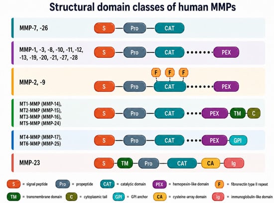
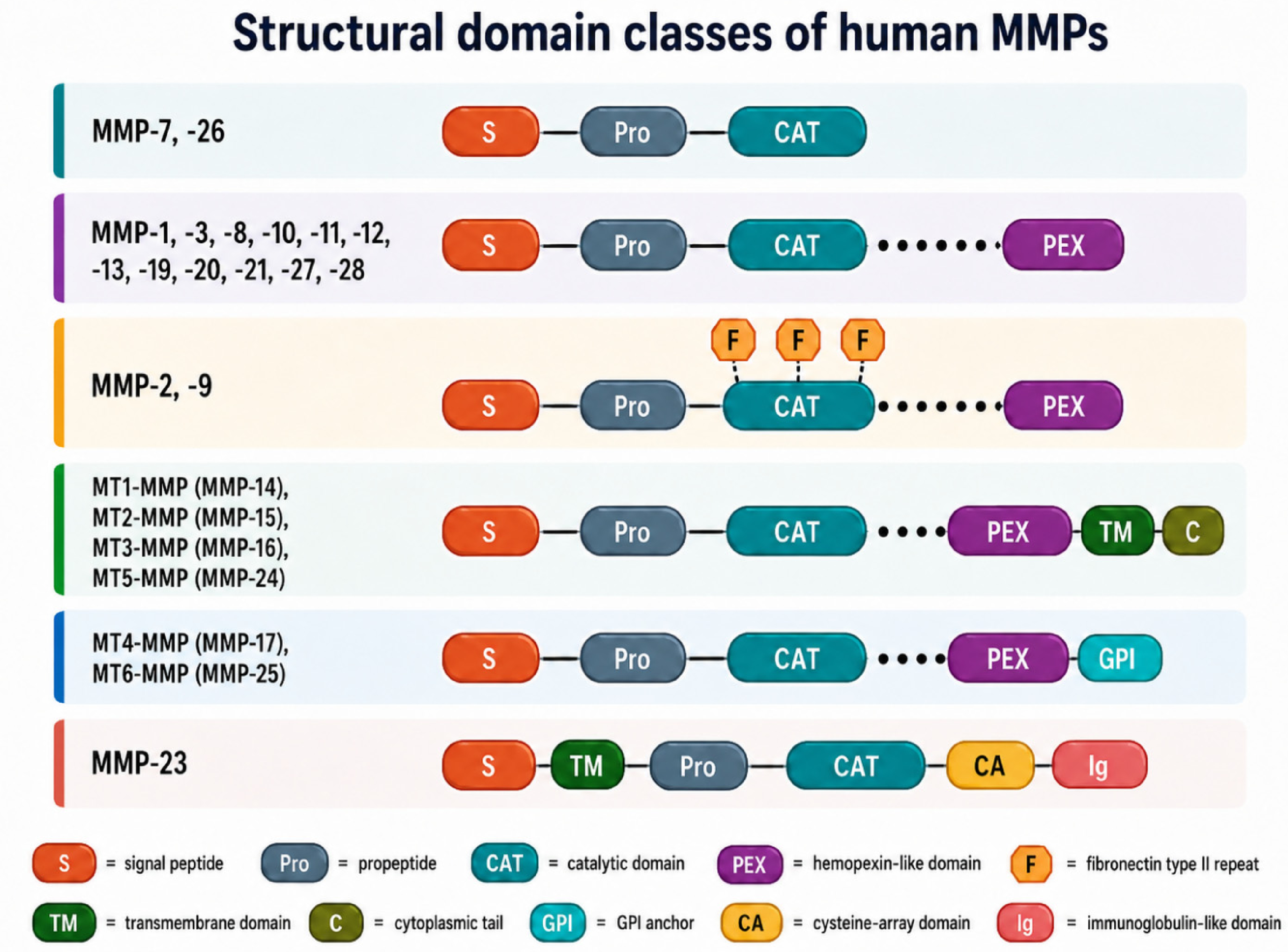
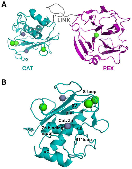
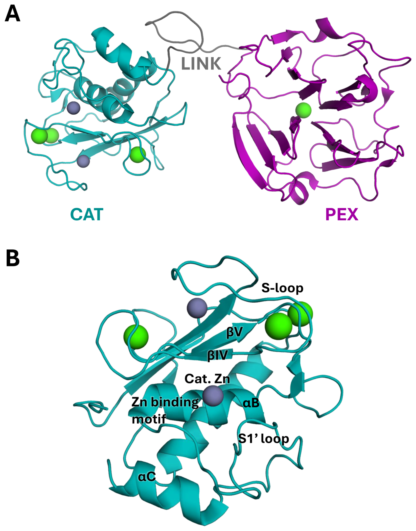
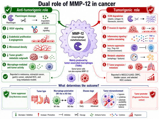
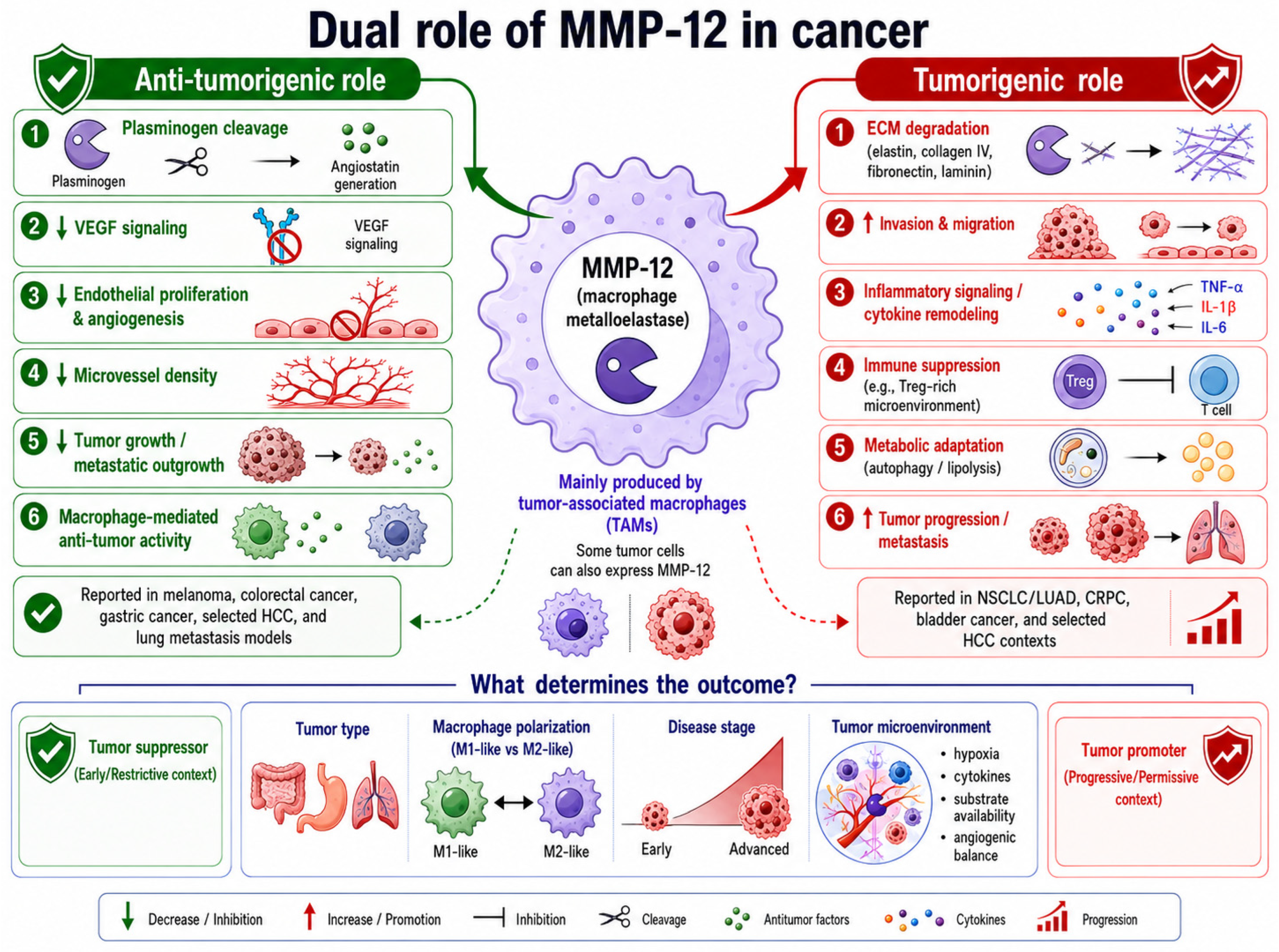
Disclaimer/Publisher’s Note: The statements, opinions and data contained in all publications are solely those of the individual author(s) and contributor(s) and not of MDPI and/or the editor(s). MDPI and/or the editor(s) disclaim responsibility for any injury to people or property resulting from any ideas, methods, instructions or products referred to in the content. |
© 2026 by the authors. Licensee MDPI, Basel, Switzerland. This article is an open access article distributed under the terms and conditions of the Creative Commons Attribution (CC BY) license.