


Abstract
Background: Food-based delivery systems are essential for the practical application of probiotics. A key challenge in translating this technology is determining whether the bacteria remain viable and biologically active after being incorporated into a real food matrix and undergoing prolonged processing. This study evaluated whether Lactiplantibacillus plantarum Lp1 retains its immunomodulatory activity when incorporated into a ripened, semi-hard cheese matrix. Methods: Female BALB/c mice were assigned to three groups: Ovalbumin-induced pneumonia (OVA); OVA plus control cheese; and OVA plus Lp1 cheese. The cheese was ripened at a low temperature for 120 days and administered at a dose of 2 g per day for four weeks. Allergic airway inflammation was induced by OVA sensitization and intranasal challenge. The following were evaluated: viable Lp1 counts after ripening, cytokine gene expression in bronchoalveolar lavage cells and spleen, total IgE levels, and lung histopathology. Results: The number of viable Lp1 decreased during ripening but remained detectable in the final cheese product at 8.7 × 10⁶ CFU/g, which corresponds to an estimated daily intake of 1.7 × 10⁷ CFU/mouse. Bronchoalveolar lavage cells from the OVA plus Lp1 cheese group showed lower expression of selected inflammatory cytokine genes, including Il1b, Il13, and Tnf, as well as higher Il10 expression, compared to the OVA plus control cheese group. Lung histopathology suggested attenuation of OVA-induced airway inflammation. Serum total IgE levels did not differ significantly among groups. Conclusions: In this mouse model, cheese containing Lactiplantibacillus plantarum Lp1 retained the potential to modulate selected inflammation-related markers after 120 days of ripening. Although total serum IgE levels were unchanged, the findings should be interpreted together with other inflammatory outcomes. These results suggest that aged cheese may preserve the biological activity of functional lactic acid bacteria, although further studies are required to establish the translational relevance of these findings.
1. Introduction
Asthma is a significant global health concern. In 2019, it affected an estimated 262 million individuals worldwide and contributed to substantial morbidity and mortality [1]. While asthma-related deaths have declined in Japan, asthma remains a significant clinical issue, especially among older adults. The disease is often complicated by multimorbidity, age-related physiological changes, and altered immune regulation in this population [2,3]. These characteristics underscore the necessity of complementary, risk-conscious strategies that can be integrated with standard pharmacotherapy, including those based on lifestyle and nutrition.
The role of diet and nutrition in asthma prevention and symptom modulation is an evolving area of research. Observational studies suggest that a Mediterranean-type diet may be associated with a lower risk of asthma or wheezing, particularly in children. However, findings in adults are inconsistent. Interventions focusing on specific nutrients, such as vitamin D and long-chain n-3 polyunsaturated fatty acids, have also been evaluated; however, their effects vary according to population, outcome, and study design [4,5]. The gut–lung axis provides a plausible biological framework mechanistically linking gut microbiota-derived metabolites and pulmonary immune regulation [6]. Therefore, probiotics have been investigated as an adjunctive approach for allergic airway diseases; however, clinical outcomes remain heterogeneous due to differences in strains, doses, intervention periods, and endpoints [7].
A key translational barrier in probiotic research is that the biological activity demonstrated using cultured bacteria is not necessarily retained when the bacteria are incorporated into a real food matrix and undergo prolonged processing. Therefore, both post-processing viability and in vivo biological activity should be confirmed before further development as a functional food is considered. Food matrices may affect probiotic survival during processing, storage, and passage through the gastrointestinal tract. Cheese is a protein-rich, palatable, nutrient-dense carrier that supports the survival of lactic acid bacteria [8]. However, manufacturing and ripening conditions may reduce bacterial viability and modify biological activity. Post-ripening verification is therefore essential.
Previous studies have shown that Lp1 modulates inflammatory responses and gut microbiota-related parameters in adult cattle [9] and attenuates pulmonary inflammation and inflammatory cytokine responses in a mouse model of ovalbumin (OVA)-induced allergic airway inflammation [10]. Dairy products, including cheese, have been investigated as delivery matrices for probiotic bacteria; however, the effects of prolonged cheese ripening on the viability and strain-specific biological activity of incorporated bacteria remain poorly understood.
In the present study, Lp1 was incorporated into a semi-hard cheese produced using a method based on the traditional manufacture of Bra Tenero (Teneréllo), a ripened cheese from northern Italy. After 120 days of low-temperature ripening, we evaluated the viability and potential immunomodulatory activity of Lp1 in the cheese. Using an OVA-induced mouse model of allergic airway inflammation, we assessed viable bacterial counts, cytokine gene expression, serum immune markers, and lung histopathological findings to determine whether Lp1-containing cheese retained biological activity after prolonged ripening.
2. Materials and Methods
2.1. Bacterial Strain and Culture Conditions
Lactiplantibacillus plantarum Lp1 (Lp1; Japanese Patent No. 5610472) was used as the probiotic strain in this study. Lp1 is a non-genetically modified lactic acid bacterium originally isolated from naturally fermented milk. The strain is characterized by resistance to gastric acid and bile salts, growth at low temperatures, and the ability to utilize a broad range of carbohydrates. Previous studies have suggested that Lp1 may modulate metabolic activity and immune responses in treated animals. Lp1 was cultured anaerobically on modified Lactobacillus Selection agar (LBS; Nissui Pharmaceutical, Tokyo, Japan) for 48 h.
A single colony was transferred to a MRS broth (Solabia Biokar Diagnostics, La Défense, France) and cultured at 37 °C with shaking. The culture was expanded sequentially from 2 mL to 1 L, after which it was centrifuged at 6000 rpm for 10 min. The supernatant was discarded, and the bacterial pellet was resuspended in 2 L of 10% skim milk. This suspension was then incubated at 37 °C for 72 h and used for cheese production. The bacterial concentration of the suspension was 3 × 10⁹ CFU/mL.
2.2. Manufacture of Lp1 Cheese and Ripening
A semi-hard ripened cheese, designated Teneréllo, was produced from pasteurized milk. Briefly, 50 L of raw milk was pasteurized at 63 °C for 30 min, after which a starter culture containing Streptococcus thermophilus and Lactobacillus delbrueckii subsp. bulgaricus was added. For production of Lp1-containing cheese, an Lp1 suspension was added to achieve a total inoculum of 8.7 × 10¹¹ colony-forming units (CFU) per 50 L of milk. The estimated initial Lp1 concentration in the cheese was 1.7 × 10⁸ CFU/g. The milk was incubated for 1 h, after which rennet was added to induce coagulation. The resulting curd was cut, drained to remove the whey, and transferred into molds. After resting overnight, the cheese was immersed in brine for 36 h.
The cheese was initially ripened at 8–9 °C for 1 week and subsequently ripened at 10 °C for 120 days. During ripening, the cheese was turned every 2 days. Control cheese was produced using the same procedure without the addition of Lp1. For enumeration of viable bacteria, three independently collected cheese samples were analyzed. Each sample was homogenized in phosphate-buffered saline, serially diluted, and plated in technical triplicate on LBS agar. Plates were incubated anaerobically at 37 °C for 48 h, after which colonies were counted. Bacterial concentrations were expressed as log10 CFU/g of cheese. For each independent sample, the mean value of the technical replicate plates was calculated. The results are presented as the mean ± standard error of the mean and the range of the independent measurements. The nutritional composition of the experimental cheese is shown in Table 1.
Table 1.
Nutritional Composition of the Experimental Cheese.
2.3. Animals and Experimental Design
Six-week-old female BALB/c mice were obtained from a commercial vendor and housed under specific pathogen-free conditions at 23 °C with a 12 h light/dark cycle. After arrival, the mice were acclimatized for 1 week and provided sterile feed and water ad libitum. As this was an exploratory study, five mice were assigned to each group based on the Reduction principle of the 3Rs and considerations of experimental feasibility; no formal a priori power calculation was performed. The mice were stratified by baseline body weight and randomly allocated to three experimental groups (n = 5 per group) using a computer-generated random-number sequence.
- OVA + control cheese (2 g/day; ripened cheese without Lp1).
- OVA + Lp1 cheese (2 g/day; ripened Lp1 cheese)
- OVA group (normal CE-2 chow only, no treatment)
Mice in the cheese-fed groups received 2 g/day of either control cheese or Lp1-containing cheese for 4 weeks. Cheese and feed intake were monitored daily, and the entire daily cheese allotment was consumed throughout the intervention period. Body weight was recorded weekly. Mean feed intake per mouse was calculated from the total amount of feed consumed by each group. Nutrient intake from the feed was estimated using the manufacturer’s nutritional information, as described below. All animal procedures were approved by the Animal Experimentation Committee of Rakuno Gakuen University and conducted in accordance with the university’s guidelines for the care and use of laboratory animals (approval no. VH24A27, approved on 21 February 2025).
2.4. OVA-Induced Allergic Airway Inflammation
Allergic airway inflammation was induced using a previously reported OVA sensitization and challenge protocol [10]. In brief, mice were sensitized via an intraperitoneal injection of 100 μg of OVA (grade V, Sigma-Aldrich, St. Louis, MO, USA) adsorbed to 20 mg of aluminum hydroxide (Pierce, Perbio Science Nederland, Etten-Leur, Netherlands) in phosphate-buffered saline. Sensitization was performed twice at one-week intervals. Two weeks after the final sensitization, the mice were challenged intranasally with 0.2 mg of OVA in 20 μL of phosphate-buffered saline on three occasions at three-day intervals.
2.5. Sample Collection
Mice were euthanized under 3% isoflurane anesthesia 48 h after the final OVA challenge. Blood was collected for serum analysis. Bronchoalveolar lavage was performed by instilling 0.2 mL of phosphate-buffered saline into the lungs. Bronchoalveolar lavage cells were recovered by centrifugation and used for RNA extraction. Spleens were also collected for RNA extraction, and the lungs were fixed in 10% neutral-buffered formalin for histopathological examination.
All samples were assigned anonymized identification codes, and investigators performing the qPCR and ELISA assays were blinded to treatment allocation. Statistical analyses were conducted using coded group labels, with treatment identities concealed until completion of the prespecified analyses. The allocation code was then unblinded, and group identities, data assignments, and figure labels were verified.
2.6. Gene Expression Analysis
Total RNA was extracted from bronchoalveolar lavage (BAL) cells and spleen tissue using the RNeasy Mini Kit (Qiagen, Hilden, Germany). RNA concentration and purity were assessed spectrophotometrically using the QIAxpert system (Qiagen). Only RNA samples with A260/A280 ratios within the predefined acceptable range were used for complementary DNA (cDNA) synthesis.
cDNA was synthesized using ReverTra Ace qPCR RT Master Mix with gDNA Remover (Toyobo Co., Ltd., Osaka, Japan) according to the manufacturer’s instructions. Quantitative PCR was performed using SYBR Green Realtime PCR Master Mix (Toyobo Co., Ltd., Osaka, Japan). Amplification specificity was assessed by melting-curve analysis, and the detection of a single peak was considered indicative of specific amplification.
Amplification efficiency was determined from the slope of a standard curve generated using serial dilutions of pooled cDNA and calculated as follows: E (%) = [10(−1/slope) − 1] × 100. Primer sets were considered acceptable when amplification efficiencies ranged from 90% to 110% and the coefficients of determination were R2 ≥ 0.98.
Each sample was analyzed in technical duplicate. The mean Cq value was used when the absolute difference between duplicate measurements was ≤0.5 cycles; samples showing a difference of >0.5 cycles were reanalyzed. The expression levels of Il1b, Il13, Il10, and Tnf were normalized to that of Gapdh. Relative gene expression was calculated using the 2−ΔΔCq method. The primer sequences are listed in Table 2.
Table 2.
Primer Sequences Used for Quantitative PCR.
2.7. Total IgE Measurements
Serum total IgE was measured using the LBIS Mouse IgE ELISA Kit (FUJIFILM Wako Pure Chemical Corporation, Osaka, Japan), following the manufacturer’s instructions. The kit’s assay range is 1–100 ng/mL. Quantitative values were calculated from the included standard curve.
2.8. Lung Histopathology and Eosinophil Quantification
Lung tissues were fixed in 10% neutral-buffered formalin, embedded in paraffin, and sectioned at a thickness of 4 µm. Sections were stained with hematoxylin and eosin for evaluation of airway inflammation. A comprehensive semiquantitative assessment of pulmonary lesions in this OVA-induced model has been reported previously [10]. In the present follow-up study, histological analysis focused on inflammatory cell infiltration as a targeted endpoint for evaluating the biological activity of Lp1-containing cheese after prolonged ripening. Epithelial alterations and peribronchiolar and perivascular inflammatory cell infiltration were assessed.
Peribronchiolar eosinophils were detected using Direct Fast Scarlet staining. Sections were incubated with Direct Fast Scarlet Solution (product no. 21422; Muto Pure Chemicals Co., Ltd., Tokyo, Japan) at 50 °C for 30 min. The sections were then rinsed in distilled water, briefly immersed in 80% ethanol containing 0.001% NaOH and 2% NaCl, rinsed again in distilled water, counterstained with Mayer’s hematoxylin for 3 min, dehydrated, cleared in xylene, and mounted. Eosinophils were counted in five microscopic fields per tissue section. Histopathological evaluation and lesion scoring were performed by an investigator blinded to treatment allocation and sample identity.
2.9. Statistical Analysis
Unless otherwise specified, normally distributed data are presented as the mean ± standard error of the mean (SEM), whereas non-normally distributed and ordinal data are presented as the median and interquartile range. Normality of model residuals was assessed using the Shapiro–Wilk test [11], and homogeneity of variance was evaluated before parametric analyses. Variables meeting the assumptions of normality and homogeneity of variance were analyzed using one-way analysis of variance (ANOVA), followed by pairwise comparisons with Holm adjustment [12]. Variables that did not meet the assumptions for parametric analysis, including non-normally distributed gene-expression and serum-marker data, were analyzed using the Kruskal–Wallis test [13], followed by Dunn’s multiple-comparison test [14] with Holm adjustment [12]. Histopathological cell-count and ordinal score data were analyzed using the Kruskal–Wallis test followed by Dunn’s test with Holm adjustment. All statistical tests were two-sided, and Holm-adjusted p values < 0.05 were considered statistically significant. Exact adjusted p values are reported whenever possible; values < 0.001 are reported as p < 0.001. Statistical analyses were performed using R software, version 4.1.0 (R Foundation for Statistical Computing, Vienna, Austria).
3. Results
3.1. Viable Lp1 Count After Ripening, Cheese Acceptance, and Body Weight
The estimated Lp1 count in the cheese before ripening was 8.23 log10 colony-forming units (CFUs)/g. After 120 days of ripening, the viable count of Lp1 in the cheese was 6.94 ± 0.09 log10 CFU/g, based on 3 independent measurements. No Lp1 colonies were detected in cheese from the control group. The average daily cheese intake per mouse was 2 g, resulting in an estimated daily Lp1 intake of 1.7 × 10⁷ CFU per mouse. In previous tests, this amount was set to ensure the bacterial count was confirmed to be effective. Both cheese formulations were readily accepted, and the entire daily cheese ration was consumed throughout the study period. At week 6, the average body weight ranged from 23.1 to 24.7 g per mouse in each group.
3.2. Nutrient Intake Estimation
We calculated the nutritional intake of the mice fed cheese based on the administered amounts. CE-2 feed was provided ad libitum, and the average intake per mouse was calculated from the feed intake of each group and was 3 g. Nutritional intake from the cheese supplement was calculated based on the measured composition of the experimental cheese. Nutritional intake from the CE-2 diet was estimated using the manufacturer’s nutritional data. The daily nutritional intake per individual is shown in Table 3.
Table 3.
Estimated Daily Nutrient Intake Per Mouse.
3.3. Cytokine Gene Expression in the Spleen and Bronchoalveolar Lavage Cells
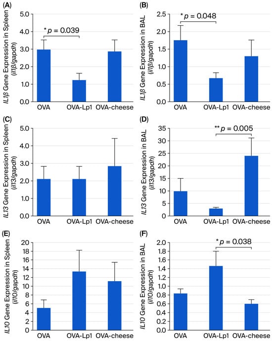
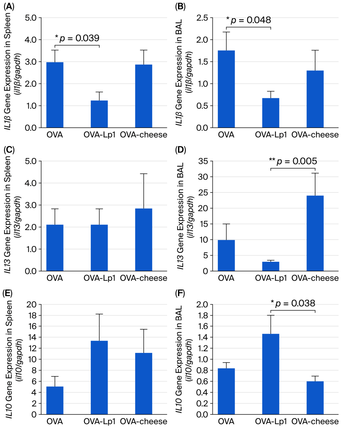
Cytokine gene expression in the spleen and bronchoalveolar lavage (BAL) cells was assessed by quantitative polymerase chain reaction (qPCR). In the spleen, Il1b expression was significantly lower in the OVA plus Lp1 cheese group than in the OVA group (95% CI, 0.967–1.387; p = 0.039; Figure 1A). No significant differences in splenic Il13 expression were observed among the groups. Splenic Il10 expression was higher in the OVA plus Lp1 cheese group than in the OVA group, although the difference did not reach statistical significance.
Figure 1.
Cytokine gene expression in the spleen and bronchoalveolar lavage cells following OVA-induced airway inflammation. The relative mRNA expression levels of Il1b (A,B), Il13 (C,D), and Il10 (E,F) were measured in the spleen (A,C,E) and bronchoalveolar lavage (BAL) cells (B,D,F) by quantitative reverse-transcription PCR. Gene expression was normalized to Gapdh. The OVA group received OVA alone, the OVA-Lp1 group received OVA and cheese containing Lactiplantibacillus plantarum Lp1, and the OVA-cheese group received OVA and control cheese without Lp1. Data are presented as the mean ± SEM (n = 5 mice per group). Statistical comparisons were performed using one-way ANOVA followed by Holm-adjusted pairwise comparisons for normally distributed data or the Kruskal–Wallis test followed by Dunn’s multiple-comparison test with Holm correction for non-normally distributed data, as appropriate. Significant differences were observed between the OVA and OVA-Lp1 groups for splenic Il1b expression (A, * p = 0.039) and BAL-cell Il1b expression (B, * p = 0.048), between the OVA-Lp1 and OVA-cheese groups for BAL-cell Il13 expression (D, ** p = 0.005), and between the OVA-Lp1 and OVA-cheese groups for BAL-cell Il10 expression (F, * p = 0.038). No statistically significant differences were detected in panels (C,E).
In BAL cells, Il1b and Il13 expression levels were significantly lower in the OVA plus Lp1 cheese group than in the OVA plus control cheese group (Il1b: 95% CI, 0.384–1.257; p = 0.048; Figure 1B; Il13: 95% CI, 1.556–27.373; p = 0.005; Figure 1D). Conversely, Il10 expression was significantly higher in the OVA plus Lp1 cheese group than in the OVA plus control cheese group (95% CI, 0.256–0.880; p = 0.038; Figure 1F). These findings indicate that administration of Lp1-containing cheese was associated with lower local expression of the inflammation-related genes Il1b and Il13 and higher expression of the regulatory cytokine gene Il10 in the airways.
In the spleen, Il10 expression was numerically higher in the OVA plus Lp1 cheese group than in the OVA group; however, the difference was not statistically significant. Thus, the present findings provide limited evidence for an effect of Lp1-containing cheese on systemic immunoregulatory responses.
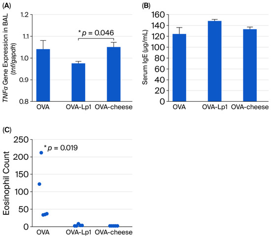
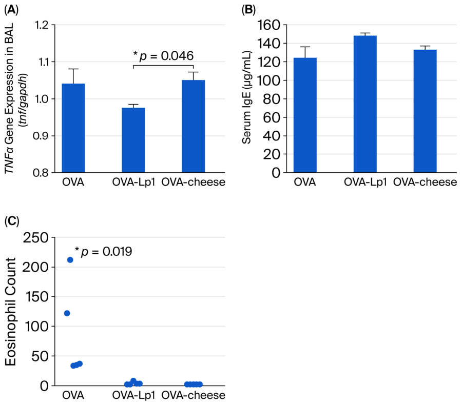
Expression of Tnf in BAL cells was also significantly lower in the OVA plus Lp1 cheese group than in the OVA plus control cheese group (95% CI, 0.002–0.213; p = 0.046; Figure 2A). Collectively, the lower expression levels of Il1b, Il13, and Tnf, together with the higher expression of Il10, are consistent with modulation of selected local inflammatory responses in the airways. In particular, reduced Il13 expression may reflect attenuation of type 2 immune responses associated with eosinophilic inflammation and mucus production, whereas reduced Il1b and Tnf expression may indicate decreased activation of innate inflammatory pathways.
Figure 2.
Tnf gene expression in bronchoalveolar lavage cells, serum total IgE concentrations, and pulmonary eosinophil counts following OVA-induced airway inflammation. (A) Relative Tnf expression in bronchoalveolar lavage (BAL) cells was measured by quantitative reverse-transcription PCR and normalized to Gapdh. (B) Serum total IgE concentrations were measured by enzyme-linked immunosorbent assay. (C) Pulmonary eosinophils were identified in direct fast scarlet-stained lung sections and counted in five microscopic fields per mouse. The OVA group received OVA alone, the OVA-Lp1 group received OVA and cheese containing Lactiplantibacillus plantarum Lp1, and the OVA-cheese group received OVA and control cheese without Lp1. Data in panels A and B are presented as the mean ± SEM, whereas individual eosinophil counts are shown in panel C (n = 5 mice per group). Statistical comparisons were performed using one-way ANOVA followed by Holm-adjusted pairwise comparisons for normally distributed data. Eosinophil counts were analyzed using the Kruskal–Wallis test followed by Dunn’s multiple-comparison test with Holm correction. BAL-cell Tnf expression was significantly lower in the OVA-Lp1 group than in the OVA-cheese group (A, * p = 0.046). Pulmonary eosinophil counts were significantly lower in the OVA-Lp1 group than in the OVA group (C, * p = 0.019). No statistically significant differences in serum total IgE concentrations were detected among the groups (B).
These transcriptional changes may be related to the reduced inflammatory cell infiltration observed in the lungs of mice administered Lp1-containing cheese. However, cytokine protein concentrations and the cellular sources of the detected transcripts were not determined. Further studies are therefore required to clarify the mechanisms and biological significance of these findings.
3.4. Lung Histopathology and Serum Total IgE
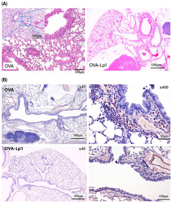
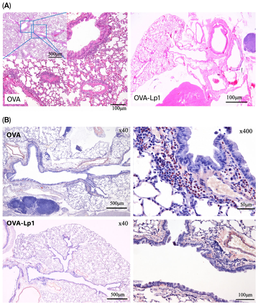
Histopathological examination revealed airway inflammatory changes in OVA-treated mice, including epithelial alterations and peribronchiolar and perivascular inflammatory cell infiltration. These changes were less prominent in the OVA plus Lp1 cheese group than in the OVA group (Figure 3A).
Figure 3.
Lung histopathology and eosinophil infiltration following OVA-induced airway inflammation. Representative lung sections from the OVA and OVA-Lp1 groups are shown. The OVA group received OVA alone, whereas the OVA-Lp1 group received OVA and cheese containing Lactiplantibacillus plantarum Lp1. (A) Hematoxylin and eosin-stained lung sections showing inflammatory cell infiltration and histopathological changes in the peribronchial and perivascular regions. The boxed areas in the low-magnification image of the OVA group are shown at higher magnification. Scale bars represent 500 μm in the low-magnification image and 100 μm in the higher-magnification images. (B) Direct fast scarlet-stained lung sections showing eosinophil infiltration. Eosinophils are identified by their red-stained cytoplasmic granules. Images of the OVA and OVA-Lp1 groups are shown at low and high magnification. Scale bars represent 500 μm in the low-magnification images, 50 μm in the high-magnification image of the OVA group, and 100 μm in the high-magnification image of the OVA-Lp1 group. Images are representative of five mice per group.
Eosinophil infiltration was quantified using Direct Fast Scarlet staining. The number of peribronchiolar eosinophils was significantly lower in the OVA plus Lp1 cheese group than in the OVA group (p = 0.019; Figure 2C). This finding was consistent with the observed cytokine gene-expression profile. In contrast, serum total IgE concentrations did not differ significantly among the OVA-treated groups, with mean values ranging from 124.2 to 148.2 μg/mL (Figure 2B). For reference, the mean serum total IgE concentration in the naïve control group was 80 ng/mL.
4. Discussion
This study examined whether Lactiplantibacillus plantarum Lp1 retained viability and biological activity after incorporation into a semi-hard cheese and prolonged low-temperature ripening. Although viable Lp1 counts decreased during the 120-day ripening period, viable bacteria remained detectable in the final product. In the present mouse model, administration of Lp1-containing cheese was associated with lower expression of Il1b, Il13, and Tnf and higher expression of Il10 in bronchoalveolar lavage (BAL) cells. These transcriptional changes were accompanied by less prominent OVA-induced inflammatory changes in the lungs [15,16]. Collectively, these findings suggest that Lp1 retained measurable biological activity after cheese processing and prolonged ripening.
A strength of this study is the use of a realistic food matrix rather than administration of cultured bacteria alone. The translational relevance of probiotic and functional-food research depends not only on the intrinsic properties of candidate strains but also on their ability to survive processing and storage and to retain biological activity within the final food product. Cheese may provide a suitable matrix because of its nutritional composition, palatability, and capacity to support the survival of lactic acid bacteria [17]. In the present study, viable Lp1 persisted after prolonged ripening, and administration of the resulting cheese was associated with modulation of selected inflammatory outcomes in vivo. However, these findings should not be interpreted as demonstrating clinical efficacy or gastrointestinal delivery, neither of which was directly assessed.
In BAL cells, Lp1-containing cheese was associated with lower expression of Il1b, Il13, and Tnf and higher expression of Il10 than control cheese. This pattern is consistent with modulation of selected inflammatory and immunoregulatory responses in the local airway environment. In contrast, Il13 expression was higher in the OVA plus control cheese group. IL-13 is a type 2 cytokine involved in allergic sensitization, eosinophilic inflammation, and mucus production. Increased IL-13 production has been reported in spleen cells and BAL fluid from OVA-sensitized BALB/c mice following antigen restimulation [15,18]. Accordingly, the increased Il13 expression observed in the spleen and BAL cells may reflect persistent or enhanced type 2 immune priming rather than established pulmonary pathology alone. The higher Il10 expression observed in BAL cells from mice administered Lp1-containing cheese may indicate enhanced activation of regulatory or anti-inflammatory pathways [19,20]. Nevertheless, because cytokine protein concentrations and the cellular sources of these transcripts were not determined, these interpretations remain provisional.
No significant differences in serum total IgE concentrations were observed among the OVA-treated groups, suggesting that Lp1-containing cheese did not markedly alter systemic IgE-associated sensitization under the present experimental conditions. IgE responses are influenced by events occurring during sensitization and by the kinetics of type 2 immune responses. Moreover, serum total IgE does not necessarily correlate directly with antigen-specific IgE, local IgE production, or the severity of inflammation in peripheral tissues [21,22]. Therefore, the absence of a significant difference in serum total IgE does not necessarily conflict with the observed changes in local airway inflammatory markers. Within the scope of this study, the findings are more appropriately interpreted as modulation of selected local inflammatory responses rather than as evidence of a direct effect on systemic IgE regulation or generalized allergic sensitization [22].
Eosinophil infiltration at sites of pulmonary inflammation was markedly lower in both cheese-administered groups than in the OVA group. The mean eosinophil counts were 88.25 ± 49.07 in the OVA group, 5.33 ± 1.17 in the OVA plus Lp1 cheese group, and 4.40 ± 0.81 in the OVA plus control cheese group. The comparable values in the two cheese-administered groups suggest that the cheese matrix itself may have contributed to the reduction in eosinophil infiltration. Bioactive peptides and other metabolites generated during cheese ripening may influence inflammatory responses and provide a biologically plausible explanation for this observation [23,24]. Under these conditions, any additional effect specifically attributable to Lp1 may have been more readily reflected in selected cytokine-related transcriptional responses than in eosinophil counts alone. However, the substantial variability in the OVA group and the small sample size warrant cautious interpretation.
The histopathological analysis in this study focused on inflammatory cell infiltration and did not include a comprehensive composite score incorporating peribronchial and perivascular inflammation, epithelial alterations, and mucus production. A detailed lung-lesion scoring system using the same experimental model has been reported previously [10]. Accordingly, the present histological findings should be interpreted as evidence of differences in inflammatory cell infiltration rather than as a comprehensive assessment of overall pulmonary lesion severity.
This study has several limitations. First, the control-cheese and Lp1-cheese groups received the same daily amount of cheese prepared using the same basic formulation; therefore, the nutritional contribution of the cheese matrix was expected to be comparable between these two groups. However, because basal-diet intake was not measured directly, equivalence of total energy intake could not be confirmed. Comparisons with the group that did not receive cheese may also have been influenced by the additional energy and nutrients supplied by the cheese. The body-weight gain observed in the OVA and control-cheese groups suggests that nutritional factors independent of Lp1 may have influenced systemic immune responses. Nevertheless, the estimated sodium intake was likely lower than that used in experimental models of high-salt-induced immune skewing [25]. Second, water intake was not measured under free-feeding conditions. Third, the study relied primarily on endpoint analyses and did not evaluate longitudinal changes in upstream immune mediators or detailed immune-cell populations. Fourth, cytokine production was assessed only at the mRNA level, and protein concentrations and cellular sources were not determined.
The small sample size of five mice per group is an additional important limitation. No formal a priori power calculation was performed, and the limited sample size may have reduced the ability to detect modest biological effects, particularly for immunological outcomes with substantial interindividual variability. Consequently, nonsignificant findings should not be interpreted as evidence of no effect, and the observed differences should be regarded as preliminary. Larger confirmatory studies with prospectively determined sample sizes, controlled nutritional conditions, and dose–response designs are required to validate the biological activity of Lp1-containing cheese and to determine whether aged dairy products can serve as effective matrices for functional lactic acid bacteria [26,27].
5. Conclusions
In conclusion, viable Lactiplantibacillus plantarum Lp1 remained detectable after incorporation into a semi-hard cheese and 120 days of low-temperature ripening. In an OVA-induced mouse model of allergic airway inflammation, administration of Lp1-containing cheese was associated with modulation of selected cytokine-related transcripts in BAL cells. However, the comparable reduction in eosinophil infiltration observed in both cheese-administered groups suggests that the cheese matrix itself may also have contributed to the histological findings. These results provide preliminary evidence that Lp1 can retain measurable biological activity after prolonged cheese ripening. Further studies incorporating protein-level measurements, larger sample sizes, and controlled nutritional conditions are required to confirm the mechanisms and translational relevance of these findings.
6. Patents
Patents: K. Hagiwara is the inventor of a patent related to Lactiplantibacillus plantarum Lp1 (Patent# 35610472). This interest is disclosed here. The authors declare no other competing interests.
Author Contributions
K.H. (Katsuro Hagiwara) was involved in the design of the study, the management of the overall project and funding, the collection of samples, animal experiments, the analysis of samples, the interpretation of data, the drafting of the manuscript, and the revision of the manuscript. M.O. contributed to the study design, sample collection, animal experiments, sample analysis, data interpretation, manuscript drafting, and manuscript revision. K.H. (Keita Hagiwara) and K.I. were involved in preparing lactic acid bacteria, producing cheese, managing its ripening, controlling its quality, interpreting cheese-related data, and preparing the manuscript. All authors have read and agreed to the published version of the manuscript.
Funding
This study was partially supported by a scholarship donation from the Scientific Feed Research Institute (No. 2023-9). The sponsor had no role in the design, execution, interpretation, or writing of the study.
Institutional Review Board Statement
The animal study protocol was approved by the Rakuno Gakuen University Laboratory Animal Committee (approval no. VH24A27, approved on 21 February 2025).
Informed Consent Statement
Not applicable.
Data Availability Statement
The results of this paper are presented in the paper itself.
Acknowledgments
We would like to thank the cheese craftsmen at Cheese Factory Shiranuka Rakkeisha Co., Ltd. for their help with this cheese-processing project.
Conflicts of Interest
Authors, Keita Hagiwara and Kazuyoshi Inoguchi, were employed by the company Cheese Factory Shiranuka rakkeisha Co., Ltd. The remaining authors declare that the research was conducted in the absence of any commercial or financial relationships that could be construed as a potential conflict of interest.
Abbreviations
The following abbreviations are used in this manuscript:
| Il1b | Interleukin 1 beta |
| Il13 | Interleukin 13 |
| Il10 | Interleukin 10 |
| Il17 | Interleukin 17A |
| tnfα | tumor necrosis factor-α |
| gapdh | Glyceraldehyde-3-phosphate dehydrogenase |
| OVA | ovalbumin |
| CFU | colony-forming units |
| LBS | Lactobacillus Selection |
References
- World Health Organization. Asthma: Fact Sheet. Updated on 6 May 2024. Available online: https://www.who.int/news-room/fact-sheets/detail/asthma (accessed on 26 March 2026).
- Kan-O, K. Asthma-related death trends and biologics use for severe asthma in the super-aged society of Japan. Respirology 2024, 29, 455–457. [Google Scholar] [CrossRef] [Scilit] [PubMed]
- Hanania, N.A.; King, M.J.; Braman, S.S.; Saltoun, C.; Wise, R.A.; Enright, P.; Falsey, A.R.; Mathur, S.K.; Ramsdell, J.W.; Rogers, L.; et al. Asthma in the elderly: Current understanding and future research needs—A report of a National Institute on Aging (NIA) workshop. J. Allergy Clin. Immunol. 2011, 128, S4–S24. [Google Scholar] [CrossRef] [Scilit] [PubMed]
- Jolliffe, D.A.; Greenberg, L.; Hooper, R.L.; Griffiths, C.J.; Camargo, C.A., Jr.; Kerley, C.P.; Jensen, M.E.; Mauger, D.; Stelmach, I.; Urashima, M.; et al. Vitamin D supplementation to prevent asthma exacerbations: A systematic review and meta-analysis of individual participant data. Lancet Respir. Med. 2017, 5, 881–890, Correction in Lancet Respir. Med. 2018, 6, E27. [Google Scholar] [CrossRef] [Scilit] [PubMed]
- Bärebring, L.; Nwaru, B.I.; Lamberg-Allardt, C.; Thorisdottir, B.; Ramel, A.; Söderlund, F.; Arnesen, E.K.; Dierkes, J.; Åkesson, A. Supplementation with long chain n-3 fatty acids during pregnancy, lactation, or infancy in relation to risk of asthma and atopic disease during childhood: A systematic review and meta-analysis of randomized controlled clinical trials. Food Nutr. Res. 2022, 66, 8842. [Google Scholar] [CrossRef] [Scilit] [PubMed]
- Yang, Z.; Mao, W.; Wang, J.; Yin, L. The gut–lung axis in asthma: Microbiota-driven mechanisms and therapeutic perspectives. Front. Microbiol. 2025, 16, 1680521. [Google Scholar] [CrossRef] [Scilit] [PubMed]
- Balan, D.; Baral, T.; Manu, M.K.; Mohapatra, A.K.; Miraj, S.S. Efficacy of probiotics as adjuvant therapy in bronchial asthma: A systematic review and meta-analysis. Allergy Asthma Clin. Immunol. 2024, 20, 60. [Google Scholar] [CrossRef] [Scilit] [PubMed]
- Desfossés-Foucault, É.; Dussault-Lepage, V.; Le Boucher, C.; Savard, P.; LaPointe, G.; Roy, D. Assessment of probiotic viability during Cheddar cheese manufacture and ripening using propidium monoazide-PCR quantification. Front. Microbiol. 2012, 3, 350. [Google Scholar] [CrossRef] [Scilit] [PubMed]
- Chida, S.; Sakamoto, M.; Takino, T.; Kawamoto, S.; Hagiwara, K. Changes in immune system and intestinal bacteria of cows during the transition period. Vet. Anim. Sci. 2021, 14, 100222. [Google Scholar] [CrossRef] [Scilit] [PubMed]
- Kishida, S.; Kato-Mori, Y.; Okamoto, M.; Hagiwara, K. Anti-inflammatory effect of a specific Lactiplantibacillus plantarum in an ovalbumin-induced asthma model. Microbiol. Immunol. 2022, 66, 442–452. [Google Scholar] [CrossRef] [Scilit] [PubMed]
- Shapiro, S.S.; Wilk, M.B. An analysis of variance test for normality (complete samples). Biometrika 1965, 52, 591–611. [Google Scholar] [CrossRef] [Scilit]
- Holm, S.A. Simple sequentially rejective multiple test procedure. Scand. J. Stat. 1979, 6, 65–70. [Google Scholar]
- Kruskal, W.H.; Wallis, W.A. Use of ranks in one-criterion variance analysis. J. Am. Stat. Assoc. 1952, 47, 583–621. [Google Scholar] [CrossRef]
- Dunn, O.J. Multiple comparisons using rank sums. Technometrics 1964, 6, 241–252. [Google Scholar] [CrossRef]
- Rael, E.L.; Lockey, R.F. Interleukin-13 signaling and its role in asthma. World Allergy Organ. J. 2011, 4, 54–64. [Google Scholar] [CrossRef] [Scilit] [PubMed]
- Juan, Z.; Zhao-Ling, S.; Ming-Hua, Z.; Chun, W.; Hai-Xia, W.; Meng-Yun, L.; Jian-Qiong, H.; Yue-Jie, Z.; Xin, S. Oral administration of Clostridium butyricum CGMCC0313-1 reduces ovalbumin-induced allergic airway inflammation in mice. Respirology 2017, 22, 898–904. [Google Scholar] [CrossRef] [Scilit] [PubMed]
- Slattery, L.; O’Callaghan, J.; Fitzgerald, G.F.; Beresford, T.; Ross, R.P. Invited review: Lactobacillus helveticus—A thermophilic dairy starter related to gut bacteria. J. Dairy Sci. 2010, 93, 4435–4454. [Google Scholar] [CrossRef] [Scilit] [PubMed]
- Bernstein, Z.J.; Shenoy, A.; Chen, A.; Heller, N.M.; Spangler, J.B. Engineering the IL-4/IL-13 axis for targeted immune modulation. Immunol. Rev. 2023, 320, 29–57. [Google Scholar] [CrossRef] [Scilit] [PubMed]
- Ağaç, D.; Estrada, L.D.; Maples, R.; Hooper, L.V.; Farrar, J.D. The β2-adrenergic receptor controls inflammation by driving rapid IL-10 secretion. Brain Behav. Immun. 2018, 74, 176–185. [Google Scholar] [CrossRef] [Scilit] [PubMed]
- Moore, K.W.; de Waal Malefyt, R.; Coffman, R.L.; O’Garra, A. Interleukin-10 and the interleukin-10 receptor. Annu. Rev. Immunol. 2001, 19, 683–765. [Google Scholar] [CrossRef] [Scilit] [PubMed]
- Davila, I.; Valero, A.; Entrenas, L.M.; Valveny, N.; Herráez, L.; SIGE Study Group. Relationship between serum total IgE and disease severity in patients with allergic asthma in Spain. J. Investig. Allergol. Clin. Immunol. 2015, 25, 120–127. [Google Scholar] [PubMed]
- De Schryver, E.; Devuyst, L.; Derycke, L.; Dullaers, M.; Van Zele, T.; Bachert, C.; Gevaert, P. Local immunoglobulin e in the nasal mucosa: Clinical implications. Allergy Asthma Immunol. Res. 2015, 7, 321–331. [Google Scholar] [CrossRef] [Scilit] [PubMed]
- Hong, W.S.; Chen, Y.P.; Dai, T.Y.; Huang, I.N.; Chen, M.J. Effect of heat-inactivated kefir-isolated Lactobacillus kefiranofaciens M1 on preventing an allergic airway response in mice. J. Agric. Food Chem. 2011, 59, 9022–9031. [Google Scholar] [CrossRef] [Scilit] [PubMed]
- Santiago-López, L.; Aguilar-Toalá, J.E.; Hernández-Mendoza, A.; Vallejo-Cordoba, B.; Liceaga, A.M.; González-Córdova, A.F. Invited review: Bioactive compounds produced during cheese ripening and health effects associated with aged cheese consumption. J. Dairy Sci. 2018, 101, 3742–3757. [Google Scholar] [CrossRef] [Scilit] [PubMed]
- Kleinewietfeld, M.; Manzel, A.; Titze, J.; Kvakan, H.; Yosef, N.; Linker, R.A.; Muller, D.N.; Hafler, D.A. Sodium chloride drives autoimmune disease by the induction of pathogenic TH17 cells. Nature 2013, 496, 518–522. [Google Scholar] [CrossRef] [Scilit] [PubMed]
- Pimentel, T.C.; Brandão, L.R.; de Oliveira, M.P.; da Costa, W.K.A.; Magnani, M. Health benefits and technological effects of Lacticaseibacillus casei-01: An overview of the scientific literature. Trends Food Sci. Technol. 2021, 114, 722–737. [Google Scholar] [CrossRef] [Scilit]
- Mounier, J.; Monnet, C.; Vallaeys, T.; Arditi, R.; Sarthou, A.-S. Microbial Interactions Within a Cheese Microbial Community. Appl. Environ. Microbiol. 2008, 74, 172–181. [Google Scholar] [CrossRef] [Scilit] [PubMed]

Disclaimer/Publisher’s Note: The statements, opinions and data contained in all publications are solely those of the individual author(s) and contributor(s) and not of MDPI and/or the editor(s). MDPI and/or the editor(s) disclaim responsibility for any injury to people or property resulting from any ideas, methods, instructions or products referred to in the content. |
© 2026 by the authors. Licensee MDPI, Basel, Switzerland. This article is an open access article distributed under the terms and conditions of the Creative Commons Attribution (CC BY) license.