


Abstract
Polystyrene microplastics (PS-MPs) are emerging environmental contaminants associated with adverse effects on human health, including renal injury. However, protective strategies against microplastic-induced nephrotoxicity remain limited. This study investigated the nephroprotective potential of panduratin A (PA) and pinostrobin (PIN) against polystyrene microplastic-induced kidney damage using human renal proximal tubular cells (HK-2) and the C57BL/6NJcL mouse model. DLS and zeta potential measurements were used to characterize the three types of particles (plain PS, PS-COOH, and PS-NH2). In deionized water, PS-NH2 had the least negative zeta potential; however, this difference was no longer observed in a medium containing FBS, since PS-NH2 in this case showed the largest increase in polydispersity index (PDI), indicating different aggregation behavior. When HK-2 cells were exposed to the three types of particles at a dose of 50 µg/mL, PS-NH2 showed greater cellular uptake and higher cytotoxicity than either plain PS or PS-COOH, as shown by increased LDH release and greater cellular accumulation. PS-NH2 was therefore selected for further investigation of its ability to produce ROS, induce apoptosis, and to assess the protective effects of PA and PIN. Treatment with either PA or PIN reduced PS-NH2 accumulation, ROS formation, and apoptosis in the HK-2 cells. In the animal study, mice were given PS-NH2 orally at a dose of 0.4 mg/day for 28 days. This exposure caused early proximal tubular injury, as evidenced by mild histopathological changes, despite no significant alterations in body weight, food intake, blood urea nitrogen, or creatinine. Pretreatment with PA (25 mg/kg BW) or PIN (40 mg/kg BW), given once daily one hour before the PS-NH2 exposure, reduced histopathological kidney injury, with PIN more effective than PA. In summary, aminated polystyrene microplastics are nephrotoxic in both in vitro and in vivo models, and panduratin A and pinostrobin reduce these adverse effects, possibly by modulating cellular uptake and intracellular stress pathways.
1. Introduction
Plastic pollution represents a major global environmental challenge, with smaller fragments of plastic in particular causing growing concerns due to their potential ecological and health impacts [1]. Among polymer types, polystyrene (PS) is one of the most widely used and most frequently detected plastic contaminants across several ecosystems [1,2]. Emerging evidence indicates that small polystyrene particles can enter the human body through several routes and accumulate in the liver, kidneys, lungs, placenta, intestines, and even the brain. This accumulation could disrupt cellular homeostasis and impair organ function [3,4,5,6,7,8]. The biological effects of small polystyrene particles are strongly influenced by their physicochemical properties, including particle size and surface functionalization [9,10]. However, these properties are not static and may change upon dispersion in protein-containing media [11]. In the kidneys, exposure to polystyrene microplastics (PS-MP) is associated with nephrotoxicity through multiple mechanisms, including oxidative stress, mitochondrial dysfunction, and inflammatory signaling, which may contribute to kidney injury and chronic kidney disease [12,13,14,15,16,17,18]. Although minimizing microplastic exposure by limiting plastic use and removing plastic particles from environmental contamination remains an important preventive strategy, it offers no therapeutic benefit once plastic particles have already accumulated in tissues and initiated pathological processes [19,20].
Recent studies have increasingly focused on natural bioactive compounds as therapeutic candidates for mitigating plastic particle-induced toxicity. Several compounds, including resveratrol, kaempferide, cyanidin-3-O-glucoside, astilbin, and nobiletin, have been reported to alleviate microplastic-induced organ damage through their antioxidant, anti-inflammatory, and cytoprotective properties [21,22,23,24,25]. Given the growing interest in utilizing natural bioactive compounds to mitigate plastic particle-induced organ injury, panduratin A (PA) and pinostrobin (PIN) merit further investigation. PA and PIN are both bioactive flavonoids isolated from Boesenbergia rotunda L. (fingerroot), which has long been used in traditional medicine and has well-documented antioxidant, anti-inflammatory, and cytoprotective properties [26,27,28,29,30,31,32]. Of particular relevance to renal protection both in vitro and in vivo, PA has been reported to attenuate cisplatin-induced renal tubular injury. At the same time, PIN has shown protective effects against cadmium- and gentamicin-induced renal toxicity, particularly by reducing oxidative stress, inflammation, and apoptosis. However, whether these compounds can protect against microplastic-induced renal injury remains unclear [30,33,34]. Among different surface-modified polystyrene microplastics, particle surface chemistry has been shown to influence cellular uptake and toxicity. In particular, aminated polystyrene (PS-NH2) has consistently demonstrated greater cytotoxicity than plain PS and carboxylated PS (PS-COOH) in previous in vitro studies, likely owing to its positively charged surface [9,10]. This comparatively greater toxicity profile makes PS-NH2 a particularly relevant and clinically meaningful model for investigating microplastic-induced renal injury and for evaluating potential protective interventions. Notably, despite the strong mechanistic overlap between the biological activities of PA and PIN and the major pathways implicated in PS-MP-induced renal injury, the potential of these compounds to mitigate PS-NH2-induced nephrotoxicity remains unexplored. Therefore, this study aimed to investigate the protective effects of PA and PIN against PS-NH2-induced kidney injury using both in vitro and in vivo models.
2. Materials and Methods
2.1. Chemical Substances
Polystyrene microplastics (PS-MP) with three types of surface functionalization were purchased from Sigma-Aldrich (St. Louis, MO, USA): plain PS (2 µm mean particle size, Cat. No. 78453, 10% w/v), PS-COOH (2 µm mean particle size, Cat. No. L4530, 2.5% w/v), and PS-NH2 (2 µm mean particle size, Cat. No. L9529, 2.5% w/v). 3-(4,5-dimethylthiazol-2-yl)-2,5-diphenyltetrazolium bromide (MTT) and dimethyl sulfoxide (DMSO) were also purchased from Sigma-Aldrich (St. Louis, MO, USA). The lactate dehydrogenase (LDH) cytotoxicity assay kit was purchased from Dojindo Molecular Technologies (Kumamoto, Japan). Cell culture reagents, including Dulbecco’s Modified Eagle’s Medium (DMEM), fetal bovine serum (FBS), penicillin, and streptomycin, were supplied by Gibco (Waltham, MA, USA). All other reagents were analytical grade unless noted otherwise.
2.2. Panduratin and Pinostrobin Preparation
Panduratin A (PA) and pinostrobin (PIN) were isolated from the rhizomes of Boesenbergia rotunda according to previously published protocols [35,36]. The purity of both compounds (>98%) was verified by HPLC analysis. PA and PIN were dissolved in DMSO and stored at −80 °C until further use.
2.3. Physicochemical Characterization of PS-MPs
Dynamic light scattering (DLS) was used to characterize the hydrodynamic diameter, polydispersity index (PDI), and zeta potential of the PS particles in different dispersion media. Plain-PS, PS-COOH, and PS-NH2 particles with nominal sizes of 2 μm were separately dispersed in distilled water (DI) and Dulbecco’s Modified Eagle Medium, low glucose (DMEM-LG) supplemented with 10% fetal bovine serum (FBS) at a final concentration of 50 μg/mL. The suspensions were vortexed and sonicated for 5 min before measurement. The hydrodynamic diameter, PDI, and zeta potential were measured using a Zetasizer Ultra (Malvern Panalytical, Malvern, UK) at the Chulalongkorn University Scientific and Technological Research Equipment Center (STREC), Chulalongkorn University, Bangkok, Thailand. Measurements were performed in triplicate immediately following particle dispersion in the respective media.
2.4. Cell Culture
The human proximal tubular epithelial cell line (HK-2-CRL2190) was obtained from the American Type Culture Collection (ATCC, Manassas, VA, USA), which certifies mycoplasma-free status at the time of original distribution. In this study, HK-2 cells at passages 14–25 were used and maintained in low-glucose DMEM supplemented with 10% heat-inactivated FBS and 1% antibiotics (100 U/mL penicillin and 100 μg/mL streptomycin). The cultures were incubated at 37 °C in a humidified atmosphere containing 5% CO2 and routinely passaged upon reaching 80–90% confluence before use.
2.5. Cell Viability Assay
Cell viability was assessed using the MTT assay. The HK-2 cells were seeded at 1 × 10⁴ cells/well (≈3.1 × 10⁴ cells/cm2) in 96-well plates and allowed to attach overnight. The cells were then treated with various concentrations of PS-MPs, PA, or PIN for the indicated periods. Following treatment, MTT solution was added to a final concentration of 0.5 mg/mL, and the cells were incubated for 4 h at 37 °C in the dark. After this, the medium was removed, and formazan crystals were dissolved in 100 μL DMSO. Absorbance was measured at 570 nm using a CLARIOstar Plus microplate reader (BMG Labtech, Ortenberg, Germany), and cell viability was calculated and expressed as a percentage of the untreated control using the following equation: Cell viability (% control) = (As − Ab)/(Ac − Ab) × 100, where As, Ab, and Ac represent the absorbance values of the treated sample, blank, and untreated control, respectively.
2.6. Lactate Dehydrogenase Assay
Cytotoxicity was further assessed by measuring extracellular lactate dehydrogenase (LDH) release as an indicator of cell membrane damage and loss of membrane integrity, using a non-homogeneous LDH assay kit (Dojindo, Kumamoto, Japan) in accordance with the manufacturer’s protocol, with modification. HK-2 cells were seeded and treated with test compounds for the specified durations. Culture supernatants were then collected and transferred to a 96-well plate. The LDH working solution was added, and the mixture was incubated for 20 min at room temperature in the dark. Absorbance was measured at 490 nm using a microplate reader. LDH release was calculated using the following equation: LDH release (% control) = (As − Ab)/(Ac − Ab) × 100, where As, Ab, and Ac represent the absorbance values of the treated sample, blank, and untreated control, respectively.
2.7. Analysis of Cellular Uptake
To assess PS-MP internalization, HK-2 cells were seeded in 6-well plates and exposed for 24 h to three types of polystyrene microparticles: unlabeled plain PS, fluorescently labeled amine-modified PS (PS-NH2), and fluorescently labeled carboxyl-modified PS (PS-COOH). The cells were then detached, centrifuged, washed, and resuspended in PBS for flow cytometric analysis. A minimum of 30,000 events per sample were acquired using a BD FACSVia Flow Cytometer (BD Biosciences, San Jose, CA, USA). Because plain PS particles were unlabeled, particle internalization in this group was evaluated solely by changes in side-scatter (SSC), which reflects increased intracellular granularity following particle uptake. For the fluorescently labeled PS-NH2 and PS-COOH particles, internalization was assessed by changes in SSC and by increases in fluorescence intensity, providing direct confirmation of particle uptake.
2.8. Measurement of Intracellular Reactive Oxygen Species (ROS)
Intracellular ROS levels were measured after 24 h of exposure to PS-NH2 alone or in combination with PA or PIN using the fluorescent probe 2′,7′-dichlorofluorescin diacetate (DCFH-DA). Following treatment, the cells were washed with PBS and incubated with DCFH-DA at 37 °C for 30 min in the dark. Fluorescence images were captured using a fluorescence microscope under identical exposure settings for all groups. Fluorescence intensity was quantified using ImageJ software version 1.54p (NIH, Bethesda, MD, USA).
2.9. Cellular Apoptosis
Cellular apoptosis was assessed using an Annexin V-FITC/PI apoptosis detection kit (Invitrogen, Thermo Fisher Scientific, Waltham, MA, USA). After treatment with 50 µg/mL PS-NH2 alone or in combination with PA or PIN, the HK-2 cells were harvested, stained with Annexin V-FITC and PI in accordance with the manufacturer’s instructions, and then analyzed using a BD FACSVia Flow Cytometer. A minimum of 30,000 events were collected per sample. Data were analyzed using BD FACSViaTM research software, version 1.1 (BD Bioscience, Sanjose, CA, USA) and cell populations were categorized as viable, early apoptotic, late apoptotic, or necrotic.
2.10. Animal Handling and Experimental Design
Twenty-four male C57BL/6NJcL mice (5 weeks old, 17–20 g) were obtained from Nomura Siam International Co., Ltd. (Bangkok, Thailand) and acclimatized for 5 days under standard laboratory conditions (22 ± 1 °C, 50–60% humidity, 12 h light/dark cycle) with free access to food and water. Mice were randomly assigned to four groups (n = 6/group): (1) vehicle control, (2) PS-NH2 (0.4 mg/day), (3) PA (25 mg/kg/day) + PS-NH2, and (4) PIN (40 mg/kg/day) + PS-NH2. PA and PIN were administered orally once daily, 1 h before PS-NH2 exposure, for 28 consecutive days. The doses of PA and PIN were based on previously reported nephroprotective effects in rodent models [35,37]. The dose and particle size of PS-NH2 were based on Wang et al. [16], who reported renal accumulation and tubular injury in C57BL/6 mice following oral administration of 2 µm PS-MPs at 0.2–0.4 mg per dose, twice weekly for 4–8 weeks. In the present study, PS-NH2 was administered at 0.4 mg daily for 28 days. Although the daily dose was within the range used by Wang et al., the more frequent administration resulted in a higher cumulative exposure over the study period. This regimen was therefore expected to increase the likelihood of detecting renal effects within a shorter exposure period. Importantly, the selected dose and duration are also consistent with previous studies by Pan et al. [38] and Xu et al. [39], which reported renal functional and structural abnormalities, including tubular and glomerular alterations and fibrotic changes, following administration of amino-modified polystyrene particles at approximately 0.5 mg/day for 28 days. At the end of the 28-day experimental period, mice were euthanized, and blood samples were collected by cardiac puncture for serum preparation and biochemical analysis of blood urea nitrogen (BUN) and creatinine. Kidneys were excised and weighed. The left kidney was fixed in 4% paraformaldehyde for histopathological examination.
2.11. Hematoxylin and Eosin (H&E) Staining and Histopathological Evaluation
Histopathological sections were prepared from the formalin-fixed left kidneys using standard tissue-processing procedures and H&E staining. The stained sections were examined under a Leica DM2000 LED light microscope (Leica Microsystems, Wetzlar, Germany), and representative micrographs were acquired using a 0.55× objective lens with a Leica MC170 HD digital camera (Leica Microsystems, Herbrug, Switzerland), operated via Leica Application Suite software (version 4.9.0). Histopathological assessment and diagnostic nomenclature were conducted in accordance with the International Harmonization of Nomenclature and Diagnostic Criteria (INHAND) guidelines, jointly established by the European (ESTP), British (BSTP), Japanese (JSTP), and North American (STP) Societies of Toxicologic Pathology. Renal lesions, excluding previously documented congenital findings, were graded semi-quantitatively on a standardized five-point severity scale by a qualified pathologist with the following classifications: minimal (+1), mild (+2), moderate (+3), severe (+4), and marked (+5).
2.12. Ethical Approval
All experimental procedures involving animals were conducted in accordance with institutional guidelines and approved by the Faculty of Science, Mahidol University Animal Care and Use Committee MUSC-IACUC review (Protocol No. MUSC68-054-824).
2.13. Statistical Analysis
Data are presented as mean ± SD from at least three independent experiments unless otherwise stated. Statistical significance was determined using one-way ANOVA followed by Tukey’s post hoc test. Statistical analyses were performed using GraphPad Prism software version 10.5.0 (GraphPad Software, Boston, MA, USA). A p-value < 0.05 was considered statistically significant.
3. Results
3.1. Characterization of PS-MPs
The physicochemical properties of the PS-MPs were examined in DI water and in DMEM-LG supplemented with FBS (see Table 1). In DI water, the hydrodynamic diameters of PS, PS-COOH, and PS-NH2 were 2041 ± 51, 2305 ± 62, and 2968 ± 78 nm, respectively; the corresponding PDI values were 0.141 ± 0.016, 0.346 ± 0.025, and 0.168 ± 0.028, respectively. The zeta potentials of PS, PS-COOH, and PS-NH2 were −63.43 ± 0.57, −36.45 ± 0.44, and −25.27 ± 0.09 mV. In DMEM-LG supplemented with FBS, all three polystyrene microplastics became less negative in zeta potential, to −14.50 ± 0.27 mV for PS, −6.22 ± 0.08 mV for PS-COOH and −7.72 ± 0.40 mV for PS-NH2. Their hydrodynamic diameters were 2415 ± 62, 2169 ± 129 and 1610 ± 20 nm, respectively, and the PDI values increased to 0.221 ± 0.006, 0.431 ± 0.106 and 0.584 ± 0.030, respectively. Overall, serum proteins reduced surface charge and increased size heterogeneity, indicating altered colloidal properties of the polystyrene microplastics.
Table 1.
Zeta potential and average particle size characterization of all polystyrene microplastics dispersed in distilled water and DMEM LG with 10% FBS. The particles were expressed as intensity-based average hydrodynamic diameter. PDI: polydispersity index.
3.2. Effects of Polystyrene Microplastics on Cell Viability, Membrane Integrity and Cellular Morphology in HK-2 Cells
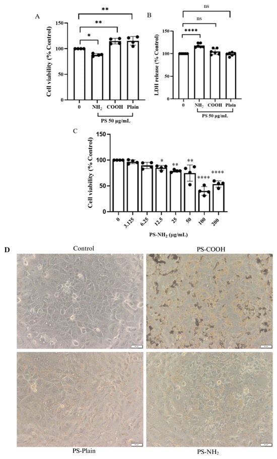
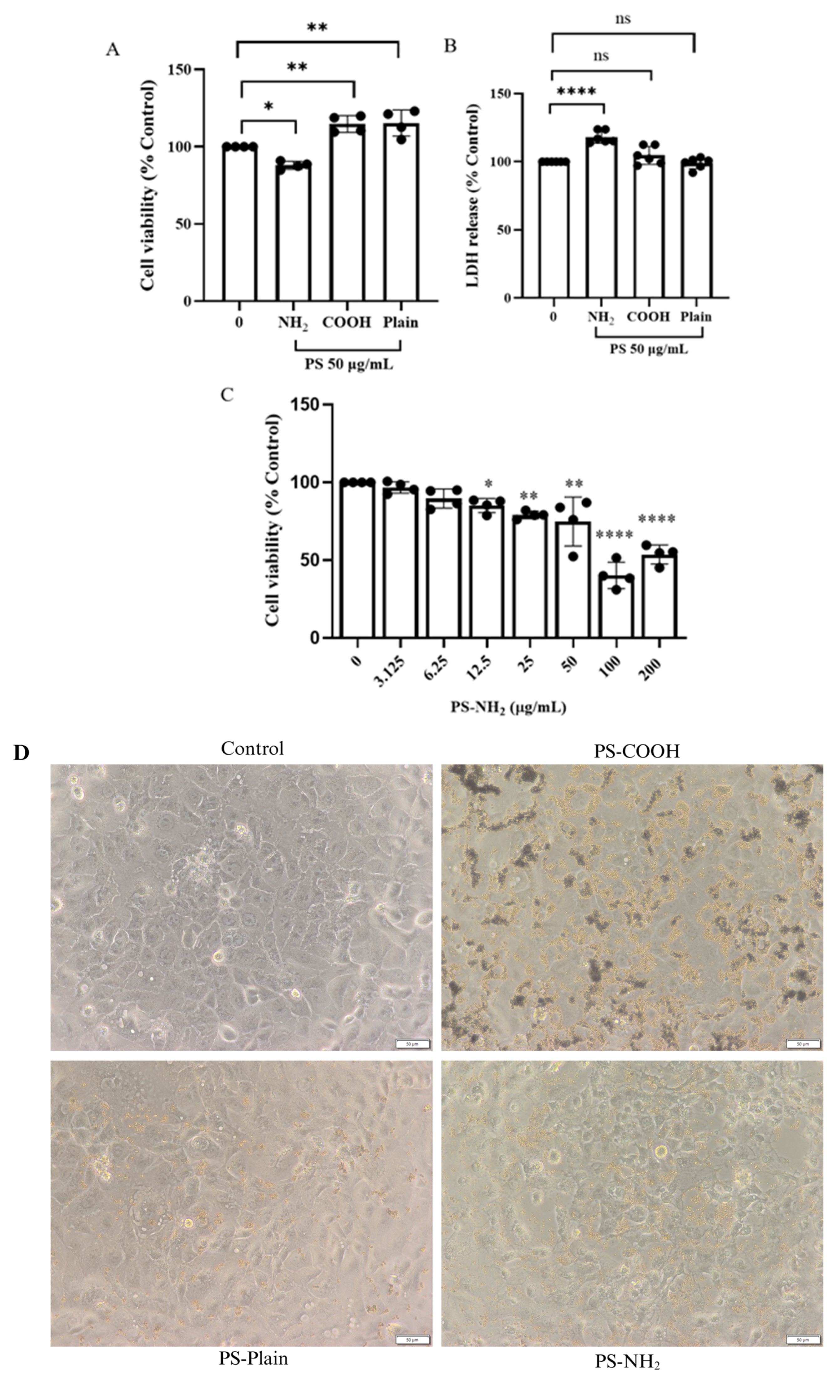
To evaluate the cytotoxic effects of polystyrene microplastics (PS-MPs), HK-2 cells were exposed to 2 μm plain (PS), amine-modified (PS-NH2), or carboxyl-modified (PS-COOH) at 50 μg/mL for 24 h. Among the tested particles, PS-NH2 induced the greatest reduction in cell viability (Figure 1A). Further analysis demonstrated a concentration-dependent decrease in cell viability following PS-NH2 exposure, with significant reductions observed from 12.5 μg/mL, and the decrease becoming more pronounced at 50–200 μg/mL (Figure 1C). Consistent with these observations, LDH release, an indicator of membrane damage, showed a significant increase in the PS-NH2-treated group, whereas plain PS and PS-COOH showed no significant change (Figure 1B). Collectively, these results indicate that PS-MP-induced cytotoxicity is strongly influenced by surface functionalization, with PS-NH2 particles exhibiting toxicity toward renal proximal tubular cells. Morphological observations further supported the differential effects of polystyrene particles, indicating that all particle types were primarily localized around the cell surface with limited structural alterations. However, PS-NH2 exposure resulted in greater cellular damage and morphological changes after 48 h (Figure 1D). Based on the cytotoxic effects, PS-NH2 at a sublethal toxicity dose (50 μg/mL) was then selected for subsequent experiments.
Figure 1.
Effects of polystyrene microplastics (PS-MPs) with different surface functionalizations on HK-2 cell viability, membrane damage, and morphology. (A) Cell viability following exposure to PS, PS-COOH, and PS-NH2 at 50 μg/mL for 24 h. (B) LDH release following exposure to PS, PS-COOH, and PS-NH2 at 50 μg/mL for 24 h. (C) Concentration-dependent effects of PS-NH2 on cell viability after 48 h of exposure. (D) Representative light microscopy images of HK-2 cells after 48 h of exposure to PS, PS-COOH, and PS-NH2 (20× objective). Data are presented as mean ± SD from at least three independent experiments. ns, not significant; * p < 0.05, ** p < 0.01 and **** p < 0.0001 indicate statistical significance.
3.3. Cytotoxicity of PA and PIN
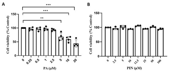
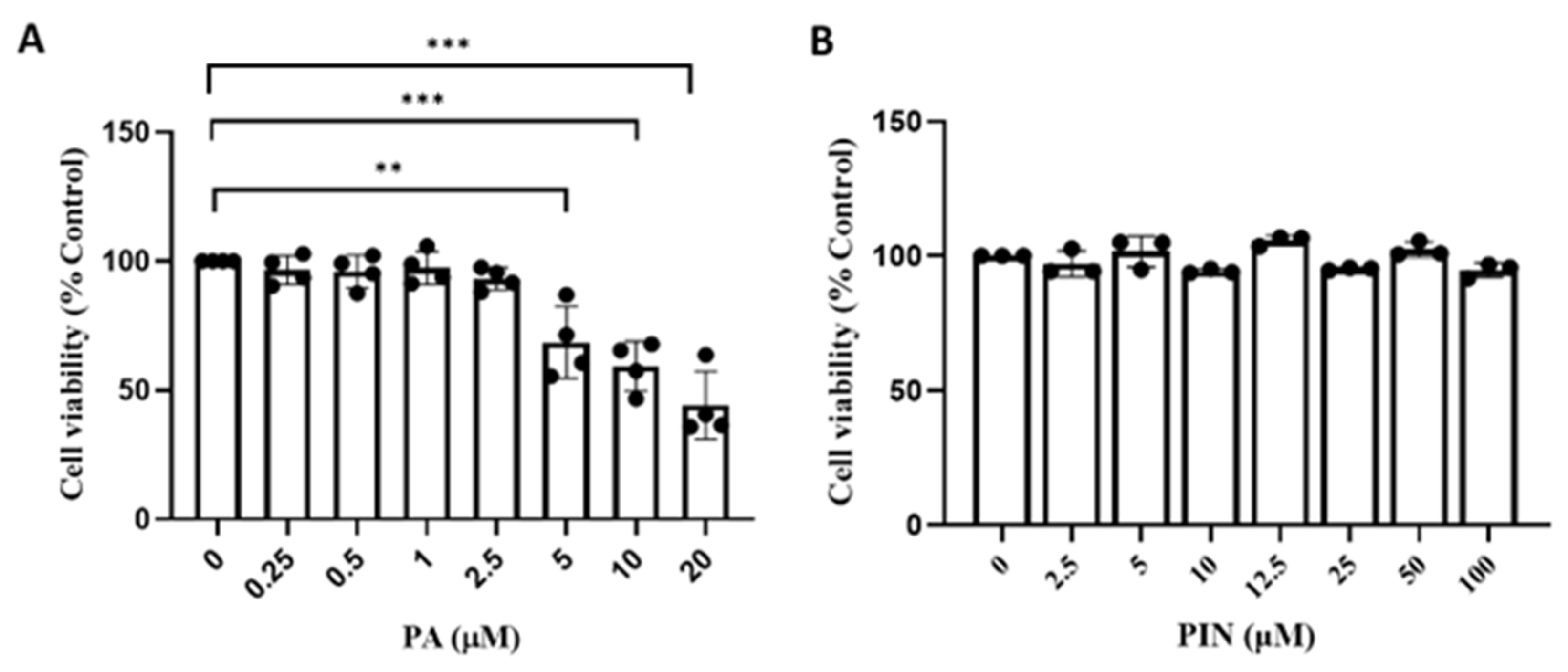
To determine non-cytotoxic concentrations of PA and PIN, the effects of PA and PIN on HK-2 cell viability were evaluated using the MTT assay. PA at concentrations of 1.0 and 2.5 µM did not significantly affect cell viability after 48 h of exposure. In contrast, concentrations of 5 µM and above induced morphological alterations, including cell shrinkage, nuclear vacuolation, and cellular rounding, indicative of cytotoxicity. PIN exhibited no cytotoxic effects over the tested concentration range of 0–100 µM. Based on these findings, PA (2.5 µM) and PIN (50 µM) were selected for subsequent experiments (Figure 2A,B).
Figure 2.
Effects of panduratin A (PA) and pinostrobin (PIN) on HK-2 cell viability. HK-2 cells were exposed to various concentrations of PA (A) or PIN (B) for 48 h, and cell viability was determined using the MTT assay. Data are presented as the mean ± SD from at least three independent experiments. ** p < 0.01 and *** p < 0.001 indicates statistical significance.
3.4. Effects of Surface Functionalization on the Cellular Uptake of PS-MPs in HK-2 Cells
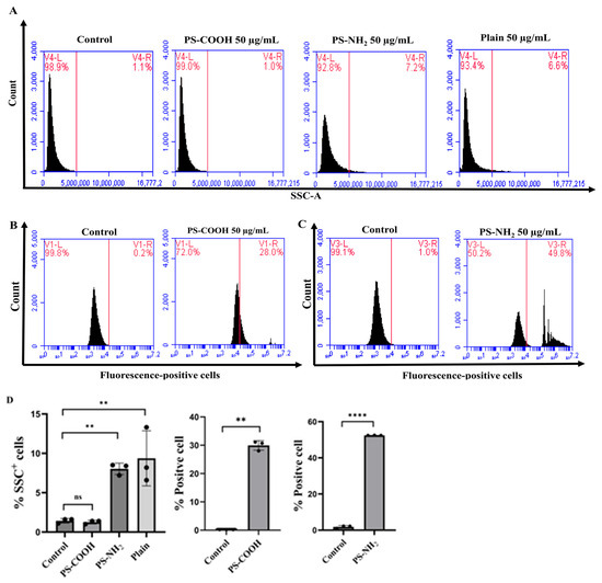
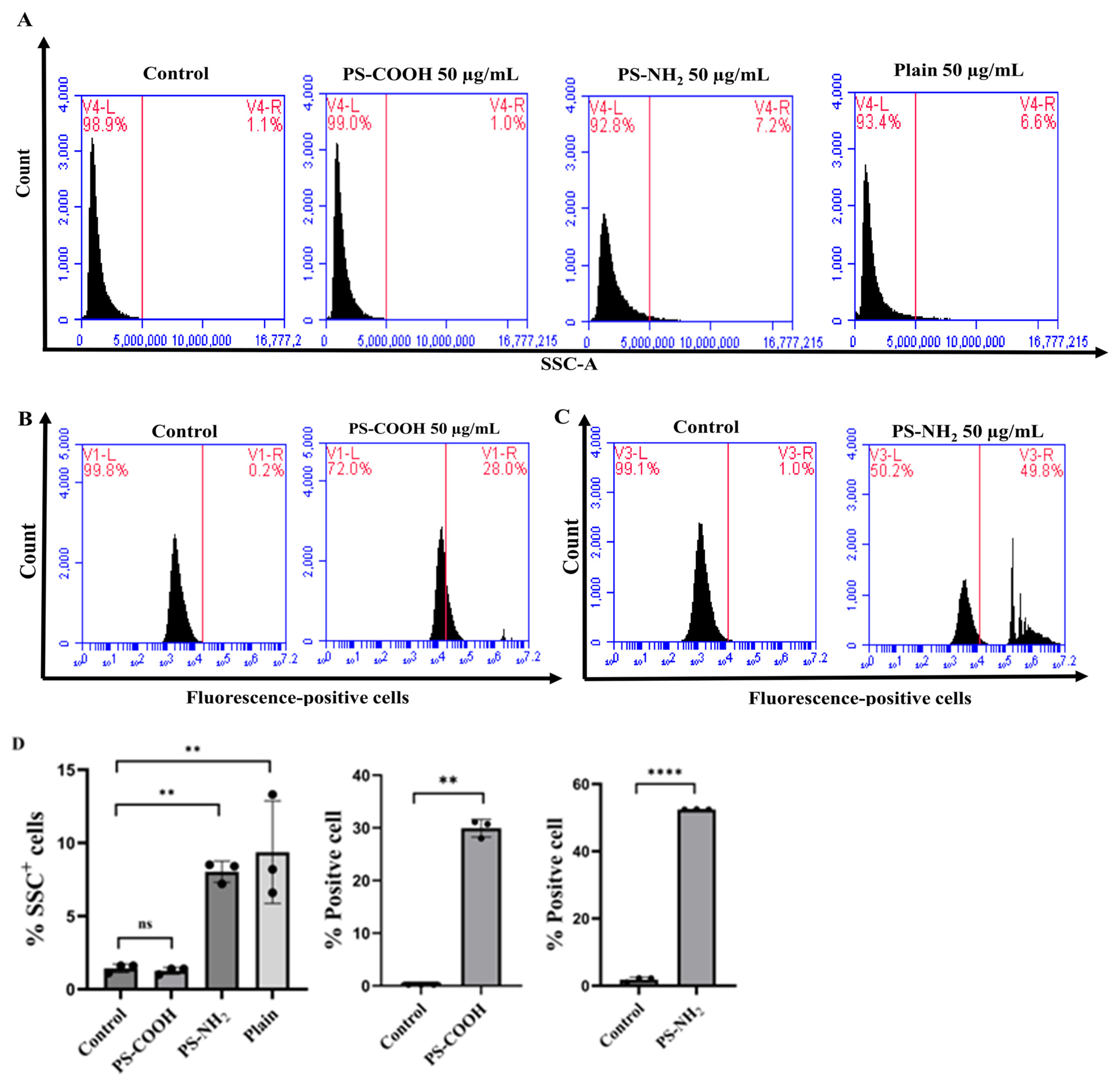
To determine whether the observed differences in cytotoxicity were associated with cellular internalization, the uptake of selected PS-MPs by HK-2 cells was investigated using flow cytometry. The 2 µm unlabeled plain PS, fluorescence-labeled PS-COOH, and fluorescence-labeled PS-NH2 particles were used. After 24 h of exposure to the respective PS-MPs at 50 µg/mL, flow cytometric analysis was performed. The results showed an increase in SSC-positive populations from 1.43 ± 0.28% to 8.03 ± 0.7% in the cells treated with PS-NH2 and to 9.36 ± 3.49% in the cells treated with Plain PS (Figure 3A). The elevated SSC signal suggests increased intracellular complexity and granularity, which indirectly reflects particle internalization. In addition, for the fluorescence-labeled particles, PS-COOH showed internalization with approximately 29.93 ± 1.6% of positive cells detected across 30,000 events. In contrast, PS-NH2 exhibited higher cellular uptake, with 52.50 ± 1.50% of cells observed under the same conditions (Figure 3B–D).
Figure 3.
Surface functionalization-dependent uptake of polystyrene microplastics. HK-2 cells were exposed to 2 μm plain, fluorescently labeled PS-COOH, or fluorescently labeled PS-NH2 particles at 50 μg/mL for 24 h. Cellular uptake was analyzed by flow cytometry. Representative flow cytometry plots (A–C) and quantitative analysis of uptake-positive cells (D) are shown. Data are presented as mean ± SD from three independent experiments. ns, not significant; ** p < 0.01 and **** p < 0.0001 indicate a statistically significant difference.
3.5. PA and PIN Inhibit PS-NH2–Induced Cellular Uptake in HK-2 Cells
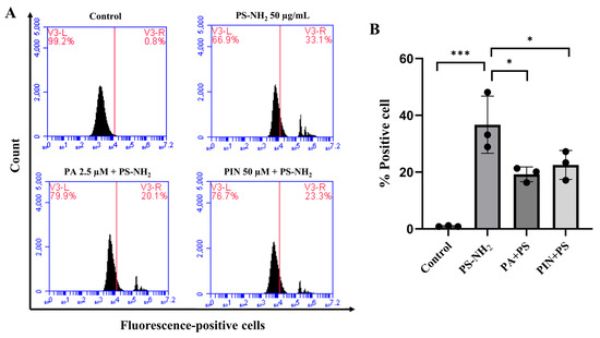
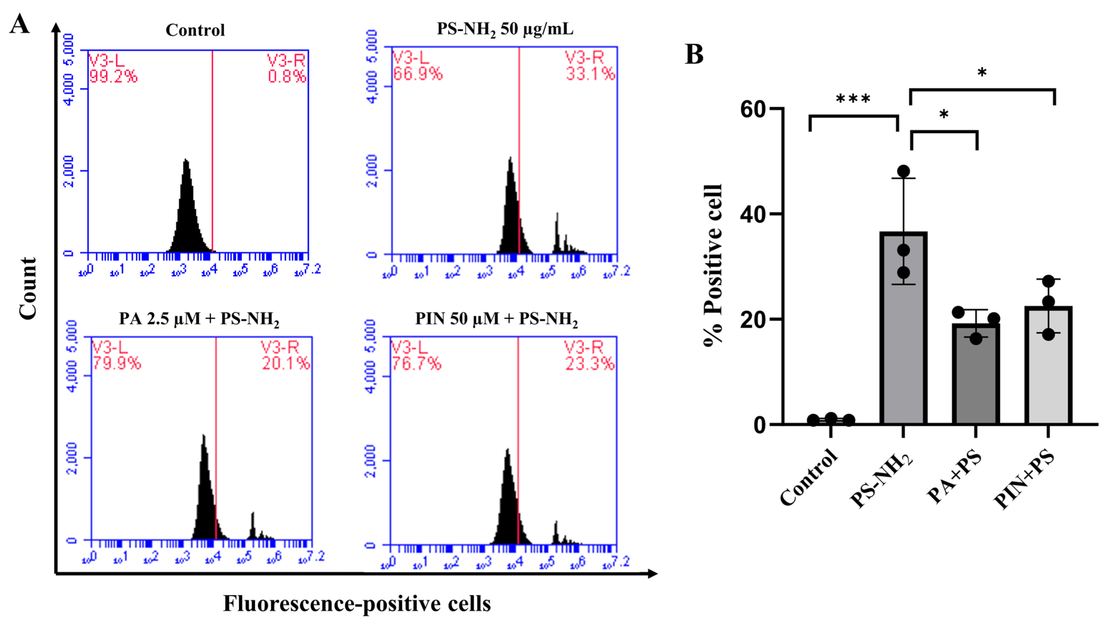
Based on the cytotoxicity and cellular uptake results, PS-NH2 exhibited the most pronounced biological effects. Therefore, further experiments were conducted to investigate whether PA and PIN could attenuate the cellular uptake of PS-NH2 particles in HK-2 cells. The results demonstrated that 36.7 ± 10.09% of PS-NH2 particles were internalized by HK-2 cells. Pretreatment with PA following co-treatment with PS-NH2 significantly reduced the uptake of micro-sized PS-NH2 particles by nearly 50% (19.23 ± 2.61), suggesting that PA may interfere with particle–cell surface interactions and membrane-associated internalization processes. Similarly, PIN treatment decreased PS-NH2 uptake from 36.7 ± 10.09% to 22.53 ±5.09, indicating its potential to limit microplastic internalization (Figure 4A,B). These findings suggest that both PA and PIN can mitigate the cellular entry of PS-NH2, which may contribute to their protective effects against PS-NH2-induced cytotoxicity.
Figure 4.
Effects of PA and PIN on the cellular uptake of PS-NH2. HK-2 cells were pretreated with PA (2.5 μM) or PIN (50 μM) for 24 h and then co-exposed to fluorescently labeled PS-NH2 particles (50 μg/mL) for 24 h. (A) Representative flow cytometry plots showing cellular uptake of PS-NH2 particles. (B) Quantification of uptake-positive cells. Data are presented as mean ± SD from three independent experiments. * p < 0.05 and *** p < 0.001 indicate a statistically significant difference.
3.6. PA and PIN Attenuate Intracellular ROS Generation Induced by PS-NH2
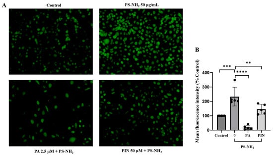
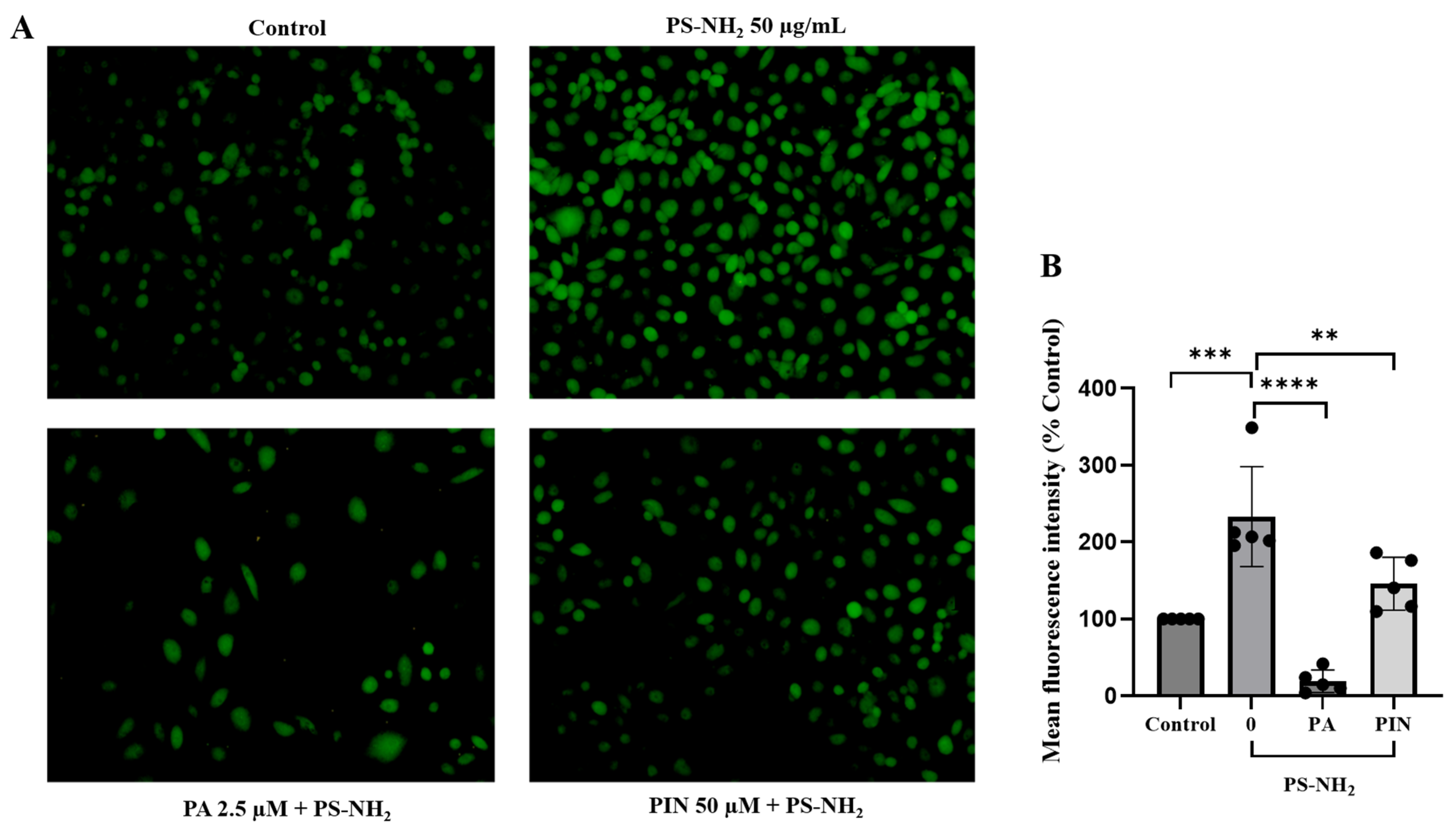
To examine the effect of PS-NH2 on intracellular ROS production, HK-2 cells were pretreated with PA or PIN for 24 h and exposed to PS-NH2 particles at 50 µg/mL for 24 h. The generation of reactive oxygen species (ROS) was assessed using DCFH-DA staining and observed under fluorescence microscopy. In the control group (0 µg/mL PS-NH2), low levels of green fluorescence were observed, indicating minimal basal intracellular ROS. Treatment with PS-NH2 markedly increased DCFH-DA fluorescence compared to the control group, reflecting elevated intracellular ROS levels. Notably, co-treatment with PA and PIN significantly reduced DCFH-DA fluorescence relative to PS-NH2 treatment alone. These findings demonstrated that PA and PIN attenuated PS-NH2–induced intracellular ROS generation in HK-2 cells (Figure 5A,B).
Figure 5.
Effect of panduratin A and pinostrobin on PS-MP (NH2)–induced intracellular ROS generation. Intracellular ROS was detected by DCFH-DA staining (green). (A) Representative images show increased ROS following PS-NH2 exposure and reduced fluorescence under PA or PIN co-treatment conditions. (B) Quantitative analysis of mean fluorescence intensity from five independent fields for each condition. Data are presented as mean ± SD. Statistical significance is indicated as ** p < 0.01, *** p < 0.001, **** p< 0.0001.
3.7. Effect of PS-NH2 on Apoptosis and Protective Role of PA and PIN
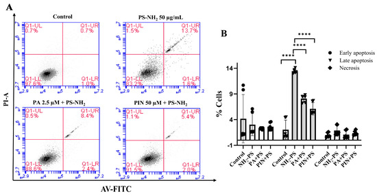
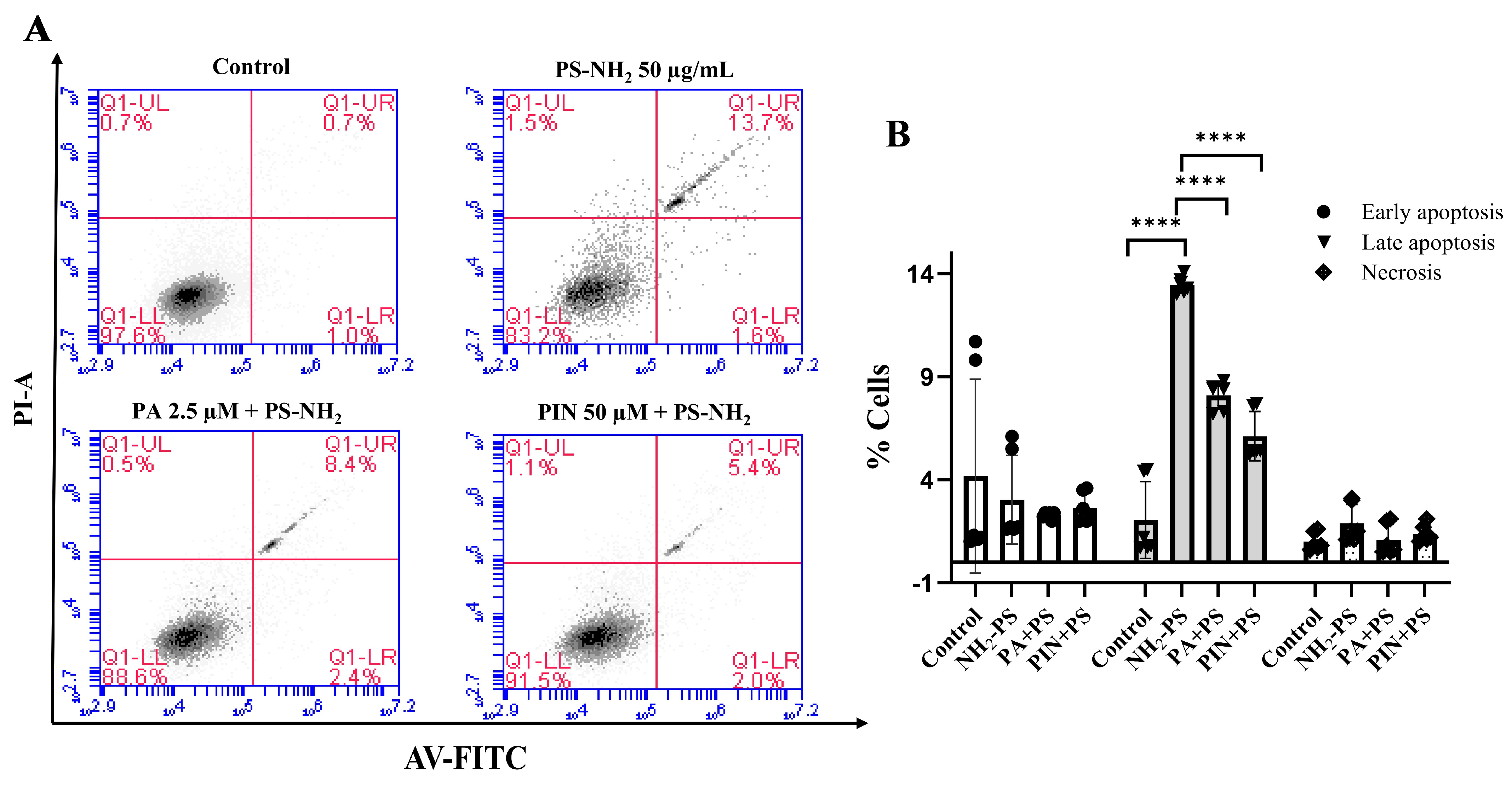
To determine whether PS-NH2 could induce cellular apoptosis, we examined the apoptosis cell population with Annexin V-FITC/PI staining. Flow cytometric analysis showed that PS-NH2 moderately increased the late-apoptotic cell population from 2.05 ± 1.86% to 13.45 ± 0.40% (Figure 6). Pretreatment with PA or PIN for 24 h and co-treatment with PS-NH2 reduced the late-apoptosis cell population to 8.10 ± 0.67% and 6.11 ± 1.19%, while early-apoptosis and necrotic cells remained negligible (Figure 6). Overall, PS-NH2 could induce apoptosis in HK-2 cells, consistent with its cellular uptake and ROS-generating capacity. Co-treatment with PA partially but not fully attenuated apoptotic cell death. The relatively modest increase in apoptosis induced by 2 µm PS-NH2 is likely attributable to its limited cellular internalization compared with smaller-sized particles.
Figure 6.
Effects of PS-NH2 on the apoptosis of renal proximal tubular cells and the protective effect of PA and PIN. HK-2 cells were pre-treated with PA or PIN for 24 h, and then co-exposed to PS-NH2 for 24 h. (A) Representative flow cytometric analysis of treatment and control conditions. (B) Quantitative analysis of cell population in live, early-apoptosis, late-apoptosis, and necrosis quadrants. Data are presented as mean ± SD. Statistical significance is considered as a p-value < 0.05. **** p < 0.0001.
3.8. Effects of PS-NH2 and Roles of PA and PIN on Physiological Parameters in C57BL/6NJcL Mice
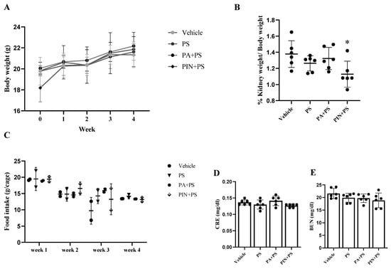
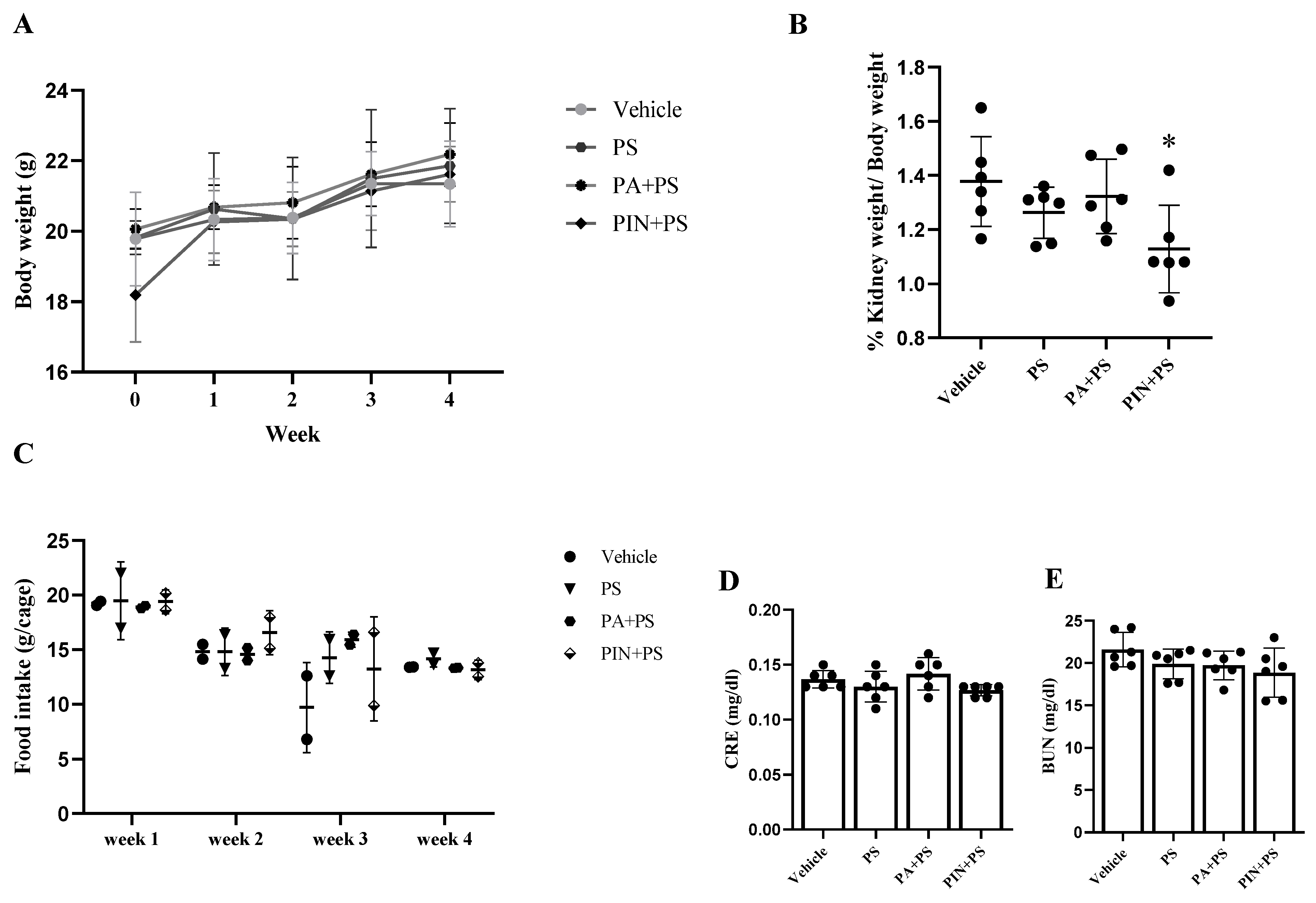
To examine the effects of PS–NH2 and the protective role of PA and PIN in the context of the whole-body system, mice were orally administered PA (25 mg/kg BW) and PIN (40 mg/kg BW) 1 h prior to exposure to PS–NH2 (0.4 mg/day) once daily for 28 days. The body weight and food intake of the mice were monitored weekly, and kidney-to-body weight ratios and renal function tests were determined at the end of the experiment. No significant differences in body weight or food intake were observed among the experimental groups throughout the study period (Figure 7A,C). Similarly, kidney-to-body weight ratios were comparable across most groups. However, a lower kidney-to-body weight ratio was observed in the PIN-treated group compared to other groups (Figure 7B). This difference may be attributed to the relatively lower initial body weight of the animals in this group rather than a treatment-related effect. No significant differences were observed in the serum levels of blood urea nitrogen (BUN) and creatinine (CRE) among the experimental groups, indicating that PS–NH2 exposure under the current conditions did not markedly affect kidney function (Figure 7D,E).
Figure 7.
Body weight, food intake, and kidney-to-body weight ratio in mice during the 28-day experimental period. (A) Body weight per mouse (g); (B) kidney-to-body weight ratio.); (C) weekly food intake was recorded in grams per cage (three mice per cage); (D,E) Serum BUN and creatinine levels. Data are presented as mean ± SD (n = 6). Statistical significance is indicated as * p < 0.05.
3.9. Effects of PS–NH2 Exposure and Effects of Co-Treatment with PA and PIN on Kidney Tissue Histopathology
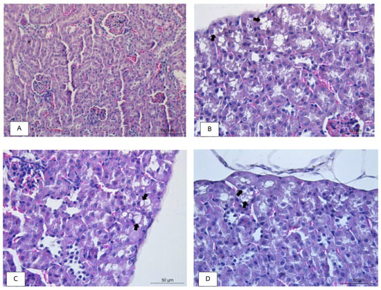
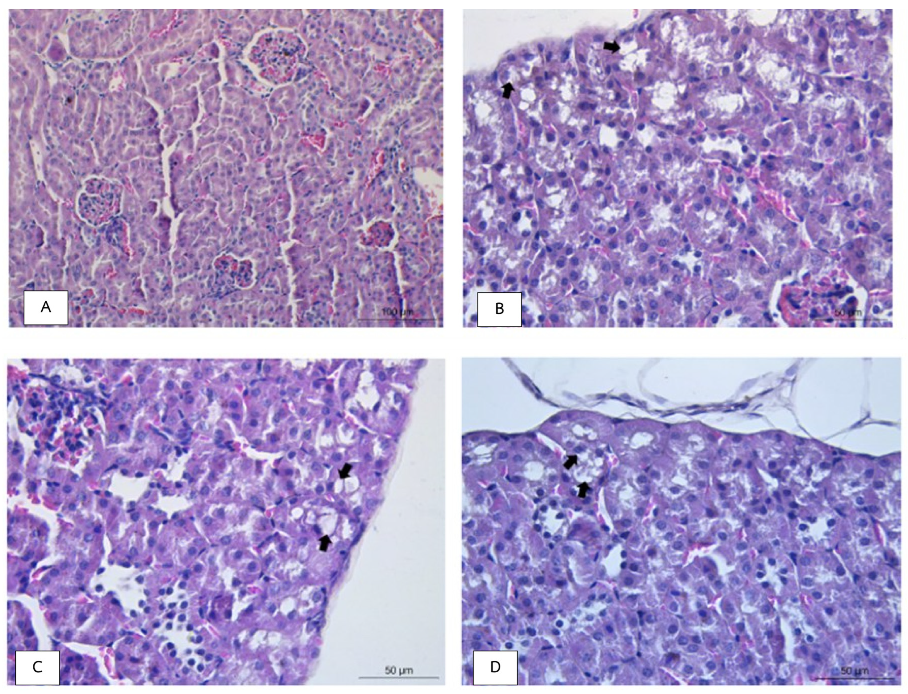
In contrast to the unchanged serum levels of BUN and CRE, histopathological analysis revealed notable alterations in kidney tissue, which was characterized by morphologic changes in renal epithelial cells, including tinctorial changes, vacuolation, and cellular sloughing. Interestingly, the H&E-stained slides of the left kidneys showed no remarkable lesions in any component of renal tissue across all six studied animals (0/6) (Table 2 and Table 3, Figure 8A). In the PS-NH2-treated group, focal to multifocal and minimal to mild degeneration of proximal tubules in the renal cortex was observed in five out of six animals (one with mild (+2) degeneration and four with minimal (+1) degeneration), with a significant increase in the average severity score of proximal tubular degeneration from 0 to 1 ± 0.58 compared to the vehicle control (Figure 8B, Table 2 and Table 3). In the PS-NH2 and PA co-treatment group, focal and minimal-to-mild degeneration of proximal tubules in the renal cortex was found in three out of six animals (one with mild (+2) degeneration and two with minimal (+1) degeneration). The average severity score in this group was 0.67 ± 0.74, indicating that pretreatment with PA tended to alleviate PS-NH2-induced renal histopathological injury (Figure 8C, Table 2 and Table 3). Surprisingly, PIN conferred a more pronounced protective effect; focal and minimal degeneration of the proximal tubules in the renal cortex was observed in only one out of six animals. The average severity score in this group was significantly reduced to 0.17 ± 0.37, which was comparable to the vehicle control condition (Figure 8D, Table 2 and Table 3). A summary of the microscopic findings and the averages of severity scores are presented in Table 2 and Table 3 and Figure 8, respectively.
Table 2.
The summary of microscopic findings.
Table 3.
The average severity score (0–5) of the lesions in the kidneys (mean ± SD).
Figure 8.
A histological study of different treatment groups. (A) Photo-micrograph of renal cortex in an animal from the vehicle control group (H&E stained, 20×), (B) Photomicrograph of mild focal proximal tubular degeneration (black arrows) in an animal from the PS-NH2 group (H&E stained, 40×), (C) Photomicrograph of minimal focal proximal tubular degeneration (black arrows) in an animal from the PS-NH2 and PA group (H&E stained, 40×), and (D) Photomicrograph of minimal focal proximal tubular degeneration (black arrows) in an animal from the PS-NH2 and PIN group (H&E stained, 40×).
4. Discussion
The kidney is considered a potentially vulnerable target of micro- and nanoplastic (MNP) exposure due to its high blood flow and critical role in filtration and clearance of circulating substances. The detection of MNPs in the systemic circulation further raises concern regarding their potential renal effects [12,40,41]. The present study demonstrates that, among the three surface-modified polystyrene microplastics (mean particle size 2 µm) examined, PS-NH2 exhibited greater cellular uptake and cytotoxicity than plain PS or PS-COOH at a similar dose (50 µg/mL). Given its greater cytotoxic potential, PS-NH2 was selected for further evaluation, revealing that it induced renal cell injury through increased particle internalization, oxidative stress, and apoptosis. Pretreatment with panduratin A (PA) or pinostrobin (PIN) following co-treatment with PS-NH2 reduced PS-NH2-induced accumulation, oxidative stress, and apoptosis in HK-2 cells. These in vitro findings were further corroborated in vivo, where oral exposure to PS-NH2 induced early-stage tubular injury in mice, which was attenuated by pretreatment with PA and PIN.
Regarding the in vitro exposure conditions, although the concentration of PS-NH2 associated with the cytotoxicity observed in this study exceeds levels typically reported in human blood (approximately 1.8–12 µg/mL), based on early estimates [40], it remains within the range (4–100 µg/mL) used in recent in vitro studies investigating the biological effects of microplastics [42]. This concentration was selected specifically to characterize cytotoxicity at the mechanistic level, rather than to model an environmentally relevant exposure.
To better understand the physicochemical basis of this differential toxicity, we characterized the dispersion behavior of the three particle types by DLS and zeta potential measurement. PS-NH2 exhibited the least negative zeta potential in deionized water (−25.27 ± 0.09 mV), compared with PS-COOH (−36.45 ± 0.44 mV) and plain PS (−63.43 ± 0.57 mV). While aminated particles are generally assumed to carry a net positive surface charge, a negative zeta potential is not uncommon in practice. Several factors, including residual sulfate ester groups introduced during manufacturing, adsorption of anionic biomolecules, and pH-dependent deprotonation, can all shift the measured charge below the theoretically predicted value. Indeed, negative zeta potentials for aminated polystyrene particles in aqueous dispersion have been reported across multiple studies, including −22.6 ± 2.6 mV and −44.90 ± 2.80 mV to −28.51 ± 27.71 mV depending on particle size [43,44]. The relatively less negative value observed for PS-NH2 compared with the other two particle types nevertheless points to partial neutralization of the polystyrene core’s intrinsic negative charge by protonated amine groups. However, this distinction was largely lost upon dispersion in complete cell culture medium: zeta potential values for all three particle types converged toward near-neutral levels (PS: −14.50 ± 0.27 mV; PS-COOH: −6.22 ± 0.08 mV; PS-NH2: −7.72 ± 0.40 mV), with PS-COOH and PS-NH2 becoming almost indistinguishable from one another. This convergence pattern, in which zeta potential differences observed in water are largely erased upon dispersion in FBS-containing medium, closely parallels findings reported for polystyrene particles of comparable surface modifications, where zeta potential values similarly converged toward near-neutral levels in FBS-containing medium (PS: −7.06 mV; PS-COOH: −7.51 mV; PS-NH2: −7.07 mV) [43]. This convergence is consistent with the known influence of serum protein adsorption on the interfacial properties and electrokinetic behavior of nanoparticles in biological media. Thus, the zeta potentials measured in complete medium should be interpreted as the effective electrokinetic potential of protein-conditioned particles rather than a direct measure of intrinsic surface charge. Because PS-COOH and PS-NH2 exhibited near-identical zeta potentials in complete medium, zeta potential alone cannot account for the markedly greater cytotoxicity of PS-NH2.
Changes in hydrodynamic size and PDI further revealed medium-dependent dispersion behavior. Plain PS showed an increase in hydrodynamic diameter and PDI upon dispersion in complete medium (from 2041 ± 51 to 2415 ± 62 nm), consistent with a previous report in which all polystyrene particle types, including those of comparable 2 µm size, showed increased hydrodynamic diameter following dispersion in FBS-containing medium relative to water [43]. In contrast, PS-COOH and PS-NH2 in the present study exhibited an apparent decrease in hydrodynamic size (PS-COOH: 2305 ± 62 to 2169 ± 129 nm; PS-NH2: 2968 ± 78 to 1610 ± 20 nm) accompanied by a pronounced increase in PDI. This divergence from the size increases typically reported for surface-modified polystyrene particles under similar conditions warrants further investigation and should not be interpreted as improved dispersion stability, but rather as a shift in the heterogeneous particle population detected by DLS following exposure to the protein-containing medium.
Notably, although PS-NH2 showed an even greater PDI increase (0.584) than PS-COOH (0.431), this was not accompanied by a similarly visible aggregation pattern (Figure 1D), consistent with a recent report showing that carboxylated polystyrene nanoparticles settle as visible clusters on the cell surface, whereas aminated particles remain more dispersed and more readily internalized [44]. Together, these findings support a model in which the visible aggregates formed by PS-COOH reduced its effective cell-surface contact and uptake, contributing to its lower cytotoxicity relative to the more finely dispersed PS-NH2. A limitation of this characterization is that particle properties were assessed at a single time point rather than continuously over the 24 h exposure period; time-resolved measurements would further strengthen confidence in particle stability under experimental conditions.
Increased uptake of PS-NH2 correlated with the enhanced cytotoxicity observed in viability assays, linking PS-NH2 uptake to a pronounced increase in intracellular ROS, suggesting that oxidative stress is a key upstream mechanism driving renal cell stress and subsequent apoptosis. This is consistent with evidence that polystyrene microplastics induce nephrotoxicity through oxidative stress, endoplasmic reticulum stress, and inflammatory pathways, including upregulation of pro-apoptotic genes such as Bax, Caspase-3, and Caspase-9, alongside downregulation of Bcl-2 [17].
PA and PIN exerted significant protective effects against PS-NH2-induced HK-2 cell damage. Treatment with both PA and PIN reduced intracellular ROS levels and attenuated the apoptotic cell death population. The cytoprotective effects of PA are consistent with its previously reported nephroprotective effects, in which PA ameliorated cisplatin-induced renal tubular cell apoptosis by suppressing the activation of ERK1/2 and caspase-3/7, reducing mitochondrial dysfunction, and restoring the expression of the anti-apoptotic protein Bcl-2 [33]. Similar to PA, PIN has demonstrated nephroprotective potential against cadmium-induced renal toxicity by reducing oxidative stress, mitochondrial dysfunction, and apoptosis [30]. At the molecular level, the antioxidant activity of PIN is partly attributable to electron delocalization within its flavanone structure, which enables direct free radical scavenging [31]. Additionally, PIN has been shown to inhibit the NF-κB signaling pathway by blocking IκB-α phosphorylation and nuclear translocation of NF-κB, thereby suppressing the production of pro-inflammatory cytokines including IL-6, TNF-α, and IL-8 [29]. This multi-target activity, encompassing both antioxidant and anti-inflammatory mechanisms, may contribute to the superior protective profile of PIN observed in vivo in the present study. Notably, the extent of ROS generation and apoptosis closely paralleled the degree of particle uptake, supporting a mechanistic link between internalization efficiency and downstream cellular responses. Taken together, these findings support a multi-step mechanism of PS-NH2-induced renal toxicity, in which particle uptake initiates oxidative stress, followed by apoptotic injury. The inhibition of cellular uptake, combined with the downstream ROS scavenging and suppression of apoptotic pathways, may therefore underlie the nephroprotective actions of PA and PIN against PS-NH2-induced cytotoxicity.
In the in vivo model, the PS-NH2 dose was based on Wang et al. [16], who found that C57BL/6 mice given oral gavage of 2 µm PS-MPs (0.2–0.4 mg per dose, twice weekly for 4 to 8 weeks) exhibited renal accumulation and tubular injury. In the current study, PS-NH2 (0.4 mg) was administered once daily for 28 days, which represents a higher total dose than that used by Wang et al., thereby increasing the likelihood of observing renal injury within a shorter exposure period. This dose and duration are in agreement with those of Pan et al. [38] and Xu et al. [39], who also observed significant renal functional and structural abnormalities—such as fibrotic changes and alterations of the tubules and glomeruli—after administering amino-modified polystyrene particles at a similar daily dose (0.5 mg/day) for 28 days.
PS-NH2 exposure induced early-stage renal injury, as evidenced by proximal tubular degeneration, predominantly at minimal to mild severity. However, these structural changes were not accompanied by alterations in renal function markers, including BUN and CRE, indicating that kidney function remained preserved at this stage. This observation is consistent with the well-recognized limitations of BUN and CRE as early biomarkers of renal tubular injury, as these markers reflect glomerular filtration rather than tubular integrity and typically rise only after substantial nephron loss [45,46]. Histopathological examination, therefore, represents a more sensitive approach to detecting early structural changes that precede functional deterioration [45,46]. Future studies incorporating tubular injury biomarkers such as kidney injury molecule-1 (KIM-1) or neutrophil gelatinase-associated lipocalin (NGAL) would provide a more comprehensive assessment of the nephrotoxic response and the protective efficacy of PA and PIN at the tubular level [45,46]. In addition, the relatively mild renal alterations observed in the present study are consistent with a previous in vivo study that demonstrated that short-term oral exposure to 1–10 μm polystyrene microplastics caused only limited tissue uptake and did not induce overt histopathological or inflammatory alterations, supporting the notion that the severity of microplastic-induced toxicity may depend on exposure duration, particle size, and dosage [47].
In the context of PA and PIN treatment, the doses of PA (25 mg/kg BW) and PIN (40 mg/kg BW) used in this study were selected based on our previous studies demonstrating their efficacy in attenuating renal pathology in rodent models of polycystic kidney disease without significant alterations in BUN or serum creatinine [35,37]. Pretreatment with PA tended to reduce the number of affected animals and lower severity scores compared with the PS-NH2 group, although these effects did not reach statistical significance. In contrast, PIN treatment significantly attenuated tubular degeneration, with severity scores comparable to those of the vehicle control group, indicating a more pronounced protective effect. The difference in protective efficacy between PA and PIN may be partly attributable to differences in their pharmacokinetic properties [48,49,50,51]. According to Kongratanapasert et al. [48], the absolute oral bioavailability of pure panduratin A in rats was approximately 9%, with a notably low kidney tissue-to-plasma ratio of 0.1-fold, the lowest among all examined organs. This limited distribution to the kidney suggests that PA may not achieve sufficient local concentrations within renal tissue to exert optimal protective effects against PS-NH2-induced histopathological alterations. Despite its low oral bioavailability, panduratin A underwent substantial biotransformation, primarily through oxidation and glucuronidation, with its metabolites predominantly excreted via the fecal route [48,49]. Furthermore, PA exhibited a prolonged time to reach maximum plasma concentration (Tmax) of approximately 8 h, whereas pinostrobin reached peak plasma concentration within 0.133 h following oral administration [50], indicating a substantially faster absorption and earlier onset of action for pinostrobin, which may be critical in attenuating the early inflammatory and oxidative processes underlying PS-NH2-induced renal injury. Additionally, pinostrobin was extensively metabolized in vivo, with less than 1.567% excreted unchanged, yielding a group of flavonoid metabolites via both Phase I and Phase II metabolic pathways. These metabolites may contribute to the overall biological activity of pinostrobin, although their specific nephroprotective effects remain to be established [51]. In contrast, panduratin A was largely excreted unchanged in feces (approximately 22–60% of the administered dose), indicating limited biotransformation and potentially reduced exposure to bioactive compounds in renal tissue [48]. Despite these histopathological changes, no significant differences in body weight or food intake were observed across experimental groups, suggesting that the treatment-related protective effects were primarily manifested at the structural level without systemic toxicity. Nevertheless, further mechanistic studies are required to clarify the precise molecular targets and tissue-specific pharmacokinetics of both compounds.
5. Conclusions
In conclusion, PS-NH2 induced renal toxicity through enhanced cellular uptake, oxidative stress, and apoptosis, and caused early-stage renal injury in vivo despite preserved kidney function markers. Both PA and PIN exerted nephroprotective effects, with PIN showing greater in vivo efficacy, highlighting the therapeutic potential of natural compounds against microplastic-induced renal damage and suggesting that limiting particle uptake and oxidative stress may be a viable mitigation strategy. Additionally, these findings not only highlight the potential renal risks of aminated microplastics but also support that both natural compounds, particularly PIN, may help mitigate microplastic-induced renal toxicity. Further studies are warranted to investigate the mechanisms and long-term effects in detail.
Author Contributions
Conceptualization, A.B. and S.S.; methodology, A.B. and N.H.; formal analysis, A.B.; investigation, A.B. and N.H.; resources, A.B. and S.S.; writing—original draft preparation, A.B.; writing—review and editing, A.B. and S.S.; supervision, S.S.; funding acquisition, A.B. All authors have read and agreed to the published version of the manuscript.
Funding
This research was funded by the National Research Council of Thailand (NRCT) under Contract No. N42A661016, and the APC was partially funded by the Faculty of Medicine, Princess of Naradhiwas University.
Institutional Review Board Statement
The animal study protocol was approved by the Faculty of Science, Mahidol University Animal Care and Use Committee MUSC-IACUC review, Mahidol University, Bangkok, Thailand on 15 January 2026 (Protocol No. MUSC68-054-824) and performed in accordance with institutional guidelines.
Informed Consent Statement
Not applicable.
Data Availability Statement
Data are included within the article. Further inquiries can be directed to the corresponding authors.
Acknowledgments
The authors gratefully acknowledge the Faculty of Medicine, Princess of Naradhiwas University, for providing technical support and valuable guidance throughout this study. The authors also thank the members of the A.B. Lab for their assistance. During the preparation of this manuscript/study, the authors used Grammarly Pro (web-based version; Grammarly, Inc.) and ChatGPT (GPT-5.3 Instant; OpenAI) solely for language editing and grammar correction. The authors have reviewed and edited the output and take full responsibility for the content of this publication.
Conflicts of Interest
The authors declare no conflicts of interest.
Abbreviations
The following abbreviations are used in this manuscript:
| ROS | Reactive oxygen species |
| PS | Polystyrene |
| PS-MP | Polystyrene microplastics |
| PS-NH2 | Amine-modified polystyrene microplastics |
| PS-COOH | Carboxyl-modified polystyrene microplastics |
| PA | Panduratin A |
| PIN | Pinostrobin |
| BUN | Blood urea nitrogen |
| CRE | Creatinine |
References
- Corcoran, P.L. Degradation of Microplastics in the Environment. In Handbook of Microplastics in the Environment; Rocha-Santos, T., Costa, M., Mouneyrac, C., Eds.; Springer International Publishing: Cham, Switzerland, 2020; pp. 1–12. [Google Scholar]
- Li, Y.; Tao, L.; Wang, Q.; Wang, F.; Li, G.; Song, M. Potential Health Impact of Microplastics: A Review of Environmental Distribution, Human Exposure, and Toxic Effect. Environ. Health 2023, 1, 249–257. [Google Scholar] [CrossRef] [Scilit] [PubMed]
- Petrov, L.; Tsvetanova, E.; Georgieva, A.; Andreeva, M.; Pramatarov, G.; Petrov, G.; Dobrev, K.; Alexandrova, A. Effect of Polystyrene Microplastic Exposure on Blood Parameters in Mice. Microplastics 2026, 5, 74. [Google Scholar] [CrossRef] [Scilit]
- Vethaak, A.D.; Legler, J. Microplastics and Human Health. Science 2021, 371, 672–674. [Google Scholar] [CrossRef] [Scilit] [PubMed]
- Kik, K.; Bukowska, B.; Sicińska, P. Polystyrene Nanoparticles: Sources, Occurrence in the Environment, Distribution in Tissues, Accumulation and Toxicity to Various Organisms. Environ. Pollut. 2020, 262, 114297. [Google Scholar] [CrossRef] [Scilit] [PubMed]
- Revel, M.; Châtel, A.; Mouneyrac, C. Micro(nano)plastics: A Threat to Human Health? Curr. Opin. Environ. Sci. Health 2018, 1, 17–23. [Google Scholar] [CrossRef] [Scilit]
- Nihart, A.J.; Garcia, M.A.; El Hayek, E.; Liu, R.; Olewine, M.; Kingston, J.D.; Castillo, E.F.; Gullapalli, R.R.; Howard, T.; Bleske, B.; et al. Bioaccumulation of Microplastics in Decedent Human Brains. Nat. Med. 2025, 31, 1114–1119, Correction in Nat. Med. 2025, 31, 1367. [Google Scholar] [CrossRef] [Scilit] [PubMed]
- Yee, M.S.; Hii, L.W.; Looi, C.K.; Lim, W.M.; Wong, S.F.; Kok, Y.Y.; Tan, B.-K.; Wong, C.-Y.; Leong, C.-O. Impact of Microplastics and Nanoplastics on Human Health. Nanomaterials 2021, 11, 496. [Google Scholar] [CrossRef] [Scilit] [PubMed]
- Banerjee, A.; Billey, L.O.; Shelver, W.L. Uptake and Toxicity of Polystyrene Micro/Nanoplastics in Gastric Cells: Effects of Particle Size and Surface Functionalization. PLoS ONE 2021, 16, e0260803. [Google Scholar] [CrossRef] [Scilit] [PubMed]
- Park, K.Y.; Kim, M.S.; Oh, N. Cytotoxicity of Amine-Modified Polystyrene MPs and NPs on Neural Stem Cells Cultured from Mouse Subventricular Zone. Heliyon 2024, 10, e30518. [Google Scholar] [CrossRef] [Scilit] [PubMed]
- Moore, T.L.; Rodriguez-Lorenzo, L.; Hirsch, V.; Balog, S.; Urban, D.; Jud, C.; Rothen-Rutishauser, B.; Lattuada, M.; Petri-Fink, A. Nanoparticle Colloidal Stability in Cell Culture Media and Impact on Cellular Interactions. Chem. Soc. Rev. 2015, 44, 6287–6305. [Google Scholar] [CrossRef] [Scilit] [PubMed]
- Massardo, S.; Verzola, D.; Alberti, S.; Caboni, C.; Santostefano, M.; Verrina, E.; Angeletti, A.; Lugani, F.; Ghiggeri, G.M.; Bruschi, M.; et al. MicroRaman Spectroscopy Detects Microplastics in Human Urine and Kidney Tissue. Environ. Int. 2024, 184, 108444. [Google Scholar] [CrossRef] [Scilit] [PubMed]
- Meng, X.; Zhang, J.; Wang, W.; Gonzalez-Gil, G.; Vrouwenvelde, J.S.; Li, Z. Effects of Nano- and Microplastics on the Kidney: Physicochemical Properties, Bioaccumulation, Oxidative Stress and Immunoreaction. Chemosphere 2022, 288, 132631. [Google Scholar] [CrossRef] [Scilit] [PubMed]
- de Oliveira, R.B.; Pelepenko, L.E.; Masaro, D.A.; Lustosa, G.M.M.; de Oliveira, M.C.; Roza, N.A.V.; Marciano, M.A.; dos Reis, L.M.; Kamel, S.; Louvet, L.; et al. Effects of Microplastics on the Kidneys: A Narrative Review. Kidney Int. 2024, 106, 400–407. [Google Scholar] [CrossRef] [Scilit] [PubMed]
- Goodman, K.E.; Hua, T.; Sang, Q.X.A. Effects of Polystyrene Microplastics on Human Kidney and Liver Cell Morphology, Cellular Proliferation, and Metabolism. ACS Omega 2022, 7, 34136–34153. [Google Scholar] [CrossRef] [Scilit] [PubMed]
- Wang, Y.-L.; Lee, Y.-H.; Hsu, Y.-H.; Chiu, I.-J.; Huang, C.C.-Y.; Huang, C.C.; Chia, Z.-C.; Lee, C.-P.; Lin, Y.-F.; Chiu, H.-W. The Kidney-Related Effects of Polystyrene Microplastics on Human Kidney Proximal Tubular Epithelial Cells HK-2 and Male C57BL/6 Mice. Environ. Health Perspect. 2021, 129, 57003. [Google Scholar] [CrossRef] [Scilit] [PubMed]
- Wang, W.; Guan, J.; Feng, Y.; Nie, L.; Xu, Y.; Xu, H.; Fu, F. Polystyrene Microplastics Induced Nephrotoxicity Associated with Oxidative Stress, Inflammation, and Endoplasmic Reticulum Stress in Juvenile Rats. Front. Nutr. 2023, 9, 1059660. [Google Scholar] [CrossRef] [Scilit] [PubMed]
- Jiang, N.; Zheng, X.; Zhang, N.; Cao, Y. The Detrimental Effects of Microplastic Exposure on Kidney Function. Front. Med. 2025, 12, 1620733. [Google Scholar] [CrossRef] [Scilit] [PubMed]
- Cui, B.; Rong, H.; Tian, T.; Guo, D.; Duan, L.; Nkinahamira, F.; Ndagijimana, P.; Yan, W.; Naidu, R. Chemical Methods to Remove Microplastics from Wastewater: A Review. Environ. Res. 2024, 249, 118416. [Google Scholar] [CrossRef] [Scilit] [PubMed]
- Anand, U.; Dey, S.; Bontempi, E.; Ducoli, S.; Vethaak, A.D.; Dey, A.; Federici, S. Biotechnological Methods to Remove Microplastics: A Review. Environ. Chem. Lett. 2023, 21, 1787–1810. [Google Scholar] [CrossRef] [Scilit] [PubMed]
- Tain, Y.L.; Chang-Chien, G.P.; Lin, S.F.; Hou, C.Y.; Hsu, C.N. Protective Effect of Resveratrol on Kidney Disease and Hypertension against Microplastics Exposure in Male Juvenile Rats. Antioxidants 2024, 13, 1457. [Google Scholar] [CrossRef] [Scilit] [PubMed]
- Ijaz, M.U.; Rafi, Z.; Hamza, A.; Sayed, A.A.; Albadrani, G.M.; Al-Ghadi, M.Q.; Abdel-Daim, M.M. Mitigative Potential of Kaempferide against Polyethylene Microplastics-Induced Testicular Damage by Activating the Nrf-2/Keap-1 Pathway. Ecotoxicol. Environ. Saf. 2024, 269, 115746. [Google Scholar] [CrossRef] [Scilit] [PubMed]
- Chen, W.; Zhu, R.; Ye, X.; Sun, Y.; Tang, Q.; Liu, Y.; Yan, F.; Yu, T.; Zheng, X.; Tu, P. Food-Derived Cyanidin-3-O-Glucoside Reverses Microplastic Toxicity via Promoting Discharge and Modulating the Gut Microbiota in Mice. Food Funct. 2022, 13, 1447–1458. [Google Scholar] [CrossRef] [Scilit] [PubMed]
- Yu, J.; Yoon, J.-H.; Park, M.; Lee, H.-J. Nobiletin-Mediated Autophagy Mitigates Nanoplastic-Induced Toxicity in Human Intestinal Caco-2 Cells. FASEB J. 2025, 39, e70452. [Google Scholar] [CrossRef] [Scilit] [PubMed]
- Rizwan, A.; Ijaz, M.U.; Hamza, A.; Anwar, H. Attenuative Effect of Astilbin on Polystyrene Microplastics-Induced Testicular Damage: Biochemical, Spermatological and Histopathological-Based Evidences. Toxicol. Appl. Pharmacol. 2023, 471, 116559. [Google Scholar] [CrossRef] [Scilit] [PubMed]
- Tan, E.-C.; Lee, Y.-K.; Chee, C.-F.; Heh, C.-H.; Wong, S.-M.; Thio, C.L.-P.; Foo, G.-T.; Khalid, N.; Abd Rahman, N.; Karsani, S.A.; et al. Boesenbergia Rotunda: From Ethnomedicine to Drug Discovery. Evid. Based Complement. Altern. Med. 2012, 2012, 473637. [Google Scholar]
- Kiatsoonthon, K.; Phimthong, N.; Potikanond, S.; Wikan, N.; Nimlamool, W. Panduratin A Inhibits TNF-α-Stimulated Endothelial Cell Activation through Suppressing the NF-κB Pathway. Biomolecules 2025, 15, 34. [Google Scholar] [CrossRef] [Scilit] [PubMed]
- Alqudah, A.; Qnais, E.; Gammoh, O.; Bseiso, Y.; Wedyan, M.; Oqal, M. Panduratin A Mitigates Inflammation and Oxidative Stress in DSS-Induced Colitis Mice Model. Future Sci. OA 2024, 10, FSO942. [Google Scholar] [CrossRef] [Scilit] [PubMed]
- Budluang, P.; Potikanond, S.; Wikan, N.; Nimlamool, W. Pinostrobin Inhibits Nuclear Factor-Kappa B Signaling and Production of Inflammatory Cytokines and Chemokines in Human Macrophages. Nutrients 2025, 17, 3589. [Google Scholar] [CrossRef] [Scilit] [PubMed]
- Ijaz, M.U.; Shahzadi, S.; Hamza, A.; Azmat, R.; Anwar, H.; Afsar, T.; Shafique, H.; Bhat, M.A.; Naglah, A.M.; Al-Omar, M.A.; et al. Alleviative Effects of Pinostrobin against Cadmium-Induced Renal Toxicity in Rats by Reducing Oxidative Stress, Apoptosis, Inflammation, and Mitochondrial Dysfunction. Front. Nutr. 2023, 10, 1175008. [Google Scholar] [CrossRef] [Scilit] [PubMed]
- González, A.S.; Tellini, V.H.S.; Gutiérrez, D.M.B. Study of the Dermal Anti-Inflammatory, Antioxidant, and Analgesic Activity of Pinostrobin. Heliyon 2022, 8, e10446. [Google Scholar] [CrossRef] [Scilit] [PubMed]
- Wang, Z.; Zhao, J.; Chen, Y.; Liu, G.; Chang, Y.; Yang, J.; Wang, X.; Cui, C. Research Progress on the Pharmacological Effects and Molecular Mechanisms of Pinostrobin. Nat. Prod. Commun. 2023, 18, 1934578X231215934. [Google Scholar] [CrossRef] [Scilit]
- Thongnuanjan, P.; Soodvilai, S.; Fongsupa, S.; Chabang, N.; Vivithanaporn, P.; Tuchinda, P.; Soodvilai, S. Protective Effect of Panduratin A on Cisplatin-Induced Apoptosis of Human Renal Proximal Tubular Cells and Acute Kidney Injury in Mice. Biol. Pharm. Bull. 2021, 44, 830–837. [Google Scholar] [CrossRef] [Scilit] [PubMed]
- Siangjong, L.; Meetam, P. Protective Effect of Pinostrobin against Gentamicin-Induced Toxicity in Human Renal Proximal Tubular Cells. Pharmacia 2024, 71, 1–7. [Google Scholar] [CrossRef] [Scilit]
- Tonum, K.; Srimai, N.; Chabang, N.; Chulikhit, Y.; Thongboonkerd, V.; Chatsudthipong, V.; Soodvilai, S. Pharmacological Effects of Panduratin A on Renal Cyst Development in In Vitro and In Vivo Models of Polycystic Kidney Disease. Int. J. Mol. Sci. 2022, 23, 4328. [Google Scholar] [CrossRef] [Scilit] [PubMed]
- Tuchinda, P.; Reutrakul, V.; Claeson, P.; Pongprayoon, U.; Sematong, T.; Santisuk, T.; Taylor, W.C. Anti-Inflammatory Cyclohexenyl Chalcone Derivatives in Boesenbergia Pandurata. Phytochemistry 2002, 59, 169–173. [Google Scholar] [CrossRef] [Scilit] [PubMed]
- Tonum, K.; Chabang, N.; Fongsupa, S.; Chantawarin, S.; Jiarpinitnun, C.; Tuchinda, P.; Soodvilai, S. Pinostrobin Inhibits Renal CFTR-Mediated Cl− Secretion and Retards Cyst Growth in Cell-Derived Cyst and Polycystic Kidney Disease Rats. J. Pharmacol. Sci. 2022, 148, 369–376. [Google Scholar] [CrossRef] [Scilit] [PubMed]
- Pan, W.; Cai, X.; Fan, Z.; Mao, W.; Shi, Y.; Wu, Y.; Liu, T.; Xu, Z.; Wang, H.; Chen, H. Polystyrene Microplastics Facilitate Renal Fibrosis through Accelerating Tubular Epithelial Cell Senescence. Food Chem. Toxicol. 2024, 191, 114888. [Google Scholar] [CrossRef] [Scilit] [PubMed]
- Xu, D.; Ma, Y.; Han, X.; Chen, Y. Systematic Toxicity Evaluation of Polystyrene Nanoplastics on Mice and Molecular Mechanism Investigation about Their Internalization into Caco-2 Cells. J. Hazard. Mater. 2021, 417, 126092. [Google Scholar] [CrossRef] [Scilit] [PubMed]
- Leslie, H.A.; van Velzen, M.J.M.; Brandsma, S.H.; Vethaak, A.D.; Garcia-Vallejo, J.J.; Lamoree, M.H. Discovery and Quantification of Plastic Particle Pollution in Human Blood. Environ. Int. 2022, 163, 107199. [Google Scholar] [CrossRef] [Scilit] [PubMed]
- Huang, H.; Hou, J.; Li, M.; Wei, F.; Liao, Y.; Xi, B. Microplastics in the Bloodstream Can Induce Cerebral Thrombosis by Causing Cell Obstruction and Lead to Neurobehavioral Abnormalities. Sci. Adv. 2025, 11, eadr8243. [Google Scholar] [CrossRef] [Scilit] [PubMed]
- Maitz, M.F.; Lenz, R.; Winkler, S.; Abschke, M.F.; Werner, C.; Fischer, D. Weathered Microplastics in Human Blood: Unraveling the Effect of Structural Changes at the Particle Surface on Coagulation and Platelet Activation. Microplast. Nanoplast. 2025, 5, 33. [Google Scholar] [CrossRef] [Scilit]
- Shi, X.; Wang, X.; Huang, R.; Tang, C.; Hu, C.; Ning, P.; Wang, F. Cytotoxicity and Genotoxicity of Polystyrene Micro- and Nanoplastics with Different Size and Surface Modification in A549 Cells. Int. J. Nanomed. 2022, 17, 4509–4523. [Google Scholar] [CrossRef] [Scilit] [PubMed]
- Chmielewska, A.J.; Klepe, A.; Špilak, A.; Leiningen-Westerburg, M.-T.; Sudheer, M.M.; Bekhit, S.; Friedl, H.-P.; Fragouli, D.; Brachner, A.; Neuhaus, W. Size- and Surface Charge-Depending Effects of Polystyrene Nanoplastics on Cells of the Neurovascular Unit. Microplastics 2026, 5, 35. [Google Scholar] [CrossRef] [Scilit]
- Bonventre, J.V.; Vaidya, V.S.; Schmouder, R.; Feig, P.; Dieterle, F. Next-Generation Biomarkers for Detecting Kidney Toxicity. Nat. Biotechnol. 2010, 28, 436–440. [Google Scholar] [CrossRef] [Scilit] [PubMed]
- Dieterle, F.; Sistare, F.; Goodsaid, F.; Papaluca, M.; Ozer, J.S.; Webb, C.P.; Baer, W.; Senagore, A.; Schipper, M.J.; Vonderscher, J.; et al. Renal Biomarker Qualification Submission: A Dialog between the FDA-EMEA and Predictive Safety Testing Consortium. Nat. Biotechnol. 2010, 28, 455–462. [Google Scholar] [CrossRef] [Scilit] [PubMed]
- Stock, V.; Böhmert, L.; Lisicki, E.; Block, R.; Cara-Carmona, J.; Pack, L.K.; Selb, R.; Lichtenstein, D.; Voss, L.; Henderson, C.J.; et al. Uptake and Effects of Orally Ingested Polystyrene Microplastic Particles In Vitro and In Vivo. Arch. Toxicol. 2019, 93, 1817–1833. [Google Scholar] [CrossRef] [Scilit] [PubMed]
- Kongratanapasert, T.; Boonyarattanasoonthorn, T.; Supannapan, K.; Hongeng, S.; Khemawoot, P. Oral Bioavailability, Tissue Distribution, Metabolism, and Excretion of Panduratin A from Boesenbergia Rotunda Extract in Healthy Rats. Drug Des. Devel. Ther. 2024, 18, 2905–2917. [Google Scholar] [CrossRef] [Scilit] [PubMed]
- Boonyarattanasoonthorn, T.; Kongratanapasert, T.; Jiso, A.; Techapichetvanich, P.; Nuengchamnong, N.; Supannapan, K.; Kijtawornrat, A.; Khemawoot, P. Absolute Oral Bioavailability and Possible Metabolic Pathway of Panduratin A from Boesenbergia Rotunda Extract in Beagle Dogs. Pharm. Biol. 2023, 61, 590–597. [Google Scholar] [CrossRef] [Scilit] [PubMed]
- Won, J.; Noh, K.; Hwang, J.-K.; Shin, B.S.; Kang, W. Pharmacokinetics of Panduratin A Following Oral Administration of a Boesenbergia pandurata Extract to Rats. J. Food Drug Anal. 2021, 29, 676–683. [Google Scholar] [CrossRef] [Scilit] [PubMed]
- Sun, X.; Liu, X.; Chen, S. The Pharmacokinetics, Tissue Distribution, Metabolism, and Excretion of Pinostrobin in Rats: Ultra-High-Performance Liquid Chromatography Coupled with Linear Trap Quadrupole Orbitrap Mass Spectrometry Studies. Front. Pharmacol. 2020, 11, 574638. [Google Scholar] [CrossRef] [Scilit] [PubMed]

Disclaimer/Publisher’s Note: The statements, opinions and data contained in all publications are solely those of the individual author(s) and contributor(s) and not of MDPI and/or the editor(s). MDPI and/or the editor(s) disclaim responsibility for any injury to people or property resulting from any ideas, methods, instructions or products referred to in the content. |
© 2026 by the authors. Licensee MDPI, Basel, Switzerland. This article is an open access article distributed under the terms and conditions of the Creative Commons Attribution (CC BY) license.