


Abstract
The rapid expansion of aquaculture has increased its reliance on plastic-based infrastructure and materials, creating opportunities for microplastic (MP) contamination from culture systems, surrounding waters, and formulated feeds. This review synthesises current evidence on MP sources, exposure pathways, biological fates, and toxicological effects in farmed fish, with emphasis on their interconnected implications for fish health and aquaculture performance. MPs enter aquaculture systems through degraded equipment, contaminated water, atmospheric deposition, and feed ingredients, including fishmeal derived from MP-exposed fish. Ingestion represents the principal exposure route, with particle size and shape, polymer composition and concentration, exposure duration, life stage, and species influencing retention, translocation, and biological responses. Although gastrointestinal accumulation is common, smaller particles may translocate to internal organs, while evidence for trophic transfer does not yet establish consistent biomagnification. Across studies, MP exposure is associated with intestinal and gill damage, altered digestive enzyme activity, impaired feeding and feed utilisation, metabolic disturbances, haematological and histopathological alterations, and reduced growth. Oxidative stress emerges as a central, but not exclusive, mechanistic pathway, linking reactive oxygen species imbalance and altered antioxidant defences with inflammation, immunotoxicity, neurotoxicity, behavioural alterations, and endocrine and reproductive disturbances. These responses are highly heterogeneous, reflecting differences in species, polymer type, particle characteristics, exposure concentration, duration, life stage, environmental conditions, and experimental design. Interactions between MPs and co-occurring contaminants may further modify their biological effects. Overall, the evidence indicates that MP exposure represents a multidimensional stressor capable of connecting environmental contamination with physiological dysfunction and impaired aquaculture performance. Understanding these interconnected responses is essential for strengthening MP risk assessment and supporting effective feed and farm management, fish health protection, food safety, and sustainable aquaculture.
1. Introduction
Aquaculture has become a central component of global aquatic food production. Farmed aquatic animals surpassed capture fisheries output in 2022 and now account for approximately 51% of global aquatic animal production, with output concentrated in Asia [1]. Alongside this expansion, plastic pollution has emerged as a transboundary environmental and socio-economic problem affecting waterways, coastal systems, seafood security, and coastal livelihoods in aquaculture-producing regions worldwide [2,3]. Within this contamination landscape, microplastics (MPs)—conventionally defined as plastic particles smaller than 5 mm—are of particular concern, being heterogeneous in size, shape, density, and polymer composition and bioavailable across the full range of feeding guilds present in cultured systems [4,5,6].
Aquaculture is distinctive among food production sectors in that it both receives and generates this contamination. Modern hatchery, pond, cage, and land-based systems depend on nets, cages, ropes, liners, tanks, pipes, floats, and packaging [7], which shed fibres and fragments under ultraviolet exposure, mechanical abrasion, weathering, and biofouling [8,9]. Facilities simultaneously receive MPs from source waters draining rivers, estuaries, coastal environments, and wastewater pathways, so environmental and farm-derived contamination cannot be cleanly separated; in intensively farmed regions such as Southeast Asia, MPs have been reported across the rivers, estuaries, coasts, and biota connected to aquaculture water supply and receiving environments [10,11]. Routine production practices add further inputs and can re-suspend legacy particles settled in pond and cage sediments, while fishmeal and shrimp meal derived from wild-caught organisms introduce MPs directly through formulated diets [8]. The resulting exposure environment is therefore cumulative rather than incidental, and differences in water exchange, infrastructure, and containment govern how particles are distributed and retained within it [8,12].
The biological consequences of this exposure depend on the interaction between particle characteristics and fish physiology, and extend well beyond the site of contact. Ingested and branchially retained particles have been associated with epithelial injury, altered digestive enzyme activity, and gut microbial disturbance, with downstream effects on redox balance, immune and metabolic regulation, endocrine and reproductive function, and behaviour [12,13,14,15,16]. Three limitations nonetheless restrict the usefulness of this literature to aquaculture. First, most available evidence derives from short-term exposures at concentrations exceeding those reported from production systems, so the extent to which such findings translate to farmed fish under commercial conditions remains unresolved. Second, although sources, uptake, and toxicity have each been reviewed in detail [8,12,13,14], they have generally been treated as separate subjects, so the pathway from a contaminated input to a measurable production outcome has not been traced end to end. Third, the dietary route—MPs carried into culture systems through fishmeal and formulated feed—has been treated as peripheral, despite being the pathway most directly amenable to control at farm level.
This review traces MP exposure in cultured finfish along a single continuous pathway: from sources and entry routes in aquaculture infrastructure, culture waters, and formulated feeds; through ingestion, branchial and dermal uptake, bioaccumulation, and co-contaminant transport; to physiological consequences at the digestive, metabolic, oxidative, haematological, histopathological, immune, neuro-behavioural, endocrine, and reproductive levels; and to their expression as impaired growth, feed efficiency, disease resistance, and reproductive performance. The modifying influence of polymer type, particle characteristics, exposure dose, and duration is considered throughout. Evidence is drawn from studies conducted globally and concerns cultured and culture-relevant finfish, with invertebrate data cited only where they establish a mechanism for which finfish evidence is unavailable. The review concludes by identifying priority knowledge gaps and evaluating the extent to which the current evidence base can support risk assessment and practical management of MP contamination in aquaculture.
2. Types and Sources of Microplastics
2.1. Classification by Origin
Microplastics are conventionally distinguished by origin. Primary microplastics are manufactured at microscopic scale for incorporation into products such as cosmetics, cleaning agents, and industrial abrasives, and enter the environment directly through use and disposal; microbeads and pre-production resin pellets are the principal examples [17,18]. Secondary microplastics arise from the progressive photodegradation, mechanical weathering, and fragmentation of larger debris, including packaging waste, abandoned or damaged fishing gear, antifouling paint flakes, and synthetic microfibres shed during domestic laundering [18,19,20]. The distinction is retained here because the two classes differ in the properties that govern biological interaction: primary particles are comparatively uniform in size and shape, whereas secondary particles are irregular and surface-weathered.
2.2. Morphology and Polymer Types of MPs Detected in Fish
Microplastics recovered from fish and other marine organisms, including sharks, are typically identified in the form of fragments, films, foams, flakes, and fibres, which are either ingested directly or adhere to gill surfaces [21,22]. The polymer composition identified across studies varies considerably [21], though spectroscopic techniques such as Fourier-transform infrared spectroscopy (FTIR) have most frequently identified polypropylene (PP) [23], polyethylene (PE) [21], nylon-6 (PN6) [24], polyamide (PA) [21], polyethylene terephthalate (PET) [21], polyvinyl chloride (PVC) [25], and polystyrene (PS) [21] as the dominant polymer types.
These morphological and polymer classes are not interchangeable descriptors; each maps onto a different point in the exposure sequence examined in the remainder of this review. Density determines whether a particle remains suspended in the water column or settles into pond and cage sediment, and therefore which species and feeding guilds encounter it: the low density of PE and PP, for example, underlies both their transport into aquatic systems and their persistence in surface waters [26], while pelagic and filter-feeding species have been reported to ingest microplastics more readily than demersal or selectively feeding species [27,28]. Morphology and size determine physical fate within the organism, governing whether a particle is retained in the intestinal lumen, egested, or translocated across the epithelium. Polymer identity determines chemical exposure, since microplastics carry manufacturing additives and can sorb environmental contaminants that are subsequently released within the tissue [29,30]. These three properties recur throughout Section 4, Section 5, Section 6 and Section 7 as the principal modifiers of toxicological outcome. The size, morphological, colour, and polymer classes by which microplastics recovered from aquatic organisms are conventionally described are summarised in Figure 1.
Figure 1.
Descriptive characteristics conventionally used to classify microplastics recovered from aquatic environments and organisms: size class (<1 µm, 1–10 µm, 10–100 µm, 100–500 µm, >500 µm), shape (fibre, fragment, film, sphere, foam), colour, and polymer type (polyethylene, polypropylene, polyvinyl chloride, polyethylene terephthalate, polystyrene, polyamide, polyester).
3. Environmental Pathways Facilitating MP Entry into Aquatic Systems
Modern aquaculture depends on polymer-based materials such as liners and tarpaulins, nets and ropes, feed bags, floats, pipes, aeration components, and plastic packaging [31]. Under ultraviolet exposure, heat, mechanical abrasion, and biofouling, these items shed microplastics, predominantly fibres and fragments, into the water column and settled sludge [32]. Aquaculture facilities additionally receive microplastics from the surface waters that supply them—rivers, lakes, shallow marine zones, and open ocean systems—through multiple interconnected pathways shaped by both anthropogenic activity and natural transport processes [33]. These pathways differ between freshwater and marine systems, although several, notably atmospheric deposition, contribute to both.
Microplastics reaching aquaculture facilities from the surrounding catchment derive from a limited number of recurring sources, summarised in Table 1. In inland systems, treated municipal sewage, agricultural runoff, and domestic laundering dominate reported inputs, delivering polypropylene (PP), polyvinyl chloride (PVC), polystyrene (PS), polyethylene (PE), and polyethylene terephthalate (PET) fragments together with synthetic fibres [19,34,35]. In coastal systems, urban stormwater, incompletely retained wastewater effluent, and the use of polyamide (PA), PP, and PE fishing and recreational gear are the principal reported inputs [36,37,38]. Two features of this input mixture are consequential for aquaculture rather than for receiving environments generally. First, the low density of PE and PP favours their retention in the surface layer, where fed fish take their ration, whereas denser polymers tend to settle towards sediment and become available principally to benthic feeders [26]. Second, catchment hydrology governs the loading regime that a facility experiences: rivers dilute and transport particles downstream, whereas lakes and impoundments act as accumulation zones, so facilities drawing from standing water may face a more persistent burden than those on flowing water at comparable upstream plastic loads.
Table 1.
Principal pathways of microplastic entry into aquaculture systems.
Two further inputs differ in kind rather than degree. Atmospheric deposition delivers fibres and fragments derived from urban activity, tyre wear, and construction over considerable distances, so inland and non-coastal facilities are not insulated from loading by location alone [39]. On-farm infrastructure is the only input generated within the production unit itself: liners, nets, ropes, floats, pipes, and packaging shed fibres and fragments continuously under ultraviolet exposure, mechanical abrasion, and biofouling, and these losses accumulate in the water column and in settled sludge [31,40]. The distinction is operationally important, because infrastructure-derived losses respond directly to material selection and replacement scheduling, whereas catchment- and atmosphere-derived inputs do not.
These pathways are not equivalent in magnitude or, more importantly for aquaculture, in the degree to which they can be managed at farm level. Ambient inputs from source water and atmospheric deposition are determined largely by catchment characteristics and lie outside operational control, whereas particles shed from on-farm infrastructure and those delivered in formulated feed are, in principle, both measurable and controllable. The available evidence does not, however, support a quantitative ranking. Pathway-specific loading rates into aquaculture systems have rarely been measured; sampling, extraction, and polymer-identification protocols differ substantially between studies; and no study identified in this review has partitioned microplastic loading within a single production system among its constituent sources. Polymer signatures offer only partial discrimination, since PE, PP, and PET recur as dominant polymers across aquaculture water, infrastructure, and feed alike [26,40,41]. Resolving the relative contribution of each pathway is therefore a prerequisite for source-directed mitigation and remains an open question.
Owing to their size relative to natural feedstuffs, fish frequently ingest microplastics after mistaking them for prey [27], and this is not restricted to carnivorous or planktivorous species: grass carp (Ctenopharyngodon idella), a herbivorous aquaculture species, has been reported to ingest particles resembling similarly sized aquatic weeds [42]. Ingestion is not necessarily selective, however, since fibres may be taken up passively during ventilation rather than actively targeted, and ingested abundance increases in the presence of food [43]. In fed aquaculture, these ambient routes operate alongside a dietary route that is specific to the production system and that is considered separately below.
3.1. Fishmeal and Formulated Feed as a Dietary Source of Microplastics
Microplastics have been detected in every commercial aquafeed and fishmeal sample examined in independent surveys conducted in Bangladesh, the United Kingdom, and multi-country collections, at reported abundances spanning approximately 124 to 14,270 particles kg−1 (Table 2). Contamination is not confined to marine-derived ingredients: soybean meal and poultry by-products also carry anthropogenic particles [44,45], indicating multiple entry points along the feed supply chain. Fibres dominate the morphotype profile in most compound-feed surveys, typically accounting for 80–90% of recovered particles, although fragment dominance has been reported in some fishmeal datasets, so morphology reflects source material and analytical method rather than any universal pattern [41,46,47,48]. Reported size distributions are concentrated below approximately 1.5 mm, most commonly between 100 and 1500 µm, and PE, PP, and PET recur as the polymers most frequently identified [41,46,49].
Table 2.
Reported microplastic abundance in commercial aquafeeds and feed ingredients.
Contamination appears to arise at two distinct stages of the feed chain. Fishmeal manufactured from wild-caught pelagic fish carries particles already ingested in the marine environment [52,53], whereas concentrations reported in processed fishmeal that exceed those measured in the captured fish themselves indicate augmentation during processing, handling, and packaging [45,51]. Consistent with the latter, anthropogenic particles in plant-derived feedstocks have been attributed to post-harvest contamination from equipment, transport, and airborne deposition rather than to uptake by the plant [44], and fishmeal produced from dried fish has been reported to carry higher loads than that produced from fresh material, plausibly reflecting prolonged open-air exposure during sun-drying and handling [48]. These stages are not mutually exclusive, and their relative contribution to the final feed burden has not been quantified.
Occurrence in feed translates into a calculable exposure estimate, but not directly into a measured body burden. Combining reported feed concentrations with feeding rates, intake over a production cycle has been estimated at the order of 102 particles for an individual tilapia over twenty weeks [50] and 103 particles for an Atlantic salmon over its commercial lifespan [44], with fishmeal-based diets yielding the largest modelled intakes among cultured taxa assessed globally [41]. These figures are exposure estimates derived from feed concentration and ration size; they assume complete consumption of the ration and uniform particle distribution within it, and they should not be read as measured tissue burdens.
Direct evidence that dietary particles reach the animal is more limited but consistent in direction. In cultured common carp, microplastic abundance in commercial fishmeal was positively related to abundance in the gastrointestinal tract and gills, with overlapping polymer signatures in meal and tissue [53]. In recirculating aquaculture systems, polymer profiles overlap between feed, culture water, and fish, a pattern compatible with dietary transfer but incapable of isolating it from waterborne uptake [54]. Gastrointestinal presence must further be distinguished from tissue accumulation, since particles recovered from fish are found predominantly in the gut and gills rather than in muscle, and retention is strongly size-dependent [55,56].
The relative contribution of this dietary route to total exposure has not, however, been established. Occurrence in feed, a calculable particle intake, and recovery of particles from the gastrointestinal tract together establish the dietary route as real and quantifiable, but they do not establish that it exceeds waterborne, sediment-associated, or trophic exposure. No study identified in this review has partitioned intake among these routes in a cultured fish under production conditions, and the polymer overlap between feed and culture water precludes apportionment by polymer signature alone [54]. The comparison is further limited by uneven evidence: waterborne exposure is well characterised toxicologically but rarely quantified at concentrations measured in production systems; sediment- and detritus-associated exposure remains largely unexamined in cultured species, despite the accumulation of legacy particles in pond and cage sediments [8]; and trophic transfer, although demonstrated from zooplankton to larval fish with associated reductions in growth and elevated mortality [57], is of uncertain relevance in intensive systems where formulated feed displaces most natural prey. Formulated feed is therefore emphasised in this review not as the dominant route, but as the one whose contamination can be measured at source and reduced through ingredient specification, processing hygiene, and packaging control—a consistently documented, quantifiable, and farm-controllable dietary exposure pathway of uncertain relative magnitude.
Biological consequences of dietary exposure are reported inconsistently, and the inconsistency is itself informative. Adverse outcomes span growth, digestive function, and tissue integrity, and include reduced weight gain, poorer feed conversion, depressed digestive enzyme activity, and gut microbiota shifts in juvenile tilapia fed PE [58]; impaired growth, reduced survival, haematological abnormalities, and intestinal lesions in juvenile striped catfish fed PA [59]; hepatic and intestinal alteration with oxidative and immune responses in European sea bass fed PVC or PE [60]; and mortality, DNA damage, and altered swimming behaviour in medaka larvae fed environmental microplastics at low dietary inclusion [61]. Other trials report little or no whole-animal effect, including low gastrointestinal retention and no clear pathology in gilt-head seabream over 45 days [62], minimal hepatic stress and no deterioration of fillet quality in rainbow trout fed comparatively large PS particles [63], and no effect on growth or survival in Atlantic salmon fed realistic dietary PP concentrations for 90 days, with oxidative-stress markers responding only at higher doses [64]. Across these studies, effect strength tends to increase with decreasing particle size, increasing dose, and increasing exposure duration, whereas larger and more inert particles are largely egested; these relationships are examined further in Section 6. Because the trials differ in polymer, particle size, inclusion rate and duration, and because few employ particles matched to those actually recovered from commercial feeds, the magnitude of dietary hazard under production conditions remains poorly constrained.
3.2. Small Pelagic Fish as a Primary Entry Point
Lower-trophic and small pelagic species have been identified as among the taxa at highest risk of microplastic contamination, plausibly owing to filter-feeding and non-selective foraging behaviour [42], and are frequently landed as bycatch and diverted into fishmeal and feed production. Aquafeed manufacture draws heavily on small pelagics, bycatch, and processing by-products [65], with species commonly used including Peruvian anchoveta, blue whiting, lesser sand eel, menhaden, Atlantic herring, mackerel, capelin, and Pacific sardine. Occurrence data from these source species are consistent with this routing: microplastics were detected in 72%, 72%, and 57% of Sardinops sagax, Etrumeus whiteheadi, and Engraulis encrasicolus, respectively, at 1.58, 1.38, and 1.13 items individual−1, with fibres the predominant morphological class [52]. Feeds formulated with a higher proportion of material derived from carnivorous species have been reported to carry elevated plastic content relative to those based on lower-trophic-level material [66]. Considered together with the direct transfer evidence summarised in Section 3.1, the overlap between microplastic-burdened wild stocks and feed-ingredient sourcing identifies fishmeal as a mechanistically plausible and partially demonstrated conduit into farmed species. The proportion of the feed-borne burden attributable to wild-stock origin rather than to processing, handling, and packaging has not, however, been resolved, and partitioning these contributions would materially improve the targeting of mitigation within the feed supply chain.
4. Key Factors Governing MP Uptake in Fish
MPs are distributed across nearly all marine habitats at a size range comparable to sediments and planktonic organisms, rendering them highly bioavailable to aquatic organisms, including fish [67]. Owing to their small size (<5 mm), fish frequently mistake MPs for natural prey items such as plankton, or ingest them incidentally when MPs are already embedded in or adhered to prey organisms. The small size, attractive colouration, and buoyancy of MPs collectively contribute to this misidentification and subsequent ingestion [27]. Uptake of MPs into fish is governed by several interacting biological and ecological factors:
Feeding behaviour is a primary determinant of MP exposure. Predatory species may accumulate MPs indirectly through consumption of MP-contaminated prey, creating potential for bioaccumulation at higher trophic levels [40], whereas filter and deposit feeders are generally considered more directly vulnerable to MP ingestion than predatory species [28]. Notably, Mizraji et al. [68] found that omnivorous intertidal fish ingested more MPs than either herbivorous or carnivorous fish when examining the relationship between feeding habit and MP uptake likelihood.
Habitat type also influences MP burden, with pelagic fish generally exhibiting higher MP ingestion than fish occupying other habitats, irrespective of predatory status [27]. This was substantiated by Phaksopa et al. [69], who reported greater MP ingestion in pelagic species (14.47%) compared to demersal species (12.63%). Additional variability in MP aggregation is attributable to species-specific traits, temporal factors, geographic distance, and exposure conditions [70].
Particle size and shape further determine MP uptake patterns. Myctophid fish, for instance, most commonly ingested MPs within the 1–2.79 mm range, closely matching the size profile of plankton, their principal food source [71]. Similarly, Oliveira et al. [72] detected MPs ranging from 1–3 mm in the stomach contents of neotropical omnivorous fish. Collectively, feeding behaviour, habitat preference, and MP size/shape emerge as the principal determinants of MP accumulation within fishmeal-producing species.
5. Routes of Exposure and Mechanisms of Toxicity
Microplastic (MP) toxicity in fish involves three distinct processes: routes of entry, mechanisms of toxicity, and in vivo fate. Routes of entry describe how MPs reach the fish, mechanisms of toxicity describe how they cause biological effects, and in vivo fate describes their subsequent distribution within the organism. These processes are therefore discussed sequentially below.
5.1. Routes of Entry
5.1.1. Ingestion
Ingestion is the principal route of MP exposure in fish. Particles taken up with food or water enter the gastrointestinal tract, where they may be retained or egested, or interact with the intestinal epithelium [27]. Consequently, ingestion has been associated with intestinal obstruction, tissue injury, histological alterations, disturbed lipid metabolism, and behavioural changes [27].
5.1.2. Branchial and Dermal Uptake
MPs may also contact fish through the gills and skin during exposure to contaminated water. Branchial retention is influenced by gill morphology, filtration mechanisms, and particle size [73]. In wild fish, MPs have been detected in the gastrointestinal tract (35%), gills (36%), and dorsal muscle (32%), with 49% of individuals showing detectable MPs [56]. The occurrence of MPs across multiple tissues indicates that branchial exposure can occur concurrently with ingestion.
5.2. Mechanisms of Toxicity
The routes above describe how MPs enter fish, whereas the following mechanisms describe how they produce biological effects. These mechanisms may act concurrently.
5.2.1. Physical Interaction and Mechanical Injury
MPs can cause physical damage through direct interaction with epithelial surfaces. Hard or angular particles may abrade the gastrointestinal or branchial tissues, while retained particles can contribute to intestinal obstruction [27]. These effects are primarily related to particle size and morphology.
5.2.2. Leaching of Polymer Additives
MPs may release additives incorporated during polymer manufacture following ingestion or branchial exposure. Compounds such as polybrominated diphenyl ethers (PBDEs), nonylphenol, bisphenol A (BPA), and octylphenol have been associated with adverse effects in fish [29]. This mechanism is therefore related primarily to polymer composition and additive characteristics.
5.2.3. Vector Transport of Sorbed Contaminants
MPs can also adsorb contaminants from the surrounding environment before biological uptake. These sorbed compounds may subsequently be delivered to fish together with the particle [30]. MPs may either enhance contaminant exposure by acting as vectors or reduce bioavailability by acting as sinks, resulting in synergistic or antagonistic effects depending on particle and contaminant properties [74]. Environmental contaminant concentrations can further influence this process [75]. These interactions may contribute to oxidative and inflammatory responses in exposed fish [76].
5.3. In Vivo Fate and Translocation
Following entry, the fate of MPs determines their distribution within the fish. MPs may remain at the initial site of exposure, pass through the gastrointestinal tract, or translocate to other tissues. Evidence indicates that MPs can be detected beyond the initial site of entry, including in the liver following intestinal exposure [27,77]. Thus, in vivo fate represents a post-entry process that determines tissue distribution rather than a route of exposure or a mechanism of toxicity. Tissue-specific distribution and accumulation are discussed in Section 6.
6. Bioaccumulation and Biomagnification of Microplastics in Farmed Fish
6.1. Bioaccumulation Pathways and Tissue-Specific Distribution
Microplastics (MPs) present in aquatic environments pose a well-recognised threat to the quality and safety of aquaculture products [78,79]. Farmed fish are primarily exposed to MPs through two principal pathways: ingestion of contaminated prey, such as plankton and benthic organisms, or feed; and direct uptake of suspended particles from the surrounding water column, with a further route of entry occurring via respiratory uptake at the gills [27,80].
Following ingestion, MPs may cross the gastrointestinal epithelium and reach other tissues, although the extent of translocation appears to depend on both particle characteristics and biological factors [27,78]. Particle size is among the more influential of these, yet the available evidence does not support universal size thresholds for intestinal retention or cross-tissue transport. Particles in the lower micrometre range appear to have the greatest potential to cross the intestinal barrier and enter systemic circulation, but the size range over which translocation has been observed varies among species and may also reflect particle shape and chemistry [81]. On this basis, larger particles—typically those above approximately 100 µm—have more often been reported to remain within the intestinal lumen and to be egested, whereas smaller particles have more frequently been associated with epithelial translocation; these represent tendencies observed across studies rather than definitive biological cut-offs [27,79,80]. Ribeiro et al. [81] further noted that ingested particles may be modified in vivo through gastrointestinal processes such as maceration and digestion, potentially altering their physical characteristics and, in turn, their capacity for translocation, although this possibility remains insufficiently investigated. Translocated MPs have been recovered from organs including the liver, kidney, muscle, and brain [27,79,80], indicating that gastrointestinal retention and systemic distribution are not mutually exclusive processes. Su et al. [55] reported MPs in the intestine of 22–100% and the gills of 22–89% of fish examined, with gill-associated particles generally smaller than those recovered from the gut; these data describe tissue-specific occurrence rather than epithelial transfer, and reflect differences in the site and mechanism of retention.
Susceptibility to bioaccumulation varies considerably across life stages, with embryos, larvae, and juveniles showing particular vulnerability owing to their small body size, elevated metabolic rates, underdeveloped physiological barriers, and immature detoxification mechanisms, all of which facilitate rapid particle uptake, tissue distribution, and amplified toxicological risk from both the particles themselves and their adsorbed contaminants [82,83]. Such early-life exposure has been linked to translocation into vital organs, including the gills, liver, and muscle, resulting in chronic effects such as developmental abnormalities, reduced organismal fitness, lipid peroxidation, neurotoxicity, stunted growth, and diminished survival [82]. Emerging evidence further points to the possibility of transgenerational transfer, with microplastics shown to translocate from maternal tissues to offspring in sea turtles [84], and nanoplastics observed to penetrate the chorionic pores of fish embryos, directly affecting early developmental processes [85].
Collectively, the bioaccumulation behaviour of MPs in farmed fish reflects the interaction of species-specific traits, such as trophic level and feeding ecology, individual behavioural patterns, life stage, and the physicochemical properties of the particles themselves, including size, polymer composition, and ambient environmental concentration [78,86]. Tissue-specific retention patterns, shaped by particle size, shape, and polymer type, therefore underscore varying degrees of physiological disruption and ecological risk across species and developmental stages.
6.2. Biomagnification Across Aquatic Trophic Levels
Distinct from bioaccumulation, which describes particle retention within a single organism, biomagnification refers to the progressive increase in contaminant concentration as it moves up successive trophic levels of a food web [87,88]. Given the ubiquitous distribution of MPs across aquatic ecosystems and their consumption by a wide range of organisms, concern has grown regarding their potential translocation through food webs, particularly along piscine food chains [87,88]. However, experimental evidence on this question remains heterogeneous, with some studies reporting trophic transfer and others finding negligible or no biomagnification, depending on species and ecosystem context [89]. The conceptual distinction between these two processes—progressive retention within an individual over its life history, and progressive concentration across successive trophic levels—is represented schematically in Figure 2.
Figure 2.
Conceptual distinction between bioaccumulation and biomagnification of microplastics. Left: bioaccumulation, the progressive increase in particle burden within a single individual with time and growth (fingerling → juvenile → adult) following uptake across the gills, the body surface, and the intestinal lumen. Right: biomagnification, the postulated increase in microplastic concentration across successive trophic levels, from phytoplankton and zooplankton through small forage fishes to higher predators. Principal sources entering the culture environment—industrial waste, aquaculture infrastructure, formulated feed, and fishing nets and ropes—are indicated at the top. Biomagnification is shown as a conceptual pathway; as discussed in Section 6, it is inconsistently observed in field studies, with several food webs instead exhibiting trophic dilution.
Evidence from marine systems illustrates this inconsistency. In the Persian Gulf, Akhbarizadeh et al. [90] reported that Penaeus semisulcatus exhibited the highest mean muscle MP concentration among assessed species (0.360 items/g), while Epinephelus coioides showed the lowest (0.158 items/g), with MP burden generally higher in gill tissue than muscle, particularly among filter feeders and scavengers such as Liza klunzingeri, Portunus armatus, and P. semisulcatus. Despite these tissue-level differences, calculated trophic magnification factor (TMF) and biomagnification factor (BMF) values indicated no significant biomagnification of MPs within the edible tissues of this food web. By contrast, biomagnification has been documented in several higher-trophic marine predators: in the Mediterranean Sea, elevated MP concentrations consistent with biomagnification have been reported in swordfish (Xiphias gladius), albacore tuna (Thunnus alalunga), and Atlantic bluefin tuna (Thunnus thynnus), while trophic transfer of MPs from Atlantic mackerel (Scomber scombrus) to grey seal (Halichoerus grypus) has also been demonstrated [91,92]. Evidence from freshwater systems remains comparatively limited. Bhatt et al. [93] examined MP burden across feeding guilds and reported a mean of 6.09 and 5.85 particles in carnivorous and omnivorous fish, respectively, compared with 1.88 in herbivorous fish, indicating a general upward trend in MP load from herbivorous to omnivorous and carnivorous species, although the authors note that differences in geography and diet composition across studies limit direct comparison.
Where biomagnification does not occur, this is generally attributed to trophic dilution, whereby rapid excretion, poor tissue assimilation, and selective feeding behaviour reduce MP concentrations at higher trophic levels [90,94]. Taken together, systematic reviews indicate that while MPs are consumed across a broad range of taxa and can be transferred to predator species, a consistent, progressive increase in concentration across trophic levels is not a general occurrence; rather, current evidence more strongly supports bioaccumulation and trophic transfer as the dominant patterns, with true biomagnification occurring only under specific ecological conditions [35,93]. Research in this area remains constrained by methodological inconsistencies, sampling biases, and limited geographic scope; addressing these gaps will require standardised assessment protocols, broader ecosystem-level monitoring, and integrative studies linking bioaccumulation and biomagnification data across trophic levels.
7. Impact of Microplastics on Fish
7.1. Gill-Mediated Effects
The gills represent an additional entry and injury site for MPs [95]. MPs trapped within gill filaments can impair respiratory function and induce hypoxia [96], and may further compromise gill integrity through filament rupture, leading to infection, respiratory distress, and mortality [56,97].
7.2. Gastrointestinal Tract
Following ingestion, a portion of the MPs is retained within the gastrointestinal (GI) tract, while the remainder is presumably egested. Retained MPs can induce false satiation and consequently reduce food intake [56], trigger digestive blockages [67], and cause structural and functional injury, with downstream consequences for nutrition and growth [97,98]. Certain MPs with hard, pointed edges cause direct mechanical damage and ulceration of the intestinal lining. Mbugani et al. [99] demonstrated that dose-dependent MP exposure significantly altered small intestinal morphology in Oreochromis urolepis larvae, including changes in villus height-to-width ratio, epithelial cell height, and the functional characteristics of epithelial, goblet, and crypt cells, alongside leucocytic infiltration and vascular congestion. Similarly, Pedà et al. [98] reported histopathological alterations in the distal intestine of MP-exposed fish, including lamina propria expansion, separation of the mucosal epithelium from the lamina propria, villus contraction and swelling, vacuolation of absorptive cells, goblet cell proliferation, and serosal structural loss. In zebrafish specifically, polystyrene (PS) exposure has been shown to rupture intestinal villi [100], while exposure to PA, PE, PP, and PVC MPs produced villus dehiscence and enterocyte lysis [101]. Reduced food intake following PE ingestion in goby fish has similarly been attributed to digestive tract obstruction [102].
7.3. Systemic Organ Pathology
Beyond the GI tract, comprehensive histological assessment has revealed MP-induced damage across multiple organ systems [103]. In the kidney, this includes capillary enlargement, inflammatory cell infiltration, basophilic cytoplasm depletion, tubular vacuolation, convoluted tubule constriction, intertubular space expansion, glomerular atrophy and vacuolation, and tubaritis. Hepatic lesions include vacuolar hydropic degeneration, necrotic foci, pyknotic nuclei, hepatocyte deformation, and dilation or obstruction of blood sinusoids. Pancreatic tissue exhibits compressed and deteriorated acini with pyknotic nuclei, haemorrhagic and necrotic areas, inflammatory infiltrate, fatty tissue deposition, and vascular engorgement. Muscle tissue shows fibre segmentation, oedema, necrosis, and inflammatory infiltration, while gill tissue exhibits vascular dilation and congestion, lamellar fusion, epithelial lifting, secondary lamellar degeneration, hyperplasia, and interlamellar MP deposition. Spinal column effects include meningeal breakdown, notochord deviation and degeneration, and spinal cord displacement. Because these lesions arise concurrently in tissues with very different functions, the organ-level distribution of microplastic-induced effects, together with the behavioural and reproductive endpoints considered later in this section, is summarised in Figure 3.
Figure 3.
Physiological cascade of microplastic toxicity in fish, summarising organ-level and systemic responses reported across the reviewed literature: gill (swelling, hyperplasia, lipid peroxidation, impaired gas exchange, filament rupture, and respiratory distress); brain (acetylcholinesterase inhibition, oxidative DNA damage, reactive oxygen species-mediated apoptosis, neurological dysfunction, and behavioural change); muscle (lipid peroxidation, fibre segmentation, oedema, necrosis, and inflammatory infiltration); liver (hepatocyte oedema, lipid accumulation, vacuolisation, glycogen depletion, single-cell necrosis, and oxidative stress); intestine (villus damage and increased mucosal permeability); gonads (delayed sexual maturity, reduced fecundity, altered gonadosomatic index, impaired gamete quality, and delayed hatching); and behaviour (hyperactivity, aggression, altered ingestion, olfactory-driven responses, and modified swimming activity). Endpoints are compiled from studies employing diverse polymer types, particle sizes, exposure routes, and concentrations; they do not all occur in any single species or exposure scenario.
7.4. Gastrointestinal Transfer and Hepatic Translocation
Although gastrointestinal MP retention is often transient, particles are frequently translocated to the liver [40]. MP-affected livers commonly display glycogen deficiency, lipid starvation, and single-cell necrosis [104]. Hepatotoxicity may result from the intestinal agglomeration of smaller MPs followed by endocytic or phagocytic uptake [73]. In a zebrafish model, seven days of PS exposure induced hepatic inflammation, oxidative stress, and disturbances in lipid and energy metabolism, including lipid accumulation and significant alterations in triglycerides and fatty acid profiles (monounsaturated fatty acids, linoleic acid, and related fatty acyl chains). This exposure also disrupted phospholipid synthesis and transport and impaired lipid metabolism via altered choline, phosphorylcholine, and cholesterol levels [100], while separately reducing branched-chain amino acids (BCAAs), which are important for fatty acid metabolism regulation [105].
7.5. Reduced Feed Utilisation Efficiency
Feed utilisation efficiency is a key determinant of both aquaculture profitability and long-term production sustainability. A growing body of evidence suggests that microplastics (MPs) compromise this efficiency by interfering with feeding behaviour, digestive processes, and metabolic function [16,106,107].
7.6. Locomotor Effects
Locomotor performance represents a further, mechanistically distinct axis of MP toxicity with direct consequences for feeding success. Swimming activity is closely tied to feed intake, as fish that swim more actively and sustain higher activity levels generally consume more feed [106,108]; accordingly, any MP-induced decline in swimming capacity would be expected to propagate into reduced feed acquisition and, ultimately, impaired growth. Consistent with this link, African catfish (Clarias gariepinus) exposed to polyethylene microplastics (PE-MPs) at 500–2000 mg/L showed significant declines in swimming speed, distance covered, and movement complexity relative to unexposed controls [107]. Locomotor and foraging capacity are similarly affected in black rockfish (Sebastes schlegelii), which exhibited reduced feeding and swimming ability under microplastic exposure [109]; a related study using fine polystyrene microplastics (PS-MPs, 0.015 mm) in the same species reported that exposure additionally triggered shoaling behaviour and reduced both swimming speed and range of activity, ultimately limiting foraging and exploratory behaviour [110]. Polyethylene exposure has likewise been shown to impair swimming performance in the common goby, compounding the predatory deficits noted above [111]. Collectively, these findings demonstrate that microplastics consistently impair swimming performance across fish species by reducing swimming speed, distance travelled, movement complexity, and behavioural range. These alterations diminish feed intake and feed utilisation efficiency, providing a mechanistic basis for the growth impairments.
7.7. Behavioural Effects
Growth impairment following MP exposure is frequently preceded by, or accompanied by, disruptions in feeding-related behaviour, indicating that behavioural change is an early and sensitive indicator of MP toxicity rather than merely a downstream consequence of it. Retained particles in the gut can generate a false sense of satiety that lowers voluntary feed intake, or, in more severe cases, causes outright gastrointestinal blockage that physically prevents normal feeding [112]. Chronic, long-term exposure produces comparable outcomes through slower-acting pathways, blunting the food-seeking response and lowering feeding rate in goldfish [16]. Sensory function underlying these feeding behaviours is also compromised. Microplastics have been shown to interfere with odorant detection in goldfish through several overlapping mechanistic pathways, thereby disrupting olfactory-guided behaviours such as foraging and predator avoidance [113]. Impairment of chemosensory and foraging cues in turn has consequences for higher-order behaviours such as predation. Juvenile common goby exposed to MPs display altered predatory feeding behaviour, raising the possibility of knock-on effects for trophic interactions and food-web dynamics [102], while polyethylene exposure has separately been shown to impair predatory success in the same species [111]. More broadly, MP exposure has been linked to disruption of both courtship and feeding behaviours across multiple species, with implications for ecological stability and food-chain integrity at the community level [114]. Collectively, these findings indicate that microplastics disrupt multiple behavioural domains, including feeding, sensory perception, predator avoidance, and social interactions, with cascading effects on trophic dynamics and ecosystem functioning.
7.8. Neurotoxic Effects
A key downstream consequence of MP-induced oxidative stress is lipid peroxidation of cell membranes, which has been shown to damage the gills, muscle, and brain [115]. In agreement with these observations, exposure to larger microplastic particles (60 μm) produced direct injury to the branchial epithelium of the African catfish, Clarias gariepinus [116]. In muscle tissue, this peroxidative damage impairs cellular energy production and neuromuscular coordination, manifesting as energy deficiency, poor movement coordination, and reduced swimming performance.
In the brain, much of this toxicity converges on acetylcholinesterase (AChE), the enzyme responsible for terminating cholinergic neurotransmission by inactivating acetylcholine at the neuromuscular junction and synapse [117]; because of this central role, AChE activity is widely used as a biomarker of neurotoxicity in ecotoxicological studies [118]. Findings on the direction of AChE disruption are not entirely consistent; Barboza et al. [56] found that MP-induced oxidative stress elevated AChE activity in fish brain, producing disorientation, elevated energy demand, and visual impairment outcomes likely to compromise individual fitness and increase susceptibility to disease. In contrast, Oliveira et al. [111] reported that pyrene-associated MPs impaired neuromuscular function through AChE inhibition rather than overactivation, while simultaneously suppressing intracellular energy metabolism via inhibition of NADP+-dependent isocitrate dehydrogenase (IDH). This apparent discrepancy likely reflects differences in plastic type, associated chemical additives, and exposure regime, and underscores that AChE dysregulation in either direction is a sensitive marker of neurotoxic insult.
The severity, and even the site, of neurological injury also appears to depend strongly on particle size. Nanoscale microplastics (24–27 nm) impair brain development in fish, producing developmental delay and structural damage [119,120] alongside reduced AChE activity [121], with particles of this size able to lodge directly within blood vessels and tissue surrounding the brain [122]. Larger particles, by contrast, tend to accumulate in the conjunctiva and perivascular tissue adjacent to the brain, where they exert a comparable, if more indirect, effect on neurological function [122]. Particles exceeding 500 nm appear to act through a still more indirect route: rather than penetrating neural tissue, they provoke immune responses and disrupt metabolism and intestinal microbiota, with these systemic disturbances thought to contribute secondarily to neurological impairment [123].
Despite this variability, inhibition of AChE remains the predominant outcome reported in the literature, having been recorded in European seabass [121], Nile tilapia (Oreochromis niloticus) following a 14-day exposure at 1, 10, and 100 μg/L [70], common carp [124], zebrafish larvae [125], juvenile common goby (Pomatoschistus microps) [126], African catfish [117], the Amazonian cichlid Symphysodon aequifasciatus [127], and goldfish [128]. The occurrence of AChE inhibition across such a wide range of fish species suggests that this response is not restricted to a particular taxon and may represent a broader neurotoxic effect. Mechanistically, this inhibition allows acetylcholine to accumulate at the synapse [70], disrupting neurotransmission and producing motor dysfunction such as muscle weakness, tremors, and impaired coordination [123].
This neurochemical disruption is mirrored at the transcriptional level. Prolonged PS exposure downregulates genes involved in glucose, lipid, and amino acid metabolism in zebrafish [129] and alters expression of both neurotransmitter-related genes (ACh, AChE, ChAT, ChE) and oxidative stress-related genes (SOD, CAT, GST) [130]. Polyethylene exposure produces a comparable phenotype of abnormal lipid metabolism and impaired neurotransmitter transmission [111]. At the subcellular level, PS nanoplastics accumulate directly within the zebrafish brain, where they induce oxidative DNA damage and apoptosis through ROS generation [131], consistent with broader evidence linking MP-induced ROS to elevated malformation rates in zebrafish embryos and juveniles [132]. Beyond AChE, MNP exposure has also been implicated in the broader suppression of several neurotransmitters and neurohormones, including dopamine, γ-aminobutyric acid, melatonin, vasopressin, serotonin, oxytocin, and kisspeptin, pointing to a wide neuroendocrine footprint of exposure.
These molecular and biochemical disruptions translate into measurable behavioural change. Exposure to 51 nm fluorescent PS nanoplastics induces oxidative stress alongside bradycardia and hyperactivity in zebrafish, impairing normal behavioural adaptation [133], while early-life PS-MP exposure produces hyperactivity that persists into adulthood, linked to altered expression of neurological (nrbf2, slc6a4b, npy) and oxidative stress-related (nrf2a, sod2) genes [134] directly connecting behavioural outcomes back to the oxidative and neurogenetic pathways described above. The severity of these effects is strongly dependent on exposure duration and concentration: brief exposure (1–10 mg/L for five days) in embryo-to-larval zebrafish produces only minor changes in swimming activity, whereas prolonged dietary exposure (1% w/w for five months) in marine medaka and zebrafish reduces growth and reproduction, causes mild premature mortality, and increases swimming activity [135], illustrating that chronic, low-dose exposure regimes may pose a distinct, and potentially greater, risk than short-term, high-dose exposure.
As summarised in Table 3, behavioural and neurophysiological endpoints respond to microplastic exposure across a broad range of species, polymer types, and exposure regimes, but the direction of response is not uniform. Effects have been reported in salmonids, cyprinids, cichlids, and medaka following both waterborne and dietary exposure, at nominal concentrations spanning three orders of magnitude and over durations from 7 to 60 days [136,137,138,139,140,141,142]. The consistent finding is that behaviour is altered, rather than that it is altered in a particular direction: hyperactivity and increased aggression are reported in some studies [136,138], whereas anxiety-like responses, reduced shoaling, and diminished foraging predominate in others [137,139,142]. Where mechanisms have been examined, they converge on cholinergic signalling, brain monoamine levels, and the brain–gut microbial axis [138,142], with nanoscale particles additionally reported to cross the blood–brain barrier [139]. Particle size in these studies ranges from the nanoscale to the upper microplastic boundary, and exposure conditions are incompletely reported in some cases, so size- and route-dependence cannot be separated. Collectively, these findings identify behaviour as an early and sensitive integrative endpoint, whose diagnostic value is presently constrained by the absence of standardised behavioural assays.
Table 3.
Behavioural and neurophysiological responses to microplastic exposure in fish.
7.9. Growth Effects
Across teleost species, microplastic (MP) exposure has been consistently linked to impaired growth performance, although the severity of this impairment is far from uniform and varies with species identity, polymer chemistry, particle size, exposure dose, and duration [143]. At a mechanistic level, ingested particles damage the gut epithelium, physically obstruct the intestinal lumen, alter feeding patterns, and interfere with nutrient absorption—a combination of effects that collectively suppresses somatic growth [143]. Because particles resist digestion, they also impose direct strain on the digestive system, reducing overall enzymatic activity while paradoxically elevating trypsin and chymotrypsin levels, a dysregulation that further compromises nutrient assimilation and, by extension, growth efficiency [144].
These physiological disruptions translate into measurable losses in energy balance and somatic growth. Sebastes schlegelii exposed to microplastics showed markedly reduced energy reserves [109], while polyethylene terephthalate (PET) particles raised metabolic energy demand and slowed growth in edible mussels [145]. In Manila clams, accumulated polystyrene particles lowered respiration, excretion, feeding, and absorption efficiency in tandem, ultimately constraining the energy budget available for growth [146]. A comparable pattern was reported in loach juveniles, in which polystyrene exposure significantly depressed both weight gain and specific growth rate [36].
Dose and exposure duration emerge as consistent modulators of severity across taxa. Leuresthes tenuis exposed for 14 days to LDPE and HDPE fragments (75–250 µm) at concentrations exceeding 1 × 10³ particles/mL exhibited pronounced growth suppression driven by both direct ingestion and trophic transfer, a finding echoed in Ctenopharyngodon idella [143,147]. Longer exposure windows appear to compound this damage: a 95-day trial exposing Ambassis dussumieri to HDPE, PVC, and PS produced severe growth impairment, consistent with cumulative, chronicity-driven toxicity [148]. In line with this dose–duration relationship, PVC particles (100–200 µm) at concentrations up to 136.65 µg/L significantly reduced weight gain in Cyprinus carpio at both 30 and 60 days of exposure [149]. At the tissue level, dietary PE-MP exposure in Oreochromis urolepis caused structural intestinal damage that impaired digestion and nutrient assimilation, manifesting as measurable growth deficits [99]. Reductions in weight gain and specific growth rate (SGR) have similarly been documented in Neogobius melanostomus [150] and Oreochromis niloticus [58,151], with the latter study additionally reporting that dietary MP inclusion of up to 10% elevated the feed conversion ratio (FCR), indicating poorer feed utilisation efficiency.
Not every study, however, reports growth impairment following MP exposure. Clarias gariepinus and Symphysodon aequifasciatus showed no significant change in growth performance, pointing to possible species- or life-stage-specific resilience [127,152], and a 150-day exposure of Oryzias latipes to polystyrene particles likewise produced no detectable effect on growth [153]. Growth suppression can also be delayed rather than immediate: Cyprinus carpio maintained stable body weight during the initial phase of polystyrene ingestion, with significant reductions emerging only after particle excretion, suggesting a latent, chronic mode of toxicity [154]. Taken together, and notwithstanding these exceptions, the weight of evidence indicates that microplastic exposure disrupts normal fish growth, acting primarily through mechanical intestinal injury, reduced feeding and digestive efficiency, lowered specific growth rate, gut microbiota disturbance, and broader systemic metabolic stress.
The studies compiled in Table 4 indicate that microplastic exposure impairs growth and feed utilisation in fish, but they also show that this impairment is registered principally through its proximate determinants rather than through direct measurement of somatic growth. Reduced growth and weakened feed utilisation have been reported in Nile tilapia after 63 days of dietary polyethylene exposure and in swordtail following acute polystyrene exposure, in both cases accompanied by suppressed digestive enzyme activity, altered energy metabolism, and reduced antioxidant capacity [58,155], while short-term exposure of African catfish to low-density polyethylene reduced swimming speed, an endpoint with indirect consequences for feed acquisition [107]. The mechanistic thread common to these studies is energetic rather than directly cytotoxic: particles compromise nutrient acquisition and assimilation, and the resulting energy deficit is expressed as reduced growth. The evidence summarised here is nonetheless limited in extent, dominated by exposures far shorter than a production cycle, and reports feed conversion only qualitatively, so the quantitative relationship between dietary microplastic load and production performance remains unresolved.
Table 4.
Growth performance and feed utilisation responses to microplastic exposure in fish.
7.10. Effects on Endocrine and Reproductive Function
Reproductive activity in fish, as in all vertebrates, is governed by a complex neuroendocrine system involving the hypothalamus–pituitary–gonad (HPG) and hypothalamus–pituitary–renal (HPR) axes [156]. Within this system, HPG–liver (HPG-L) signalling coordinates gonad development, gametogenesis, vitellogenesis, steroidogenesis, and, in some species, gonadal sex change [157]. Briefly, hypothalamic GnRH stimulates pituitary release of FSH and LH, which in turn drive gonadal production of the dominant sex steroids 17β-oestradiol and 11-ketotestosterone in fish via receptor-mediated signalling [158].
Microplastics (MPs) disrupt this axis primarily by acting as vectors for endocrine-disrupting chemicals (EDCs), either adsorbed from the surrounding environment or incorporated as manufacturing additives. These compounds can competitively bind sex-steroid hormone receptors, interfering with normal reproductive signalling [159,160]. A well-established biomarker of this disruption is the upregulation of vitellogenin (Vtg) in male fish: EDCs can either mimic oestrogen directly or promote the conversion of testosterone to oestradiol via the CYP19A1 aromatase enzyme, driving Vtg induction and feminisation of males [161].
This mechanism has been demonstrated across several species. Dietary PE exposure in Japanese medaka (Oryzias latipes) over two months impaired endocrine function and caused abnormal proliferation of spermatogonia and spermatocytes, alongside broader shifts in gene expression [162]. A separate 60-day dietary PE exposure in the same species downregulated transcript levels of vitellogenin and choriogenin precursor proteins for egg yolk and egg envelope formation, respectively, again pointing to disrupted reproductive signalling [104]. Exposure to microfibres similarly altered HPG axis-related gene expression and promoted vitellogenesis in male medaka, indicating endocrine disruption and irregular maturation [163]. In African catfish, brief (96-h) exposure to polyethylene MPs (<60 μm) altered transcription of reproductive-axis genes [116], while longer dietary exposure (500 mg/kg for 15 days) caused testicular damage, reduced sperm quality and viability, and suppressed hormone profiles [164]. PE-MP exposure in male tilapia at 10 mg/L likewise triggered hormonal disruption, testicular damage, and induction of testis-ova [165]. In marine medaka, PE-MPs interfered with HPG axis gene expression, producing compromised seminiferous lobule structure, reduced seminal fluid in males, fewer mature spawning follicles, and an increased proportion of early vitellogenic oocytes in females [166]. Gonadal growth is affected in invertebrates, too: in whiteleg shrimp, environmentally relevant MP doses alone or in combination with bisphenol A (BPA) inhibited gonadal development, likely through combined disruption of endocrine and metabolic regulation [167].
Beyond gene- and tissue-level effects, MP exposure can impair reproductive performance, with reported effects including altered maturation and spawning, reduced fecundity, gonadosomatic index, fertilisation rate, and gamete quality [168]. Fertilisation success has been shown to decline to 56–58% following PS-MP exposure [169], consistent with evidence from broadcast-spawning bivalves showing impaired gamete interaction following MP exposure [170]. Similarly, PS-MP exposure adversely affected energy allocation and reproductive health in oysters [171], while BPA leached from MPs disrupted gonadal and reproductive organ development in carp [172]. In male medaka, MP exposure increased mortality and reduced body length, courtship displays, sperm bundles, and sexual activity [173]. Developmental effects have also been reported, including delayed hatching in fish [174]. Collectively, these findings indicate that MP exposure can disrupt fish reproduction across multiple stages, from gamete function and reproductive behaviour to gonadal development and early-life development.
Oxidative stress appears to be a key mechanism underlying these reproductive impacts, given its well-established role in ovarian and testicular dysfunction through disruption of the balance between reactive oxygen species (ROS) and antioxidant capacity [175]. In zebrafish, 21-day exposure to 100 µg/L PS-MPs thinned the testis basement membrane via elevated ROS production [176]. A related sub-chronic exposure trial using 1 µm PS particles found that ROS generation increased in both ovary and testis at 100 and 1000 µg/L, with spermatocyte apoptosis rising at the higher concentration; this was accompanied by elevated expression of pro-apoptotic markers (p53, Bax, and multiple caspases) alongside further thinning of the basement membrane [176]. A longer, 35-day PS-MP exposure in zebrafish similarly altered metabolic processes and gene regulation, with the effects again attributed to ROS generation [125]. These reproductive effects are not necessarily confined to the exposed generation. Toxic effects can be transmitted to offspring via germ cells, producing intergenerational, multigenerational, or transgenerational impacts [177], including increased embryonic mortality, delayed hatching, malformations, and developmental or behavioural abnormalities [178]. Parental exposure of marine medaka to PS-MPs, for instance, was associated with elevated heart rate, premature hatching, and reduced growth in F1 offspring, linked to disrupted sex steroid biosynthesis and cytochrome P450 signalling [179]. However, evidence on this point is not fully consistent: some studies suggest that environmentally realistic levels of virgin MPs induce physiological stress in parental fish without necessarily affecting the reproductive output of their offspring [180], and this remains a notable gap in the transgenerational toxicology literature.
As shown in Table 5, reproductive and developmental endpoints are among the most consistently affected of those reviewed, and the reported effects resolve into three distinct levels of biological organisation. Disruption of the hypothalamic–pituitary–gonadal axis is evident transcriptionally as altered gonadotropin-releasing hormone, luteinising hormone receptor, vitellogenin, and steroidogenic gene expression [163,181]; histologically as gonadal degeneration, oocyte vacuolation, testicular necrosis, and testis–ova formation [182,183]; and functionally as reduced circulating sex steroids, impaired sperm quality, and diminished reproductive output [164,184]. This convergence holds across polyethylene, polystyrene, polyvinyl chloride, and microfibre exposures and across cyprinids, cichlids, catfish, and small model species, indicating a response that is neither polymer- nor taxon-specific. Gonadal oxidative injury is the mechanism most frequently invoked and is supported by direct measurement of reactive oxygen species and apoptosis within the gonad [176,184]. Notably, effects are reported at concentrations as low as 10–100 µg/L [176,181], well below those eliciting growth or haematological responses, identifying reproduction as a comparatively sensitive endpoint of direct relevance to broodstock and hatchery performance.
Table 5.
Reproductive and developmental effects of microplastic exposure in fish.
7.11. Digestive and Enzymatic Effects
A substantial body of work confirms that MPs accumulate within the fish gut, where they can cause physical obstruction, suppress digestive enzyme secretion, and impair digestive function more broadly [78,185]. In large yellow croaker (Larimichthys crocea), a 14-day exposure to PS-MPs (488–518 nm; 104–106 particles/L) significantly reduced intestinal lipase and trypsin activity, although α-amylase activity was unaffected [185]. A comparable enzymatic effect was reported in blue mussels (Mytilus galloprovincialis), where exposure to 0.01 mm PS-MPs significantly lowered amylase activity [186]. At the metabolite level, zebrafish exposed to PS-MPs showed disrupted amino acid metabolism, with significant shifts in intestinal leucine, proline, threonine, lysine, glutamine, phenylalanine, alanine, and tyrosine [187]. In black rockfish, PS-MP exposure additionally produced structural abnormalities in the bile, liver, and intestinal lumen, together with a significant rise in ammonia excretion—evidence of physiological stress during nutrient assimilation [110].
The evidence summarised in Table 6 identifies the intestine as an early and convergent target of microplastic exposure, with injury manifesting simultaneously as structural damage, oxidative imbalance, and microbial dysbiosis rather than as any one of these in isolation [147,188,189]. Effects are reported at concentrations from 100 µg/L upward and after exposures as short as 48 h, indicating rapid onset, and the accompanying shifts in digestion-related transcript abundance [188] provide a plausible link between intestinal injury and the impaired nutrient utilisation considered in Section 7.5. Two constraints qualify this evidence. All three studies employed polystyrene, so polymer-dependence cannot be assessed from this dataset; and one used co-exposure with oxytetracycline [189], in which the contribution attributable to microplastics alone cannot be isolated. The emerging issue represented by this table is therefore the coupling of mechanical and microbial injury within the gut, together with the need for exposure studies employing the polymer types actually recovered from aquaculture feeds and culture waters.
Table 6.
Digestive and intestinal effects of microplastic exposure in fish.
7.12. Metabolic and Molecular Effects
Beyond digestion and absorption, MPs interfere with energy metabolism, driving dysfunction in both liver and pancreas. In zebrafish, polystyrene (PS) exposure suppressed hepatic glucose and energy metabolism, reflected in reduced glucose, α-ketoglutarate, and isocitrate dehydrogenase levels. Key glycolytic genes phosphoenolpyruvate carboxykinase (pepckc), glycerol kinase (gk), and pyruvate kinase (pk) were largely downregulated, with the exception of hexokinase (hk1), which was upregulated at lower exposure concentrations [129]. Similarly, lipid biosynthesis was inhibited, marked by declining total lipid and cholesterol levels and reduced expression of the key lipogenic genes carnitine palmitoyltransferase 1 (cpt1), peroxisome proliferator-activated receptor alpha (pparα), acetyl-CoA carboxylase 1 (acc1), fatty acid synthase (fas), sterol regulatory element-binding protein 1 alpha (srebp1α), and peroxisome proliferator-activated receptor gamma (pparγ), while genes governing lipid oxidation and transport, including acyl-CoA oxidase (aco) and fatty acid-binding protein 6 (fabp6), were upregulated [129]. The above evidence indicates that MPs reduce feed utilisation efficiency in farmed fish through three interconnected pathways: suppression of feeding behaviour via impaired swimming capacity and reduced feed intake; inhibition of digestive enzyme activity, leading to intestinal obstruction and disrupted nutrient processing; and disturbance of glycolytic and fatty acid metabolism through altered gene expression and metabolite profiles.
As compiled in Table 7, microplastic exposure perturbs hepatic energy metabolism and the associated cellular stress machinery across several species and polymer types [163,168,190,191,192]. The reported changes span lipid metabolism and the expression of genes governing digestion, transport, and absorption [168], respiratory function and tricarboxylic acid cycle intermediates [190], xenobiotic biotransformation through cyp1 induction [191], and oxidative and apoptotic signalling accompanied by genotoxic damage [163,192]. Taken together, these observations indicate that metabolic disturbance is not a discrete effect but the biochemical expression of a generalised stress response, with induction of biotransformation enzymes suggesting that the associated chemical burden, rather than the particle alone, contributes to the outcome. The studies differ by three orders of magnitude in exposure concentration and employ both waterborne and dietary routes, yet report broadly comparable directions of change, indicating a response that is qualitatively robust but not yet quantitatively predictable. Its practical significance for aquaculture lies in the diversion of energy from production towards maintenance and repair.
Table 7.
Metabolic and molecular disturbances following microplastic exposure in fish.
7.13. Impact of Microplastics on Fish Haematology
Fish, like other aquatic organisms, are highly susceptible to anthropogenic contaminants such as microplastics (MPs) [79]. Haematobiochemical profiling is a cornerstone diagnostic tool in fish health assessment, routinely used in the laboratory, and these parameters also function as sensitive bioindicators of exposure to a range of environmental pollutants [193]. Once MPs enter systemic circulation, they can disrupt blood chemistry and trigger toxicity across multiple physiological levels, altering haemoglobin (Hb), haematocrit (Hct), mean corpuscular volume (MCV), mean corpuscular haemoglobin (MCH), mean corpuscular haemoglobin concentration (MCHC), and thrombocyte (TC) counts, collectively signalling compromised physiological and pathological states [79]. In particular, MP-induced damage to erythrocyte membranes, haemolysis, and impaired erythropoiesis may compromise oxygen-carrying capacity and contribute to altered erythrocyte-related indices [193,194]. Beyond the particles themselves, MPs and their adsorbed chemical contaminants entering the bloodstream have also been shown to alter immune function and gene expression in exposed fish [194].
7.13.1. Red Blood Cell Indices
Several studies converge on a pattern of MP-induced haematopoietic disruption, oxidative stress, inflammation, and chemical toxicity, manifesting as significantly reduced red blood cell (RBC) counts, Hb, and Hct, alongside shifting proportions of lymphocytes, eosinophils, and neutrophils [139,143,195]. MCV and MCH, by contrast, have been reported to rise with increasing MP concentration [195], a pattern attributed to MP-driven iron deficiency, nutritional stress, impaired erythropoiesis, and oxidative damage to red blood cells. A notable exception is a sharp decline in Hb observed when Nile tilapia (Oreochromis niloticus) was exposed to polyamide (PA) across multiple dose levels [196]. MCHC, meanwhile, tends to fall following MP exposure, attributed to red cell damage and Hb breakdown, while packed cell volume (PCV) similarly declines as a result of red cell destruction coupled with impaired production [195].
7.13.2. White Blood Cell and Immune Indices
Exposure to polystyrene (PS), polyethylene terephthalate (PET), low-density polyethylene (LDPE), and polyamide (PA) across varying doses has been linked to significant elevations in white blood cell (WBC) counts and blood glucose, consistent with immune activation, inflammatory signalling, and physiological responses to oxidative stress [195,196,197]. The elevation in WBC counts may reflect the ability of microplastics (MPs) to translocate across biological barriers and reach the circulatory system, where they can disturb physiological homeostasis and stimulate oxidative and inflammatory responses, thereby promoting leukocyte mobilisation. Concurrent alterations in the gut microbiota may reflect microbial dysbiosis and impaired intestinal homeostasis, potentially increasing susceptibility to inflammation and disease development [198]. In contrast, prolonged or high-dose exposure in common carp (Cyprinus carpio) and pearl spot (Etroplus suratensis) exposed to polyethylene (PE) and polyvinyl chloride (PVC) resulted in marked reductions in WBC counts, which have been attributed to immunosuppression, haematopoietic tissue toxicity, and oxidative-stress-induced cellular damage [199,200]. These contrasting haematological responses suggest that the effects of MPs on immune status may depend on polymer type, exposure concentration and duration, and the physiological capacity of the exposed fish to compensate for persistent stress.
7.13.3. Biochemical Parameters
MP exposure also disturbs broader biochemical indices, including blood urea, glucose, and cholesterol. In yellow catfish (Pseudobagrus fulvidraco), polyethylene (PE) exposure raised all three, reflecting underlying kidney dysfunction, hepatic damage, and oxidative stress [143,201,202]. Plasma electrolyte balance was likewise significantly disrupted in African catfish (Clarias gariepinus) following PE exposure [203]. In rockfish (Sebastes schlegeli), PE exposure at concentrations of 400, 800, 1600, and 3200 µg/L significantly lowered total protein, calcium, and magnesium levels, consistent with liver damage, kidney dysfunction, gill injury, disrupted homeostasis, and hormonal imbalance [204]. Beyond these indices, MP-induced inflammation can elevate circulating markers such as C-reactive protein (CRP) and cytokines. MPs have also been shown to disrupt lipid metabolism, altering the blood lipid profile through increased total cholesterol, triglycerides, and low-density lipoprotein (LDL) cholesterol, alongside decreased high-density lipoprotein (HDL) cholesterol. Endocrine interference is a further route by which MPs alter blood biochemistry: exposure has been linked to disruption of the thyroid hormone system, with corresponding changes in circulating thyroid hormone levels [205]. Liver function is similarly affected, with downstream consequences for blood biochemical parameters [118,206]. With a few exceptions, the collective evidence indicates that MP exposure in fish lowers RBC count, Hb, Hct, thrombocytes, MCHC, and PCV, while raising WBC, MCH, MCV, glucose, and total protein a pattern consistent with combined stress and immune-activation responses to microplastic contamination.
As summarised in Table 8, the haematological evidence is limited in extent but internally consistent: polyethylene exposure reduced erythrocyte counts, haemoglobin, and haematocrit in both species examined, accompanied by declining leucocyte populations and antioxidant capacity in Nile tilapia and by altered plasma protein and electrolyte status in yellow catfish [143,201]. The concordant direction of these responses—suppression of erythroid indices together with evidence of oxidative and osmoregulatory disturbance—supports the interpretation. Microplastics compromise oxygen-carrying capacity through combined haemolytic, haematopoietic, and oxidative mechanisms rather than through a single pathway. The dataset is nonetheless too small to resolve polymer-, dose-, or duration-dependence, and exposure conditions were not reported in one of the two studies. Haematological indices accordingly remain a promising but under-characterised set of biomarkers for routine health surveillance of microplastic-exposed stocks.
Table 8.
Haematological and biochemical alterations following microplastic exposure in fish.
7.14. Histopathological Impact of Microplastic on Fish
The gastrointestinal tract is consistently identified as a primary target of microplastic (MP) toxicity in fish, with damage ranging from villous injury to inflammatory and degenerative changes in the intestinal wall. In adult zebrafish (Danio rerio), a mixed-polymer exposure comprising polyamide, polyethylene, polypropylene, and polyvinyl chloride particles (~70 μm) for 10 days produced cracking of intestinal villi and splitting of enterocytes [101], establishing that intestinal injury can occur rapidly and across multiple polymers types. Comparable, concentration-dependent degeneration of the small-intestinal wall has been documented in Wami tilapia (Oreochromis urolepis) following polyethylene MP exposure, and a related trial in the same species found that after a 65-day exposure followed by a 60-day depuration period, intestinal wall degeneration was still evident, indicating that recovery from MP-induced injury is slow and may be incomplete [99]. Longer-term exposure appears to deepen this injury rather than resolve it. In European sea bass (Dicentrarchus labrax) fed a PVC-MP diet for up to 90 days, the distal intestine showed the most pronounced alterations, including detachment of the mucosal epithelium from the lamina propria, fusion of villi, and hyperplasia of mucus-secreting goblet cells [98]. Using a semi-quantitative lesion-index approach, Ahrendt et al. [207] further showed that intestinal damage in the intertidal fish Girella laevifrons, including hyperemia, leukocyte infiltration, and loss of crypt and villous cells, increased in severity with MP concentration, reinforcing a dose–response relationship across taxonomically distinct species.
Beyond the intestine itself, MP exposure has been linked to histopathological injury across multiple organs and to functional consequences for the fish. In juvenile Nile tilapia (Oreochromis niloticus), a 15-day exposure produced lesions not only in the intestine but also in the liver, kidney, gills, pancreas, and muscle, and a subsequent 15-day clearance period resolved only some of this damage, leaving other lesions persistent [103]. This partial reversibility, together with the incomplete recovery reported in O. urolepis after depuration, suggests that once a threshold of exposure is exceeded, MP-induced tissue injury can outlast the exposure period itself. Because the intestine mediates nutrient absorption and energy acquisition, this damage has been proposed to compromise digestive efficiency and, ultimately, fish welfare [187]. These findings collectively frame the intestine as a sensitive and slow-recovering target of MP toxicity—one whose vulnerability may be compounded when MPs co-occur with other contaminants. Recent studies in zebrafish indicate that co-exposure to microplastics (MPs) and copper can intensify gastrointestinal damage, producing more severe effects than exposure to either stressor alone [208]. Such interactions are particularly relevant to aquaculture because MPs can act as carriers or co-stressors in contaminated aquatic environments. Overall, the available evidence indicates that MP exposure can disrupt intestinal architecture through epithelial injury, villous degeneration, inflammatory responses, and cellular degeneration, with the magnitude and persistence of these effects depending on MP characteristics, exposure concentration and duration, and the presence of additional environmental stressors.
The studies presented in Table 9 indicate that microplastic-induced histopathology is multi-organ rather than confined to the site of entry, with lesions reported in liver, gill, and intestine following exposure to polyvinyl chloride and polyamide particles [149,209]. Two features distinguish this dataset from the others compiled here. First, the particle sizes involved are the largest reviewed, extending from 100 µm to several millimetres, indicating that structural injury does not require epithelial translocation and can be produced by particles that remain within the lumen. Second, hepatic vacuolation and inflammatory infiltration occur alongside altered antioxidant gene expression [149], linking the histological changes to the oxidative mechanisms considered in Section 7.14. Exposures ranged from 15 to 60 days and encompassed both dietary inclusion at percentage levels and waterborne exposure, so the lesions reported reflect sustained rather than acute insult. Collectively, the table indicates that histopathological assessment provides an integrative record of cumulative exposure, complementing biochemical markers that respond more rapidly but less durably.
Table 9.
Histopathological alterations following microplastic exposure in fish.
7.15. Oxidative Stress and Its Downstream Toxic Effects in Fishes
Microplastics have been widely detected in aquaculture organisms, including fish, mussels, shrimp, and crabs [210]. Their accumulation has been associated with diverse toxicological responses, with oxidative stress being one of the most prominent effects, along with disturbances in behaviour, growth, and reproductive performance. Collectively, these effects may compromise animal health and productivity, potentially resulting in reduced economic returns from aquaculture production. Microplastics (MPs) are readily taken up across the intestinal epithelium of fish, from where they translocate into internal organs and systemic circulation and ultimately infiltrate individual cells; this cellular infiltration is now recognised as the principal route through which MPs exert cytotoxicity, largely by triggering oxidative stress [78]. Under normal physiological conditions, reactive oxygen species (ROS) produced in response to endogenous or external stressors are effectively controlled by the organism’s antioxidant defence system. However, when ROS production exceeds the capacity of these protective mechanisms, redox homeostasis is disrupted, leading to oxidative stress and subsequent inflammatory responses [211]. Polystyrene microplastics (PS-MPs), in particular, interfere with intracellular metabolic processes such as mitochondrial electron transport, driving excessive ROS production. The resulting radicals attack lipids, proteins, and nucleic acids, producing widespread cellular damage [125,212]. Elevated ROS levels are regarded as an early, sensitive biomarker of MP induced tissue injury in fish [79]. This oxidative cascade is a major mechanism contributing to the neurotoxic, immunotoxic, and behavioural effects associated with microplastic (MP) exposure.
7.15.1. Induction of Oxidative Stress and Antioxidant Enzyme Responses
MP exposure elevates classical oxidative stress markers, most notably superoxide dismutase (SOD) and catalase (CAT) activity [100]. Redox homeostasis is maintained jointly by enzymatic antioxidants catalase (CAT), glutathione peroxidase (Gpx), and superoxide dismutase (SOD), and non-enzymatic components such as glutathione (GSH) and glutathione S-transferase (GST), which together scavenge free radicals and limit oxidative damage [125]. The magnitude and direction of this antioxidant response, however, are highly tissue- and species-specific. In the Korean bullhead (Pseudobagrus fulvidraco), increasing MP concentrations produced dose-dependent increase in SOD, CAT, and GPX activity in the gills, liver, and intestine, while Gst activity declined over the same range [213]. Yellowtail kingfish (Seriola lalandi) and Ambon damselfish (Pomacentrus amboinensis) both showed coordinated increases in CAT, GPx, SOD, GSH, and GST activity relative to controls, accompanied by elevated lipid peroxidation [214,215]. A broadly similar pattern of raised Cat/Sod activity, together with elevated malondialdehyde (MDA), a lipid peroxidation end-product, has been reported in the liver and brain of Nile tilapia, and the liver and intestine of blunt snout bream (Megalobrama amblycephala) [216,217].
Not all species exhibit a straightforward dose-dependent response. In goldfish and crucian carp (Carassius carassius) exposed to polyethylene microplastics, antioxidant responses followed a biphasic pattern, with SOD, CAT, and GST activities increasing at lower concentrations but declining at higher exposures. In contrast, histological tissue damage continued to intensify with increasing exposure [86,139,218]. This pattern suggests that low levels of MP exposure act as a mild stressor capable of stimulating antioxidant defences up to a physiological threshold, and, beyond this threshold, ROS production outpaces the capacity of the antioxidant system, leading to enzymatic suppression and progressively worsening oxidative injury [211,218].
As shown in Table 10, oxidative stress is the most broadly replicated response considered in this review, reported across six polymer types—including ethylene, propylene, polystyrene, polyacrylamide, polyethylene and polypropylene—and in marine and freshwater species spanning flatfish, sparids, cichlids, cyprinids, and percids [219,220,221,222,223,224]. Elevated reactive oxygen species and malondialdehyde are near-universal, and are accompanied in several studies by DNA damage, lipid peroxidation, protein ubiquitination, and apoptosis [222,223], indicating that oxidative injury propagates to macromolecular damage rather than remaining a transient biochemical signal. The antioxidant response itself is not unidirectional: superoxide dismutase, catalase, and glutathione are elevated in some exposures and depressed in others [220,221], a divergence consistent with the concentration-dependent threshold behaviour described above, in which defences are induced at low exposure and overwhelmed at higher loads. Tissue distribution is likewise uneven, with gills more affected than liver in at least one species [219].
Table 10.
Oxidative stress and antioxidant responses to microplastic exposure in fish.
Microplastic exposure exerts broader effects on fish metabolism, influencing lipid metabolism, carbohydrate metabolism, and toxin excretion, of which oxidative stress induction remains the most extensively studied [225]. Exposure has been shown to shift the overall metabolic state of fish and promote ROS production, thereby triggering oxidative stress responses [226]; the resulting ROS elevation adversely affects circulating cholesterol levels and the activity of enzymes governing lipid metabolism [119,227]. This response is not limited to polystyrene or polyethylene, as polyvinyl chloride (PVC) and polyethylene terephthalate (PET) particles (40–150 μm) also induced oxidative cellular damage in sea bream and sea bass [60], suggesting that oxidative stress is a common response across different plastic polymers.
7.15.2. Immunotoxic Effects
Substantial evidence indicates that MP-induced behavioural and growth impairments are underpinned, at least in part, by oxidative-stress-mediated immunotoxicity. Oxidative stress arising from microplastic (MP) exposure is not confined to neural pathways; it also drives inflammation, genotoxicity, and immunotoxicity, and is frequently accompanied by shifts in gut microbiota composition and corresponding changes in swimming activity and feeding behaviour [228], linking the immunotoxic and behavioural axes of MP toxicity mechanistically rather than merely correlatively.
The pathway connecting oxidative stress to immune activation has been characterised at the molecular signalling level. In fish hepatocytes, MP-elevated mitochondrial reactive oxygen species (ROS) upregulate Toll-like receptor 2 (TLR2) signalling, initiating a cascade through myeloid differentiation factor 88 and TNF receptor-associated factor 6 that culminates in NF-κB phosphorylation [229]. A parallel pathway operates in common carp (Cyprinus carpio), in which PS-MP exposure induces gill apoptosis and immune-inflammatory responses via oxidative stress, NF-κB pathway activation, and NLRP3 inflammasome triggering [230]. In zebrafish, PS exposure similarly induces oxidative stress, activates innate immune responses, disrupts gut microflora, and provokes inflammatory reactions [231], reinforcing NF-κB and inflammasome signalling as conserved mechanistic hubs of MP-induced immunotoxicity across finfish species.
These immunotoxicological effects are not restricted to finfish but extend across cultured aquatic taxa more broadly, underscoring their relevance to aquaculture health and productivity as a whole. In mussels, microplastic exposure has been linked to inflammation of the digestive system [232], while in sea urchins, it destabilises phagocyte lysosomal membranes and induces apoptosis [144]. Elevated oxidative stress markers, including superoxide dismutase (SOD) activity and lipid peroxidation, have similarly been recorded in mussels (Perna canaliculus) co-exposed to microplastics and the antimicrobial compound triclosan [233], indicating that MPs can potentiate immunotoxic responses when co-occurring with other environmental contaminants. This potentiating effect is further corroborated in the blood clam Tegillarca granosa, where microplastics were shown to enhance the immunotoxicity of the pharmaceutical sertraline [113]. Collectively, this evidence indicates that oxidative-stress-mediated immune disruption spanning TLR2/NF-κB and NLRP3 inflammasome-mediated inflammatory signalling, gill and digestive tissue inflammation, apoptosis, and contaminant-potentiated immunotoxicity is a broadly conserved response to microplastic exposure across cultured finfish and shellfish alike, with direct implications for disease susceptibility, aquaculture productivity, and economic sustainability.
The studies compiled in Table 11 indicate that microplastic exposure elicits active immune engagement rather than simple immunosuppression, at least over the short exposures examined [230,234,235]. Reported responses include gill apoptosis and inflammation mediated through oxidative stress and NLRP3 inflammasome activation in common carp [230], accumulation of particles in spleen and kidney accompanied by increased melanomacrophage activity in spotted seabass [234], and upregulation of immune-related genes with elevated hepatic metabolic activity in rockfish after only three days of exposure [235]. Two points follow. First, the recovery of particles from spleen and kidney links immune activation directly to the tissue distribution considered in Section 6, indicating that the response is mounted against internalised material and not solely at epithelial surfaces. Second, the activation reported here contrasts with the leucocyte depletion after prolonged or higher-dose exposure, a discrepancy best interpreted as a biphasic response in which early inflammatory activation gives way to functional exhaustion. Distinguishing these phases is central to determining whether microplastic exposure increases disease susceptibility under production conditions.
Table 11.
Immune responses and immunological alterations following microplastic exposure in fish.
8. Conclusions
Microplastic contamination in aquaculture represents a production concern rather than a peripheral environmental issue. Cultured fish may be exposed through contaminated source waters, plastic-based infrastructure, and, importantly, feed ingredients, including fishmeal derived from MP-exposed fish. Following ingestion, MPs can impair intestinal integrity, digestive enzyme activity, feeding, and nutrient utilisation, while smaller particles may translocate to internal tissues. Oxidative stress emerges as an important mechanistic pathway linking these effects with inflammation, immune and haematological disturbances, neurotoxicity, endocrine and metabolic disruption, and altered reproductive function, with potential consequences for growth, feed efficiency, disease resistance, and aquaculture productivity. However, responses remain highly variable among species and exposure conditions, and the extent of tissue translocation and biomagnification remains uncertain. Chronic exposure, environmentally relevant low concentrations, and interactions with co-occurring contaminants further complicate interpretation of MP toxicity. These uncertainties are compounded by methodological heterogeneity and differences in particle characteristics, exposure regimes, and experimental design. Overall, the evidence supports an integrated view of MPs as a multidimensional stressor affecting both fish health and production performance, highlighting the importance of consistent assessment approaches, monitoring of feed and water inputs, and management of plastic sources within aquaculture systems to strengthen risk assessment and support sustainable aquaculture.
Author Contributions
A.R.: writing—original draft, visualisation, funding; K.S.: writing—original draft; A.U., A.J.T., and R.K.: writing—review and editing. All authors have read and agreed to the published version of the manuscript.
Funding
The corresponding author and Principal Investigator, Amit Ranjan, gratefully acknowledges the research funding support provided by the Directorate of Technical Education through Chief Minister’s Research Grant (CMRG), Government of Tamil Nadu (Project ID: CMRG25-UE-0074).
Institutional Review Board Statement
Not applicable.
Informed Consent Statement
Not applicable.
Data Availability Statement
No new data were created or analysed in this study. Data sharing is not applicable to this article.
Acknowledgments
The authors would like to thank the Dean (Basic Sciences), Institute of Fisheries, Post Graduate Studies, Chennai for providing resources to accomplish this work.
Conflicts of Interest
The authors declare no conflicts of interest.
Abbreviations
The following abbreviations are used in this manuscript:
| MPS | Microplastics |
| PE | Polyethylene |
| PP | Polypropylene |
| PS | Polystyrene |
| PVC | Polyvinyl chloride |
| PET | Polyethylene terephthalate |
| PA | Polyamide |
| PN6 | Nylon-6 |
| LDPE | Low-density polyethylene |
| HDPE | High-density polyethylene |
| PS-MPs | Polystyrene microplastics |
| PVC-MPs | Polyvinyl chloride microplastics |
| PE-MPs | Polyethylene microplastics |
| FTIR | Fourier-transform infrared spectroscopy |
| UV | Ultraviolet |
| NF-kB | Nuclear factor kappa B |
| GnRH | Gonadotropin-releasing hormone |
| FSH | Follicle-stimulating hormone |
| HPG | Hypothalamus–pituitary–gonad |
| BMF | Biomagnification factor |
| TMF | Trophic magnification factor |
| HSI | Hepatosomatic index |
| GSI | Gonadosomatic index |
| RBC | Red blood cell |
| WBC | White blood cell |
| Hb | Haemoglobin |
| Hct | Haematocrit |
| MCV | Mean corpuscular volume |
| MCH | Mean corpuscular haemoglobin |
| MCHC | Mean corpuscular haemoglobin concentration |
| TC | Thrombocyte count |
| CYP19A1 | Cytochrome P450 family 19 subfamily A member 1 |
| Vtg | Vitellogenin |
| EDCs | Endocrine-disrupting chemicals |
| TNF | Tumour necrosis factor |
References
- Food and Agriculture Organization of the United Nations (FAO). FAO Report: Global Fisheries and Aquaculture ProductionReaches a New Record High; FAO: Rome, Italy, 2024. [Google Scholar]
- Pu, C.; Sun, M.; Shang, H.; Pan, X.; Liu, J. Enhancing Coastal Ecosystem Resilience Amid Transboundary Marine Pollution Through Collaborative Governance: An Evolutionary Game Analysis. Mar. Pollut. Bull. 2025, 212, 117565. [Google Scholar] [CrossRef] [Scilit] [PubMed]
- OECD. Regional Plastics Outlook for Southeast and East Asia; OECD Publishing: Paris, France, 2025. [Google Scholar]
- Mathew, J.T.; Inobeme, A.; Adetuyi, B.O.; Adetunji, C.O.; Popoola, O.A.; Olaitan, F.Y.; Yerima, M.B. General introduction of microplastic: Uses, types, and generation. In Microplastic Pollution; Springer Nature Singapore: Singapore, 2024; pp. 3–21. [Google Scholar]
- Kaundal, M.; Singh, J. MPs Properties, Classification, Characterisation, Sources, Environmental and Health Impact. In Microplastics (MPs) in Wastewater: Determination–Treatment Methods and Effects on Climate Change; Springer: Cham, Switzerland, 2025; pp. 1–15. [Google Scholar] [CrossRef] [Scilit]
- Desidery, L.; Lanotte, M. Polymers and Plastics: Types, Properties, and Manufacturing. In Plastic Waste for Sustainable Asphalt Roads; Woodhead Publishing: Cambridge, UK, 2022; pp. 3–28. [Google Scholar] [CrossRef] [Scilit]
- Ta, A.T.; Babel, S.; Nguyen, L.T.P.; Sembiring, E. Microplastic Pollution in High Population Density Zones of Selected Rivers From Southeast Asia. Bull. Environ. Contam. Toxicol. 2024, 112, 73. [Google Scholar] [CrossRef] [Scilit] [PubMed]
- Wu, H.; Hou, J.; Wang, X. A Review of Microplastic Pollution in Aquaculture: Sources, Effects, Removal Strategies and Prospects. Ecotoxicol. Environ. Saf. 2023, 252, 114567. [Google Scholar] [CrossRef] [Scilit] [PubMed]
- Mohd Harun, M.H.Z.; Azam, M.A.; Zainuddin, M.I.F. Microplastics in Aquaculture Wastewater in Southeast Asia: Sources, Fate, and Treatment Technologies: A Review. Water Environ. Res. 2026, 98, e70407. [Google Scholar] [CrossRef] [Scilit] [PubMed]
- Tang-Siri, J.; Vibhatabandhu, P.; Srithongouthai, S. Occurrence of Microplastics and Ecological Risk Assessment During Tidal Changes in the Chao Phraya River Estuary, Thailand. Mar. Environ. Res. 2024, 200, 106647. [Google Scholar] [CrossRef] [Scilit] [PubMed]
- Nguyen, T.T.; Lebarillier, S.; Vu, T.K.; Wong-Wah-Chung, P.; Fauvelle, V.; Malleret, L. Spatial and seasonal abundance and characteristics of microplastics along the Red River to the Gulf of Tonkin, Vietnam. Sci. Total Environ. 2024, 957, 177778. [Google Scholar] [CrossRef] [Scilit] [PubMed]
- Xie, Q.; Chen, F.; Huang, X.; Wang, J.; Xie, D. Advances in Understanding Micro- and Nanoplastic Toxicity on Farmed Fish and Emerging Nutritional Interventions. Rev. Aquac. 2026, 18, e70128. [Google Scholar] [CrossRef] [Scilit]
- Hasan, A.M.; Hamed, M.; Hasan, J.; Martyniuk, C.J.; Niyogi, S.; Chivers, D.P. A review of the neurobehavioural, physiological, and reproductive toxicity of microplastics in fishes. Ecotoxicol. Environ. Saf. 2024, 282, 116712. [Google Scholar] [CrossRef] [Scilit] [PubMed]
- Siddique, M.A.U.Z.; Sheikh, M.S.; Shahjahan, M.; Al-Emran, M. Microplastics Induced Dysfunctions in Physiology and Behavior of Fish: A Comprehensive Review. J. Toxicol. 2026, 2026, 4669316. [Google Scholar] [CrossRef] [Scilit] [PubMed]
- Patra, I.; Huy, D.T.N.; Alsaikhan, F.; Opulencia, M.J.C.; Van Tuan, P.; Nurmatova, K.C.; Karbalaei, S. Toxic effects on enzymatic activity, gene expression and histopathological biomarkers in organisms exposed to microplastics and nanoplastics: A review. Environ. Sci. Eur. 2022, 34, 80. [Google Scholar] [CrossRef] [Scilit]
- Liang, W.; Li, B.; Jong, M.C.; Ma, C.; Zuo, C.; Chen, Q.; Shi, H. Process-oriented impacts of microplastic fibers on behavior and histology of fish. J. Hazard. Mater. 2023, 448, 130856. [Google Scholar] [CrossRef] [Scilit] [PubMed]
- Cole, M.; Lindeque, P.; Halsband, C.; Galloway, T.S. Microplastics as contaminants in the marine environment: A review. Mar. Pollut. Bull. 2011, 62, 2588–2597. [Google Scholar] [CrossRef] [Scilit] [PubMed]
- Boucher, J.; Friot, D. Primary Microplastics in the Oceans: A Global Evaluation of Sources; IUCN: Gland, Switzerland, 2017; Available online: https://portals.iucn.org/library/sites/library/files/documents/2017-002-En.pdf (accessed on 19 September 2026).
- Napper, I.E.; Thompson, R.C. Release of synthetic microplastic plastic fibres from domestic washing machines: Effects of fabric type and washing conditions. Mar. Pollut. Bull. 2016, 112, 39–45. [Google Scholar] [CrossRef] [Scilit] [PubMed]
- Muller-Karanassos, C.; Turner, A.; Arundel, W.; Vance, T.; Lindeque, P.K.; Cole, M. Antifouling paint particles in intertidal estuarine sediments from southwest England and their ingestion by the harbour ragworm, Hediste diversicolor. Environ. Pollut. 2019, 249, 163–170. [Google Scholar] [CrossRef] [Scilit] [PubMed]
- Galafassi, S.; Campanale, C.; Massarelli, C.; Uricchio, V.F.; Volta, P. Do freshwater fish eat microplastics? A review with a focus on effects on fish health and predictive traits of MPs ingestion. Water 2021, 13, 2214. [Google Scholar] [CrossRef] [Scilit]
- Pasalari, M.; Esmaeili, H.R.; Keshavarzi, B.; Busquets, R.; Abbasi, S.; Momeni, M. Microplastic footprints in sharks and rays: First assessment of microplastic pollution in two cartilaginous fishes, hardnose shark and whitespotted whipray. Mar. Pollut. Bull. 2025, 212, 117350, Corrigendum in Mar. Pollut. Bull. 2025, 211, 117543. [Google Scholar] [CrossRef] [Scilit] [PubMed]
- Evode, N.; Qamar, S.A.; Bilal, M.; Barceló, D.; Iqbal, H.M. Plastic waste and its management strategies for environmental sustainability. Case Stud. Chem. Environ. Eng. 2021, 4, 100142. [Google Scholar] [CrossRef] [Scilit]
- Devi, S.S.; Sreedevi, A.V.; Kumar, A.B. First report of microplastic ingestion by the alien fish Pirapitinga (Piaractus brachypomus) in the Ramsar site Vembanad Lake, south India. Mar. Pollut. Bull. 2020, 160, 111637. [Google Scholar] [CrossRef] [Scilit] [PubMed]
- Slootmaekers, B.; Carteny, C.C.; Belpaire, C.; Saverwyns, S.; Fremout, W.; Blust, R.; Bervoets, L. Microplastic contamination in gudgeons (Gobio gobio) from Flemish rivers (Belgium). Environ. Pollut. 2019, 244, 675–684. [Google Scholar] [CrossRef] [Scilit] [PubMed]
- Rhodes, C.J. Solving the plastic problem: From cradle to grave, to reincarnation. Sci. Prog. 2019, 102, 218–248. [Google Scholar] [CrossRef] [Scilit] [PubMed]
- Jovanović, B. Ingestion of microplastics by fish and its potential consequences from a physical perspective. Integr. Environ. Assess. Manag. 2017, 13, 510–515. [Google Scholar] [CrossRef] [Scilit] [PubMed]
- Wesch, C.; Bredimus, K.; Paulus, M.; Klein, R. Towards the suitable monitoring of ingestion of microplastics by marine biota: A review. Environ. Pollut. 2016, 218, 1200–1208. [Google Scholar] [CrossRef] [Scilit] [PubMed]
- Alberghini, L.; Truant, A.; Santonicola, S.; Colavita, G.; Giaccone, V. Microplastics in fish and fishery products and risks for human health: A review. Int. J. Environ. Res. Public Health 2022, 20, 789. [Google Scholar] [CrossRef] [Scilit] [PubMed]
- Batel, A.; Linti, F.; Scherer, M.; Erdinger, L.; Braunbeck, T. Transfer of benzo [a] pyrene from microplastics to Artemia nauplii and further to zebrafish via a trophic food web experiment: CYP1A induction and visual tracking of persistent organic pollutants. Environ. Toxicol. Chem. 2016, 35, 1656–1666. [Google Scholar] [CrossRef] [Scilit] [PubMed]
- Fred-Ahmadu, O.H.; Ahmadu, F.O.; Adedapo, A.E.; Oghenovo, I.; Ogunmodede, O.T.; Benson, N.U. Microplastics and Chemical Contamination in Aquaculture Ecosystems: The Role of Climate Change and Implications for Food Safety—A Review. Environ. Sci. Eur. 2024, 36, 181. [Google Scholar] [CrossRef] [Scilit]
- Md Amin, R.; Sohaimi, E.S.; Anuar, S.T.; Bachok, Z. Microplastic Ingestion by Zooplankton in Terengganu Coastal Waters, Southern South China Sea. Mar. Pollut. Bull. 2020, 150, 110616. [Google Scholar] [CrossRef] [Scilit] [PubMed]
- Niu, L.; Wang, Y.; Li, Y.; Lin, L.; Chen, Y.; Shen, J. Occurrence, degradation pathways, and potential synergistic degradation mechanism of microplastics in surface water: A review. Curr. Pollut. Rep. 2023, 9, 312–326. [Google Scholar] [CrossRef] [Scilit]
- Kataoka, T.; Nihei, Y.; Kudou, K.; Hinata, H. Assessment of the sources and inflow processes of microplastics in the river environments of Japan. Environ. Pollut. 2019, 244, 958–965. [Google Scholar] [CrossRef] [Scilit] [PubMed]
- Gao, S.; Zhang, Y.; Zhang, X.L.; Liu, P.X. Research status on distribution, hazards and removal of microplastics in tap-water. Mod. Chem. Ind. 2024, 44, 66–69. [Google Scholar] [CrossRef]
- Wang, C.; O’Connor, D.; Wang, L.; Wu, W.-M.; Luo, J.; Hou, D. Microplastics in urban runoff: Global occurrence and fate. Water Res. 2022, 225, 119129. [Google Scholar] [CrossRef] [Scilit] [PubMed]
- Freeman, S.; Booth, A.M.; Sabbah, I.; Tiller, R.; Dierking, J.; Klun, K.; Angel, D.L. Between source and sea: The role of wastewater treatment in reducing marine microplastics. J. Environ. Manag. 2020, 266, 110642. [Google Scholar] [CrossRef] [Scilit] [PubMed]
- Jambeck, J.; Moss, E.; Dubey, B.; Arifin, Z.; Godfrey, L.; Hardesty, B.D.; Woodall, L. Leveraging Multi-Target Strategies to Address Plastic Pollution in the Context of an Already Stressed Ocean. In The Blue Compendium: From Knowledge to Action for a Sustainable Ocean Economy; Springer International Publishing: Cham, Switzerland, 2023; pp. 141–184. [Google Scholar]
- Ioannidis, I.; Kinigopoulou, V.; Giannakoudakis, D.A.; Arkas, M.; Anastopoulos, I.; Triantafyllidis, K.S.; Pashalidis, I. Microplastics and disposable face masks as “Trojan Horse” for radionuclides pollution in water bodies—A review with emphasis on the involved interactions. Sustain. Chem. Environ. 2023, 1, 100005. [Google Scholar] [CrossRef] [Scilit]
- Lusher, A.; Hollman, P.; Mendoza-Hill, J. Microplastics in Fisheries and Aquaculture: Status of Knowledge on Their Occurrence and Implications for Aquatic Organisms and Food Safety; FAO: Rome, Italy, 2017. [Google Scholar]
- Wang, Q.; Li, J.; Zhu, X.; Sun, C.; Teng, J.; Chen, L.; Shan, E.; Zhao, J. Microplastics in fish meals: An exposure route for aquaculture animals. Sci. Total Environ. 2021, 807, 151049. [Google Scholar] [CrossRef] [Scilit] [PubMed]
- Walkinshaw, C.; Lindeque, P.K.; Thompson, R.; Tolhurst, T.; Cole, M. Microplastics and seafood: Lower trophic organisms at highest risk of contamination. Ecotoxicol. Environ. Saf. 2020, 190, 110066. [Google Scholar] [CrossRef] [Scilit] [PubMed]
- Liu, S.; Shi, J.; Wang, J.; Dai, Y.; Li, H.; Li, J.; Zhang, P. Interactions between microplastics and heavy metals in aquatic environments: A review. Front. Microbiol. 2021, 12, 652520. [Google Scholar] [CrossRef] [Scilit] [PubMed]
- Walkinshaw, C.; Tolhurst, T.J.; Lindeque, P.K.; Thompson, R.; Cole, M. Detection and Characterisation of Microplastics and Microfibres in Fishmeal and Soybean Meal. Mar. Pollut. Bull. 2022, 185, 114189. [Google Scholar] [CrossRef] [Scilit] [PubMed]
- Mohsen, M.; Lin, J.; Lu, K.; Wang, L.; Zhang, C. Microplastic pollution in aquafeed of diverse aquaculture animals. Heliyon 2024, 10, e37370. [Google Scholar] [CrossRef] [Scilit] [PubMed]
- Siddique, M.A.M.; Tahsin, T.; Hossain, I.; Hossain, M.S.; Shazada, N.E. Microplastic contamination in commercial fish feeds: A major concern for sustainable aquaculture from a developing country. Ecotoxicol. Environ. Saf. 2023, 267, 115659. [Google Scholar] [CrossRef] [Scilit] [PubMed]
- Devi, S.; Jayan, S.; Kumar, A.N.B. Microplastic assessment in aquaculture feeds: Analyzing polymer variability across commercial fishfeeds from three continents. J. Hazard. Mater. 2024, 479, 135621. [Google Scholar] [CrossRef] [Scilit] [PubMed]
- Jeyasanta, I.; Sathish, M.N.; Patterson, J.; Esmeralda, V.G.; R.L, L. Microplastics contamination in commercial fish meal and feed: A major concern in the cultured organisms. Chemosphere 2024, 363, 142832. [Google Scholar] [CrossRef] [Scilit] [PubMed]
- Nyem, S.A.A.; Saifullah, M.; Jahan, I.; Chowdhury, G.; Khatun, M.; Toha, M.; Rahman, M.M.; Huque, R.; Hossain, M.A.; Kabir, M.A.; et al. Microplastics in Commercial Fish Feed in Bangladesh: An Emergent Risk Factor for Aquaculture. Aquac. Res. 2025, 2025, 5591001. [Google Scholar] [CrossRef] [Scilit]
- Muhib, M.I.; Rahman, M.M. Microplastics contamination in fish feeds: Characterisation and potential exposure risk assessment for cultivated fish of Bangladesh. Heliyon 2023, 9, e19789. [Google Scholar] [CrossRef] [Scilit] [PubMed]
- Thiele, C.J.; Hudson, M.; Russell, A.E.; Saluveer, M.; Sidaoui-Haddad, G. Microplastics in fish and fishmeal: An emerging environmental challenge? Sci. Rep. 2021, 11, 2045. [Google Scholar] [CrossRef] [Scilit] [PubMed]
- Bakir, A.; Van der Lingen, C.D.; Preston-Whyte, F.; Bali, A.; Geja, Y.; Barry, J.; Maes, T. Microplastics in commercially important small pelagic fish species from South Africa. Front. Mar. Sci. 2020, 7, 574663. [Google Scholar] [CrossRef] [Scilit]
- Hanachi, P.; Karbalaei, S.; Walker, T.R.; Cole, M.; Hosseini, S.V. Abundance and properties of microplastics found in commercial fish meal and cultured common carp (Cyprinus carpio). Environ. Sci. Pollut. Res. 2019, 26, 23777–23787. [Google Scholar] [CrossRef] [Scilit] [PubMed]
- Zhou, Z.; Song, X.-F.; Dong, D.; Li, X.; Sun, Y.; Wang, L.; Huang, Z.; Li, M. Occurrence, distribution and sources of microplastics in typical marine recirculating aquaculture system (RAS) in China: The critical role of RAS operating time and microfilter. Water Res. 2024, 255, 121476. [Google Scholar] [CrossRef] [Scilit] [PubMed]
- Su, L.; Deng, H.; Li, B.; Chen, Q.; Pettigrove, V.; Wu, C.; Shi, H. The occurrence of microplastic in specific organs in commercially caught fishes from coast and estuary area of east China. J. Hazard. Mater. 2019, 365, 716–724. [Google Scholar] [CrossRef] [Scilit] [PubMed]
- Barboza, L.G.A.; Lopes, C.; Oliveira, P.; Bessa, F.; Otero, V.; Henriques, B.; Guilhermino, L. Microplastics in wild fish from North East Atlantic Ocean and its potential for causing neurotoxic effects, lipid oxidative damage, and human health risks associated with ingestion exposure. Sci. Total Environ. 2020, 717, 134625. [Google Scholar] [CrossRef] [Scilit] [PubMed]
- Uy, C.A.; Johnson, D.W. Effects of microplastics on the feeding rates of larvae of a coastal fish: Direct consumption, trophic transfer, and effects on growth and survival. Mar. Biol. 2021, 169, 27. [Google Scholar] [CrossRef] [Scilit] [PubMed]
- Lu, X.; Zhang, J.X.; Zhang, L.; Wu, D.; Tian, J.; Yu, L.J.; Jiang, M. Comprehensive understanding the impacts of dietary exposure to polyethylene microplastics on genetically improved farmed tilapia (Oreochromis niloticus): Tracking from growth, microbiota, metabolism to gene expressions. Sci. Total Environ. 2022, 841, 156571. [Google Scholar] [CrossRef] [Scilit] [PubMed]
- Shahriar, S.I.; Islam, N.; Emon, F.J.; Ashaf-Ud-Doulah, M.; Khan, S.; Shahjahan, M. Size dependent ingestion and effects of microplastics on survivability, hematology and intestinal histopathology of juvenile striped catfish (Pangasianodon hypophthalmus). Chemosphere 2024, 356, 141827. [Google Scholar] [CrossRef] [Scilit] [PubMed]
- Espinosa, C.; Esteban, M.Á.; Cuesta, A. Dietary administration of PVC and PE microplastics produces histological damage, oxidative stress and immunoregulation in European sea bass (Dicentrarchus labrax L.). Fish Shellfish Immunol. 2019, 95, 574–583. [Google Scholar] [CrossRef] [Scilit] [PubMed]
- Pannetier, P.; Morin, B.; Dubreil, L.; Clérandeau, C.; Chouvellon, F.; Danion, M.; Cachot, J. Environmental samples of microplastics induce significant toxic effects in fish larvae. Environ. Int. 2019, 134, 105047. [Google Scholar] [CrossRef] [Scilit] [PubMed]
- Jovanović, B.; Emre, Y.; Whitley, E.; Kideys, A. Virgin microplastics are not causing imminent harm to fish after dietary exposure. Mar. Pollut. Bull. 2018, 130, 123–131. [Google Scholar] [CrossRef] [Scilit] [PubMed]
- Ašmonaitė, G.; Larsson, K.; Undeland, I.; Sturve, J.; Almroth, B.C.M. Size Matters: Ingestion of Relatively Large Microplastics Contaminated with Environmental Pollutants Posed Little Risk for Fish Health and Fillet Quality. Environ. Sci. Technol. 2018, 52, 14381–14391. [Google Scholar] [CrossRef] [Scilit] [PubMed]
- Varshney, S.; Berntssen, M.; Refosco, A.; Gjerde, J.; Bienfait, A.M.; Ali, A.; Lie, K.; Olsvik, P.A. Toxicological impact of dietary exposure to polypropylene microplastics in Atlantic salmon (Salmo salar). Sci. Total Environ. 2025, 1004, 180770. [Google Scholar] [CrossRef] [Scilit] [PubMed]
- FAO. The State of World Fisheries and Aquaculture 2022; FAO: Rome, Italy, 2022; Available online: https://www.fao.org/3/cc0461en/online/sofia/2022/utilization-processing-fisheries-production.html (accessed on 15 May 2026).
- Gündoğdu, S.; Eroldoğan, O.T.; Evliyaoğlu, E.; Turchini, G.M.; Wu, X.G. Fish out, plastic in: Global pattern of plastics in commercial fishmeal. Aquaculture 2021, 534, 736316. [Google Scholar] [CrossRef] [Scilit]
- Wang, S.; Zhang, C.; Pan, Z.; Sun, D.; Zhou, A.; Xie, S.; Zou, J. Microplastics in wild freshwater fish of different feeding habits from Beijiang and Pearl River Delta regions, south China. Chemosphere 2020, 258, 127345. [Google Scholar] [CrossRef] [Scilit] [PubMed]
- Mizraji, R.; Ahrendt, C.; Perez-Venegas, D.; Vargas, J.; Pulgar, J.; Aldana, M.; Galbán-Malagón, C. Is the feeding type related with the content of microplastics in intertidal fish gut? Mar. Pollut. Bull. 2017, 116, 498–500. [Google Scholar] [CrossRef] [Scilit] [PubMed]
- Phaksopa, J.; Sukhsangchan, R.; Keawsang, R.; Tanapivattanakul, K.; Thamrongnawasawat, T.; Worachananant, S.; Sreesamran, P. Presence and characterisation of microplastics in coastal fish around the eastern coast of Thailand. Sustainability 2021, 13, 13110. [Google Scholar] [CrossRef] [Scilit]
- Ding, J.; Zhang, S.; Razanajatovo, R.M.; Zou, H.; Zhu, W. Accumulation, Tissue Distribution, and Biochemical Effects of Polystyrene Microplastics in the Freshwater Fish Red Tilapia (Oreochromis niloticus). Environ. Pollut. 2018, 238, 1–9. [Google Scholar] [CrossRef] [Scilit] [PubMed]
- Boerger, C.M.; Lattin, G.L.; Moore, S.; Moore, C.J. Plastic ingestion by planktivorous fishes in the North Pacific Central Gyre. Mar. Pollut. Bull. 2010, 60, 2275–2778. [Google Scholar] [CrossRef] [Scilit] [PubMed]
- Oliveira, C.W.d.S.; Corrêa, C.d.S.; Smith, W.S. Food Ecology and Presence of Microplastic in the Stomach Content of Neotropical Fish in an Urban River of the Upper Paraná River Basin. Rev. Ambient. Água 2020, 15, e2551. [Google Scholar] [CrossRef] [Scilit]
- Collard, F.; Gilbert, B.; Eppe, G.; Roos, L.; Compère, P.; Das, K.; Parmentier, E. Morphology of the filtration apparatus of three planktivorous fishes and relation with ingested anthropogenic particles. Mar. Pollut. Bull. 2017, 116, 182–191. [Google Scholar] [CrossRef] [Scilit] [PubMed]
- Ding, T.; Wei, L.; Hou, Z.; Li, J.; Zhang, C.; Lin, D. Microplastics altered contaminant behavior and toxicity in natural waters. J. Hazard. Mater. 2022, 425, 127908. [Google Scholar] [CrossRef] [Scilit] [PubMed]
- Verla, A.W.; Enyoh, C.E.; Verla, E.N.; Nwarnorh, K.O. Microplastic–toxic chemical interaction: A review study on quantified levels, mechanism and implication. SN. Appl. Sci. 2019, 1, 1400. [Google Scholar] [CrossRef] [Scilit]
- Pandey, S.; Parvez, S.; Ansari, R.A.; Ali, M.; Kaur, M.; Hayat, F.; Raisuddin, S. Effects of exposure to multiple trace metals on biochemical, histological and ultrastructural features of gills of a freshwater fish, Channa punctata Bloch. Chem. Biol. Interact. 2008, 174, 183–192. [Google Scholar] [CrossRef] [Scilit]
- Dong, X.; Liu, X.; Hou, Q.; Wang, Z. From natural environment to animal tissues: A review of microplastics (nanoplastics) translocation and hazards studies. Sci. Total Environ. 2023, 855, 158686. [Google Scholar] [CrossRef] [Scilit]
- Cao, Y.; Bi, L.; Chen, Q.; Liu, Y.; Zhao, H.; Jin, L.; Peng, R. Understanding the links between micro/nanoplastics-induced gut microbes dysbiosis and potential diseases in fish: A review. Environ. Pollut. 2024, 352, 124103. [Google Scholar] [CrossRef] [Scilit]
- Guerrera, M.C.; Aragona, M.; Porcino, C.; Fazio, F.; Laurà, R.; Levanti, M.; Germanà, A. Micro and nano plastics distribution in fish as model organisms: Histopathology, blood response and bioaccumulation in different organs. Appl. Sci. 2021, 11, 5768. [Google Scholar] [CrossRef] [Scilit]
- Shazani, S.; Azman, S.; Said, M.I.M.; Jamal, M.H. Microplastics in Freshwater Ecosystems: A Recent Review of Occurrence, Analysis, Potential Impacts, and Research Needs. Environ. Sci. Pollut. Res. Int. 2021, 28, 1341–1356. [Google Scholar] [CrossRef] [Scilit]
- Ribeiro, F.; O’Brien, J.W.; Galloway, T.; Thomas, K.V. Accumulation and fate of nano-and micro-plastics and associated contaminants in organisms. TrAC Trends Anal. Chem. 2019, 111, 139–147. [Google Scholar] [CrossRef] [Scilit]
- Do Nascimento, L.S.; De Oliveira, S.L.; Da Costa, C.C.; Aracati, M.F.; Rodrigues, L.F.; Charlie-Silva, I.; Conde, G.; Mansano, C.F.M.; Andreani, D.I.K.; de Andrade Belo, M.A. Deleterious effects of polypropylene microplastic ingestion in nile Tilapia (Oreochromis niloticus). Bull. Environ. Contam. Toxicol. 2023, 111, 13. [Google Scholar] [CrossRef] [Scilit]
- Ghosh, T. Microplastics Bioaccumulation in Fish: Its Potential Toxic Effects on Hematology, Immune Response, Neurotoxicity, Oxidative Stress, Growth, and Reproductive Dysfunction. Toxicol. Rep. 2025, 14, 101854. [Google Scholar] [CrossRef] [Scilit]
- Curl, L.F.; Hurst, S.A.; Pomory, C.M.; Lamont, M.M.; Janosik, A.M. Assessing Microplastics Contamination in Unviable Loggerhead Sea Turtle Eggs. Sci. Total Environ. 2024, 912, 169434. [Google Scholar] [CrossRef] [Scilit]
- Ma, C.; Chen, Q.; Li, J.; Li, B.; Liang, W.; Su, L.; Shi, H. Distribution and translocation of micro-and nanoplastics in fish. Crit. Rev. Toxicol. 2021, 51, 740–753. [Google Scholar] [CrossRef] [Scilit]
- Li, X.; Huang, Y.; Li, W.; Deng, C.; Cao, W.; Yao, Y. Effects of Polystyrene Microplastic Exposure on Liver Cell Damage, Oxidative Stress, and Gene Expression in Juvenile Crucian Carp (Carassius auratus). Toxics 2025, 13, 53. [Google Scholar] [CrossRef] [Scilit]
- Yadav, D.K.; Samantaray, B.P.; Kumar, R. Effect of alternative natural diet on microplastic ingestion, functional responses and trophic transfer in a tri-trophic coastal pelagic food web. Sci. Total Environ. 2024, 950, 174999. [Google Scholar] [CrossRef] [Scilit]
- Gong, N.; Wang, Z.; Wang, X.; Shao, K. Uptake, removal and trophic transfer of fluorescent polyethylene microplastics by freshwater model organisms: The impact of particle size and food availability. Aquat. Toxicol. 2024, 277, 107165. [Google Scholar] [CrossRef] [Scilit]
- Pei, Y.; Lin, Y.; Guo, J.; Luo, K.; Wu, J.; Wu, J.; Gao, J. Microplastics in wild fish in the Three Gorges Reservoir, China: A detailed investigation of their occurrence, characteristics, biomagnification and risk. J. Hazard. Mater. 2024, 478, 135551. [Google Scholar] [CrossRef] [Scilit]
- Akhbarizadeh, R.; Moore, F.; Keshavarzi, B. Investigating microplastics bioaccumulation and biomagnification in seafood from the Persian Gulf: A threat to human health? Food Addit. Contam. Part A 2019, 36, 1696–1708. [Google Scholar] [CrossRef] [Scilit]
- Miller, M.E.; Hamann, M.; Kroon, F.J. Bioaccumulation and biomagnification of microplastics in marine organisms: A review and meta-analysis of current data. PLoS ONE 2020, 15, e0240792. [Google Scholar] [CrossRef] [Scilit]
- Wang, F.; Zhang, Q.; Cui, J.; Bao, B.; Deng, X.; Liu, L.; Guo, M.Y. Polystyrene microplastics induce endoplasmic reticulum stress, apoptosis and inflammation by disrupting the gut microbiota in carp intestines. Environ. Pollut. 2023, 323, 121233. [Google Scholar] [CrossRef] [Scilit]
- Bhatt, V.; Chauhan, J.S. Microplastic in freshwater ecosystem: Bioaccumulation, trophic transfer, and biomagnification. Environ. Sci. Pollut. Res. 2023, 30, 9389–9400. [Google Scholar] [CrossRef] [Scilit]
- Covernton, G.A.; Davies, H.L.; Cox, K.D.; El-Sabaawi, R.; Juanes, F.; Dudas, S.E.; Dower, J.F. A Bayesian analysis of the factors determining microplastics ingestion in fishes. J. Hazard. Mater. 2021, 413, 125405. [Google Scholar] [CrossRef] [Scilit]
- Imhof, H.K.; Laforsch, C. Hazardous or not–are adult and juvenile individuals of Potamopyrgus antipodarum affected by non-buoyant microplastic particles? Environ. Pollut. 2016, 218, 383–391. [Google Scholar] [CrossRef] [Scilit]
- Movahedinia, A.; Abtahi, B.; Bahmani, M. Gill histopathological lesions of the sturgeons. Asian J. Anim. Vet. Adv. 2012, 7, 710–717. [Google Scholar] [CrossRef] [Scilit]
- Jabeen, K.; Li, B.; Chen, Q.; Su, L.; Wu, C.; Hollert, H.; Shi, H. Effects of virgin microplastics on goldfish (Carassius auratus). Chemosphere 2018, 213, 323–332. [Google Scholar] [CrossRef] [Scilit]
- Pedà, C.; Caccamo, L.; Fossi, M.C.; Gai, F.; Andaloro, F.; Genovese, L.; Perdichizzi, A.; Romeo, T.; Maricchiolo, G. Intestinal alterations in European sea bass Dicentrarchus labrax (Linnaeus, 1758) exposed to microplastics: Preliminary results. Environ. Pollut. 2016, 212, 251–256. [Google Scholar] [CrossRef] [Scilit]
- Mbugani, J.J.; Machiwa, J.F.; Shilla, D.A.; Joseph, D.; Kimaro, W.H.; Khan, F.R. Impaired Growth Performance of Wami Tilapia Juveniles (Oreochromis urolepis) (Norman, 1922) Due to Microplastic Induced Degeneration of the Small Intestine. Microplastics 2022, 1, 334–345. [Google Scholar] [CrossRef] [Scilit]
- Lu, Y.F.; Zhang, Y.; Deng, Y.F.; Jiang, W.; Zhao, Y.P.; Geng, J.J.; Ding, L.L.; Ren, H.Q. Uptake and Accumulation of Polystyrene Microplastics in Zebrafish (Danio rerio) and Toxic Effects in Liver. Environ. Sci. Technol. 2016, 50, 4054–4060. [Google Scholar] [CrossRef] [Scilit] [PubMed]
- Lei, L.L.; Wu, S.Y.; Lu, S.B.; Liu, M.T.; Song, Y.; Fu, Z.H.; Shi, H.H.; Raley-Susman, K.M.; He, D.F. Microplastic particles cause intestinal damage and other adverse effects in zebrafish Danio rerio and nematode Caenorhabditis elegans. Sci. Total Environ. 2018, 619–620, 1–8. [Google Scholar] [CrossRef] [Scilit]
- de Sá, L.C.; Luís, L.G.; Guilhermino, L. Effects of microplastics on juveniles of the common goby (Pomatoschistus microps): Confusion with prey, reduction of the predatory performance and efficiency, and possible influence of developmental conditions. Environ. Pollut. 2015, 196, 359–362. [Google Scholar] [CrossRef] [Scilit]
- Hamed, M.; Soliman, H.A.; Badrey, A.E.; Osman, A.G. Microplastics induced histopathological lesions in some tissues of tilapia (Oreochromis niloticus) early juveniles. Tissue Cell 2021, 71, 101512. [Google Scholar] [CrossRef] [Scilit]
- Rochman, C.M.; Hoh, E.; Kurobe, T.; Teh, S.J. Ingested plastic transfers hazardous chemicals to fish and induces hepatic stress. Sci. Rep. 2013, 3, 3263. [Google Scholar] [CrossRef] [Scilit]
- Newgard, C.B. Interplay between lipids and branched-chain amino acids in development of insulin resistance. Cell Metab. 2012, 15, 606–614. [Google Scholar] [CrossRef] [Scilit]
- Mas-Muñoz, J.; Komen, H.; Schneider, O.; Visch, S.W.; Schrama, J.W. Feeding Behaviour, Swimming Activity and Boldness Explain Variation in Feed Intake and Growth of Sole (Solea solea) Reared in Captivity. PLoS ONE 2011, 6, e21393. [Google Scholar] [CrossRef] [Scilit]
- Tongo, I.; Erhunmwunse, N.O. Effects of Ingestion of Polyethylene Microplastics on Survival Rate, Opercular Respiration Rate and Swimming Performance of African Catfish (Clarias gariepinus). J. Hazard. Mater. 2022, 423, 127237. [Google Scholar] [CrossRef] [Scilit]
- da Silva Souza, J.G.; Libeck, L.T.; do Carmo Rodrigues Virote, B.; Egger, R.C.; de Sá, G.C.R.; Machado, G.J.; Murgas, L.D.S. A method to analyze the relationship between locomotor activity and feeding behaviour in larvae of Betta splendens. Aquac. Int. 2020, 28, 1141–1152. [Google Scholar] [CrossRef] [Scilit]
- Yin, L.; Chen, B.; Xia, B.; Shi, X.; Qu, K. Polystyrene microplastics alter the behavior, energy reserve and nutritional composition of marine jacopever (Sebastes schlegelii). J. Hazard. Mater. 2018, 360, 97–105. [Google Scholar] [CrossRef] [Scilit]
- Yin, L.; Liu, H.; Cui, H.; Chen, B.; Li, L.; Wu, F. Impacts of Polystyrene Microplastics on the Behavior and Metabolism in a Marine Demersal Teleost, Black Rockfish (Sebastes schlegelii). J. Hazard. Mater. 2019, 380, 120861. [Google Scholar] [CrossRef] [Scilit]
- Oliveira, M.; Ribeiro, A.; Hylland, K.; Guilhermino, L. Single and combined effects of microplastics and pyrene on juveniles (0+group) of the common goby Pomatoschistus microps (Teleostei, Gobiidae). Ecol. Indic. 2013, 34, 641–647. [Google Scholar] [CrossRef] [Scilit]
- Colferai, A.S.; Silva, R.P.; Martins, A.M.; Bugoni, L. Distribution pattern of anthropogenic marine debris along the gastrointestinal tract of green turtles (Chelonia mydas) as implications for rehabilitation. Mar. Pollut. Bull. 2017, 119, 231–237. [Google Scholar] [CrossRef] [Scilit]
- Shi, W.; Sun, S.; Han, Y.; Tang, Y.; Zhou, W.; Du, X.; Liu, G. Microplastics impair olfactory-mediated behaviors of goldfish Carassius auratus. J. Hazard. Mater. 2021, 409, 125016. [Google Scholar] [CrossRef] [Scilit]
- Ratnasari, A.; Zainiyah, I.F.; Hadibarata, T.; Yan, L.Y.; Sharma, S.; Thakur, S.S. The Crucial Nexus of Microplastics on Ecosystem and Climate Change: Types, Source, Impacts, and Transport. Water. Air. Soil Pollut. 2024, 235, 315. [Google Scholar] [CrossRef] [Scilit]
- Vieira, L.; Gravato, C.; Soares, A.; Morgado, F.; Guilhermino, L. Acute effects of copper and mercury on the estuarine fish Pomatoschistus microps: Linking biomarkers to behaviour. Chemosphere 2009, 76, 1416–1427. [Google Scholar] [CrossRef] [Scilit]
- Karami, A.; Romano, N.; Galloway, T.; Hamzah, H. Virgin microplastics cause toxicity and modulate the impacts of phenanthrene on biomarker responses in African catfish (Clarias gariepinus). Environ. Res. 2016, 151, 58–70. [Google Scholar] [CrossRef] [Scilit]
- Hamed, M.; Soliman, H.A.M.; Eid, Z.; Al Naggar, Y. Dietary Feeding Lycopene, Citric Acid, and Chlorella Alleviated the Neurotoxicity of Polyethylene Microplastics in African Catfish (Clarias gariepinus). Front. Environ. Sci. 2022, 10, 869727. [Google Scholar] [CrossRef] [Scilit]
- Hu, J.; Zuo, J.; Li, J.; Zhang, Y.; Ai, X.; Zhang, J.; Sun, D. Effects of secondary polyethylene microplastic exposure on crucian (Carassius carassius) growth, liver damage, and gut microbiome composition. Sci. Total Environ. 2022, 802, 149736. [Google Scholar] [CrossRef] [Scilit] [PubMed]
- Wan, Z.; Wang, C.; Zhou, J.; Shen, M.; Wang, X.; Fu, Z.; Jin, Y. Effects of polystyrene microplastics on the composition of the microbiome and metabolism in larval zebrafish. Chemosphere 2019, 217, 646–658. [Google Scholar] [CrossRef] [Scilit]
- LeMoine, C.M.; Kelleher, B.M.; Lagarde, R.; Northam, C.; Elebute, O.O.; Cassone, B.J. Transcriptional effects of polyethylene microplastics ingestion in developing zebrafish (Danio rerio). Environ. Pollut. 2018, 243, 591–600. [Google Scholar] [CrossRef] [Scilit]
- Barboza, L.G.A.; Vieira, L.R.; Guilhermino, L. Single and Combined Effects of Microplastics and Mercury on Juveniles of the European Seabass (Dicentrarchus labrax): Changes in Behavioural Responses and Reduction of Swimming Velocity and Resistance Time. Environ. Pollut. 2018, 236, 1014–1019. [Google Scholar] [CrossRef] [Scilit]
- Schür, C.; Rist, S.; Baun, A.; Mayer, P.; Hartmann, N.B.; Wagner, M. When fluorescence is not a particle: The tissue translocation of microplastics in Daphnia magna seems an artifact. Environ. Toxicol. Chem. 2019, 38, 1495–1503. [Google Scholar] [CrossRef] [Scilit] [PubMed]
- Chen, Q.; Gundlach, M.; Yang, S.; Jiang, J.; Velki, M.; Yin, D.; Hollert, H. Quantitative investigation of the mechanisms of microplastics and nanoplastics toward zebrafish larvae locomotor activity. Sci. Total Environ. 2017, 584, 1022–1031. [Google Scholar] [CrossRef] [Scilit]
- Banaee, M.; Soltanian, S.; Sureda, A.; Gholamhosseini, A.; Haghi, B.N.; Akhlaghi, M.; Derikvandy, A. Evaluation of single and combined effects of cadmium and micro-plastic particles on biochemical and immunological parameters of common carp (Cyprinus carpio). Chemosphere 2019, 236, 124335. [Google Scholar] [CrossRef] [Scilit]
- Umamaheswari, S.; Priyadarshinee, S.; Bhattacharjee, M.; Kadirvelu, K.; Ramesh, M. Exposure to Polystyrene Microplastics Induced Gene Modulated Biological Responses in Zebrafish (Danio rerio). Chemosphere 2021, 281, 128592. [Google Scholar] [CrossRef] [Scilit]
- Fonte, E.; Ferreira, P.; Guilhermino, L. Temperature rise and microplastics interact with the toxicity of the antibiotic cefalexin to juveniles of the common goby (Pomatoschistus microps): Post-exposure predatory behaviour, acetylcholinesterase activity and lipid peroxidation. Aquat. Toxicol. 2016, 180, 173–185. [Google Scholar] [CrossRef] [Scilit]
- Wen, B.; Jin, S.R.; Chen, Z.Z.; Gao, J.Z.; Liu, Y.N.; Liu, J.H.; Feng, X.S. Single and combined effects of microplastics and cadmium on the cadmium accumulation, antioxidant defence and innate immunity of the discus fish (Symphysodon aequifasciatus). Environ. Pollut. 2018, 243, 462–471. [Google Scholar] [CrossRef] [Scilit]
- Yang, H.; Xiong, H.; Mi, K.; Xue, W.; Wei, W.; Zhang, Y. Toxicity Comparison of Nano-Sized and Micron-Sized Microplastics to Goldfish Carassius auratus Larvae. J. Hazard. Mater. 2020, 388, 122058. [Google Scholar] [CrossRef] [Scilit]
- Zhao, Y.; Bao, Z.W.; Wan, Z.Q.; Fu, Z.W.; Jin, Y.X. Polystyrene microplastic exposure disturbs hepatic glycolipid metabolism at the physiological, biochemical, and transcriptomic levels in adult zebrafish. Sci. Total Environ. 2020, 710, 136279. [Google Scholar] [CrossRef] [Scilit]
- Ding, P.; Xiang, C.D.; Li, X.T.; Chen, H.B.; Shi, X.X.; Li, X.; Huang, C.S.; Yu, Y.J.; Qi, J.Y.; Li, A.J.; et al. Photoaged microplastics induce neurotoxicity via oxidative stress and abnormal neurotransmission in zebrafish larvae (Danio rerio). Sci. Total Environ. 2023, 881, 163480. [Google Scholar] [CrossRef] [Scilit]
- Baran, A.; Köktürk, M.; Atamanalp, M.; Ceyhun, S.B. Determination of developmental toxicity of zebrafish exposed to propyl gallate dosed lower than ADI (Acceptable Daily Intake). Regul. Toxicol. Pharm. 2018, 94, 16–21. [Google Scholar] [CrossRef] [Scilit]
- Li, C.X.; Gao, Y.X.; Zhang, J.Q.; Xu, L.X.; Liu, Z.L.; Miao, L.Z.; Hou, J. Structure and community composition of algae attached to different microplastic substrates. China Environ. Sci. 2020, 40, 3360–3366. [Google Scholar]
- Pitt, J.A.; Kozal, J.S.; Jayasundara, N.; Massarsky, A.; Trevisan, R.; Geitner, N.; Wiesner, M.; Levin, E.D.; Giulio, R.T.D. Uptake, tissue distribution, and toxicity of polystyrene nanoparticles in developing zebrafish (Danio rerio). Aquat. Toxicol. 2018, 194, 185–194. [Google Scholar] [CrossRef] [Scilit]
- Im, J.; Eom, H.J.; Choi, J. Effect of early-life exposure of polystyrene microplastics on behavior and DNA methylation in later life stage of zebrafish. Arch. Environ. Contam. Toxicol. 2022, 82, 558–568. [Google Scholar] [CrossRef] [Scilit]
- Cormier, B.; Cachot, J.; Blanc, M.; Cabar, M.; Clérandeau, C.; Dubocq, F.; Cousin, X. Environmental microplastics disrupt swimming activity in acute exposure in Danio rerio larvae and reduce growth and reproduction success in chronic exposure in D. rerio and Oryzias melastigma. Environ. Pollut. 2022, 308, 119721. [Google Scholar] [CrossRef] [Scilit]
- Hollerova, A.; Hodkovicova, N.; Blahova, J.; Faldyna, M.; Franc, A.; Pavlokova, S.; Svobodova, Z. Polystyrene microparticles can affect the health status of freshwater fish–Threat of oral microplastics intake. Sci. Total Environ. 2023, 858, 159976. [Google Scholar] [CrossRef] [Scilit]
- Félix, L.; Carreira, P.; Peixoto, F. Effects of chronic exposure of naturally weathered microplastics on oxidative stress level, behaviour, and mitochondrial function of adult zebrafish (Danio rerio). Chemosphere 2023, 310, 136895. [Google Scholar] [CrossRef] [Scilit]
- Yu, H.; Chen, Q.; Qiu, W.; Ma, C.; Gao, Z.; Chu, W.; Shi, H. Concurrent water- and foodborne exposure to microplastics leads to differential microplastic ingestion and neurotoxic effects in zebrafish. Water Res. 2022, 219, 118582. [Google Scholar] [CrossRef] [Scilit]
- Yu, Y.-B.; Choi, J.H.; Choi, C.Y.; Kang, J.C.; Kim, J.H. Toxic Effects of Microplastic (Polyethylene) Exposure: Bioaccumulation, Hematological Parameters, and Antioxidant Responses in Crucian Carp, Carassius carassius. Chemosphere 2023, 332, 138801. [Google Scholar] [CrossRef] [Scilit]
- Li, B.; Liang, W.; Fu, S.; Fu, C.; Cai, Z.; Munson, A.; Shi, H. Swimming behavior affects ingestion of microplastics by fish. Aquat. Toxicol. 2024, 266, 106798. [Google Scholar] [CrossRef] [Scilit]
- Gorule, P.A.; Šmejkal, M.; Tapkir, S.; Stepanyshyna, Y.; Stejskal, V.; Follesa, M.C.; Cau, A. Long-term sublethal exposure to polyethylene and tire wear particles: Effects on risk-taking behaviour in invasive and native fish. Sci. Total Environ. 2024, 908, 168233. [Google Scholar] [CrossRef] [Scilit]
- Tamura, Y.; Takai, Y.; Miyamoto, H.; SeokHyun, L.; Liu, Y.; Qiu, X.; Oshima, Y. Alteration of shoaling behavior and dysbiosis in the gut of medaka (Oryzias latipes) exposed to 2-μm polystyrene microplastics. Chemosphere 2024, 353, 141643. [Google Scholar] [CrossRef] [Scilit]
- Lee, J.H.; Kim, J.H. Toxic effects of microplastic (Polyethylene) on fish: Accumulation, hematological parameters and antioxidant responses in Korean Bullhead, Pseudobagrus fulvidraco. Sci. Total Environ. 2023, 877, 162874. [Google Scholar] [CrossRef] [Scilit] [PubMed]
- Romano, N.; Ashikin, M.; Teh, J.C.; Syukri, F.; Karami, A. Effects of pristine polyvinyl chloride fragments on whole body histology and protease activity in silver barb Barbodes gonionotus fry. Environ. Pollut. 2018, 237, 1106–1111. [Google Scholar] [CrossRef] [Scilit]
- Détrée, C.; Gallardo-Escárate, C. Single and repetitive microplastics exposures induce immune system modulation and homeostasis alteration in the edible mussel Mytilus galloprovincialis. Fish Shellfish Immunol. 2018, 83, 52–60. [Google Scholar] [CrossRef] [Scilit]
- Jiang, W.; Fang, J.; Du, M.; Gao, Y.; Fang, J.; Jiang, Z. Microplastics influence physiological processes, growth and reproduction in the Manila clam, Ruditapes philippinarum. Environ. Pollut. 2022, 293, 118502. [Google Scholar] [CrossRef] [Scilit]
- Hao, Y.; Sun, Y.; Li, M.; Fang, X.; Wang, Z.; Zuo, J.; Zhang, C. Adverse effects of polystyrene microplastics in the freshwater commercial fish, grass carp (Ctenopharyngodon idella): Emphasis on physiological response and intestinal microbiome. Sci. Total Environ. 2023, 856, 159270. [Google Scholar] [CrossRef] [Scilit]
- Naidoo, T.; Glassom, D. Decreased Growth and Survival in Small Juvenile Fish, After Chronic Exposure to Environmentally Relevant Concentrations of Microplastic. Mar. Pollut. Bull. 2019, 145, 254–259. [Google Scholar] [CrossRef] [Scilit]
- Xia, X.; Sun, M.; Zhou, M.; Chang, Z.; Li, L. Polyvinyl chloride microplastics induce growth inhibition and oxidative stress in Cyprinus carpio var. larvae. Sci. Total Environ. 2020, 716, 136479. [Google Scholar] [CrossRef] [Scilit]
- D’Avignon, G.; Wang, D.; Reid, H.B.; Gregory-Eaves, I.; Ricciardi, A. Effects of Elevated Temperature and Microplastic Exposure on Growth and Predatory Performance of a Freshwater Fish. Limnol. Oceanogr. 2023, 68, 2245–2260. [Google Scholar] [CrossRef] [Scilit]
- Mahmood, M.; Hussain, S.M.; Sarker, P.K.; Ali, S.; Arif, M.S.; Nazish, N.; Naeem, A. Toxicological assessment of dietary exposure of polyethylene microplastics on growth, nutrient digestibility, carcass and gut histology of Nile Tilapia (Oreochromis niloticus) fingerlings. Ecotoxicology 2024, 33, 296–304. [Google Scholar] [CrossRef] [Scilit]
- Permatasari, D.; Fadjar, M.; Yuniarti, A. Survival Rate and Growth Length of Catfish (Clarias gariepinus) Exposed to Microplastics. J. Aquac. Fish Health 2023, 12, 86–93. [Google Scholar] [CrossRef] [Scilit]
- Gonzalez-Doncel, M.; Garcia-Maurino, J.E.; Beltran, E.M.; Fernandez Torija, C.; Andreu Sanchez, O.; Pablos, M.V. Effects of Life Cycle Exposure to Polystyrene Microplastics on Medaka Fish (Oryzias latipes). Environ. Pollut. 2022, 311, 120001. [Google Scholar] [CrossRef] [Scilit]
- Ouyang, M.Y.; Feng, X.S.; Li, X.X.; Wen, B.; Liu, J.H.; Huang, J.N.; Chen, Z.Z. Microplastics intake and excretion: Resilience of the intestinal microbiota but residual growth inhibition in common carp. Chemosphere 2021, 276, 130144. [Google Scholar] [CrossRef] [Scilit]
- Zhang, Y.K.; Yang, B.K.; Zhang, C.N.; Xu, S.X.; Sun, P. Effects of polystyrene microplastics acute exposure in the liver of swordtail fish (Xiphophorus helleri) revealed by LC-MS metabolomics. Sci. Total Environ. 2022, 850, 157772. [Google Scholar] [CrossRef] [Scilit]
- Faheem, M.; Bhandari, R.K. Detrimental effects of bisphenol compounds on physiology and reproduction in fish: A literature review. Environ. Toxicol. Pharmacol. 2021, 81, 103497. [Google Scholar] [CrossRef] [Scilit] [PubMed]
- Borella, M.I.; Chehade, C.; Costa, F.G.; De Jesus, L.W.O.; Cassel, M.; Batlouni, S.R. The brain-pituitary-gonad axis and the gametogenesis. In Biology and Physiology of Freshwater Neotropical Fish; Academic Press: Cambridge, MA, USA, 2000; pp. 315–341. [Google Scholar] [CrossRef] [Scilit]
- Hachfi, L.; Couvray, S.; Simide, R.; Tarnowska, K.; Pierre, S.; Gaillard, S.; Richard, S.; Coupé, S.; Grillasca, J.-P.; Prévot-D’Alvise, N. Impact of Endocrine Disrupting Chemicals [EDCs] on Hypothalamic-Pituitary-Gonad-Liver [HPGL] Axis in Fish. World J. Fish Mar. Sci. 2012, 4, 14–30. [Google Scholar]
- Celino-Brady, F.T.; Lerner, D.T.; Seale, A.P. Experimental approaches for characterizing the endocrine-disrupting effects of environmental chemicals in fish. Front. Endocrinol. 2021, 11, 619361. [Google Scholar] [CrossRef] [Scilit]
- Sayed, A.E.D.H.; Hamed, M.; Badrey, A.E.A.; Soliman, H.A.M. Bioremediation of hemotoxic and oxidative stress induced by polyethylene microplastic in Clarias gariepinus using lycopene, citric acid, and chlorella. Comp. Biochem. Physiol. Part C Toxicol. Pharmacol. 2021, 250, 109189. [Google Scholar] [CrossRef] [Scilit]
- Molina, A.M.; Abril, N.; Morales-Prieto, N.; Monterde, J.G.; Lora, A.J.; Ayala, N.; Moyano, R. Evaluation of toxicological endpoints in female zebrafish after bisphenol A exposure. Food Chem. Toxicol. 2018, 112, 19–25. [Google Scholar] [CrossRef] [Scilit]
- Rochman, C.M.; Hentschel, B.T.; Teh, S.J. Long-term sorption of metals Is similar among plastic types: Implications for plastic debris in aquatic environments. PLoS ONE 2014, 9, e85433. [Google Scholar] [CrossRef] [Scilit] [PubMed]
- Kim, M.J.; Kim, J.A.; Song, J.A.; Kho, K.H.; Choi, C.Y. Synthetic microfiber exposure negatively affects reproductive parameters in male medaka (Oryzias latipes). Gen. Comp. Endocrinol. 2023, 334, 114216. [Google Scholar] [CrossRef] [Scilit]
- Sayed, A.E.D.H.; Hamed, M.; Ismail, R.F. Natural antioxidants can improve microplastics-induced male reproductive impairment in the African Catfish (Clarias gariepinus). Front. Environ. Sci. 2022, 9, 811466. [Google Scholar] [CrossRef] [Scilit]
- Ismail, R.F.; Saleh, N.E.; Sayed, A.E.D.H. Impacts of microplastics on reproductive performance of male tilapia (Oreochromis niloticus) pre-fed on Amphora coffeaeformis. Environ. Sci. Pollut. Res. 2021, 28, 68732–68744. [Google Scholar] [CrossRef] [Scilit]
- Wang, J.; Li, Y.; Lu, L.; Zheng, M.; Zhang, X.; Tian, H.; Ru, S. Polystyrene microplastics cause tissue damages, sex-specific reproductive disruption and transgenerational effects in marine medaka (Oryzias melastigma). Environ. Pollut. 2019, 254, 113024. [Google Scholar] [CrossRef] [Scilit]
- Han, Y.; Shi, W.; Tang, Y.; Zhou, W.; Sun, H.; Zhang, J.; Yan, M.; Hu, L.; Liu, G. Microplastics and bisphenol A hamper gonadal development of whiteleg shrimp (Litopenaeus vannamei) by interfering with metabolism and disrupting hormone regulation. Sci. Total Environ. 2022, 810, 152354. [Google Scholar] [CrossRef] [Scilit]
- Zhang, J.; Meng, H.; Kong, X.; Cheng, X.; Ma, T.; He, H.; Du, W.; Yang, S.; Li, S.; Zhang, L. Combined effects of polyethylene and organic contaminant on zebrafish (Danio rerio): Accumulation of 9-Nitroanthracene, biomarkers and intestinal microbiota. Environ. Pollut. 2021, 277, 116767. [Google Scholar] [CrossRef] [Scilit]
- Martinez-Gomez, C.; Leon, V.M.; Calles, S.; Gomariz-Olcina, M.; Vethaak, A.D. The adverse effects of virgin microplastics on the fertilization and larval development of sea urchins. Mar. Environ. Res. 2017, 130, 69–76. [Google Scholar] [CrossRef] [Scilit]
- Shi, W.; Sun, S.; Han, Y.; Tang, Y.; Zhou, W.; Zhang, W.; Du, X.; Huang, L.; Liu, G. Microplastics hamper the fertilization success of a broadcast spawning bivalve through reducing gamete collision and gamete fusion efficiency. Aquat. Toxicol. 2022, 242, 106049. [Google Scholar] [CrossRef] [Scilit]
- Sussarellu, R.; Suquet, M.; Thomas, Y.; Lambert, C.; Fabioux, C.; Pernet, M.E.J.; Le Goic, N.; Quillien, V.; Mingant, C.; Epelboin, Y.; et al. Oyster reproduction is affected by exposure to polystyrene microplastics. Proc. Natl. Acad. Sci. USA 2016, 113, 2430–2435. [Google Scholar] [CrossRef] [Scilit]
- Mandich, A.; Bottero, S.; Benfenati, E.; Cevasco, A.; Erratico, C.; Maggioni, S.; Massari, A.; Pedemonte, F.; Viganò, L. In Vivo Exposure of Carp to Graded Concentrations of Bisphenol A. Gen. Comp. Endocrinol. 2007, 153, 15–24. [Google Scholar] [CrossRef] [Scilit]
- Rahman, M.M.; Ferdouse, Z.; Nur, N.; Islam, M.N.; Rouf, M.A.; Arafat, S.T.; Mustafizur Rahman, S.; Rahman, M.M. Microplastic ingestion alters the expression of some sexually selected traits in a model fish guppy (Poecilia reticulata Peters 1859). Mar. Freshw. Behav. Physiol. 2022, 55, 87–106. [Google Scholar] [CrossRef] [Scilit]
- Bonfanti, P.; Colombo, A.; Saibene, M.; Motta, G.; Saliu, F.; Catelani, T.; Mehn, D.; La Spina, R.; Ponti, J.; Cella, C.; et al. Microplastics from Miscellaneous Plastic Wastes: Physico-Chemical Characterisation and Impact on Fish and Amphibian Development. Ecotoxicol. Environ. Saf. 2021, 225, 112775. [Google Scholar] [CrossRef] [Scilit]
- Barati, E.; Nikzad, H.; Karimian, M. Oxidative stress and male infertility: Current knowledge of pathophysiology and role of antioxidant therapy in disease management. Cell. Mol. Life Sci. 2020, 77, 93–113. [Google Scholar] [CrossRef] [Scilit]
- Qiang, L.; Cheng, J. Exposure to polystyrene microplastics impairs gonads of zebrafish (Danio rerio). Chemosphere 2021, 263, 128161. [Google Scholar] [CrossRef] [Scilit] [PubMed]
- Bringer, A.; Cachot, J.; Dubillot, E.; Prunier, G.; Huet, V.; Clérandeau, C.; Evin, L.; Thomas, H. Intergenerational effects of environmentally aged microplastics on the Crassostrea gigas. Environ. Pollut. 2022, 294, 118600. [Google Scholar] [CrossRef] [Scilit]
- Zhou, R.; Lu, G.; Yan, Z.; Jiang, R.; Bao, X.; Lu, P. A review of the influences of microplastics on toxicity and transgenerational effects of pharmaceutical and personal care products in aquatic environment. Sci. Total Environ. 2020, 732, 139222. [Google Scholar] [CrossRef] [Scilit]
- Wang, J.; Zheng, M.; Lu, L.; Li, X.; Zhang, Z.; Ru, S. Adaptation of life-history traits and trade-offs in marine medaka (Oryzias melastigma) after whole life-cycle exposure to polystyrene microplastics. J. Hazard. Mater. 2021, 414, 125537. [Google Scholar] [CrossRef] [Scilit]
- Qiang, L.; Lo, L.S.H.; Gao, Y.; Cheng, J. Parental exposure to polystyrene microplastics at environmentally relevant concentrations has negligible transgenerational effects on zebrafish (Danio rerio). Ecotoxicol. Environ. Saf. 2020, 206, 111382. [Google Scholar] [CrossRef] [Scilit]
- Lin, W.; Luo, H.; Wu, J.; Liu, X.; Cao, B.; Liu, Y.; Yang, J. Polystyrene microplastics enhance the microcystin-LR-induced gonadal damage and reproductive endocrine disruption in zebrafish. Sci. Total Environ. 2023, 876, 162664. [Google Scholar] [CrossRef] [Scilit]
- Das, A. The emerging role of microplastics in systemic toxicity: Involvement of reactive oxygen species (ROS). Sci. Total Environ. 2023, 895, 165076. [Google Scholar] [CrossRef] [Scilit]
- Liu, X.; Liang, C.; Fan, J.; Zhou, M.; Chang, Z.; Li, L. Polyvinyl chloride microplastics induce changes in gene expression and organ histology along the HPG axis in Cyprinus carpio var. larvae. Aquat. Toxicol. 2023, 258, 106483. [Google Scholar] [CrossRef] [Scilit]
- Xia, X.; Guo, W.; Ma, X.; Liang, N.; Duan, X.; Zhang, P.; Zhang, X. Reproductive toxicity and cross-generational effect of polyethylene microplastics in Paramisgurnus dabryanus. Chemosphere 2023, 313, 137440. [Google Scholar] [CrossRef] [Scilit]
- Gu, W.; Liu, S.; Chen, L.; Liu, Y.; Gu, C.; Ren, H.Q.; Wu, B. Single-cell RNA sequencing reveals size-dependent effects of polystyrene microplastics on immune and secretory cell populations from zebrafish intestines. Environ. Sci. Technol. 2020, 54, 3417–3427. [Google Scholar] [CrossRef] [Scilit]
- O’Brien, C.J.; Hong, H.C.; Bryant, E.E.; Connor, K.M. The Observation of Starch Digestion in Blue Mussel Mytilus galloprovincialis Exposed to Microplastic Particles under Varied Food Conditions. PLoS ONE 2021, 16, e0253802. [Google Scholar] [CrossRef] [Scilit]
- Qiao, R.; Deng, Y.; Zhang, S.; Wolosker, M.B.; Zhu, Q.; Ren, H.; Zhang, Y. Accumulation of different shapes of microplastics initiates intestinal injury and gut microbiota dysbiosis in the gut of zebrafish. Chemosphere 2019, 236, 124334. [Google Scholar] [CrossRef] [Scilit] [PubMed]
- Zhang, C.; Wang, F.; Wang, Q.; Zou, J.; Zhu, J. Species-specific effects of microplastics on juvenile fishes. Front. Physiol. 2023, 14, 1256005. [Google Scholar] [CrossRef] [Scilit]
- Kong, J.; Fan, B.Y.; Yuan, J.L.; Yu, L.Q. Combined effects of environmental concentration of oxytetracycline and polystyrene microplastics on intestinal tract of juvenile yellow catfish (Pelteobagrus fulvidraco). Asian J. Ecotoxicol. 2023, 18, 426–439. [Google Scholar] [CrossRef]
- Zheng, S.; Tang, B.Z.; Wang, W. Microplastics and nanoplastics induced differential respiratory damages in tilapia fish Oreochromis niloticus. J. Hazard. Mater. 2024, 465, 133181. [Google Scholar] [CrossRef] [Scilit]
- Martínez-Álvarez, I.; Le Menach, K.; Cajaraville, M.P.; Budzinski, H.; Orbea, A. Effects of polystyrene nano- and microplastics and of microplastics with sorbed polycyclic aromatic hydrocarbons in adult zebrafish. Sci. Total Environ. 2024, 927, 172380. [Google Scholar] [CrossRef] [Scilit]
- Jeyavani, J.; Sibiya, A.; Stalin, T.; Vigneshkumar, G.; Al-Ghanim, K.A.; Riaz, M.N.; Vaseeharan, B. Biochemical, genotoxic and histological implications of polypropylene microplastics on freshwater fish Oreochromis mossambicus: An aquatic eco-toxicological assessment. Toxics 2023, 11, 282. [Google Scholar] [CrossRef] [Scilit]
- Hamed, M.; Soliman, H.A.M.; Osman, A.G.M.; Sayed, A.E.-D.H. Assessment the Effect of Exposure to Microplastics in Nile Tilapia (Oreochromis niloticus) Early Juvenile: I. Blood Biomarkers. Chemosphere 2019, 228, 345–350. [Google Scholar] [CrossRef] [Scilit]
- Kim, J.-H.; Yu, Y.-B.; Choi, J.H. Toxic Effects on Bioaccumulation, Hematological Parameters, Oxidative Stress, Immune Responses and Neurotoxicity in Fish Exposed to Microplastics: A Review. J. Hazard. Mater. 2021, 413, 125423. [Google Scholar] [CrossRef] [Scilit] [PubMed]
- Rashid, E.; Hussain, S.M.; Sarker, P.K.; Ali, S.; Naeem, A.; Al-Ghanim, K.A. Evaluating the Influence of Aquatic Environmental Hazard, Polylactic Acid Microplastics, on Labeo rohita. Environ. Pollut. Bioavailab. 2025, 37, 2467745. [Google Scholar] [CrossRef] [Scilit]
- Islam, N.; Shahriar, S.I.M.; Noor, S.A.; Paray, B.A.; Zahangir, M.M.; Shahjahan, M. Alteration of Growth, Hematology, Histopathology of Tissues and Immune Antioxidant Genes Expression in Nile Tilapia Following Co Exposure of Hexavalent Chromium and Polyamide Microplastics. Ecotoxicology 2025, 34, 1300–1311. [Google Scholar] [CrossRef] [Scilit]
- Fatema, K.; Auditi, T.I.; Biswas, S.; Ayesha, S.B.; Uddin, M.H.; Sumon, K.A.; Rashid, H. Investigations of hemato-biochemical and histopathological parameters, and growth performance of walking catfish (Clarias batrachus) exposed to PET and LDPE microplastics. Environ. Toxicol. Pharmacol. 2023, 102, 104250. [Google Scholar] [CrossRef] [Scilit]
- Sharpton, T.J. Role of the gut microbiome in vertebrate evolution. Msystems 2018, 3, 10–1128. [Google Scholar] [CrossRef] [Scilit]
- Ammar, E.; Hamed, M.; Mohamed, M.S.; Sayed, A.E.D.H. The Synergetic E7ectsof4-nonylphenol and Polyethylene Microplastics in Cyprinus carpio Juveniles Using Blood Biomarkers. Sci. Rep. 2023, 13, 11635. [Google Scholar] [CrossRef] [Scilit]
- Vijayaraghavan, G.; Neethu, K.V.; Aneesh, B.P.; Suresh, A.; Saranya, K.S.; Bijoy Nandan, S.; Sharma, K.V. Evaluation of toxicological impacts of Polyvinyl Chloride (PVC) microplastics on fish, Etroplus suratensis (Bloch, 1790), Cochin estuary, India. Toxicol. Environ. Health Sci. 2022, 14, 131–140. [Google Scholar] [CrossRef] [Scilit]
- Sabra, M.S.; Sayed, A.E.D.H.; Idriss, S.K.A.; Soliman, H.A.M. Single and combined toxicity of tadalafil (Cilais) and microplastic in Tilapia fish (Oreochromis niloticus). Sci. Rep. 2024, 14, 14576. [Google Scholar] [CrossRef] [Scilit]
- Shahriar, S.I.M.; Zannat, M.M.; Zahangir, M.M.; Everman, E.R.; Ando, H.; Shahjahan, M. Co-exposure to Hexavalent Chromium and Polyamide Microplastics Alters Growth, Hematology, Histopathology of Tissues and Immune Genes Expression in Striped Cat fish (Pangasianodon hypophthalmus). Biol. Trace Elem. Res. 2025, 204, 4038–4052. [Google Scholar] [CrossRef] [Scilit]
- Soliman, H.A.M.; Salaah, S.M.; Hamed, M.; Sayed, A.E.-D.H. Toxicity of Co-Exposure of Microplastics and Lead in African Catfish (Clarias gariepinus). Front. Vet. Sci. 2023, 10, 1279382. [Google Scholar] [CrossRef] [Scilit]
- Kang, J.-H.; Ko, J.Y.; Yu, Y.-B.; Choi, J.; Lee, J.; Kang, J.-C. Toxic Effects of Microplastic on Hematological Parameters, Plasma Components, and Antioxidant Responses in the Korean Rockfish Sebastes schlegelii. Korean J. Fish. Aquat. Sci. 2024, 57, 227–238. [Google Scholar] [CrossRef]
- Banaei, M.; Forouzanfar, M.; Jafarinia, M. Toxic effects of polyethylene microplastics on transcriptional changes, biochemical response, and oxidative stress in common carp (Cyprinus carpio). Comp. Biochem. Physiol. Part C Toxicol. Pharmacol. 2022, 261, 109423. [Google Scholar] [CrossRef] [Scilit]
- Yin, J.; Ju, Y.; Qian, H.; Wang, J.; Miao, X.; Zhu, Y.; Ye, L. Nanoplastics and microplastics may be damaging our livers. Toxics 2022, 10, 586. [Google Scholar] [CrossRef] [Scilit]
- Ahrendt, C.; Perez-Venegas, D.J.; Urbina, M.; Gonzalez, C.; Echeveste, P.; Aldana, M.; Pulgar, J.; Galban-Malagon, C. Microplastic ingestion cause intestinal lesions in the intertidal fish Girella laevifrons. Mar. Pollut. Bull. 2020, 151, 110795. [Google Scholar] [CrossRef] [Scilit]
- Santos, D.; Luzio, A.; Sousa, J.; Pinto, A.R.; Cabecinha, E.; Varandas, S.; Monteiro, S.M.; Saavedra, M.J. Microplastics and Copper Co-Exposure Induces Intestinal Damage, Gut Dysbiosis, and Antimicrobial Resistance in Zebrafish (Danio rerio). Microplastics 2026, 5, 77. [Google Scholar] [CrossRef] [Scilit]
- Hasan, J.; Siddik, M.A.; Ghosh, A.K.; Mesbah, S.B.; Sadat, M.A.; Shahjahan, M. Increase in temperature increases ingestion and toxicity of polyamide microplastics in Nile tilapia. Chemosphere 2023, 327, 138502. [Google Scholar] [CrossRef] [Scilit]
- Rezania, S.; Park, J.; Din, M.F.M.; Taib, S.M.; Talaiekhozani, A.; Yadav, K.K.; Kamyab, H. Microplastics pollution in different aquatic environments and biota: A review of recent studies. Mar. Pollut. Bull. 2018, 133, 191–208. [Google Scholar] [CrossRef] [Scilit]
- Zhang, Y.; Roh, Y.J.; Han, S.J.; Park, I.; Lee, H.M.; Ok, Y.S.; Lee, S.R. Role of selenoproteins in redox regulation of signaling and the antioxidant system: A review. Antioxidants 2020, 9, 383. [Google Scholar] [CrossRef] [Scilit]
- Zheng, J.L.; Wang, D.; Chen, X.; Song, H.Z.; Xiang, L.P.; Yu, H.X.; Zhu, Q.L. Nutritional-status dependent effects of microplastics on activity and expression of alkaline phosphatase and alpha-amylase in Brachionus rotundiformis. Sci. Total Environ. 2022, 806, 150213. [Google Scholar] [CrossRef] [Scilit]
- Jo, A.H.; Yu, Y.B.; Choi, J.H.; Lee, J.H.; Choi, C.Y.; Kang, J.C.; Kim, J.H. Microplastics induce toxic effects in fish: Bioaccumulation, hematological parameters and antioxidant responses. Chemosphere 2025, 375, 144253. [Google Scholar] [CrossRef] [Scilit] [PubMed]
- Kelly, E.R.M.; Trujillo, J.E.; Setiawan, A.; Pether, S.; Burritt, D.; Allan, B.J.M. Investigating the Metabolic and Oxidative Stress Induced by Biofouled Microplastics Exposure in Seriola lalandi (Yellowtail kingfish). Mar. Pollut. Bull. 2024, 203, 116438, Corrigendum in Mar. Pollut. Bull. 2024, 206, 116718. [Google Scholar] [CrossRef] [Scilit]
- Mannering, A.M.; Burritt, D.J.; Komyakova, V.; Ferrari, M.C.; Vamvounis, G.; Gulizia, A.M.; Allan, B.J. Microplastic consumption elevates fish oxidative stress but does not affect predator-driven mortality. Sci. Total Environ. 2025, 983, 179644. [Google Scholar] [CrossRef] [Scilit]
- Chen, F.; Li, Z.; Su, Z. Toxic Impacts of Polystyrene Nanoplastics and PCB77 in Blunt Snout Bream: Evidence From Tissue Morphology, Oxidative Stress and Intestinal Microbiome. Geosci. Front. 2025, 16, 102005. [Google Scholar] [CrossRef] [Scilit]
- Zitouni, N.; Bousserrhine, N.; Missawi, O. Uptake, Tissue Distribution and Toxicological Effects of Environmental Microplastics in Early Juvenile Fish Dicentrarchus labrax. J. Hazard. Mater. 2021, 403, 124055. [Google Scholar] [CrossRef] [Scilit]
- Romano, N.; Renukdas, N.; Fischer, H.; Shrivastava, J.; Baruah, K.; Egnew, N.; Sinha, A.K. Differential modulation of oxidative stress, antioxidant defense, histomorphology, ion-regulation and growth marker gene expression in goldfish (Carassius auratus) following exposure to different dose of virgin microplastics. Comp. Biochem. Physiol. Part C Toxicol. Pharmacol. 2020, 238, 108862. [Google Scholar] [CrossRef] [Scilit]
- Köktürk, M.; Özgeriş, F.B.; Atamanalp, M.; Ucar, A.; Özdemir, S.; Parlak, V.; Alak, G. Microplastic-induced oxidative stress response in turbot and potential intake by humans. Drug Chem. Toxicol. 2024, 47, 296–305. [Google Scholar] [CrossRef] [Scilit]
- Del Piano, F.; Lama, A.; Monnolo, A.; Pirozzi, C.; Piccolo, G.; Vozzo, S.; Ferrante, M.C. Subchronic exposure to polystyrene microplastic differently affects redox balance in the anterior and posterior intestine of Sparus aurata. Animals 2023, 13, 606. [Google Scholar] [CrossRef] [Scilit]
- Raza, T.; Rasool, B.; Asrar, M.; Manzoor, M.; Javed, Z.; Jabeen, F.; Younis, T. Exploration of polyacrylamide microplastics and evaluation of their toxicity on multiple parameters of Oreochromis niloticus. Saudi J. Biol. Sci. 2023, 30, 103518. [Google Scholar] [CrossRef] [Scilit]
- Bobori, D.C.; Dimitriadi, A.; Feidantsis, K.; Samiotaki, A.; Fafouti, D.; Sampsonidis, I.; Kaloyianni, M. Differentiation in the expression of toxic effects of polyethylene-microplastics on two freshwater fish species: Size matters. Sci. Total Environ. 2022, 830, 154603. [Google Scholar] [CrossRef] [Scilit]
- Bobori, D.C.; Feidantsis, K.; Dimitriadi, A.; Datsi, N.; Ripis, P.; Kalogiannis, S.; Kaloyianni, M. Dose-dependent cytotoxicity of polypropylene microplastics (PP-MPs) in two freshwater fishes. Int. J. Mol. Sci. 2022, 23, 13878. [Google Scholar] [CrossRef] [Scilit]
- Yedier, S.; Yalçınkaya, S.K.; Bostancı, D. Exposure to polypropylene microplastics via diet and water induces oxidative stress in Cyprinus carpio. Aquat. Toxicol. 2023, 259, 106540. [Google Scholar] [CrossRef] [Scilit]
- Jacob, H.; Besson, M.; Swarzenski, P.W.; Lecchini, D.; Metian, M. Effects of vrgin micro- and nanoplastics on fish: Trends, meta-analysis, and perspectives. Environ. Sci. Technol. 2020, 54, 4733–4745. [Google Scholar] [CrossRef] [Scilit]
- Jeong, C.-B.; Kang, H.-M.; Lee, M.-C.; Kim, D.-H.; Han, J.; Hwang, D.-S.; Souissi, S.; Lee, S.-J.; Shin, K.-H.; Park, H.G.; et al. Adverse effects of microplastics and oxidative stress-induced MAPK/Nrf2 pathway-mediated defense mechanisms in the marine copepod Paracyclopina nana. Sci. Rep. 2017, 7, srep41323. [Google Scholar] [CrossRef] [Scilit]
- Cedervall, T.; Hansson, L.-A.; Lard, M.; Frohm, B.; Linse, S. Food chain transport of nanoparticles affects behaviour and fat metabolism in fish. PLoS ONE 2012, 7, e32254. [Google Scholar] [CrossRef] [Scilit] [PubMed]
- Subaramaniyam, U.; Allimuthu, R.S.; Vappu, S.; Ramalingam, D.; Balan, R.; Paital, B.; Sahoo, D.K. Effects of microplastics, pesticides and nano-materials on fish health, oxidative stress and antioxidant defense mechanism. Front. Physiol. 2023, 14, 1217666. [Google Scholar] [CrossRef] [Scilit] [PubMed]
- Sun, Y.; Deng, Q.F.; Zhang, Q.R.; Zhou, X.; Chen, R.H.; Li, S.Y.; Wu, Q.; Chen, H.G. Hazards of microplastics exposure to liver function in fishes: A systematic review and meta-analysis. Mar. Environ. Res. 2024, 196, 106423. [Google Scholar] [CrossRef] [Scilit]
- Cao, J.; Xu, R.; Wang, F.; Geng, Y.; Xu, T.; Zhu, M.; Guo, M.Y. Polyethylene microplastics trigger cell apoptosis and inflammation via inducing oxidative stress and activation of the NLRP3 inflammasome in carp gills. Fish Shellfish Immunol. 2023, 132, 108470. [Google Scholar] [CrossRef] [Scilit]
- Pei, X.; Heng, X.; Chu, W.H. Polystyrene nano/microplastics induce microbiota dysbiosis, oxidative damage, and innate immune disruption in zebrafish. Microb. Pathog. 2022, 163, 105387. [Google Scholar] [CrossRef] [Scilit]
- von Moos, N.; Burkhardt-Holm, P.; Kohler, A. Uptake and effects of microplastics on cells and tissue of the blue mussel Mytilus edulis L. after an Experimental Exposure. Environ. Sci. Technol. 2012, 46, 11327–11335. [Google Scholar] [CrossRef] [Scilit]
- Webb, S.; Gaw, S.; Marsden, I.D.; McRae, N.K. Biomarker responses in New Zealand green-lipped mussels Perna canaliculus exposed to microplastics and triclosan. Ecotoxicol. Environ. Saf. 2020, 201, 110871. [Google Scholar] [CrossRef] [Scilit]
- Yan, L.L.; Yao, X.X.; Wang, P.F.; Zhao, C.; Zhang, B.; Qiu, L.H. Effect of polypropylene microplastics on virus resistance in spotted sea bass (Lateolabrax maculatus). Environ. Pollut. 2024, 342, 123054. [Google Scholar] [CrossRef] [Scilit]
- Choi, K.M.; Mun, S.H.; Shin, D.; Kim, C.H.; Kim, T.H.; Jung, J.H. The toxic effects of exposure to fibrous and fragmented microplastic in juvenile rockfish based on two omics approach. Chemosphere 2024, 367, 143541. [Google Scholar] [CrossRef] [Scilit]
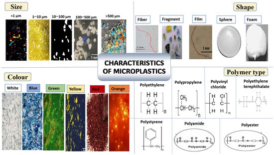
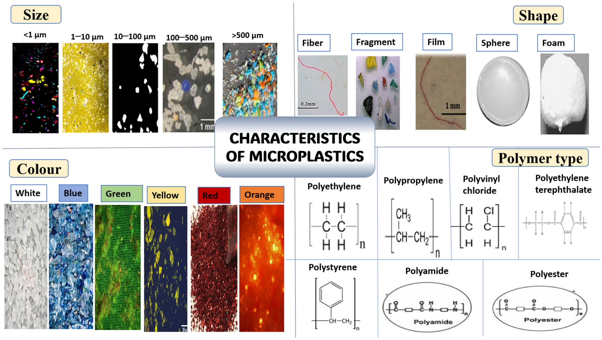
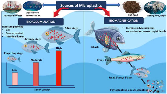
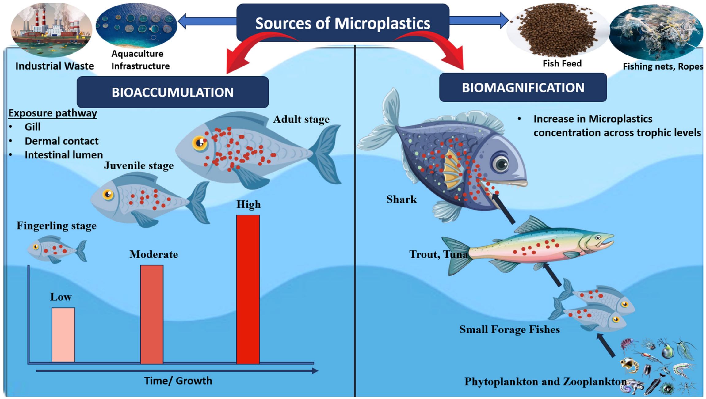
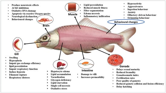
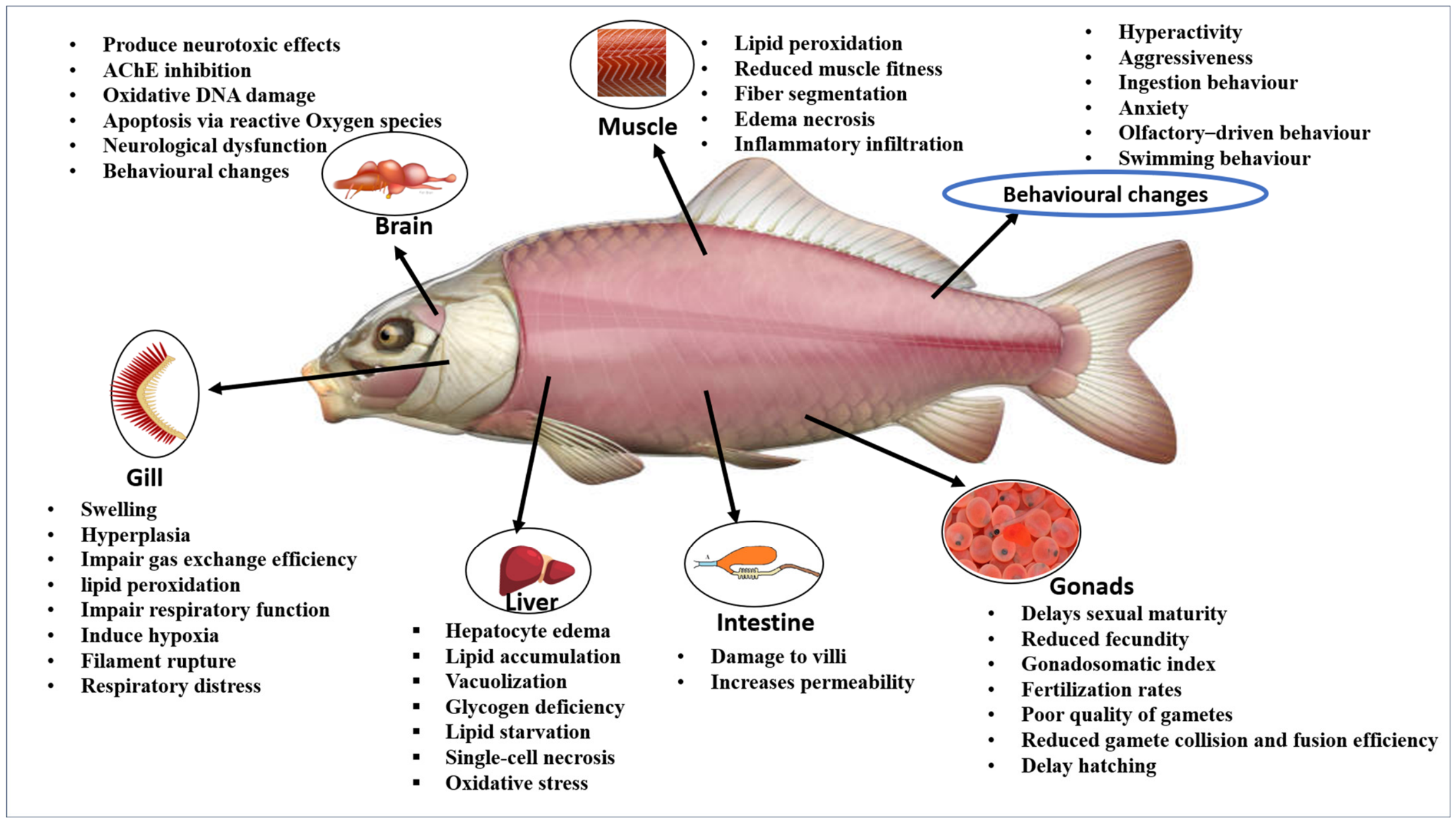

Disclaimer/Publisher’s Note: The statements, opinions and data contained in all publications are solely those of the individual author(s) and contributor(s) and not of MDPI and/or the editor(s). MDPI and/or the editor(s) disclaim responsibility for any injury to people or property resulting from any ideas, methods, instructions or products referred to in the content. |
© 2026 by the authors. Licensee MDPI, Basel, Switzerland. This article is an open access article distributed under the terms and conditions of the Creative Commons Attribution (CC BY) license.