


Abstract
Microplastic (MP) analysis in various environmental media currently faces significant challenges due to the complex and transportable nature of these materials. This study employed Fourier-transform infrared spectroscopy (FTIR) to investigate the presence of MPs in caddisfly larval cases, which incorporate vegetative fragments, sediment grains, and MP particles as building materials. The analysis of MPs in 2049 caddisfly larval cases revealed a total of 7553 items, resulting in an average of 3.7 items/case. The lowest recorded rate was 0.8 items/case, while the highest reached 18.2 items/case. Fiber MPs comprised 39.2% of the total, followed by fragment MPs at 30.8% and spherical MPs at 30%. Among the colors analyzed, blue MPs were the most prevalent, accounting for 36.6%, with white/transparent MPs at 22.1% and red MPs at 14.1%. MPs smaller than 100 µm were the most common, accounting for 37%, followed by MPs in the 100–250 µm range at 23.9%, larger than 500 µm at 22.3%, and those between 250 and 500 µm at 15.8%. Cellulose acetate was discovered to be the most abundant MP among different polymer types, followed by cellulose acetate butyrate, polyethylene terephthalate, poly(vinyl propionate), poly(ethylene glycol), poly(acrylonitrile-co-butadiene), cellulose propionate, hydroxyethyl cellulose, polyvinyl alcohol, glycerol triacetate, polystyrene, and poly(propylene glycol) methacrylate. These findings show the existence of MP in biotic components of these ecosystems, which has implications for aquatic biota health and freshwater quality, particularly in places influenced by human activity.
1. Introduction
Numerous researchers have expressed interest in the subject of MP contamination, including its incidence, source, and potential consequences, and its existence in the environment has been thoroughly documented. These ubiquitous pollutants are widely distributed in freshwaters, which can be influenced by anthropogenic factors (e.g., proximity to urban areas, low efficiency of wastewater treatment plants (WWTPs), and the use of sewage debris for fields), as well as natural factors (e.g., wind, storms, and floods). MPs (MPs < 5 mm) can negatively impact aquatic animals at the individual, cellular, and molecular levels. The most prevalent consequence of MPs is a decrease in food absorption, which is most likely caused by false food satiation and is most noticeable when combined with other pollutants. MPs can act as carriers for environmental contaminants and disease-causing microbes, raising ecological concerns due to their propensity to adsorb toxic compounds. Furthermore, the use of additives in plastic products to improve polymer properties poses many chemical risks to living organisms [1].
MP debris pollutes freshwater environments worldwide, including streams and rivers [1]. MPs (particles < 5 mm) come in many shapes (fragment, film, pellet [spherical], foam, and fiber [2]), colors, and chemical compositions. They are classed as primary or secondary MPs [3]. Primary MPs, such as spherical microbeads, are created in extremely small sizes. These spheres have a variety of applications, such as exfoliating agents in cosmetics, and may end up in the environment because wastewater treatment plants are frequently ineffective at removing them [4]. Secondary MPs, such as fragments, films, and fibers, are formed when larger plastic waste is physically or photochemically broken into smaller plastic bits [3]. Wind, rain drainage, wastewater, and inappropriate plastic waste disposal can all move plastic fragments from land to streams and rivers. Industrial mass production of plastics began in the 1940s [3], and MPs were discovered in the waters as early as the 1970s [5,6]. Since then, numerous studies have investigated the impact of MPs on marine life, including vertebrates [7] and invertebrates [8,9]. Rivers play a crucial role as sources of marine MPs, as they transport plastic debris from inland waters to the ocean [10]. Despite their important pathway, freshwater MPs have received far less attention than MPs in the marine environment [1]. Recent research on the interactions between MPs and freshwater organisms has mainly concentrated on the ingestion of MPs by vertebrates, such as fish [11,12], and invertebrates, including mollusks [13] and crustaceans [14]. This research also examines the resulting physiological effects [15,16]. Despite aquatic insects’ ubiquity and ecological importance [17], MP research on these animals is still in its early stages, with an emphasis on MP dietary intake [18,19,20].
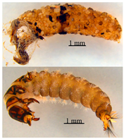
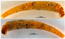
In addition to ingestion, MPs can be integrated into animal structures. For example, the marine tube-building polychaete Gunnarea gaimardi (Quatrefages, 1848) incorporates MPs into its housing [21], stabilizing MP particles in a biological structure. Similar biological structures in freshwater settings include larval cases built by various epibenthic caddisfly (Trichoptera) species, such as Limnephilus sp. (Family: Limnephilidae) caddisfly larvae [22,23]. Although it has been suggested that MPs may be fixed in caddisfly larval cases, no analysis such as micro-Fourier-transform infrared (μFTIR) spectroscopy has been performed to verify that the particles observed in caddisfly larval cases were MPs and not parts of ceramics or cardboard [24]. To investigate the incorporation of freshwater MPs into caddisfly larval cases, we initially assessed the presence of MPs within these cases, as well as their characteristics, including form, polymer type, color, and size.
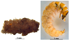
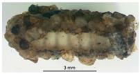
Caddisfly larvae form cases to defend themselves from predators and environmental factors [25,26]. As the larvae develop, additional material is added to the front end of the casing and fixed by silk filaments produced by the larvae [27]. Caddisfly larvae actively collect various biotic (e.g., leaves; [28,29] and abiotic (e.g., sediment grains; [30,31] resources for case building, and some species use both [32]. Caddisfly species have different preferences for particular case-building materials [33]. Several possible sentinel species for MP contamination in freshwater environments have been proposed recently, including tadpoles [34] and Asian clams [13], although none have yet been established. As a result, we address the possible use of caddisfly larval cases as bioindicators for freshwater MPs, which could aid in MP assessment in streams and rivers. MPs are incorporated into marine polychaete tubes [21], and freshwater sediments are a sink for MPs [35]. We hypothesized that MPs would be fixed in caddisfly larval cases, which are constructed in water using mineral grains, coarse particulate organic matter (detritus and leaf fragments), and other natural item particles. We conducted observational field research to evaluate our hypothesis.
2. Materials and Methods
2.1. Study Area
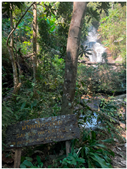
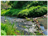
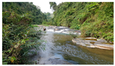
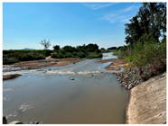
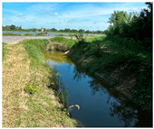
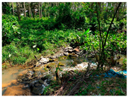
This study utilized museum-preserved caddisfly samples collected from streams across various regions of Thailand that were deposited at the Zoology Area in the Department of Science and Bioinnovation of Kasetsart University, Kamphaeng Saen Campus, Nakhon Pathom, Thailand. The specimens were preserved in ethanol. Information regarding the sample collection locations can be found in Table 1.
Table 1.
Characteristics of streams that were sampled for larval and casing-order Trichoptera.
2.2. Aquatic Insect Sampling and Laboratory Examination
Caddisfly larvae and pupae were sampled from all sample locations (Table 1). Kick sampling and hand picking were used to gather samples from the upper and lower surfaces of stones, stone crevices, gravel, woody debris, and other stable substrates for use with attached filter nets and pupal chambers. Caddisfly larvae and pupae were collected and preserved in 95% ethyl alcohol before being transferred to a 70% ethyl alcohol solution for preservation and transported to the laboratory. Adults were gathered near the stream margin in the same areas where larvae and pupae were collected, using black light traps that were set up after nightfall until morning. The metamorphotype approach was used to connect immature forms with adults, which involves collecting a pharate male in its pupal exuviae and shed larval sclerites within the pupal case or shelter [36,37].
2.3. MP Extraction and Characterization
The MPs in the caddisfly larval case were isolated following the protocols outlined by Maneechan & Prommi [38] and Vitheepradit & Prommi [39]. Individuals’ cases from each species were separated to conduct MP analysis (see Table 1). Individuals from each caddisfly larval case sample were placed in a 100 mL Erlenmeyer flask and digested with 20 mL of 30% hydrogen peroxide (H2O2) (Port Adelaide, Australia) to break down the biological tissues. To aid digestion, materials were heated in a water bath shaker at 50 °C for three hours and subsequently filtered through 0.45 μm filter paper (Whatman, Kent, UK). The filter papers containing MP precipitates were then placed in Petri dishes lined with aluminum foil and dried in a cabinet at 50 °C for two days prior to the experiment.
The number, shape, and color of the MPs were determined using a dissecting microscope (Leica EZ4E) (Leica, Wetzlar and Mannheim, Germany) and a digital camera. To evaluate the chemical composition of the MPs, the selected particles were examined with a PerkinElmer spectrum–Fourier-transform infrared spectrometer (FT–IR) (Bruker Daltonik, Billerica, MA, USA) in attenuated total reflection (ATR) mode. The spectral range was 4000–500 cm−1, with a spectral resolution of 32 cm−1 and 16 co-scans per measurement. The functional group characterization and polymer type analyses were both compared using the Bruker spectrum library. Woodall et al. [40] recommended spectra matching with a quality index ≥ 0.7.
2.4. MPs and Laboratory Quality Assurance and Control (QA/QC)
To prevent contamination from airborne MPs, all containers and equipment were washed with distilled water and wrapped in aluminum foil when not in use. The laboratory only used nitrile gloves, steel, and glass instruments. Before beginning lab work, the scissors and forceps were washed three times with deionized water. The lab surfaces were carefully cleaned with 70% ethanol. Blanks were run without tissues at each stage of the analysis in the same manner as the samples. All experimental procedures were completed as quickly as possible.
Due to the limitations of museum-preserved materials, various issues may arise, including contamination from long-term storage, alcohol contamination, air precipitation during storage, and alterations during preservation. This investigation used samples stored in 70% ethanol from a museum, which were not contaminated. Importantly, the caddisfly casings were thoroughly cleaned with clean water prior to MP examination.
2.5. Data Analysis
The primary characteristics of MP content in the samples were summarized using descriptive statistics, specifically the mean and standard deviation (SD). Data analysis was conducted using Microsoft Excel 2013 to create visual graphics. The data from larval cases and MP samples were evaluated using a series of steps. Before beginning the study, necessary data checks were conducted, including normality and homogeneity tests, to ensure the data met acceptable standards. The Kruskal–Wallis test was then applied to evaluate differences across various locations.
3. Results and Discussion
3.1. The Diversity of MPs in the Order Trichoptera
Research efforts to analyze MP pollution in freshwater macroinvertebrate populations are rising globally [41]; therefore, detecting MPs in insects with aquatic larvae is not unusual. Despite this, there are very few studies that document the existence of MPs in various Trichoptera species; only five of them focus on instances collected in the field, and only one quantifies the MPs, which appear to vary greatly depending on the site [1]. Furthermore, as far as we know, simultaneous control of different caddisfly families in riverine habitats has never been accomplished.
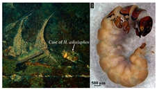
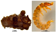
MPs were identified in 130 samples of Hydromanicus malayanus larval cases collected from the Khao Kho Hong stream. This resulted in a total of 103 MP items, yielding an average of 0.8 items/case (6.9 ± 4.5). MPs were discovered in 207 Trichomacronema paniae samples obtained at Sai Yoi Waterfall, for a total of 373 items, or 1.8 items/case (8.9 ± 5.6). MPs were also discovered in 215 samples of Hydropsyche askalaphos larval cases from the Mae-Ngim stream, for a total of 1593 items, or 7.4 items/case (37.0 ± 28.4). Similarly, 3733 MP items were found in 260 samples of Hydropsyche pallipenne larval cases from Mae-Ngim stream, averaging 14.4 items/case (533.3 ± 170.1). Furthermore, 70 samples of Amphipsyche meridiana larval cases from Khlong Hoi Khong Stream contained 300 items, or 4.3 items/case (6.0 ± 5.2). A total of 1145 larval case samples of Amphipsyche meridiana were found in an irrigation canal. In these samples, 1102 MP items were identified, averaging 0.9 items/case (52.6 ± 37.4). Additionally, MPs were detected in the larval cases of Amphipsyche meridiana at the inlet of the Kasiow Dam (5 cases), with a total of 92 items found, averaging 18.2 items/case (18.4 ± 8.4). Similarly, MPs were discovered in the larval cases of Macrostemum indistinctum at the same site (5 cases), totaling 73 items (average 14.6 ± 6.3 items/case). Finally, MPs were discovered in the larval cases of Marilia sumatrana in the Nam Phuai stream (12 samples), totaling 184 items (15.3 items/case, average 15.3 ± 3.9). The Kruskal–Wallis H test revealed significant differences in the mean abundance of MP particles among sample sites [chi-squared = 130.834 (fragments), 77.623 (fibers), 100.357 (spheres), 106.531 (<100 µm), 101.213 (100–250 µm), 106.743 (250–500 µm), 65.374 (>500 µm), 82.885 (blue), 91.591 (violet), 61.745 (black), 73.730 (red), 30.073 (green), 85.200 (yellow), 27.649 (pink), 99.001 (white), 51.535 (orange), 18.128 (multicolor), 27.667 (brown), df = 8, p < 0.05)] (Table 2).
Table 2.
MPs in caddisfly larval cases of the order Trichoptera.
In summary, an examination of MPs in 2049 larval cases of the order Trichoptera revealed a total of 7553 MP items found in the larval cases. This results in an average of 3.7 items/case. The smallest recorded amount was 0.8 items/case, while the largest was 18.2 items/case (Table 2 and see also Table S1 in supplementary materials).
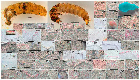
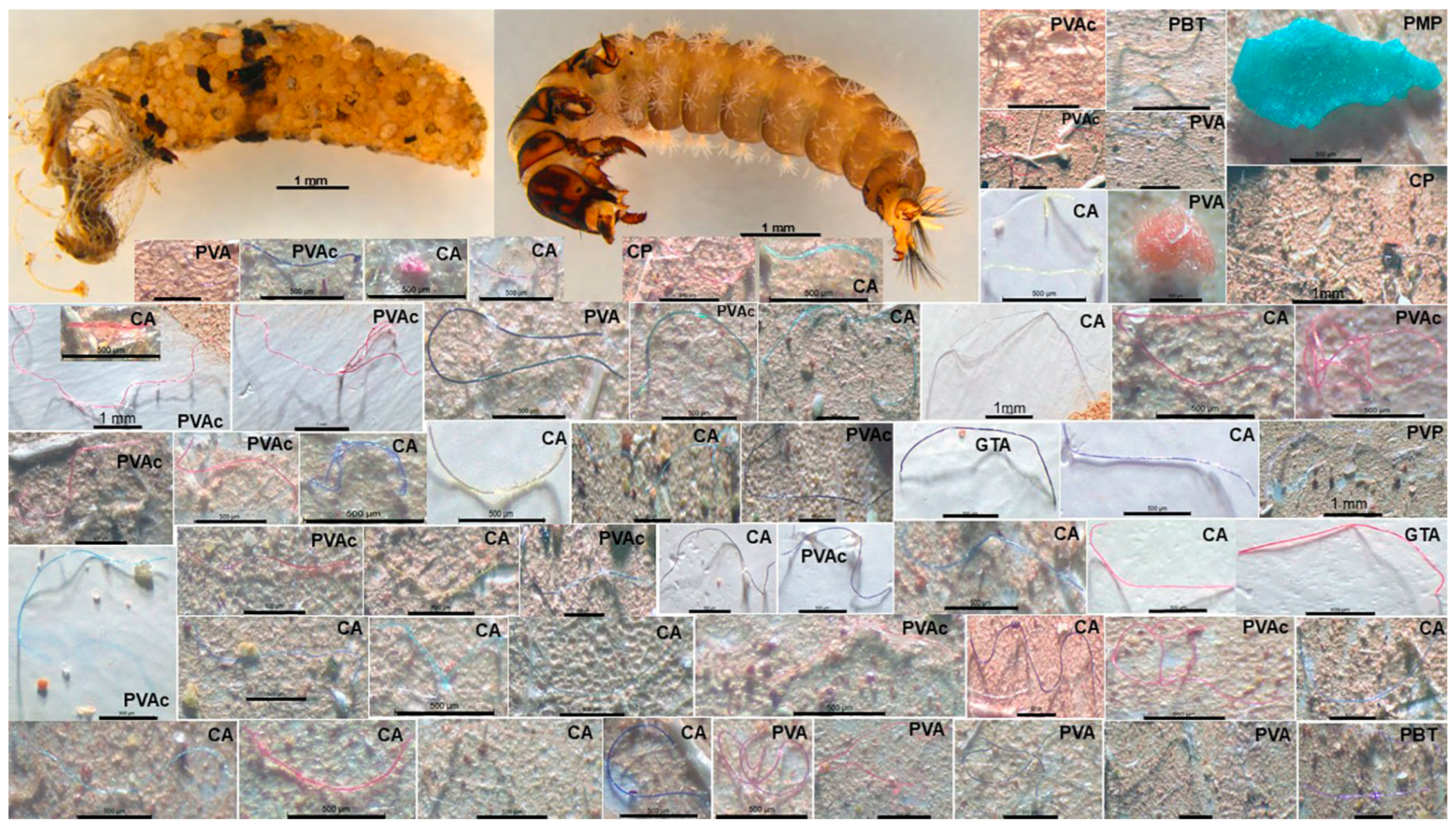
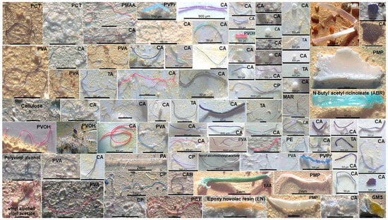
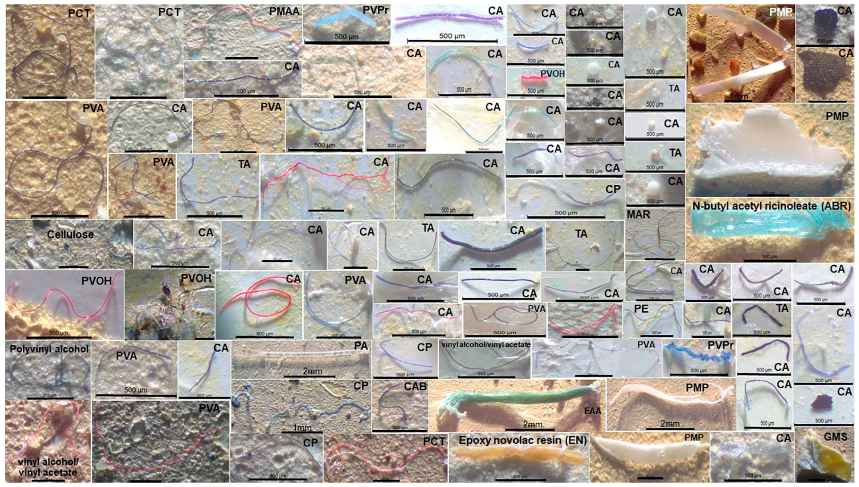
Figure 1 and Figure 2 illustrate the types, sizes, and colors of MPs found in the larval cases of various species within the order Trichoptera.
Figure 1.
Types, sizes, and colors of MPs found in the larval cases of the Trichomacronema paniae at the Sai Yoi Waterfall. The abbreviations for the polymer and plasticizer are presented in Table 3. Scale bar = 500 µm.
Figure 2.
Types, sizes, and colors of MPs found in the larval cases of Hydropsyche askalaphos in the Mae-Ngim stream. The abbreviations for the polymer and plasticizer are presented in Table 3. Scale bar = 500 µm.
3.2. MP Types
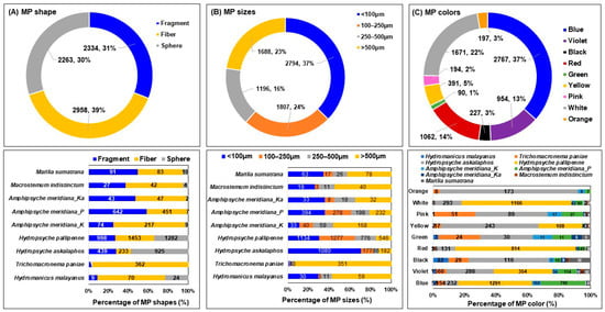
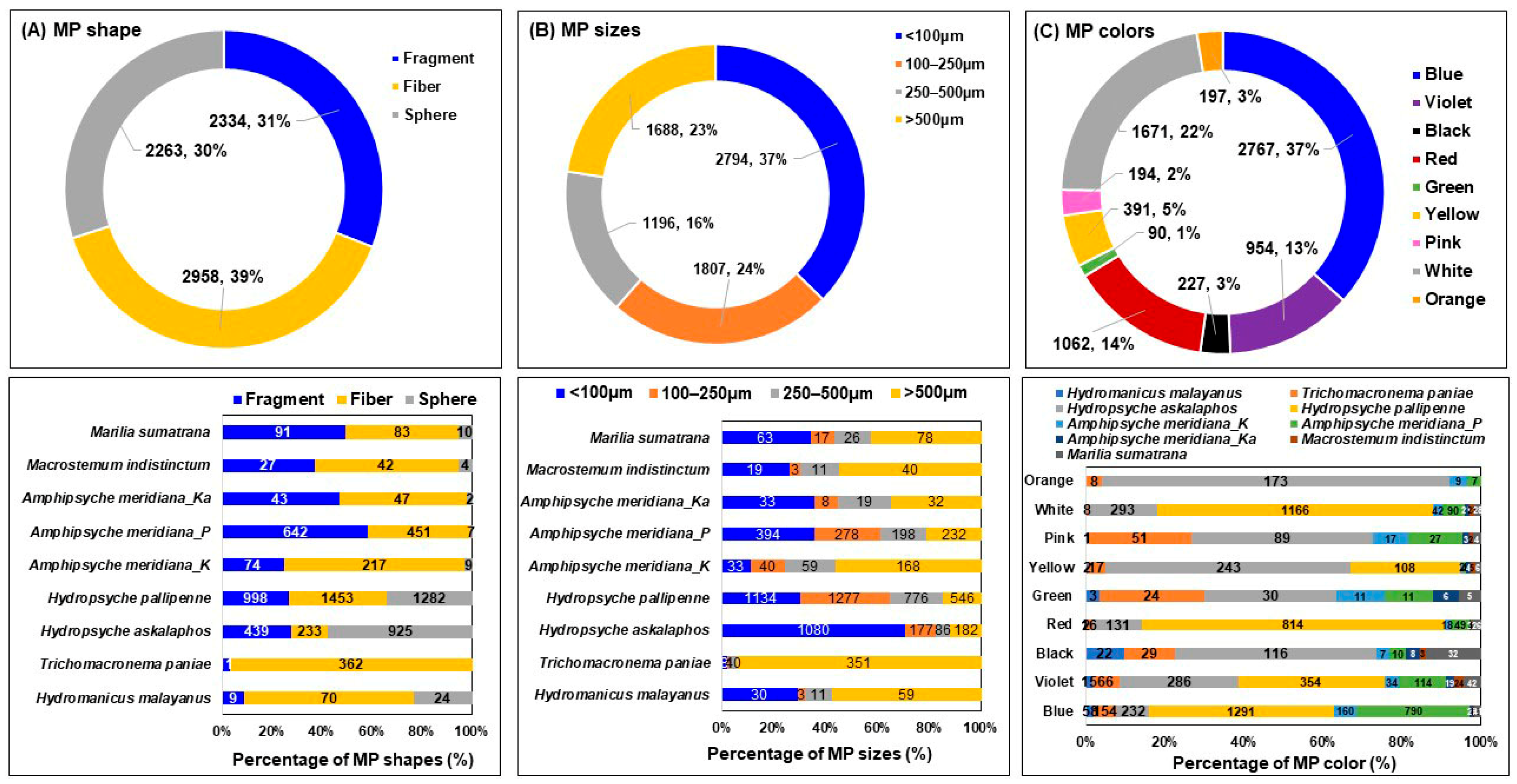
MPs of three types were identified in the larval cases of all species of aquatic insects in the Trichoptera order: fibers (39.2%), fragments (30.8%), and spheres (30%) (Figure 3). Fiber forms are widely recognized as the most prevalent type of MPs, accounting for 90% of all MPs [42]. MP fibers are the most common because they easily degrade in clothing and furnishings, such as curtains and fabrics [43]. MPs’ initial shape is determined by degradative and erosion processes [44]. Clothing and textiles can easily release a substantial amount of fiber material during wear and washing [44]. Fishing is a major source of fiber MPs [45]. Furthermore, MP fibers are abundant in the atmosphere [46]. Because the sampling stations were along roadsides, tire wear immediately introduced MPs [47]. Tire wear releases 6.1 million tons of MPs into the environment each year, 75.87% of which are fibers [48]. There were no MP analyses in water or sediment in this study. The distribution of fiber material in water and sediments at that time can be seen in the larval cases of the order Trichoptera, which account for 32% of the MPs’ morphology. Fibers having a diameter of less than 100 µm were discovered to aggregate readily in aquatic insects. However, fibers have been identified as the most common type of MP found in most freshwater invertebrates, accounting for 48.5% of total MPs [49].
Figure 3.
Photographs illustrating the various morphotypes, sizes, and colors of MPs found within the larval cases of caddisfly species.
The type of MP particle indicates whether it is primary or secondary [50]. According to Lehtiniemi et al. [51], primary MPs are typically symmetrical shapes, such as spheres. This shape has been observed in the larval cases of Hydropsyche askalaphos and H. pallipenne found in the Mae-Ngim stream in northern Thailand, as well as in Hydromanicus malayanus in the Khao Kho Hong stream in Songkhla Province, southern Thailand, where local people use the water for bathing and cleaning. In contrast, secondary MPs tend to be asymmetrical. Secondary MPs are more commonly found in aquatic environments than primary MPs [51,52]. Fibers are an example of primary MPs because they are frequently derived from clothes and other textiles or byproducts [50]. The presence of fragments suggests the possibility of secondary MPs because these fragments are frequently formed during the breakdown or disintegration of larger plastic materials [3]. The majority of the MPs found during this examination were secondary, in the form of fragments. This distribution indicates that MPs in the area are primarily caused by the decomposition of larger polymers. The high number of fibers and fragments (70% combined) in this study suggests that textile fibers produced during domestic washing, wastewater treatment, or fishing play a substantial role. Visual morphological examination contributes valuable information to MP assessment by identifying the source of contamination and, ultimately, its environmental impact [53]. Trichoptera larval cases include fibers from a variety of sources, including residential wastewater, untreated textile wastewater, fishing nets, and ropes. The breakdown of large plastic objects discarded in water may also cause fragmentation. Further research is needed to determine the origins of MPs detected in Trichoptera larval cases.
3.3. MP Sizes
All of the larval cases of the order Trichoptera contained 7553 suspected MP particles. The most common size range was <100 µm, which accounted for 37.0% of the identified MPs, followed by 100–250 µm (23.9%), >500 µm (22.3%), and 250–500 µm (15.8%) (see Figure 3). Particle size may indicate the distance from the source of pollution. Small MPs may indicate more distant sources of pollution, as they are byproducts of ongoing degradation [54] caused by exposure to various processes (mechanical or chemical) and prolonged water movement, whereas larger particles may be linked to direct primary MP intake from human activities such as fishing. The distance between the stream and communities, as well as human activities, may account for size variations. Kang et al. [55] discovered that MPs smaller than 2 mm were the most commonly identified size at all sampling sites along Korea’s southeastern coast. The size of plastic particles influences how they are absorbed and accumulated by organisms [56,57]. However, the size of organisms and their feeding habits can influence the types of plastic particles that accumulate within them. Larger plastic fragments are more likely to be ingested because of the anatomical features of their oral structures, which allow for the swallowing of larger particles and entry into the digestive tract [58]. Plastic fragments accumulate in prey and the surrounding environment in predators, implying that size is a limiting factor in plastic fragment ingestion [59].
3.4. The Color of MPs
All Trichoptera larval cases were found to have 7553 suspected MP particles. These particles can be classified into nine colors: blue, violet, black, red, green, yellow, pink, white/transparent, and orange. Blue fragments accounted for the most (2767 items, 36.6%), followed by white/transparent (1671 items, 22.1%), red (1062 items, 14.1%), violet (954 items, 12.6%), yellow (391 items, 5.2%), black (227 items, 3.0%), orange (197 items, 2.61%), and pink (194 items, 2.57%) (Figure 3). The color-based identification of MP particles is crucial for determining their origin and potential for ingestion by other organisms. Color is an effective measure of the increasing demand and use of plastic goods in our daily lives, which contributes to massive amounts of plastic waste [60]. The majority of plastics used in Thai shopping bags, packaging, fabrics, nets, and fishing gear are blue, white, or transparent and can be consumed by fish. MP bioaccumulation is highly influenced by particle size and exposure time [61].
3.5. Chemical Composition of MPs
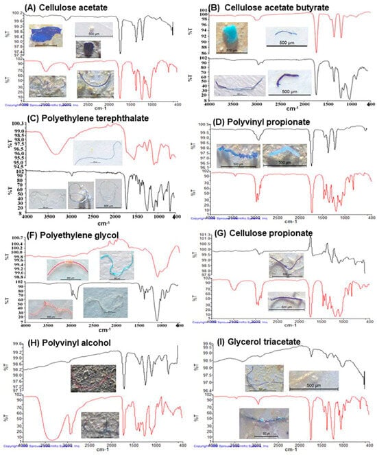
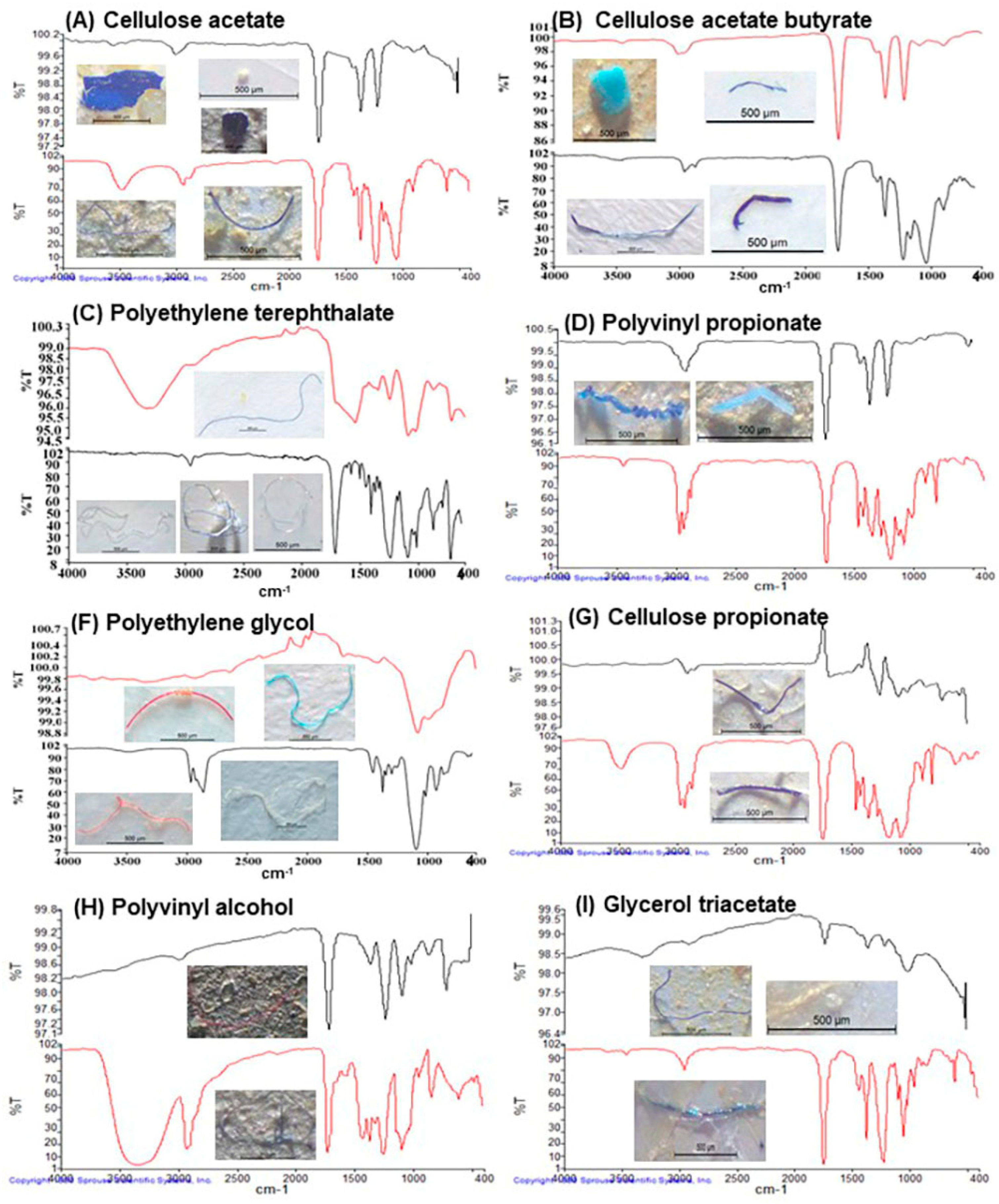
A total of 7553 suspected MP particles were discovered in the larval cases of the order Trichoptera; 856 (11.3%) of these particles were randomly selected for type identification using FTIR. Table 3 lists 43 different types of polymers (21 types), plasticizers (11 types), and non-MPs (11 types). The most frequent MP was cellulose acetate (321 items), followed by cellulose acetate butyrate (80 items), polyethylene terephthalate (75 items), poly(vinyl propionate) (49 items), poly(ethylene glycol) (40 items), and other MPs (Table 3, Figure 4). The composition of plastic can help identify the sources of MPs. Various types of polymers can produce MPs with similar color schemes; therefore, MPs that look alike may originate from multiple sources [62]. Cellulose acetate is classified as a type of MP. It is a man-made, semi-synthetic polymer, not a purely natural material. As a result, the small fibers and particles it breaks down into in the environment are categorized as MPs [63]. Cellulose acetate butyrate (CAB) is a synthetic/semi-synthetic cellulose ester commonly found in aquatic habitats as microfibers derived from cigarette filters, coatings, and industrial polymers. While generally non-toxic and largely biodegradable, CAB particles remain in water and sediment, posing physical ingestion concerns to aquatic life [63]. The plastic used to manufacture water bottles and synthetic textiles, known as polyethylene terephthalate (PET), poses a serious threat to aquatic environments. It breaks down into MPs, which aquatic organisms mistakenly ingest as food. This ingestion can lead to fatal satiation, metabolic abnormalities, reproductive toxicity, and oxidative stress in both marine and freshwater species [64].
Table 3.
Summarizes the various types of polymers/plasticizers found in the order Tricoptera larval cases. HA = Hydropsyche askalaphos; TP = Trichomacronema paniae; HM = Hydromanicuc malananus; AM = Amphipsyche meridiana; MI = Macrostemum indistinctum; MS = Marilia sumatrana. * = MP polymer, ** = plasticizers, and *** = non-MP particles.
Figure 4.
FTIR spectra of typical MP polymers. The black spectrum is the result of the FT–IR measurement, while the red spectrum is the reference spectrum from the Bruker library.
This study found both primary (spherical) and secondary (fiber and fragment) MPs embedded in the larval casings of all examined Trichoptera species. However, no spherical MPs were found in the larval casings of Trichomacronema paniae collected near Sai Yoi Waterfall. This absence is attributed to the sheer cliff face of the sampling site, which limits access for bathing and swimming. Spherical MPs were found to be more prevalent in the larval cases of Hydropsyche askalaphos collected from the Mae–Ngim Stream compared to other Trichoptera species examined across all sites. These spherical MPs represented 30% of all the MPs identified and resembled those commonly found in personal care products [65]. As the Mae-Ngim Stream lies outside the boundaries of the national park, local residents utilize its water for their livelihoods. Secondary MPs, which include fragments and fibers detected in Trichoptera larvae, are formed when larger plastic fragments break down into smaller pieces [3]. Each caddisfly case we tested had primary and secondary MPs of varying shapes, polymer types, sizes, and colors fixed within. The findings were validated by FTIR analysis [66]. As a result, we showed that caddisfly larval cases contain freshwater MPs with varied characteristics.
Although this study did not analyze MPs in water or sediment, the caddisfly cases contain materials that may be present in water and sediment. As MPs were found in caddisfly cases in this study, they must be part of the MPs suspended in streams. Thus, caddisfly case analysis can offer information on the various MPs found in a caddisfly’s habitat. Silk is used to fix the MP particles in the caddisfly larval case matrix, while mineral cases are extremely stable [67]. Caddisfly mineral cases contain a smaller fraction of organic material and fewer sand grains than sediment samples, so a filter produced by caddisfly case processing is easier to visually assess than a filter with a sediment MP fraction. The latter is frequently characterized by partially digested organic remains and microscopic sand grains that density separation cannot completely remove [68].
The disintegration of the cases allowed us to evaluate all MPs found in the caddisfly cases. Some MPs, potentially originating from the inner case wall or previously obscured by sediment grains within the case matrix, would have remained undetected if the cases had not been dissolved. Furthermore, the use of a digital microscope enabled us to record MP particles that are ordinarily invisible to the naked eye. Nuelle et al. [69] found that a 7-day treatment with 35% H2O2 effectively digests organic materials, but may cause color and size loss in some polymer types. The robust colors of MPs detected in caddisfly cases indicate that there was no bleaching owing to the use of H2O2, possibly facilitated by MP protection inside the caddisfly case matrix during digestion. The cases contained MPs with various chemical compositions (Table 3). Low-density MPs like ABS can float in the water column [70], but high-density MPs like PVC [71] are denser than freshwater and accumulate in sediments [72]. The caddisfly cases contained both high-density plastics like PVC and low-density MPs including ABS, PE, and PP [71].
As a result, it is likely that variables such as turbulence [70] or an increase in MP density caused by a biofilm cover [73,74] resulted in low-density MP sedimentation, making it available to caddisfly larvae. Similarly, we discovered PE in caddisfly larval case samples. The fact that the sediment had higher MP burdens than the water samples supports the hypothesis that the MPs in the examined caddisfly cases came from stream sediments. However, because water flows through the caddisfly case [75], it is possible that some MPs inside the caddisfly case originate in the water column.
4. Conclusions
Our findings indicate that the most commonly used plastic types, which are also likely the most widespread in the environment, are absorbed into caddisfly cases. This research suggests that caddisfly cases could be employed as quantitative bioindicators for MPs. However, future research should compare MP burdens in caddisfly larval cases from streams with varying levels of plastic pollution to explore their potential as quantitative freshwater MP bioindicators. Furthermore, future research could investigate whether MP burdens in caddisfly larval cases vary with seasonal flow rates. For example, the number of MPs ingested by chironomid larvae exhibits the same seasonal changes as sediment MP loads [19].
Future studies should also examine the role of MPs in insect constructs as indirect stressors. MPs in caddisfly cases may harm caddisfly larvae because they frequently contain hazardous additives [76], raise the risk of cancer [77] and carry pathogenic germs [78]. For example, plastic leachates impair brown mussel larvae [79], and PVC (as reported in the caddisfly cases) increases mortality in barnacle larvae [80]. We conclude that caddisfly larval cases have the potential to serve as freshwater MP bioindicators. However, future research should examine the MP content of cases from various caddisfly species and settings with varying amounts of plastic pollution.
Supplementary Materials
The following supporting information can be downloaded at https://www.mdpi.com/article/10.3390/microplastics5030170/s1, Table S1. MPs in caddisfly larval cases.
Author Contributions
Conceptualization: T.O.P.; methodology: T.O.P.; sampling: T.O.P.; processing and analysis: K.L., K.C. and P.D.; writing—original draft preparation: T.O.P.; review and editing: T.O.P., K.L., K.C. and P.D.; supervision: T.O.P.; project administration: T.O.P. All authors have read and agreed to the published version of the manuscript.
Funding
This research was financially supported by the Kasetsart University Research and Development Institute (KURDI), Bangkok, Thailand (Grant No. FF(KU) 52.69), as well as the Science Classroom in University Affiliated School (SCiUS), which is funded by the Ministry of Higher Education, Science, Research, and Innovation. The authors are appreciative of the resources provided by the Department of Science and Bioinnovation, Faculty of Liberal Arts and Science, Kasetsart University, Kamphaeng Saen Campus.
Institutional Review Board Statement
All animal procedures were approved by the Kasetsart University Animal Care and Use for Scientific Research Committee (ACKU68-ETC-004) on 13 May 2025.
Data Availability Statement
The raw data supporting the conclusions of this article will be made available by the authors on request.
Acknowledgments
Generative AI technologies were utilized to help with language editing and clarity. The authors produced and validated the scientific content, data interpretation, and conclusions and accept full responsibility for the manuscript’s accuracy and integrity.
Conflicts of Interest
The authors declare no conflicts of interest.
References
- Ehlers, S.M.; Manz, W.; Koop, J.H. Microplastics of different characteristics are incorporated into the larval cases of the freshwater caddisfly Lepidostoma basale. Aquat. Biol. 2019, 28, 67–77. [Google Scholar] [CrossRef] [Scilit]
- Free, C.M.; Jensen, O.P.; Mason, S.A.; Eriksen, M.; Williamson, N.J.; Boldgiv, B. High levels of microplastic pollution in a large, remote, mountain lake. Mar. Pollut. Bull. 2014, 85, 156–163. [Google Scholar] [CrossRef] [Scilit] [PubMed]
- Cole, M.; Lindeque, P.; Halsband, C.; Galloway, T.S. Microplastics as contaminants in the marine environment: A review. Mar. Pollut. Bull. 2011, 62, 2588–2597. [Google Scholar] [CrossRef] [Scilit] [PubMed]
- Rochman, C.M.; Kross, S.M.; Armstrong, J.B.; Bogan, M.T.; Darling, E.S.; Green, S.J.; Smyth, A.R.; Veríssimo, D. Scientific evidence supports a ban on microbeads. Environ. Sci. Technol. 2015, 49, 14740. [Google Scholar] [CrossRef] [Scilit] [PubMed]
- Carpenter, E.J.; Anderson, S.J.; Harvey, G.R.; Miklas, H.P.; Peck, B.B. Polystyrene spherules in coastal waters. Science 1972, 178, 749–750. [Google Scholar] [CrossRef] [Scilit] [PubMed]
- Carpenter, E.J.; Smith, K.L., Jr. Plastics on the Sargasso Sea surface. Science 1972, 175, 1240–1241. [Google Scholar] [CrossRef] [Scilit] [PubMed]
- Germanov, E.S.; Marshall, A.D.; Bejder, L.; Fossi, M.C.; Loneragan, N.R. Microplastics: No small problem for filter-feeding megafauna. Trends Ecol. Evol. 2018, 33, 227–232. [Google Scholar] [CrossRef] [Scilit] [PubMed]
- Cole, M.; Lindeque, P.; Fileman, E.; Halsband, C.; Goodyear, R.; Moger, J.; Galloway, T.S. Microplastic ingestion by zooplankton. Environ. Sci. Technol. 2013, 47, 6646–6655. [Google Scholar] [CrossRef] [Scilit] [PubMed]
- Kaposi, K.L.; Mos, B.; Kelaher, B.P.; Dworjanyn, S.A. Ingestion of microplastic has limited impact on a marine larva. Environ. Sci. Technol. 2014, 48, 1638–1645. [Google Scholar] [CrossRef] [Scilit] [PubMed]
- Lebreton, L.C.M.; van der Zwet, J.; Damsteeg, J.W.; Slat, B.; Andrady, A.; Reisser, J. River plastic emissions to the world’s oceans. Nat. Commun. 2017, 8, 15611. [Google Scholar] [CrossRef] [Scilit] [PubMed]
- Horton, A.A.; Jürgens, M.D.; Lahive, E.; van Bodegom, P.M.; Vijver, M.G. The influence of exposure and physiology on microplastic ingestion by the freshwater fish Rutilus rutilus (roach) in the River Thames, UK. Environ. Pollut. 2018, 236, 188–194. [Google Scholar] [CrossRef] [Scilit] [PubMed]
- McNeish, R.E.; Kim, L.H.; Barrett, H.A.; Mason, S.A.; Kelly, J.J.; Hoellein, T.J. Microplastic in riverine fish is connected to species traits. Sci. Rep. 2018, 8, 11639. [Google Scholar] [CrossRef] [Scilit] [PubMed]
- Su, L.; Cai, H.; Kolandhasamy, P.; Wu, C.; Rochman, C.M.; Shi, H. Using the Asian clam as an indicator of microplastic pollution in freshwater ecosystems. Environ. Pollut. 2018, 234, 347–355. [Google Scholar] [CrossRef] [Scilit] [PubMed]
- Weber, A.; Scherer, C.; Brennholt, N.; Reifferscheid, G.; Wagner, M. PET microplastics do not negatively affect the survival, development, metabolism and feeding activity of the freshwater invertebrate Gammarus pulex. Environ. Pollut. 2018, 234, 181–189. [Google Scholar] [CrossRef] [Scilit] [PubMed]
- Ding, J.; Zhang, S.; Razanajatovo, R.M.; Zou, H.; Zhu, W. Accumulation, tissue distribution, and biochemical effects of polystyrene microplastics in the freshwater fish red tilapia (Oreochromis niloticus). Environ. Pollut. 2018, 238, 1–9. [Google Scholar] [CrossRef] [Scilit] [PubMed]
- Redondo-Hasselerharm, P.E.; Falahudin, D.; Peeters, E.T.H.M.; Koelmans, A.A. Microplastic effect thresholds for freshwater benthic macroinvertebrates. Environ. Sci. Technol. 2018, 52, 2278–2286. [Google Scholar] [CrossRef] [Scilit] [PubMed]
- Suter, G.W.; Cormier, S.M. Why care about aquatic insects: Uses, benefits, and services. Integr. Environ. Assess. Manag. 2015, 11, 188–194. [Google Scholar] [CrossRef] [Scilit] [PubMed]
- Kim, S.W.; Kim, D.; Chae, Y.; An, Y.J. Dietary uptake, bio distribution, and depuration of microplastics in the freshwater diving beetle Cybister japonicus: Effects on predacious behavior. Environ. Pollut. 2018, 242, 839–844. [Google Scholar] [CrossRef] [Scilit] [PubMed]
- Nel, H.A.; Dalu, T.; Wasserman, R.J. Sinks and sources: Assessing microplastic abundance in river sediment and deposit feeders in an Austral temperate urban river system. Sci. Total Environ. 2018, 612, 950–956. [Google Scholar] [CrossRef] [Scilit] [PubMed]
- Windsor, F.M.; Tilley, R.M.; Tyler, C.R.; Ormerod, S.J. Microplastic ingestion by riverine macroinvertebrates. Sci. Total Environ. 2019, 646, 68–74. [Google Scholar] [CrossRef] [Scilit] [PubMed]
- Nel, H.A.; Froneman, P.W. Presence of microplastics in the tube structure of the reef-building polychaete Gunnarea gaimardi (Quatrefages 1848). Afr. J. Mar. Sci. 2018, 40, 87–89. [Google Scholar] [CrossRef] [Scilit]
- Pfister, H.; D’Avignon, G.; Wang, A.; Felismino, M.E.L.; Ricciardi, A. Caddisfly larvae (Trichoptera: Limnephilidae) facilitate the uptake of microplastics by a freshwater fish (Ameiurus nebulosus). Environ. Pollut. 2025, 385, 127133. [Google Scholar] [CrossRef] [Scilit] [PubMed]
- Arias-Paco, A.; Springer, M. Patterns of microplastic incorporation in larval and pupal cases of Limnephilus hamifer (Trichoptera: Limnephilidae). Rev. Biol. Trop. 2025, 73, e63696. [Google Scholar] [CrossRef] [Scilit]
- Tibbetts, J.; Krause, S.; Lynch, I.; Sambrook Smith, G.H. Abundance, distribution, and drivers of microplastic contamination in urban river environments. Water 2018, 10, 1597. [Google Scholar] [CrossRef] [Scilit]
- Boyero, L.; Rincón, P.A.; Bosch, J. Case selection by a limnephilid caddisfly [Potamophylax latipennis (Curtis)] in response to different predators. Behav. Ecol. Sociobiol. 2006, 59, 364–372. [Google Scholar] [CrossRef] [Scilit]
- Zamora Muñoz, C.; Svensson, B.W. Survival of caddis larvae in relation to their case material in a group of temporary and permanent pools. Freshw. Biol. 1996, 36, 23–31. [Google Scholar] [CrossRef] [Scilit]
- Stewart, R.J.; Wang, C.S. Adaptation of caddisfly larval silks to aquatic habitats by phosphorylation of h-fibroin serines. Biomacromolecules 2010, 11, 969–974. [Google Scholar] [CrossRef] [Scilit] [PubMed]
- Sheath, R.G.; Müller, K.M.; Larson, D.J.; Cole, K.M. Incorporation of freshwater Rhodophyta into the cases of caddisflies (Trichoptera) from North America. J. Phycol. 1995, 31, 889–896. [Google Scholar] [CrossRef] [Scilit]
- Moretti, M.S.; Loyola, R.D.; Becker, B.; Callisto, M. Leaf abundance and phenolic concentrations codetermine the selection of case-building materials by Phylloicus sp. (Tricho ptera, Calamoceratidae). Hydrobiologia 2009, 630, 199–206. [Google Scholar] [CrossRef] [Scilit]
- Gaino, E.; Cianficconi, F.; Rebora, M.; Todini, B. Case-building of some Trichoptera larvae in experimental conditions: Selectivity for calcareous and siliceous grains. Ital. J. Zool. 2002, 69, 141–145. [Google Scholar] [CrossRef] [Scilit]
- Okano, J.I.; Kikuchi, E.; Sasaki, O.; Ohi, S. Mineralogical composition of sediment determines the preference for smooth particles by caddisfly larvae during case construction. Ecol. Entomol. 2012, 37, 426–434. [Google Scholar] [CrossRef] [Scilit]
- Hansell, M.H. Case building behaviour of the caddisfly larva, Lepidostoma hirtum. J. Zool. 1972, 167, 179–192. [Google Scholar] [CrossRef] [Scilit]
- Hanna, H.M. Selection of materials for case-building by larvae of caddisflies (Trichoptera). Proc. R. Entomol. Soc. Lond. Ser. A Gen. Entomol. 1961, 36, 37–47. [Google Scholar] [CrossRef] [Scilit]
- Hu, L.; Chernick, M.; Hinton, D.E.; Shi, H. Microplastics in small waterbodies and tadpoles from Yangtze River Delta, China. Environ. Sci. Technol. 2018, 52, 8885–8893. [Google Scholar] [CrossRef] [Scilit] [PubMed]
- Castañeda, R.A.; Avlijas, S.; Simard, M.A.; Ricciardi, A. Microplastic pollution in St. Lawrence river sediments. Can. J. Fish. Aquat. Sci. 2014, 71, 1767–1771. [Google Scholar] [CrossRef] [Scilit]
- Milne, M.J. The metamorphotype method in Trichoptera. J. N. Y. Entomol. Soc. 1938, 46, 435–437. [Google Scholar]
- Wiggins, G.B. Larvae of the North American Caddisfly Genera (Trichoptera), 2nd ed.; The University of Toronto Press: Toronto, ON, Canada, 1996; 457p. [Google Scholar]
- Maneechan, W.; Prommi, T. Proximate composition, fatty acid profile, and microplastic contamination of edible odonate larvae (Aeshnidae: Anax sp.) in rice fields. Jordan J. Biol. Sci. 2023, 16, 649–654. [Google Scholar] [CrossRef] [Scilit]
- Vitheepradit, A.; Prommi, T. Microplastics in surface water and tissue of white leg shrimp, Litopenaeus vannamei, in a cultured pond in Nakhon Pathom Province, Central Thailand. AIMS Environ. Sci. 2023, 10, 478–503. [Google Scholar] [CrossRef] [Scilit]
- Woodall, L.C.; Sanchez-Vidal, A.; Canals, M.; Paterson, G.L.J.; Coppock, R.; Sleight, V.; Calafat, A.; Rogers, A.D.; Narayanaswamy, B.E.; Thompson, R.C. The deep sea is a major sink for microplastic debris. R. Soc. Open Sci. 2014, 1, 140317. [Google Scholar] [CrossRef] [Scilit] [PubMed]
- Cera, A.; Cesarini, G.; Scalici, M. Microplastics in freshwater: What is the news from the world? Diversity 2020, 12, 276. [Google Scholar] [CrossRef] [Scilit]
- Harris, P.T. The fate of microplastic in marine sedimentary environments: A review and synthesis. Mar. Pollut. Bull. 2020, 158, 111398. [Google Scholar] [CrossRef] [Scilit] [PubMed]
- Dris, R.; Gasperi, J.; Mirande, C.; Mandin, C.; Guerrouache, M.; Langlois, V.; Tassin, B. A first overview of textile fibers, including microplastics, in indoor and outdoor environments. Environ. Pollut. 2017, 221, 453–458. [Google Scholar] [CrossRef] [Scilit] [PubMed]
- Zhu, J.; Liu, S.; Wang, H.; Wang, D.; Zhu, Y.; Wang, J.; He, Y.; Zheng, Q.; Zhan, X. Microplastic particles alter wheat rhizosphere soil microbial community composition and function. J. Hazard. Mater. 2022, 436, 129176. [Google Scholar] [CrossRef] [Scilit] [PubMed]
- Yuan, W.; Liu, X.; Wang, W.; Di, M.; Wang, J. Microplastic abundance, distribution and composition in water, sediments, and wild fish from Poyang Lake, China. Ecotoxicol. Environ. Saf. 2019, 170, 180–187. [Google Scholar] [CrossRef] [Scilit] [PubMed]
- Liu, K.; Wu, T.; Wang, X.; Song, Z.; Zong, C.; Wei, N.; Li, D. Consistent transport of terrestrial microplastics to the ocean through atmosphere. Environ. Sci. Technol. 2019, 53, 10612–10619. [Google Scholar] [CrossRef] [Scilit] [PubMed]
- Bouredji, A.; Pourchez, J.; Forest, V. Biological effects of tire and road wear particles (TRWP) assessed by in vitro and in vivo studies—A systematic review. Sci. Total Environ. 2023, 894, 164989. [Google Scholar] [CrossRef] [Scilit] [PubMed]
- Baensch-Baltruschat, B.; Kocher, B.; Stock, F.; Reifferscheid, G. Tyre and road wear particles (TRWP)-A review of generation, properties, emissions, human health risk, ecotoxicity, and fate in the environment. Sci. Total Environ. 2020, 733, 137823. [Google Scholar] [CrossRef] [Scilit] [PubMed]
- Bertoli, M.; Pastorino, P.; Lesa, D.; Renzi, M.; Anselmi, S.; Prearo, M.; Pizzul, E. Microplastics accumulation in functional feeding guilds and functional habit groups of freshwater macrobenthic invertebrates: Novel insights in a riverine ecosystem. Sci. Total Environ. 2022, 804, 150207. [Google Scholar] [CrossRef] [Scilit] [PubMed]
- Park, T.-J.; Lee, S.-H.; Lee, M.-S.; Lee, J.-K.; Lee, S.-H.; Zoh, K.-D. Occurrence of microplastics in the Han River and riverine fish in South Korea. Sci. Total Environ. 2020, 708, 134535. [Google Scholar] [CrossRef] [Scilit] [PubMed]
- Lehtiniemi, M.; Hartikainen, S.; Näkki, P.; Engstrom-Öst, J.; Koistinen, A.; Setälä, O. Size matters more than shape: Ingestion of primary and secondary microplastics by small predators. Food Webs 2018, 17, e00097. [Google Scholar] [CrossRef] [Scilit]
- Galgani, F.; Hanke, G.; Maes, T. Global Distribution, Composition and Abundance of Marine Litter. In Marine Anthropogenic Litter; Bergmann, M., Gutow, L., Klages, M., Eds.; Springer: Cham, Switzerland, 2015; pp. 29–56. [Google Scholar]
- Banda, M.H.T.; Olayon, M.C.; Inocente, S.A.; Segovia, J.L.; Bedoya, N.; Bigcas, E.; Lomantong, A.N.; Bacsarpa, C.; Aporbo, I.C.C.; Reble, D.; et al. Microplastic pollution in mindanao’s taguibo river watershed forest reserve: Characterization, and distribution patterns, and implications for freshwater ecosystem conservation. Appl. Sci. Eng. Prog. 2024, 17, 7416. [Google Scholar] [CrossRef] [Scilit]
- Pan, Z.; Guo, H.; Chen, H.; Wang, S.; Sun, X.; Zou, Q.; Huang, J. Microplastics in the Northwestern Pacific: Abundance, distribution, and characteristics. Sci. Total Environ. 2019, 650, 1913–1922. [Google Scholar] [CrossRef] [Scilit] [PubMed]
- Kang, J.H.; Kwon, O.Y.; Lee, K.W.; Song, Y.K.; Shim, W.J. Marine neustonic microplastics around the southeastern coast of Korea. Mar. Pollut. Bull. 2015, 96, 304–312. [Google Scholar] [CrossRef] [Scilit] [PubMed]
- Alaraby, M.; Abass, D.; Domenech, J.; Hernández, A.; Marcos, R. Hazard assessment of ingested polystyrene nanoplastics in Drosophila larvae. Environ. Sci. Nano. 2022, 9, 1845–1857. [Google Scholar] [CrossRef] [Scilit]
- Liu, W.; Liao, H.; Wei, M.; Junaid, M.; Chen, G.; Wang, J. Biological uptake, distribution and toxicity of micro (nano) plastics in the aquatic biota: A special emphasis on size-dependent impacts. TrAC Trends Anal. Chem. 2023, 170, 117477. [Google Scholar] [CrossRef] [Scilit]
- Scherer, C.; Brennholt, N.; Reifferscheid, G.; Wagner, M. Feeding type and development drive the ingestion of microplastics by freshwater invertebrates. Sci. Rep. 2017, 7, 17006. [Google Scholar] [CrossRef] [Scilit] [PubMed]
- Coughlan, N.E.; Stevens, A.L.; Kelly, T.C.; Dick, J.T.; Jansen, M.A. Zoochorous Dispersal of Freshwater Bivalves: An Overlooked Vector in Biological Invasions? Knowl. Manag. Aquat. Ecosyst. 2017, 418, 42. [Google Scholar] [CrossRef] [Scilit]
- Zhang, B.; Yang, X.; Chen, L.; Chao, J.; Teng, J.; Wang, Q. Microplastics in soils: A review of possible sources, analytical methods and ecological impacts. J. Chem. Technol. Biotechnol. 2020, 95, 2052–2068. [Google Scholar] [CrossRef] [Scilit]
- Sun, Y.; Li, X.; Cao, N.; Duan, C.; Ding, C.; Huang, Y.; Wang, J. Biodegradable microplastics enhance soil microbial network complexity and ecological stochasticity. J. Hazard. Mater. 2022, 439, 129610. [Google Scholar] [CrossRef] [Scilit] [PubMed]
- Jenner, L.C.; Sadofsky, L.R.; Danopoulos, E.; Rotchell, J.M. Household indoor microplastics within the Humber region (United Kingdom): Quantification and chemical characterization of particles present. Atmos. Environ. 2021, 259, 118512. [Google Scholar] [CrossRef] [Scilit]
- Tanaka, K.; Takada, H. Microplastic fragments and microbeads in digestive tracts of planktivorous fish from urban coastal waters. Sci. Rep. 2016, 6, 34351. [Google Scholar] [CrossRef] [Scilit] [PubMed]
- Hidalgo-Ruz, V.; Gutow, L.; Thompson, R.C.; Thiel, M. Microplastics in the marine environment: A review of the methods used for identification and quantification. Environ. Sci. Technol. 2016, 46, 3060–3075. [Google Scholar]
- Otto, C.; Svensson, B.S. The significance of case material selection for the survival of caddis larvae. J. Anim. Ecol. 1980, 49, 855–865. [Google Scholar] [CrossRef] [Scilit]
- Mathalon, A.; Hill, P. Microplastic fibers in the intertidal ecosystem surrounding Halifax Harbor, Nova Scotia. Mar. Pollut. Bull. 2014, 81, 69–79. [Google Scholar] [CrossRef] [Scilit] [PubMed]
- Nuelle, M.T.; Dekiff, J.H.; Remy, D.; Fries, E. A new analytical approach for monitoring microplastics in marine sediments. Environ. Pollut. 2014, 184, 161–169. [Google Scholar] [CrossRef] [Scilit] [PubMed]
- McCormick, A.; Hoellein, T.J.; Mason, S.A.; Schluep, J.; Kelly, J.J. Microplastic is an abundant and distinct microbial habitat in an urban river. Environ. Sci. Technol. 2014, 48, 11863–11871. [Google Scholar] [CrossRef] [Scilit] [PubMed]
- Avio, C.G.; Gorbi, S.; Regoli, F. Plastics and microplastics in the oceans: From emerging pollutants to emerged threat. Mar. Environ. Res. 2017, 128, 2–11. [Google Scholar] [CrossRef] [Scilit] [PubMed]
- Wright, S.L.; Thompson, R.C.; Galloway, T.S. The physical impacts of microplastics on marine organisms: A review. Environ. Pollut. 2013, 178, 483–492. [Google Scholar] [CrossRef] [Scilit] [PubMed]
- Lagarde, F.; Olivier, O.; Zanella, M.; Daniel, P.; Hiard, S.; Caruso, A. Microplastic interactions with freshwater microalgae: Hetero-aggregation and changes in plastic density appear strongly dependent on polymer type. Environ. Pollut. 2016, 215, 331–339. [Google Scholar] [CrossRef] [Scilit] [PubMed]
- Rummel, C.D.; Jahnke, A.; Gorokhova, E.; Kühnel, D.; Schmitt-Jansen, M. Impacts of biofilm formation on the fate and potential effects of microplastic in the aquatic environment. Environ. Sci. Technol. Lett. 2017, 4, 258–267. [Google Scholar] [CrossRef] [Scilit]
- Williams, D.D.; Tavares, A.F.; Bryant, E. Respiratory device or camouflage? A case for the caddisfly. Oikos 1987, 50, 42–52. [Google Scholar] [CrossRef] [Scilit]
- Hermabessiere, L.; Dehaut, A.; Paul-Pont, I.; Lacroix, C.; Jezequel, R.; Soudant, P.; Duflos, G. Occurrence and effects of plastic additives on marine environments and organisms: A review. Chemosphere 2017, 182, 781–793. [Google Scholar] [CrossRef] [Scilit] [PubMed]
- Erren, T.; Zeuß, D.; Steffany, F.; Meyer-Rochow, V.B. Oceans of plastics: Possible risks of cancer in marine wildlife and humans. In Exploring Themes on Aquatic Toxicology; Allodi, S., Nazari, E.M., Eds.; Research Signpost: Trivandrum, India, 2013; pp. 51–74. [Google Scholar]
- McCormick, A.R.; Hoellein, T.J.; London, M.G.; Hittie, J.; Scott, J.W.; Kelly, J.J. Microplastic in surface waters of urban rivers: Concentration, sources, and associated bacterial assemblages. Ecosphere 2016, 7, e01556. [Google Scholar] [CrossRef] [Scilit]
- Gandara e Silva, P.P.; Nobre, C.R.; Resaffe, P.; Pereira, C.D.S.; Gusmão, F. Leachate from microplastics impairs larval development in brown mussels. Water. Res. 2016, 106, 364–370. [Google Scholar] [CrossRef] [Scilit] [PubMed]
- Li, H.X.; Getzinger, G.J.; Ferguson, P.L.; Orihuela, B.; Zhu, M.; Rittschof, D. Effects of toxic leachate from commercial plastics on larval survival and settlement of the barnacle Amphibalanus amphitrite. Environ. Sci. Technol. 2016, 50, 924–931. [Google Scholar] [CrossRef] [Scilit] [PubMed]
- Abbasi, F.; De-la-Torre, G.E.; Renner, G.; Senga Green, D.; Schmidt, T.C.; Dobaradaran, S. A review of occurrence and concentrations of cellulose acetate and other artificial cellulose microfibers in aquatic environmental matrices: An indicator of cigarette butts’ contamination? J. Hazard. Mater. Adv. 2025, 19, 100786. [Google Scholar] [CrossRef] [Scilit]
- Serra, T.; Vilaseca, F.; Colomer, J. The chronic effects of polyethylene terephthalate and biodegradable polyhydroxybutyrate microplastics on Daphnia magna. Environ. Res. 2025, 274, 121281. [Google Scholar] [CrossRef] [Scilit] [PubMed]

Disclaimer/Publisher’s Note: The statements, opinions and data contained in all publications are solely those of the individual author(s) and contributor(s) and not of MDPI and/or the editor(s). MDPI and/or the editor(s) disclaim responsibility for any injury to people or property resulting from any ideas, methods, instructions or products referred to in the content. |
© 2026 by the authors. Licensee MDPI, Basel, Switzerland. This article is an open access article distributed under the terms and conditions of the Creative Commons Attribution (CC BY) license.