


Abstract
Global plastic production and inadequate waste management have led to widespread environmental contamination with microplastics (MPs), now detected in food, water, and air. Their small size, diverse polymer composition, and capacity to carry chemical additives and co-pollutants facilitate intestinal uptake and raise concerns about their potential impact on gut microbiota. This review synthesizes current evidence on how MPs influence gut microbial composition and function, gut barrier integrity, and associated inflammatory and metabolic pathways. We conducted a narrative review of in vivo animal studies, in vitro simulated gut systems, and human observational studies that assessed MP exposure, gut microbiota profiles, and downstream toxicological outcomes. MPs originate from primary and secondary sources and can act as vectors for metals and organic pollutants. Following ingestion, they may cross the intestinal barrier via endocytic and persorption routes, acquire a protein corona, and be recognized by immune cells, activating TLR/NF-κB, and MAPK pathways alongside oxidative stress. In these models, MP exposure induces dysbiosis, characterized by loss of beneficial SCFA-producing bacteria (e.g., Bifidobacterium, Lactobacillus, Bacteroides) and expansion of pathobionts (e.g., Escherichia/Shigella, Staphylococcus, Enterobacteriaceae), accompanied by altered bile acid metabolism. These microbiota and metabolic alterations are linked to increased gut permeability, intestinal inflammation, metabolic dysfunction, and, in some studies, reproductive and neurobehavioral effects. Current evidence supports MPs as emerging modulators of gut microbial and intestinal homeostasis. However, heterogeneity across experimental models, reliance on high exposure doses, and lack of standardized MP characterization limit robust risk assessment. These limitations underscore the need for harmonized methodologies, longitudinal large-scale human studies, and the development of targeted mitigation strategies.
1. Introduction
Since plastic products began to be distributed on a large scale in the 1950s, global plastic production has increased substantially, from 2 million tons per year in 1950 to 430.9 million tons by 2024 [1,2]. Due to its slow degradation, plastic accumulates in the environment [3,4]. Once there, it is subjected to ultraviolet radiation, friction, and various physical, chemical, and biological processes, which degrade it into smaller fragments [5,6]. Environmental plastic debris is commonly classified by size; however, definitions vary slightly among regulatory bodies and scientific sources, particularly regarding the boundary between nanoplastics and microplastics. In this review, we adopt the widely used classification in which nanoplastics (NPs) are defined as particles <1 µm in diameter, while microplastics (MPs) range from 1 µm to 5 mm [7,8].
Alarming amounts of MPs have been observed in recent years [9], and they have been detected in various environments, ranging from the depths of the ocean to the Earth’s atmosphere [10]. These plastic particles not only pollute water and soil but also enter the food chain, thereby affecting a variety of organisms, including humans [11]. Humans can be exposed to microplastics through three major routes: ingestion, inhalation, and dermal contact [12]. Among these, ingestion is the most common method of exposure, as MPs can be found in contaminated food and drinks such as processed food, shellfish, fish, sea salt, and drinking water [11,13].
Recent research has shown that microplastics may impact the health of organisms by causing inflammation and other adverse effects, including changes in gut microbiota [10]. The relevance of the gut microbiota stems from its numerous physiological roles, including nutrient digestion and absorption. Furthermore, the intestinal microbiota acts as a barrier to pathogens, preventing their colonization and growth in the gut [14]. However, various environmental factors can influence the composition and function of microbiota. Among these factors, diet, antibiotic use, lifestyle, and exposure to environmental contaminants play crucial roles [15,16].
The gut microbiota comprises not only bacteria but also viruses, fungi, and protozoa, forming a complex and dynamic ecosystem. Among these, bacteria are the most prevalent and extensively studied microorganisms, constituting over 90% of the gut microbiota, which predominantly consists of the phyla Firmicutes, Bacteroidetes, Actinobacteria, and Proteobacteria [17].
Although the gut microbiota includes bacterial, viral, fungal, and protozoal communities, current evidence regarding the effects of microplastics is largely restricted to bacterial microbiota analyses. Data on the gut virome and mycobiome remain scarce, limiting our understanding of how microplastic exposure may affect these microbial components. Therefore, this review primarily focuses on bacterial community alterations and their implications for intestinal homeostasis.
Recently, the MPs in microbiota dysbiosis have gained attention. It has been proposed that MPs could act as vectors, delivering toxic agents or pathological microorganisms into the human body [18]. Consequently, overexposure to these agents could disrupt the normal gut microbiota, leading to microbiota dysbiosis and increased intestinal permeability, and triggering several chronic diseases such as obesity, cancer, and inflammatory bowel disease [19,20,21].
In this context, this review aims to describe the effects of microplastic exposure on gut microbiota composition and function across in vivo animal models, in vitro gastrointestinal simulation systems, and available human studies. Integrating findings from these approaches helps clarify the potential of MPs to disrupt intestinal microbial ecology and underscores the need for improved plastic waste management to reduce environmental and food chain contamination.
For this narrative review, a literature search was conducted using PubMed, Scopus, Web of Science, and Google Scholar for publications from 2008 to 2026, with emphasis on studies from the last decade and recent evidence from 2020 onward. Earlier key studies were included when they provided relevant conceptual, methodological, or mechanistic support. Search terms included combinations of “microplastics”, “nanoplastics”, “gut microbiota”, “intestinal microbiota”, “dysbiosis”, “intestinal barrier”, “gut permeability”, “inflammation”, “oxidative stress”, “short-chain fatty acids”, “bile acids”, “in vivo”, “in vitro”, and “human studies”. Studies were selected according to their relevance to microplastic exposure, gut microbiota composition, intestinal epithelial integrity, immune activation, and toxicological outcomes, prioritizing original experimental studies, in vitro gastrointestinal models, animal studies, human observational studies, and relevant reviews. No formal systematic review protocol, risk-of-bias assessment, or meta-analysis was performed, consistent with the narrative nature of this review.
2. Microplastic Definition, Size, Origin and Classification
Plastics are synthetic materials composed of organic polymers embedded with chemical additives to impart specific properties, including color, UV and thermal resistance, transparency, and durability [7]. Over recent decades, plastics have emerged as ubiquitous materials across diverse commercial sectors, driven primarily by their cost-effectiveness, versatility, and manufacturing efficiency. Consequently, this exponential rise in production and consumption has culminated in a substantial accumulation of plastic waste throughout global ecosystems [22].
Driven by this widespread utilization and subsequent environmental persistence, MPs have become pervasive pollutants [23]. Based on their origin, MPs are categorized into primary and secondary variants [24]. Primary MPs are intentionally manufactured in sub-millimeter dimensions for specific formulations, including cosmetics, detergents, and industrial applications [25]. Conversely, secondary MPs arise from the weathering and fragmentation of macroplastics (e.g., bottles and packaging) driven by environmental forces [1,25,26].
Morphologically, MPs are classified into five major types: fragments, spheres (microbeads), fibers, granules (pellets), and films [11]. Microbeads are manufactured plastic spheres that frequently occur in sizes below 1 mm in commercial formulations, though they can reach up to 5 mm under general regulatory and scientific classifications [7,8,27]. These particles are commonly found in personal care items like exfoliating scrubs, toothpaste, and cosmetics [27]. Fibers represent elongated, thread-like structures (<10 μm diameter) used in various applications, including clothing, household items, and cleaning products [24]. In addition, fragments are characterized by irregular morphologies and rigid structures, often resulting from the breakdown of larger plastic items [28]. Moreover, microplastic pellets are small plastic particles (1–5 mm) created from recycled plastic and frequently utilized as raw materials in the manufacturing of new plastic products [29]. Finally, the fragmentation of plastic bags, plastic packaging, and low-density plastic creates thin, transparent, and soft films [30].
Plastic degradation proceeds via biotic (biodegradation) and abiotic pathways [31]. Biotic degradation is mediated by microbial communities that enzymatically fragment polymers, whereas abiotic mechanisms encompass thermal, physical, photo-oxidative, and hydrolytic processes [5,32].
These degradation processes alter the mechanical properties of plastic particles, increase their specific surface area, modify their physicochemical reactivity, and induce unique interactions with living organisms, potentially enhancing their toxicity [33,34]. Owing to their reduced dimensions, these particles exhibit enhanced bioavailability and translocation capabilities, enabling them to cross physiological barriers—including the blood–brain and placental barriers—thereby compromising systemic biological functions [35].
3. Physicochemical Properties of Microplastics
MPs are composed of various synthetic polymers, each consisting of long molecular chains with repeating units. The chemical structure of MPs depends on the specific polymer used in their creation [36]. Key polymers found in MPs include polyethylene (PE), commonly used in packaging materials, bottles, and a range of plastic products; polypropylene (PP), found in packaging, textiles, and plastic containers; polyethylene terephthalate (PET), frequently used in beverage bottles, food containers, and synthetic fibers; polyvinyl chloride (PVC), present in construction materials, pipes, and various types of packaging; polystyrene (PS), often used in foam packaging, disposable utensils, and insulation materials; polyurethane (PU), used in foams, coatings, adhesives, and flexible plastics; nylon, used in textiles, fishing nets, and some plastic components; and acrylic, found in transparent plastics, lenses, and signage [7,21]. These polymers are resistant to biodegradation, contributing to the longevity of MPs in the environment. Currently, fossil fuel-derived plastics are the most common, with biodegradable plastics accounting for less than 1% of the total [26].
Additionally, during the plastic production process, various organic and inorganic additives are incorporated into plastic polymers to achieve unique properties such as color and transparency, or to enhance mechanical, thermal, and durability characteristics [37]. These additives, which include thousands of chemical products used in plastic manufacturing, are often toxic, carcinogenic, or have endocrine-disrupting effects, leading to a wide range of potential dangers [31]. Numerous organic additives, such as polyamide amine-epichlorohydrins (PAEs), brominated flame retardants (BFRs), organophosphate flame retardants (OPFRs), and bisphenol A (BPA), have been extensively researched for their impacts on different species, including endocrine disruption, immunotoxicity, neurotoxicity, and developmental and reproductive toxicity [38].
Furthermore, inorganic additives based on metal and metalloid compounds, such as arsenic, cadmium, chromium VI, lead, and silicon dioxide, are commonly incorporated into plastics to enhance their resistance to deformation and physical durability [39]. The environmental and human health impacts of these substances are not well understood. However, these additives can be released from plastic materials, persist in the environment, and potentially exert toxic effects [40,41].
In addition to leaching chemical additives, MPs effectively adsorb hydrophobic organic contaminants and heavy metals from the surrounding matrix [42]. MPs are lipophilic and possess extensive hydrophobic surfaces, enabling them to attract and retain emerging contaminants such as polycyclic aromatic hydrocarbons (PAHs), polybrominated diphenyl ethers (PBDEs), polychlorinated biphenyls (PCBs), and dichlorodiphenyltrichloroethane (DDT) [43]. These organic pollutants attach more readily to MPs than to larger plastic debris due to their greater specific surface area, extent of weathering, polarity, and hydrophobic nature [38,44,45].
Several studies have shown that microplastics can act as carriers for transporting various trace toxic elements, such as heavy metals, in aquatic and sedimentary environments, thereby facilitating the accumulation of these contaminants. MPs function like “magnets” for metals, concentrating them to elevated levels that can be toxic to ecosystems [46]. The adsorption or desorption of contaminants associated with plastic particles in the environment can be influenced by various factors, including environmental conditions, surface area, dissolved organic matter, and microbial activity [47]. Thus, these particles can function like Trojan horses, transporting and releasing contaminants across different environments via air or water, and they can also carry and release them within biological systems [48]. There is even the possibility that the combination of MPs and the adsorbed chemicals could be more toxic than the individual substances on their own [5].
Several factors influence the adsorption of pollutants on MPs. Fragmentation impacts physicochemical properties like size, crystallinity, and oxygen-containing groups, altering the adsorption profile of contaminants [49]. Similarly, UV radiation exposure increases surface area and pore volume, enhancing pollutant adsorption [50,51]. In the same way, aging leads to surface oxidation, increasing affinity for metals [52]. Moreover, environmental factors such as salinity, temperature, and pH also affect adsorption and desorption of contaminants [47,53,54].
4. Sources and Environmental Distribution of Plastic Waste
The growth in plastic production and consumption has significantly increased the annual amount of plastic waste entering the environment worldwide. In 2000, the volume of plastic waste generated was 156 million tons, whereas by 2019, it had grown to 353 million tons [55]. Of this plastic waste, only 9% is recycled, 19% is incinerated, approximately 50% ends up in controlled landfills, and the remaining 22% is discarded in uncontrolled landfills, burned openly, or released into the environment [24,56].
Each year, approximately 8 million tons of plastic waste enter the oceans, with over 250,000 tons floating on the surface [57]. Plastics can break down into MPs through processes such as microbial degradation and physical abrasion. It is estimated that 80% of MPs originate on land, while less than 20% come from the sea, and due to their low density, these particles can disperse globally [33]. Although MPs are prevalent in the environment, their specific health risks to organisms, including humans, are not yet well understood [5].
This widespread environmental dispersion of plastic waste directly translates into measurable human exposure. The contamination of seafood, drinking water, table salt, and even air contributes to an estimated uptake of 74,000–121,000 microplastic particles per year [58]. Airborne exposure also plays a role, as indoor environments can contain 1.6–12.6 fibers/m3, leading to inhalation of thousands of particles daily [59]. These findings demonstrate that environmental distribution is closely linked to human exposure, supporting the need for risk assessment.
5. Routes of Microplastic Exposure
5.1. Invisible Airborne Threats: Microplastic Inhalation
Airborne MPs, originating from plastic dust pollution, can be inhaled [33,60]. These tiny plastic fibers present a significant risk to respiratory health as they can become lodged in the lungs [12,61]. The alveolar region of the lungs, with its large surface area and thin barrier, facilitates the penetration of nanoparticles, potentially causing pulmonary inflammation and genotoxic effects. Furthermore, these particles may enter the bloodstream, disseminating throughout the body [33,57].
5.2. Dermal Infiltration: Microplastic Entry via Skin
Cutaneous contact, though less studied, is a significant pathway for exposure to MPs. The stratum corneum, the outermost layer of the skin, serves as a protective barrier against injuries and harmful agents. However, its hydrophobic nature may permit absorption through sweat glands or hair follicles [24]. UV exposure can compromise this barrier, increasing the risk of MP penetration. Additionally, various compounds, such as alcohols and alpha-hydroxy acids, can enhance skin permeability, potentially facilitating the entry of MPs [62]. These particles are primarily found in cosmetics and personal care products, such as shaving creams, toothpaste, facial soaps, and anti-wrinkle creams, but also in contaminated water and soil [12,13].
5.3. Ingesting the Invisible: Microplastic Oral Exposure
Ingestion is the primary route through which MPs enter the human body [42]. These particles can be consumed via contaminated food and beverages. Rubio-Armendáriz et al. (2022) estimated that the potential daily dietary intake of microplastics could reach approximately 34,254 particles/day [63]. This calculation integrates multiple dietary sources, including 33,626 particles/day from the consumption of 2 L/day of drinking water, 212 particles/day from an average intake of 70.68 g/day of crustaceans/mollusks, 409.94 particles/day from 70.68 g/day of fish, and 6.40 particles/day from the consumption of 9.4 g/day of table salt [63]. However, additional studies indicate that MPs present in soil can translocate from plant roots to stems, leaves, fruits, and seeds, representing another dietary source of human exposure [39,64,65].
However, a critical gap remains when contrasting these estimated human intake pro-files with the exposure regimens utilized in the primary literature. While real-world hu-man ingestion is characterized by chronic, low-dose exposure to highly heterogeneous particles, most current in vivo animal models rely on acute or sub-chronic designs using specific polystyrene spheres at concentrations orders of magnitude higher than realistic environmental levels. Consequently, the biological disruptions reported in animal models must be interpreted with caution, as it remains uncertain whether these pathophysiological cascades occur under environmentally relevant human exposure scenarios.
Despite providing valuable quantitative estimates, this assessment relies exclusively on particle counts and does not include key characteristics such as particle size, mass, morphology, or polymer composition. The absence of these parameters limits the toxicological relevance of the exposure data, as particle properties play a crucial role in determining biological interactions, uptake, and potential health effects [66,67].
Following ingestion, MPs in the digestive tract interact with the intestinal mucosa and epithelium, which together form a significant barrier against the absorption of xenobiotics [57]. Research involving animals and in vitro intestinal models has demonstrated that MPs can cross the intestinal barrier via enterocytes, entering the lymphatic system or bloodstream and subsequently reaching various organs and tissues [6], where they may accumulate or impact the immune system and cellular viability [34,57].
Once ingested, MPs can influence the gut microbiota, triggering various responses, including modifications in community structure, population levels, and function [21,68]. These impacts on the gut microbiota are significant, as they can alter the balance of microbial communities, potentially leading to health issues.
5.4. Intestinal Translocation, Cellular Uptake, and Immune Recognition of MPs
Ingestion is considered the primary route through which MPs enter the human body. After ingestion, particles interact with the intestinal mucosa and the epithelial barrier. This barrier is primarily formed by enterocytes, but its function is structurally and immunologically supported by specialized cell types, including goblet cells, Paneth cells, and microfold (M) cells, which are specialized in transporting luminal antigens to the underlying lymphoid structures [69,70].
When MPs encounter the gastrointestinal epithelium, several pathways for uptake and translocation have been proposed. One potential, though functionally limited, route is paracellular passage through tight junctions; however, this mechanism is constrained by the extremely small pore size of these channels (approximately 1.5 nm) [33,34]. Instead, current evidence indicates that transcellular transport represents the predominant mechanism. This process is mediated primarily by endocytic and phagocytic pathways, including specialized uptake by M cells located in Peyer’s patches [33,71].
Persorption represents another potential entry route across the intestinal barrier. This mechanism is particularly relevant for NPs up to 130 nm in size and involves the passage of particles through transient gaps formed during physiological epithelial cell shedding. Through these discontinuities, NPs can access the underlying tissue, where they may be engulfed by dendritic cells and subsequently transported toward the lymphatic vessels and the systemic circulation (Figure 1) [34].
Figure 1.
Mechanisms of intestinal translocation, immune recognition, and inflammatory activation induced by microplastics (MPs) and nanoplastics (NPs). This figure illustrates the main pathways by which MPs and NPs (white circles) traverse the intestinal barrier and trigger inflammatory responses within the lamina propria. MPs and NPs may cross the epithelium through three mechanisms: (1) paracellular passage (blue arrows); (2) transcellular transport (black arrow); and (3) persorption (magenta arrows). After translocation, plastic particles interact with macrophages and dendritic cells, promoting their activation and the release of pro-inflammatory cytokines (red circles), including IL-1α, IL-1β, IL-2, IL-5, IL-6, and TNF-α. These mediators amplify epithelial permeability and initiate intracellular signaling cascades such as NF-κB activation. Persistent cytokine exposure drives differentiation of naïve T cells into Th1 and Th17 subtypes, with Th17 cells further contributing to neutrophil recruitment and tissue injury. Together, epithelial barrier disruption, immune activation, and sustained cytokine production establish a chronic inflammatory microenvironment characteristic of prolonged exposure to MPs and NPs. Created by the authors using Adobe Photoshop, version 25.0 (Adobe Inc., San Jose, CA, USA).
Once across the intestinal barrier, MPs can be internalized by cells through multiple endocytic routes, with uptake strongly influenced by particle size, surface chemistry, and the composition of the protein corona [33,72]. Distinct cell types employ different internalization mechanisms. MPs may enter through (1) general endocytic pathways, in which MPs and NPs interact with membrane channels, transporter proteins, or, in rare cases, passively cross the plasma membrane, and (2) specialized routes such as clathrin-dependent and caveolae-dependent endocytosis, macropinocytosis, and phagocytosis [73]. Smaller particles (50–200 nm) are preferentially taken up by clathrin-mediated endocytosis, whereas larger particles (>500 nm) rely primarily on macropinocytic and phagocytic pathways [74].
During digestion, MPs can bind to circulating proteins and other biomolecules, forming a “protein corona” that alters their biological identity and modulates how they interact with cells—either masking reactive surfaces or enhancing uptake through receptor-mediated processes [73,75,76]. Immune-related proteins such as immunoglobulins, complement factors, apolipoproteins, coagulation mediators, and acute-phase proteins commonly adsorb onto particle surfaces and contribute to this corona [77]. Several of these proteins act as opsonins, facilitating recognition and internalization by phagocytic cells [76].
Opsonins such as antibodies, fibronectin, complement components, milk fat globulin (lactadherin), and mannose-binding lectin [78] can adsorb onto the surface of MPs and serve as molecular tags that enhance their recognition by phagocytic cells. Once bound, these proteins interact with specific receptors on macrophages and other phagocytes, thereby facilitating particle uptake [76,78,79]. The most relevant receptors include Fc receptors, which bind the Fc region of IgG or IgA, and complement receptors, which recognize activated complement fragments such as iC3b. Engagement of these receptors by opsonin-coated particles initiates intracellular signaling cascades that reorganize the actin cytoskeleton, driving membrane extension around the particle. The phagocytic cup then closes distally to form a phagosome, thereby internalizing the particle for subsequent processing [78,79].
5.5. Pathophysiological Cascade Initiated by MPs in the Intestinal Epithelium
Exposure to MPs and NPs initiates a pathophysiological cascade in the gut by disrupting epithelial homeostasis and triggering innate immune activation. One of the earliest events involves the recognition of foreign particles by intestinal immune cells, which respond by releasing cytokines that increase epithelial permeability. This heightened permeability facilitates the passage of MPs, NPs, and luminal antigens into deeper tissue layers, where they contribute to the disorganization of tight-junction proteins, including ZO-1, ZO-2, claudins, and occludins, ultimately weakening barrier integrity and enhancing paracellular transport [18,69,70].
Once these particles and microbial components reach the lamina propria, they engage pattern-recognition receptors (PRRs) expressed on epithelial cells, macrophages, and dendritic cells. This interaction activates canonical innate immune signaling pathways, particularly the TLR–MyD88–IRAK–TRAF6–IKKβ axis and parallel MAPK cascades (ERK1/2, JNK, p38). The convergence of these pathways leads to NF-κB nuclear translocation and AP-1 activation, promoting the transcription of pro-inflammatory cytokines (TNF, IL-1β, IL-6), effector enzymes (COX-2, iNOS), and chemokines that further amplify immune activation. These processes are tightly coupled to increased production of reactive oxygen species (ROS), generated through both NADPH oxidase activity and mitochondrial dysfunction. Elevated ROS levels reinforce NF-κB and MAPK signaling, creating a self-sustaining feedback loop that perpetuates inflammation and oxidative stress [18,69,70].
Sustained release of inflammatory mediators by activated macrophages and T and B lymphocytes drives the differentiation of naïve T cells into Th1 and Th17 subsets, both central to mucosal inflammation. Th17 cells, in particular, secrete cytokines that recruit neutrophils, thereby exacerbating tissue damage and contributing to ongoing epithelial injury. Collectively, these events promote progressive barrier deterioration and thickening of the lamina propria, establishing a chronic inflammatory microenvironment characteristic of prolonged exposure to MPs and NPs (Figure 1) [18,69,70].
6. Microplastic Intrusion: Disrupting the Balance of the Intestinal Microbiota
Recent studies have demonstrated the effects of microplastics on the gut microbiota, highlighting their role in microbial dysbiosis and their potential contribution to various diseases. The investigations summarized in Table 1 include multiple studies involving animal models (mice and pigs), in vitro systems, and human cohorts. These findings reveal significant changes in composition and abundance of specific bacterial genera, including Bacteroides, Lactobacillus, Firmicutes, and Escherichia, due to exposure to certain MPs such as polystyrene (PS), polyethylene (PE), and polyethylene terephthalate (PET).
However, the available evidence remains inconsistent regarding the relationship between gut microbiota alterations and microplastic exposure. Some studies have reported reductions in taxa such as Bacteroides, Lactobacillales/Lactobacillus, and Parabacteroides, whereas others have observed increases in Firmicutes and Actinobacteria/Actinobacteriota in different in vivo and in vitro models and under different exposure conditions [80]. These differences may be partly explained by particle size, exposure level, exposure duration, and polymer type, including PS, PE, and PET [81]. These factors may influence how microplastics interact with the intestinal mucosa and the gut microbiota [81,82]. In particular, smaller particles may have stronger effects on inflammation and microbiota disruption because they are more likely to interact with epithelial cells or cross the intestinal barrier, while larger particles may remain mainly within the intestinal lumen and have a lower probability of reaching systemic circulation [83].
These alterations emerged under different exposure conditions. For instance, in one animal model, exposure to small PS particles (0.5 µm) resulted in a reduction of beneficial Bacteroides and an increase in Bifidobacterium [68]. In another study using a similar model, exposure to larger PS particles (10–150 µm) led to a notable increase in Staphylococcus and a decrease in Parabacteroides, suggesting that particle size or the specific characteristics of the microplastic could exacerbate gut microbial imbalance [18]. Moreover, some of these studies report a shift towards pathogenic or opportunistic bacteria. For instance, exposure to PS microplastics has been associated with a decrease in beneficial Bacteroides and an increase in harmful genera such as Staphylococcus and Escherichia [84,85].
These findings highlight the need for further investigation into the broader implications of microplastic exposure, particularly regarding gut microbiome stability, which is essential for overall health and homeostasis [86,87].
Table 1.
Effects of microplastic exposure on gut microbiota across different species and experimental models.
7. Effects of MPs on Gut Microbiota
7.1. In Vivo Studies: Results in Animal Models
The effect on the gut microbiota after MP ingestion has been assessed in several animal models. For instance, in a mouse in vivo study, several PE-MP concentrations were evaluated for their impact on the gut microbiota and inflammatory response. It was observed that mice under the higher MP concentration showed an increased bacterial alpha diversity, along with greater proinflammatory cytokine secretion, triggering gut tissue inflammation. Moreover, an increase in pathogenic bacteria such as Staphylococcus and a decrease in other bacteria such as Parabacteroides was observed. This microbial change also leads to the expression of pro-inflammatory cytokines through the activation of the Toll-like receptor (TLR) pathway [89].
Another mice model study showed that exposure to MPs combined with several phthalate esters led to a greater gut accumulation of the combination MPs + di-(2-ethylhexyl) phthalate. This combination also promoted alterations in intestinal gene expression of oxidative response, lipid metabolism, immune response, and hormone metabolism. In contrast with the previous study, the alpha diversity was not different between exposed groups; however, there were differences in bacterial abundance like changes of microbes involved in energy metabolism and immune function (Adlercreutzia, Butyricimonas, Prevotella, and Ruminococcus). Moreover, the inflammatory response was also present in mice treated with MPs + di-(2-ethylhexyl) phthalate, which was related to an increased intestinal permeability effect that promotes bacterial colonization, immune response, and intestinal inflammation [101].
Gut microbiota dysbiosis and intestinal inflammation have also been described in zebrafish model studies. In these cases, Bacteroidetes and Proteobacteria showed a decreased abundance after PS-MP exposure. In these studies, the accumulation of MPs in the gut of zebrafish was also described as the main cause of intestinal inflammation, which led to an increased intestinal permeability and bacterial colonization [102]. As seen in mouse models, exposure to MPs also promoted the expression of pro-inflammatory cytokines, which led to oxidative stress and gut inflammatory diseases. Moreover, bacterial dysbiosis was related to the altered metabolism of carbohydrates, cholesterol, amino acids, and bile acids, which could be related to metabolic disorders [102].
Therefore, MP consumption is involved in microbiota modifications and gut inflammation in murine models (Figure 1). Although microbial diversity differed among experimental models, these differences may be related to the specific MPs assessed. This difference in microbiota diversity could be related to its susceptibility to MP components, which could exert several effects on microbiota composition. Therefore, the molecular mechanisms underlying these microbiota alterations should be investigated using controlled and standardized animal models of MP exposure. Moreover, MP additives and their ability to serve as a vector for other toxic compounds as well as pathogenic bacteria could be involved in gut microbiota modification and inflammation. Bacterial dysbiosis related to altered biomolecule metabolism (carbohydrates, cholesterol, and amino acids) is also a critical approach that could be related to a lower absorption of nutrients, which could lead to greater susceptibility to infections or metabolic diseases in individuals with high amounts of MPs accumulated in the intestine.
7.2. In Vitro Studies: Key Observations and Findings
In vitro studies have also been performed to assess the effect of MPs on a simulated human gut. For instance, a study used the simgi® system, a computer-controlled gastrointestinal in vitro model designed to reproduce the colonic microbiota and different colonic compartments, to assess the PET-MPs’ effect on microbiota composition and MP modification after colonic fermentation. This study found that MPs were structurally degraded after digestion. Regarding the alpha diversity of microbiota, it decreased after MP exposure. Moreover, the bacterial relative abundances changed at the colonic compartments; for example, Bacteroides and Parabacteroides decreased in the transverse and descending colon, while Escherichia and Shigella increased at the same gut compartments. This in vitro study suggested that a decrease in Bacteroides and Parabacteroides could be associated with less production of anti-inflammatory molecules, such as polysaccharide A or sphingolipids, while the increase in pathological bacteria (Escherichia/Shigella) could trigger inflammatory bowel disease due to the lack of endogenous antibacterial molecules [93]. Another study that simulated a human colon, but with infant microbiota, demonstrated an increase in alpha diversity, mainly harmful pathobionts (Enterobacteriaceae), and a decrease in butyrate production, but no disturbances in the gut barrier, after PE MP exposure [86].
Similarly to in vivo models, there were differences in alpha diversity after MP exposure. In this case, this outcome could be due to the donors who provided the microbiota sample, since the first one was an adult healthy volunteer, while the second was an infant’s microbiota sample. Therefore, the behavior of microbiota under MP exposure, its effect on the gut barrier, and the inflammatory process could depend on microbiota maturity, although more standardized protocols are needed to assess these approaches.
Another previous in vitro study performed an artificial in vitro digestion protocol to assess the effect of synthetic saliva and artificial gastric and intestinal juices on several MPs (PS, PP, PE, PVC, and PET). Artificial digestion only affected the shape and size of PS-MPs, while the rest were not remarkably changed. Moreover, changes observed in PE particles were associated with the binding of organic matter to the particle surface. Lastly, the deposition of organic matter in the small PS particles was associated with its possible role in the particle’s uptake by the human gut [103].
These in vitro studies also demonstrated microbiota modification after MP exposure. Moreover, MP modifications are primarily due to bacterial metabolism instead of gastrointestinal fluids (Figure 2). These MP modifications produce the smallest particles (PS), which could participate as a vector agent due to their ability to be taken up by the human gut, and could serve as a vector of pathogenic agents or toxic elements like heavy metals. However, these approaches also need to be evaluated in validated protocols.
Figure 2.
Microbiota dysbiosis associated with microplastic exposure in in vivo studies (left panel), including human microbiota and animal models, and in vitro studies (right panel), including colonic cultures and gastrointestinal tract simulations. The figure summarizes microbial taxa reported to increase (green arrows) or decrease (orange arrows) following exposure to polystyrene (PS), polyethylene (PE), and polyurethane (PU) microplastics. Created in BioRender. Ruiz-Pozo, V.A. (2026) https://BioRender.com/10973o4 (accessed on 12 July 2026).
In this context, a virus or multi-resistant bacteria anchored to MP particles could reach the lungs and promote chronic obstructive pulmonary disease [104]. This approach could be applicable to any pathological agent, like mycobacteria, enterobacteria, and viruses, among others. It has been suggested that the smallest MP particles could have a worse effect, due to their capacity to enter more easily into cells and trigger inflammation [105,106]. Therefore, investigating these agents in MPs obtained from human or animal tissues could determine their effects at the tissue level and could help to improve health policies for the management of plastic residues to prevent the expansion of potentially epidemic diseases.
7.3. Human Studies: Correlations and Epidemiological Findings
There are few studies about microbiota alterations after MP exposure in the human intestine. The main studies are observational studies that describe the effects on the microbiota in a high-exposure population versus low-exposure population. For instance, the gut microbiota was analyzed in individuals who worked and lived in a plastic industrial area in China. This group was catalogued as high exposure, while individuals who only worked in that industrial area were catalogued as low exposure. In this case, high-exposure individuals showed more kinds of MPs (PU, silicone resin, ethylene-vinyl acetate copolymer, and PE) in their fecal samples than low-exposure individuals. The alpha microbial gut diversity was no different between high- and low-exposure individuals. However, at the phylum level, the relative abundance of Proteobacteria and Campylobacterota was lower in the high-exposure individuals, while at the genus level, the relative abundances of Bifidobacterium, Streptococcus, and Sphingomonas were less abundant, and [Ruminococcus]_torques_group, Dorea, Fusobacterium, and Coprococcus were more abundant in high-exposure individuals [92]. Therefore, human gut microbiota dysbiosis could occur after chronic MP exposure, although more observational cross-sectional studies could help to assess the effect of MPs in several human tissues that have direct exposure to MPs, like lungs, skin, buccal, and even genitourinary tissue. Moreover, the effect on microbiota alpha diversity could also indicate that MP additives may influence microbiota behavior. This hypothesis must be proved in validated experiments since the outcomes observed in animals or simulated gut models showed different microbiota behavior.
8. Alterations in Microbiota Function
The results described above showed microbiota modification after MP exposure that alters the number of beneficial bacteria (Bifidobacterium and Lactobacillus) involved in the production of short-chain fatty acids and anti-inflammatories, which could lead to the development of inflammatory diseases [107,108]. Although there is no clear mechanism involved in the decrease in specific probiotic human gut bacteria, it could be due to the toxic effect of MP components [105]. It has been noted that MPs can carry heavy metals or organic pollutants [109], and alter the microbial community of soil by disruption of pathways involved in organic acids and lipid metabolism [110,111]. Therefore, the presence of heavy metals or other pollutants anchored to MP particles could disturb the bacterial pathways responsible for energy production and bacterial homeostasis in mammals, although this hypothesis must be assessed.
Decreased gut microbiota diversity could impair nutrient absorption because of the reduced abundance of bacteria involved in the breakdown of dietary components. Moreover, this nutrient deficit could prevent an adequate immune response and promote inflammatory diseases. For instance, there could be an accumulation of MPs in the intestinal microvilli, which could trigger intestinal inflammation and increased gut permeability [18]. The accumulation of MPs in the intestinal epithelium promotes cell apoptosis by reactive oxygen species overproduction [106]. Moreover, intestinal inflammation has also been associated with MP exposure by the overexpression of TLR [103]. Considering that MPs could carry toxic elements or pathological microorganisms, these elements could worsen the inflammatory state and promote cell necrosis and translocation of these elements to the circulation [104].
The chronic exposure to MPs has allowed the adaptation of gut microbiota to an enhanced metabolism of styrene and nitrotoluene through degradation pathways [92]. Hence, the degradation of MPs by gut microbiota may generate metabolites that could be absorbed by enterocytes and reach distant organs like the heart, brain, lungs, and liver, among others [105]. Therefore, the assessment of microplastic products of degradation in these organs could be proved in validated experiments, which could also help to elucidate the way in which these MPs interplay with the human organism. At the immune level, several signaling pathways like MAPK, PPAR, Ras, TLR, NF-κB, and NLRP3 have been associated with promoting inflammation after MP exposure [104,105].
9. Clinical Relevance
Emerging evidence suggests that MP exposure may be clinically relevant to gastrointestinal and systemic alterations through gut dysbiosis, impairment of the intestinal epithelial barrier, oxidative stress associated with ROS production, inflammation, and the accumulation of MPs in distal tissues. These processes may contribute to immune modulation, reproductive and cardiovascular effects, neurotoxicity and neurodevelopmental alterations, pulmonary toxicity, and carcinogenesis [112,113,114]. Experimental and early human studies have linked microplastics with altered gut microbial composition, reduced short-chain fatty acid metabolism, tight-junction disruption, and mucosal immune activation, mechanisms that are particularly relevant to inflammatory bowel disease, intestinal fibrosis, colorectal carcinogenesis, metabolic disorders, liver disease, and immune-mediated conditions [65,114]. Together, these findings support an integrated mechanistic framework in which MP exposure may contribute to downstream intestinal and systemic effects through sequential alterations involving intestinal lumen interaction, gut microbiota dysbiosis, epithelial barrier dysfunction, particle uptake or translocation, oxidative stress, and immune-inflammatory activation (Figure 3). However, current evidence remains largely mechanistic or associative, and direct causal relationships in humans have not been established.
Figure 3.
Mechanistic framework of microplastic-induced intestinal toxicity and downstream biological effects. Following oral exposure, microplastics (MPs) interact with the intestinal lumen, where particle size, polymer type, additives, and adsorbed pollutants may influence their biological behavior. These interactions may alter gut microbial composition and function, promoting dysbiosis characterized by reduced beneficial taxa, increased pathobionts, decreased short-chain fatty acid production, and altered bile acid metabolism. In parallel, MPs may disrupt the mucus layer and epithelial barrier, impair tight-junction integrity, and increase intestinal permeability. Particle uptake or translocation may occur through paracellular passage, transcellular transport, M cell-mediated uptake, and persorption. Once internalized or translocated, MPs can activate oxidative stress and innate immune pathways, including ROS production, TLR/NF-κB signaling, MAPK activation, and pro-inflammatory cytokine release. Together, these mechanisms may contribute to intestinal inflammation, epithelial injury, metabolic dysfunction, immune modulation, and potential systemic effects. The numbered panels indicate the proposed sequence of events. Upward and downward arrows indicate increases and decreases, respectively. Solid arrows indicate particle movement, cellular uptake, or sequential signaling events, whereas dashed arrows indicate indirect or proposed mechanistic links; the dashed bidirectional arrow indicates crosstalk between Th1 and Th17 responses. White circles represent MPs/NPs; green wavy lines represent mucin; green bacteria represent beneficial taxa; red bacteria represent pathobionts; blue dots represent SCFAs; green hexagons represent bile acids; and orange and purple symbols represent additives or adsorbed pollutants. Created by the authors using Adobe Photoshop, version 25.0 (Adobe Inc., San Jose, CA, USA).
10. Limitations, Challenges and Future Directions
Current research on the interactions between gut microbiota and MPs faces several methodological, conceptual, and translational limitations that hinder the establishment of definitive conclusions. One major constraint is the inherent complexity and interindividual variability of the gut microbiota. Microbial composition differs widely among hosts due to genetic background, diet, lifestyle, environmental exposures, and health status [115,116]. This heterogeneity complicates the standardization of experimental conditions and limits the comparability of findings across studies. In addition, the findings summarized in Table 1 should be interpreted in the context of methodological differences across studies. In some cases, sample size, sequencing depth, quality-control procedures, or bioinformatic pipelines were not clearly reported, limiting direct quantitative comparison of microbiota-related outcomes [117].
Another critical challenge is the lack of standardized protocols for detecting and characterizing MPs, exposure assessment, data reporting, and quantifying MPs and NPs in biological samples. MPs vary markedly in size, morphology, polymer type, surface properties, additives, exposure duration, and concentration units, all of which influence their biological behavior and make direct comparisons difficult. Moreover, exposure is reported in several units such as mass concentration, particle number, surface area, and nominal dose, while polymer identification may rely on different analytical techniques with distinct detection limits and error rate, which could influence toxicity estimation, microbiota alterations, and gut barrier effects. Current analytical techniques also struggle to distinguish MPs from environmental contaminants introduced during collection, processing, or instrumentation, often resulting in inconsistent or contradictory outcomes [117]. The absence of validated reference materials further limits reproducibility and cross-study comparisons.
In addition to methodological limitations, mechanistic understanding of how MPs influence gut microbial composition and function remains incomplete. MPs can alter microbial communities through several pathways, including direct physical interactions, induction of oxidative and inflammatory responses, and release of adsorbed chemical additives or environmental pollutants [118,119]. However, the relative contribution of each mechanism, as well as their context-specific effects, remains poorly defined.
Long-term and low-dose exposures represent another major knowledge gap. Most available data arise from acute, high-dose studies in animal models, which may not accurately reflect human exposure scenarios. Longitudinal human cohorts, environmentally relevant exposure models, and chronic toxicity studies are needed to assess the long-term consequences of MP ingestion on gut microbiota homeostasis, metabolic function, and systemic health outcomes [18,120].
To address these challenges, future research must adopt methodological and analytical strategies that improve comparability, reproducibility, and mechanistic understanding. A priority is the development and validation of standardized workflows for sampling, extraction, purification, and quantification of MPs and NPs in biological matrices. Harmonized procedures, together with certified reference materials that reflect realistic polymer types, surface chemistries, degradation states, and size distributions, will minimize variability across laboratories. These efforts should be complemented by strict contamination controls and interlaboratory proficiency testing to ensure the reliability of reported concentrations [6]. Therefore, gut microbiota disruption by MPs warrants further validation through longitudinal and standardized methods for quantifying individual MP exposures, and evaluation of the clinically meaningful exposure thresholds must be performed in clinically human studies.
Advancing mechanistic insight will also require the integration of multi-omics approaches with high-resolution imaging techniques. Combining metagenomics, metabolomics, transcriptomics, and proteomics with Raman or FTIR microimaging, for example, would allow researchers to identify how MPs alter microbial metabolic pathways, functional gene expression, and host–microbe interactions. Techniques such as stable isotope tracing can further clarify whether MPs disrupt nutrient utilization or metabolite production within the gut ecosystem [6].
Another essential direction is the establishment of physiologically relevant exposure models. Humanized microbiota animal models, gut-on-a-chip platforms, and intestinal organoids offer more accurate representations of human gastrointestinal physiology and exposure profiles. These systems should be used in tandem with chronic, low-dose exposure experiments that reflect real-world ingestion patterns rather than relying solely on acute, high-concentration exposures. Such models will improve the translational value of preclinical findings [6].
In addition to characterizing risks, it is important to evaluate strategies that may mitigate MP-induced alterations in the gut microbiota. Potential interventions include the use of probiotics or synbiotics designed to restore microbial balance, dietary antioxidants that reduce oxidative stress, and detoxification strategies that support endogenous clearance pathways [18,121]. Fecal microbiota transplantation (FMT) may also represent a promising therapeutic avenue and warrants investigation in controlled experimental settings [18].
Advancing these lines of investigation will not only strengthen methodological consistency but also generate the mechanistic evidence required to clarify how microplastic exposure shapes gut microbial ecology and host physiology. Such progress is essential for producing reliable risk assessments, guiding the development of effective mitigation strategies, and informing regulatory policies that aim to reduce human exposure and safeguard gastrointestinal and systemic health.
11. Conclusions
Microplastics are persistent, bioactive contaminants whose small size, large surface area, and capacity to carry chemical additives, heavy metals, and microorganisms transform them into complex exposure mixtures. Humans are exposed to different microplastic mixtures mainly through ingestion, but also via inhalation and dermal contact, positioning the gastrointestinal tract as a central target organ.
Evidence from animal models, in vitro gut simulations, and limited human studies converges to demonstrate that microplastics can disturb gut microbial composition, reduce beneficial taxa, disrupt barrier integrity, and activate oxidative and inflammatory pathways such as TLR, NF-κB, and MAPK. These alterations are accompanied by changes in bile acids, lipids, amino acids, and xenobiotic metabolism, providing mechanistic links between microplastic exposure, microbiota dysbiosis, chronic intestinal inflammation, and metabolic disturbances.
Current knowledge remains constrained by high-dose experimental designs and substantial variation in microplastic characteristics. To determine whether the observed preclinical effects translate to humans, future research must adopt standardized analytical workflows, chronic low-dose exposure models, and longitudinal human cohorts.
Author Contributions
Conceptualization: V.A.R.-P., L.V., R.T.-T. and E.P.-C.; Methodology: V.A.R.-P., L.V., R.T.-T., C.M.-M., C.A. and E.P.-C.; Investigation: V.A.R.-P., L.V., R.T.-T., C.M.-M., C.A. and E.P.-C.; Data curation: V.A.R.-P., L.V., R.T.-T., C.M.-M., C.A. and E.P.-C.; Formal analysis: V.A.R.-P., L.V., R.T.-T., C.M.-M., C.A. and E.P.-C.; Writing—original draft: V.A.R.-P., L.V., R.T.-T., C.M.-M., C.A. and E.P.-C.; Writing—review and editing: V.A.R.-P., L.V., R.T.-T., C.M.-M., C.A. and E.P.-C.; Funding acquisition: E.P.-C.; Supervision: E.P.-C. All authors have read and agreed to the published version of the manuscript.
Funding
Universidad UTE funded the article processing charge (APC). No funding number is applicable.
Data Availability Statement
No new data were created or analyzed in this study.
Acknowledgments
The authors are grateful to Universidad UTE for its support. During the preparation of this manuscript, the authors used ChatGPT (OpenAI, San Francisco, CA, USA; GPT-5.5) for English language editing and polishing. The authors have reviewed and edited the output and take full responsibility for the content of this publication.
Conflicts of Interest
The authors declare no conflicts of interest.
Abbreviations
AP-1, Activator Protein 1; BFRs, Brominated Flame Retardants; BPA, Bisphenol A; COX-2, Cyclooxygenase-2; DDT, Dichlorodiphenyltrichloroethane; ERK1/2, Extracellular Signal-Regulated Kinases 1/2; FMT, Fecal Microbiota Transplantation; FTIR, Fourier-Transform Infrared Spectroscopy; HDPE, High-Density Polyethylene; IKKβ, Inhibitor of Nuclear Factor Kappa B Kinase Subunit Beta; iNOS, Inducible Nitric Oxide Synthase; IRF5, Interferon Regulatory Factor 5; JNK, c-Jun N-terminal Kinase; M cells, Microfold Cells; MAPK, Mitogen-Activated Protein Kinase; MHET, Mono-(2-hydroxyethyl) terephthalate; MPs, Microplastics; MyD88, Myeloid Differentiation Primary Response 88; NF-κB, Nuclear Factor Kappa B; NLRP3, NOD-, LRR- and Pyrin Domain-Containing Protein 3; NPs, Nanoplastics; OPFRs, Organophosphate Flame Retardants; PA6, Polyamide 6 (Nylon 6); PAEs, Phthalic Acid Esters (Phthalates); PAHs, Polycyclic Aromatic Hydrocarbons; PBDEs, Polybrominated Diphenyl Ethers; PCBs, Polychlorinated Biphenyls; PE, Polyethylene; PET, Polyethylene Terephthalate; PP, Polypropylene; PPAR, Peroxisome Proliferator-Activated Receptor; PRRs, Pattern Recognition Receptors; PS, Polystyrene; PU, Polyurethane; PVC, Polyvinyl Chloride; ROS, Reactive Oxygen Species; SCFAs, Short-Chain Fatty Acids; TLR, Toll-Like Receptor; TPA, Terephthalic acid; TRAF6, TNF Receptor Associated Factor 6; TBT, Tributyltin; UV, Ultraviolet; ZO-1/ZO-2, Zonula Occludens-1/Zonula Occludens-2.
References
- Barnes, D.K.A.; Galgani, F.; Thompson, R.C.; Barlaz, M. Accumulation and Fragmentation of Plastic Debris in Global Environments. Philos. Trans. R. Soc. B Biol. Sci. 2009, 364, 1985. [Google Scholar] [CrossRef] [Scilit] [PubMed]
- Plastics Europe AISBL Plastics the Fast Facts 2025. Available online: https://plasticseurope.org/es/knowledge-hub/plastics-the-fast-facts-2025/ (accessed on 4 July 2026).
- Dissanayake, P.D.; Kim, S.; Sarkar, B.; Oleszczuk, P.; Sang, M.K.; Haque, M.N.; Ahn, J.H.; Bank, M.S.; Ok, Y.S. Effects of Microplastics on the Terrestrial Environment: A Critical Review. Environ. Res. 2022, 209, 112734. [Google Scholar] [CrossRef] [Scilit] [PubMed]
- Issac, M.N.; Kandasubramanian, B. Effect of Microplastics in Water and Aquatic Systems. Environ. Sci. Pollut. Res. 2021, 28, 19544–19562. [Google Scholar] [CrossRef] [Scilit] [PubMed]
- Jiang, B.; Kauffman, A.E.; Li, L.; McFee, W.; Cai, B.; Weinstein, J.; Lead, J.R.; Chatterjee, S.; Scott, G.I.; Xiao, S. Health Impacts of Environmental Contamination of Micro- and Nanoplastics: A Review. Environ. Health Prev. Med. 2020, 25, 29. [Google Scholar] [CrossRef] [Scilit] [PubMed]
- Food and Agriculture Organization of the United Nations (FAO). The Impact of Microplastics on the Gut Microbiome and Health; Food Safety and Quality Series; FAO: Rome, Italy, 2023. [Google Scholar] [CrossRef] [Scilit]
- Gigault, J.; ter Halle, A.; Baudrimont, M.; Pascal, P.Y.; Gauffre, F.; Phi, T.L.; El Hadri, H.; Grassl, B.; Reynaud, S. Current Opinion: What Is a Nanoplastic? Environ. Pollut. 2018, 235, 1030–1034. [Google Scholar] [CrossRef] [Scilit] [PubMed]
- Wang, L.; Wu, W.M.; Bolan, N.S.; Tsang, D.C.W.; Li, Y.; Qin, M.; Hou, D. Environmental Fate, Toxicity and Risk Management Strategies of Nanoplastics in the Environment: Current Status and Future Perspectives. J. Hazard. Mater. 2021, 401, 123415. [Google Scholar] [CrossRef] [Scilit] [PubMed]
- Wu, P.; Huang, J.; Zheng, Y.; Yang, Y.; Zhang, Y.; He, F.; Chen, H.; Quan, G.; Yan, J.; Li, T.; et al. Environmental Occurrences, Fate, and Impacts of Microplastics. Ecotoxicol. Environ. Saf. 2019, 184, 109612. [Google Scholar] [CrossRef] [Scilit] [PubMed]
- Ghosh, S.; Sinha, J.K.; Ghosh, S.; Vashisth, K.; Han, S.; Bhaskar, R. Microplastics as an Emerging Threat to the Global Environment and Human Health. Sustainability 2023, 15, 10821. [Google Scholar] [CrossRef] [Scilit]
- Ziani, K.; Ioniță-Mîndrican, C.B.; Mititelu, M.; Neacșu, S.M.; Negrei, C.; Moroșan, E.; Drăgănescu, D.; Preda, O.T. Microplastics: A Real Global Threat for Environment and Food Safety: A State of the Art Review. Nutrients 2023, 15, 617. [Google Scholar] [CrossRef] [Scilit] [PubMed]
- Domenech, J.; Marcos, R. Pathways of Human Exposure to Microplastics, and Estimation of the Total Burden. Curr. Opin. Food Sci. 2021, 39, 144–151. [Google Scholar] [CrossRef] [Scilit]
- Lee, Y.; Cho, J.; Sohn, J.; Kim, C. Health Effects of Microplastic Exposures: Current Issues and Perspectives in South Korea. Yonsei Med. J. 2023, 64, 301. [Google Scholar] [CrossRef] [Scilit] [PubMed]
- Rowland, I.; Gibson, G.; Heinken, A.; Scott, K.; Swann, J.; Thiele, I.; Tuohy, K. Gut Microbiota Functions: Metabolism of Nutrients and Other Food Components. Eur. J. Nutr. 2017, 57, 1–24. [Google Scholar] [CrossRef] [Scilit] [PubMed]
- Karahan, F. Environmental Factors Affecting the Gut Microbiota and Their Consequences. Nat. Cell Sci. 2024, 2, 133–140. [Google Scholar] [CrossRef] [Scilit]
- Tasnim, N.; Abulizi, N.; Pither, J.; Hart, M.M.; Gibson, D.L. Linking the Gut Microbial Ecosystem with the Environment: Does Gut Health Depend on Where We Live? Front. Microbiol. 2017, 8, 1935. [Google Scholar] [CrossRef] [Scilit] [PubMed]
- Qin, J.; Li, R.; Raes, J.; Arumugam, M.; Burgdorf, K.S.; Manichanh, C.; Nielsen, T.; Pons, N.; Levenez, F.; Yamada, T.; et al. A Human Gut Microbial Gene Catalogue Established by Metagenomic Sequencing. Nature 2010, 464, 59–65. [Google Scholar] [CrossRef] [Scilit] [PubMed]
- Bora, S.S.; Gogoi, R.; Sharma, M.R.; Anshu; Borah, M.P.; Deka, P.; Bora, J.; Naorem, R.S.; Das, J.; Teli, A.B. Microplastics and Human Health: Unveiling the Gut Microbiome Disruption and Chronic Disease Risks. Front. Cell. Infect. Microbiol. 2024, 14, 1492759. [Google Scholar] [CrossRef] [Scilit] [PubMed]
- Durack, J.; Lynch, S.V. The Gut Microbiome: Relationships with Disease and Opportunities for Therapy. J. Exp. Med. 2019, 216, 20. [Google Scholar] [CrossRef] [Scilit] [PubMed]
- Geirnaert, A.; Calatayud, M.; Grootaert, C.; Laukens, D.; Devriese, S.; Smagghe, G.; De Vos, M.; Boon, N.; Van De Wiele, T. Butyrate-Producing Bacteria Supplemented in Vitro to Crohn’s Disease Patient Microbiota Increased Butyrate Production and Enhanced Intestinal Epithelial Barrier Integrity. Sci. Rep. 2017, 7, 11450. [Google Scholar] [CrossRef] [Scilit] [PubMed]
- Demarquoy, J. Microplastics and Microbiota: Unraveling the Hidden Environmental Challenge. World J. Gastroenterol. 2024, 30, 2191. [Google Scholar] [CrossRef] [Scilit] [PubMed]
- Evode, N.; Qamar, S.A.; Bilal, M.; Barceló, D.; Iqbal, H.M.N. Plastic Waste and Its Management Strategies for Environmental Sustainability. Case Stud. Chem. Environ. Eng. 2021, 4, 100142. [Google Scholar] [CrossRef] [Scilit]
- Matavos-Aramyan, S. Addressing the Microplastic Crisis: A Multifaceted Approach to Removal and Regulation. Environ. Adv. 2024, 17, 100579. [Google Scholar] [CrossRef] [Scilit]
- Emenike, E.C.; Okorie, C.J.; Ojeyemi, T.; Egbemhenghe, A.; Iwuozor, K.O.; Saliu, O.D.; Okoro, H.K.; Adeniyi, A.G. From Oceans to Dinner Plates: The Impact of Microplastics on Human Health. Heliyon 2023, 9, e20440. [Google Scholar] [CrossRef] [Scilit] [PubMed]
- Cortés, C.; Domenech, J.; Salazar, M.; Pastor, S.; Marcos, R.; Hernández, A. Nanoplastics as a Potential Environmental Health Factor: Effects of Polystyrene Nanoparticles on Human Intestinal Epithelial Caco-2 Cells. Environ. Sci. Nano 2020, 7, 272–285. [Google Scholar] [CrossRef] [Scilit]
- Jiménez-Arroyo, C.; Tamargo, A.; Molinero, N.; Moreno-Arribas, M.V. The Gut Microbiota, a Key to Understanding the Health Implications of Micro(Nano)Plastics and Their Biodegradation. Microb. Biotechnol. 2023, 16, 34–53. [Google Scholar] [CrossRef] [Scilit] [PubMed]
- Da Costa, J.P.; Avellan, A.; Mouneyrac, C.; Duarte, A.; Rocha-Santos, T. Plastic Additives and Microplastics as Emerging Contaminants: Mechanisms and Analytical Assessment. TrAC Trends Anal. Chem. 2023, 158, 116898. [Google Scholar] [CrossRef] [Scilit]
- Huber, M.; Archodoulaki, V.M.; Pomakhina, E.; Pukánszky, B.; Zinöcker, E.; Gahleitner, M. Environmental Degradation and Formation of Secondary Microplastics from Packaging Material: A Polypropylene Film Case Study. Polym. Degrad. Stab. 2022, 195, 109794. [Google Scholar] [CrossRef] [Scilit]
- Almeida, C.C.; De Araújo Cavalcante, W.; Brito, C.D.A.; Silva, L.N.G.; Bochow, M.; Santaella, S.T. Establishing a Standard-Basis for the Characterization of Marine Microp Lastic-Pellets. J. Integr. Coast. Zone Manag. 2022, 22, 193–205. [Google Scholar] [CrossRef] [Scilit]
- Prapanchan, V.N.; Kumar, E.; Subramani, T.; Sathya, U.; Li, P. A Global Perspective on Microplastic Occurrence in Sediments and Water with a Special Focus on Sources, Analytical Techniques, Health Risks, and Remediation Technologies. Water 2023, 15, 1987. [Google Scholar] [CrossRef] [Scilit]
- Sutkar, P.R.; Gadewar, R.D.; Dhulap, V.P. Recent Trends in Degradation of Microplastics in the Environment: A State-of-the-Art Review. J. Hazard. Mater. Adv. 2023, 11, 100343. [Google Scholar] [CrossRef] [Scilit]
- Dube, E.; Okuthe, G.E. Plastics and Micro/Nano-Plastics (MNPs) in the Environment: Occurrence, Impact, and Toxicity. Int. J. Environ. Res. Public Health 2023, 20, 6667. [Google Scholar] [CrossRef] [Scilit] [PubMed]
- Yee, M.S.L.; Hii, L.W.; Looi, C.K.; Lim, W.M.; Wong, S.F.; Kok, Y.Y.; Tan, B.K.; Wong, C.Y.; Leong, C.O. Impact of Microplastics and Nanoplastics on Human Health. Nanomaterials 2021, 11, 496. [Google Scholar] [CrossRef] [Scilit] [PubMed]
- Wright, S.L.; Kelly, F.J. Plastic and Human Health: A Micro Issue? Environ. Sci. Technol. 2017, 51, 6634–6647. [Google Scholar] [CrossRef] [Scilit] [PubMed]
- Shi, Q.; Tang, J.; Liu, R.; Wang, L. Toxicity in Vitro Reveals Potential Impacts of Microplastics and Nanoplastics on Human Health: A Review. Crit. Rev. Environ. Sci. Technol. 2022, 52, 3863–3895. [Google Scholar] [CrossRef] [Scilit]
- Andrady, A.L. The Plastic in Microplastics: A Review. Mar. Pollut. Bull. 2017, 119, 12–22. [Google Scholar] [CrossRef] [Scilit] [PubMed]
- Campanale, C.; Massarelli, C.; Savino, I.; Locaputo, V.; Uricchio, V.F. A Detailed Review Study on Potential Effects of Microplastics and Additives of Concern on Human Health. Int. J. Environ. Res. Public Health 2020, 17, 1212. [Google Scholar] [CrossRef] [Scilit] [PubMed]
- Chouchene, K.; da Costa, J.P.; Chamkha, M.; Ksibi, M.; Sayadi, S. Effects of Microplastics’ Physical and Chemical Properties on Aquatic Organisms: State-of-the-Art and Future Research Trends. TrAC Trends Anal. Chem. 2023, 166, 117192. [Google Scholar] [CrossRef] [Scilit]
- Ebrahimi, P.; Abbasi, S.; Pashaei, R.; Bogusz, A.; Oleszczuk, P. Investigating Impact of Physicochemical Properties of Microplastics on Human Health: A Short Bibliometric Analysis and Review. Chemosphere 2022, 289, 133146. [Google Scholar] [CrossRef] [Scilit] [PubMed]
- Turner, A.; Filella, M. Hazardous Metal Additives in Plastics and Their Environmental Impacts. Environ. Int. 2021, 156, 106622. [Google Scholar] [CrossRef] [Scilit] [PubMed]
- Bouwmeester, H.; Hollman, P.C.H.; Peters, R.J.B. Potential Health Impact of Environmentally Released Micro- and Nanoplastics in the Human Food Production Chain: Experiences from Nanotoxicology. Environ. Sci. Technol. 2015, 49, 8932–8947. [Google Scholar] [CrossRef] [Scilit] [PubMed]
- Domenech, J.; Cortés, C.; Vela, L.; Marcos, R.; Hernández, A. Polystyrene Nanoplastics as Carriers of Metals. Interactions of Polystyrene Nanoparticles with Silver Nanoparticles and Silver Nitrate, and Their Effects on Human Intestinal Caco-2 Cells. Biomolecules 2021, 11, 859. [Google Scholar] [CrossRef] [Scilit] [PubMed]
- Joo, S.H.; Liang, Y.; Kim, M.; Byun, J.; Choi, H. Microplastics with Adsorbed Contaminants: Mechanisms and Treatment. Environ. Chall. 2021, 3, 100042. [Google Scholar] [CrossRef] [Scilit] [PubMed]
- Bradney, L.; Wijesekara, H.; Palansooriya, K.N.; Obadamudalige, N.; Bolan, N.S.; Ok, Y.S.; Rinklebe, J.; Kim, K.H.; Kirkham, M.B. Particulate Plastics as a Vector for Toxic Trace-Element Uptake by Aquatic and Terrestrial Organisms and Human Health Risk. Environ. Int. 2019, 131, 104937. [Google Scholar] [CrossRef] [Scilit] [PubMed]
- Nayanathara Thathsarani Pilapitiya, P.G.C.; Ratnayake, A.S. The World of Plastic Waste: A Review. Clean. Mater. 2024, 11, 100220. [Google Scholar] [CrossRef] [Scilit]
- Teuten, E.L.; Saquing, J.M.; Knappe, D.R.U.; Barlaz, M.A.; Jonsson, S.; Björn, A.; Rowland, S.J.; Thompson, R.C.; Galloway, T.S.; Yamashita, R.; et al. Transport and Release of Chemicals from Plastics to the Environment and to Wildlife. Philos. Trans. R. Soc. 2009, 364, 2027–2045. [Google Scholar] [CrossRef] [Scilit] [PubMed]
- Holmes, L.A.; Turner, A.; Thompson, R.C. Interactions between Trace Metals and Plastic Production Pellets under Estuarine Conditions. Mar. Chem. 2014, 167, 25–32. [Google Scholar] [CrossRef] [Scilit]
- Bhat, M.A.; Gedik, K.; Gaga, E.O. Atmospheric Micro (Nano) Plastics: Future Growing Concerns for Human Health. Air Qual. Atmos. Health 2022, 16, 233–262. [Google Scholar] [CrossRef] [Scilit] [PubMed]
- Liu, S.; Wu, X.; Gu, W.; Yu, J.; Wu, B. Influence of the Digestive Process on Intestinal Toxicity of Polystyrene Microplastics as Determined by in Vitro Caco-2 Models. Chemosphere 2020, 256, 127204. [Google Scholar] [CrossRef] [Scilit] [PubMed]
- Li, J.; Zhang, K.; Zhang, H. Adsorption of Antibiotics on Microplastics. Environ. Pollut. 2018, 237, 460–467. [Google Scholar] [CrossRef] [Scilit] [PubMed]
- Velez, J.F.M.; Shashoua, Y.; Syberg, K.; Khan, F.R. Considerations on the Use of Equilibrium Models for the Characterisation of HOC-Microplastic Interactions in Vector Studies. Chemosphere 2018, 210, 359–365. [Google Scholar] [CrossRef] [Scilit] [PubMed]
- Andrady, A.L. Microplastics in the Marine Environment. Mar. Pollut. Bull. 2011, 62, 1596–1605. [Google Scholar] [CrossRef] [Scilit] [PubMed]
- Yu, F.; Yang, C.; Zhu, Z.; Bai, X.; Ma, J. Adsorption Behavior of Organic Pollutants and Metals on Micro/Nanoplastics in the Aquatic Environment. Sci. Total Environ. 2019, 694, 133643. [Google Scholar] [CrossRef] [Scilit] [PubMed]
- Anderson, J.C.; Park, B.J.; Palace, V.P. Microplastics in Aquatic Environments: Implications for Canadian Ecosystems. Environ. Pollut. 2016, 218, 269–280. [Google Scholar] [CrossRef] [Scilit] [PubMed]
- Kumar, R.; Verma, A.; Shome, A.; Sinha, R.; Sinha, S.; Jha, P.K.; Kumar, R.; Kumar, P.; Shubham; Das, S.; et al. Impacts of Plastic Pollution on Ecosystem Services, Sustainable Development Goals, and Need to Focus on Circular Economy and Policy Interventions. Sustainability 2021, 13, 9963. [Google Scholar] [CrossRef] [Scilit]
- OECD Global Plastics Outlook. Available online: https://www.oecd.org/en/publications/global-plastics-outlook_de747aef-en.html (accessed on 2 April 2025).
- Banerjee, A.; Shelver, W.L. Micro- and Nanoplastic Induced Cellular Toxicity in Mammals: A Review. Sci. Total Environ. 2021, 755, 142518. [Google Scholar] [CrossRef] [Scilit] [PubMed]
- Cox, K.D.; Covernton, G.A.; Davies, H.L.; Dower, J.F.; Juanes, F.; Dudas, S.E. Human Consumption of Microplastics. Environ. Sci. Technol. 2019, 53, 7068–7074. [Google Scholar] [CrossRef] [Scilit] [PubMed]
- Prata, J.C. Airborne Microplastics: Consequences to Human Health? Environ. Pollut. 2018, 234, 115–126. [Google Scholar] [CrossRef] [Scilit] [PubMed]
- Abbasi, S. Routes of Human Exposure to Micro(Nano)Plastics. Curr. Opin. Toxicol. 2021, 27, 41–46. [Google Scholar] [CrossRef] [Scilit]
- Surya, G.; Lombardi, L.G.; Di Russo, M.; Zjalic, D.; Lanza, T.; Simmons, M.; Moscato, U.; Ricciardi, W.; Chiara, C. Microplastics Inhalation and Their Effects on Human Health: A Systematic Review. Eur. J. Public Health 2022, 32, ckac131.152. [Google Scholar] [CrossRef] [Scilit]
- Kumar, R.; Manna, C.; Padha, S.; Verma, A.; Sharma, P.; Dhar, A.; Ghosh, A.; Bhattacharya, P. Micro(Nano)Plastics Pollution and Human Health: How Plastics Can Induce Carcinogenesis to Humans? Chemosphere 2022, 298, 134267. [Google Scholar] [CrossRef] [Scilit] [PubMed]
- Rubio-Armendáriz, C.; Alejandro-Vega, S.; Paz-Montelongo, S.; Gutiérrez-Fernández, Á.J.; Carrascosa-Iruzubieta, C.J.; Hardisson-de la Torre, A. Microplastics as Emerging Food Contaminants: A Challenge for Food Safety. Int. J. Environ. Res. Public Health 2022, 19, 1174. [Google Scholar] [CrossRef] [Scilit] [PubMed]
- Akbari, A.; Taghavi, K.; Jaafari, J. Sources of Microplastics in the Environment and Human Exposure Routes: A Review. Casp. J. Health Res. 2024, 9, 163–176. [Google Scholar] [CrossRef] [Scilit]
- Li, Y.; Tao, L.; Wang, Q.; Wang, F.; Li, G.; Song, M. Potential Health Impact of Microplastics: A Review of Environmental Distribution, Human Exposure, and Toxic Effects. Environ. Health 2023, 1, 249–257. [Google Scholar] [CrossRef] [Scilit] [PubMed]
- Hartmann, N.B.; Hüffer, T.; Thompson, R.C.; Hassellöv, M.; Verschoor, A.; Daugaard, A.E.; Rist, S.; Karlsson, T.; Brennholt, N.; Cole, M.; et al. Are We Speaking the Same Language? Recommendations for a Definition and Categorization Framework for Plastic Debris. Environ. Sci. Technol. 2019, 53, 1039–1047. [Google Scholar] [CrossRef] [Scilit] [PubMed]
- Pletz, M. Ingested Microplastics: Do Humans Eat One Credit Card per Week? J. Hazard. Mater. Lett. 2022, 3, 100071. [Google Scholar] [CrossRef] [Scilit]
- Jiang, P.; Yuan, G.H.; Jiang, B.R.; Zhang, J.Y.; Wang, Y.Q.; Lv, H.J.; Zhang, Z.; Wu, J.L.; Wu, Q.; Li, L. Effects of Microplastics (MPs) and Tributyltin (TBT) Alone and in Combination on Bile Acids and Gut Microbiota Crosstalk in Mice. Ecotoxicol. Environ. Saf. 2021, 220, 112345. [Google Scholar] [CrossRef] [Scilit] [PubMed]
- van Wijngaarden, E.W.; Arias, S.L.; Rhee, M.; Silberstein, M.N.; Brito, I.L. The Role of Human Intestinal Mucus in the Prevention of Microplastic Uptake and Cell Damage. Biomater. Sci. 2025, 13, 1010–1020. [Google Scholar] [CrossRef] [Scilit] [PubMed]
- Prata, J.C. Microplastics and Human Health: Integrating Pharmacokinetics. Crit. Rev. Environ. Sci. Technol. 2023, 53, 1489–1511. [Google Scholar] [CrossRef] [Scilit]
- Popa, R.P.; Tabaran, A.F. A Systematic Review of the Toxicokinetics of Micro- and Nanoplastics in Mammals Following Digestive Exposure. Appl. Sci. 2025, 15, 6135. [Google Scholar] [CrossRef] [Scilit]
- Khan, A.; Jia, Z. IScience Recent Insights into Uptake, Toxicity, and Molecular Targets of Microplastics and Nanoplastics Relevant to Human Health Impacts. iScience 2023, 26, 106061. [Google Scholar] [CrossRef] [Scilit] [PubMed]
- Kaidashev, I.; Rybalchenko, Y. The Micronanoplastics-Immune Axis across Organ Systems: Towards a Research Agenda. Discov. Med. 2025, 2, 306. [Google Scholar] [CrossRef] [Scilit]
- Lin, R.H.; Chen, H.T.; Lee, I.T.; Vo, T.T.T.; Wang, Y.L. Microplastics and Nanoplastics in Health Concerning Cellular Toxicity Mechanisms, Exposure Pathways, and Global Mitigation Strategies. Life 2025, 15, 1449. [Google Scholar] [CrossRef] [Scilit] [PubMed]
- Brouwer, H.; Porbahaie, M.; Boeren, S.; Busch, M.; Bouwmeester, H. The in Vitro Gastrointestinal Digestion-Associated Protein Corona of Polystyrene Nano- and Microplastics Increases Their Uptake by Human THP-1-Derived Macrophages. Part. Fibre Toxicol. 2024, 21, 4. [Google Scholar] [CrossRef] [Scilit] [PubMed]
- Ramsperger, A.F.R.M.; Narayana, V.K.B.; Gross, W.; Mohanraj, J.; Thelakkat, M.; Greiner, A.; Schmalz, H.; Kress, H.; Laforsch, C. Environmental Exposure Enhances the Internalization of Microplastic Particles into Cells. Sci. Adv. 2020, 6, eabd1211. [Google Scholar] [CrossRef] [Scilit] [PubMed]
- Yang, W.; Jannatun, N.; Zeng, Y.; Liu, T.; Zhang, G.; Chen, C.; Li, Y. Impacts of Microplastics on Immunity. Front. Toxicol. 2022, 4, 956885. [Google Scholar] [CrossRef] [Scilit] [PubMed]
- Rosales, C.; Uribe-Querol, E. Phagocytosis: A Fundamental Process in Immunity. BioMed Res. Int. 2017, 2017, 9042851. [Google Scholar] [CrossRef] [Scilit] [PubMed]
- Uribe-Querol, E.; Rosales, C. Phagocytosis: Our Current Understanding of a Universal Biological Process. Front. Immunol. 2020, 11, 1066. [Google Scholar] [CrossRef] [Scilit] [PubMed]
- Thin, Z.S.; Chew, J.; Ong, T.Y.Y.; Raja Ali, R.A.; Gew, L.T. Impact of Microplastics on the Human Gut Microbiome: A Systematic Review of Microbial Composition, Diversity, and Metabolic Disruptions. BMC Gastroenterol. 2025, 25, 583. [Google Scholar] [CrossRef] [Scilit] [PubMed]
- Zhang, Z.; Xu, M.; Wang, L.; Gu, W.; Li, X.; Han, Z.; Fu, X.; Wang, X.; Li, X.; Su, Z. Continuous Oral Exposure to Micro- and Nanoplastics Induced Gut Microbiota Dysbiosis, Intestinal Barrier and Immune Dysfunction in Adult Mice. Environ. Int. 2023, 182, 108353. [Google Scholar] [CrossRef] [Scilit] [PubMed]
- Djouina, M.; Vignal, C.; Dehaut, A.; Caboche, S.; Hirt, N.; Waxin, C.; Himber, C.; Beury, D.; Hot, D.; Dubuquoy, L.; et al. Oral Exposure to Polyethylene Microplastics Alters Gut Morphology, Immune Response, and Microbiota Composition in Mice. Environ. Res. 2022, 212, 113230. [Google Scholar] [CrossRef] [Scilit] [PubMed]
- Zhang, Z.; Chen, W.; Chan, H.; Peng, J.; Zhu, P.; Li, J.; Jiang, X.; Zhang, Z.; Wang, Y.; Tan, Z.; et al. Polystyrene Microplastics Induce Size-Dependent Multi-Organ Damage in Mice: Insights into Gut Microbiota and Fecal Metabolites. J. Hazard. Mater. 2024, 461, 132503. [Google Scholar] [CrossRef] [Scilit] [PubMed]
- Qiao, J.; Chen, R.; Wang, M.; Bai, R.; Cui, X.; Liu, Y.; Wu, C.; Chen, C. Perturbation of Gut Microbiota Plays an Important Role in Micro/Nanoplastics-Induced Gut Barrier Dysfunction. Nanoscale 2021, 13, 8806–8816. [Google Scholar] [CrossRef] [Scilit] [PubMed]
- Zhai, Z.; Yang, Y.; Chen, S.; Wu, Z. Long-Term Exposure to Polystyrene Microspheres and High-Fat Diet–Induced Obesity in Mice: Evaluating a Role for Microbiota Dysbiosis. Environ. Health Perspect. 2024, 132, 097002. [Google Scholar] [CrossRef] [Scilit] [PubMed]
- Fournier, E.; Ratel, J.; Denis, S.; Leveque, M.; Ruiz, P.; Mazal, C.; Amiard, F.; Edely, M.; Bezirard, V.; Gaultier, E.; et al. Exposure to Polyethylene Microplastics Alters Immature Gut Microbiome in an Infant in Vitro Gut Model. J. Hazard. Mater. 2023, 443, 130383. [Google Scholar] [CrossRef] [Scilit] [PubMed]
- Hasegawa, Y.; Okamura, T.; Ono, Y.; Ichikawa, T.; Saijo, Y.; Nakanishi, N.; Sasano, R.; Hamaguchi, M.; Takano, H.; Fukui, M. Oral Exposure to High Concentrations of Polystyrene Microplastics Alters the Intestinal Environment and Metabolic Outcomes in Mice. Front. Immunol. 2024, 15, 1407936. [Google Scholar] [CrossRef] [Scilit] [PubMed]
- Tu, P.; Xue, J.; Niu, H.; Tang, Q.; Mo, Z.; Zheng, X.; Wu, L.; Chen, Z.; Cai, Y.; Wang, X. Deciphering Gut Microbiome Responses upon Microplastic Exposure via Integrating Metagenomics and Activity-Based Metabolomics. Metabolites 2023, 13, 530. [Google Scholar] [CrossRef] [Scilit] [PubMed]
- Li, B.; Ding, Y.; Cheng, X.; Sheng, D.; Xu, Z.; Rong, Q.; Wu, Y.; Zhao, H.; Ji, X.; Zhang, Y. Polyethylene Microplastics Affect the Distribution of Gut Microbiota and Inflammation Development in Mice. Chemosphere 2020, 244, 125492. [Google Scholar] [CrossRef] [Scilit] [PubMed]
- Huang, W.; Yin, H.; Yang, Y.; Jin, L.; Lu, G.; Dang, Z. Influence of the Co-Exposure of Microplastics and Tetrabromobisphenol A on Human Gut: Simulation in Vitro with Human Cell Caco-2 and Gut Microbiota. Sci. Total Environ. 2021, 778, 146264. [Google Scholar] [CrossRef] [Scilit] [PubMed]
- Sun, H.; Chen, N.; Yang, X.; Xia, Y.; Wu, D. Effects Induced by Polyethylene Microplastics Oral Exposure on Colon Mucin Release, Inflammation, Gut Microflora Composition and Metabolism in Mice. Ecotoxicol. Environ. Saf. 2021, 220, 112340. [Google Scholar] [CrossRef] [Scilit] [PubMed]
- Zhang, X.; Wang, H.; Peng, S.; Kang, J.; Xie, Z.; Tang, R.; Xing, Y.; He, Y.; Yuan, H.; Xie, C.; et al. Effect of Microplastics on Nasal and Intestinal Microbiota of the High-Exposure Population. Front. Public Health 2022, 10, 1005535. [Google Scholar] [CrossRef] [Scilit] [PubMed]
- Tamargo, A.; Molinero, N.; Reinosa, J.J.; Alcolea-Rodriguez, V.; Portela, R.; Bañares, M.A.; Fernández, J.F.; Moreno-Arribas, M.V. PET Microplastics Affect Human Gut Microbiota Communities during Simulated Gastrointestinal Digestion, First Evidence of Plausible Polymer Biodegradation during Human Digestion. Sci. Rep. 2022, 12, 528. [Google Scholar] [CrossRef] [Scilit] [PubMed]
- Gałęcka, I.; Rychlik, A.; Całka, J. Influence of Selected Dosages of Plastic Microparticles on the Porcine Fecal Microbiome. Sci. Rep. 2025, 15, 1269. [Google Scholar] [CrossRef] [Scilit] [PubMed]
- Zhou, H.; Shi, S.; You, Q.; Zhang, K.; Chen, Y.; Zheng, D.; Sun, J. Polyethylene Terephthalate Hydrolases in Human Gut Microbiota and Their Implications for Human Health. Microorganisms 2024, 12, 138. [Google Scholar] [CrossRef] [Scilit] [PubMed]
- Ke, D.; Zheng, J.; Liu, X.; Xu, X.; Zhao, L.; Gu, Y.; Yang, R.; Liu, S.; Yang, S.; Du, J.; et al. Occurrence of Microplastics and Disturbance of Gut Microbiota: A Pilot Study of Preschool Children in Xiamen, China. eBioMedicine 2023, 97, 104828. [Google Scholar] [CrossRef] [Scilit] [PubMed]
- Nugrahapraja, H.; Sugiyo, P.W.W.; Putri, B.Q.; Ni’matuzahroh; Fatimah; Huang, L.; Hafza, N.; Götz, F.; Santoso, H.; Wibowo, A.T.; et al. Effects of Microplastic on Human Gut Microbiome: Detection of Plastic-Degrading Genes in Human Gut Exposed to Microplastics—Preliminary Study. Environments 2022, 9, 140. [Google Scholar] [CrossRef] [Scilit]
- Zaheer, J.; Kim, H.; Ko, I.O.; Jo, E.K.; Choi, E.J.; Lee, H.J.; Shim, I.; Woo, H.J.; Choi, J.; Kim, G.H.; et al. Pre/Post-Natal Exposure to Microplastic as a Potential Risk Factor for Autism Spectrum Disorder. Environ. Int. 2022, 161, 107121. [Google Scholar] [CrossRef] [Scilit] [PubMed]
- Chi, J.; Patterson, J.S.; Jin, Y.; Kim, K.J.; Lalime, N.; Hawley, D.; Lewis, F.; Li, L.; Wang, X.; Campen, M.J.; et al. Metabolic Reprogramming in Gut Microbiota Exposed to Polystyrene Microplastics. Biomedicines 2025, 13, 446. [Google Scholar] [CrossRef] [Scilit] [PubMed]
- Wen, S.; Zhao, Y.; Liu, S.; Yuan, H.; You, T.; Xu, H. Microplastics-Perturbed Gut Microbiota Triggered the Testicular Disorder in Male Mice: Via Fecal Microbiota Transplantation. Environ. Pollut. 2022, 309, 119789. [Google Scholar] [CrossRef] [Scilit] [PubMed]
- Deng, Y.; Yan, Z.; Shen, R.; Wang, M.; Huang, Y.; Ren, H.; Zhang, Y.; Lemos, B. Microplastics Release Phthalate Esters and Cause Aggravated Adverse Effects in the Mouse Gut. Environ. Int. 2020, 143, 105916. [Google Scholar] [CrossRef] [Scilit] [PubMed]
- Jin, Y.; Xia, J.; Pan, Z.; Yang, J.; Wang, W.; Fu, Z. Polystyrene Microplastics Induce Microbiota Dysbiosis and Inflammation in the Gut of Adult Zebrafish. Environ. Pollut. 2018, 235, 322–329. [Google Scholar] [CrossRef] [Scilit] [PubMed]
- Stock, V.; Fahrenson, C.; Thuenemann, A.; Dönmez, M.H.; Voss, L.; Böhmert, L.; Braeuning, A.; Lampen, A.; Sieg, H. Impact of Artificial Digestion on the Sizes and Shapes of Microplastic Particles. Food Chem. Toxicol. 2020, 135, 111010. [Google Scholar] [CrossRef] [Scilit] [PubMed]
- Ali, N.; Katsouli, J.; Marczylo, E.L.; Gant, T.W.; Wright, S.; Bernardino de la Serna, J. The Potential Impacts of Micro-and-Nano Plastics on Various Organ Systems in Humans. eBioMedicine 2024, 99, 104901. [Google Scholar] [CrossRef] [Scilit] [PubMed]
- Zhao, B.; Rehati, P.; Yang, Z.; Cai, Z.; Guo, C.; Li, Y. The Potential Toxicity of Microplastics on Human Health. Sci. Total Environ. 2024, 912, 168946. [Google Scholar] [CrossRef] [Scilit] [PubMed]
- Liang, B.; Zhong, Y.; Huang, Y.; Lin, X.; Liu, J.; Lin, L.; Hu, M.; Jiang, J.; Dai, M.; Wang, B.; et al. Underestimated Health Risks: Polystyrene Micro- and Nanoplastics Jointly Induce Intestinal Barrier Dysfunction by ROS-Mediated Epithelial Cell Apoptosis. Part. Fibre Toxicol. 2021, 18, 20. [Google Scholar] [CrossRef] [Scilit] [PubMed]
- Fusco, W.; Lorenzo, M.B.; Cintoni, M.; Porcari, S.; Rinninella, E.; Kaitsas, F.; Lener, E.; Mele, M.C.; Gasbarrini, A.; Collado, M.C.; et al. Short-Chain Fatty-Acid-Producing Bacteria: Key Components of the Human Gut Microbiota. Nutrients 2023, 15, 2211. [Google Scholar] [CrossRef] [Scilit] [PubMed]
- Imaoka, A.; Shima, T.; Kato, K.; Mizuno, S.; Uehara, T.; Matsumoto, S.; Setoyana, H.; Hara, T.; Umesaki, Y. Anti-Inflammatory Activity of Probiotic Bifidobacterium: Enhancement of IL-10 Production in Peripheral Blood Mononuclear Cells from Ulcerative Colitis Patients and Inhibition of IL-8 Secretion in HT-29 Cells. World J. Gastroenterol. 2008, 14, 2511. [Google Scholar] [CrossRef] [Scilit] [PubMed]
- Nair, H.T.; Sivaraman, R.; Ponnusamy, S.; Palanisamy, R.; Perumal, S. Microplastic Particles as a Vector for Toxic-Heavy Metals: A Study on Selective Aquatic and Terrestrial Ecosystems. Sustain. Chem. One World 2025, 7, 100097. [Google Scholar] [CrossRef] [Scilit]
- Yuan, Y.; Yang, L.; Wan, X.; Zhao, Y.; Gong, Y.; Xing, W.; Xue, T.; Tao, J. Microplastics in Heavy Metal-Contaminated Soil Drives Bacterial Community and Metabolic Changes. Sci. Total Environ. 2024, 948, 174770. [Google Scholar] [CrossRef] [Scilit] [PubMed]
- Shang, X.C.; Zhao, L.P.; Xiong, Y.; Zhang, Z.; Li, Y.; Yang, L.; Zhang, M.M.; Hou, X. Traditional Microplastics Alter Microbial Community, Metabolites and Nutrition in Heavy Metal-Contaminated Coastal Saline Soil. Commun. Earth Environ. 2025, 6, 867. [Google Scholar] [CrossRef] [Scilit]
- Lamoree, M.H.; van Boxel, J.; Nardella, F.; Houthuijs, K.J.; Brandsma, S.H.; Béen, F.; van Duursen, M.B.M. Health Impacts of Microplastic and Nanoplastic Exposure. Nat. Med. 2025, 31, 2873–2887. [Google Scholar] [CrossRef] [Scilit] [PubMed]
- Ririe, A.K.; Fatema, N.; Dina, T.J.; Kuchipudi, J.D.; Tamanna, P.; Libriansyah, L.; Akter, T.; Kaderi, N. Impact of Microplastic Exposure on Human Health: A Systematic Review of Mechanisms, Biomarkers, and Clinical Outcomes. Cureus 2025, 17, e100295. [Google Scholar] [CrossRef] [Scilit] [PubMed]
- Snehamayee, N.; Somya, S.; Kumar, S.C.; Niranjan, M.; Ranjan, S.B.; Kumar, M.N. Microplastics and Human Health: A Comprehensive Review on Exposure Pathways, Toxicity, and Emerging Risks. Microplastics 2026, 5, 8. [Google Scholar] [CrossRef] [Scilit]
- Hirt, N.; Body-Malapel, M. Immunotoxicity and Intestinal Effects of Nano- and Microplastics: A Review of the Literature. Part. Fibre Toxicol. 2020, 17, 57. [Google Scholar] [CrossRef] [Scilit] [PubMed]
- Djouina, M.; Loison, S.; Body-Malapel, M. Recent Progress in Intestinal Toxicity of Microplastics and Nanoplastics: Systematic Review of Preclinical Evidence. Microplastics 2024, 3, 217–233. [Google Scholar] [CrossRef] [Scilit]
- Shoopman, C.H.; Pan, X. Microplastics: A Review of Methodology for Sampling and Characterizing Environmental and Biological Samples. Methods Mol. Biol. 2021, 2326, 339–359. [Google Scholar] [CrossRef] [Scilit] [PubMed]
- Gross, N.; Muhvich, J.; Ching, C.; Gomez, B.; Horvath, E.; Nahum, Y.; Zaman, M.H. Effects of Microplastic Concentration, Composition, and Size on Escherichia Coli Biofilm-Associated Antimicrobial Resistance. Appl. Environ. Microbiol. 2025, 91, e02282-24. [Google Scholar] [CrossRef] [Scilit] [PubMed]
- Cheng, Y.; Yang, Y.; Bai, L.; Cui, J. Microplastics: An Often-Overlooked Issue in the Transition from Chronic Inflammation to Cancer. J. Transl. Med. 2024, 22, 959. [Google Scholar] [CrossRef] [Scilit] [PubMed]
- Cho, Y.M.; Choi, K.H. The Current Status of Studies of Human Exposure Assessment of Microplastics and Their Health Effects: A Rapid Systematic Review. Environ. Anal. Health Toxicol. 2021, 36, e2021004. [Google Scholar] [CrossRef] [Scilit] [PubMed]
- Bazeli, J.; Banikazemi, Z.; Hamblin, M.R.; Sharafati Chaleshtori, R. Could Probiotics Protect against Human Toxicity Caused by Polystyrene Nanoplastics and Microplastics? Front. Nutr. 2023, 10, 1186724. [Google Scholar] [CrossRef] [Scilit] [PubMed]
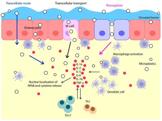
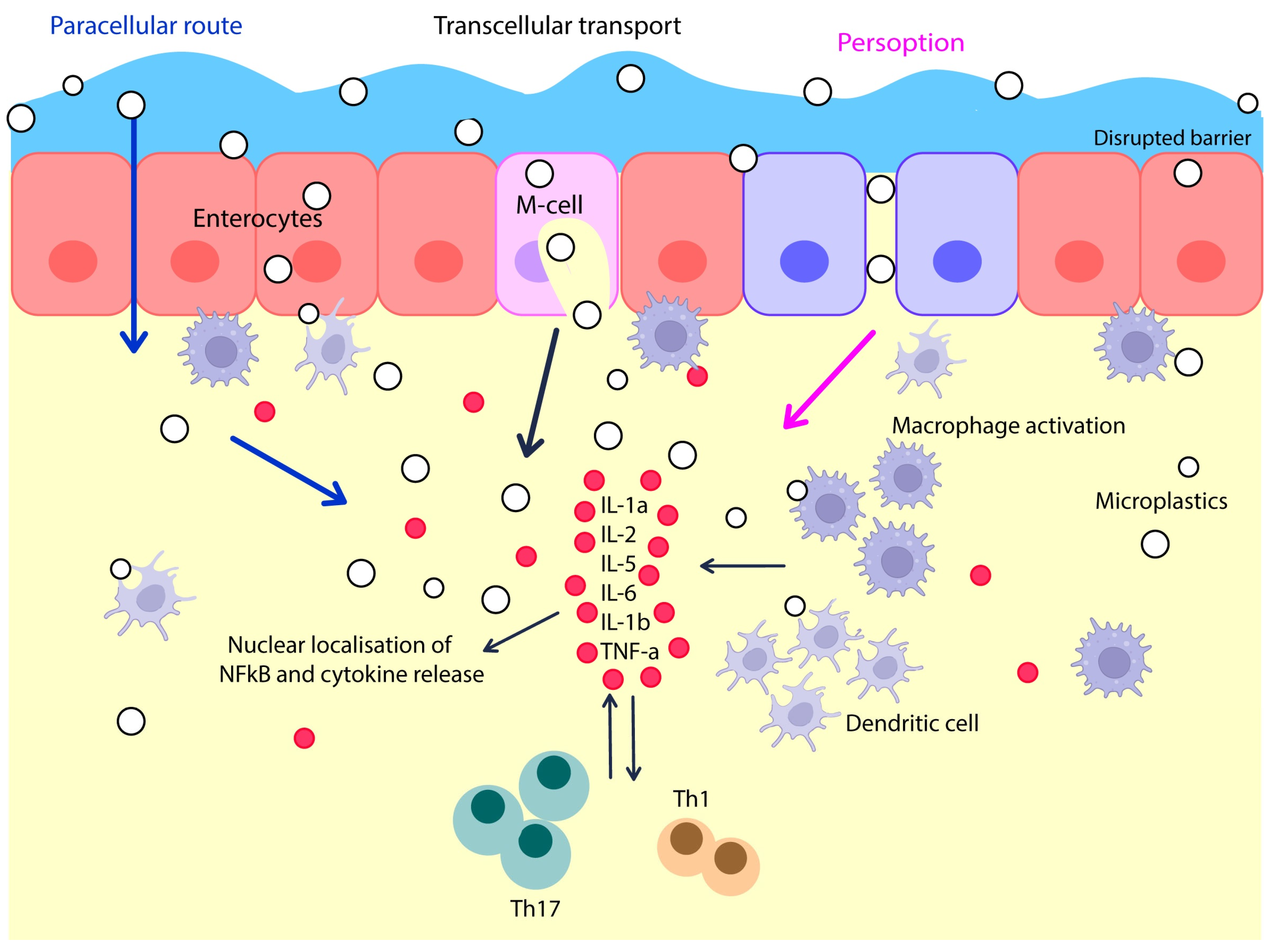
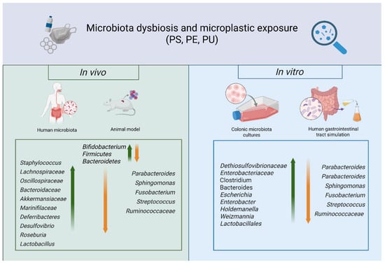
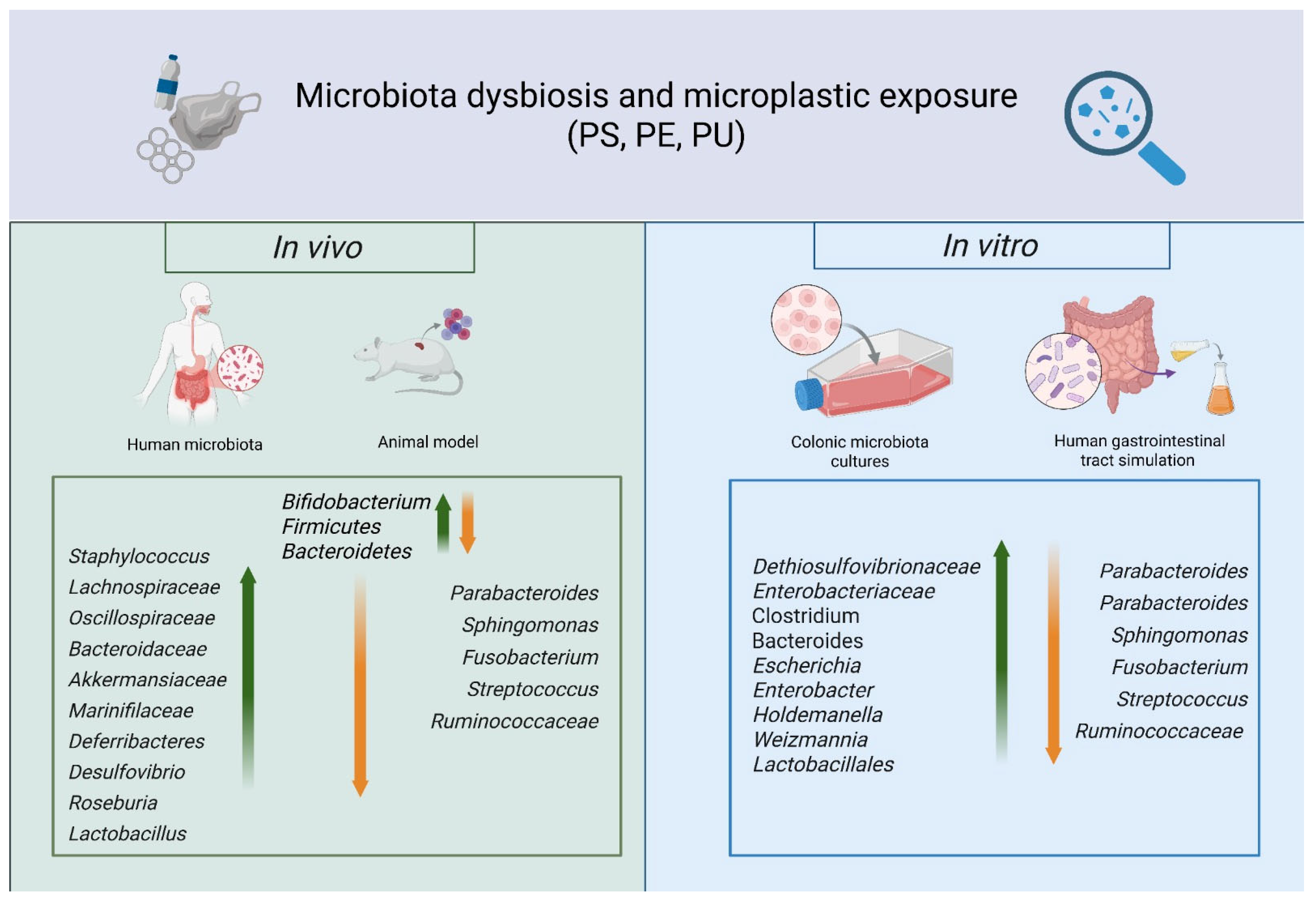
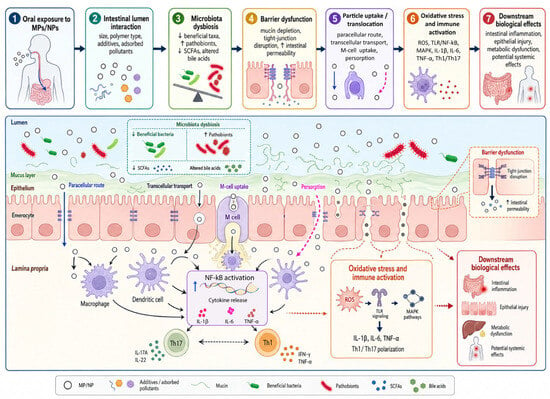
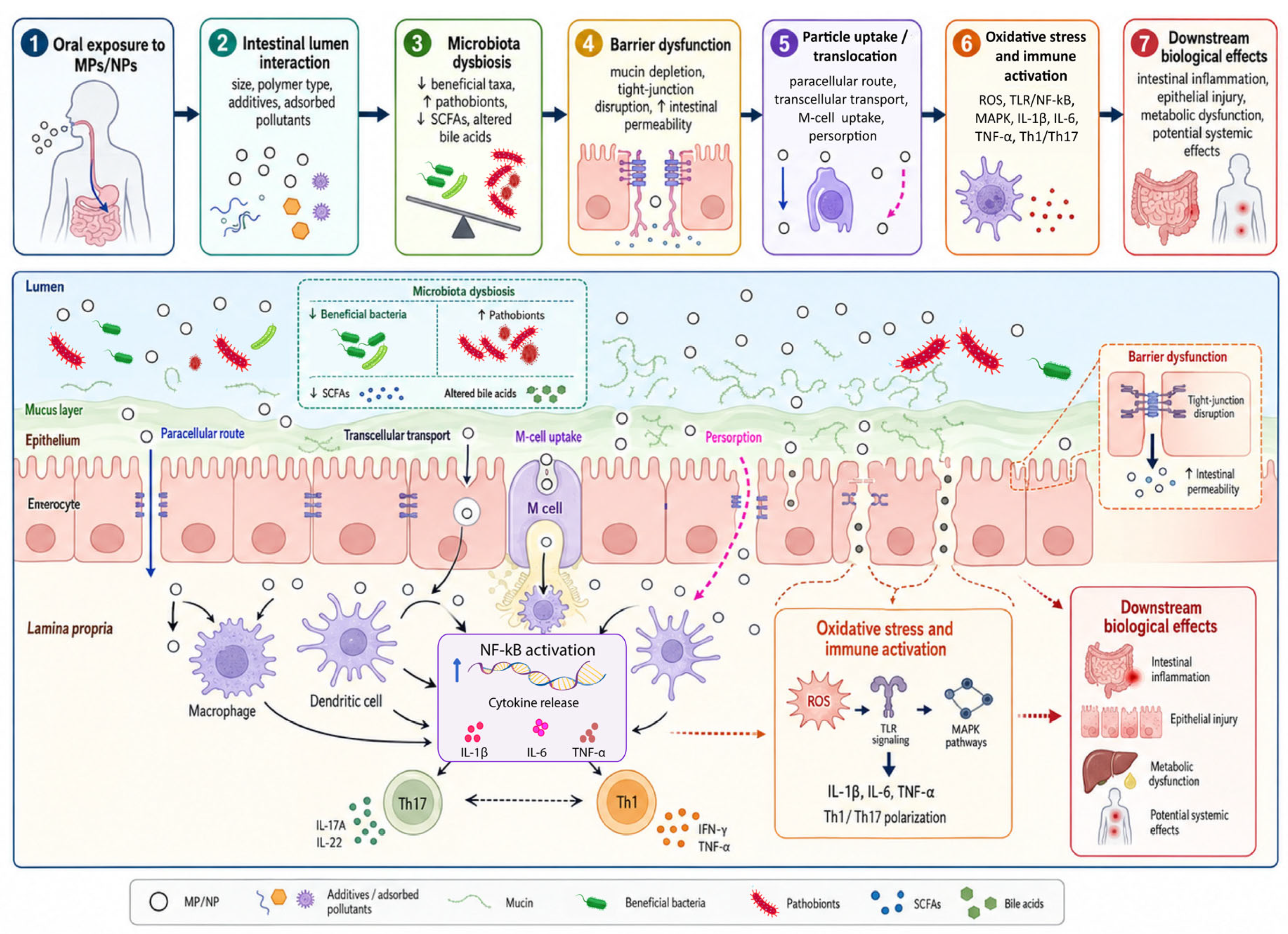
Disclaimer/Publisher’s Note: The statements, opinions and data contained in all publications are solely those of the individual author(s) and contributor(s) and not of MDPI and/or the editor(s). MDPI and/or the editor(s) disclaim responsibility for any injury to people or property resulting from any ideas, methods, instructions or products referred to in the content. |
© 2026 by the authors. Licensee MDPI, Basel, Switzerland. This article is an open access article distributed under the terms and conditions of the Creative Commons Attribution (CC BY) license.