


Abstract
Progressive familial intrahepatic cholestasis (PFIC) encompasses genetically heterogeneous disorders affecting bile transport, membrane organisation, epithelial polarity and bile-acid signalling. Current medical and surgical treatments can improve symptoms and disease course in selected patients, but substantial unmet need remains, particularly in severe early-onset disease and multisystem disorders. This review considers the thirteen numbered PFIC subtypes and selected related monogenic cholestatic conditions, relating their mechanisms to gene addition, genome editing, splice correction, messenger RNA replacement and cell-based strategies. Published PFIC intervention evidence is predominantly preclinical, with proof of concept in selected mouse models and in vitro systems. Clinical experience in other liver diseases provides relevant delivery and safety precedents but does not establish efficacy in PFIC. Platform selection will depend on molecular mechanism, target tissue, therapeutic threshold, disease stage and the durability required during liver growth. Severe disease identifies unmet need rather than a proven optimal platform. We propose a framework for research prioritisation and identify the studies needed to establish sustained benefit, genomic and immunological safety, and clinically meaningful outcomes.
1. Introduction
Bile formation depends on coordinated canalicular transport, membrane lipid composition, intracellular trafficking, tight-junction integrity and transcriptional regulation. Disruption can cause retention of bile constituents, inflammation, fibrosis and cirrhosis. PFIC comprises severe inherited cholestatic disorders that commonly present in infancy or childhood with jaundice, pruritus, malabsorption and growth failure, although age at onset and progression vary substantially [1,2,3,4,5].
The classical disorders caused by ATP8B1, ABCB11 and ABCB4 mutations have been joined by conditions affecting junctional proteins, nuclear receptors, trafficking machinery and ciliary or signalling pathways [6,7,8,9,10,11,12,13]. Gene-based nomenclature is useful because a single gene can produce a spectrum extending beyond its numbered PFIC phenotype. Here, we consider the thirteen numbered PFIC subtypes together with selected related disorders involving VIPAS39, CLDN1, UNC45A and SLC51B mutations. These additional conditions illustrate the limitations of liver-restricted treatment and the importance of identifying the molecular mechanism. The review is not intended as an exhaustive catalogue of genetic cholestasis.
UDCA, antipruritic treatment, nutritional support and surgical interruption of enterohepatic circulation can improve outcomes in selected patients [14,15,16]. IBAT inhibitors represent a major targeted pharmacological advance, with randomised evidence of improvement in pruritus and serum bile acids in defined trial populations [17,18]. Treatment response remains heterogeneous, and transplantation does not correct extrahepatic disease in disorders such as FIC1 deficiency or ARC syndrome [19]. Early HCC and poor native-liver survival in severe BSEP deficiency provide additional reasons to develop disease-corrective treatments [20,21].
Reliable population-based incidence estimates for individual PFIC genotypes remain scarce. The often-cited frequency of approximately one affected child per 50,000–100,000 births is a historical estimate rather than a robust measurement of global prevalence [5,22]. Early recognition of infantile cholestasis and timely molecular diagnosis are therefore important for both clinical care and trial development. Disease stage is likely to influence treatment response, but the fibrosis threshold beyond which gene-based treatment would be ineffective has not been established.
Several delivery systems are available for gene-based treatment of a liver disorder, and their properties determine which disease mechanisms each can address. Recombinant AAV is a small non-enveloped vector whose capsid determines tissue tropism, and AAV8 and AAV5 are the serotypes conventionally described as hepatotropic. Its genome persists mainly as episomes rather than integrating, which limits insertional risk but allows expression to be diluted as hepatocytes divide, and a packaging limit of approximately 4.7 kb constrains the transgene and regulatory sequence it can carry. Integrating lentiviral vectors accept larger cargo and are retained through cell division, while non-viral delivery of mRNA in lipid nanoparticles avoids genomic alteration but gives only transient expression. Hepatotropism, a low insertional risk relative to integrating vectors, and the clinical experience accumulated in other monogenic liver disorders account for the predominance of AAV among the published PFIC intervention studies. The disclosed industry programmes in PFIC, Vivet’s vectors targeting ABCB11 and ABCB4, are also AAV-based [23,24]. Each platform, and the evidence supporting its use, is considered with the relevant literature in Section 4.
AAV-mediated gene addition has demonstrated efficacy in selected PFIC mouse models, and sponsor-disclosed programmes remain in preclinical development [23,24]. Earlier reviews have summarised current management or the gene-therapy approaches available at the time [25,26]. This review updates that evidence and relates intervention choice to mechanism, tissue distribution and disease course. Human efficacy in other indications is distinguished from PFIC intervention studies, while proposed platform–genotype matches are identified as research hypotheses.
This is a narrative review based on targeted literature retrieval and reference checking, updated on 26 September 2026. Primary disease-gene reports, natural-history studies and intervention experiments were prioritised, with reviews used for context. ClinicalTrials.gov was searched using PFIC with gene therapy, gene transfer, ABCB11 or ABCB4, and the programme names VTX-802 and VTX-803. No interventional PFIC gene-addition or editing trial was identified in those searches. This statement is limited to the registry and search date and does not imply exhaustive coverage of all trial registries [27].
2. Molecular Pathogenesis and Therapeutic Implications
PFIC mechanisms can be considered in six overlapping groups: canalicular transport and membrane lipid organisation, tight-junction regulation, transcriptional control, intracellular trafficking and polarity, intestinal basolateral bile-acid transport, and ciliary or signalling abnormalities. Figure 1 uses these groups to organise the principal proteins and compartments. Table 1 summarises the numbered subtypes and selected related conditions. Grouping by mechanism is intended to guide research questions rather than to imply a validated hierarchy of treatment difficulty.
2.1. Classical Canalicular Transporter Defects
ATP8B1 mutations cause PFIC1, in which FIC1, a P4-ATPase phospholipid flippase involved in membrane lipid asymmetry, is deficient. Structural work supports broad glycerophospholipid substrate specificity [2,28]. FIC1 is expressed at apical membranes in several epithelial tissues, including the hepatobiliary and digestive systems [29,30]. Diarrhoea, hearing impairment, pancreatitis and poor growth distinguish its broader phenotype from liver-restricted transporter disease [19,31]. Hepatocyte-directed correction could improve the hepatic component, but would not directly correct intestinal or other extrahepatic defects. Persistent or worsened diarrhoea after liver transplantation illustrates why improvement in cholestasis does not resolve the overall disease burden [19].
ABCB11 mutations cause PFIC2, in which BSEP-mediated canalicular bile-salt export is lost or reduced [3]. Severe disease can produce early fibrosis and HCC, but genotype and residual function strongly influence the course [20,21]. In the original 264-patient NAPPED cohort, median native-liver survival was 20.4 years in BSEP1, 7.0 years in BSEP2 and 3.5 years in BSEP3 [21]. These were study-defined categories of predicted functional severity, not direct measurements of protein function in every patient.
The original BSEP1 category included patients with at least one p.E297G or p.D482G variant. Subsequent analysis showed the importance of the second allele. Patients with one of these recurrent variants and a predicted protein-truncating variant, designated BSEP1/3, had a severe course, with 10-year native-liver survival of 21%, compared with 75% in BSEP1/1 and 23% in BSEP3/3 [32]. The presence of a recurrent missense variant therefore cannot by itself establish a mild phenotype or suitability for a particular intervention. Native-liver survival should also be distinguished from overall survival.
ABCB4 mutations cause PFIC3, in which MDR3-mediated phosphatidylcholine secretion into bile is impaired [4]. Reduced biliary phospholipid content increases the injurious effects of bile acids and contributes to biliary inflammation and fibrosis. Some residual-function genotypes respond to UDCA, while genotypes causing complete loss of MDR3 function can progress rapidly [33,34]. Preclinical studies suggest that partial restoration of phospholipid secretion can confer substantial benefit [23,35]. The required extent and distribution of correction in patients remain unknown. Both ABCB4- and ABCB11-related diseases span severe and less progressive phenotypes, so urgency should follow individual disease severity rather than the gene name alone.
2.2. Tight Junction and Regulatory Defects
TJP2 mutations cause PFIC4, in which a scaffold connecting tight-junction components with the cytoskeleton is disrupted. Defective epithelial barrier integrity is thought to contribute to bile-constituent leakage and hepatocellular injury [6]. Severe early cholestasis and childhood HCC have been reported [36]. Restoring TJP2 expression must also restore its appropriate localisation and interactions. It is plausible that the spatial distribution of corrected cells will matter, but no comparative study establishes a higher therapeutic correction threshold than for transporter deficiency.
CLDN1 mutations cause NISCH syndrome, a related disorder rather than a numbered PFIC subtype. Claudin-1 is a transmembrane component of tight-junction strands, and loss of function can increase hepatic paracellular permeability [37,38]. Liver involvement ranges from absent or transient cholestasis to progressive sclerosing cholangiopathy. GGT is variable and can be elevated. Ichthyosis, hypotrichosis and other ectodermal findings support the syndromic diagnosis [39]. Liver-directed treatment would not be expected to correct the skin phenotype.
NR1H4 mutations cause PFIC5, in which FXR, a nuclear receptor central to bile-acid homeostasis, is deficient [7]. FXR regulates BSEP, MDR3 and enterohepatic signalling pathways [40,41,42]. Replacing NR1H4 itself could potentially restore several downstream functions, rather than requiring separate replacement of each regulated transporter. Expression level, physiological regulation and the relative contributions of hepatic and intestinal FXR will need to be resolved experimentally.
USP53 mutations cause PFIC7, which presents with variable low-GGT cholestasis [43]. Although historically classified as a catalytically inactive protein, USP53 has been shown to possess K63-linkage-directed deubiquitinase activity. Disease-associated variants can impair catalysis, and USP53 depletion alters the ubiquitination of tricellular junction components [44]. The mechanism should therefore not be described solely as loss of a structural scaffold. A relatively mild or intermittent course occurs in some patients, but this cannot be assumed for every genotype or used alone to defer treatment development.
2.3. Basolateral Intestinal Transport Failure
SLC51A mutations cause PFIC6, in which OSTα is deficient. OSTα forms a heteromeric transporter with OSTβ, encoded by SLC51B. The complex mediates basolateral export of reabsorbed bile acids from ileal enterocytes [45,46]. SLC51B mutations have also been described in children with congenital diarrhoea and features of cholestasis, and OSTβ deficiency is included here as a related disorder [47]. OSTα/β is not exclusively intestinal, and adaptive hepatic expression further complicates a strict division between intestinal and hepatic disease.
The combination of enteropathy and cholestasis suggests that intestinal correction may be important, but neither the necessary tissue distribution nor the therapeutic threshold has been established. Durable intestinal gene transfer would need to address rapid epithelial turnover and access to long-lived stem or progenitor compartments. These challenges differ from those of hepatocyte-directed treatment.
2.4. Intracellular Trafficking and Polarity Defects
KIF12 mutations cause PFIC8, which is linked to disturbed hepatocyte polarity and microtubule-associated trafficking [9]. MYO5B mutations cause PFIC10, in which myosin Vb, an actin-based motor involved in apical recycling, is deficient, with altered localisation of canalicular proteins [8]. Myosin Vb deficiency phenotypes range from liver-predominant cholestasis to MVID with severe intestinal involvement. Effects of particular variants and retained protein interactions may be important, so these conditions should not all be treated as equivalent examples of absent protein.
VPS33B and VIPAR, encoded by VPS33B and VIPAS39 respectively, participate in the CHEVI trafficking complex and maintenance of epithelial polarity [12,13,48]. Biallelic variants can cause multisystem ARC syndrome, with renal, musculoskeletal, skin, immunological and platelet manifestations. Attenuated VPS33B deficiency with isolated low-GGT cholestasis provides the basis for the PFIC12 phenotype [49]. An adult ARC transplant report illustrates a broader age range but should not be taken as proof of an isolated hepatic presentation [50]. ARC syndrome caused by VIPAS39 mutations is designated ARCS2 and is not assigned a PFIC number in this review.
Single-gene replacement can restore function when one component of the VPS33B–VIPAR complex is deficient. AAV and LV studies have demonstrated hepatic rescue in liver-specific Vps33b knockout models [51,52]. These findings do not show that either approach corrects multisystem ARC disease. Tissue coverage is a separate problem from vector cargo capacity or the number of proteins in the complex.
UNC45A mutations cause OOHE syndrome, with congenital diarrhoea, cholestasis, hearing impairment and bone fragility [53,54]. UNC45A acts as a myosin co-chaperone. A recent mechanistic study identified MYO5B as a client and linked an OOHE-associated UNC45A variant to defective canalicular BSEP trafficking [55]. This provides a direct connection with MYO5B-associated cholestasis, while reinforcing the need to distinguish hepatic rescue from correction of intestinal, auditory and skeletal disease.
2.5. Ciliary and Signalling Disorders
ZFYVE19 mutations cause PFIC9, which links high-GGT cholestasis, congenital hepatic fibrosis and sclerosing cholangiopathy to disturbed cellular organisation [10]. Subsequent functional work has connected ZFYVE19 deficiency with abnormal ciliary and cell-division processes [56]. PSKH1 mutations cause PFIC13, which has been reported as a hepatorenal ciliopathy supported by human genetic and experimental evidence [57]. The cell populations and level of correction required for treatment remain uncertain in both conditions.
SEMA7A mutations cause PFIC11, which should be distinguished from these ciliary disorders. The original homozygous p.R148W report proposed a gain-of-function mechanism involving increased PKC activity and reduced canalicular Bsep and Mrp2 in experimental models [11]. The index child had no reported pruritus and also carried a homozygous SLC10A1 p.S267F variant. The investigators used mouse and cell studies to examine causality, but the limited clinical series constrains generalisation. Wild-type gene addition cannot be assumed to rescue a gain-of-function mechanism. Variant correction or suppression represents a possible research direction, with no established therapeutic rescue for this disorder.
2.6. Lessons for Therapy Design
Mechanism determines whether supplementation, correction, splice modulation or suppression is appropriate [25,26]. For membrane transporters and junctional proteins, expression must occur in the relevant cells and subcellular compartments [2,3,4,37,51]. Conventional cross-correction by secretion of a functional enzyme is not available, making the corrected-cell fraction and its spatial distribution important. Comparative correction thresholds across PFIC subtypes have not been established. Multisystem disease adds a further requirement to define whether liver-only benefit is sufficient or whether several tissues must be reached [12,13,19].
Figure 1.
Molecular compartments in which PFIC and related cholestatic disease genes act, and the consequences of their disruption for bile formation. The numbered groups distinguish (1) canalicular transport and membrane lipid organisation, (2) tight-junction regulation, (3) FXR-dependent transcription, (4) trafficking and polarity, (5) intestinal basolateral bile-acid transport and (6) ciliary or signalling abnormalities. FIC1 maintains lipid asymmetry, whereas BSEP and MDR3 support bile-salt and phosphatidylcholine secretion, respectively. The junctional proteins have distinct scaffold, transmembrane and deubiquitinase functions. OSTα and OSTβ, encoded by SLC51A and SLC51B, form a heteromeric exporter. ASBT is shown as physiological context, not as a numbered PFIC gene. The cilium is drawn on a generic ciliated cell without assigning a specific hepatic cell type. The proposed gain-of-function signalling mechanism associated with SEMA7A p.R148W is shown separately. Tissue requirements for therapy are unresolved. Human gene symbols are italicised, and protein names are in roman type. Original schematic based on the cited literature; not to scale [2,3,4,6,7,8,9,10,11,12,13,28,37,44,45,46,55,56,57]. FIC1, ATP8B1-encoded phospholipid flippase; PC, phosphatidylcholine; BSEP, bile-salt export pump; FXR, farnesoid X receptor; MDR3, multidrug resistance protein 3; OST, organic solute transporter; PKC, protein kinase C; ASBT, apical sodium-dependent bile-acid transporter.
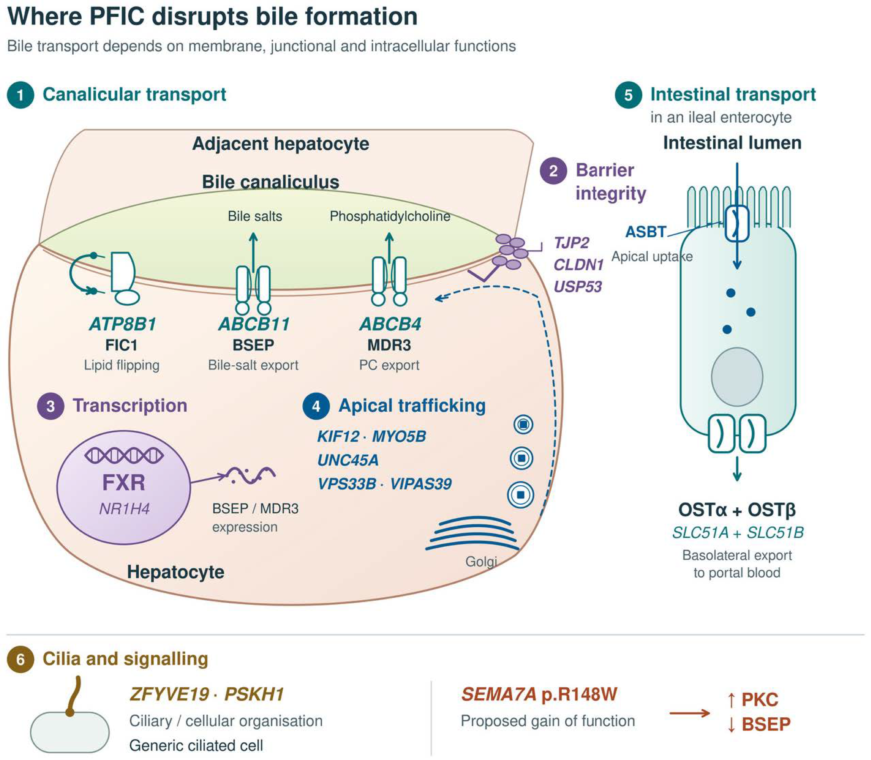
Table 1.
Thirteen numbered PFIC subtypes and four selected related monogenic cholestatic disorders.
3. Current Therapy and the Residual Unmet Need
UDCA remains useful in selected cholestatic disorders, particularly some ABCB4 genotypes with residual MDR3 function. Antipruritic treatment, nutritional support and correction of fat-soluble vitamin deficiency are central to care [14,15,16,34]. Surgical biliary diversion can improve native-liver survival in selected BSEP-deficient patients, although benefit varies by genotype and postoperative biochemical response [21,32]. These interventions can modify disease course without correcting the causal variant.
Odevixibat and maralixibat inhibit IBAT, also termed ASBT, in the terminal ileum and interrupt enterohepatic bile-acid circulation. The mechanism has preclinical support, while pivotal randomised trials demonstrate clinical benefit in defined PFIC populations [17,18,58]. Indirect comparisons have also been reported, but they should not be interpreted as head-to-head evidence [59]. Long-term outcomes and responses across the full genetic spectrum require further study [22].
The mechanism depends on sufficient delivery of bile acids to the intestine. The pivotal odevixibat trial excluded patients predicted to have a complete absence of functional BSEP, and product information states that this group will not respond [17,60]. Maralixibat product information does not recommend treatment for specific ABCB11 variants associated with non-functional or absent BSEP. Its small BSEP3 subgroup did not show improvement in pruritus [18,61]. Trial exclusion, mechanistic prediction and observed subgroup response are distinct forms of evidence and should not be conflated.
Liver transplantation replaces the affected hepatic compartment but requires lifelong immunosuppression and does not correct extrahepatic disease [19,34]. In severe BSEP deficiency, de novo alloimmune anti-BSEP responses can cause recurrent cholestasis and graft injury [62]. This raises a relevant question about immune tolerance to therapeutic BSEP after gene transfer, although transplantation experience cannot determine the incidence of transgene immunity with a particular vector.
4. Gene-Based Therapeutic Platforms
The relative merits of AAV, LV, genome editing, RNA therapeutics and corrected-cell transplantation depend on mechanism, cargo, tissue access, expression level and duration. Table 2 presents published PFIC-relevant intervention studies and separates them from historical sponsor disclosures. Figure 2 compares the platforms and identifies the evidence level supporting their application to PFIC.
4.1. AAV-Mediated Gene Addition
AAV is a clinically established platform for hepatic gene transfer in other indications, including haemophilia A and B [63,64,65,66]. Its application to PFIC is most directly supported by mouse studies of ABCB4 and ABCB11 gene addition [23,35,67]. AAV packaging capacity is approximately 4.7 kb for the complete vector genome, including inverted terminal repeats and regulatory sequences. ABCB11 and ABCB4 coding sequences leave limited space for those elements, making compact cassette design important [26,35,67].
For PFIC3, AAV-mediated ABCB4 delivery restored biliary phospholipid secretion and improved disease in several mouse experiments, with effects dependent on model, sex, age, dose and administration schedule [23,35]. In infant Abcb4-null mice, co-administration with rapamycin-containing tolerogenic nanoparticles, termed ImmTOR, enabled repeat AAV dosing and sustained correction during the reported follow-up [68]. This is a mouse redosing strategy, not an established clinical solution. A hybrid AAV–piggyBac approach achieved stable expression by deliberate genomic integration and reduced tumour development in an Abcb4-null model [69]. Its efficacy should therefore be distinguished from conventional predominantly episomal AAV, and its insertional safety assessed separately.
For PFIC2, the VTX-802 study demonstrated canalicular BSEP expression and partial phenotypic correction in Abcb11-null mice [67]. In the dose-range study, five-week-old female mice received 6 × 10¹³ to 1.8 × 10¹⁴ vector genomes/kg and were assessed 15 weeks later. Transaminases, hepatomegaly and bile-acid secretion improved, but serum bile acids and overall fibrosis were not uniformly corrected. The findings establish preclinical efficacy while leaving major questions about dose, durability and translation to the human bile-acid environment.
Recombinant AAV genomes can persist as episomal chromatin, with foundational evidence from muscle and extensive subsequent hepatic application [63,70,71]. Hepatocyte proliferation during growth or regeneration can reduce vector abundance. Whether this leads to loss of clinical benefit depends on initial transduction, expression level, turnover and the therapeutic threshold. Adult cholestatic liver disease may also involve regeneration, so the limitation is not confined to infants. Random integration is not a dependable therapeutic durability mechanism, and predominantly nonintegrating AAV still requires evaluation of genomic safety [68,69,72].
Pre-existing or treatment-induced antibodies can limit transduction or prevent effective readministration. Capsid-related immune responses and liver enzyme elevations are recognised in clinical hepatic AAV therapy [63,65]. Mouse tropism, bile-acid composition and the severity of a particular disease model may differ substantially from the human setting. These differences preclude choosing AAV solely on the basis of age or a gene fitting within the vector.
4.2. Integrating Lentiviral Vectors
Integration allows an LV transgene to be retained in daughter cells and therefore offers a rationale for sustained expression during liver growth. The extent of benefit still depends on initial transduction, expression, cell survival and safety. Preclinical hepatic LV studies have treated haemophilia B dogs, corrected haemophilia A mice and achieved normal-range factor VIII activity in non-human primates [73,74]. The primate study should be interpreted as an expression of therapeutic activity, rather than a correction of a haemophilia phenotype in affected primates.
Transient macrophage depletion with clodronate enhanced neonatal or juvenile hepatic LV transduction in mice [75]. This remains an experimental conditioning strategy with implications for infection defence, tissue homeostasis and clinical feasibility. In a liver-specific Vps33b knockout model, neonatal LV administration with transient macrophage depletion improved survival, growth, biochemical function and canalicular architecture under a cholic-acid challenge [52]. The model provides direct cholestasis efficacy evidence but does not demonstrate correction of renal, platelet or musculoskeletal ARC manifestations.
LV cargo capacity can accommodate constructs larger than those suited to a conventional AAV cassette, although payload size affects vector production and titre. Additional coding capacity does not itself provide multisystem delivery. Hepatic programmes must address scalable manufacturing, dose, biodistribution, off-target transduction and the clinical acceptability of any conditioning regimen [73,74,75].
Genotoxicity remains a central concern. Historical gamma-retroviral HSC experience, experimental comparisons of vector designs and more recent LV-associated haematological cancers show why insertional effects cannot be dismissed [76,77,78,79]. Risks observed with an HSC product cannot be transferred quantitatively to hepatocytes, but they support product-specific assessment rather than a class-wide assumption of safety.
In the Vps33b liver-model safety experiment, five animals per vector group were followed for 36 weeks. Liver tumours occurred in two of five EF1-VPS and three of five EF1-GFP recipients. The combined tumour set included adenomas and HCCs, whereas none were observed with the tested LP1 constructs [52]. The findings support a role for promoter and vector design in that model. They do not prove absence of insertional risk or establish promoter choice as the sole determinant of long-term oncological outcome. Preclinical genotoxicity testing, product release controls and post-treatment monitoring should be considered separately. Integration-site clonality in a patient’s liver cannot be a pre-dose manufacturing release criterion, and serial liver-wide assessment presents substantial sampling limitations.
4.3. Genome Editing
Base and prime editing offer routes to correct selected variants at their endogenous loci, potentially preserving physiological regulation. Cytosine and adenine base editors mediate specified transition changes, while prime editing can in principle install a broader range of substitutions and small insertions or deletions. Eligibility depends on the exact sequence, editor, target-access requirements, efficiency and unintended-edit profile [80,81]. These editors derive from CRISPR–Cas systems, in which a Cas protein whose nuclease activity is disabled or limited to a single-strand nick is directed to a chosen sequence and fused to a deaminase or a reverse transcriptase, so that correction does not require a double-strand break. Conventional homology-directed correction following a nuclease-induced double-strand break uses a donor DNA template to introduce the intended change. In our view the efficiency currently achievable in the liver, and the risk of unintended rearrangement at the cut site, make it a less promising route for PFIC than the editors above, and we do not consider it further. Zinc-finger nucleases and TALENs preceded CRISPR-based editing and have been largely superseded by it in liver-directed research, and are therefore also outside the scope of this review.
The recurrent ABCB11 p.E297G and p.D482G variants illustrate possible research targets but are not validated PFIC editing candidates [21,32]. Their coding changes, c.890A>G and c.1445A>G, respectively, require G-to-A repair on the coding strand. Cytosine editing of the opposite strand could in principle achieve this, depending on the available target site and bystander constraints. A specific therapeutic design requires transcript and sequence validation and demonstration of restored BSEP function.
Delivery requirements differ between viral systems, editor proteins or ribonucleoproteins, and LNP-delivered editor mRNA with guide RNA. Guide-RNA chemical modifications can improve stability and editing activity, but do not solve every co-delivery problem [82]. The individualised in vivo base-editing treatment of an infant with CPS1 deficiency provides an important clinical liver-delivery precedent, with early follow-up in one patient [83]. It does not establish PFIC efficacy, lifetime durability or general applicability across target variants. Relevant risks include off-target and bystander editing, unintended on-target changes, immunogenicity and the consequences of long-lived edited-cell populations [80,81]. Genome editing also includes targeted gene insertion, so the absence of a transgene-integration requirement applies specifically to suitable endogenous correction strategies.
4.4. Antisense Oligonucleotides and Splice Correction
Splice-switching ASOs are relevant to selected variants that produce a correctable RNA-processing defect. ASOs have other mechanisms as well, so the mutation-specific scope described here should not be applied to the entire class [84]. Evidence across PFIC genes is at different stages. An ABCB4 study predicted effects of synonymous variants computationally, whereas ATP8B1 splice rescue was demonstrated using adapted U1 small nuclear RNA, a distinct intervention [85,86].
For ABCB11, the intronic c.76+29T>G variant caused the retention of an additional sequence in a minigene assay, and an ASO restored the splicing pattern. The index child also carried c.390-2A>G, which caused exon skipping, but the reported ASO rescue concerned c.76+29T>G [87]. No patient administration was reported. Endogenous transcript correction, functional rescue in relevant cells and effective delivery remain necessary steps before clinical translation.
GalNAc conjugation provides a well-established principle for hepatocyte-directed oligonucleotide delivery, with supporting work in both siRNA and gapmer ASOs [88,89]. These results cannot directly establish the potency or nuclear delivery of a PFIC splice-switching construct. Nusinersen provides a clinical splice modulation precedent in another organ system, and other neurological and ocular studies show the range of experimental and early clinical applications [90,91,92,93]. Lumasiran and its preclinical programme provide additional hepatic oligonucleotide experience through RNA interference, not ASO-mediated splice correction [94,95].
Repeated treatment may permit dose adjustment or discontinuation, but entails delivery, monitoring and access burdens. Dosing in a growing child will need empirical pharmacokinetic and pharmacodynamic assessment rather than an assumed linear relationship with age or weight. Genotype-specific eligibility, retained correctable transcript and the functional consequence of the restored RNA are central to patient selection.
4.5. mRNA Replacement and LNP Delivery
ABCB4 mRNA–LNP administration restored functional protein expression and biliary phospholipid secretion in a severe BALB/c.Abcb4-null model. Five doses over two weeks improved inflammation, ductular reaction and fibrosis in young mice [96]. This establishes repeated-dose preclinical efficacy, while leaving the optimal maintenance schedule and long-term treatment burden unresolved.
mRNA replacement avoids a requirement for permanent genomic alteration and can be adjusted through repeat dosing. However, transient expression does not establish intrinsically low toxicity or uncomplicated redosing. Cumulative exposure, infusion reactions, innate immune activation, formulation tolerability and sustained clinical benefit require evaluation [97,98]. Depending on these results, mRNA could serve as a bridge or as continuing replacement therapy rather than necessarily preceding a permanent intervention.
LNP composition can influence tissue delivery and intracellular processing. Siloxane-containing formulations demonstrate that distinct designs can alter delivery to tissues including liver, spleen and lung [99]. Such findings do not establish a single formulation capable of correcting all affected compartments in multisystem PFIC-related disease. Cell-type specificity and functional expression at the correct membrane remain essential.
4.6. Cell-Based Approaches
Ex vivo correction of expanded patient-derived primary hepatocytes offers the possibility of characterising a cell product before transplantation. CRISPR-mediated correction and transplantation have been studied in other inherited liver diseases [100]. Engraftment, expansion, product consistency and the replacement fraction required for benefit remain major challenges [101].
Selective advantage is highly dependent on disease and model. FAH-based selection can produce substantial repopulation experimentally, but cannot be assumed to translate to PFIC [102]. Conversely, the absence of selective advantage should not be asserted for every cholestatic model. In Gunn rats, normal transplanted hepatocytes reduced hyperbilirubinaemia after preparative irradiation and partial hepatectomy [103]. This was neither gene-edited-cell treatment nor a clinical PFIC conditioning study. Any conditioning strategy must be evaluated against its own risks, particularly in young children with liver disease.
Figure 2.
Candidate gene-based platforms and the evidence supporting their application to PFIC. Evidence labels refer to PFIC-relevant intervention studies, not the maturity of a platform in other indications. The rows are grouped by the route taken to restore function rather than by molecular class, so they are not mutually exclusive categories. AAV and LV are viral-derived delivery vehicles, and lipid nanoparticles are a non-viral one; antisense oligonucleotides and messenger RNA are therapeutic cargo rather than vehicles; genome editing is a mode of correction that either type of vehicle can deliver; and corrected-cell transplantation delivers cells rather than nucleic acid. The rows illustrate routes under investigation for PFIC and are not an exhaustive list. AAV and LV pathways illustrate gene addition; hybrid integrating AAV–piggyBac is discussed separately in the text. AAV capacity is approximately 4.7 kb for the complete vector genome, including regulatory sequences and inverted terminal repeats. Durability depends on adequate functional correction, cell survival and disease context; no platform has established lifetime PFIC efficacy. Editing feasibility and splice correction depend on the exact variant. Repeated mRNA replacement could provide continuing treatment if benefit and treatment burden are acceptable. The cell-transplantation pathway refers to expanded primary hepatocytes and extrapolation from other liver diseases. These are research options, not clinical prescriptions by age or genotype. The editing carrier is neutral because viral and non-viral delivery are possible. Molecular symbols are schematic. Original schematic based on published studies and mechanistic reviews [23,35,51,52,67,80,81,87,96,100,101]. AAV, adeno-associated virus; LV, lentiviral vector; ASO, antisense oligonucleotide; LNP, lipid nanoparticle; mRNA, messenger RNA.
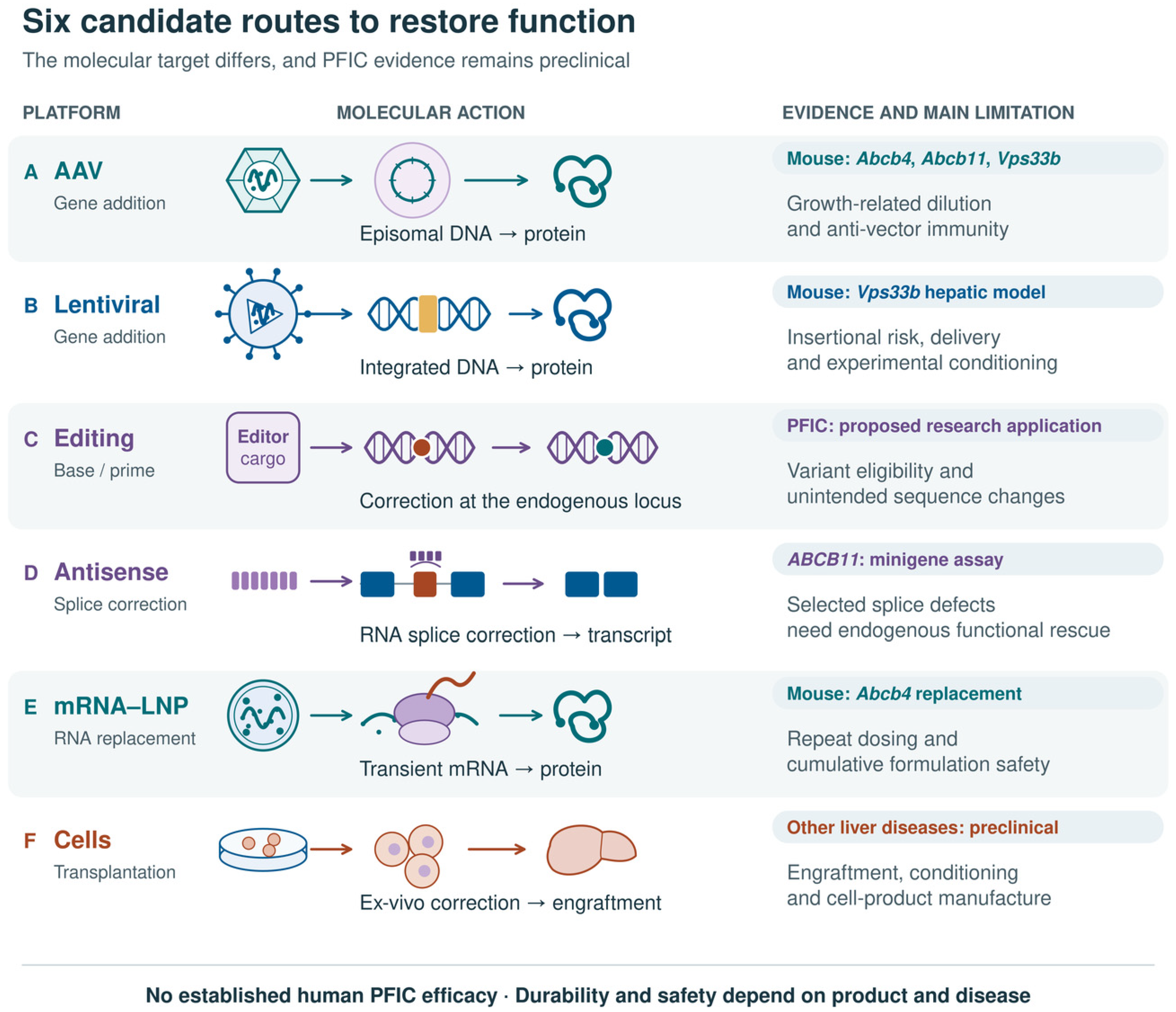
Table 2.
Published preclinical intervention studies relevant to PFIC. Different experiments are listed separately; rows are not independent commercial programmes.
5. Clinical Priorities and a Genotype-Guided Research Framework
Severe early-onset disease, limited benefit from existing treatment and poor native-liver survival identify major unmet need. These features make severe BSEP deficiency, TJP2 deficiency, FXR deficiency and selected phenotypes caused by VPS33B and VIPAS39 mutations compelling research priorities. They do not establish which group will benefit most from an untested intervention. Advanced disease, cancer risk, immune tolerance, tissue access and delivery efficiency may complicate the very conditions in greatest need.
Within BSEP deficiency, both the original NAPPED analysis and its subsequent refinement should inform research stratification [21,32]. Figure 3 presents their distinct cohort-level outcomes without converting them into a clinical gene-therapy algorithm. In particular, the severe course of BSEP1/3 disease cautions against assigning low priority on the basis of one recurrent missense allele. Current phenotype and response to treatment remain important alongside genotype.
Figure 3.
ABCB11 genotype evidence and a framework for research stratification. (A) Median native-liver survival in the original 264-patient NAPPED cohort: BSEP1, 20.4 years (n = 72); BSEP2, 7.0 years (n = 136); BSEP3, 3.5 years (n = 56). Original BSEP1 included at least one p.E297G or p.D482G variant; BSEP2 comprised other missense-containing genotypes outside BSEP1; BSEP3 comprised genotypes predicted to produce no functional BSEP [21]. (B) A subsequent refined analysis reported 10-year native-liver survival of 75% in BSEP1/1 (n = 31), 21% in BSEP1/3 (n = 30) and 23% in BSEP3/3 (n = 77). BSEP1/1 denotes homozygosity for p.E297G or p.D482G, BSEP1/3 combines one recurrent variant with a predicted protein-truncating variant, and BSEP3/3 denotes two predicted protein-truncating variants [32]. Panels (A,B) use different group definitions and endpoints; they are separate analyses, not independent treatment-comparison cohorts. Bars reproduce published point estimates without adding unreported uncertainty intervals. (C) Proposed research considerations combine complete genotype with disease burden, stage, tissue involvement and intervention risks. The figure is neither an individual prognostic calculator nor a validated clinical gene-therapy algorithm. BSEP, bile-salt export pump; NAPPED, Natural course and Prognosis of PFIC and the effect of biliary Diversion.
PFIC3 is comparatively well represented in preclinical intervention studies, but this does not make every patient a lower clinical priority or justify risk solely for platform development [23,35,68,69,96]. Some residual-function phenotypes may remain well controlled with conventional treatment, while severe MDR3 deficiency has substantial unmet need. Similar caution applies to the variable course of cholestasis caused by USP53 mutations.
Platform selection should therefore be a conditional research decision. Gene addition may suit loss-of-function disorders if the appropriate cells can be reached. Editing and splice modulation depend on variant-specific feasibility. Suppression may be relevant to gain-of-function mechanisms. Repeated RNA replacement may offer sustained benefit if its clinical burden is acceptable. Integration provides a durability rationale but not a universal requirement for infants, and no direct comparison establishes age-based superiority of AAV, LV or editing across PFIC.
6. Translational Challenges and Future Directions
Durability must be studied in the context of the growing or regenerating liver. Comparative studies in relevant disease models should measure vector or edit persistence, functional protein localisation and the threshold for continued benefit. Large-animal studies may inform delivery and scaling, but will not reproduce every human genotype or bile-acid environment. Apparent success in a short mouse experiment should not be equated with correction throughout childhood.
Safety assessment must address both platform and disease. AAV requires attention to immunity, hepatic toxicity, readministration and potential integration. LV and integrating hybrid systems require product-specific assessment of insertional effects and clonal selection. Editing requires the evaluation of intended and unintended sequence changes [52,63,72,80,81]. Long-term follow-up is particularly important in children and in disorders with an intrinsic malignancy risk. Biochemical normalisation has not been shown to eliminate human PFIC-associated HCC risk, so appropriate imaging-based surveillance should continue. AFP may contribute to surveillance but should not be its sole component.
Susceptibility to hepatotoxicity may itself be disease-specific, which has direct relevance to PFIC. Safety data generated in structurally normal livers cannot be assumed to transfer to an organ that is already cholestatic, fibrotic and under regenerative stress. X-linked myotubular myopathy provides the closest available illustration. In a zebrafish model, loss of mtm1 impairs bile flow and reduces canalicular Bsep and Mdr1 protein without a corresponding reduction in transcript, together with dispersal of Rab11-positive recycling endosomes, indicating an intrinsic trafficking-related cholestatic phenotype rather than incidental liver involvement [105]. This parallels the trafficking and polarity defects considered earlier in this review, in which canalicular transporter delivery rather than transporter synthesis is the primary abnormality, as in disease caused by VPS33B and VIPAS39 mutations. In the ASPIRO trial of AAV8-mediated MTM1 replacement at doses of 1.3 × 10¹⁴ and 3.5 × 10¹⁴ vg/kg, four of 24 dosed boys died, all with cholestatic liver failure at the time of death and with evidence of cholestasis that pre-dated dosing, and the investigators concluded that the trial had revealed a previously unrecognised proclivity for cholestatic liver disease in this disorder. One death in the untreated control group was attributed to haemorrhage presumed to be related to hepatic peliosis, indicating that the hepatobiliary phenotype can arise without vector exposure [106]. A controlled comparison points the same way: chimpanzees given portal-vein injection of a recombinant AAV2 vector showed substantially greater rises in aspartate aminotransferase, alanine aminotransferase and creatine phosphokinase when infected with hepatitis C virus than did uninfected controls, which the authors interpreted as increased susceptibility to subclinical liver toxicity in the presence of infection. Enzyme elevations in that study followed pre-treatment liver biopsy as well as vector injection, and the respective contributions of vector and infusion procedure were not separated [107].
The mechanisms of vector-associated injury are varied and are not limited to direct hepatocellular toxicity. Innate sensing of capsid and vector genomes can provoke a systemic inflammatory response, and reported manifestations include thrombotic microangiopathy, in which complement activation is accompanied by platelet aggregation and endothelial activation and injury, and haemophagocytic lymphohistiocytosis. Capillary leak syndrome has also been reported after gene therapy, although the cases described followed a complement C3 inhibitor given as prophylaxis against thrombotic microangiopathy and the mechanism remains uncertain. Adaptive responses include capsid-directed cytotoxic T-cell responses against transduced hepatocytes, typically presenting as delayed transaminitis, and humoral responses that generate neutralising antibodies and preclude readministration [63,108]. Each has a plausible interaction with the PFIC liver. Complement and endothelial activation would occur in an organ whose sinusoidal architecture may already be altered by fibrosis; cytotoxic T-cell responses would remove the corrected hepatocytes on which benefit depends; and an antibody response forecloses repeat dosing in a child whose transgene copy number will fall as the liver grows. Comparable questions apply to non-viral platforms, since lipid nanoparticle components and oligonucleotide chemistries carry their own hepatic and innate immune liabilities. Risk–benefit assessment for PFIC should therefore be made disease-specifically rather than extrapolated from programmes conducted in structurally normal livers.
Integration warrants brief mention alongside these mechanisms. Recombinant AAV persists predominantly as episomes, but a fraction integrates. In neonatal mice, hepatocellular carcinoma was dose-dependent and was eliminated by changing the enhancer–promoter of the vector rather than the transgene carried. The chicken beta-actin and thyroxine-binding globulin enhancer–promoter elements were associated with tumours, whereas no tumours were observed in the ten animals given a human alpha-1 antitrypsin promoter [72,109]. The first human case has now been reported: a neuroepithelial tumour diagnosed four years after intracisternal magna delivery of an AAV9 vector to a child treated at 13 months of age for mucopolysaccharidosis type I, in which rearranged vector sequences carrying a duplicated cytomegalovirus enhancer and chicken beta-actin promoter had integrated into intron 4 of PLAG1 and generated a chimeric AAV-PLAG1 transcript [110]. The authors set this against more than 6000 patients treated without evidence of major long-term toxic effects, note that the risk may be modulated by vector design, dose, route of administration and the proliferative state of the tissue, identify host factors as a possible further contributor, and distinguish this configuration from conditions treatable by low-dose systemic delivery with tissue-specific promoters. A hepatic PFIC programme using a liver-specific promoter shares that one design feature, although that does not by itself establish that it meets the low-dose condition, and the preclinical data indicate that liver specificity alone is not sufficient reassurance. Promoter choice and dose should therefore be treated as safety parameters alongside expression, and imaging-based hepatic surveillance should continue in a disorder that already carries an intrinsic carcinoma risk.
Endpoints should reflect the phenotype and treatment mechanism. Serum bile acids, bilirubin, synthetic function, growth, validated pruritus measures and quality of life can provide complementary information. GGT is not a useful universal primary response marker in low-GGT PFIC. Biliary composition, histology and detailed protein localisation may support mechanistic studies but can require invasive sampling. Durable outcomes such as native-liver survival require longer follow-up and well-characterised natural-history comparators [21,22,32,34]. Vector copy number or integration-site analyses apply to particular platforms and must be interpreted in light of tissue access and sampling limitations.
International collaboration is necessary because individual genotypes are rare and heterogeneous. Shared registries and harmonised outcomes could support subtype-stratified protocols and carefully justified adaptive designs. Pooling genotypes should depend on a defensible common mechanism and outcome definition, rather than an assumption that all PFIC is biologically interchangeable. Early engagement with families is also needed to assess acceptable uncertainty, treatment burden and meaningful benefit.
Regulatory incentives should be distinguished from expedited-development pathways. Orphan designation provides specified incentives and is separate from marketing approval. Rare-paediatric-disease designation may support eligibility for a priority-review voucher after approval under the applicable programme criteria, but does not itself accelerate review of the designated therapy. Fast Track, Breakthrough Therapy and other expedited routes have separate requirements [111,112,113]. Access also depends on manufacturing feasibility, price and confidence in durable benefit. The health-technology assessment of haemophilia gene therapy illustrates how uncertainty in long-term outcomes affects economic evaluation [114]. PFIC programmes should address these questions alongside natural history, biomarkers and trial design, without assuming that a successful efficacy study establishes cost-effectiveness.
Model limitations are real and are acknowledged above, but we do not regard them as the principal reason that no PFIC gene-addition or editing trial was identified in the registry searches described in Section 1. Models of several subtypes exist and have supported the intervention studies summarised in Table 2. Three other constraints appear to us to weigh more heavily. That ordering is our judgement rather than a comparative measurement. Rare disorders attract limited commercial investment, and PFIC is subdivided into genotypes that are individually rarer still, so the case for any single programme is weak. The liver of a child with early-onset disease is also a difficult target, because it increases several-fold in mass during the period when treatment would be given, so an episomal vector is unlikely to provide a lifelong solution, while progressive fibrosis makes efficient hepatocyte delivery harder as disease advances. The platforms best matched to these constraints, integrating vectors and endogenous-locus editing, remain in development for liver-directed use in young children. Better models would help, but we expect these factors to continue to set the pace.
These considerations can be stated as what a gene-based therapy would have to achieve in PFIC. In our view, four conditions must be met, with a fifth that would substantially lower the threshold for the others if it proves to hold. First, the correction must persist for life, since the infant liver treated will increase several-fold in mass, and an episomal genome that does not replicate with the hepatocyte will be diluted. Second, a sufficient proportion of the cells that express the affected gene must be corrected, and the restored protein must reach the compartment in which it functions. For canalicular transporters that means the apical membrane, for trafficking and polarity disorders it extends to the delivery machinery itself, for a nuclear receptor such as FXR it means the nucleus, with the relative contributions of hepatic and intestinal expression still to be resolved, and for intestinal basolateral bile-acid transport the relevant cells are not hepatocytes at all. Third, intervention must precede the point at which fibrosis becomes irreversible, which is undefined for every PFIC subtype, so early genetic diagnosis matters as much as vector properties. Fourth, selection should follow genotype and residual function rather than diagnostic label, since the ABCB11 data in Figure 3 show that the second allele materially alters native-liver survival. The fifth, desirable but not essential, is that the corrected cells would ideally hold a selective advantage, since hepatocytes that regain bile-acid export may tolerate the prevailing bile-acid environment better than their uncorrected neighbours, which would allow a modest corrected fraction to expand. Whether this occurs is unresolved, and it is among the more consequential open questions, because a platform able to rely on it would clear a substantially lower initial threshold.
No platform currently satisfies these requirements. AAV-mediated gene addition has the strongest preclinical record in PFIC models but is the most exposed to dilution in the growing liver. Integrating vectors and endogenous-locus editing address durability directly, at the cost of risks that require long-term follow-up in children. RNA replacement avoids both genomic risks but commits the patient to indefinite repeated dosing. Corrected-cell transplantation remains limited by engraftment efficiency and manufacturing capability. Our expectation is that the first durable clinical benefit in PFIC will come from a platform achieving stable correction in a self-renewing hepatocyte population, applied before fibrosis becomes irreversible and in a genotype-selected group, rather than from improvements in transduction efficiency alone.
7. Conclusions
PFIC is a group of molecularly distinct disorders in which mechanism, tissue involvement and disease stage have direct implications for therapeutic development. Gene-based interventions have established proof of concept in selected models, particularly for hepatic disease caused by ABCB4, ABCB11 and VPS33B mutations. These findings justify further development but do not yet establish human PFIC efficacy or an optimal platform for a given age or genotype.
Progress will require demonstration that sufficient functional correction can be achieved in the appropriate cells, maintained during growth and regeneration, and delivered with an acceptable long-term risk–benefit balance. Gene addition, editing, splice modulation, RNA replacement and corrected-cell transplantation should be assessed against these requirements. Severe genotypes identify unmet need, while the choice and timing of intervention must remain grounded in disease-specific evidence.
Author Contributions
Conceptualisation, P.G., C.A.C. and Z.J.; investigation (literature review), Z.J.; writing—original draft preparation, Z.J.; writing—review and editing, Z.J., S.T.R., P.G. and C.A.C.; visualisation, Z.J.; supervision, S.T.R., P.G. and C.A.C. All authors have read and agreed to the published version of the manuscript.
Funding
S.T.R. is supported by the Medical Research Council (UKRI1972; MR/Z504427/1), the Rosetrees Trust (PGL24/100047) and the Alpha-1 Foundation (PB1089). P.G. and C.A.C. are supported by LifeArc (grant 586434) and Action Medical Research (GN2983). Z.J. is supported by a Translational Research (Non-Clinical) PhD Studentship from the National Institute for Health and Care Research (NIHR) Great Ormond Street Hospital Biomedical Research Centre and by an International Scholar Award for Doctoral Training from University College London. The views expressed are those of the authors and not necessarily those of the NHS, the NIHR or the Department of Health and Social Care.
Institutional Review Board Statement
Not applicable.
Informed Consent Statement
Not applicable.
Data Availability Statement
No new experimental datasets were generated. The published sources underlying this review are cited in the reference list.
Acknowledgments
During the preparation of this manuscript, the authors used ChatGPT (OpenAI, GPT-6.1 Sol) and Claude (Anthropic, Claude Opus 5 and Claude Fable 5.1) for manuscript editing, reference verification and figure preparation. The authors have reviewed and edited the output and take full responsibility for the content of this publication.
Conflicts of Interest
The authors declare no conflicts of interest. The funders had no role in the preparation of this review or in the decision to submit it for publication.
Abbreviations
AAV, adeno-associated virus. ARC, arthrogryposis–renal dysfunction–cholestasis. ASBT, apical sodium-dependent bile-acid transporter. ASO, antisense oligonucleotide. BSEP, bile-salt export pump. FXR, farnesoid X receptor. GalNAc, N-acetylgalactosamine. GGT, gamma-glutamyl transferase. HCC, hepatocellular carcinoma. IBAT, ileal bile-acid transporter. LNP, lipid nanoparticle. LV, lentiviral vector. MDR3, multidrug resistance protein 3. MVID, microvillus inclusion disease. NAPPED, Natural course and Prognosis of PFIC and the effect of biliary Diversion. NISCH, neonatal ichthyosis–sclerosing cholangitis. OOHE, osteo-oto-hepato-enteric. OST, organic solute transporter. PFIC, progressive familial intrahepatic cholestasis. UDCA, ursodeoxycholic acid. VIPAR, VPS33B-interacting protein, apical–basolateral polarity regulator.
References
- Clayton, R.J.; Iber, F.L.; Ruebner, B.H.; McKusick, V.A. Byler disease. Fatal familial intrahepatic cholestasis in an Amish kindred. Am. J. Dis. Child. 1969, 117, 112–124. [Google Scholar]
- Bull, L.N.; van Eijk, M.J.; Pawlikowska, L.; DeYoung, J.A.; Juijn, J.A.; Liao, M.; Klomp, L.W.; Lomri, N.; Berger, R.; Scharschmidt, B.F.; et al. A gene encoding a P-type ATPase mutated in two forms of hereditary cholestasis. Nat. Genet. 1998, 18, 219–224. [Google Scholar] [CrossRef] [Scilit]
- Strautnieks, S.S.; Bull, L.N.; Knisely, A.S.; Kocoshis, S.A.; Dahl, N.; Arnell, H.; Sokal, E.; Dahan, K.; Childs, S.; Ling, V.; et al. A gene encoding a liver-specific ABC transporter is mutated in progressive familial intrahepatic cholestasis. Nat. Genet. 1998, 20, 233–238. [Google Scholar] [CrossRef] [Scilit]
- de Vree, J.M.; Jacquemin, E.; Sturm, E.; Cresteil, D.; Bosma, P.J.; Aten, J.; Deleuze, J.F.; Desrochers, M.; Burdelski, M.; Bernard, O.; et al. Mutations in the MDR3 gene cause progressive familial intrahepatic cholestasis. Proc. Natl. Acad. Sci. USA 1998, 95, 282–287. [Google Scholar] [CrossRef] [Scilit]
- Baker, A.; Kerkar, N.; Todorova, L.; Kamath, B.M.; Houwen, R.H.J. Systematic review of progressive familial intrahepatic cholestasis. Clin. Res. Hepatol. Gastroenterol. 2019, 43, 20–36. [Google Scholar] [CrossRef] [Scilit]
- Sambrotta, M.; Strautnieks, S.; Papouli, E.; Rushton, P.; Clark, B.E.; Parry, D.A.; Logan, C.V.; Newbury, L.J.; Kamath, B.M.; Ling, S.; et al. Mutations in TJP2 cause progressive cholestatic liver disease. Nat. Genet. 2014, 46, 326–328. [Google Scholar] [CrossRef] [Scilit]
- Gomez-Ospina, N.; Potter, C.J.; Xiao, R.; Manickam, K.; Kim, M.S.; Kim, K.H.; Shneider, B.L.; Picarsic, J.L.; Jacobson, T.A.; Zhang, J.; et al. Mutations in the nuclear bile acid receptor FXR cause progressive familial intrahepatic cholestasis. Nat. Commun. 2016, 7, 10713. [Google Scholar] [CrossRef] [Scilit]
- Gonzales, E.; Taylor, S.A.; Davit-Spraul, A.; Thébaut, A.; Thomassin, N.; Guettier, C.; Whitington, P.F.; Jacquemin, E. MYO5B mutations cause cholestasis with normal serum gamma-glutamyl transferase activity in children without microvillous inclusion disease. Hepatology 2017, 65, 164–173. [Google Scholar] [CrossRef] [Scilit]
- Stalke, A.; Sgodda, M.; Cantz, T.; Skawran, B.; Lainka, E.; Hartleben, B.; Baumann, U.; Pfister, E.D. KIF12 Variants and Disturbed Hepatocyte Polarity in Children with a Phenotypic Spectrum of Cholestatic Liver Disease. J. Pediatr. 2022, 240, 284–291.e9. [Google Scholar] [CrossRef] [Scilit]
- Luan, W.; Hao, C.Z.; Li, J.Q.; Wei, Q.; Gong, J.Y.; Qiu, Y.L.; Lu, Y.; Shen, C.H.; Xia, Q.; Xie, X.B.; et al. Biallelic loss-of-function ZFYVE19 mutations are associated with congenital hepatic fibrosis, sclerosing cholangiopathy and high-GGT cholestasis. J. Med. Genet. 2021, 58, 514–525. [Google Scholar] [CrossRef] [Scilit]
- Pan, Q.; Luo, G.; Qu, J.; Chen, S.; Zhang, X.; Zhao, N.; Ding, J.; Yang, H.; Li, M.; Li, L.; et al. A homozygous R148W mutation in Semaphorin 7A causes progressive familial intrahepatic cholestasis. EMBO Mol. Med. 2021, 13, e14563. [Google Scholar] [CrossRef] [Scilit]
- Gissen, P.; Johnson, C.A.; Morgan, N.V.; Stapelbroek, J.M.; Forshew, T.; Cooper, W.N.; McKiernan, P.J.; Klomp, L.W.; Morris, A.A.; Wraith, J.E.; et al. Mutations in VPS33B, encoding a regulator of SNARE-dependent membrane fusion, cause arthrogryposis-renal dysfunction-cholestasis (ARC) syndrome. Nat. Genet. 2004, 36, 400–404. [Google Scholar] [CrossRef] [Scilit]
- Cullinane, A.R.; Straatman-Iwanowska, A.; Zaucker, A.; Wakabayashi, Y.; Bruce, C.K.; Luo, G.; Rahman, F.; Gürakan, F.; Utine, E.; Ozkan, T.B.; et al. Mutations in VIPAR cause an arthrogryposis, renal dysfunction and cholestasis syndrome phenotype with defects in epithelial polarization. Nat. Genet. 2010, 42, 303–312. [Google Scholar] [CrossRef] [Scilit]
- Beuers, U. Drug insight: Mechanisms and sites of action of ursodeoxycholic acid in cholestasis. Nat. Clin. Pract. Gastroenterol. Hepatol. 2006, 3, 318–328. [Google Scholar] [CrossRef] [Scilit]
- Huang, Y.; Luo, E.P.; Li, M.; Yang, J.; Gan, J.H.; Zhao, W.F. Two novel ATP8B1 mutations involved in progressive familial intrahepatic cholestasis type 1 that is ameliorated by rifampicin: A case report. J. Dig. Dis. 2022, 23, 124–129. [Google Scholar] [CrossRef] [Scilit]
- Gunaydin, M.; Bozkurter Cil, A.T. Progressive familial intrahepatic cholestasis: Diagnosis, management, and treatment. Hepat. Med. 2018, 10, 95–104. [Google Scholar] [CrossRef] [Scilit]
- Thompson, R.J.; Arnell, H.; Artan, R.; Baumann, U.; Calvo, P.L.; Czubkowski, P.; Dalgic, B.; D’Antiga, L.; Durmaz, Ö.; Fischler, B.; et al. Odevixibat treatment in progressive familial intrahepatic cholestasis: A randomised, placebo-controlled, phase 3 trial. Lancet Gastroenterol. Hepatol. 2022, 7, 830–842. [Google Scholar] [CrossRef] [Scilit]
- Miethke, A.G.; Moukarzel, A.; Porta, G.; Covarrubias Esquer, J.; Czubkowski, P.; Ordonez, F.; Mosca, A.; Aqul, A.A.; Squires, R.H.; Sokal, E.; et al. Maralixibat in progressive familial intrahepatic cholestasis (MARCH-PFIC): A multicentre, randomised, double-blind, placebo-controlled, phase 3 trial. Lancet Gastroenterol. Hepatol. 2024, 9, 620–631. [Google Scholar] [CrossRef] [Scilit]
- Pfister, E.D.; Dröge, C.; Liebe, R.; Stalke, A.; Buhl, N.; Ballauff, A.; Cantz, T.; Bueltmann, E.; Stindt, J.; Luedde, T.; et al. Extrahepatic manifestations of progressive familial intrahepatic cholestasis syndromes: Presentation of a case series and literature review. Liver Int. 2022, 42, 1084–1096. [Google Scholar] [CrossRef] [Scilit]
- Knisely, A.S.; Strautnieks, S.S.; Meier, Y.; Stieger, B.; Byrne, J.A.; Portmann, B.C.; Bull, L.N.; Pawlikowska, L.; Bilezikçi, B.; Ozçay, F.; et al. Hepatocellular carcinoma in ten children under five years of age with bile salt export pump deficiency. Hepatology 2006, 44, 478–486. [Google Scholar] [CrossRef] [Scilit]
- van Wessel, D.B.E.; Thompson, R.J.; Gonzales, E.; Jankowska, I.; Sokal, E.; Grammatikopoulos, T.; Kadaristiana, A.; Jacquemin, E.; Spraul, A.; Lipiński, P.; et al. Genotype correlates with the natural history of severe bile salt export pump deficiency. J. Hepatol. 2020, 73, 84–93. [Google Scholar] [CrossRef] [Scilit]
- Horslen, S.P.; Vogel, G.; Terner-Rosenthal, J.; Zadok, N.; D’Antiga, L. Progressive familial intrahepatic cholestasis disease burden and clinical approaches: A systematic review. J. Comp. Eff. Res. 2026, e260047. [Google Scholar] [CrossRef] [Scilit]
- Weber, N.D.; Odriozola, L.; Martínez-García, J.; Ferrer, V.; Douar, A.; Bénichou, B.; González-Aseguinolaza, G.; Smerdou, C. Gene therapy for progressive familial intrahepatic cholestasis type 3 in a clinically relevant mouse model. Nat. Commun. 2019, 10, 5694. [Google Scholar] [CrossRef] [Scilit]
- Vivet Therapeutics. Vivet Therapeutics. VTX-802 Programme Information. Available online: https://www.vivet-therapeutics.com/ (accessed on 26 September 2026).
- Mkarem, L.E.; Batika, M.A.H.; Bitar, R. New hope in treating progressive familial intrahepatic cholestasis in children. World J. Hepatol. 2025, 17, 108253. [Google Scholar] [CrossRef] [Scilit]
- Bosma, P.J.; Wits, M.; Oude-Elferink, R.P. Gene Therapy for Progressive Familial Intrahepatic Cholestasis: Current Progress and Future Prospects. Int. J. Mol. Sci. 2021, 22, 273. [Google Scholar] [CrossRef] [Scilit]
- US National Library of Medicine. ClinicalTrials.gov. PFIC Gene-Therapy Registry Searches. Available online: https://clinicaltrials.gov/search?cond=Progressive+Familial+Intrahepatic+Cholestasis&intr=Gene+Therapy (accessed on 26 September 2026).
- Dieudonné, T.; Kümmerer, F.; Laursen, M.J.; Stock, C.; Flygaard, R.K.; Khalid, S.; Lenoir, G.; Lyons, J.A.; Lindorff-Larsen, K.; Nissen, P. Activation and substrate specificity of the human P4-ATPase ATP8B1. Nat. Commun. 2023, 14, 7492. [Google Scholar] [CrossRef] [Scilit]
- Eppens, E.F.; van Mil, S.W.; de Vree, J.M.; Mok, K.S.; Juijn, J.A.; Oude Elferink, R.P.; Berger, R.; Houwen, R.H.; Klomp, L.W. FIC1, the protein affected in two forms of hereditary cholestasis, is localized in the cholangiocyte and the canalicular membrane of the hepatocyte. J. Hepatol. 2001, 35, 436–443. [Google Scholar] [CrossRef] [Scilit]
- van Mil, S.W.; van Oort, M.M.; van den Berg, I.E.; Berger, R.; Houwen, R.H.; Klomp, L.W. Fic1 is expressed at apical membranes of different epithelial cells in the digestive tract and is induced in the small intestine during postnatal development of mice. Pediatr. Res. 2004, 56, 981–987. [Google Scholar] [CrossRef] [Scilit]
- Pawlikowska, L.; Strautnieks, S.; Jankowska, I.; Czubkowski, P.; Emerick, K.; Antoniou, A.; Wanty, C.; Fischler, B.; Jacquemin, E.; Wali, S.; et al. Differences in presentation and progression between severe FIC1 and BSEP deficiencies. J. Hepatol. 2010, 53, 170–178. [Google Scholar] [CrossRef] [Scilit]
- Felzen, A.; van Wessel, D.B.E.; Gonzales, E.; Thompson, R.J.; Jankowska, I.; Shneider, B.L.; Sokal, E.; Grammatikopoulos, T.; Kadaristiana, A.; Jacquemin, E.; et al. Genotype-phenotype relationships of truncating mutations, p.E297G and p.D482G in bile salt export pump deficiency. JHEP Rep. 2023, 5, 100626. [Google Scholar] [CrossRef] [Scilit]
- Al-Hussaini, A.; Lone, K.; Bashir, M.S.; Alrashidi, S.; Fagih, M.; Alanazi, A.; AlYaseen, S.; Almayouf, A.; Alruwaithi, M.; Asery, A. ATP8B1, ABCB11, and ABCB4 Genes Defects: Novel Mutations Associated with Cholestasis with Different Phenotypes and Outcomes. J. Pediatr. 2021, 236, 113–123.e2. [Google Scholar] [CrossRef] [Scilit]
- European Association for the Study of the Liver. EASL Clinical Practice Guidelines on genetic cholestatic liver diseases. J. Hepatol. 2024, 81, 303–325. [Google Scholar] [CrossRef] [Scilit]
- Aronson, S.J.; Bakker, R.S.; Shi, X.; Duijst, S.; Ten Bloemendaal, L.; de Waart, D.R.; Verheij, J.; Ronzitti, G.; Oude Elferink, R.P.; Beuers, U.; et al. Liver-directed gene therapy results in long-term correction of progressive familial intrahepatic cholestasis type 3 in mice. J. Hepatol. 2019, 71, 153–162. [Google Scholar] [CrossRef] [Scilit]
- Zhou, S.; Hertel, P.M.; Finegold, M.J.; Wang, L.; Kerkar, N.; Wang, J.; Wong, L.J.; Plon, S.E.; Sambrotta, M.; Foskett, P.; et al. Hepatocellular carcinoma associated with tight-junction protein 2 deficiency. Hepatology 2015, 62, 1914–1916. [Google Scholar] [CrossRef] [Scilit]
- Hadj-Rabia, S.; Baala, L.; Vabres, P.; Hamel-Teillac, D.; Jacquemin, E.; Fabre, M.; Lyonnet, S.; De Prost, Y.; Munnich, A.; Hadchouel, M.; et al. Claudin-1 gene mutations in neonatal sclerosing cholangitis associated with ichthyosis: A tight junction disease. Gastroenterology 2004, 127, 1386–1390. [Google Scholar] [CrossRef] [Scilit]
- Grosse, B.; Cassio, D.; Yousef, N.; Bernardo, C.; Jacquemin, E.; Gonzales, E. Claudin-1 involved in neonatal ichthyosis sclerosing cholangitis syndrome regulates hepatic paracellular permeability. Hepatology 2012, 55, 1249–1259. [Google Scholar] [CrossRef] [Scilit]
- Ghosh, U.; Agrawal, A.; Srinivasan, V.M.; Manisha, R.; Shukla, U.; Jain, V.; Nilay, M.; Kumar, H. Neonatal ichthyosis-sclerosing cholangitis syndrome caused by a novel CLDN1 mutation: A case report and literature review. Clin. Exp. Pediatr. 2025, 68, 858–867. [Google Scholar] [CrossRef] [Scilit]
- Byun, S.; Kim, D.H.; Ryerson, D.; Kim, Y.C.; Sun, H.; Kong, B.; Yau, P.; Guo, G.; Xu, H.E.; Kemper, B.; et al. Postprandial FGF19-induced phosphorylation by Src is critical for FXR function in bile acid homeostasis. Nat. Commun. 2018, 9, 2590. [Google Scholar] [CrossRef] [Scilit]
- Ananthanarayanan, M.; Balasubramanian, N.; Makishima, M.; Mangelsdorf, D.J.; Suchy, F.J. Human bile salt export pump promoter is transactivated by the farnesoid X receptor/bile acid receptor. J. Biol. Chem. 2001, 276, 28857–28865. [Google Scholar] [CrossRef] [Scilit]
- Huang, L.; Zhao, A.; Lew, J.L.; Zhang, T.; Hrywna, Y.; Thompson, J.R.; de Pedro, N.; Royo, I.; Blevins, R.A.; Peláez, F.; et al. Farnesoid X receptor activates transcription of the phospholipid pump MDR3. J. Biol. Chem. 2003, 278, 51085–51090. [Google Scholar] [CrossRef] [Scilit]
- Zhang, J.; Yang, Y.; Gong, J.Y.; Li, L.T.; Li, J.Q.; Zhang, M.H.; Lu, Y.; Xie, X.B.; Hong, Y.R.; Yu, Z.; et al. Low-GGT intrahepatic cholestasis associated with biallelic USP53 variants: Clinical, histological and ultrastructural characterization. Liver Int. 2020, 40, 1142–1150. [Google Scholar] [CrossRef] [Scilit]
- Wendrich, K.; Gallant, K.; Recknagel, S.; Petroulia, S.; Kazi, N.H.; Hane, J.A.; Führer, S.; Bezstarosti, K.; O’Dea, R.; Demmers, J.; et al. Discovery and mechanism of K63-linkage-directed deubiquitinase activity in USP53. Nat. Chem. Biol. 2025, 21, 746–757. [Google Scholar] [CrossRef] [Scilit]
- Gao, E.; Cheema, H.; Waheed, N.; Mushtaq, I.; Erden, N.; Nelson-Williams, C.; Jain, D.; Soroka, C.J.; Boyer, J.L.; Khalil, Y.; et al. Organic Solute Transporter Alpha Deficiency: A Disorder With Cholestasis, Liver Fibrosis, and Congenital Diarrhea. Hepatology 2020, 71, 1879–1882. [Google Scholar] [CrossRef] [Scilit]
- Dawson, P.A.; Hubbert, M.; Haywood, J.; Craddock, A.L.; Zerangue, N.; Christian, W.V.; Ballatori, N. The heteromeric organic solute transporter alpha-beta, Ostalpha-Ostbeta, is an ileal basolateral bile acid transporter. J. Biol. Chem. 2005, 280, 6960–6968. [Google Scholar] [CrossRef] [Scilit]
- Sultan, M.; Rao, A.; Elpeleg, O.; Vaz, F.M.; Abu-Libdeh, B.; Karpen, S.J.; Dawson, P.A. Organic solute transporter-β (SLC51B) deficiency in two brothers with congenital diarrhea and features of cholestasis. Hepatology 2018, 68, 590–598. [Google Scholar] [CrossRef] [Scilit]
- Rogerson, C.; Gissen, P. The CHEVI tethering complex: Facilitating special deliveries. J. Pathol. 2016, 240, 249–252. [Google Scholar] [CrossRef] [Scilit]
- Qiu, Y.L.; Liu, T.; Abuduxikuer, K.; Hao, C.Z.; Gong, J.Y.; Zhang, M.H.; Li, L.T.; Yan, Y.Y.; Li, J.Q.; Wang, J.S. Novel missense mutation in VPS33B is associated with isolated low gamma-glutamyltransferase cholestasis: Attenuated, incomplete phenotype of arthrogryposis, renal dysfunction, and cholestasis syndrome. Hum. Mutat. 2019, 40, 2247–2257. [Google Scholar] [CrossRef] [Scilit]
- Moustafa, M.; Peng, L.; Chi, Z. Histomorphological Features of a Liver Explant From an Adult With Arthrogryposis-Renal Dysfunction-Cholestasis Syndrome: A Case Report and Literature Review. Int. J. Surg. Pathol. 2026, 34, 1649–1653. [Google Scholar] [CrossRef] [Scilit]
- Hanley, J.; Dhar, D.K.; Mazzacuva, F.; Fiadeiro, R.; Burden, J.J.; Lyne, A.M.; Smith, H.; Straatman-Iwanowska, A.; Banushi, B.; Virasami, A.; et al. Vps33b is crucial for structural and functional hepatocyte polarity. J. Hepatol. 2017, 66, 1001–1011. [Google Scholar] [CrossRef] [Scilit]
- Cozmescu, C.A.; Nazari, M.; Touramanidou, L.; Gurung, S.; Perocheau, D.; Sebire, N.; Hu, Y.T.; Goldsworthy, S.; Burden, J.J.; Khalil, Y.; et al. Safety and efficacy analysis of in vivo lentiviral gene therapy in pre-clinical ARC syndrome models. Nat. Commun. 2026, 17, 5074. [Google Scholar] [CrossRef] [Scilit]
- Esteve, C.; Francescatto, L.; Tan, P.L.; Bourchany, A.; De Leusse, C.; Marinier, E.; Blanchard, A.; Bourgeois, P.; Brochier-Armanet, C.; Bruel, A.L.; et al. Loss-of-Function Mutations in UNC45A Cause a Syndrome Associating Cholestasis, Diarrhea, Impaired Hearing, and Bone Fragility. Am. J. Hum. Genet. 2018, 102, 364–374. [Google Scholar] [CrossRef] [Scilit]
- Sun, Z.; Song, Q.; Zhang, Z.; Liu, S.; Yu, R.; Chen, Y.; Liu, H.; Wang, L. A Case Report and Literature Review on Osteo-Oto-Hepato-Enteric Syndrome in Premature Infants Caused by UNC45A Deficiency. Mol. Genet. Genom. Med. 2025, 13, e70149. [Google Scholar] [CrossRef] [Scilit]
- Zhou, Z.; Lin, Y.; Imhann, F.; Verkade, H.J.; Li, Q.; van IJzendoorn, S.C.D. A Molecular Mechanism of Intrahepatic Cholestasis in Osteo-Oto-Hepato-Enteric Syndrome. Cell Mol. Gastroenterol. Hepatol. 2026, 20, 101805. [Google Scholar] [CrossRef] [Scilit]
- Yang, J.; Zhang, Y.N.; Wang, R.X.; Hao, C.Z.; Qiu, Y.; Chi, H.; Luan, W.S.; Tang, H.; Zhang, X.J.; Sun, X.; et al. ZFYVE19 deficiency: A ciliopathy involving failure of cell division, with cell death. J. Med. Genet. 2024, 61, 750–758. [Google Scholar] [CrossRef] [Scilit]
- Maddirevula, S.; Shagrani, M.; Ji, A.R.; Horne, C.R.; Young, S.N.; Mather, L.J.; Alqahtani, M.; McKerlie, C.; Wood, G.; Potter, P.K.; et al. Large-scale genomic investigation of pediatric cholestasis reveals a novel hepatorenal ciliopathy caused by PSKH1 mutations. Genet. Med. 2024, 26, 101231. [Google Scholar] [CrossRef] [Scilit]
- Baghdasaryan, A.; Fuchs, C.D.; Österreicher, C.H.; Lemberger, U.J.; Halilbasic, E.; Påhlman, I.; Graffner, H.; Krones, E.; Fickert, P.; Wahlström, A.; et al. Inhibition of intestinal bile acid absorption improves cholestatic liver and bile duct injury in a mouse model of sclerosing cholangitis. J. Hepatol. 2016, 64, 674–681. [Google Scholar] [CrossRef] [Scilit]
- Lacey, G.; Gosden, T.; Darlington, O.; Evers, E.; Howard, R.; Quadrado, L. Indirect Comparison of Maralixibat and Odevixibat for the Treatment of Progressive Familial Intrahepatic Cholestasis. Clin. Ther. 2025, 47, 1016–1023. [Google Scholar] [CrossRef] [Scilit]
- Electronic Medicines Compendium. Bylvay: Summary of Product Characteristics. Available online: https://www.medicines.org.uk/emc/product/15893/smpc (accessed on 26 September 2026).
- DailyMed. LIVMARLI (Maralixibat): US Prescribing Information. Available online: https://dailymed.nlm.nih.gov/dailymed/fda/fdaDrugXsl.cfm?setid=64000394-1ef6-4e76-8ba8-11f25ba1b167 (accessed on 26 September 2026).
- Keitel, V.; Burdelski, M.; Vojnisek, Z.; Schmitt, L.; Häussinger, D.; Kubitz, R. De novo bile salt transporter antibodies as a possible cause of recurrent graft failure after liver transplantation: A novel mechanism of cholestasis. Hepatology 2009, 50, 510–517. [Google Scholar] [CrossRef] [Scilit]
- Mücke, M.M.; Fong, S.; Foster, G.R.; Lillicrap, D.; Miesbach, W.; Zeuzem, S. Adeno-associated viruses for gene therapy—Clinical implications and liver-related complications, a guide for hepatologists. J. Hepatol. 2024, 80, 352–361. [Google Scholar] [CrossRef] [Scilit]
- Ozelo, M.C.; Mahlangu, J.; Pasi, K.J.; Giermasz, A.; Leavitt, A.D.; Laffan, M.; Symington, E.; Quon, D.V.; Wang, J.D.; Peerlinck, K.; et al. Valoctocogene Roxaparvovec Gene Therapy for Hemophilia A. N. Engl. J. Med. 2022, 386, 1013–1025. [Google Scholar] [CrossRef] [Scilit]
- Nathwani, A.C.; Tuddenham, E.G.; Rangarajan, S.; Rosales, C.; McIntosh, J.; Linch, D.C.; Chowdary, P.; Riddell, A.; Pie, A.J.; Harrington, C.; et al. Adenovirus-associated virus vector-mediated gene transfer in hemophilia B. N. Engl. J. Med. 2011, 365, 2357–2365. [Google Scholar] [CrossRef] [Scilit]
- Pipe, S.W.; Leebeek, F.W.G.; Recht, M.; Key, N.S.; Castaman, G.; Miesbach, W.; Lattimore, S.; Peerlinck, K.; Van der Valk, P.; Coppens, M.; et al. Gene Therapy with Etranacogene Dezaparvovec for Hemophilia B. N. Engl. J. Med. 2023, 388, 706–718. [Google Scholar] [CrossRef] [Scilit]
- Molina, A.; Trigueros-Motos, L.; Molina, M.; Martínez-García, J.; Palomo, L.; Pérez, G.; Odriozola, L.; Gázquez, C.; Tamarit, B.; Van Faassen, M.; et al. Liver-directed gene therapy results in amelioration of progressive familial intrahepatic cholestasis type 2 in mice. Hepatol. Commun. 2026, 10, e1027. [Google Scholar] [CrossRef] [Scilit]
- Weber, N.D.; Odriozola, L.; Ros-Gañán, I.; García-Porrero, G.; Salas, D.; Argemi, J.; Combal, J.P.; Kishimoto, T.K.; González-Aseguinolaza, G. Rescue of infant progressive familial intrahepatic cholestasis type 3 mice by repeated dosing of AAV gene therapy. JHEP Rep. 2023, 5, 100713. [Google Scholar] [CrossRef] [Scilit]
- Siew, S.M.; Cunningham, S.C.; Zhu, E.; Tay, S.S.; Venuti, E.; Bolitho, C.; Alexander, I.E. Prevention of Cholestatic Liver Disease and Reduced Tumorigenicity in a Murine Model of PFIC Type 3 Using Hybrid AAV-piggyBac Gene Therapy. Hepatology 2019, 70, 2047–2061. [Google Scholar] [CrossRef] [Scilit]
- Penaud-Budloo, M.; Le Guiner, C.; Nowrouzi, A.; Toromanoff, A.; Chérel, Y.; Chenuaud, P.; Schmidt, M.; von Kalle, C.; Rolling, F.; Moullier, P.; et al. Adeno-associated virus vector genomes persist as episomal chromatin in primate muscle. J. Virol. 2008, 82, 7875–7885. [Google Scholar] [CrossRef] [Scilit]
- Duan, D.; Sharma, P.; Yang, J.; Yue, Y.; Dudus, L.; Zhang, Y.; Fisher, K.J.; Engelhardt, J.F. Circular intermediates of recombinant adeno-associated virus have defined structural characteristics responsible for long-term episomal persistence in muscle tissue. J. Virol. 1998, 72, 8568–8577. [Google Scholar] [CrossRef] [Scilit]
- Chandler, R.J.; LaFave, M.C.; Varshney, G.K.; Trivedi, N.S.; Carrillo-Carrasco, N.; Senac, J.S.; Wu, W.; Hoffmann, V.; Elkahloun, A.G.; Burgess, S.M.; et al. Vector design influences hepatic genotoxicity after adeno-associated virus gene therapy. J. Clin. Investig. 2015, 125, 870–880. [Google Scholar] [CrossRef] [Scilit]
- Cantore, A.; Ranzani, M.; Bartholomae, C.C.; Volpin, M.; Valle, P.D.; Sanvito, F.; Sergi, L.S.; Gallina, P.; Benedicenti, F.; Bellinger, D.; et al. Liver-directed lentiviral gene therapy in a dog model of hemophilia B. Sci. Transl. Med. 2015, 7, 277ra28. [Google Scholar] [CrossRef] [Scilit]
- Milani, M.; Canepari, C.; Liu, T.; Biffi, M.; Russo, F.; Plati, T.; Curto, R.; Patarroyo-White, S.; Drager, D.; Visigalli, I.; et al. Liver-directed lentiviral gene therapy corrects hemophilia A mice and achieves normal-range factor VIII activity in non-human primates. Nat. Commun. 2022, 13, 2454. [Google Scholar] [CrossRef] [Scilit]
- Touramanidou, L.; Gurung, S.; Cozmescu, C.A.; Perocheau, D.; Moulding, D.; Finn, P.F.; Frassetto, A.; Waddington, S.N.; Gissen, P.; Baruteau, J. Macrophage Inhibitor Clodronate Enhances Liver Transduction of Lentiviral but Not Adeno-Associated Viral Vectors or mRNA Lipid Nanoparticles in Neonatal and Juvenile Mice. Cells 2024, 13, 1979. [Google Scholar] [CrossRef] [Scilit]
- Hacein-Bey-Abina, S.; Von Kalle, C.; Schmidt, M.; McCormack, M.P.; Wulffraat, N.; Leboulch, P.; Lim, A.; Osborne, C.S.; Pawliuk, R.; Morillon, E.; et al. LMO2-associated clonal T cell proliferation in two patients after gene therapy for SCID-X1. Science 2003, 302, 415–419. [Google Scholar] [CrossRef] [Scilit]
- Nienhuis, A.W.; Dunbar, C.E.; Sorrentino, B.P. Genotoxicity of retroviral integration in hematopoietic cells. Mol. Ther. 2006, 13, 1031–1049. [Google Scholar] [CrossRef] [Scilit]
- Montini, E.; Cesana, D.; Schmidt, M.; Sanvito, F.; Ponzoni, M.; Bartholomae, C.; Sergi Sergi, L.; Benedicenti, F.; Ambrosi, A.; Di Serio, C.; et al. Hematopoietic stem cell gene transfer in a tumor-prone mouse model uncovers low genotoxicity of lentiviral vector integration. Nat. Biotechnol. 2006, 24, 687–696. [Google Scholar] [CrossRef] [Scilit]
- Duncan, C.N.; Bledsoe, J.R.; Grzywacz, B.; Beckman, A.; Bonner, M.; Eichler, F.S.; Kühl, J.S.; Harris, M.H.; Slauson, S.; Colvin, R.A.; et al. Hematologic Cancer after Gene Therapy for Cerebral Adrenoleukodystrophy. N. Engl. J. Med. 2024, 391, 1287–1301. [Google Scholar] [CrossRef] [Scilit]
- Pacesa, M.; Pelea, O.; Jinek, M. Past, present, and future of CRISPR genome editing technologies. Cell 2024, 187, 1076–1100. [Google Scholar] [CrossRef] [Scilit]
- Anzalone, A.V.; Koblan, L.W.; Liu, D.R. Genome editing with CRISPR-Cas nucleases, base editors, transposases and prime editors. Nat. Biotechnol. 2020, 38, 824–844. [Google Scholar] [CrossRef] [Scilit]
- Hendel, A.; Bak, R.O.; Clark, J.T.; Kennedy, A.B.; Ryan, D.E.; Roy, S.; Steinfeld, I.; Lunstad, B.D.; Kaiser, R.J.; Wilkens, A.B.; et al. Chemically modified guide RNAs enhance CRISPR-Cas genome editing in human primary cells. Nat. Biotechnol. 2015, 33, 985–989. [Google Scholar] [CrossRef] [Scilit]
- Musunuru, K.; Grandinette, S.A.; Wang, X.; Hudson, T.R.; Briseno, K.; Berry, A.M.; Hacker, J.L.; Hsu, A.; Silverstein, R.A.; Hille, L.T.; et al. Patient-Specific In Vivo Gene Editing to Treat a Rare Genetic Disease. N. Engl. J. Med. 2025, 392, 2235–2243. [Google Scholar] [CrossRef] [Scilit]
- Lauffer, M.C.; van Roon-Mom, W.; Aartsma-Rus, A.; N = 1 Collaborative. Possibilities and limitations of antisense oligonucleotide therapies for the treatment of monogenic disorders. Commun. Med. 2024, 4, 6. [Google Scholar] [CrossRef] [Scilit]
- Khabou, B.; Siala-Sahnoun, O.; Gargouri, L.; Mkaouar-Rebai, E.; Keskes, L.; Hachicha, M.; Fakhfakh, F. In silico investigation of the impact of synonymous variants in ABCB4 gene on mRNA stability/structure, splicing accuracy and codon usage: Potential contribution to PFIC3 disease. Comput. Biol. Chem. 2016, 65, 103–109. [Google Scholar] [CrossRef] [Scilit]
- van der Woerd, W.L.; Mulder, J.; Pagani, F.; Beuers, U.; Houwen, R.H.; van de Graaf, S.F. Analysis of aberrant pre-messenger RNA splicing resulting from mutations in ATP8B1 and efficient in vitro rescue by adapted U1 small nuclear RNA. Hepatology 2015, 61, 1382–1391. [Google Scholar] [CrossRef] [Scilit]
- Zheng, Y.; Zhou, C.; Zheng, B.; Hu, G.; Wang, C.; Zhou, W.; Lu, Y.; Zhang, Z.; Lin, Q.; Guo, H.; et al. Antisense oligonucleotides rescue an intronic splicing variant in the ABCB11 gene that causes progressive familial intrahepatic cholestasis type 2. Dig. Liver Dis. 2022, 54, 1541–1547. [Google Scholar] [CrossRef] [Scilit]
- Nair, J.K.; Willoughby, J.L.; Chan, A.; Charisse, K.; Alam, M.R.; Wang, Q.; Hoekstra, M.; Kandasamy, P.; Kel’in, A.V.; Milstein, S.; et al. Multivalent N-acetylgalactosamine-conjugated siRNA localizes in hepatocytes and elicits robust RNAi-mediated gene silencing. J. Am. Chem. Soc. 2014, 136, 16958–16961. [Google Scholar] [CrossRef] [Scilit]
- Prakash, T.P.; Graham, M.J.; Yu, J.; Carty, R.; Low, A.; Chappell, A.; Schmidt, K.; Zhao, C.; Aghajan, M.; Murray, H.F.; et al. Targeted delivery of antisense oligonucleotides to hepatocytes using triantennary N-acetyl galactosamine improves potency 10-fold in mice. Nucleic Acids Res. 2014, 42, 8796–8807. [Google Scholar] [CrossRef] [Scilit]
- Finkel, R.S.; Mercuri, E.; Darras, B.T.; Connolly, A.M.; Kuntz, N.L.; Kirschner, J.; Chiriboga, C.A.; Saito, K.; Servais, L.; Tizzano, E.; et al. Nusinersen versus Sham Control in Infantile-Onset Spinal Muscular Atrophy. N. Engl. J. Med. 2017, 377, 1723–1732. [Google Scholar] [CrossRef] [Scilit]
- Hua, Y.; Sahashi, K.; Hung, G.; Rigo, F.; Passini, M.A.; Bennett, C.F.; Krainer, A.R. Antisense correction of SMN2 splicing in the CNS rescues necrosis in a type III SMA mouse model. Genes. Dev. 2010, 24, 1634–1644. [Google Scholar] [CrossRef] [Scilit]
- Cideciyan, A.V.; Jacobson, S.G.; Drack, A.V.; Ho, A.C.; Charng, J.; Garafalo, A.V.; Roman, A.J.; Sumaroka, A.; Han, I.C.; Hochstedler, M.D.; et al. Effect of an intravitreal antisense oligonucleotide on vision in Leber congenital amaurosis due to a photoreceptor cilium defect. Nat. Med. 2019, 25, 225–228. [Google Scholar] [CrossRef] [Scilit]
- Dulla, K.; Slijkerman, R.; van Diepen, H.C.; Albert, S.; Dona, M.; Beumer, W.; Turunen, J.J.; Chan, H.L.; Schulkens, I.A.; Vorthoren, L.; et al. Antisense oligonucleotide-based treatment of retinitis pigmentosa caused by USH2A exon 13 mutations. Mol. Ther. 2021, 29, 2441–2455. [Google Scholar] [CrossRef] [Scilit]
- Liebow, A.; Li, X.; Racie, T.; Hettinger, J.; Bettencourt, B.R.; Najafian, N.; Haslett, P.; Fitzgerald, K.; Holmes, R.P.; Erbe, D.; et al. An Investigational RNAi Therapeutic Targeting Glycolate Oxidase Reduces Oxalate Production in Models of Primary Hyperoxaluria. J. Am. Soc. Nephrol. 2017, 28, 494–503. [Google Scholar] [CrossRef] [Scilit]
- Garrelfs, S.F.; Frishberg, Y.; Hulton, S.A.; Koren, M.J.; O’Riordan, W.D.; Cochat, P.; Deschênes, G.; Shasha-Lavsky, H.; Saland, J.M.; Van’t Hoff, W.G.; et al. Lumasiran, an RNAi Therapeutic for Primary Hyperoxaluria Type 1. N. Engl. J. Med. 2021, 384, 1216–1226. [Google Scholar] [CrossRef] [Scilit]
- Wei, G.; Cao, J.; Huang, P.; An, P.; Badlani, D.; Vaid, K.A.; Zhao, S.; Wang, D.Q.; Zhuo, J.; Yin, L.; et al. Synthetic human ABCB4 mRNA therapy rescues severe liver disease phenotype in a BALB/c.Abcb4-/- mouse model of PFIC3. J. Hepatol. 2021, 74, 1416–1428. [Google Scholar] [CrossRef] [Scilit]
- Qin, S.; Tang, X.; Chen, Y.; Chen, K.; Fan, N.; Xiao, W.; Zheng, Q.; Li, G.; Teng, Y.; Wu, M.; et al. mRNA-based therapeutics: Powerful and versatile tools to combat diseases. Signal Transduct. Target. Ther. 2022, 7, 166. [Google Scholar] [CrossRef] [Scilit]
- Navid Talemi, M.; Ramezani Farani, M.; Alipour Eskandani, N.; Mirzaee, D.; Alipourfard, I.; Huh, Y.S. Programmable lipid nanoparticles for RNA therapeutics: Design principles and clinical translation. Mater. Today Bio 2026, 37, 102774. [Google Scholar] [CrossRef] [Scilit]
- Xue, L.; Zhao, G.; Gong, N.; Han, X.; Shepherd, S.J.; Xiong, X.; Xiao, Z.; Palanki, R.; Xu, J.; Swingle, K.L.; et al. Combinatorial design of siloxane-incorporated lipid nanoparticles augments intracellular processing for tissue-specific mRNA therapeutic delivery. Nat. Nanotechnol. 2025, 20, 132–143. [Google Scholar] [CrossRef] [Scilit]
- Zhang, K.; Wan, P.; Wang, L.; Wang, Z.; Tan, F.; Li, J.; Ma, X.; Cen, J.; Yuan, X.; Liu, Y.; et al. Efficient expansion and CRISPR-Cas9-mediated gene correction of patient-derived hepatocytes for treatment of inherited liver diseases. Cell Stem Cell 2024, 31, 1187–1202.e8. [Google Scholar] [CrossRef] [Scilit]
- Soltys, K.A.; Soto-Gutiérrez, A.; Nagaya, M.; Baskin, K.M.; Deutsch, M.; Ito, R.; Shneider, B.L.; Squires, R.; Vockley, J.; Guha, C.; et al. Barriers to the successful treatment of liver disease by hepatocyte transplantation. J. Hepatol. 2010, 53, 769–774. [Google Scholar] [CrossRef] [Scilit]
- Paulk, N.K.; Wursthorn, K.; Haft, A.; Pelz, C.; Clarke, G.; Newell, A.H.; Olson, S.B.; Harding, C.O.; Finegold, M.J.; Bateman, R.L.; et al. In vivo selection of transplanted hepatocytes by pharmacological inhibition of fumarylacetoacetate hydrolase in wild-type mice. Mol. Ther. 2012, 20, 1981–1987. [Google Scholar] [CrossRef] [Scilit]
- Guha, C.; Parashar, B.; Deb, N.J.; Garg, M.; Gorla, G.R.; Singh, A.; Roy-Chowdhury, N.; Vikram, B.; Roy-Chowdhury, J. Normal hepatocytes correct serum bilirubin after repopulation of Gunn rat liver subjected to irradiation/partial resection. Hepatology 2002, 36, 354–362. [Google Scholar] [CrossRef] [Scilit]
- Vivet Therapeutics. Vivet Therapeutics Receives US and EU Orphan Drug Designation for VTX-803 to Treat PFIC3. 2020. Available online: https://www.vivet-therapeutics.com/wp-content/uploads/2022/05/PR_US-EU-ODD-VTX-803_06-01-2020-final2.pdf (accessed on 26 September 2026).
- Karolczak, S.; Deshwar, A.R.; Aristegui, E.; Kamath, B.M.; Lawlor, M.W.; Andreoletti, G.; Volpatti, J.; Ellis, J.L.; Yin, C.; Dowling, J.J. Loss of Mtm1 causes cholestatic liver disease in a model of X-linked myotubular myopathy. J. Clin. Investig. 2023, 133, e166275. [Google Scholar] [CrossRef] [Scilit]
- Shieh, P.B.; Kuntz, N.L.; Dowling, J.J.; Müller-Felber, W.; Bönnemann, C.G.; Seferian, A.M.; Servais, L.; Smith, B.K.; Muntoni, F.; Blaschek, A.; et al. Safety and efficacy of gene replacement therapy for X-linked myotubular myopathy (ASPIRO): A multinational, open-label, dose-escalation trial. Lancet Neurol. 2023, 22, 1125–1139. [Google Scholar] [CrossRef] [Scilit]
- Flotte, T.R.; Goetzmann, J.; Caridi, J.; Paolillo, J.; Conlon, T.J.; Potter, M.; Mueller, C.; Byrne, B.J. Apparently nonspecific enzyme elevations after portal vein delivery of recombinant adeno-associated virus serotype 2 vector in hepatitis C virus-infected chimpanzees. Hum. Gene Ther. 2008, 19, 681–689. [Google Scholar] [CrossRef] [Scilit]
- Jeyaraj, R.; Brown, L.-A.K.; Jagadisan, B.; Muntoni, F.; Baruteau, J.; Gupte, G.; Breuer, J.; Gissen, P.; Oo, Y.H.; Dhawan, A. Safety of in vivo gene therapy in children: Mechanisms and management of liver injury. Lancet Child. Adolesc. Health 2026, 10, 448–461. [Google Scholar] [CrossRef] [Scilit]
- Donsante, A.; Miller, D.G.; Li, Y.; Vogler, C.; Brunt, E.M.; Russell, D.W.; Sands, M.S. AAV vector integration sites in mouse hepatocellular carcinoma. Science 2007, 317, 477. [Google Scholar] [CrossRef] [Scilit]
- Ahrens-Nicklas, R.C.; Kotch, C.; Roche, A.M.; Everett, J.K.; Reddy, S.; Santi, M.; Madsen, P.J.; Martos-Rus, C.; Yrigollen, C.M.; Small, J.C.; et al. Neuroepithelial tumor with AAV integration after intracisternal magna vector delivery. N. Engl. J. Med. 2026, 394, 2126–2133. [Google Scholar] [CrossRef] [Scilit]
- US Food and Drug Administration. Designating an Orphan Product: Drugs and Biological Products. Available online: https://www.fda.gov/industry/medical-products-rare-diseases-and-conditions/designating-orphan-product-drugs-and-biological-products (accessed on 26 September 2026).
- US Food and Drug Administration. Rare Pediatric Disease Designation and Priority Review Voucher Programs. Available online: https://www.fda.gov/industry/medical-products-rare-diseases-and-conditions/rare-pediatric-disease-designation-and-priority-review-voucher-programs (accessed on 26 September 2026).
- US Food and Drug Administration. Guidance Documents for Rare Disease Drug Development. Available online: https://www.fda.gov/drugs/guidances-drugs/guidance-documents-rare-disease-drug-development (accessed on 26 September 2026).
- National Institute for Health and Care Excellence. Etranacogene Dezaparvovec for Treating Moderately Severe or Severe Haemophilia B (TA989). 2024. Available online: https://www.nice.org.uk/guidance/ta989 (accessed on 26 September 2026).
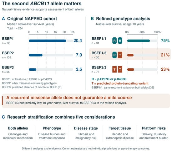
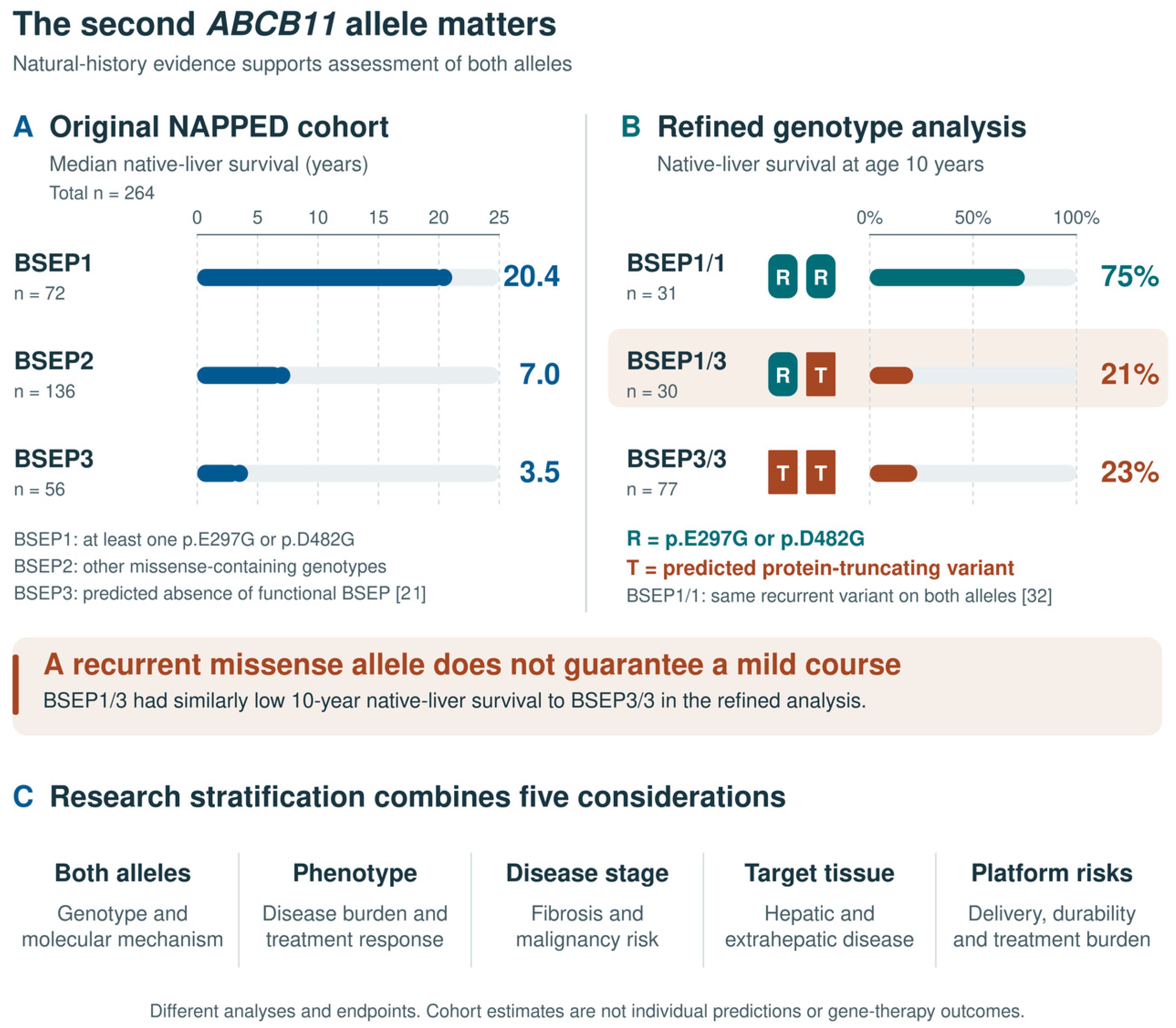
Disclaimer/Publisher’s Note: The statements, opinions and data contained in all publications are solely those of the individual author(s) and contributor(s) and not of MDPI and/or the editor(s). MDPI and/or the editor(s) disclaim responsibility for any injury to people or property resulting from any ideas, methods, instructions or products referred to in the content. |
© 2026 by the authors. Licensee MDPI, Basel, Switzerland. This article is an open access article distributed under the terms and conditions of the Creative Commons Attribution (CC BY) license.