


Abstract
Background/Objectives: Bulgarian Rose Essential Oil (BREO), obtained from Rosa damascena Mill., is a complex mixture of bioactive compounds with diverse pharmacological properties. This study investigated the bioactivities of BREO in cancer models using experimental and in silico approaches. Methods: Phytochemical composition of BREO was characterized by GC-FID. Antiproliferative activity was evaluated against HeLa, PC-3, and Ehrlich Ascites Carcinoma (EAC) cells, with HaCaT keratinocytes as non-tumorigenic reference. Apoptosis and cellular responses were assessed in EAC cells by fluorescence microscopy. Toxicity and anticancer effects of BREO were investigated in murine model of Ehrlich carcinoma using histopathological, hematological, and immunohistochemical analyses. Serum Interleukin-1β and Interleukin-17α levels were determined. Molecular docking of three major BREO constituents, geraniol, citronellol, and nerol, against IKKβ, HER2, and ERα was performed. Results: BREO exhibited concentration-dependent antiproliferative activity, with EAC cells showing the greatest sensitivity with IC50 value of 195 µg/mL. Fluorescence microscopy revealed morphological changes consistent with cell death and alterations in mitochondrial membrane potential. In vivo, BREO treatment was associated with extensive necrotic and apoptotic areas within tumors and changes in Ki-67 and hormone receptor expression. Serum IL-1β levels were comparable between BREO- and 5-fluorouracil-treated groups, whereas no significant differences in IL-17α levels were observed. Molecular docking revealed favorable binding of all three constituents within active sites of the respective targets with binding energies of −5.3 kcal/mol, −6.3 kcal/mol, and −6.0 kcal/mol, respectively. Conclusions: BREO exhibited anticancer potential, accompanied by cellular, morphological, and molecular changes associated with cancer cell death, warranting further mechanistic studies.
1. Introduction
Global cancer statistics estimate that approximately 20% of men or women develop neoplastic diseases, with lung, breast, colorectum, prostate, and stomach carcinomas representing the major global causes of morbidity and mortality worldwide [1,2]. Treatment advances in conventional chemotherapy and targeted therapies often are limited by severe adverse effects, drug resistance, and tumor recurrence. These data determine the high level of interest in the identification and development of natural bioactive compounds with favorable safety profiles and multimodal mechanisms of action that could complement or enhance existing therapeutic strategies [3].
Rosa damascena Mill. has been cultivated for centuries for its aromatic and therapeutic properties [4]. Rose essential oil has been used in folk medicine for its anti-inflammatory, analgesic, and mood-enhancing effects [5,6]. Recent pharmacological investigations have revealed that rose essential oil possesses antioxidant, antimicrobial, and anticancer activities, largely attributed to its rich content of bioactive monoterpenes, including geraniol, citronellol, and nerol [7,8]. However, widely varying active concentrations of the essential oil and its isolated monoterpenes have been reported across different studies [4,5]. These discrepancies stem from multiple factors, including variations in geographic origin, climatic conditions, and extraction methods, as well as differences in application forms, bioavailability, and the experimental model systems employed. Although geraniol, citronellol, and nerol represent the primary bioactive constituents, the overall pharmacological response relies on the complex interplay and potential synergistic effects among the oil’s components. Consequently, comprehensive phytochemical characterization is essential to reliably correlate the chemical composition of the oil with its observed biological properties.
The current evidence regarding the anticancer potential of Rosa damascena essential oil relies primarily on preliminary in vitro screening, where relatively high concentrations are often required to achieve these bioactivities. Furthermore, the clinical translation of these natural monoterpenes remains challenging due to their modest oral bioavailability, rapid metabolic degradation, and potential batch-to-batch compositional variability [4,5]. Thus, transitioning from isolated cellular models to in vivo tumor environments is required to validate whether these natural bioactive compounds can exert their pharmacological effects at physiologically relevant doses.
Accumulating evidence has demonstrated antiproliferative effects of essential oil and extracts obtained from R. damascena on various cancer cell lines [9,10,11,12,13,14]. In our previous study, we reported that BREO exerts selective antiproliferative activity against colorectal adenocarcinoma cells (HT-29 and HCT-8) with low cytotoxicity toward non-tumorigenic MCF-12F cells, and further demonstrated its ability to suppress colony formation and induce apoptosis [9]. Complementary in silico molecular docking analysis predicted potential protein targets underlying these effects, including MAPK p38α [9]. However, these studies have focused solely on direct cytotoxicity in isolated cancer cell cultures, overlooking the complex interplay between cancer cells and the host immunity, e.g., the tumor microenvironment (TME), which is a critical determinant of cancer progression and therapeutic outcome [15,16,17]. The TME has been in the spotlight in recent years for its crucial role in tumor development and the release of pro-inflammatory molecules related to tumor progression and response to therapy. It is a dynamic system composed of cancer cells, immune infiltrates (macrophages, T-cells, myeloid-derived suppressor cells), stromal cells, and a complex network of chemokines and cytokines [16]. Chronic inflammation within the TME, driven by pro-inflammatory mediators such as interleukin-1β (IL-1β) and interleukin-17α (IL-17α), promotes tumor angiogenesis, metastasis, and immunosuppression, thereby fostering tumor growth and resistance to therapy [17]. Conversely, agents capable of modulating these inflammatory cues and reprogramming the TME toward an antitumor state hold considerable therapeutic promise, because in many cases, the TME exhibits strong immunosuppressive properties, posing a major obstacle to successful therapeutic intervention [18].
Ehrlich Ascites Carcinoma (EAC) is a widely recognized experimental murine cancer model for preclinical assessment of potential anticancer compounds [19]. Originally, a spontaneous murine mammary adenocarcinoma, EAC can be propagated in both ascites and solid forms, depending on the route of inoculation. The solid form of EAC permits non-invasive monitoring of tumor growth over time, while the ascites form enables rapid propagation, and the cell line derived from it offers the significant advantage of using the same tumor cells for complementary in vitro experimental approaches [19,20]. This model has been extensively utilized in studies on the antitumor potential of natural and synthetic compounds, tumor-associated enzyme activity, and the immunological aspects of tumor–host interactions [19,20,21,22,23,24].
To date, the potential of BREO to exert immunomodulatory effects within the TME has not been systematically investigated. Furthermore, the in vivo antitumor efficacy of BREO and its systemic impact on immune-related parameters remain unexplored.
In this study, we hypothesized that BREO exerts its anticancer activity not only through direct cytotoxicity but also via modulation of the inflammatory tumor microenvironment. To systematically evaluate the anticancer and immunomodulatory potential of BREO, this study was designed as an integrated, step-wise strategy. First, the volatile phytochemical composition was characterized via GC-FID to identify the major constituents. Next, in vitro screening across human and murine cancer cell lines (HeLa, PC-3, and EAC) alongside a non-tumorigenic reference (HaCaT) was performed to determine selective antiproliferative and pro-apoptotic effects. To translate these cellular findings to a complex biological system, an in vivo solid EAC murine model was employed for assessment of systemic safety and tumor growth inhibition, combined with immunohistochemical marker analysis (Ki-67, ERα, PR, HER2) and serum cytokine profiling (IL-1β, IL-17α) to evaluate TME-related inflammatory responses. Complementing these experimental approaches, the three most abundant constituents—geraniol, citronellol, and nerol—were subjected to molecular docking against IKKβ, HER2, and ERα, respectively. These targets were selected based on their established roles in inflammation, cellular proliferation, and hormone signaling, thereby bridging the observed phenotypic effects with potential underlying molecular pathways.
This multi-target approach aimed to explore distinct molecular pathways that may contribute to the broad spectrum of BREO activity. By bridging direct antiproliferative effects with systemic immunomodulatory observations, this work aims to provide a comprehensive preclinical evaluation of BREO and lay the groundwork for future studies into its role as a regulator of antitumor immunity.
2. Materials and Methods
2.1. Materials and Chemicals
Bulgarian rose essential oil (BREO) was obtained from the Institute of Roses and Aromatic Plants, Agricultural Academy. Cell culture reagents, Dulbecco’s Modified Eagle Medium (DMEM) and fetal bovine serum, were supplied by Gibco-Invitrogen (Leicestershire, UK). The solution of penicillin and streptomycin was obtained from Lonza (Verviers, Belgium). Phosphate-buffered saline, dimethylsulfoxide, and trypsin–ethylenediaminetetraacetic acid solution were provided from AppliChem (Darmstadt, Germany). The MTT reagent (3-(4,5-dimethylthiazol-2-yl)-2,5-diphenyltetrazolium bromide) was obtained from Sigma-Aldrich Chemie GmbH (Darmstadt, Germany). DAPI (4′,6-diamidino-2-phenylindole) fluorescent stain, acridine orange (AO), and ethidium bromide (EB) were delivered from Merck (Darmstadt, Germany). The sterile disposable plastic consumables were obtained from Orange Scientific (Braine-l’Alleud, Belgium).
2.2. Cell Lines
Human tumor cell lines HeLa (cervical carcinoma) and PC-3 (prostate carcinoma) were purchased from the American Type Culture Collection (ATCC, Manassas, VA, USA). The non-cancerous human keratinocyte cell line HaCaT was obtained from CLS Cell Lines Service (Eppelheim, Germany). The murine Ehrlich Ascites Carcinoma (EAC) cell line was derived in our laboratory from Ehrlich tumor-bearing mice. All cell lines were routinely maintained as monolayers in high glucose DMEM cell culture medium supplemented with 10% fetal bovine serum, 50 U/mL penicillin, and 50 mg/mL streptomycin and 2 mM L-glutamine. Cells were cultured at 37 °C in a humidified atmosphere containing 5% CO2 and were subcultured two or three times per week to preserve exponential growth. At 60–80% confluency, cells were detached using 0.25% trypsin-EDTA in PBS (pH 7.4), counted, and resuspended in fresh complete medium. For the respective experiments, cells were seeded into sterile 96-well or 24-well plates at the required densities. For fluorescence microscopy assays, sterile glass coverslips with 13 mm diameter were placed on the bottom of 24-well plates before cell seeding.
2.3. Laboratory Animals
A total of 32 female ICR mice, 3 months old and weighing approximately 20 g, were obtained from Experimental breeding base for laboratory animals, Institute of Neurobiology, Bulgarian Academy of Sciences. The animals were housed for a standard quarantine period under species-appropriate environmental conditions, including controlled temperature and light/dark cycles, in spacious metal cages and unrestricted access to food and drinking water. All animal experiments were reviewed and approved by the institutional committee and performed in accordance with the national regulations on laboratory animals and animal welfare (Regulation No. 20/01.11.2012) and Directive 2010/63/EU of the European Parliament and of the Council. The animal study was approved by the Ethical Committee of IEMPAM, BAS, and the Institutional Review Board of the Bulgarian Food Safety Agency, approval No. 459, issued on 18 December 2025. Mice were allocated to experimental groups by simple randomization generated by number. Four animals per group were used in this preliminary-stage study and no animals were excluded from the groups. A total of 16 mice were used in the toxicological evaluation and 16 were used in the antitumor effect evaluation, described in detail in the relevant sections. All treatments were conducted after the cages were numbered, and the animals were marked. During treatment, animals were processed sequentially (by number) in the presence of two people to avoid errors. All data obtained from each single animal were included when drawing conclusions.
2.4. Extraction and Phytochemical Analysis
To guarantee the authenticated origin and high quality of the raw material, Rosa damascena Mill. flowers were harvested from the experimental fields of the Institute of Roses, Aromatic and Medicinal Plants (Agricultural Academy, Kazanlak, Bulgaria). Manual picking was conducted during the peak flowering season in May and June, specifically in the early morning hours (04:00–08:00 a.m.) when the blossoms were at the IV–V developmental stage (semi-opened to fully opened). To preserve the volatile phytochemical profile, the freshly collected flowers were immediately processed using water–steam microdistillation in a Clevenger-type apparatus to isolate the Bulgarian Rose Essential Oil (BREO). Subsequently, the extracted BREO was subjected to gas chromatography with flame ionization detection (GC-FID) for the identification and quantification of its volatile constituents, as previously described [9]. Briefly, an Agilent 7820A GC-FID system equipped with a non-polar capillary column (EconoCap EC-5, 30 m × 0.32 mm × 0.25 μm) was used for the analysis. An n-alkane reference mixture (C8–C27) was used for quantification of major and minor components. The split ratio was 1:10, the inlet temperature was 250 °C, and the FID temperature was programmed from 60 °C to 300 °C. Data were processed using Agilent OpenLab CDS software (version 9.2).
2.5. Assessment of Cell Viability
The antiproliferative activity of Bulgarian rose essential oil (BREO) was evaluated using the MTT colorimetric assay. Cells were counted using a hemocytometer and seeded at a density of 1 × 10⁵ cells/mL of complete culture medium into 96-well cell culture plates. After overnight incubation, cells were treated with increasing concentrations of BREO (7.8–1000 μg/mL) for 72 h. Untreated cells maintained under identical culture conditions were used as negative controls. The HaCaT cell line was included as a non-tumorigenic reference to evaluate the relative selectivity of the treatment toward malignant cells. Following exposure, the cells were rinsed with PBS and incubated with 5 mg/mL MTT solution at 37 °C for 2–3 h. A solution of DMSO and ethanol (1:1, v/v) was used to dissolve the resulting formazan crystals, and optical density was recorded at 570 nm with a microplate reader (TECAN, Sunrise™, Männedorf, Switzerland). Cell viability was calculated as a percentage of the corresponding untreated control. IC50 values were estimated by nonlinear regression analysis. Each concentration was analyzed using six replicates and experiments were independently repeated three times.
2.6. Acridine Orange/Ethidium Bromide Fluorescent Staining
Changes in cellular morphology and membrane integrity following BREO treatment were examined by dual acridine orange/ethidium bromide (AO/EB) fluorescent staining. EAC cells were seeded onto 13 mm diameter coverslips placed in 24-well plates and incubated overnight to allow cell attachment and spreading. Cells were subsequently treated with BREO at 125 μg/mL for 72 h. Cells exposed to 5-FU and untreated cells were included as positive and negative controls, respectively. Following treatment, coverslips were gently washed with PBS and stained with AO and EB, each at a concentration of 5 μg/mL in PBS, for 5 min. Analysis was performed immediately after sample preparation using a fluorescence microscope (Leica DM 5000B, Wetzlar, Germany) equipped with the appropriate fluorescence filter set. AO/EB staining was used to distinguish viable cells with predominantly green fluorescence from cells exhibiting apoptotic and/or membrane-compromised phenotypes characterized by yellow, orange, or red fluorescence.
2.7. DAPI Fluorescent Staining
Nuclear morphological changes associated with cell death were further analyzed after staining with 4′,6-diamidino-2-phenylindole dihydrochloride (DAPI). EAC cells were cultured on glass coverslips and exposed to BREO (125 μg/mL) for 72 h. Untreated cells and 5-FU-treated cells were used as negative and positive controls, respectively. Following treatment, cells were fixed for 10 min with 3% paraformaldehyde and washed with PBS. The cell samples were then incubated with DAPI solution (1 μg/mL) in the dark, at room temperature for 15 min. After staining, the coverslips were mounted using Mowiol® and examined by fluorescence microscopy (Leica DM 5000B, Wetzlar, Germany). Nuclear morphology was evaluated with particular attention to nuclear size reduction, chromatin condensation, and margination, as well as nuclear fragmentation, which are characteristic features associated with apoptotic cell death.
2.8. Evaluation of Mitochondrial Membrane Potential
Alterations in mitochondrial membrane potential (ΔΨm) were assessed in EAC cells using the fluorescent probe JC-1. Cells were seeded onto 13 mm diameter glass coverslips in 24-well plates and incubated overnight to allow cell spread and attachment. The cells were subsequently exposed to BREO at 125 μg/mL for 72 h. Untreated cells and cells treated with 5-FU (50 μM) served as negative and positive controls of the experiments, respectively. After treatment, cells were carefully washed with PBS and incubated with 5 μg/mL JC-1 for 20 min in the dark at 37 °C. Fluorescence was subsequently visualized using fluorescence microscopy. Changes in ΔΨm were evaluated according to the relative distribution of red-fluorescent JC-1 aggregates, characteristic of polarized mitochondria, and green-fluorescent JC-1 monomers, associated with mitochondrial depolarization. Image analysis was performed using ImageJ, 1.49v software. Fifty cells from each experimental group were analyzed, and fluorescence intensity was normalized to the respective cell area. The mean red/green fluorescence ratio was calculated from three independent experiments and used as a measure of mitochondrial membrane polarization.
2.9. Toxicological Evaluation
Four groups of healthy mice (Groups A, B, C, D; n = 4) were formed. The animals were treated with BREO six times every other day with the fowling single doses: Group A: 1720 mg/kg b.w.; Group B: 1290 mg/kg b.w. BREO; Group C: 860 mg/kg b.w. BREO; Group 430 mg/kg b.w. For all groups, the BREO was diluted 1:1 with 5% Tween 20 in saline. Throughout the study, animals had unrestricted access to food and drinking water. The experimental animals from all groups were sacrificed by anesthetic overdose at the end of the experiment and tissue samples were taken for histological analyses.
2.10. Evaluation of In Vivo Antitumor Effect
Three groups of mice (Groups 1, 2, and 3; n = 4) were formed. All mice were inoculated in the hind right limb with a suspension of Ehrlich’s carcinoma cells to develop solid tumors, and treated for 15 days as follows: Group 1: mice inoculated with 1 × 10⁶ Ehrlich carcinoma cells and treated with 0.02 mL saline every other day; Group 2: mice inoculated with 1 × 10⁶ Ehrlich carcinoma cells and treated subcutaneously (s.c.) with 0.02 mL BREO diluted 1:1 with 5% Tween 20 in saline at a dose of 430 mg/kg every other day; Group 3: mice inoculated with 1 × 10⁶ Ehrlich carcinoma cells and treated subcutaneously (s.c.) with 0.02 mL 5-fluorouracil (5-FU) solution in saline at a dose of 15 mg/kg body weight every other day.
2.11. Hematological Analysis
Complete blood counts of the collected blood from all tumor-bearing mice were measured at day 15 of the experiment for assessment of antitumor effect using automatic hematology analyzer (BC-2800 Vet (Mindray, Shenzhen, China)). Blood smears were stained by May Grunwald Giemsa—rapid method (Diapath, Martinengo, Italy) and were examined by light microscope Leica DM5000B, (Leica Microsystems, Wetzlar, Germany).
2.12. Histopathological Examination
Tissue samples from kidney, liver, and spleen (for toxicological and immunological assessment) and tumors (for antitumor effect study) were collected from all mice in the experiments. All samples were fixed in 10% neutral buffered formalin, followed by dehydration, xylene clearing, and paraffin embedding. Paraffin blocks were sectioned at 3–5 μm, and the resulting sections were stained with hematoxylin and eosin (H&E). Histopathological examination was performed using a Leica DM 5000B light microscope (Wetzlar, Germany).
2.13. Immunohistochemistry
Tissue samples from all tumor-bearing mice were obtained. Tissue sections from 4 μm thick were cut and were pretreated for antigen retrieval with citrate buffer in high temperature Zytomed Systems OmniPrep Solution pH 6.0 ZUC079-500 (Zytomed Syst. GmbH, Berlin, Germany). The endogenous peroxidase activity was blocked for 10 min by Peroxide block ZUC019-100 (Zytomed Syst. GmbH, Berlin, Germany). The samples were incubated with primary mouse antibodies: Ki67 (clone MM1, RTU, Leica Biosystems, PA0118), Estrogen Receptor (Clone F611, RTU, Leica Biosystems, PA0151), and Progesterone Receptor (Clone 16, RTU, Leica Biosystems, PA0312). The reaction was visualized with polymer conjugated horseradish peroxidase kit (ZytoChem Plus (HRP) Polymer Kit, POLHRP-100, Zytomed Syst. GmbH Berlin), developed with 3,3′-diaminobenzidine (DAB) (DABPLUS, Zytomed Syst. GmbH Berlin), and counterstained with Mayer’s hematoxylin (Zytomed Syst. GmbH Berlin). For positive controls of the reaction, human tonsils were used; for negative controls, specimens were used, incubated with diluent instead of the first antibody. Slides were observed under Leica DM 5000B light microscope (Wetzlar, Germany).
2.14. IL Screening by ELISA
Serum samples were isolated from all tumor-bearing mice from Groups 1–3 and four healthy controls by centrifugation at 1000× g, for 20 min and cryopreserved at −80 °C. Serum concentrations of interleukin-1β (IL-1β) (Cat. No. abx050111, Abbexa) and interleukin-17α (IL-17α) (Cat. No. EM1624, Fine Test) were quantified according to the manufacturers’ protocols. Samples and standards were added to antibody-coated 96-well microplates and incubated with biotinylated detection antibodies followed by horseradish peroxidase-conjugated reagents. After color development and reaction termination, absorbance was measured at 450 nm using an ELISA microplate reader. Each serum sample was analyzed in three technical replicates. Cytokine concentrations were determined from standard curves (7.81–500 pg/mL) and expressed as pg/mL.
2.15. Molecular Docking
Molecular docking was empoyed to predict the binding conformations and affinities of the three major BREO constituents—geraniol, citronellol, and nerol—against selected target proteins. All docking simulations were performed using the CB-Dock2 web-based platform (https://cadd.labshare.cn/cb-dock2/, accessed on 17 September 2026), which combines curvature-based cavity detection (CurPocket), molecular docking, and template-based cavity fitting (FitPocket) into a unified platform [25,26].
2.15.1. Protein Preparation
The three-dimensional crystal structures of the selected protein targets were obtained from the RCSB Protein Data Bank (PDB). The following structures were selected based on their relevance to the studied cancer cell lines and the availability of co-crystallized reference ligands: IKKβ (Inhibitor of κB kinase β); HER2 (Human epidermal growth factor receptor 2); and ERα (Estrogen receptor α). The crystal structures of human IKKβ (PDB ID: 3RZF [27]), the Kinase domain of Human HER2 (erbB2) (PDB ID: 3PP0 [28]), and Human Estrogen Receptor Ligand—Binding Domain (PDB ID: 1ERE [29]) (accessed on 17 September 2026) were used as the receptors for molecular docking. The protein structures were prepared in advance using AutoDockTools 1.5.7, which included the removal of crystallographic water molecules, heteroatoms, and the original co-crystallized ligands.
2.15.2. Ligand Preparation
- The three major BREO constituents—geraniol (PubChem CID: 637566), citronellol (PubChem CID: 8842), and nerol (PubChem CID: 643820) were accessed on 17 September 2026. The 3D molecular structures of the selected ligands were retrieved from PubChem in SDF format. For positive control validation, the following reference inhibitors were used:
- N-(6-chloro-7-methoxy-9H-beta-carbolin-8-yl)-2-methylnicotinamide—(MLN120B) (inhibitor of IKKβ)—(PubChem CID: 9929127). Characteristics: ATP-competitive, Ki/IC50 for IKK—60 nM, >1000-fold selectivity relative to 30 representative tyrosine and serine/threonine kinases; good oral bioavailability; Pre-Clinical Therapeutic Efficacy—Multiple myeloma [30], arthritis [31];
- 4-{[4-(4-Chlorophenyl)pyrimidin-2-Yl]amino}phenyl) [4-(2-Hydroxyethyl)piperazin-1-Yl]methanone—(XNM)—(PubChem CID:52914873)—co-crystallized ligand;
- Lapatinib (PubChem CID:208908)—Positive control and co-crystallized ligand;
- 4-Hydroxytamoxifen (PubChem CID: 449459)—Active metabolite of tamoxifen; FDA-approved for breast cancer.
2.15.3. Grid Box and Docking Parameters
Molecular docking was performed using the CB-Dock2 online server. For both blind and targeted docking, the server automatically defined a standard grid box of 24 × 24 × 24 Å centered on the identified active sites to fully encompass the binding pockets. All other parameters were set to the default values of the CB-Dock2 platform.
2.15.4. Docking Validation
Docking validation was performed using two approaches: (i) docking of the known IKKβ inhibitor MLN120B as a positive control to benchmark the binding affinity of geraniol and (ii) re-docking of the co-crystallized ligands (XNM for IKKβ, Lapatinib for HER2, and 4-Hydroxytamoxifen for ERα) into their native binding sites to confirm the accuracy of the docking protocol.
2.15.5. Interaction Analysis and Visualization
The protein–ligand interactions and binding orientations were visualized and analyzed using BIOVIA Discovery Studio Visualizer 2025 (v25.1.0.0). The corresponding 2D interaction maps were constructed to characterize hydrogen bonds, hydrophobic interactions (alkyl and π-alkyl), and other non-covalent contacts. Three-dimensional binding poses were rendered to illustrate the spatial orientation of each ligand within its respective binding pocket.
2.16. Statistical Analysis
All data are presented as mean ± SD and the statistical significance was analyzed by one-way ANOVA followed by Bonferroni’s post hoc test, Tukey’s multiple comparisons test, and Dennett’s multiple comparisons test, as needed, using GraphPad Prism Software, Version 7 (San Diego, CA, USA).
3. Results
3.1. Phytochemical Composition of BREO
The phytochemical composition of BREO was analyzed by GC-FID, and the identification of compounds was based on retention time comparisons with authentic references and retention indices from the NIST database. Quantification was carried out by peak area percentage without correction factors. Table 1 presents the retention times, relative percentages, and calculated/literature retention indices for 20 identified compounds, including the main constituents specified in ISO 9842:2024 [32,33].
Table 1.
Retention time, relative percentage, and retention index for 20 BREO compounds analyzed on a non-polar column EconoCap EC-5 with GC-FID.
The geraniol content determined in the present study (32.79%) exceeds the reference range specified in ISO 9842:2024 (14.0–22.0%). This deviation is consistent with a trend of increasing geraniol levels observed in Bulgarian rose oil over the past four harvest years, including the 2025 harvest analyzed here. This shift is attributed to climatic variations affecting the experimental fields of the Institute of Roses and Aromatic Plants, Agricultural Academy in Bulgaria, where the plant material was cultivated under controlled and uniform conditions. These observations suggest that the current ISO reference values may require revision to reflect ongoing environmental changes. The plant material used in this study was obtained from the Institute’s experimental field, which owns the intellectual property rights to the cultivated varieties, as listed in the Official Variety List of the Republic of Bulgaria for 2025 [34].
3.2. In Vitro Effects of BREO in Cancer Cell Models
3.2.1. Cell Viability Assessment
The antiproliferative effect of Bulgarian rose essential oil was initially evaluated in vitro using the MTT assay in three tumor cell lines, HeLa, PC-3, and EAC, and in the non-tumorigenic HaCaT cell line. BREO produced a concentration-dependent decrease in cell viability, although the magnitude of the response differed considerably among the tested cell models (Figure 1). HeLa cells showed a pronounced response at higher BREO concentrations, with cell viability decreasing markedly at 250 μg/mL and above. A similar, and more pronounced, response was observed in EAC cells, in which a substantial reduction in viability was already evident at 125 μg/mL and became progressively stronger with increasing concentrations. In contrast, PC-3 cells remained comparatively resistant to BREO up to 500 μg/mL, with a pronounced loss of viability observed only at 1000 μg/mL. HaCaT cells also displayed relatively high viability at concentrations up to 250 μg/mL, followed by a progressive decrease at higher doses.
Figure 1.
Cell viability in HeLa, PC-3, EAC, and HaCaT cells after treatment with Bulgarian rose essential oil. Cells were treated with BREO for 72 h. Data are presented as percentages relative to untreated controls. Values are mean ± SD from six replicates; one-way ANOVA with Bonferroni’s post hoc test (n = 6); ** p < 0.01, and *** p < 0.001 denote significant differences relative to the negative control.
Among the tumor cell lines, EAC cells exhibited a particularly pronounced sensitivity to BREO, whereas PC-3 cells were the least responsive. The comparatively lower effect observed in HaCaT cells within the lower concentration range further supported the selection of EAC cells for subsequent mechanistic investigations. Based on this screening, BREO at 125 μg/mL was selected for the subsequent fluorescence-based analyses.
The calculated IC50 values and selectivity indices (SI) further quantified the differential response of the tested cell lines to BREO (Table 2).
Table 2.
Inhibitory concentrations (IC50) of BREO in cancer and non-cancerous cell lines and selectivity indices.
The lowest IC50 values were established in EAC cells (195.0 μg/mL) and HeLa cells (279.6 μg/mL), whereas substantially higher concentrations were required to reduce cell viability in PC-3 and HaCaT cells, with IC50 values of 740.2 and 673.0 μg/mL, respectively. The selectivity indices relative to HaCaT cells were 3.5, 2.4, and 0.9 for EAC, HeLa, and PC-3 cells, respectively. The determined IC50 values and selectivity indices confirmed the differences in cellular responsiveness observed across the tested cancer cell models.
3.2.2. Cytomorphological Alterations in BREO-Treated Cancer Cells
The marked antiproliferative response of EAC cells to BREO prompted further investigation of treatment-associated morphological changes. Since the cytotoxic effect at 125 μg/mL was sufficiently pronounced while retaining a substantial viable cell population, this concentration was selected for fluorescence microscopy analysis using AO/EB staining (Figure 2).
Figure 2.
Morphological alterations in EAC cells treated with Bulgarian rose essential oil for 72 h. Representative fluorescence microscopy images of control cells (a), BREO-treated cells (b), and 5-FU-treated (c) cells after AO/EB staining. Scale bar = 20 μm.
Control cells predominantly showed uniform green fluorescent staining, consistent with preserved membrane integrity and viable cellular morphology. Following exposure to BREO, a reduction in cell density and changes in cellular morphology became apparent, accompanied by an increased number of cells displaying yellowish to orange/red fluorescence. These changes were indicative of progressive alterations in membrane integrity and cell death. The effect was more prominant in the 5-FU-treated cells, which showed a higher reduction in the cell number and a larger proportion of orange/red fluorescent cells. The morphological alterations observed after BREO treatment were consistent with the appearance of apoptotic and late-stage cell death phenotypes. The presence of both green/yellow fluorescent cells and cells with pronounced orange/red fluorescence suggests that the treatment affected cells at different stages of the cell-death process.
3.2.3. Nuclear Morphological Changes
The effect of BREO on nuclear morphology was further examined using DAPI staining (Figure 3).
Figure 3.
Nuclear morphology alterations in EAC cells following treatment with BREO (125 μg/mL) for 72 h. Representative DAPI fluorescence images of control (a), BREO-treated (b), and 5-FU-treated (c) cells. Scale bar = 20 μm.
Untreated EAC cells displayed predominantly intact, round, or oval nuclei with relatively homogeneous fluorescence and no obvious signs of nuclear fragmentation. In contrast, BREO-treated cells exhibited alterations in nuclear morphology, including increased fluorescence heterogeneity, chromatin condensation, nuclear shrinkage, and the appearance of fragmented nuclear structures. These changes were more evident in cells exposed to 5-FU, which showed pronounced nuclear alterations and a lower overall cell density. The DAPI findings therefore supported the morphological observations obtained by AO/EB staining and indicated that BREO treatment was associated with nuclear changes characteristic of apoptotic cell death.
3.2.4. Mitochondrial Membrane Potential
The effect of BREO on mitochondrial membrane polarization of EAC cells was evaluated using JC-1 staining and compared with untreated and 5-FU-treated cells (Figure 4). JC-1 accumulates in polarized mitochondria in the form of red-fluorescent aggregates, whereas mitochondrial depolarization is associated with the appearance of green-fluorescent monomers.
Figure 4.
Effect of BREO on mitochondrial membrane potential (ΔΨm) in EAC cells. Representative fluorescence images of JC-1 stained control (a), BREO-treated (b), and 5-FU-treated (c) cells. Scale bar = 20 μm. Fluorescence intensity was measured using ImageJ. Data are presented as mean ± SD. One-way ANOVA with Bonferroni’s post hoc test; *** p < 0.001 indicate significant differences relative to the negative control.
Control EAC cells displayed green fluorescence with numerous bright red punctata, whereas BREO treatment resulted in a distinct alteration in JC-1 fluorescence distribution, with changes in the intensity and pattern of mitochondrial-associated fluorescence. The BREO-treated cells exhibited a shift toward a weaker red component and more prominent green fluorescence compared with the control group, indicating a reduction in mitochondrial membrane polarization. The effect was more evident in the positive control group, which displayed the strongest alteration in JC-1 fluorescence and the greatest reduction in the red component. Quantitative image analysis of the red/green fluorescence ratio further supported the treatment-associated reduction in mitochondrial polarization. These findings indicate that BREO at 125 μg/mL affects mitochondrial membrane potential in EAC cells. The observed mitochondrial depolarization, together with the cytomorphological and nuclear changes detected by AO/EB and DAPI staining, suggests that mitochondrial dysfunction may contribute to the cell-death response induced by BREO.
3.3. In Vivo Effects of BREO
3.3.1. Clinical Observations and Gross Pathology—Toxicity Study
The toxicological study revealed clinical manifestation of signs of moderate indisposition at doses of BREO 860, 1290, and 1720 mg/kg b.w. The observed effects included transient signs of pain at the site of inoculation and behavioral changes, manifested as lying on the side or abdomen for approximately 2–3 min before resuming normal activity. No animals died during the experiment, and the lethal doses LD100, LD50, and LD05 were not reached. At the lowest dose (430 mg/kg b.w.), no significant deviations from the normal clinical condition of the mice were observed, apart from occasional transient lying on the floor for several seconds. Gross pathological examination revealed no abnormalities in any of the experimental groups.
3.3.2. Clinical Observations and Gross Pathology—Antitumor Effect Study
Two control groups were included in the experiment to monitor tumor development: a negative control group consisting of mice bearing solid carcinomas and receiving no treatment, and a positive control group of tumor-bearing mice treated with the standard anticancer agent 5-FU, which is used for breast carcinoma treatment, including in experimental mouse models. The clinical condition deteriorated in all three treatment groups, with the observed behavioral changes, particularly reduced and impaired movement, being associated with solid tumor development. Difficulty in moving the affected hind limb was observed as a consequence of tumor growth between the muscle fibers. No other deviations from the normal clinical condition or signs of local pain at the inoculation site were observed. Gross pathological examination revealed splenic congestion and splenomegaly in all groups, findings consistent with Ehrlich carcinoma development, although these changes were more pronounced in the saline-treated group. Neither tumor necrosis nor fistulation was observed in any of the animals. No other macroscopic abnormalities of the examined organs were detected.
3.3.3. Hematology
Hematological analyses showed marked leukocytosis in Group 1 (Control saline-treated EAC-bearing mice), with WBC counts ranging from 8.9 to 14.5 × 10⁹/L (mean ± SD: 12.7 ± 2.58 × 10⁹/L), compared with the reference range of 0.8–6.8 × 10⁹/L. This finding is characteristic of the advanced stages of tumor development and may be associated with central tumor necrosis and inflammatory processes. In Groups 2 (BREO-treated) and 3 (5-FU-treated), WBC counts were also slightly above the upper reference limit but significantly lower than in Group 1, ranging from 6.4 to 9.2 × 10⁹/L (mean ± SD: 7.4 ± 1.24 × 10⁹/L; p = 0.0337 vs. Group 1) and from 6.6 to 11.3 × 10⁹/L (mean ± SD: 7.9 ± 2.27 × 10⁹/L, p = 0.0260 vs. Group 1), respectively. Blood smear examination revealed normal morphology of erythrocytes and leukocytes in all examined groups (Figure 5).
Figure 5.
Blood smears of mice from: (a) Group 1, treated with saline; (b) Group 2, treated with BREO; (c) Group 3, treated with 5-FU; (d) qualitative analysis of WBC counts; one-way ANOVA, Tukey’s multiple comparisons test (n = 4); * p < 0.05.
3.3.4. Histopathology
The effects of rose oil BREO on the parenchymal organs were evaluated by histopathological examination in both the toxicity study and the antitumor effect study. In agreement with the clinical observations, the higher doses of parenterally administered BREO (Groups A and B) were associated with slight alterations in the normal histological architecture of the liver and kidneys, as shown in Figure 6. In the kidneys, changes in the tubulointerstitial architecture and focal glomerular collapse were observed. In the liver, focal lesions affecting the centrilobular parenchyma were detected, characterized by alterations in cellular arrangement, limited cytoplasmic hydropic changes, and vacuolization, as well as single pyknotic nuclei of condensed apoptotic cells. The centrilobular regions are metabolically active areas of the liver and are considered particularly susceptible to toxic injury. No pathological lesions were observed in the spleens of the animals examined for BREO toxicity.
Figure 6.
Histopathology of parenchymal organs in mice: (a,b) kidney and liver in mice treated s.c. with BREO 1720 mg/kg b.w.; (c,d) normal histology of kidney and liver; yellow arrows—glomeruli, green arrows—apoptotic hepatocytes. H&E staining.
In tumor-bearing mice from all groups, increased spleen weight and size were observed. Histopathological examination revealed alterations in splenic architecture, characterized by a loss of clear demarcation between the red and white pulp, as well as a reduction in the number of lymphoid follicles. These changes were most pronounced in the saline-treated group (Figure 7).
Figure 7.
Histopathology of spleen in mice, bearing the solid form of Ehrlich carcinoma, treated s.c. with the following: (a) saline; (b) BREO 430 mg/kg b.w.; (c) 5-FU 15 mg/kg b.w.; blue arrows—megakaryocyte cells, yellow arrows—splenic nodules (follicles). H&E staining.
In the BREO- and 5-FU-treated groups, areas of preserved and immunologically active white pulp were observed (Figure 7b,c). Megakaryocytes were normally present in the spleens of all groups, taking into account increased splenic localization in Groups 2 and 3 (BREO- and 5-FU-treated groups, respectively). No treatment-related histopathological alterations were observed in the kidneys. In the liver of the saline-treated group, only mild parenchymal sinusoidal dilatation was observed (Figure 8).
Figure 8.
Histopathology of liver in mice, bearing the solid form of Ehrlich carcinoma, treated s.c. with the following: (a) saline—mild sinusoidal dilatation; (b) 430 mg/kg BREO; (c) 5-FU 15 mg/kg b.w. H&E staining.
Notable differences were observed in the tumors developing in the hindlimb muscles among the experimental groups (Figure 9). In saline-treated mice, extensive tumor masses were observed, infiltrating the muscle fibers and occupying almost the entire musculoskeletal compartment of the limb (approximately 90%). The tumor cells exhibited predominantly elliptical, basophilic nuclei and acidophilic cytoplasm (Figure 9a,b). The nuclear hyperchromasia and cellular morphology were consistent with the malignant and poorly differentiated characteristics of the tumor tissue. Focal hemorrhagic necrotic areas were found in the central regions of the tumors, which may be associated with local hypoxia. In the BREO- and 5-FU-treated groups (Figure 9c,d and Figure 9e,f, respectively), hemorrhagic necrotic areas were also observed in the central tumor regions, together with extensive central vacuolization and numerous apoptotic tumor cells. These changes were particularly evident in the BREO-treated group (Figure 9d). Compared with the saline-treated group, destructive changes within the tumors were more extensively pronounced in both treatment groups, with necrotic and apoptotic areas accounting for approximately 30–40% of the tumor occupied tissue.
Figure 9.
Histopathology of tumor masses in mice, bearing the solid form of Ehrlich carcinoma, treated s.c. with the following: (a,b) saline; inclusion: intact tumor cells; (c,d) BREO 430 mg/kg; inclusion: apoptotic tumor cells; (e,f) 5-FU 15 mg/kg b.w. Blue arrows—muscle fibers, surrounded by tumor cells; black arrows—tumor cells; white arrows—focal hemorrhagic necrosis; yellow arrows—adipocytes; red arrows—apoptotic tumor cells. H&E staining.
The histopathological findings in the parenchymal organs and tumor tissues of mice from the three experimental groups were semi-quantitatively scored and summarized in Table 3.
Table 3.
Histopathology scoring of lesions observed in different organs and tissues of Ehrlich tumor-bearing mice treated with BREO.
3.3.5. Immunohistochemistry
Immunolocalization of targeted markers is presented on Figure 10. Positive expression of Ki 67 was evident in Group 1 along with negative results (receptor loss, triple negative) for ERα, PR, and HER2. Moreover, in our case, immunohistochemistry results showed receptor discordance or conversional status of estrogen, progesterone, and HER 2 receptors, expressed in unstable positive status after treatment of mice from Group 2 with BREO. Regarding Ki-67 expression, it showed negligible immunolocalization (isolated single cell marking) in samples from Group 2, with significantly reduced intensity compared to those mice treated with saline.
Figure 10.
Immunolocalization of Ki 67, ER α, PR, HER 2 receptors in tumors of mice treated with saline (first line) and treated with BREO (second line). Red arrows point to positive staining.
3.3.6. ELISA Screening
The values of IL-1β and IL-17α were evaluated in the sera of the tumor-bearing mice, treated with saline, BREO, 5-FU compared to that of four healthy mice (from the same breeder), used as assay controls. IL-1β beta screenings of BREO-treated mice were comparable to those in 5-FU and slightly elevated than those in the controls. Significantly elevated values, compared to the reference and to those of the controls (p = 0.0090), were maintained in the Group 1 (Figure 11a). Significantly elevated IL-17α levels assessed in all tumor-bearing mice (Saline vs. Controls: p < 0.0001; BREO vs. Controls: p = 0.0001; 5-FU vs. Controls: p = 0.0007) were linked to expressed sensitivity within the tumor microenvironment and to the chronic inflammation of tumor masses during the period of experimental ending and were not influenced by the treatment, as the statistics show (Figure 11b).
Figure 11.
Analysis of IL-1β (a) and IL-17α (b) in the sera of mice with Ehrlich solid tumors and treatment (saline, BREO, 5-FU) and control healthy mice without tumors. One-way ANOVA Dunnett’s multiple comparisons test (n = 4); ** p < 0.01; *** p < 0.001; **** p < 0.0001.
3.4. Molecular Docking
3.4.1. Docking of Geraniol into IKKβ (3RZF)
Docking validation was performed using two approaches: (i) docking of the known IKKβ inhibitor MLN120B as a positive control, and (ii) re-docking of the co-crystallized ligand XNM to confirm the accuracy of the docking protocol.
The positive control MLN120B bound to the same ATP-binding pocket with a binding energy of −4.6 kcal/mol. The re-docked co-crystallized ligand XNM reproduced its experimentally determined binding pose with a binding energy of −8.6 kcal/mol, confirming the validity of the docking protocol (Table 4).
Table 4.
Docking analysis of geraniol and reference ligands against IKKβ: binding energies and interacting residues of geraniol, the positive control MLN120B, and the re-docked co-crystallized ligand XNM in the ATP-binding pocket.
Following validation, molecular docking of geraniol into the IKKβ structure (PDB: 3RZF) was performed using CB-Dock2. Blind docking identified three potential binding pockets, of which Pocket C2, corresponding to the ATP-binding site, was selected as the most biologically relevant based on the presence of the key hinge region and catalytic residues.
Geraniol bound to the ATP-binding site with a binding energy of −5.3 kcal/mol. Key interactions included hydrogen bonding with Glu97, Tyr98, and Cys99 (hinge region residues), as well as hydrophobic contacts with Leu21, Val29, Ala42, Met96, Val74, and Val152 (Table 4).
The binding energy of geraniol (−5.3 kcal/mol) was comparable to that of the positive control MLN120B (−4.6 kcal/mol), suggesting a favorable and biologically relevant interaction with the ATP-binding site of IKKβ.
3.4.2. Docking of Citronellol into HER2 (3PP0)
Docking validation was performed using Lapatinib, the FDA-approved HER2/EGFR inhibitor and co-crystallized ligand of 3PP0, as a positive control. Lapatinib bound to the same ATP-binding pocket with a binding energy of −9.9 kcal/mol (Table 5).
Table 5.
Docking analysis of citronellol and reference ligand against HER2: binding energies and interacting residues of citronellol and the positive control Lapatinib in the ATP-binding pocket.
Following validation, molecular docking of citronellol into the HER2 structure (PDB: 3PP0) was performed using CB-Dock2. Blind docking identified multiple potential binding pockets, of which Pocket C2, corresponding to the ATP-binding site of the tyrosine kinase domain, was selected as the most biologically relevant based on the presence of key hinge region and catalytic residues.
Citronellol bound to the ATP-binding site with a binding energy of −6.3 kcal/mol. Key interactions included hydrogen bonding with Met801 and Cys805 (hinge region residues), as well as hydrophobic contacts with Leu726, Val734, Ala751, Ile752, Lys753, Met774, Leu796, Val797, Thr798, Leu800, Leu852, Thr862, Asp863, and Phe864 (Table 5).
Citronellol achieved 64% of the binding energy of Lapatinib. For a small natural monoterpene, this is a favorable result and supports the hypothesis that citronellol contributes to the anticancer activity of BREO.
3.4.3. Docking of Nerol into ERα (1ERE)
Docking validation was performed using 4-hydroxytamoxifen, the active metabolite of tamoxifen and a known ERα modulator, as a positive control. 4-Hydroxytamoxifen bound to the same ligand-binding pocket with a binding energy of −6.1 kcal/mol (Table 6).
Table 6.
Docking analysis of nerol and reference ligand against ERα: binding energies and interacting residues of nerol and the positive control 4-hydroxytamoxifen in the ligand-binding domain.
Following validation, molecular docking of nerol into the ERα structure (PDB: 1ERE) was performed using CB-Dock2. Blind docking identified multiple potential binding pockets, of which Pocket C3, corresponding to the ligand-binding domain (LBD), was selected as the most biologically relevant based on the presence of key anchor residues (Glu353, Arg394, His524).
Nerol bound to the ligand-binding domain with a binding energy of −6.0 kcal/mol. Key interactions included hydrogen bonding with Glu353, Arg394, and His524 (critical anchor residues), as well as hydrophobic contacts with Leu346, Leu384, Leu387, Met388, Leu391, Phe404, Met421, Phe425, Leu525, and Leu540 (Table 6).
Nerol achieved 98% of the binding energy of 4-hydroxytamoxifen. This strongly suggests that nerol is a promising natural alternative for ERα modulation and supports the hypothesis that nerol contributes to the anticancer activity of BREO.
The docking results demonstrate that each of the three major BREO constituents interacts favorably with a distinct molecular target, supporting a multi-target mechanism of action for BREO. The detailed 2D interaction maps and 3D binding poses for all three ligand–target complexes are shown in Figure 12.
Figure 12.
Molecular docking of the three major BREO constituents into their respective target proteins. (a,c,e) Two-dimensional interaction maps showing hydrogen bonds (green dashed lines), hydrophobic interactions (pink/purple), and key amino acid residues within the binding pocket. (b,d,f) Three-dimensional binding poses of the ligands within the ATP-binding sites. (a,b) Geraniol docked into IKKβ (PDB: 3RZF); (c,d) citronellol docked into HER2 (PDB: 3PP0); (e,f) nerol docked into ERα (PDB: 1ERE). Binding energies are indicated for each ligand–target complex.
4. Discussion
The evidence-based use of chemicals derived from plants, as well as the synergistic action of phytomedicines with conventional treatments, has become a very promising cancer therapy strategy [35,36]. The present study provides a comprehensive evaluation of BREO bioactivities, integrating in vitro assays for antiproliferative activity in a panel of human and murine cancer cell lines and a non-tumorigenic human cell line, with in vivo toxicity, antitumor, and immunomodulatory assessments in the Ehrlich carcinoma solid tumor model, and in silico molecular docking of the three major BREO constituents. As critical challenges in the translation from research to clinical application, the tumor heterogeneity, the need for predictive biomarkers, and patient-specific TME phenotypic diversity are crucial for addressing treatment resistance [37]. The findings in this study demonstrate that BREO exerts its anticancer activity through both direct cytotoxicity and modulation of the tumor microenvironment. Furthermore, distinct molecular targets for the major components of BREO—geraniol, citronellol, and nerol—possibly contributing to detected bioactivities were identified using in silico analyses.
The results of in vitro analyses indicated that BREO exhibited concentration-dependent antiproliferative activity in all cancer cell lines tested. EAC cells were the most sensitive, followed by HeLa cells, whereas PC-3 cells exhibited the lowest sensitivity among the cancer cell lines. The non-tumorigenic HaCaT cells displayed relatively high viability and the established IC50 value was higher than that observed for EAC and HeLa cells, suggesting a degree of selectivity towards mammary and cervical carcinoma cells. The antiproliferative activity detected in this study is in line with previous reports describing the cytotoxic effects of R. damascena essential oil and related rose preparations in various cancer cell lines. In our previous study, antiproliferative activity of BREO against HT-29 and HCT-8 colorectal adenocarcinoma cells was reported [9]. Similarly, the non-soluble fraction R. damascena essential oil was reported to inhibit the of cell growth of SW742 colon cancer cells [13]. Free R. damascena essential oil and its nanostructured lipid carrier formulation inhibited the proliferation of MDA-MB-231 breast carcinoma cells [10]. In another study, a nanoemulsion of R. damascena essential oil also showed strong cytotoxic activity against HCT116 and HeLa cells [11]. Hexane and methanolic extracts of R. damascena exhibited cytotoxic effects against MCF-7, A-549, and HeLa cells, while the methanolic extract triggered oxidative stress and apoptotic processes in HeLa cells [12]. Our findings support and complement the available in vitro evidence for the anticancer activity of R. damascena preparations across diverse cancer cell models.
Fluorescence microscopy analyses provided strong evidence that the antiproliferative activity of BREO is associated with pro-apoptotic effects. AO/EB staining showed increased numbers of cells with yellowish to orange/red fluorescence, indicative of progressive membrane alterations and cell death. DAPI staining showed chromatin condensation, nuclear shrinkage, and nuclear fragmentation—classical hallmarks of apoptotic cell death. JC-1 staining revealed mitochondrial membrane depolarization, with a transition from red-emitting aggregates to green-emitting monomers, reflecting mitochondrial membrane depolarization. These morphological and functional changes align with previously pro-apoptotic activities of rose essential oil and its constituents [38,39]. Geraniol, a major BREO component, has been shown to trigger apoptosis through mitochondrial dysfunction, reactive oxygen species (ROS) production, and caspase activation in various cancer cell lines [40,41]. Similarly, citronellol induced apoptosis through inhibition of anti-apoptotic proteins and activation of caspase-dependent pathways [42,43]. The mitochondrial depolarization observed in our study supports the role of mitochondrial-mediated dysfunction in the apoptotic pathways, which is consistent with previous reports that essential oils trigger apoptosis, DNA damage, and cell cycle arrest [44,45,46].
Hematological analyses and splenic histopathology provided additional evidence for immunomodulation. Tumor-bearing mice from all groups exhibited splenomegaly and architectural changes, with more pronounced red pulp/white pulp zoning loss in the saline group. In contrast, BREO- and 5-FU-treated mice maintained immune-active white pulp, suggesting preservation of splenic immune function despite tumor burden. Megakaryocytes, which have been recently associated with immune response modulation and phagocytosis, were present in all groups. Megakaryocytes, located in the sinusoids of the spleen, besides production of thrombocytes, VEGF, TGF-β, etc., have been recently widely associated with an immune response role, mainly phagocytosis and the modulating of inflammation. These findings support the hypothesis that BREO exerts systemic immunomodulatory effects that may contribute to its antitumor activity.
The immunomodulatory effects of BREO were additionally evaluated through serum cytokine profiling. High IL-1β levels usually are accepted as pro-inflammatory markers, indicating active inflammation, which means that the organism tries to control the tumor state. Chronic IL-1β high values trigger a cascade that can cause tissue damage and metabolic distress and are connected with the enhancement of tumor growth in the tumor microenvironment, as this promotes tumor angiogenesis and metastasis. IL-1β is accepted as a key alarm and pro-inflammatory cytokine within the tumor microenvironment, of which elevated levels are associated with poor prognosis in various cancers [47,48]. In our experiment serum, IL-1β levels in BREO-treated mice were comparable to those in 5-FU-treated mice and the heathy controls, which were significantly lower than those in saline-treated controls, suggesting that BREO modulates the chronic inflammatory state associated with tumor progression. The reduction in IL-1β levels by BREO suggests a dampening of the tumor-promoting inflammatory microenvironment.
In contrast to IL-1β levels, IL-17α levels were not significantly different between treatment groups, indicating that BREO does not broadly suppress all inflammatory mediators but rather exerts a selective regulatory effect. As previously reported, in the context of cancer, Th17 cells, producing IL-17, exhibit extraordinary plasticity and adapt their phenotype and function in response to the tumor microenvironment, which influences IL-17 interactions in tumor growth regulation and metastasis [49,50]. The lack of significant IL-17α modulation suggests that BREO’s anti-inflammatory effects may be primarily directed at the IL-1β/NF-κB axis rather than at the IL-17 pathway.
Morphological examinations of the organs, isolated from the tumor-bearing mice, also supported the conclusions drawn from the data presented above. In the solid tumor model, BREO treatment significantly suppressed tumor growth compared to saline-treated controls. Histopathological examination revealed that BREO-treated tumors exhibited extensive apoptotic and necrotic regions, with a remarkable density of apoptotic cell vacuolization and pyknoses. These findings are in agreement with the antiproliferative and pro-apoptotic effects observed the in vitro antitumor efficacy of BREO.
The phenomenon that chemotherapy can cause tumor hormone receptors to change by shifting their status is well known—the so-called receptor discordance or conversion. Clinically, chemotherapy has been shown to alter hormone receptor status in 15–33% of breast cancer cases, with discordance rates ranging from 5.3% for ERα to 43.7% for HER2 [51,52,53]. The EAC model, while traditionally considered ERα, PR, HER2 hormone-receptor-positive, displayed varied receptor phenotypes under treatment, mirroring the clinical heterogeneity observed in human breast cancers [54,55]. In our case, results showed the unstable status of estrogen, progesterone, and HER2 receptors and the turns from negative to positive status after treatment with BREO, while Ki-67 expression showed the opposite trend. This also highlights a critical and challenging reality in tumor biology dynamics and the role of therapeutic pressure in driving receptor conversion (discordance) [56,57]. Previous investigations have indicated that receptor loss (conversion from positive to negative status) occurs more frequently than receptor gain (transition from negative to positive status), but such a shift from negative to positive, although rare, was nonetheless documented, a phenomenon perhaps connected with intratumoral heterogeneity or dynamic phenotypic shift [58]. After all, the observed reduction in Ki-67 staining in BREO-treated tumors correlates with the suppression of tumor growth and confirms that BREO inhibits cancer cell proliferation in vivo.
This observation underscores the importance of monitoring receptor status during therapy and suggests that BREO, like other conventional chemotherapeutic agents, may influence hormone receptor expression in the tumor microenvironment with corresponding applications in the therapeutic protocol.
Molecular docking analysis provided mechanistic insights into the distinct targets of the three major BREO constituents. Geraniol bound to the ATP-binding site of IKKβ with a binding energy of −5.3 kcal/mol, interacting with critical hinge region residues (Glu97, Tyr98, Cys99) and hydrophobic pocket residues. IKKβ is a central kinase in the NF-κB pathway, and its inhibition prevents IκBα phosphorylation and subsequent NF-κB nuclear translocation, thereby reducing transcription of pro-inflammatory cytokines including IL-1β [59,60]. Geraniol has been shown to modulate the Nrf2/HO-1 ↔ NF-κB axis, lowering the levels of pro-inflammatory cytokines, including TNF-α, IL-1β, and IL-6 [60]. The docking result provides a molecular basis for the reduced serum IL-1β levels observed in BREO-treated mice.
Citronellol bound to the ATP-binding site of HER2 with a binding energy of −6.3 kcal/mol, interacting with hinge region residues Met801 and Cys805. HER2 is overexpressed in 20–30% of breast cancers and is linked with poor prognosis. Citronellol has been reported to exhibit cytotoxic activity against HER2-positive breast cancer models and to induce apoptosis through mitochondrial pathways [61]. The docking result points to a potential role for citronellol as an ATP-competitive inhibitor of HER2, contributing to the antiproliferative effects observed in vitro and in vivo.
Nerol bound to the ligand-binding domain of ERα with a binding energy of −6.0 kcal/mol, achieving 98% of the binding energy of 4-hydroxytamoxifen, the active metabolite of tamoxifen. Nerol interacted with the critical anchor residues Glu353, Arg394, and His524, which are essential for hydrogen bonding with the hydroxyl group of estradiol. Previous studies have demonstrated that nerol and geraniol interact with the ligand-binding domain of ERα and exhibit estrogenic activity at high concentrations [62]. Molecular graphics analyses have confirmed that the hydroxyl substituents of nerol may interact with Arg394 of the estrogen receptor [63]. These data suggest that nerol may act as a competitive ERα modulator, interfering with estrogen signaling and contributing to the observed changes in hormone receptor status.
The selection of distinct targets for each BREO constituent—IKKβ for geraniol, HER2 for citronellol, and ERα for nerol—reflects a multi-target mechanism of action that may explain the broad-spectrum anticancer activity of the essential oil. This multi-target approach is consistent with the complex phytochemical composition of BREO and the known pharmacological promiscuity of monoterpenes [61,64]. The convergence of these three constituents on pathways involved in inflammation (IKKβ/NF-κB), proliferation (HER2), and hormone signaling (ERα) provides a comprehensive mechanistic framework for the observed anticancer and immunomodulatory effects [65,66,67,68]. The Ehrlich Ascites Carcinoma model, which shares key biological features with human breast cancer, provides a relevant in vivo system for evaluating these multi-target effects [69].
Despite the promising findings, several limitations of this study should be acknowledged. The molecular mechanisms underlying BREO activity remain insufficiently characterized, and the apoptotic response was not quantitatively assessed. The in vivo model used did not allow reliable measurement of tumor volume or weight, and the experiments included a limited number of animals. Furthermore, the assessment of systemic inflammation was restricted to a limited number of cytokines (IL-1β and IL-17α). The molecular docking results, while validated, are computational predictions that require experimental confirmation through biochemical assays. Additionally, the current study does not include Molecular Dynamics (MD) simulations or MM/PBSA binding free energy calculations, which are computationally intensive methods. Future studies should focus on exploring the molecular pathways that contribute to the observed antiproliferative effects, particularly the potential involvement of HER2, ER, and PR signaling, and quantitatively characterize apoptosis using complementary methods. Broader cytokine profiling and analysis of the tumor immune microenvironment would provide a more comprehensive assessment of inflammatory responses. Further in vivo investigations using alternative tumor models, administration strategies and dosing regimens, alongside pharmacokinetic and bioavailability studies, would further confirm the antitumor potential of BREO. Experimental validation of the predicted molecular targets would help to elucidate the mechanisms underlying the biological activities of BREO.
5. Conclusions
The present study provides comprehensive assessment of the biological activity of Bulgarian rose essential oil (BREO) in experimental cancer models, combining phytochemical characterization with in vitro, in vivo, histopathological, immunohistochemical, and in silico approaches. In vitro studies demonstrated that BREO induces concentration-dependent inhibition of the cancer cell growth. Cell lines EAC and HeLa exhibited the highest sensitivity and selectivity relative to non-tumorigenic cells (SI = 3.5 and SI = 2.4, respectively), whereas PC-3 cells were considerably less affected. In EAC cells, the antiproliferative effects were accompanied by mitochondrial depolarization and morphological changes consistent with apoptosis. In the solid EAC model, histopathological examination revealed marked morphological alterations with extensive necrotic and apoptotic areas within the tumor tissue. The white blood cell counts were significantly reduced in BREO-treated mice compared to untreated tumor-bearing controls, and serum IL-1β levels were broadly comparable between BREO- and 5-fluorouracil-treated groups, while IL-17A levels did not differ significantly among experimental groups. Immunohistochemical examination showed increased ER, PR, and HER2 immunoreactivity and decreased Ki-67 immunostaining following treatment. In silico, molecular docking predicted favorable interactions of between the three major BREO constituents—geraniol, citronellol, and nerol—and their respective targets: IKKβ (−5.3 kcal/mol), HER2 (−6.3 kcal/mol), and ERα (−6.0 kcal/mol), suggesting these proteins as potential molecular targets contributing to the observed bioactivities. These findings support further investigation of BREO’s antitumor potential and provide a rationale for future mechanistic and translational studies.
Author Contributions
Conceptualization, R.N. and K.T.; methodology, R.N., K.T., A.G. and Z.P.; software, R.N. and A.G.; validation, R.N., A.D. and I.I.; formal analysis, R.N., K.T. and A.G.; investigation, R.N., K.T., A.G., R.M. and I.I.; resources, R.N., Z.P. and I.I.; data curation, R.N., Z.P. and R.M.; writing—original draft preparation, R.N., K.T. and A.G.; writing—review and editing, K.T. and A.G.; visualization, R.N., K.T. and A.G.; supervision, A.D. and I.I. All authors have read and agreed to the published version of the manuscript.
Funding
This research received no external funding.
Institutional Review Board Statement
The experimental in vivo study was approved by the institutional committee and conducted in accordance with the national regulations on laboratory animals and animal welfare (Regulation No. 20/01.11.2012) and Directive 2010/63/EU of the European Parliament and of the Council. The study was approved by the Institutional Review Board of the Bulgarian Food Safety Agency, approval No. 459, date of approval 18 December 2025.
Data Availability Statement
The original contributions presented in this study are included in the article. Further inquiries can be directed to the corresponding authors.
Conflicts of Interest
The authors declare no conflicts of interest.
References
- Sung, H.; Ferlay, J.; Siegel, R.L.; Laversanne, M.; Soerjomataram, I.; Jemal, A.; Bray, F. Global cancer statistics 2020: GLOBOCAN estimates of incidence and mortality worldwide for 36 cancers in 185 countries. CA Cancer J. Clin. 2021, 71, 209–249. [Google Scholar] [CrossRef] [Scilit] [PubMed]
- Bray, F.; Laversanne, M.; Sung, H.; Ferlay, J.; Siegel, R.L.; Soerjomataram, I.; Jemal, A. Global cancer statistics 2022: GLOBOCAN estimates of incidence and mortality worldwide for 36 cancers in 185 countries. CA Cancer J. Clin. 2024, 74, 229–263. [Google Scholar] [CrossRef] [Scilit] [PubMed]
- Bansal, N.; Alaseem, A.M.; Babu, A.M.; Alasiri, G.; Singh, T.G.; Tyagi, Y.; Sharma, G. Unveiling the anticancer potential of medicinal plants: Metabolomics and analytical tools in phytomedicine. Mol. Divers. 2026, 30, 3219–3258. [Google Scholar] [CrossRef] [Scilit] [PubMed]
- Nayebi, N.; Khalili, N.; Kamalinejad, M.; Emtiazy, M. A systematic review of the efficacy and safety of Rosa damascena Mill. with an overview on its phytopharmacological properties. Complement. Ther. Med. 2017, 34, 129–140. [Google Scholar] [CrossRef] [Scilit] [PubMed]
- Akram, M.; Riaz, M.; Munir, N.; Akhter, N.; Zafar, S.; Jabeen, F.; Ali Shariati, M.; Akhtar, N.; Riaz, Z.; Altaf, S.H.; et al. Chemical constituents, experimental and clinical pharmacology of Rosa damascena: A literature review. J. Pharm. Pharmacol. 2020, 72, 161–174. [Google Scholar] [CrossRef] [Scilit] [PubMed]
- Antonova, D.V.; Medarska, Y.N.; Stoyanova, A.S.; Nenov, N.S.; Slavov, A.M.; Antonov, L.M. Chemical profile and sensory evaluation of Bulgarian rose (Rosa damascena Mill.) aroma products, isolated by different techniques. J. Essent. Oil Res. 2021, 33, 171–181. [Google Scholar] [CrossRef] [Scilit]
- Dobreva, A.; Getchovska, K.; Nedeltcheva-Antonova, D. A comparative study of Saudi Arabia and Bulgarian rose oil chemical profile: The effect of the technology and geographic origin. Flavour Fragr. J. 2020, 35, 584. [Google Scholar] [CrossRef] [Scilit]
- Carnesecchi, S.; Bras-Gonçalves, R.; Bradaia, A.; Zeisel, M.; Gossé, F.; Poupon, M.F.; Raul, F. Geraniol, a component of plant essential oils, modulates DNA synthesis and potentiates 5-fluorouracil efficacy on human colon tumor xenografts. Cancer Lett. 2004, 215, 53–59. [Google Scholar] [CrossRef] [Scilit] [PubMed]
- Nenova, R.; Kalinov, K.; Nedeva, D.; Dobreva, A.; Vilhelmova-Ilieva, N.; Georgieva, A.; Iliev, I. Evaluation of the safety and antiproliferative activity of Bulgarian rose essential oil: An in vitro and in silico model of colorectal adenocarcinoma. Curr. Issues Mol. Biol. 2025, 47, 649. [Google Scholar] [CrossRef] [Scilit] [PubMed]
- Yari, E.; Sari, S.; Kelidari, H.; Asare-Addo, K.; Nokhodchi, A. Effect of Rosa damascena essential oil loaded in nanostructured lipid carriers on the proliferation of human breast cancer cell line MDA-MB-231 in comparison with cisplatin. J. Pharm. Innov. 2024, 19, 4. [Google Scholar] [CrossRef] [Scilit]
- Saffari, I.; Motallebi Moghanjoghi, A.; Sharafati Chaleshtori, R.; Ataee, M.; Khaledi, A. Nanoemulsification of Rose (Rosa damascena) Essential Oil: Characterization, Anti-Salmonella, In Vitro Cytotoxicity to Cancer Cells, and Advantages in Sheep Meat Application. J. Food Qual. 2023, 2023, 6665799. [Google Scholar] [CrossRef] [Scilit]
- Al-Oqail, M.M.; Farshori, N.N.; Al-Sheddi, E.S.; Al-Massarani, S.M.; Saquib, Q.; Siddiqui, M.A.; Al-Khedhairy, A.A. Oxidative stress mediated cytotoxicity, cell cycle arrest, and apoptosis induced by Rosa damascena in human cervical cancer HeLa cells. Oxid. Med. Cell. Longev. 2021, 2021, 6695634. [Google Scholar] [CrossRef] [Scilit] [PubMed]
- Rezaie-Tavirani, M.; Fayazfar, S.; Heydari-Keshel, S.; Rezaee, M.B.; Zamanian-Azodi, M.; Rezaei-Tavirani, M.; Khodarahmi, R. Effect of essential oil of Rosa damascena on human colon cancer cell line SW742. Gastroenterol. Hepatol. Bed Bench 2013, 6, 25–31. [Google Scholar] [PubMed]
- Mileva, M.; Ilieva, Y.; Jovtchev, G.; Gateva, S.; Zaharieva, M.M.; Georgieva, A.; Dimitrova, L.; Dobreva, A.; Angelova, T.; Vilhelmova-Ilieva, N.; et al. Rose flowers–A delicate perfume or a natural healer? Biomolecules 2021, 11, 127. [Google Scholar] [CrossRef] [Scilit] [PubMed]
- Sharma, M.; Grewal, K.; Jandrotia, R.; Batish, D.R.; Singh, H.P.; Kohli, R.K. Essential Oils as Anticancer Agents: Potential Role in Malignancies, Drug Delivery Mechanisms, and Immune System Enhancement. Biomed. Pharmacother. 2022, 146, 112514. [Google Scholar] [CrossRef] [Scilit] [PubMed]
- Tang, R.; Liu, C.; Zhang, Y. Immune cells in the tumor microenvironment: Mechanisms of immune resistance and therapeutic advances. Mini Rev. Med. Chem. 2026, 26, 1642–1656. [Google Scholar] [CrossRef] [Scilit] [PubMed]
- Zhang, C.; Liu, W.; Yang, P.; Lin, R.; Pu, L.; Zhang, H. Dual roles of innate immune cells and cytokines in shaping the breast cancer microenvironment. Front. Immunol. 2025, 16, 1654947. [Google Scholar] [CrossRef] [Scilit] [PubMed]
- Essa, M.E.A.; Elbadri, S.; Ahmed, A.A.; Noori, H. Reprogramming the tumour microenvironment: Emerging strategies to overcome immunotherapy resistance. Clin. Transl. Discov. 2025, 5, e70098. [Google Scholar] [CrossRef] [Scilit]
- Sulikovska, I.; Georgieva, A.; Djeliova, V.; Todorova, K.; Vasileva, A.; Ivanov, I.; Dimitrova, M. Anticancer Activity of Ethyl Acetate/Water Fraction from Tanacetum vulgare L. Leaf and Flower Extract. Curr. Issues Mol. Biol. 2025, 47, 215. [Google Scholar] [CrossRef] [Scilit] [PubMed]
- Todorova, K.; Ivanov, I.; Iliev, I.; Kirazov, L.; Dimitrova, M. Biological activity of orally given ethyl acetate extract from Cotinus coggygria in albino mice with solid and ascites forms of Ehrlich’s tumor. Acta Morphol. Anthropol. 2021, 28, 3–9. [Google Scholar]
- Georgieva, A.; Iliev, I.; Topashka-Ancheva, M.; Kraicheva, I.; Tsacheva, I.; Tashev, E.; Tosheva, T.; Kril, A. In vitro antitumour activity, safety testing and subcellular distribution of two poly [oxyethylene (aminophosphonate-co-H-phosphonate)]s in Ehrlich ascites carcinoma and BALB/c 3T3 cell culture systems. Biotechnol. Biotechnol. Equip. 2016, 30, 192–196. [Google Scholar] [CrossRef] [Scilit]
- Dimitrova, M.; Iliev, I.; Pavlova, V.; Mitev, V.; Ivanov, I. Study on Fibroblast Activation Protein-α Activity in an In Vivo Mouse Model of Ehrlich Ascites Carcinoma. Acta Morphol. Anthropol. 2015, 22, 9–14. [Google Scholar]
- Botev, B.; Marholev, L.; Zaharieva, E.; Golemanova, E. Study on immunological reactivity of mice treated with antigen from ascitic form of Ehrlich carcinoma. C. R. Acad. Bulg. Sci. 1970, 23, 455–456. [Google Scholar] [PubMed]
- Dimitrova, M.; Todorova, K.; Iliev, I.; Sulikovska, I.; Kirazov, L.; Ivanov, I. Effects of Geranium sanguineum ethanol extract after i.p. application in a mouse model of Ehrlich’s breast cancer. Acta Morphol. Anthropol. 2022, 29, 29–32. [Google Scholar] [CrossRef] [Scilit]
- Liu, Y.; Yang, X.; Gan, J.; Chen, S.; Xiao, Z.-X.; Cao, Y. CB-Dock2: Improved protein–ligand blind docking by integrating cavity detection, docking and homologous template fitting. Nucleic Acids Res. 2022, 50, W159–W164. [Google Scholar] [CrossRef] [Scilit] [PubMed]
- Yang, X.; Liu, Y.; Gan, J.; Xiao, Z.X.; Cao, Y. FitDock: Protein–ligand docking by template fitting. Brief. Bioinform. 2022, 23, bbac087. [Google Scholar] [CrossRef] [Scilit] [PubMed]
- Xu, G.; Lo, Y.C.; Li, Q.; Napolitano, G.; Wu, X.; Jiang, X.; Dreano, M.; Karin, M.; Wu, X. Crystal structure of inhibitor of κB kinase β. Nature 2011, 472, 325–330. [Google Scholar] [CrossRef] [Scilit] [PubMed]
- Aertgeerts, K.; Skene, R.; Yano, J.; Sang, B.-C.; Zou, H.; Snell, G.; Jennings, A.; Iwamoto, K.; Habuka, N.; Hirokawa, A.; et al. Structural analysis of the mechanism of inhibition and allosteric activation of the kinase domain of HER2 protein. J. Biol. Chem. 2011, 286, 18756–18765. [Google Scholar] [CrossRef] [Scilit] [PubMed]
- Brzozowski, A.M.; Pike, A.C.-W.; Dauter, Z.; Hubbard, R.E.; Bonn, T.; Engström, O.; Öhman, L.; Greene, G.L.; Gustafsson, J.-Å.; Carlquist, M. Molecular basis of agonism and antagonism in the oestrogen receptor. Nature 1997, 389, 753–758. [Google Scholar] [CrossRef] [Scilit] [PubMed]
- Hideshima, T.; Neri, P.; Tassone, P.; Yasui, H.; Ishitsuka, K.; Raje, N.; Chauhan, D.; Podar, K.; Mitsiades, C.; Dang, L.; et al. MLN120B, a novel IκB kinase beta inhibitor, blocks multiple myeloma cell growth in vitro and in vivo. Clin. Cancer Res. 2006, 12, 5887–5894. [Google Scholar] [CrossRef] [Scilit] [PubMed]
- Schopf, L.; Savinainen, A.; Anderson, K.; Kujawa, J.; DuPont, M.; Silva, M.; Siebert, E.; Chandra, S.; Morgan, J.; Gangurde, P.; et al. IKKbeta inhibition protects against bone and cartilage destruction in a rat model of rheumatoid arthritis. Arthritis Rheum. 2006, 54, 3163–3173. [Google Scholar] [CrossRef] [Scilit] [PubMed]
- ISO 9842:2024; Essential Oil of Rose (Rosa x Damascena Miller). International Organization for Standardization: Geneva, Switzerland, 2024. Available online: https://www.iso.org/standard/86897.html (accessed on 4 December 2024).
- Raeber, J.; Favrod, S.; Steuer, C. Determination of major, minor and chiral components as quality and authenticity markers of Rosa damascena oil by GC-FID. Plants 2023, 12, 506. [Google Scholar] [CrossRef] [Scilit] [PubMed]
- Executive Agency for Variety Testing, Field Inspection and Seed Control. Official Variety List of Varieties of Tobacco and Vine, Fruit, Medicinal and Aromatic Plant Species Accepted for Certification and Marketing on the Territory of the Republic of Bulgaria 2025; Ministry of Agriculture and Food: Sofia, Bulgaria, 2025. Available online: https://iasas.government.bg/att/OSL%202%20-%202025%20May%201.pdf (accessed on 12 May 2025).
- Sharma, S.; Yadav, C.; Chawla, S.; Jha, S.K. Beyond chemotherapy: Exploring the potential of phytomedicine in oncology. Ayush J. Integr. Oncol. 2025, 2, 133–145. [Google Scholar] [CrossRef] [Scilit] [PubMed]
- Lesgards, J.-F.; Baldovini, N.; Vidal, N.; Pietri, S. Anticancer activities of essential oils constituents and synergy with conventional therapies: A review. Phytother. Res. 2014, 28, 1423–1446. [Google Scholar] [CrossRef] [Scilit] [PubMed]
- Feng, Y.; Wang, Y.; Li, X.; Wang, Y.; Zhang, M. Immune regulatory mechanisms in the tumor microenvironment and their applications in cancer therapy: From basic research to clinical translation. Front. Immunol. 2026, 17, 1810065. [Google Scholar] [CrossRef] [Scilit] [PubMed]
- Gautam, N.; Mantha, A.K.; Mittal, S. Essential oils and their constituents as anticancer agents: A mechanistic view. Biomed Res. Int. 2014, 2014, 154106. [Google Scholar] [CrossRef] [Scilit] [PubMed]
- Silva, B.I.M.; Nascimento, E.A.; Silva, C.L.; Silva, T.G.; Aguiar, J.S. Anticancer activity of monoterpenes: A systematic review. Mol. Biol. Rep. 2021, 48, 5775–5785. [Google Scholar] [CrossRef] [Scilit] [PubMed]
- Qi, F.; Yan, Q.; Zheng, Z.; Liu, J.; Chen, Y.; Zhang, G. Geraniol and geranyl acetate induce potent anticancer effects in colon cancer Colo-205 cells by inducing apoptosis, DNA damage and cell cycle arrest. J. BUON 2018, 23, 346–352. [Google Scholar] [PubMed]
- Yang, X.; Zhang, Y.; Cui, J.; Hao, Z. Apoptosis-mediated anticancer activity of geraniol inhibits NF-κB, MAPK, and JAK-STAT-3 signaling pathways in human thyroid cancer cells. Pharmacogn. Mag. 2022, 18, 1183–1189. [Google Scholar]
- Malik, M.N.H.; Ali, S.; Ali, A.; Alanzi, A.R.; Atif, M.; Alharbi, H.A.; Wang, B.; Raza, M.; Maqbool, T.; Anjum, I.; et al. Citronellol induces apoptosis via differential regulation of caspase-3, NF-κB, and JAK2 signaling pathways in glioblastoma cell line. Food Sci. Nutr. 2025, 13, e4678. [Google Scholar] [CrossRef] [Scilit] [PubMed]
- Ho, Y.; Suphrom, N.; Daowtak, K.; Potup, P.; Thongsri, Y.; Usuwanthim, K. Anticancer effect of Citrus hystrix DC. leaf extract and its bioactive constituents citronellol and citronellal on the triple negative breast cancer MDA-MB-231 cell line. Pharmaceuticals 2020, 13, 476. [Google Scholar] [CrossRef] [Scilit] [PubMed]
- Kim, H.; Li, S.; Nilkhet, S.; Baek, S.J. Anti-cancer activity of rose-geranium essential oil and its bioactive compound geraniol in colorectal cancer cells. Appl. Biol. Chem. 2025, 68, 30. [Google Scholar] [CrossRef] [Scilit]
- Agrawal, M.; Saxena, A.K.; Agrawal, S.K. Essential oil from Ocimum carnosum induces ROS mediated mitochondrial dysfunction and intrinsic apoptosis in HL-60 cells. Toxicol. Vitro 2025, 104, 105988. [Google Scholar] [CrossRef] [Scilit] [PubMed]
- Pavithra, P.S.; Mehta, A.; Verma, R.S. Essential oils: From prevention to treatment of skin cancer. Drug Discov. Today 2019, 24, 644–655. [Google Scholar] [CrossRef] [Scilit] [PubMed]
- Diep, S.; Maddukuri, M.; Yamauchi, S.; Geshow, G.; Delk, N.A. Interleukin-1 and nuclear factor kappa B signaling promote breast cancer progression and treatment resistance. Cells 2022, 11, 1673. [Google Scholar] [CrossRef] [Scilit] [PubMed]
- Briukhovetska, D.; Dörr, J.; Endres, S.; Libby, P.; Dinarello, C.A.; Kobold, S. Interleukins in cancer: From biology to therapy. Nat. Rev. Cancer 2021, 21, 481–499. [Google Scholar] [CrossRef] [Scilit] [PubMed]
- Gorczynski, R.M. IL-17 signaling in the tumor microenvironment. Adv. Exp. Med. Biol. 2020, 1240, 47–58. [Google Scholar] [CrossRef] [Scilit] [PubMed]
- Sutanto, H.; Ningtyas, M.C.; Rachma, B.; Pratiwi, L.; Fetarayani, D. Th17 cells in cancer: Plasticity-driven immunopathology and therapeutic opportunity. Immunol. Cell Biol. 2025, 103, 696–722. [Google Scholar] [CrossRef] [Scilit] [PubMed]
- Dieci, M.V.; Barbieri, E.; Piacentini, F.; Ficarra, G.; Bettelli, S.; Dominici, M.; Conte, P.F.; Guarneri, V. Discordance in receptor status between primary and recurrent breast cancer has a prognostic impact: A single-institution analysis. Ann. Oncol. 2013, 24, 101–108. [Google Scholar] [CrossRef] [Scilit] [PubMed]
- Chong, E.G.; Joung, B.; Guo, M.; Jeon, W.J.; Bera, K.; Lee, J.; Moon, J.H.F.; Nagaraj, G. Biomarker discordance after neoadjuvant systemic therapy and associated patterns of recurrence. J. Clin. Oncol. 2024, 42, 3149. [Google Scholar] [CrossRef] [Scilit]
- Nakatani, S.; Hayashi, T.; Hosaka, A.; Maeda, H. Prognostic value of HER2 discordance between primary and metastatic or recurrent breast cancer: A systematic review and meta-analysis. Crit. Rev. Oncol. Hematol. 2026, 217, 105016. [Google Scholar] [CrossRef] [Scilit] [PubMed]
- Wang, J.; Long, X.; Tang, M.; Xiao, X. HER2 and hormone receptor conversion after neoadjuvant therapy for breast cancer. Front. Oncol. 2025, 15, 1522460. [Google Scholar] [CrossRef] [Scilit] [PubMed]
- Radulski, D.R.; Stipp, M.C.; Galindo, C.M.; Acco, A. Features and applications of Ehrlich tumor model in cancer studies: A literature review. Transl. Breast Cancer Res. 2023, 4, 22. [Google Scholar] [CrossRef] [Scilit] [PubMed]
- Ravishankar, M.C.; Toi, P.C.; Dubashi, B.; Dharanipragada, K. Changes in ER, PR, HER2, and their association with disease outcome in invasive breast carcinoma (IBC) patients post-neo adjuvant chemotherapy (NAC) and surgery. Breast Cancer Basic Clin. Res. 2025, 19, 11782234251342463. [Google Scholar] [CrossRef] [Scilit] [PubMed]
- Kahlon, N.; Baddam, S.; Bansal, N.; Qureshi, Z.; Maradana, J. Dynamic changes in breast cancer receptor status: A case report highlighting the importance of repeat biopsies in guiding treatment strategies. J. Investig. Med. High Impact Case Rep. 2025, 13, 23247096251362974. [Google Scholar] [CrossRef] [Scilit] [PubMed]
- Lee, S.M.; Kang, E.J.; Kim, J.H.; Yun, J.M.; Sun, D.S.; Ko, Y.H.; Won, H.S. Triple-negative breast cancer that progressed as estrogen receptor-positive skin metastases. Korean J. Intern. Med. 2025, 30, 411. [Google Scholar]
- Zhou, Y.; Bai, M.; Chen, L.; Han, H.; Hou, S.; Wang, X.; Fan, P.; Han, S.; Chen, F.; Li, J.; et al. Targeting inflammatory microenvironments: Overcoming therapy resistance and immunosuppression. Mol. Cancer 2026, 25, 81. [Google Scholar] [CrossRef] [Scilit] [PubMed]
- Parsa, S.; Sarkani, E.O.; Chaleshtori, Z.M.; Darash, S.; Fotros, N.; Iraj, K.M.; Ehsani, F.Z.; Naeini, Y.M.; Yahyazadeh, S.; Fatehpour, M.; et al. Drugs and natural bioactive products in modulation of inflammatory pathways in cancer: Preclinical and clinical evidence of inflammasomes, cytokine networks, and macrophage polarization. Int. Immunopharmacol. 2026, 176, 116454. [Google Scholar] [CrossRef] [Scilit] [PubMed]
- Saini, D.; Chaudhary, P.K.; Chaudhary, J.K.; Kaur, H.; Verma, G.K.; Pramanik, S.D.; Roy, P.; Mirza-Shariff, A.A.; Prasad, R. Molecular mechanisms of antiproliferative and pro-apoptotic effects of essential oil active constituents in MCF7 and T24 cancer cell lines: In vitro insights and in silico modelling of proapoptotic gene product-compound interactions. Apoptosis 2025, 30, 805–825. [Google Scholar] [CrossRef] [Scilit] [PubMed]
- Howes, M.J.; Houghton, P.J.; Barlow, D.J.; Pocock, V.J.; Milligan, S.R. Assessment of estrogenic activity in some common essential oil constituents. J. Pharm. Pharmacol. 2002, 54, 1521–1528. [Google Scholar] [CrossRef] [Scilit] [PubMed]
- Turini, S.; Dunjic, M.; Nejkovic, L.; Pikula, A.; Novakovic, T.; Dunjic, M.; Dunjic, K. Comparative molecular docking analysis of natural and synthetic ligands targeting BRCA1, BRCA2, ER, and PR in breast cancer treatment. Asian Pac. J. Cancer Prev. 2025, 26, 4349–4361. [Google Scholar] [CrossRef] [Scilit] [PubMed]
- Lu, D.; Zhang, L.; Akdaş, E.Y.; Roberts, E.L.; Mai, H.; Weiser, T.; Multhoff, G.; Dezfouli, A.B.; Wollenberg, B. 1,8-Cineole as a multi-target anticancer candidate integrating molecular mechanisms, pharmacokinetics and translational perspectives. Biomed. Pharmacother. 2026, 199, 119467. [Google Scholar] [CrossRef] [Scilit] [PubMed]
- Yang, L.; Shao, Y.; Zhang, Z.; Li, Y.; Wang, F.; Yu, H. Metabolic insights into TAMs and the tumor immune microenvironment: Regulatory mechanisms and therapeutic interventions. Biochim. Biophys. Acta Rev. Cancer 2025, 1880, 189411. [Google Scholar] [CrossRef] [Scilit] [PubMed]
- Li, Y.; Wang, L. Bidirectional regulation in the tumour microenvironment: The interaction between tumour-associated macrophages and T cells reshapes the paradigm of cancer immunotherapy. Immunology 2026, 178, 1–22. [Google Scholar] [CrossRef] [Scilit] [PubMed]
- Zhu, Q.; Zhang, R.; Zhao, Z.; Xie, T.; Sui, X. Harnessing phytochemicals: Innovative strategies to enhance cancer immunotherapy. Drug Resist. Updat. 2025, 79, 101206. [Google Scholar] [CrossRef] [Scilit] [PubMed]
- Wang, R.; Tan, H.; Li, Z.; Li, D.; Pan, Y.; He, Z. Immunomodulatory functions of growth differentiation factor 15 in skin aging and inflammatory dermatoses. Front. Immunol. 2026, 17, 1902574. [Google Scholar] [CrossRef] [Scilit] [PubMed]
- Ozaslan, M.; Karagoz, I.D.; Kilic, I.H.; Guldur, M.E. Ehrlich ascites carcinoma. Afr. J. Biotechnol. 2011, 10, 2375–2378. [Google Scholar]
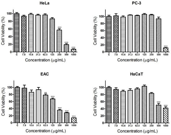
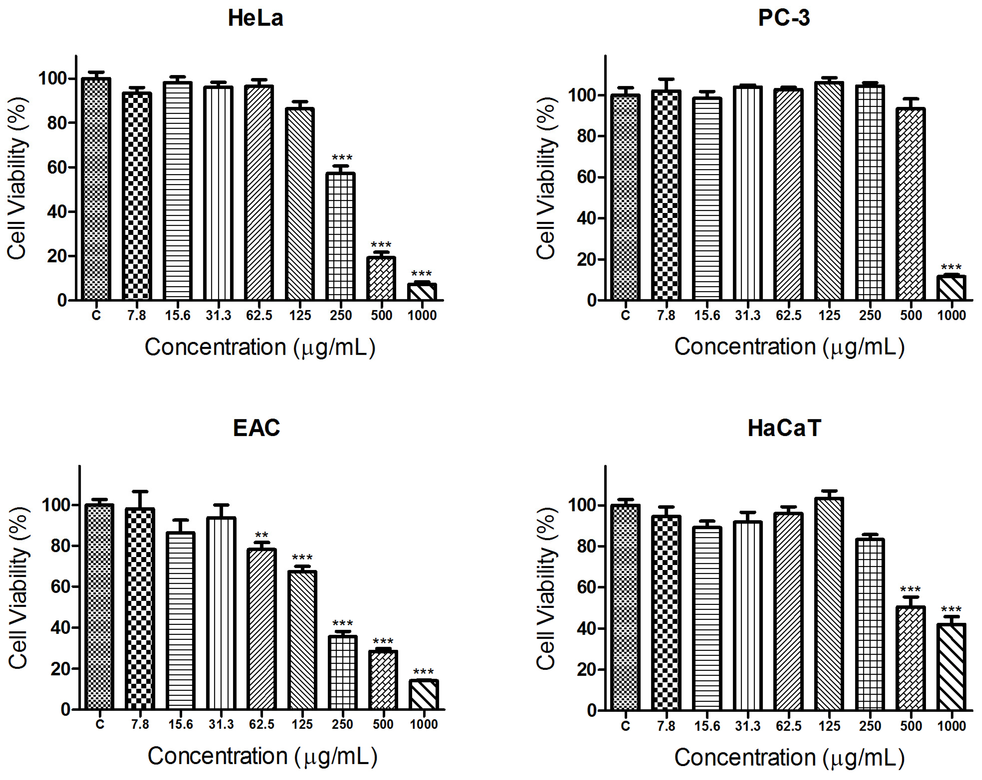
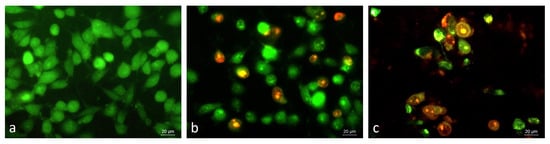
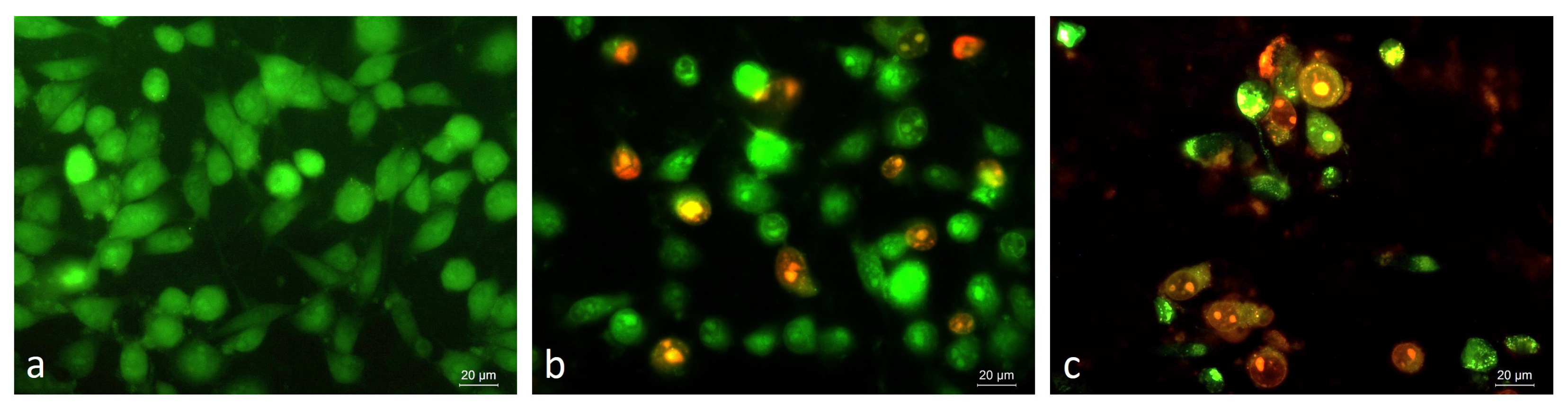
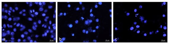
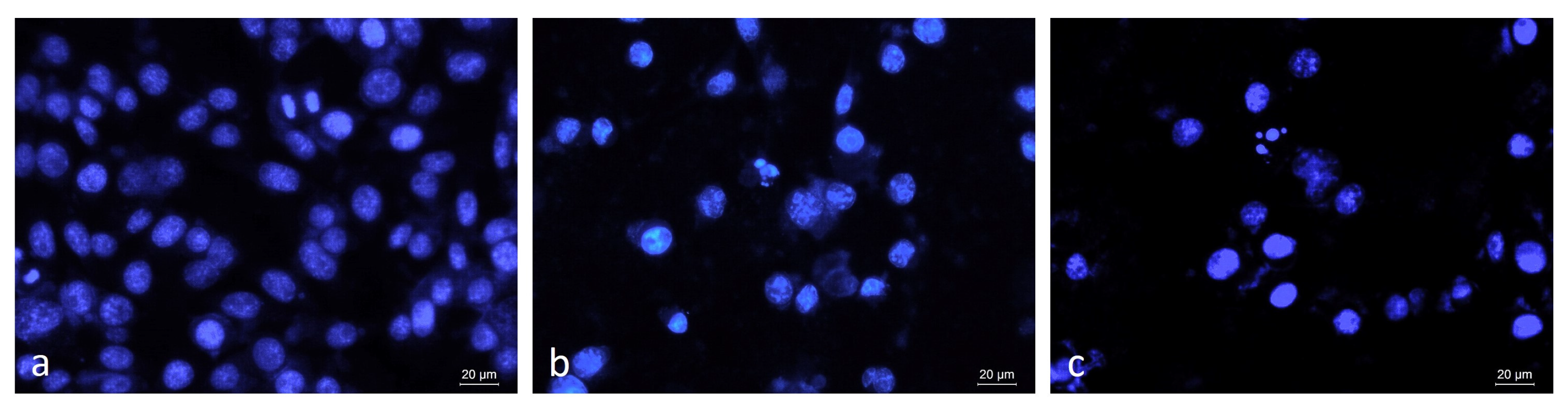
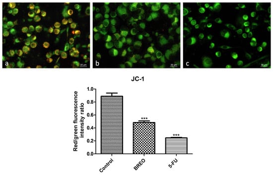
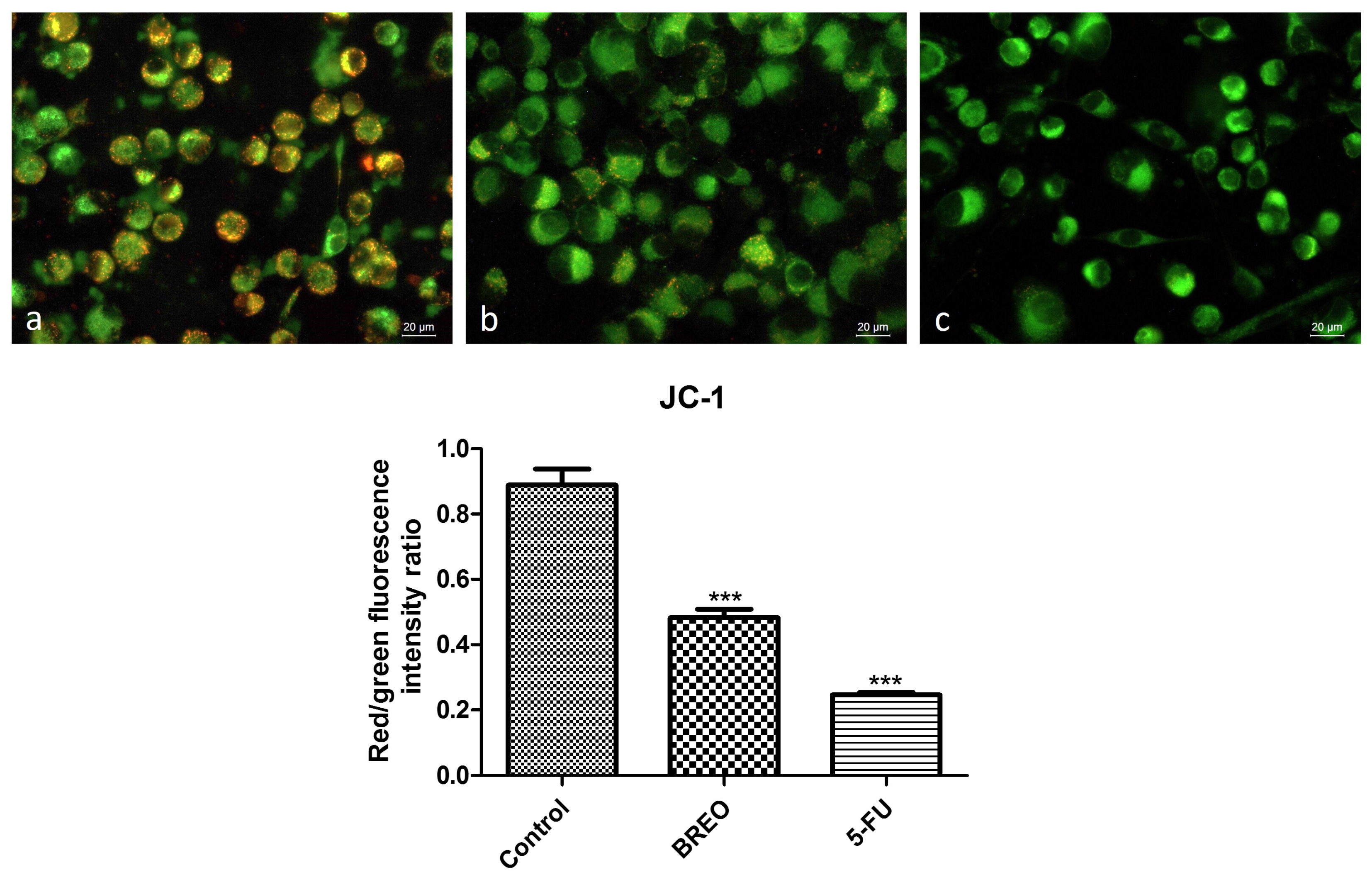
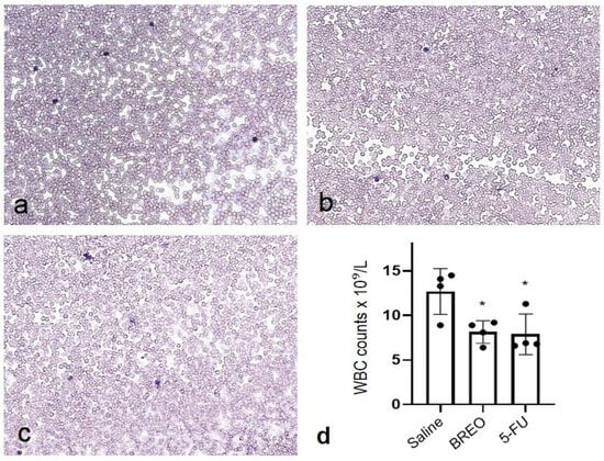
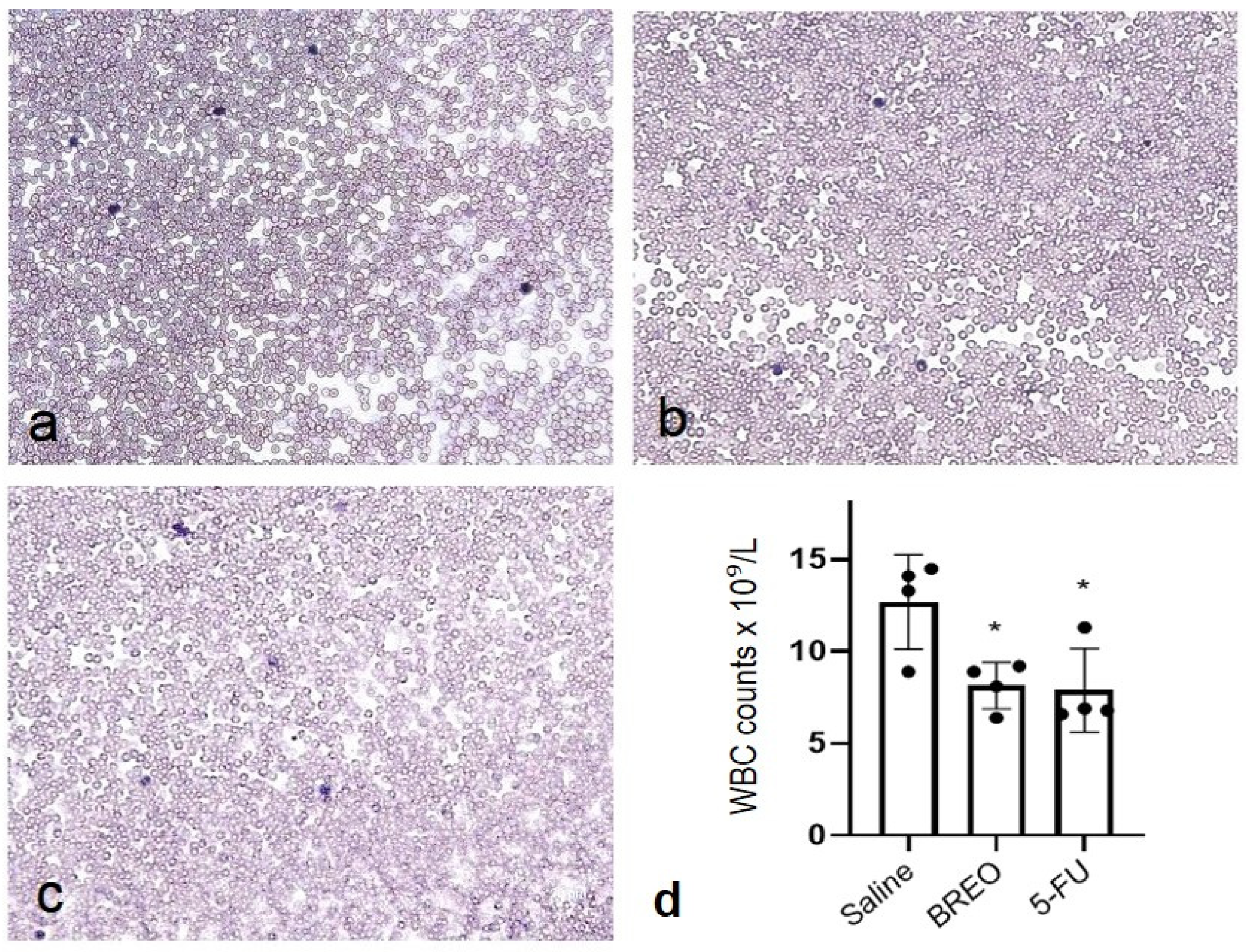
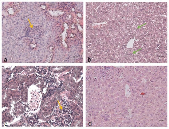
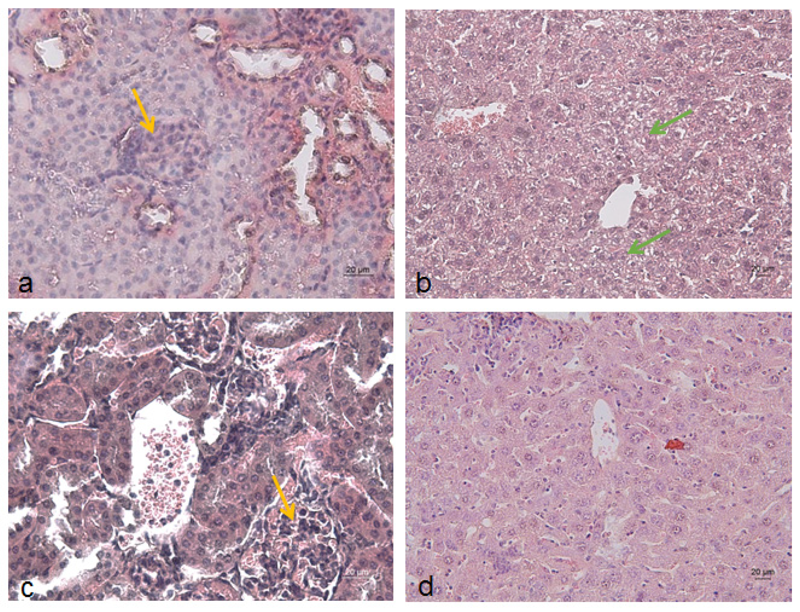
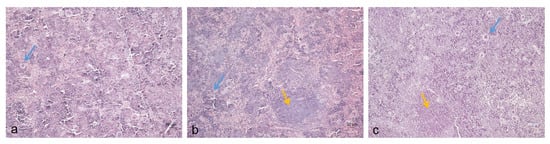
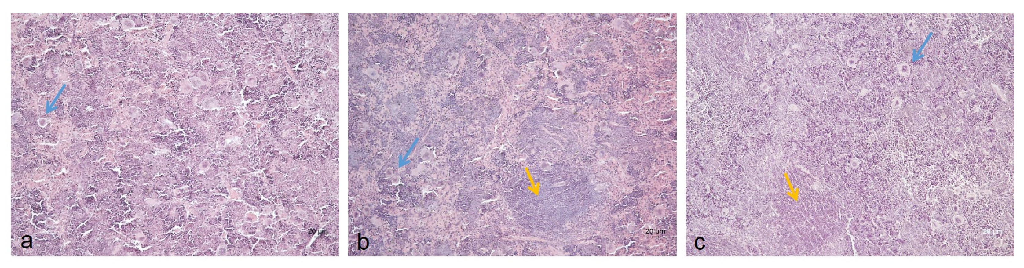
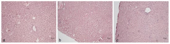
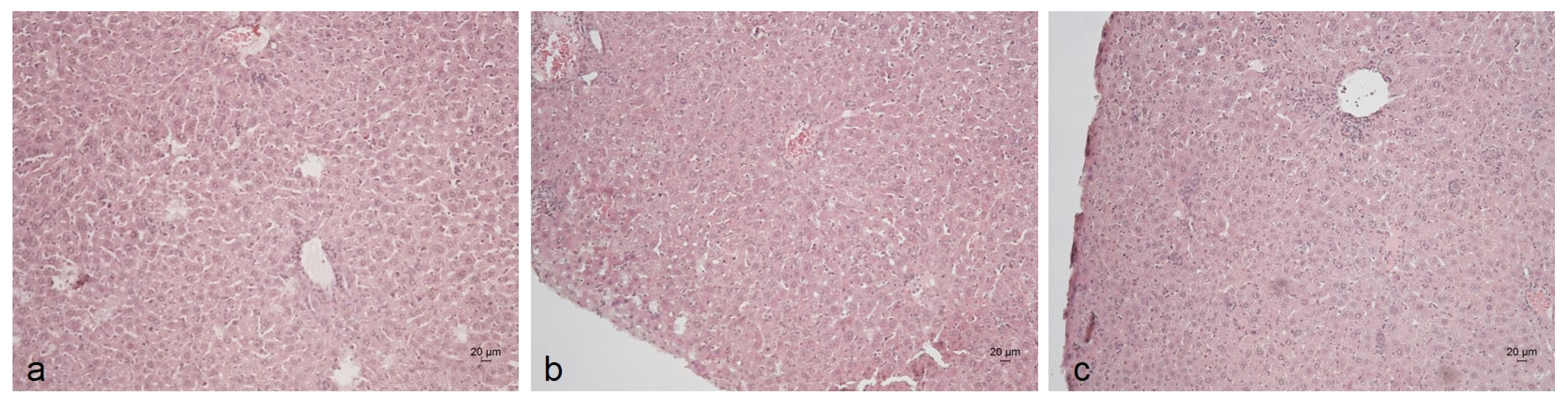
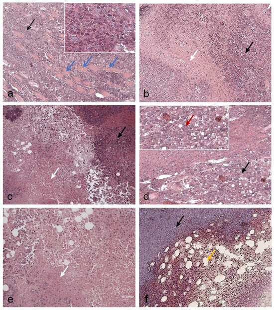
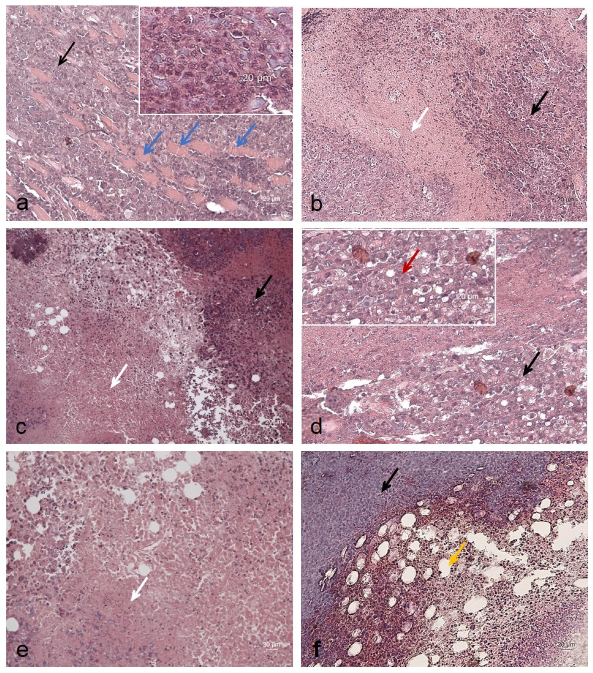
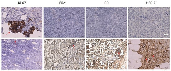
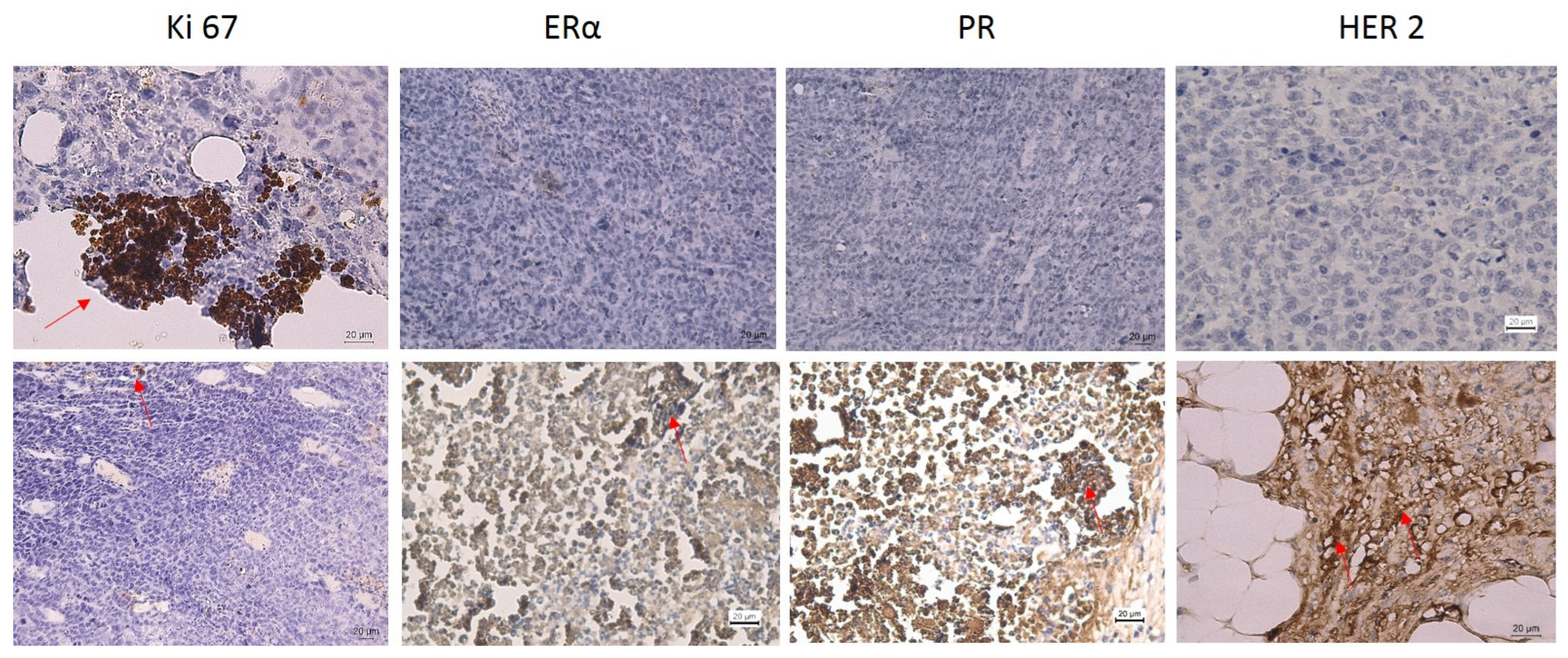
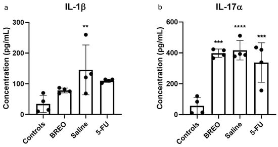
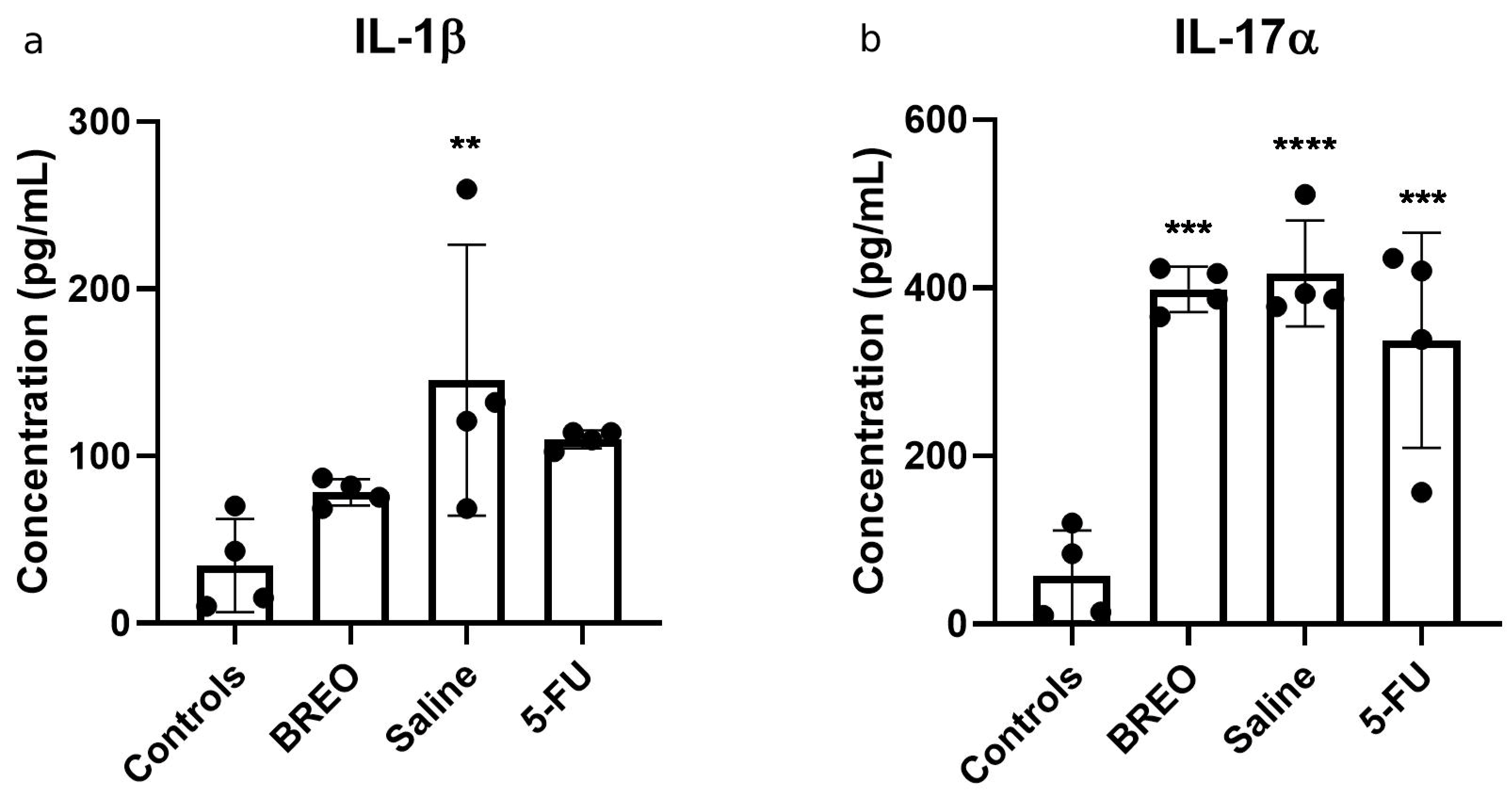
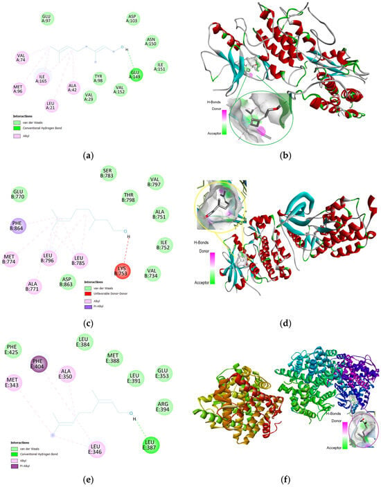
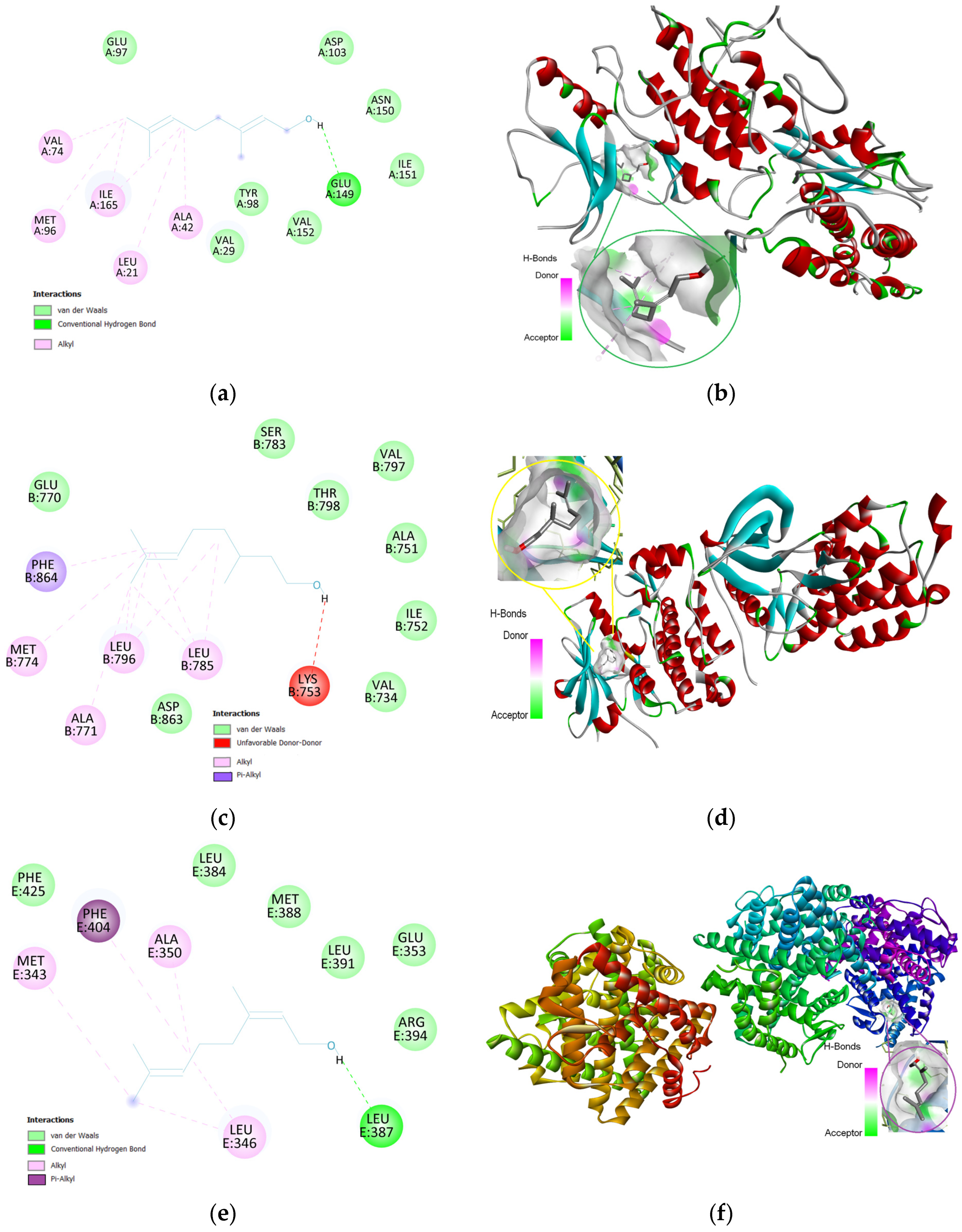

Disclaimer/Publisher’s Note: The statements, opinions and data contained in all publications are solely those of the individual author(s) and contributor(s) and not of MDPI and/or the editor(s). MDPI and/or the editor(s) disclaim responsibility for any injury to people or property resulting from any ideas, methods, instructions or products referred to in the content. |
© 2026 by the authors. Licensee MDPI, Basel, Switzerland. This article is an open access article distributed under the terms and conditions of the Creative Commons Attribution (CC BY) license.