


Abstract
Background: Psoriatic arthritis (PsA) is associated with hepatic risk, but longitudinal data during methotrexate (MTX) and biologic therapy are limited. We evaluated two-year changes in age-fixed FIB-4 and APRI and their associations with cumulative MTX exposure and biologic class. Methods: This retrospective cohort included 74 patients receiving continuous biologic therapy for 24 months. Baseline age was used to calculate FIB-4 at both time points. Logistic regression assessed factors associated with FIB-4 ≥ 1.3 at follow-up. Results: Forty-five patients received anti-TNF therapy and 29 received an IL-17 inhibitor. Age-fixed FIB-4 increased from 1.26 ± 0.44 to 1.72 ± 0.73 (p < 0.001); APRI did not change significantly (0.21 ± 0.08 to 0.23 ± 0.15; p = 0.155). No significant differences in either index were detected between biologic groups. Each 500-mg increase in documented cumulative MTX exposure was associated with higher odds of FIB-4 ≥ 1.3 (OR 2.164, 95% CI 1.04–4.41; p = 0.038). Conclusions: Greater documented cumulative MTX exposure was associated with FIB-4 ≥ 1.3 in an exploratory model. The findings neither establish a causal MTX effect nor demonstrate equivalence between biologic classes and require prospective validation against elastography or histology.
1. Introduction
Psoriatic arthritis (PsA) is a chronic immune-mediated inflammatory disease characterized by peripheral arthritis, axial involvement, enthesitis, dactylitis, and cutaneous psoriasis. Beyond its musculoskeletal and dermatological manifestations, PsA is increasingly recognized as a systemic disease associated with cardiometabolic and hepatic comorbidities. Among these, metabolic dysfunction-associated steatotic liver disease (MASLD), formerly known as non-alcoholic fatty liver disease, is clinically important because of its high prevalence, frequent asymptomatic course, and potential association with progressive liver fibrosis and liver-related complications [1,2,3,4].
The relationship between psoriatic disease and MASLD is multifactorial. Shared metabolic risk factors, including obesity, diabetes mellitus, insulin resistance, dyslipidemia, and hypertension, are common in PsA and may contribute to hepatic steatosis and fibrosis risk [1,5]. In parallel, chronic systemic inflammation may influence hepatic injury pathways through pro-inflammatory cytokines such as tumor necrosis factor-alpha, interleukin-6, and interleukin-17 [6,7,8]. Therefore, liver risk assessment in PsA should be considered within the broader context of systemic inflammation, metabolic dysfunction, and treatment exposure.
Methotrexate (MTX) remains widely used in PsA, either alone or in combination with biologic therapies [9]. However, the hepatic safety of long-term MTX exposure has been debated for decades. Historically, cumulative MTX dose was considered a major determinant of liver fibrosis risk, leading to intensive monitoring strategies in patients with psoriasis and inflammatory arthritis [10]. More recent studies have suggested that metabolic comorbidities may be stronger drivers of liver fibrosis than MTX exposure itself [11,12]. Nevertheless, uncertainty persists in routine rheumatology practice, particularly among patients exposed to MTX over long periods and treated with biologic agents.
Biologic therapies, including anti-tumor necrosis factor (anti-TNF) agents and interleukin-17 inhibitors (IL-17i), have improved PsA outcomes and are widely used in clinical practice [13]. Their relationship with hepatic risk remains incompletely understood. Because IL-17 signaling may be involved in hepatic inflammation and fibrogenesis, IL-17 blockade has theoretical relevance to hepatic outcomes [7,8]. Recent real-world data suggest that IL-17 inhibitors do not worsen non-invasive fibrosis indices during follow-up and may have a neutral hepatic safety profile [14,15]. However, comparative longitudinal data evaluating biologic treatment class together with cumulative MTX exposure remain limited.
Non-invasive liver risk assessment is increasingly important in routine practice. Liver biopsy is unsuitable for screening because of its invasiveness, cost, sampling variability, and limited feasibility [16]. Transient elastography and magnetic resonance elastography provide more direct information on liver stiffness but are not universally available in rheumatology clinics. Therefore, simple laboratory-based indices such as the Fibrosis-4 index (FIB-4) and the aspartate aminotransferase-to-platelet ratio index (APRI) are attractive tools for first-line hepatic risk stratification [17,18,19]. FIB-4 incorporates age, aspartate aminotransferase, alanine aminotransferase, and platelet count, whereas APRI is based on aspartate aminotransferase and platelet count. These indices are not diagnostic substitutes for elastography or histology, but they may support non-invasive risk monitoring and identify patients requiring further evaluation.
In routine practice, FIB-4 and APRI provide inexpensive and readily available first-line risk stratification using standard laboratory parameters. Their principal clinical value lies in excluding advanced fibrosis in patients with low scores and identifying patients with intermediate- or high-risk results who require secondary assessment using transient elastography, other validated non-invasive tests, or specialist referral. Current guidance recommends secondary risk assessment when FIB-4 is ≥1.3, whereas values >2.67 indicate an increased likelihood of clinically significant fibrosis. Higher FIB-4 and APRI values have also been associated with cirrhosis, liver-related complications, and mortality in chronic liver disease. Neither index directly diagnoses portal hypertension; rather, results suggesting advanced fibrosis or cirrhosis should prompt confirmatory assessment and evaluation for portal hypertension-related complications when clinically indicated [20,21].
The use of laboratory-based fibrosis indices has also extended to chronic rheumatic diseases. In rheumatoid arthritis, FIB-4 has been evaluated particularly among patients receiving MTX and has been proposed as a practical marker for identifying patients who require additional hepatic assessment. Longitudinal data have also suggested that rheumatoid arthritis disease activity may influence changes in FIB-4 independently of cumulative MTX exposure. In systemic lupus erythematosus, a large multicenter cohort demonstrated an association between higher FIB-4 values and increased overall mortality. Nevertheless, rheumatology-specific evidence remains more extensive for FIB-4 than for APRI, and neither index should be interpreted as independent confirmation of hepatic fibrosis [22,23,24].
The combined assessment of FIB-4 and APRI may be useful in patients with PsA exposed to MTX and biologic therapies. Recent PsA-specific data further support FIB-4 as a practical first-line triage tool, particularly for ruling out clinically significant liver disease, while emphasizing the need for confirmatory elastography when scores are elevated [25,26]. FIB-4 may be more dynamic for longitudinal risk stratification, whereas APRI may provide supportive concordance using routinely available laboratory parameters. However, the relative contribution of cumulative MTX exposure and biologic treatment class to longitudinal FIB-4 and APRI changes has not been sufficiently clarified. In addition, clinically interpretable MTX exposure thresholds for closer hepatic surveillance remain uncertain.
Therefore, this study aimed to evaluate two-year longitudinal changes in FIB-4 and APRI in patients with PsA receiving biologic therapy with cumulative MTX exposure. The primary objective was to determine whether cumulative MTX dose and biologic therapy class were associated with moderate-to-high FIB-4-based risk at 24 months. Secondary objectives were to evaluate longitudinal APRI changes, assess concordance between APRI and FIB-4-based risk classification, examine the relationship between MTX exposure and radiologic hepatic steatosis, and explore a cumulative MTX threshold for closer non-invasive hepatic monitoring.
2. Materials and Methods
2.1. Study Design and Population
This retrospective longitudinal cohort study included patients with PsA followed in the rheumatology outpatient clinic between 2020 and 2024. Baseline was defined as the date of biologic therapy initiation. A total of 300 patients with PsA were screened for eligibility. Patients were included if they were aged ≥18 years, fulfilled the Classification Criteria for Psoriatic Arthritis (CASPAR), had documented current or previous MTX exposure, received biologic therapy continuously throughout the 24-month follow-up, and had complete clinical, laboratory, and hepatic assessment data at baseline and 24 months [27]. Thus, the minimum duration of biologic exposure required for inclusion was 24 months.
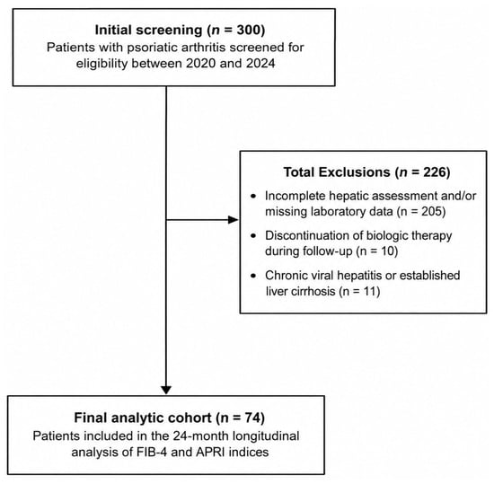
Patients were excluded if they had incomplete hepatic assessment and/or missing laboratory data required for FIB-4/APRI calculation (n = 205), discontinued biologic therapy during follow-up (n = 10), or had chronic viral hepatitis, established liver cirrhosis, or other clinically significant liver disorders that could substantially affect liver biochemistry or non-invasive fibrosis indices (n = 11). Hepatic exclusion criteria included chronic viral hepatitis, established cirrhosis, known autoimmune liver disease, alcohol-related liver disease, decompensated liver disease, hepatic malignancy, or other documented chronic liver disorders when available in medical records. After applying the inclusion and exclusion criteria, 74 patients were included in the final analysis.
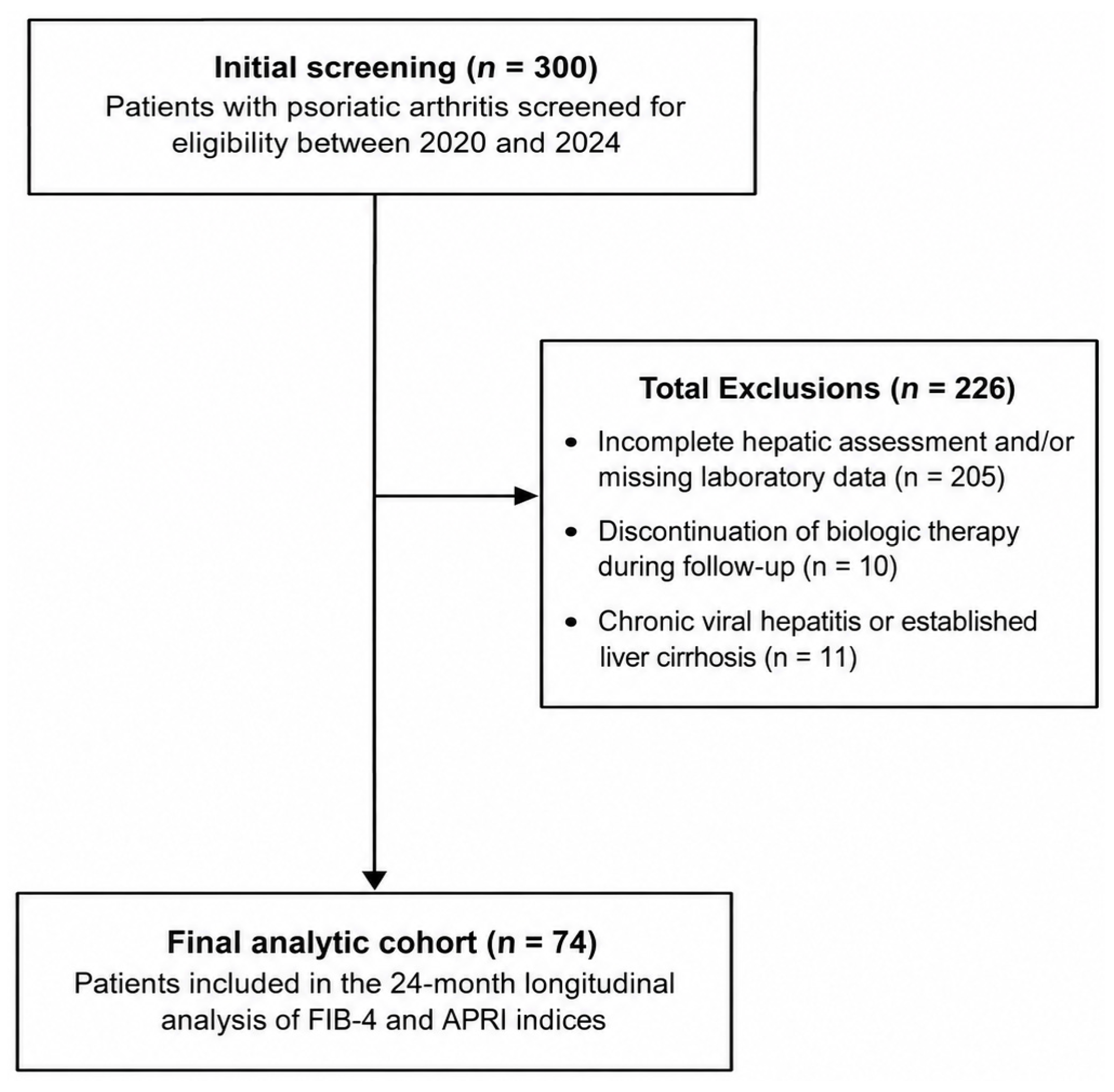
The patient selection process is summarized in Figure 1.
Figure 1.
Study flow diagram. A total of 300 patients with psoriatic arthritis were screened for eligibility between 2020 and 2024. After exclusion of 226 patients because of incomplete hepatic assessment and/or missing laboratory data required for FIB-4/APRI calculation, discontinuation of biologic therapy during follow-up, chronic viral hepatitis, established liver cirrhosis, or other clinically significant liver disorders, 74 patients were included in the final analytic cohort.
To evaluate possible selection bias, available baseline characteristics of included patients were compared with those of excluded patients. Age, sex distribution, biologic treatment class, and MTX exposure did not differ significantly between included and excluded patients with available data.
2.2. Clinical and Demographic Data
Demographic characteristics, including sex, age at the beginning of follow-up, age at PsA onset, disease duration at baseline, and baseline body mass index (BMI), were obtained from electronic medical records. Disease duration was calculated as the interval between PsA onset and baseline. BMI was calculated as weight in kilograms divided by height in meters squared. Obesity was defined as BMI ≥ 30 kg/m2 [28]. Clinical PsA-related variables included enthesitis, dactylitis, radiographic sacroiliitis, HLA-B27 status, and concomitant medication use. Disease activity was assessed using the Disease Activity Index for Psoriatic Arthritis (DAPSA) [29]. Dyslipidemia was defined by a documented physician diagnosis or lipid-lowering therapy. Hyperuricemia was defined by a documented diagnosis or a serum urate value above the sex-specific upper reference limit of the institutional laboratory, and gout was defined by a documented physician diagnosis.
Laboratory parameters, including aspartate aminotransferase (AST), alanine aminotransferase (ALT), gamma-glutamyl transferase (GGT), platelet count, C-reactive protein (CRP), erythrocyte sedimentation rate (ESR), fasting plasma glucose, and glycated hemoglobin (HbA1c), were recorded at baseline and at the 24-month follow-up visit. Diabetes mellitus was identified from a documented diagnosis or glucose-lowering treatment. In patients without a previously documented diagnosis, diabetes was considered present when HbA1c was ≥6.5% or fasting plasma glucose was ≥126 mg/dL, when available, in accordance with the American Diabetes Association criteria [30].
2.3. Treatment Exposure
Biologic treatment class was categorized as anti-TNF therapy or IL-17 inhibitor therapy. Anti-TNF agents included biologics used in routine PsA care, whereas IL-17 inhibitors included secukinumab and ixekizumab. Biologic exposure was continuous throughout the 24-month observation period; patients who discontinued biologic therapy were excluded. Current concomitant MTX use and previous MTX exposure without current use were recorded separately.
Cumulative MTX exposure was reconstructed from documented MTX dose schedules and treatment durations before biologic initiation and during the 24-month observation period. MTX received before referral or prescribed outside the institution was included only when both dose and duration were explicitly documented; undocumented exposure was neither estimated nor imputed. No minimum MTX duration or cumulative dose threshold was specified because exposure was analyzed quantitatively as cumulative dose. For regression analyses, cumulative MTX exposure was rescaled per 500-mg increments to improve clinical interpretability.
Concomitant medications with potential hepatic effects, including corticosteroids and non-steroidal anti-inflammatory drugs, were considered during chart review when documented. However, because of the retrospective design and inconsistent recording of dose and duration, these variables could not be incorporated reliably into the primary multivariable model.
2.4. Assessment of Liver Fibrosis Risk
Non-invasive liver fibrosis risk was assessed using FIB-4 and APRI. FIB-4 and APRI were calculated using laboratory parameters obtained during the same routine clinical assessment whenever available. Laboratory measurements were performed as part of routine clinical follow-up in the institutional laboratory system according to standard clinical procedures. For each time point, laboratory values closest to the baseline and 24-month clinical assessment were used.
FIB-4 was calculated using age, AST, ALT, and platelet count according to the standard formula [17]. For the longitudinal analysis, age at the beginning of follow-up was used in the FIB-4 calculation at both baseline and 24 months. Thus, the age component was held constant to prevent the two-year increase in chronological age from mechanically influencing the longitudinal change:
Baseline FIB-4 = (baseline age × baseline AST)/(baseline platelet count × √baseline ALT)
Age-fixed FIB-4 at 24 months = (baseline age × 24-month AST)/(24-month platelet count × √24-month ALT)
APRI was calculated using AST, the upper limit of normal for AST, and platelet count according to the standard formula [18]:
APRI = [(AST/upper limit of normal AST)/platelet count] × 100
FIB-4 values were categorized using established thresholds: <1.3 was considered low risk, 1.3–2.67 intermediate (moderate) risk, and >2.67 high risk [20]. Because few patients met the high-risk threshold, the intermediate- and high-risk categories were combined as FIB-4 ≥ 1.3 for inferential analyses. APRI was analyzed as a continuous complementary index, and no APRI risk categories were used.
2.5. Radiologic Liver Assessment
Abdominal ultrasonography findings and their original interpretations were extracted from contemporaneous radiology reports in the electronic medical records. Images were not retrieved or re-evaluated specifically for this study. Hepatic steatosis was categorized as absent or present. For sensitivity analyses, radiologic hepatic steatosis was defined as an original ultrasonographic report of fatty liver, and advanced steatosis was considered when the report indicated grade ≥ 2 steatosis. The examinations and interpretations had been performed by radiologists as part of routine clinical care, before the present study and without knowledge of its objectives.
Transient elastography was not routinely available for the full cohort and was not performed systematically as part of the study protocol. Therefore, elastography data were not included in the analyses.
2.6. Study Outcomes
The primary outcome was moderate-to-high FIB-4-based risk at 24 months, defined as FIB-4 ≥ 1.3. Secondary outcomes included longitudinal changes in FIB-4 and APRI over 24 months, differences in liver fibrosis indices according to biologic treatment class, the association between cumulative MTX exposure and fibrosis indices, the performance of cumulative MTX dose for identifying moderate-to-high FIB-4-based risk, and the concordance between APRI and FIB-4-based risk categories.
Sensitivity analyses were performed using alternative outcomes independent of the primary FIB-4-based categorization, including radiologic hepatic steatosis and longitudinal change in FIB-4 score.
2.7. Statistical Analysis
Statistical analyses were performed using IBM SPSS Statistics for Windows, Version 27.0 (IBM Corp., Armonk, NY, USA). The normality of continuous variables was assessed using visual methods and appropriate normality tests. Continuous variables were expressed as mean ± standard deviation or median with interquartile range, depending on data distribution. Categorical variables were presented as frequencies and percentages.
Baseline and 24-month values were compared using paired-samples tests or repeated-measures models, as appropriate. Between-group comparisons according to biologic treatment class were performed using independent-samples tests for continuous variables and chi-square or Fisher’s exact tests for categorical variables. Because only 14 patients had FIB-4 ≥ 1.3 at 24 months, two-tailed Fisher’s exact tests were used for exploratory comparisons of dyslipidemia, obesity, hyperuricemia, and gout across FIB-4 risk groups.
Available baseline characteristics of included and excluded patients were compared to evaluate potential selection bias. Variables used for this comparison included age, sex, biologic treatment class, and MTX exposure.
Multivariable logistic regression analysis was performed to identify factors associated with FIB-4 ≥ 1.3 at 24 months. Because only 14 outcome events occurred, the model was considered exploratory and was restricted to prespecified clinically relevant variables: documented cumulative MTX dose per 500-mg increment, sex, diabetes mellitus, and biologic treatment class. The possibility of model overfitting was considered when interpreting the results. Dyslipidemia, obesity, hyperuricemia, and gout were reported and compared between risk groups but were not entered individually into the multivariable model because doing so would further increase the risk of overfitting. Age was not included as an independent variable because baseline age was already incorporated into the FIB-4 outcome.
Receiver operating characteristic curve analysis was performed to evaluate the discriminative ability of cumulative MTX dose for identifying moderate-to-high FIB-4-based risk. The optimal cut-off value was determined using the Youden index. Additional ROC analyses were performed to evaluate the concordance between APRI and FIB-4-based risk classification and to explore the relationship between FIB-4 and radiologic hepatic steatosis.
A two-sided p value < 0.05 was considered statistically significant.
The study was conducted in accordance with the Declaration of Helsinki and approved by the local Non-Interventional Research Ethics Committee (Approval No: 2024/400; Meeting No: 20; Approval Date: 13 November 2024). Patient consent was waived because of the retrospective design and the use of anonymized clinical data.
3. Results
3.1. Patient Selection and Baseline Characteristics
The mean age at the beginning of follow-up was 43.34 ± 6.51 years, and 49 patients (66.2%) were male. The mean age at PsA onset was 36.25 ± 7.42 years, the mean disease duration was 7.09 ± 4.15 years, and the mean baseline BMI was 28.45 ± 4.23 kg/m2. Forty-five patients (60.8%) received anti-TNF therapy and 29 (39.2%) received IL-17 inhibitor therapy. All patients had documented current or previous MTX exposure: 30 (40.5%) were receiving concomitant MTX at baseline and 44 (59.5%) had previous exposure without current MTX use. Dyslipidemia, obesity, hyperuricemia, and gout were present in 28 (37.8%), 22 (29.7%), 14 (18.9%), and 5 (6.8%) patients, respectively. HLA-B27 positivity was present in 12 patients (16.2%), while enthesitis, dactylitis, and radiographic sacroiliitis were present in 19 (25.7%), 10 (13.5%), and 8 (10.8%) patients, respectively. Baseline characteristics are summarized in Table 1. Dyslipidemia, obesity, hyperuricemia, and gout were numerically more frequent in the FIB-4 ≥ 1.3 group, but none of the exploratory comparisons were statistically significant (all p > 0.05; Supplementary Table S5).
Table 1.
Baseline demographic and clinical characteristics of the study population.
3.2. Longitudinal Changes in Disease Activity and Laboratory Parameters
During the 24-month follow-up period, disease activity and systemic inflammatory markers improved significantly. The mean DAPSA score decreased from 30.47 ± 4.16 at baseline to 3.65 ± 1.07 at 24 months (p < 0.001). Similarly, CRP decreased from 6.35 ± 9.74 mg/L to 1.72 ± 2.65 mg/L (p < 0.001), and ESR decreased from 15.76 ± 10.29 mm/h to 9.50 ± 6.15 mm/h (p < 0.001).
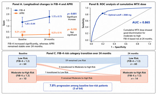
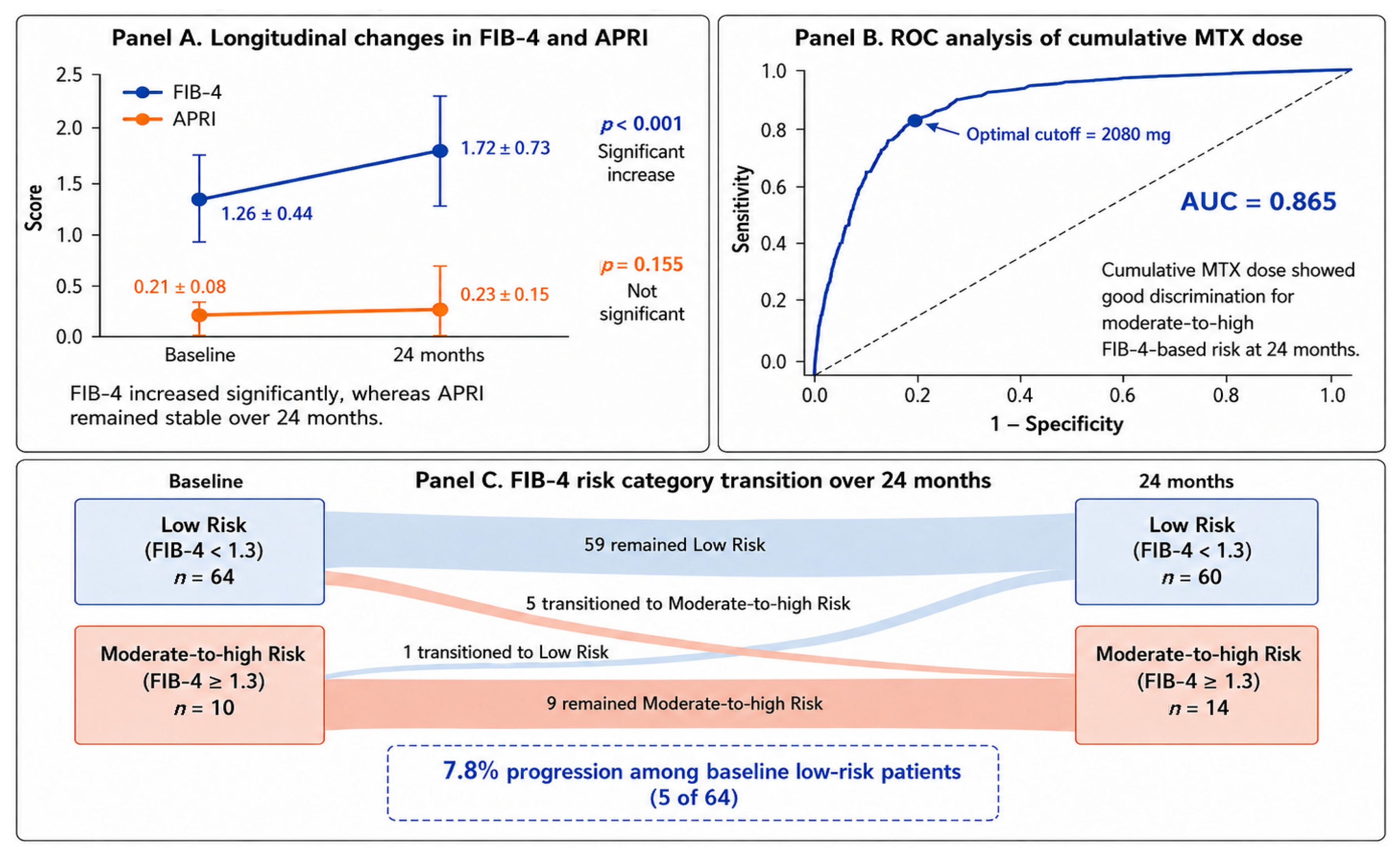
With baseline age held constant in the FIB-4 calculation at both time points, mean FIB-4 increased from 1.26 ± 0.44 at baseline to 1.72 ± 0.73 at 24 months (p < 0.001). Therefore, this increase was not mechanically attributable to chronological aging during follow-up. In contrast, APRI, which does not include age, showed no statistically significant change (0.21 ± 0.08 vs. 0.23 ± 0.15, p = 0.155). ALT increased numerically from 22.81 ± 13.01 U/L to 37.18 ± 97.39 U/L, but the change was not statistically significant (p = 0.148). The wide variability in 24-month ALT values suggested a skewed distribution driven by outlying values; therefore, ALT findings were interpreted cautiously. Detailed longitudinal changes are presented in Table 2.
Table 2.
Two-year longitudinal changes in clinical and laboratory parameters.
3.3. Categorical Changes in FIB-4-Based Risk and Radiologic Hepatic Findings
At baseline, 64 patients (86.5%) were classified as low risk according to FIB-4, whereas 10 patients (13.5%) were classified as moderate-to-high risk. At 24 months, 60 patients (81.1%) were in the low-risk category, whereas 14 patients (18.9%) were classified as moderate-to-high risk. Among patients who were initially classified as low risk, 5 of 64 patients (7.8%) transitioned to the moderate-to-high FIB-4-based risk category during follow-up (Figure 2C).
Figure 2.
Longitudinal monitoring of liver fibrosis risk in psoriatic arthritis. (A) Longitudinal changes in FIB-4 and APRI scores over 24 months. With baseline age held constant, FIB-4 increased significantly from baseline to 24 months (1.26 ± 0.44 vs. 1.72 ± 0.73, p < 0.001), whereas APRI showed a nonsignificant change (0.21 ± 0.08 vs. 0.23 ± 0.15, p = 0.155). Data are presented as mean ± standard deviation. (B) Receiver operating characteristic curve of cumulative MTX dose for identifying moderate-to-high FIB-4-based risk at 24 months. Cumulative MTX dose demonstrated good discriminative ability, with an AUC of 0.865. The optimal threshold determined by the Youden index was 2080 mg. This threshold should be interpreted as exploratory rather than clinically validated. (C) Transition of FIB-4 risk categories from baseline to 24 months. Among 64 patients initially classified as low risk, 59 remained low risk and 5 transitioned to moderate-to-high risk. Among 10 patients initially classified as moderate-to-high risk, 9 remained in the same category and 1 transitioned to low risk.
At baseline, abdominal ultrasonography showed normal liver findings in 55 patients (74.3%), whereas hepatic steatosis was present in 19 patients (25.7%). At 24 months, normal ultrasonographic findings were observed in 50 patients (67.6%), whereas hepatic steatosis was detected in 24 patients (32.4%). The categorical distribution of FIB-4-based risk and hepatic ultrasonography findings is shown in Supplementary Table S2.
3.4. Comparison According to Biologic Treatment Class
At 24 months, there were no statistically significant differences between anti-TNF and IL-17 inhibitor groups in terms of non-invasive fibrosis indices. The mean FIB-4 score was 1.80 ± 0.69 in the anti-TNF group and 1.59 ± 0.78 in the IL-17 inhibitor group (p = 0.078). The mean APRI score was 0.22 ± 0.13 and 0.23 ± 0.18 in the anti-TNF and IL-17 inhibitor groups, respectively (p = 0.335).
Cumulative MTX dose was also similar between the two biologic treatment groups (1541.6 ± 441.9 mg vs. 1483.7 ± 291.7 mg, p = 0.678), and no significant between-group difference was observed in 24-month DAPSA scores (3.57 ± 1.02 vs. 3.78 ± 1.15, p = 0.418). Thus, no statistically significant between-group differences were detected for the evaluated indices or disease activity; these nonsignificant results should not be interpreted as evidence of equivalence between biologic classes (Table 3).
Table 3.
Comparison of liver-related markers according to biologic treatment class.
3.5. Correlation Analyses
Cumulative MTX dose showed a weak but statistically significant positive correlation with 24-month FIB-4 score (r = 0.231, p = 0.043). Baseline DAPSA score was also positively correlated with longitudinal FIB-4 change (ΔFIB-4) (r = 0.229, p = 0.043). In contrast, 24-month CRP was not significantly correlated with 24-month FIB-4 score (r = 0.056, p = 0.622).
A very strong positive correlation was observed between 24-month APRI and 24-month FIB-4 scores (r = 0.958, p < 0.001), supporting concordance between these two laboratory-based non-invasive indices. However, because APRI and FIB-4 share common laboratory components, this finding was interpreted as concordance rather than independent diagnostic validation. Correlation analyses are summarized in Supplementary Table S3.
3.6. Multivariable Logistic Regression Analysis
In the exploratory multivariable logistic regression model, each 500-mg increase in documented cumulative MTX exposure was associated with higher odds of FIB-4 ≥ 1.3 at 24 months (OR: 2.164, 95% CI: 1.04–4.41, p = 0.038). This estimate was based on only 14 outcome events and should therefore be interpreted cautiously.
Male sex was not significantly associated with FIB-4 ≥ 1.3 (OR: 1.842, 95% CI: 0.76–4.45, p = 0.174). Diabetes mellitus showed a nonsignificant trend (OR: 1.955, 95% CI: 0.88–4.34, p = 0.098). No statistically significant association was detected for IL-17 inhibitor therapy compared with anti-TNF therapy (OR: 0.540, 95% CI: 0.22–1.31, p = 0.172). Given the limited number of events, the model was considered potentially susceptible to overfitting and unable to establish equivalence or exclude clinically relevant differences between biologic classes (Table 4).
Table 4.
Multivariable logistic regression model for moderate-to-high FIB-4-based risk at 24 months.
3.7. Sensitivity and ROC Analyses
Sensitivity analyses used liver-related outcomes independent of the primary FIB-4 risk classification. Documented cumulative MTX dose was not significantly associated with ultrasonographic steatosis (OR per 500-mg increase: 1.082, p = 0.345). When longitudinal FIB-4 change was evaluated as a continuous outcome, documented cumulative MTX exposure was positively associated with ΔFIB-4 (β = 0.224, p = 0.045). These exploratory findings indicate statistical associations with FIB-4-based outcomes but do not establish MTX-related fibrosis progression.
ROC analysis demonstrated that cumulative MTX dose had good discriminative ability for identifying patients with moderate-to-high FIB-4-based risk at 24 months, with an AUC of 0.865 (Figure 2B). The optimal cumulative MTX threshold determined by the Youden index was 2080 mg. However, because this threshold was derived from a relatively small cohort with a limited number of outcome events, it should be interpreted as an exploratory finding rather than a validated clinical cut-off.
APRI showed high concordance with FIB-4-based risk classification, with an AUC of 0.970 for identifying patients classified as moderate-to-high risk according to FIB-4. However, because FIB-4 and APRI share common laboratory components, this analysis was interpreted as concordance between non-invasive indices rather than as proof of diagnostic superiority. Detailed ROC analyses are shown in Supplementary Table S4.
The longitudinal trajectories of FIB-4 and APRI, the ROC analysis of cumulative MTX exposure, and the transition of FIB-4-based risk categories are summarized in Figure 2.
4. Discussion
In this two-year retrospective longitudinal cohort study, age-fixed FIB-4 increased, whereas APRI showed no statistically significant longitudinal change. Greater documented cumulative MTX exposure was associated with FIB-4 ≥ 1.3 in an exploratory adjusted model. No statistically significant differences in FIB-4 or APRI were detected between the anti-TNF and IL-17 inhibitor groups; however, the study was not powered to establish equivalence or to demonstrate that the effect of biologic class was weaker than that of MTX exposure. APRI showed strong concordance with FIB-4-based classification, and the exploratory cumulative MTX threshold of approximately 2080 mg requires external validation.
The significant increase in FIB-4 despite marked improvement in DAPSA, CRP, and ESR is clinically relevant. It suggests that improvement in articular disease activity and systemic inflammatory markers does not necessarily eliminate the need for hepatic risk monitoring in PsA. However, this finding should be interpreted as an increase in FIB-4-based risk rather than confirmed liver fibrosis. FIB-4 is a laboratory-based screening and risk stratification tool, not a diagnostic confirmation of fibrosis or liver injury. Therefore, patients with elevated or increasing FIB-4 should undergo further evaluation using metabolic risk assessment, repeated non-invasive indices, and, when available, transient elastography or hepatology consultation.
Greater documented cumulative MTX exposure was associated with FIB-4 ≥ 1.3 in the adjusted model, but this finding rests on a single estimate from a model with only 14 outcome events and a relatively wide confidence interval. It should therefore be considered exploratory and hypothesis-generating. The study cannot establish that MTX exposure causes liver fibrosis. Moreover, retrospective reconstruction may have incompletely captured MTX received before referral or prescribed elsewhere. Because undocumented exposure was not estimated, cumulative MTX dose may have been underestimated; although such misclassification was likely nondifferential, its direction and magnitude cannot be determined. Higher documented cumulative exposure may identify a subgroup warranting closer non-invasive hepatic surveillance rather than demonstrate a causal MTX effect.
The exploratory cumulative MTX threshold of approximately 2080 mg may have practical relevance, but it should not be considered a validated clinical cut-off. The threshold was derived from a relatively small cohort and a limited number of outcome events. Therefore, it should be viewed as hypothesis-generating and should not be incorporated into clinical decision-making without external validation. Larger prospective studies incorporating elastography-based reference standards are needed to determine whether cumulative MTX exposure thresholds can meaningfully guide hepatic monitoring strategies in PsA.
No statistically significant differences were detected between anti-TNF and IL-17 inhibitor groups in 24-month FIB-4, APRI, documented cumulative MTX exposure, or disease activity, and biologic class was not statistically significant in the exploratory multivariable model. These null findings should not be interpreted as proof of comparable hepatic effects or true equivalence. The sample was small and the confidence interval for biologic class was wide; therefore, a clinically relevant class effect cannot be excluded.
The role of IL-17 in liver disease is biologically complex. IL-17 signaling has been implicated in hepatic inflammation, stellate cell activation, and fibrogenesis [7,8], but clinical data remain limited and heterogeneous. The present study neither demonstrates a hepatoprotective effect of IL-17 inhibition nor excludes a difference between IL-17 inhibitors and anti-TNF therapy. Larger adequately powered comparative studies are required.
APRI was evaluated as a complementary index in this study. Although APRI did not increase significantly over time, it showed strong concordance with 24-month FIB-4 values and FIB-4-based risk classification. This may support its use as an additional laboratory-based index in hepatic risk monitoring. However, APRI and FIB-4 share common laboratory components, particularly AST and platelet count. Therefore, the observed APRI–FIB-4 concordance should not be interpreted as independent diagnostic validation or superiority. Instead, APRI may provide supportive information when interpreted together with FIB-4 and clinical risk factors.
The relationship between FIB-4 and ultrasonographic steatosis also requires careful interpretation. Cumulative MTX exposure was not significantly associated with radiologic hepatic steatosis, whereas it remained associated with longitudinal FIB-4 change. This suggests that FIB-4-based risk and ultrasonographic steatosis may capture different aspects of hepatic involvement. Conventional ultrasonography is useful for detecting steatosis but is limited for early fibrosis assessment, whereas FIB-4 is designed to estimate fibrosis risk rather than quantify hepatic fat accumulation [16,19]. Therefore, the absence of a strong association between FIB-4 and steatosis should not be considered contradictory.
Residual confounding by metabolic and hepatic factors remains important. Baseline BMI and documented dyslipidemia, obesity, hyperuricemia, and gout were reported and compared across FIB-4 risk groups, with no statistically significant univariable associations. Nevertheless, only 14 outcome events precluded their reliable simultaneous inclusion in the multivariable model. Other potential determinants, including hypertension, waist circumference, detailed alcohol intake, and the dose and duration of concomitant medications with hepatic effects, were not uniformly available. Consequently, residual confounding cannot be excluded.
Selection bias should also be considered. The final analytic cohort included 74 of 300 screened patients, mainly because many patients lacked complete hepatic assessment or laboratory data required for FIB-4/APRI calculation. To address this concern, available baseline characteristics of included and excluded patients were compared, and no significant differences were observed in age, sex distribution, biologic treatment class, or MTX exposure. Nevertheless, patients with complete laboratory and hepatic monitoring may differ from those with incomplete data in unmeasured ways, and the generalizability of the findings remains limited.
Chronological age is incorporated directly into the standard FIB-4 formula and may therefore mechanically increase FIB-4 during longitudinal follow-up. In the present analysis, baseline age was held constant when calculating FIB-4 at both time points. Consequently, the observed increase cannot be explained by the two-year increase in chronological age and reflects changes in the laboratory components of the index. Nevertheless, FIB-4 remains an indirect surrogate measure, and its longitudinal increase should not be interpreted as definitive evidence of fibrosis progression without confirmation by elastography, other imaging modalities, or histopathology.
This study has several strengths. First, it provides two-year longitudinal real-world data on FIB-4 and APRI in patients with PsA receiving biologic therapy. Second, it evaluates cumulative MTX exposure and biologic treatment class together, which reflects routine clinical practice. Third, MTX exposure was analyzed using clinically interpretable 500-mg increments. Fourth, sensitivity analyses were performed using outcomes independent of the primary FIB-4-based classification, including radiologic steatosis and longitudinal FIB-4 change. Finally, the study explicitly evaluates APRI as a complementary index for concordance with FIB-4-based risk.
Several limitations should be acknowledged. First, the retrospective single-center design and small sample limited statistical power, subgroup analyses, and generalizability. Second, the multivariable model included only 14 outcome events and was susceptible to overfitting; its MTX estimate is exploratory, and the nonsignificant biologic-class result cannot establish equivalence. Third, all participants had documented current or previous MTX exposure, so an MTX-naïve comparator group was unavailable. Fourth, cumulative MTX exposure may have been incompletely captured for treatment before referral or outside the institution, and residual metabolic and medication-related confounding remains possible. Fifth, liver fibrosis was assessed using laboratory-based indices rather than elastography, magnetic resonance elastography, or biopsy. Although baseline age was fixed in the longitudinal FIB-4 calculations to remove the mechanical effect of aging, this age-fixed approach and the use of FIB-4 thresholds remain surrogate risk assessments rather than confirmation of fibrosis. Finally, the 2080-mg cumulative MTX threshold was derived from this cohort and should not guide clinical decisions without external validation.
5. Conclusions
In this exploratory cohort of patients with PsA receiving biologic therapy, greater documented cumulative MTX exposure was associated with FIB-4 ≥ 1.3 at 24 months. No statistically significant differences in FIB-4 or APRI were detected between anti-TNF and IL-17 inhibitor groups. Given the small number of outcome events, potential exposure misclassification, residual confounding, and limited statistical power, these findings neither establish a causal MTX effect nor demonstrate equivalence between biologic classes. FIB-4 and APRI may support non-invasive hepatic risk stratification, but prospective validation against elastography or histology is required.
Supplementary Materials
The following supporting information can be downloaded at: https://www.mdpi.com/article/10.3390/biologics6030025/s1. Table S1: Comparison of available baseline characteristics between included and excluded patients; Table S2: Categorical distribution of FIB-4-based risk and radiologic hepatic steatosis; Table S3: Correlations between fibrosis indices and clinical parameters; Table S4: ROC curve analyses; Table S5: Distribution of additional comorbidities according to 24-month FIB-4 risk category.
Author Contributions
Conceptualization, M.S.K. and S.E.M.A.; methodology, M.S.K., S.E.M.A. and A.Y.T.; formal analysis, A.Y.T. and T.A.; investigation, M.S.K., S.E.M.A. and O.V.Y.; data curation, M.S.K., S.E.M.A. and O.V.Y.; writing—original draft preparation, M.S.K.; writing—review and editing, M.S.K., S.E.M.A., O.V.Y., A.Y.T. and T.A.; supervision, T.A. All authors have read and agreed to the published version of the manuscript.
Funding
This research received no external funding.
Institutional Review Board Statement
The study was conducted in accordance with the Declaration of Helsinki and approved by the local Non-Interventional Research Ethics Committee (Approval No: 2024/400; Meeting No: 20; Approval Date: 13 November 2024).
Informed Consent Statement
Patient consent was waived because of the retrospective design of the study and the use of anonymized clinical data.
Data Availability Statement
The datasets generated and/or analyzed during the current study are not publicly available due to institutional and ethical restrictions related to retrospective clinical data. De-identified data supporting the findings of this study may be available from the corresponding author upon reasonable request and with permission of the relevant institutional authorities.
Acknowledgments
The authors thank the clinical staff of the physical therapy and rehabilitation outpatient clinic for their support in patient follow-up and data collection.
Conflicts of Interest
The authors declare no conflicts of interest.
Abbreviations
The following abbreviations are used in this manuscript:
| ALT | Alanine aminotransferase |
| APRI | Aspartate aminotransferase-to-platelet ratio index |
| AST | Aspartate aminotransferase |
| AUC | Area under the curve |
| CASPAR | Classification Criteria for Psoriatic Arthritis |
| CRP | C-reactive protein |
| DAPSA | Disease Activity Index for Psoriatic Arthritis |
| ESR | Erythrocyte sedimentation rate |
| FIB-4 | Fibrosis-4 index |
| GGT | Gamma-glutamyl transferase |
| IL-17 | Interleukin-17 |
| MASLD | Metabolic dysfunction-associated steatotic liver disease |
| MTX | Methotrexate |
| PsA | Psoriatic arthritis |
| ROC | Receiver operating characteristic |
| TNF | Tumor necrosis factor |
| USG | Ultrasonography |
References
- Ogdie, A.; Grewal, S.K.; Noe, M.H.; Shin, D.B.; Takeshita, J.; Fuxench, Z.C.C.; Carr, R.M.; Gelfand, J.M. Risk of incident liver disease in patients with psoriasis, psoriatic arthritis, and rheumatoid arthritis: A population-based study. J. Investig. Dermatol. 2018, 138, 760–767. [Google Scholar] [CrossRef] [Scilit] [PubMed]
- Rinella, M.E.; Lazarus, J.V.; Ratziu, V.; Francque, S.M.; Sanyal, A.J.; Kanwal, F.; Romero, D.; Abdelmalek, M.F.; Anstee, Q.M.; Arab, J.P.; et al. A multisociety Delphi consensus statement on new fatty liver disease nomenclature. Hepatology 2023, 78, 1966–1986. [Google Scholar] [CrossRef] [Scilit] [PubMed]
- Miele, L.; Vallone, S.; Cefalo, C.; La Torre, G.; Di Stasi, C.; Vecchio, F.M.; D’agostino, M.; Gabrieli, M.L.; Vero, V.; Biolato, M.; et al. Prevalence, characteristics and severity of non-alcoholic fatty liver disease in patients with chronic plaque psoriasis. J. Hepatol. 2009, 51, 778–786. [Google Scholar] [CrossRef] [Scilit] [PubMed]
- James, L.; Diamond, C.; Al-Mossawi, H.; Andersson, A.; Pandya, P.; Shumbayawonda, E.; Firouzabadi, L.I.; Watson, L.; Savage, L.J.; Thomaides-Brears, H.; et al. High prevalence of MASLD in psoriasis and psoriatic arthritis assessed with multiparametric magnetic resonance imaging. Rheumatology 2025, 64, 5741–5750. [Google Scholar] [CrossRef] [Scilit] [PubMed]
- Watabe, H.; Miyagaki, T.; Nakajima, K.; Go, K.; Okano, T.; Kadono, T. Clinical profile of psoriasis patients with comorbid fatty liver and risk factors for advanced liver fibrosis in those with metabolic dysfunction-associated steatotic liver disease. J. Dermatol. 2026, 53, 237–244. [Google Scholar] [CrossRef] [Scilit] [PubMed]
- Boehncke, W.H.; Schön, M.P. Psoriasis. Lancet 2015, 386, 983–994. [Google Scholar] [CrossRef] [Scilit] [PubMed]
- Takamura, S.; Teraki, Y.; Katayama, E.; Kawaguchi, T.; Kawaguchi, M.; Nakano, D.; Tsutsumi, T.; Nagoshi, S.; Nakama, T.; Torimura, T. Effects of interleukin-17 inhibitors on hepatic fibrosis index in patients with psoriasis and metabolic dysfunction-associated fatty liver disease: Directed acyclic graphs. Clin. Mol. Hepatol. 2022, 28, 269–272. [Google Scholar] [CrossRef] [Scilit] [PubMed]
- Heitmann, J.; Frings, V.G.; Geier, A.; Goebeler, M.; Kerstan, A. Non-alcoholic fatty liver disease and psoriasis—Is there a shared proinflammatory network? J. Dtsch. Dermatol. Ges. 2021, 19, 517–528. [Google Scholar] [CrossRef] [Scilit] [PubMed]
- Gossec, L.; Baraliakos, X.; Kerschbaumer, A.; de Wit, M.; McInnes, I.; Dougados, M.; Primdahl, J.; McGonagle, D.G.; Aletaha, D.; Balanescu, A.; et al. EULAR recommendations for the management of psoriatic arthritis with pharmacological therapies: 2019 update. Ann. Rheum. Dis. 2020, 79, 700–712. [Google Scholar] [CrossRef] [Scilit] [PubMed]
- Maybury, C.M.; Jabbar-Lopez, Z.K.; Wong, T.; Dhillon, A.P.; Barker, J.N.; Smith, C.H. Methotrexate and liver fibrosis in people with psoriasis: A systematic review of observational studies. Br. J. Dermatol. 2014, 171, 17–29. [Google Scholar] [CrossRef] [Scilit] [PubMed]
- Kharouf, F.; Mehta, P.; Abarza, V.C.; Gao, S.; Pereira, D.; Gladman, D.D.; Poddubnyy, D.; Chandran, V. The lack of association between cumulative MTX dose and liver fibrosis in PsA: A cohort study. Rheumatology 2025, 64, 5090–5095. [Google Scholar] [CrossRef] [Scilit] [PubMed]
- Cheema, H.I.; Haselow, D.; Dranoff, J.A. Review of existing evidence demonstrates that methotrexate does not cause liver fibrosis. J. Investig. Med. 2022, 70, 1452–1460. [Google Scholar] [CrossRef] [Scilit] [PubMed]
- Kharouf, F.; Gladman, D.D. Advances in the management of psoriatic arthritis in adults. BMJ 2024, 387, e081860. [Google Scholar] [CrossRef] [Scilit] [PubMed]
- Bakay, U.; Duran, T.I.; Bakay, O.S.K.; Ekmekcioglu, R. Hepatic safety of IL-17 inhibitors in psoriasis and psoriatic arthritis: A 12-month retrospective cohort evaluation using the FIB-4 index. Dermatol. Pract. Concept. 2026, 16, 6746. [Google Scholar] [CrossRef] [Scilit] [PubMed]
- Fernández, J.G.; Prieto-Torres, L.; Martín, M.A.; Martínez-Domínguez, S.J. MASLD and liver fibrosis in patients with psoriasis receiving IL-17 or IL-23 inhibitors: A systematic review. Ther. Adv. Gastroenterol. 2025, 18, 17562848251335824. [Google Scholar] [CrossRef] [Scilit] [PubMed]
- Castera, L.; Friedrich-Rust, M.; Loomba, R. Noninvasive assessment of liver disease in patients with nonalcoholic fatty liver disease. Gastroenterology 2019, 156, 1264–1281.e4. [Google Scholar] [CrossRef] [Scilit] [PubMed]
- Sterling, R.K.; Lissen, E.; Clumeck, N.; Sola, R.; Correa, M.C.; Montaner, J.; Sulkowski, M.S.; Torriani, F.J.; Dieterich, D.T.; Thomas, D.L.; et al. Development of a simple noninvasive index to predict significant fibrosis in patients with HIV/HCV coinfection. Hepatology 2006, 43, 1317–1325. [Google Scholar] [CrossRef] [Scilit] [PubMed]
- Wai, C.-T.; Greenson, J.K.; Fontana, R.J.; Kalbfleisch, J.D.; Marrero, J.A.; Conjeevaram, H.S.; Lok, A.S.-F. A simple noninvasive index can predict both significant fibrosis and cirrhosis in patients with chronic hepatitis C. Hepatology 2003, 38, 518–526. [Google Scholar] [CrossRef] [Scilit] [PubMed]
- Wattacheril, J.J.; Abdelmalek, M.F.; Lim, J.K.; Sanyal, A.J. AGA Clinical Practice Update on the role of noninvasive biomarkers in the evaluation and management of nonalcoholic fatty liver disease: Expert review. Gastroenterology 2023, 165, 1080–1088. [Google Scholar] [CrossRef] [Scilit] [PubMed]
- Rinella, M.E.; Neuschwander-Tetri, B.A.; Siddiqui, M.S.; Abdelmalek, M.F.; Caldwell, S.; Barb, D.; Kleiner, D.E.; Loomba, R. AASLD Practice Guidance on the clinical assessment and management of nonalcoholic fatty liver disease. Hepatology 2023, 77, 1797–1835. [Google Scholar] [CrossRef] [Scilit] [PubMed]
- Lee, J.; Vali, Y.; Boursier, J.; Spijker, R.; Anstee, Q.M.; Bossuyt, P.M.; Zafarmand, M.H. Prognostic accuracy of FIB-4, NAFLD fibrosis score and APRI for NAFLD-related events: A systematic review. Liver Int. 2021, 41, 261–270. [Google Scholar] [CrossRef] [Scilit] [PubMed]
- Miyata, M.; Kuroda, M.; Unakami, M.; Tasaki, K.; Migita, K.; Ohira, H. Validation of the fibrosis-4 (FIB-4) index in the diagnosis of liver disease of rheumatoid arthritis patients treated with methotrexate. Mod. Rheumatol. 2019, 29, 936–942. [Google Scholar] [CrossRef] [Scilit] [PubMed]
- Namura, N.; Kamada, K.; Hagiwara, T.; Takahashi, K.; Matsui, K. Disease activity and changes in the fibrosis-4 index in patients with rheumatoid arthritis treated with methotrexate for a short period. Arch. Rheumatol. 2025, 40, 53–62. [Google Scholar] [CrossRef] [Scilit] [PubMed]
- Jin, Z.; Feng, X.; Wang, D.; Pan, W.; Liu, L.; Wu, M.; Hu, H.; Ding, X.; Wei, H.; Zou, Y.; et al. FIB-4 index associated with mortality risk of patients with systemic lupus erythematosus: A large retrospective cohort study. Arch. Med. Sci. 2026, 22, 141–154. [Google Scholar] [CrossRef] [Scilit] [PubMed]
- Meridor, K.; Harrison, S.R.; Parker, R.; Chimakurthi, C.; Laws, P.M.; McGonagle, D.; Barr, A.; Vandevelde, C.Y.; Marzo-Ortega, H.; Freeston, J.E. FIB-4 as an effective screening tool in psoriatic arthritis patients at high-risk for liver disease: A cross-sectional study using FibroScan. Rheumatology 2026, 65, keaf640. [Google Scholar] [CrossRef] [Scilit] [PubMed]
- Mehta, P.; Kharouf, F. Screening for liver fibrosis in psoriatic arthritis: Emphasizing metabolic risk. Rheumatology 2026, 65, keag132. [Google Scholar] [CrossRef] [Scilit] [PubMed]
- Taylor, W.; Gladman, D.; Helliwell, P.; Marchesoni, A.; Mease, P.; Mielants, H. Classification criteria for psoriatic arthritis: Development of new criteria from a large international study. Arthritis Rheum. 2006, 54, 2665–2673. [Google Scholar] [CrossRef] [Scilit] [PubMed]
- World Health Organization. Obesity: Preventing and Managing the Global Epidemic. Report of a WHO Consultation; WHO Technical Report Series 894; World Health Organization: Geneva, Switzerland, 2000. [Google Scholar]
- Schoels, M.M.; Aletaha, D.; Alasti, F.; Smolen, J.S. Disease activity in psoriatic arthritis: Defining remission and treatment success using the DAPSA score. Ann. Rheum. Dis. 2016, 75, 811–818. [Google Scholar] [CrossRef] [Scilit] [PubMed]
- American Diabetes Association Professional Practice Committee for Diabetes. 2. Diagnosis and Classification of Diabetes: Standards of Care in Diabetes—2026. Diabetes Care 2026, 49, S27–S49. [Google Scholar] [CrossRef] [Scilit] [PubMed]

Disclaimer/Publisher’s Note: The statements, opinions and data contained in all publications are solely those of the individual author(s) and contributor(s) and not of MDPI and/or the editor(s). MDPI and/or the editor(s) disclaim responsibility for any injury to people or property resulting from any ideas, methods, instructions or products referred to in the content. |
© 2026 by the authors. Licensee MDPI, Basel, Switzerland. This article is an open access article distributed under the terms and conditions of the Creative Commons Attribution (CC BY) license.