


Abstract
The repair of critical bone defects remains a substantial clinical challenge. A regenerative material must support osteogenesis, provide appropriate mechanical function, and degrade at a rate compatible with tissue formation. Hydroxyapatite (HA) is osteoconductive and chemically similar to bone mineral, but its brittleness and slow resorption limit its use as a stand-alone load-bearing material. Silsesquioxanes, particularly polyhedral oligomeric silsesquioxanes (POSS), provide molecularly defined silicon–oxygen frameworks with modifiable organic substituents. These can be incorporated as fillers or covalent network components. This review critically evaluates silsesquioxane-based biomaterials for bone regeneration. It distinguishes direct evidence from HA-containing systems from mechanistic analogues. These analogues include silicate-substituted HA, bioactive glass, dental POSS composites, and related polymer–POSS materials. Sol–gel processing, in situ mineralisation, particle incorporation, surface grafting, and network formation are assessed in relation to interfacial bonding, apatite nucleation, mechanical behaviour, and degradation. Current evidence indicates that reinforcement depends on cage functionality, dispersion, loading, matrix chemistry, and interfacial integration. Direct evidence that implanted HA–POSS systems release orthosilicic acid at biologically active concentrations remains unavailable. Cross-system comparisons further show that reported mechanical values cannot be ranked without accounting for physical form, porosity, test mode, and environmental conditions. The principal translational barriers are (i) the scarcity of load-bearing in vivo studies, (ii) uncertain degradation-product fate, and (iii) sterilisation sensitivity in some POSS-modified polymers. Further barriers are (iv) limited manufacturing standardisation and (v) the absence of clinically validated HA–POSS implants. The most defensible rationale for these hybrids is therefore their capacity for multifunctional interfacial design rather than universal mechanical superiority.
1. Introduction
Bone is one of the few tissues that can heal without scar formation, but this capacity is limited. When loss follows high-energy injury, oncological resection, osteomyelitis, or a congenital defect, the defect often exceeds the critical size the tissue can bridge unaided [1,2]. Harvested autologous bone remains the benchmark, because it is osteoconductive, osteoinductive, and osteogenic. However, it is available only in limited quantity, and its retrieval causes donor-site morbidity. Allograft is more plentiful but raises concerns over immune reaction, disease transfer, and variable incorporation [1,3,4,5].
Bone defines the design requirements for a regenerative scaffold. As a material, it is a nanocomposite ordered across many scales, in which a resilient collagen framework is stiffened by a hard, fracture-prone carbonated apatite [6,7]. The combined straining of these two constituents across scales makes bone both strong and damage-tolerant [8,9]. No single-phase material reproduces this behaviour. Hydroxyapatite (HA) contributes bioactivity and rigidity but is brittle under tensile loading [10]. Polymers contribute toughness but generally lack bioactivity and stiffness. This limitation has motivated the development of organic–inorganic hybrids in which complementary phases are integrated across multiple length scales [11,12].
Among the available inorganic partners, the silsesquioxanes have drawn steady interest [13,14]. These organosilicon species have the formula RSiO1.5, and their best-characterised representatives are the polyhedral oligomeric silsesquioxanes (POSS). A POSS cage is essentially a molecular-scale siloxane cage within a tunable organic shell. It can be dispersed through a matrix as a nanofiller or incorporated covalently as a network junction. Hydrolysis or degradation of the Si–O–Si framework may generate soluble silicon-containing species (orthosilicic acid and/or partially condensed siloxane species; see Section 5.3). Whether intact POSS-based bone materials release orthosilicic acid at biologically active concentrations has not been demonstrated. HA is a mineral chemically related to bone apatite, and coupling it with silsesquioxanes combines it with a versatile nanoscale modifier that is reported to be bioactive (Figure 1). This rationale is attractive, but the extent to which direct experimental evidence supports the proposed advantages remains to be critically assessed.
Figure 1.
The design rationale for hydroxyapatite–silsesquioxane hybrids, as it is usually presented. Hydroxyapatite’s bioactivity is weighed against its brittleness, and the silsesquioxane cage is credited with toughness, tunable functionality, and orthosilicic acid release.
1.1. What This Review Adds
POSS in bone tissue engineering was reviewed as recently as 2025 [13], and POSS-based hybrid biomaterials for tissue engineering and regenerative medicine were surveyed in the same year [15]. The latter review covers the preparation, structure, mechanical and degradation properties, and applications of POSS-based hybrids, including bone and cartilage regeneration [15]. The role of POSS in nanomedicine and delivery has been reviewed several times [14,16,17], and sol–gel silica biomaterials have their own review literature [12,18,19]. These surveys are largely descriptive and application oriented. They summarise the studies in which POSS improved a property and generally conclude that POSS is promising. To the best of our knowledge, previous reviews have not systematically examined negative findings, quantitatively benchmarked POSS against competing materials, or critically assessed the limited clinical translation of POSS-based systems. This includes the case in which a POSS-based scaffold was implanted in human patients.
The present review therefore provides a critical, evidence-weighted synthesis. Where the literature reports an improvement, the conditions under which that improvement fails are examined (Section 5.1). For each presumed mechanism, the review asks whether it has been demonstrated (Section 5.3). Where superiority is implied, it is examined in context against bioactive glass, mesoporous silica, collagen, graphene, silk fibroin, degradable polyesters, and sol–gel organosilica hybrids, while recognising differences in physical form and test method (Section 7). Where translation is invoked, the one documented clinical use of a POSS-based scaffold identified in our search is examined (Section 8.2). A testable hypothesis is also proposed. The dispersion state that dictates whether POSS reinforces a matrix may be the same parameter that governs NLRP3 inflammasome activation (Section 5.5). Table 1 sets these contributions beside the standing literature. None of these amounts to a case against HA–silsesquioxane hybrids. Instead, the review recognises that an evidence base dominated by positive reports requires experiments designed to detect null or adverse outcomes. Several hybrids merit further investigation, and their capacity to combine properties that competing systems do not combine is emphasised throughout.
1.2. Scope and Literature Selection
The literature was assembled through iterative searches of PubMed, Scopus and Web of Science, from database inception through September 2026. The search combined the terms “hydroxyapatite”, “silsesquioxane”, “POSS”, “bone”, “scaffold”, “bioactivity” and “orthosilicic acid”. It was supplemented by citation tracking forward and backward from key papers and by targeted searches for competing hybrid systems (bioactive glass, mesoporous silica, collagen, graphene, silk fibroin, polyester and sol–gel organosilica). No lower date limit was imposed, because several foundational mechanistic and methodological references remain the primary sources of their respective claims and continue to be cited in current work. Examples are the 2003 dose–response study of Reffitt et al. [20] and the 2009 critique of Bohner [21]. Where a more recent primary study delivered the same or better evidence, it was preferred. After screening and citation tracking, 125 references, including standards, were retained. Approximately 25 of these are primary studies on HA–silsesquioxane or POSS-based bone or scaffold systems. The remainder serve as proxy evidence, benchmarking comparators, mechanistic references, or reviews of adjacent fields. The small number of primary HA–POSS bone studies is itself a finding relevant to Section 5 and Section 8.
Studies were retained if they met any of three criteria. Criterion (i) was primary quantitative mechanical, physicochemical, or biological data on an HA–silsesquioxane, HA–silica, or POSS-based system directly relevant to bone regeneration. Criterion (ii) was a negative, null, or non-superior finding relevant to the mechanical, biological, or translational claims under critical evaluation. Such findings were actively sought because of their under-representation in the field. Criterion (iii) was a mechanistic or benchmarking observation that qualifies or bounds a central claim of the field. Reviews and secondary sources were used for orientation and are cited in that role. Non-peer-reviewed sources were excluded, except for international standards, which are cited for the procedural requirements they define.
Primary HA–POSS bone studies are scarce. This review therefore distinguishes direct evidence (a) from four adjacent evidence families (b)–(e), each used for a specific and bounded purpose. (a) Primary studies of HA-containing silsesquioxane or POSS hybrids (HA–POSS) provide the direct evidence about the target class. POSS-based bone materials that contain no pre-incorporated HA are closely related but distinct evidence (“POSS, no HA”). They include POSS-derived hybrid glasses that subsequently nucleate apatite, POSS hydrogels and POSS–polymer scaffolds, and they are labelled as such in the text and tables. (b) Silicate-substituted hydroxyapatite and silicate bioactive glass studies are used to bound, rather than to establish, claims about the biological performance of released silicon in bone. Section 5.3 makes explicit how the local environment of Si in these materials differs from that in POSS. (c) Sol–gel silica and bioactive glass hybrids serve as mechanistic analogues for interfacial architecture (Class I versus Class II) and are used in Section 4.1 in that role only. (d) Dental methacrylate–POSS composites are used only to illustrate conditional mechanical behaviour and the experimental controls needed to evaluate it. General principles of dispersion and interfacial load transfer apply across composite classes. However, their quantitative consequences, including optimum loading and failure thresholds, remain strongly dependent on the matrix, interfacial chemistry, and processing conditions. Dental-composite findings are therefore not treated as direct evidence for HA–POSS bone materials. (e) The POSS-modified poly(carbonate-urea) urethane (POSS-PCU) tracheal-graft literature is used only in Section 8.2. It serves as a methodological precedent for caution and not as evidence about degradable HA–POSS bone materials, whose chemistry, mechanical role, degradation behaviour and clinical setting are fundamentally different. Where a claim in Section 5, Section 6, Section 7 and Section 8 rests on a proxy family rather than on HA–POSS bone data, the source is flagged inline.
A formal risk-of-bias instrument of the kind developed for clinical or animal-intervention meta-analyses (ROBINS-I, SYRCLE, Cochrane) is not directly applicable here. The literature is a heterogeneous body of materials-characterisation, in vitro, and small-animal pilot studies. Instead, and to make the evaluative work of the review transparent, each central claim in Section 5, Section 7 and Section 8 is appraised against five criteria. These ask whether (i) a systematic loading series was reported, (ii) a POSS-free (or otherwise unfilled) control was included, and (iii) cage functionality and connectivity were specified. They also ask whether (iv) dispersion state was characterised in the relevant medium rather than dry, and (v) the study extends beyond in vitro/simulated-body-fluid endpoints to a load-bearing in vivo model. These criteria correspond to the study-design gaps identified in Section 8.1 as diagnostic of the field’s uniformity. They are stated here at the front of the review so that the weight given to each cited claim is clear in advance.
Priority was given to primary studies reporting quantitative mechanical or biological data, to reports containing negative or contradictory results, and to the small body of in vivo work. Dental-composite studies were included where they provided the systematic loading series and negative controls that remain uncommon in the bone literature. Of the 125 cited sources, 89 (71%) were published in 2015 or later, 49 (39%) in 2020 or later, and 19 in 2023 or later. Nineteen pre-2010 publications were retained selectively. They provide foundational mechanistic, methodological, or historical evidence that has not been superseded by more recent primary studies. Additional targeted searches for POSS-free comparator controls, POSS modification of HA surfaces, and silicon dissolution or release from Si-HA identified further primary studies, which are included [22,23,24,25,26,27]. This is a critical narrative synthesis and not a systematic review or meta-analysis. No review protocol was therefore registered, and, for the reasons given above, no formal risk-of-bias instrument was applied to individual studies. Study quality was instead appraised narratively and made explicit alongside each finding. This covered sample size, the presence or absence of a material-free or POSS-free control, in vitro versus in vivo status, and statistical significance where reported. That appraisal underlies the conditional or negative language used throughout Section 5, Section 6, Section 7 and Section 8. The term “key papers” denotes the primary reports that first established a mechanism, material system, or quantitative benchmark central to this review (for example, refs. [20,21,28,29,30,31]). Forward and backward citation tracking then proceeded from these reports. The search was iterative and was performed without dual independent screening. Statements below that a study, control, or experiment “was not identified” or “could not be located” therefore refer to the sources retrieved by this strategy.
Table 1.
The scope of this review is set beside the coverage of the existing review literature on hydroxyapatite-, silsesquioxane- and silica-based bone biomaterials.
The article proceeds as follows. Section 2 revisits hydroxyapatite and its shortcomings. Section 3 introduces only the silsesquioxane chemistry that is directly relevant to bone hybrids. Section 4 describes the synthetic routes and the classification of the organic–inorganic interface. Section 5 critically appraises structure–property relationships, including evidence against the accepted view. Section 6 weighs the applications by physical form. Section 7 benchmarks HA–POSS against competing systems. Section 8 addresses translation, and Section 9 concludes.
2. Bioactivity and Limits of Hydroxyapatite
2.1. Structure and Bioactivity
Hydroxyapatite, Ca10(PO4)6(OH)2, is the closest well-defined synthetic analogue of the mineral phase of bone, but it is not identical to it. Biological apatite is nanocrystalline, calcium-deficient, and carbonate-substituted, and it also contains minor Na+, Mg2+, and other substituents. It has lower crystallinity and higher solubility than stoichiometric HA [39]. Stoichiometric HA is nevertheless similar enough in composition and crystal habit to biological apatite that it bonds to host bone in vivo rather than becoming encapsulated in fibrous tissue [10,40]. This bond-forming capacity, together with osteoconductivity, has kept HA central to graft substitutes, dental materials and implant coatings [41,42]. The synthetic repertoire is well developed. Precipitation, hydrothermal, sol–gel and solid-state methods each govern stoichiometry, crystallinity and morphology [40,41], and ionic substitution tunes solubility and biological response [42]. Natural feedstocks are also used. For example, phase-pure HA has been recovered from crocodile eggshell, and the phosphate precursor determined the phase assemblage [43]. Reducing the crystallite to the nanoscale brings the material closer to the platelets of biological apatite and enhances protein adsorption and osteoblast attachment [44,45].
Osteoconductivity (the capacity of HA surfaces to support the ingress, attachment, and proliferation of osteogenic cells and the deposition of new bone matrix) has been documented across a wide range of geometries and is central to HA’s continued clinical use [10,40]. Submicron and nanoscale HA has been reported to stimulate osteoblastic proliferation and differentiation, plausibly through a combination of surface chemistry, protein adsorption, and local ion supersaturation [44,45]. HA is well established as osteoconductive, while osteoinductive behaviour has been reported under particular compositional, structural, and implantation conditions [44,45].
In this review, osteoconduction means support for bone ingrowth along a surface. Osteoinduction means the induction of osteogenic differentiation of undifferentiated cells in a non-osteogenic (for example, ectopic) context. The terms “supports osteogenic differentiation” and “osteogenic response” are used only where increased ALP activity, RUNX2 expression, osteocalcin or mineralisation was measured, often in cells that were already osteogenically committed. Such data do not by themselves demonstrate osteoinductivity. This review deliberately focuses on the mechanical and chemical determinants of hybrid performance, because the reinforcement rationale for silsesquioxane incorporation (Section 3, Section 4 and Section 5) is decided there. It does not attempt a comprehensive account of the dose-dependent osteogenic signalling of HA [46].
2.2. Two Limitations of Hydroxyapatite
The strengths of HA set out in Section 2.1 are the reason it is used at all. These are osteoconductivity, bioactivity, and the capacity of nano-HA surfaces to support osteogenic cell responses. This section addresses the two limitations that constrain that use. The first limitation is resorption. Stoichiometric HA has relatively low solubility and generally resorbs more slowly than new bone is formed, so unresorbed ceramic can linger and obstruct remodelling [10]. Combining HA with β-tricalcium phosphate yields biphasic ceramics whose dissolution tracks the β-TCP content [47,48,49].
The second limitation is mechanical, and it is the more important one. HA is hard, rigid, and brittle, with poor fracture toughness. This is the opposite of the damage-tolerant behaviour of bone, whose toughness arises from extrinsic mechanisms distributed across its hierarchy [6,9]. Under load, dense HA performs poorly, and introducing the interconnected macroporosity that cells require reduces its strength further [2,50]. Monolithic HA is thus limited in practice to sites that bear no load [2]. This deficit, and not slow resorption, drives hybridisation. Any candidate hybrid must therefore be judged first on whether it addresses the mechanical shortfall without sacrificing bioactivity. Section 7 applies exactly that test.
2.3. Established Mitigation Strategies
Three strategies partly address these limitations. They are highlighted here because each is well represented in the peer-reviewed literature, by materials that combine hydroxyapatite with a load-bearing, carrier, or bioactivity-enhancing function while retaining bone-bonding capacity. They illustrate the strategies pursued for HA-based bone biomaterials that meet this criterion, and they do not catalogue them exhaustively. As a coating, HA imparts bioactivity to a mechanically sound metal, although the crystallinity and adhesion of the layer remain problematic [51,52]. As a filler, HA is blended with collagen or synthetic polymers to recover some of the organic–inorganic duality of bone [11,53,54]. As a carrier, nanostructured HA sustains the release of antibiotics and growth factors [55,56]. None of these three strategies, taken on its own, produces a material that is at once tough, bioactive, and appropriately resorbable. This is the gap that the hybrid strategy aims to fill. Several composite and hybrid systems come closer to satisfying all three criteria simultaneously than any single-phase or two-phase mitigation reviewed here. Section 7 examines those systems quantitatively and uses them as the benchmark against which silsesquioxane hybrids are compared.
3. Silsesquioxane Features Relevant to Bone
Silsesquioxane chemistry is a broad subject, and a full account is beyond the scope of this review. This section is confined to the three structural facts that bear directly on hybrid bone materials. Dedicated reviews cover the synthetic particulars [13,14,57,58].
3.1. Three Bone-Relevant Facts
The first is that silsesquioxanes are structurally definable. This matters because, as Section 5.1 shows, the mechanical effect of a cage depends on how it is joined to the matrix and not on its mere presence. The family runs from random networks through ladder motifs to discrete polyhedral cages [57,59,60]. Purified molecular POSS cages can be compositionally well defined, although commercial products may contain mixtures. The archetypal T8 siloxane core (Si8O12) bears eight organic substituents. Its overall dimension depends strongly on those substituents and is commonly in the nanometre range. Cage-rearrangement routes open access to the larger T10 and T12 homologues [61] and to frameworks as large as Si18O27 [62].
The second is that the number and reactivity of the corner groups are important design variables. Inert substituents leave the cage as a dispersed filler. Reactive substituents such as amine, methacrylate, thiol, vinyl or isocyanate turn it into a covalent network junction [13,14]. Section 5.1 will show that these two situations yield mechanical effects of opposite sign within the same matrix. Any statement about “POSS” that leaves functionality unspecified is therefore under-determined.
The third is that anisotropically functionalised architectures exist. Tetrafunctional double-decker silsesquioxane (DDSQ) derivatives can offer four addressable sites on a rigid, well-defined core [63], while Janus cages and rings carry chemically distinct substituents on opposing faces [64,65]. Such architectures are, in principle, what a bifunctional organic–inorganic interface requires. One face could bind the polymer and the other could nucleate or anchor the apatite. As Section 8.6 argues, bone materials rarely use these architectures and still rely on plain octafunctional POSS. Figure 2 gathers the accessible architectures.
Figure 2.
Silsesquioxane architectures pertinent to hybrid design. (a) Random networks are ill-defined. (b) Ladder silsesquioxanes are rigid and thermally robust. (c) The T8 POSS cage is a discrete molecular siloxane core with eight tunable substituents. Its overall size depends on substituent identity. (d) Double-decker silsesquioxanes (DDSQ) act as tetratopic nodes. (e) Cage rearrangement affords the larger T10, T12, and Si18O27 cores. (f) Janus architectures display two orthogonally addressable faces.
3.2. Biological Tolerance, and Its Limits
POSS-bearing materials are generally described as well tolerated, and POSS has been reviewed as a scaffold for nanomedicine and delivery [14,16,17,66]. POSS-modified poly(carbonate-urea) urethane (POSS-PCU) and POSS-modified poly(caprolactone-urea) urethane (POSS-PCLU) have both been advanced as implantable materials in cardiovascular and airway settings [67,68]. Yet the evidence is less reassuring than these summaries suggest. Gamma-irradiation sterilisation of POSS-PCU and POSS-PCLU (designated POSS-PCL in the cited study) causes minor degradation and makes the material cytotoxic toward endothelial progenitor cells, and POSS-PCLU loses its structural integrity on autoclaving [69]. This finding reflects sterilisation conditions rather than physiological degradation (Section 5.4). The developers of POSS-PCU reported that its integration with host tissue was “problematic”. Its in vitro cytocompatibility is no better than moderate. The porosity that vascular ingrowth demands also conflicts with the surface continuity that epithelial cover requires, and this tension remains unresolved [37]. Section 8.2 describes what happened when the material was nonetheless implanted in patients.
4. Design and Synthesis of HA–Silsesquioxane Hybrids
4.1. Class I and Class II Interfaces
Because the literature specifically combining HA with molecular silsesquioxanes remains limited, selected sol–gel hybrid systems are discussed here as mechanistic analogues rather than as direct examples of HA–POSS materials. Throughout Section 4 and Section 5, the originating material system is named at first mention of each cited finding. The system may be a direct HA–POSS hybrid, a POSS-only or silsesquioxane sol–gel system, a dental-composite POSS formulation, silicate-substituted HA, or bioactive glass. This lets the reader judge at each step whether direct HA–POSS evidence or an extrapolation from an analogous system is being invoked. Section 5.1 (mechanical reinforcement) and Section 5.3 (silicon signalling) both state this boundary explicitly, where the extrapolation is load bearing for the argument.
A hybrid’s behaviour depends more on how the two phases are bound together than on their co-presence. Class I hybrids associate their components predominantly through non-covalent interactions, including hydrogen bonding, van der Waals forces, and chain entanglement. Class II hybrids contain covalent coupling between organic and inorganic domains [12], although the specific linkage and its hydrolytic stability must be reported for each material. These categories describe a continuum of interfacial integration rather than a universal binary predictor of performance. Covalent coupling can improve load transfer and resistance to phase separation, but the outcome also depends on network architecture, dispersion, processing, and test conditions. Figure 3 shows both classes together with the principal synthetic routes.
Figure 3.
Synthesis and interfacial architecture. (a) Class I hybrids, bound only by physical interaction (weak interactions), are prone to phase separation and poor load transfer. Class II hybrids contain covalent interdomain linkages whose identity and hydrolytic stability must be established for each system. The covalent linkage drawn in panel (a) is illustrative. Covalent modification of an HA surface [70] and covalent polymer–POSS integration are not automatically the same interface. MPS, 3-(trimethoxysilyl)propyl methacrylate. (b) Sol–gel processing, (c) in situ mineralisation, and (d) grafting with fibre spinning each terminate in an organic–inorganic hybrid network. Section 5.1 shows this distinction decides whether silsesquioxane incorporation strengthens a matrix or weakens it.
Two studies from the same group [24,70] are, to our knowledge, the most direct experimental examinations of the HA–POSS interface identified in this review. They are therefore weighted more heavily here than the non-POSS silica analogues discussed below. In the first, methacrylate-functionalised POSS was attached to the HA surface by alternative modification pathways, reaching a grafting efficiency of approximately 78.6% and changing the mineral from hydrophilic to hydrophobic [70]. This is the kind of covalently coupled, Class II HA–POSS system that the framework above calls for, and its rarity illustrates the gap that Section 8.6 identifies. In the second, the POSS-modified HA was used as a filler in experimental dental resin composites. Compared with composites containing unmodified HA, the modified filler changed the particle surface from hydrophilic to hydrophobic and improved mass stability after 2 and 12 weeks of aqueous incubation. It also gave more balanced calcium release, while degree of conversion, depth of cure, and mechanical properties remained comparable [24]. Both studies concern dental resin matrices, and neither addresses osteogenic or in vivo bone responses. They show that covalent modification of an HA surface with POSS is chemically feasible. They do not show that it improves bone regeneration. Modification of an HA surface is also not equivalent to covalent integration of POSS into a bone-relevant polymer network.
4.2. Sol–Gel Processing
The sol–gel route is still the standard means of building silsesquioxane hybrids. Alkoxysilane precursors are hydrolysed and condensed under gentle conditions alongside the organic component, so that the phases interweave at the nanometre scale [12,18]. When the precursor bears a coupling group, condensation forms covalent links and a true Class II hybrid. Polyvinyl alcohol–borosilicate hybrids made this way settle into a single interpenetrating phase and transform to apatite. They outperformed the bare polymer in bridging a critical-sized femoral condyle defect [71]. Silica–gelatin hybrids first established the guiding principle that covalent coupling lets mechanical and dissolution properties be tuned independently [72]. Chitosan–silica hybrids show gains in strength and biomineralisation [73,74].
Sol–gel chemistry also governs hierarchical porosity. This matters because interconnected macropores on the order of 100 μm or larger are often considered favourable for vascular and cellular ingrowth. The effective range, however, depends on pore geometry, interconnectivity, material, degradation, and model [75]. More recent work indicates that pores of about 50–100 μm may favour cell attachment, whereas larger interconnected pores of about 200–400 μm can improve nutrient transport and angiogenesis. It also indicates that hierarchical pore distributions may be advantageous [76]. Three-dimensionally ordered macroporous–mesoporous bioactive glasses of 45S5 and S53P4 composition combine colloidal-crystal templating with sol–gel chemistry and show strong apatite-forming capacity [77]. Frameworks of this kind also provide drug-loading capacity [78].
4.3. In Situ Mineralisation and Particle Incorporation
A second set of strategies brings in the apatite phase directly. Preformed HA particles such as nano-HA or silicon-substituted HA may be seeded through a POSS or polymer network, increasing stiffness and buffering acidic hydrolysis by-products that could otherwise contribute to local inflammation [79,80]. Within a POSS–poly(lactide) shape-memory system, HA loadings reaching 60 wt.% raised the glassy-state storage modulus in networks with shorter PEG crosslinkers while mitigating, in vitro, the negative effect of degradation on the osteogenic differentiation of bone marrow stromal cells [81]. The alternative is to grow the mineral in place (Figure 3c). POSS-derived hybrid glasses and oligo(ethylene glycol)–POSS hydrogels contain POSS but no pre-incorporated HA. Both nucleate apatite in simulated body fluid, and the mineral’s habit depends on spacer length [28,29].
4.4. Grafting, Fibre Fabrication and Network Formation
Where the object is a coating or a fibrous scaffold, silsesquioxanes are grafted on or blended in before spinning. POSS/PCL nanofibres have been reported to improve the low cell adhesion and what the original authors describe as the acidic degradation of unmodified poly(ε-caprolactone) (PCL). Here, POSS/PCL means POSS combined with PCL and should not be confused with the POSS-PCLU polyurethane of Section 3.2. The nanofibres also sustain osteogenic differentiation of mesenchymal stem cells [82]. Functionalisation strategies for electrospun nanofibres in bone tissue engineering more generally are reviewed in [83]. One clarification is needed. PCL degrades by hydrolysis of its ester linkages to 6-hydroxyhexanoic acid, which is acidic but comparatively mild. This acid is released slowly relative to the lactic and glycolic acid liberated by faster-degrading polyesters such as PLGA (Section 5.4). Acidic degradation products may contribute to local inflammatory responses, although this concern is generally less pronounced for slowly degrading PCL than for faster-degrading PLGA. Thiol–ene chemistry lets POSS cages be incorporated into covalently crosslinked networks whose interfacial character depends on tether length [29]. Freeze-drying and porogen leaching turn POSS–polymer blends into macroporous sponges [79], and macroporous HA–POSS–polymer scaffolds have also been produced by crosslinking in 3D-printed sacrificial moulds [81].
5. Structure–Property Relationships and Critical Appraisal
Four mechanisms are used to account for these hybrids. They are reinforcement, apatite nucleation, silicon signalling, and controlled degradation, as shown in Figure 4. Each is assessed by how well it withstands scrutiny, not only by whether it has been reported. A fifth consideration is added: the agglomeration–inflammation axis, which the literature has yet to connect.
Figure 4.
The four mechanisms customarily invoked for silsesquioxane hybrids. (a) A covalently integrated cage diverts cracks and dissipates energy, reaching bone-range fracture toughness, but only under the conditions laid out in Section 5.1. Reproduced with permission from ref. [28]. Copyright © 2015 Elsevier. (b) Silanols nucleate apatite from body fluid. After 7 days, the SEM images and the corresponding EDX spectra showed apatite formation on the surface of OEGn–POSS materials. Reproduced with permission from ref. [29]. Copyright © 2019 American Chemical Society. (c) Liberated orthosilicic acid is proposed to stimulate osteoblasts and suppress osteoclasts. This mechanism remains contested, as discussed in Section 5.3. (d) The ideal implant resorbs as bone forms (solid lines). Hydroxyapatite resorbs too slowly and polyesters too quickly, and the mismatch is still unresolved. The right insert shows micro-CT three-dimensional representations of the OEG1–POSS and OEG2–POSS implants (red) placed in the rat femoral condyles after 6 weeks of implantation. Reproduced with permission from ref. [29]. Copyright © 2019 American Chemical Society.
5.1. Mechanical Reinforcement as a Conditional Effect
The strong positive results below (fracture toughness, cement modulus, shape-memory storage modulus) come from POSS-based bone-hybrid studies. Only one of these studies contains pre-incorporated HA, the shape-memory storage modulus [81]. The fracture toughness comes from a POSS-derived hybrid glass without pre-incorporated HA [28], and the cement modulus from a POSS–PEG system without HA [84]. The three subsequent strands of evidence that establish reinforcement as conditional, and Table 3, come from dental methacrylate–POSS composites, not from HA–POSS bone hybrids. They are used here to show that the general principles of agglomeration and interfacial load transfer operate across composite systems. Their magnitude, optimum loading, and consequences depend strongly on matrix chemistry, interfacial compatibility, processing conditions, and testing environment. They are not used as evidence about biological performance in bone.
Reinforcement is the central mechanical claim for these hybrids, and in its strongest form it holds. Within a biomimetic POSS-derived hybrid bioactive glass (no pre-incorporated HA, apatite-forming), covalent coupling let the cage flex elastically. The bulk material reached a fracture toughness of 3.56 ± 0.25 MPa·m1/2, which lies inside the range cited for natural bone (2.4–5.3 MPa·m1/2) [28]. The authors derived the toughness from compressive stress–strain data on small cylindrical specimens, as the square root of the product of Young’s modulus and the strain-energy release rate. They did not use a standard fracture-mechanics test [28]. This value is mechanistically informative but is not direct mechanical evidence for an HA–POSS composite. POSS–PEG cements were closer to cancellous bone in modulus than commercial acrylic cements while holding comparable strength [84]. HA–POSS–poly(lactide) shape-memory composites pair gigapascal storage moduli with shape recovery above 95% [81]. The moduli were measured by dynamic mechanical analysis rather than as a compressive modulus. The quantitative values appear in Table 2.
It would nonetheless be misleading to present these outcomes as if they generalised. Three strands of evidence show that the effect is conditional, and Table 3 lays them out. The first is agglomeration. In POSS-hybrid bioactive glass dental composites, mechanical properties and double-bond conversion increased with POSS content (2–8 wt.% tested) only up to roughly 4 wt.%. Beyond that, the authors attributed the subsequent deterioration to agglomeration [30]. The same pattern recurs in experimental methacrylate matrices. Flexural strength peaked at 2 wt.% M-POSS (99.9 MPa) and fell to 79.2 MPa at 15 wt.%, and flexural modulus decreased from 2390 to 1660 MPa. In the corresponding composites, microscopy showed visible surface irregularities in the groups with higher M-POSS content (10 and 15 wt.%) [85]. Reinforcement in these systems therefore follows an optimum rather than a steady trend, and reports that probe a single favourable loading cannot tell the two apart. The optimum of about 2–4 wt.% seen in these methacrylate systems is specific to the matrix and processing route and should not be read as a general agglomeration threshold for POSS. Whether and where agglomeration limits reinforcement depends on cage functionality, grafting density, polymer chemistry and molecular weight, solvent, curing route, viscosity, interfacial coupling, and how loading is defined (cage or particle mass fraction). None of these is constant across HA–POSS systems. In a chitosan matrix, for example, the compression modulus of dry OctaTMA-POSS scaffolds increased with POSS content up to about 20 wt.% and decreased above that loading. The authors attributed the decrease to POSS agglomeration [86].
The second is functionality. Compared within a single matrix, monofunctional POSS aggregated and crystallised. Its mechanical properties and wear resistance fell monotonically as content rose, and the aggregates served as “mechanical weak points”. Multifunctional POSS, by contrast, stayed miscible and reinforcing. Even so, it lost flexural strength above about 3 wt.% through phase separation, and the authors concluded that “there is a limited range in which the reinforcement properties of MA-POSS can operate” [31]. The identical cage, differing only in how many corners were reactive, produced mechanical effects of opposite sign. This is a direct experimental reading of the Class I–Class II distinction of Section 4.1. It also means that a blanket assertion that “POSS improves mechanical properties” is imprecise and potentially wrong.
The third, and most challenging, finding is that POSS does not always outperform an unfilled control. In a systematic study of experimental methacrylate materials, the gains obtained in the unfilled matrix did not carry over into the filled composites. The POSS-free composite returned the highest flexural modulus (4174 MPa) and the highest diametral tensile strength (37.5 MPa) of any group tested. The authors report, however, that neither the fall in diametral tensile strength nor the effect on composite bending strength reached statistical significance. They also report that polymerisation contraction stress was significantly reduced at 2 and 10 wt.% M-POSS [85]. The finding is therefore that POSS was not demonstrably better than no additive on these mechanical endpoints, although it gave a real benefit by reducing polymerisation contraction stress. It does not show that POSS performs worse. We did not identify a comparably designed test of a POSS-free control across a systematic loading series in the HA–POSS bone literature. The scarcity of such controls limits how far the independent contribution of POSS can be established. POSS-free controls are not entirely absent from the wider POSS-based bone literature. Chen et al. compared Gel–POSS hydrogels with POSS-free gelatin hydrogels and reported enhanced mechanical and degradation properties, mesenchymal stem cell responses, and bone repair in a rat calvarial defect [22]. That study, however, concerns a POSS hydrogel without pre-incorporated HA and reports a positive rather than a null effect of POSS. It shows that the contribution of POSS can be isolated in a suitable matrix, but not whether the same holds for HA–POSS systems.
Some of the positive results also appear to be driven by factors other than the cage. In POSS–poly(lactide) networks, swelling and hydrolytic degradation rise with the length of the polyethylene glycol crosslinker and fall with the density of POSS netpoints [81]. These two variables covary in that design, so the contribution of the cage cannot be isolated. In chitosan, octafunctional POSS raises the compression modulus of hydrated scaffolds from 56 kPa to 78–107 kPa, whereas, in the dry state, the POSS scaffolds remained less stiff than neat chitosan, even though their modulus rose with POSS content up to about 20 wt.% (see above). The hydrated gain is real but modest, 1.4-to-1.9-fold, and it was measured in kilopascals [86]. Section 7 puts that figure in perspective. Functional POSS–PLA systems likewise show that the biological and mechanical response depends on cage chemistry and matrix integration rather than the POSS label alone [87].
One asymmetry should be conceded before these findings are carried across. All three strands come from dental composites rather than from HA–POSS bone hybrids, and extrapolating between material systems is a practice that Section 5.3 criticises. The extrapolation is nonetheless defensible on a narrow ground. The general physical principles of filler agglomeration and of load transfer across a particle–matrix interface are shared by composite systems, although their quantitative thresholds do not transfer between matrices. Biological signalling, in contrast, is strongly context dependent. The dental literature therefore does not supply a result about bone hybrids. It shows that the experiments capable of qualifying a reinforcement claim are feasible and informative, and in the bone literature they remain uncommon.
Table 2.
Quantitative performance reported for hydroxyapatite- and silsesquioxane-based hybrids. Values are as stated in the cited studies. Because test geometries differ, entries are not strictly comparable across rows. This interpretive limitation is itself a finding (Section 8.5). Evidence category (Section 1.2) is given in parentheses.
Table 3.
Evidence that silsesquioxane reinforcement is conditional. These findings come largely from the dental-composite literature, where systematic loading series and POSS-free controls are routine. The bone literature rarely reports either and offers no comparable body of negative evidence.
5.2. Bioactivity and Apatite Nucleation
Among the mechanisms commonly proposed, apatite nucleation on hydrolysed siloxane-derived surfaces is supported by a relatively substantial body of evidence. Silanol groups generated as the Si–O network hydrolyses are recognised nucleation sites for apatite, and POSS-based materials form hydroxyapatite in simulated body fluid even without added HA [28,29,84]. The mineral’s habit responds to composition. In oligo(ethylene glycol)–POSS hydrogels, it followed the molecular weight of the organic spacer [29]. Combining the silsesquioxane’s intrinsic bioactivity with incorporated HA thus provides two complementary routes to mineralisation. Several reported systems show enhanced biomineralisation and osteogenic differentiation [28,81]. Alkaline phosphatase activity and osteocalcin secretion were increased in osteoblast-like cells [86]. POSS-loaded gelatin hydrogels further encourage angiogenesis during vascularised bone regeneration, which matters because vascular invasion is often the rate-limiting step in large defects [89].
Two cautions are due, nonetheless. Apatite formation in simulated body fluid (SBF) is an initial screening observation only. ISO 23317:2025 presents the method as an initial screening test. Its scope and introduction were revised to clarify the limitations of the SBF test. The method is intended for bulky solid materials rather than porous materials, particulate materials, or solute species [90]. Because much of the POSS literature concerns porous scaffolds, sponges, hydrogels and cements, the bone-bonding ability of these formats cannot be inferred from SBF apatite formation. SBF apatite formation should not be used as a primary endpoint for them. The familiar osteogenic marker panel is also read almost entirely in vitro. Section 8.1 and Section 8.7 consider how little of it has been confirmed in animals and what a more complete biological evaluation would include.
5.3. Silicon Signalling as a Contested Mechanism
Three chemically and structurally distinct classes of silicon-containing biomaterial are discussed in this review, and their local silicon environments and release kinetics are not interchangeable. (i) In silicate-substituted hydroxyapatite (Si-HA), silicon substitutes as SiO44– on isolated phosphate sites within an apatite lattice and is liberated slowly as the surrounding apatite dissolves. (ii) In silicate bioactive glasses, silicon is a network-forming component within an amorphous silicate network. Ion exchange and network hydrolysis lead to soluble silicic species together with the formation of a silica-rich surface layer. The kinetics depend strongly on glass composition and network connectivity. (iii) In silsesquioxanes and POSS-based hybrids, silicon is locked in a T3 RSiO1.5 cage or ladder framework, with an organic substituent on every silicon. Its release requires cleavage or extensive restructuring of the cage framework. This is a chemically distinct process whose rate depends on cage functionality, defects, and the surrounding matrix. The three systems therefore differ in dissolution kinetics. They also differ in the local concentration and time course of release and in the accompanying organic species. Critically for Bohner’s critique [21], they differ in whether the released silicon reaches concentrations at which any bioactive dose–response has been observed in vivo. Accordingly, evidence from Si-HA and bioactive glass systems is used in what follows as a bound on plausibility and not as evidence about HA–POSS hybrids by itself.
The silicon-containing species that may be present in, or released from, these materials are chemically different, so this review does not use them interchangeably. Six forms are distinguished: (i) total dissolved silicon, the sum of all soluble Si species, measured, for example, by ICP-OES or ICP-MS; (ii) orthosilicic acid, Si(OH)4; (iii) silicate anions, which are minor at pH 7.4; (iv) partially condensed siloxane or oligosilicate species, which, for a degrading POSS network, may retain organic substituents; (v) silicon incorporated in Si-HA, either in the apatite lattice or as amorphous silica-gel domains; and (vi) amorphous silica or silica-gel phases. Orthosilicic acid is the predominant monomeric form of dissolved silicon at physiological pH (pKa ≈ 9.8) [91] and the species used in the cell studies cited below. The dose–response and signalling studies cited below used orthosilicic acid. Their findings can be transferred to a POSS-based material only if the silicon actually released is present as orthosilicic acid at comparable local concentrations. Where “released silicon” is used without qualification below, it refers to total soluble silicon.
The claim that silsesquioxane hybrids aid bone because they release orthosilicic acid is often invoked as a biological mechanism. On present evidence, it is the least secure of the four mechanisms considered here. Some supporting evidence exists. Orthosilicic acid activates osteoblasts via the PI3K–Akt–mTOR pathway [92]. It also drives collagen-I and osteocalcin synthesis through BMP-2/Smad1,5/RUNX2 signalling, and noggin abolishes this effect [93]. Orthosilicic acid inhibits the formation of TRAP-positive multinucleated osteoclasts from RANKL-stimulated precursors within the first day [94] and reverses ovariectomy-induced bone loss in vivo [95]. Later work confirmed this inhibitory effect in human cells. Orthosilicic acid suppressed TRAP-positive osteoclast formation from primary CD14+ monocyte precursors and reduced resorption pit formation by roughly two-thirds [96]. A POSS-containing hydrogel has likewise been reported to inhibit osteoclastogenesis [23], but such an effect cannot be attributed to released silicon without release and speciation data. Silicate substitution into HA improves bone apposition [97,98]. Together, these results are commonly read as showing that silicon release is osteogenic and anti-resorptive, and therefore that a slowly hydrolysing silsesquioxane is a sustained source of a beneficial signal.
Four objections stand against that inference, and they remain insufficiently resolved for POSS-based bone materials. Three of the four draw on evidence from silicate-substituted hydroxyapatite and bioactive glass, not from POSS-based hybrids specifically. This risks the cross-system extrapolation that this review otherwise criticises (Section 5.1), so the asymmetry is addressed here. As argued in Section 5.1, extrapolating a physical phenomenon such as filler agglomeration or interfacial load transfer across matrices rests on comparatively firm ground. On this basis, the dental-composite studies show that conditional reinforcement is experimentally plausible, but they do not define the loading threshold or mechanical response of HA–POSS bone materials. Extrapolating a biological or pharmacological effect is far less secure, because such effects depend strongly on the chemical environment set out above. Examples include whether silicon is released at an active concentration and where it resides. The four objections should therefore be read in a narrower sense. They show that the release mechanism is unproven in silicon-bearing calcium phosphates generally, which shifts rather than removes the burden of proof for POSS specifically. They do not show that the mechanism is absent from HA–POSS hybrids, only that it has not been shown to operate in them.
First, and most basically, Bohner has held the consensus to be “controversial and somehow misleading”. He gave two reasons. The first was that “there is no experimental evidence that Si ions are released from Si-substituted calcium phosphates at therapeutic concentrations”. The second was that “there is no study linking the improved biological performance of Si-substituted calcium phosphates to Si release” [21]. Published in 2009, the objection has since been partly addressed for Si-HA, but not for silsesquioxane hybrids. In vivo, Porter et al. compared the dissolution of HA, 0.8 wt.% Si-HA, and 1.5 wt.% Si-HA and observed greater dissolution with increasing Si substitution [25]. A later ultrastructural study reported preferential Si-associated dissolution and differences between SBF and in vivo dissolution and apatite precipitation [26]. Guth et al. measured silicon release from microporous Si-HA into culture medium. Release was detectable within 24 h but fell towards zero by day 10 under semi-dynamic conditions [27]. These studies show that Si-HA can dissolve and release silicon, so the categorical statement that there is no experimental evidence for Si release from Si-substituted calcium phosphates is too strong. They do not show that the silicon reaches a biologically active local concentration in vivo. They also do not identify the species released or link improved biological performance to that release. In addition, they concern Si-HA rather than POSS-based hybrids. The objection therefore stands in a narrower form. It concerns the supposition that a silicon-bearing biomaterial actually delivers orthosilicic acid at active concentrations, and it does not concern the biology of orthosilicic acid itself. Reviews that present Si release as an established mechanism without addressing this limitation risk overstating the available evidence [21].
Second, the reported dose–response depends on concentration, and the weaker effect at the highest concentration tested does not by itself define a therapeutic window. The study most often cited in support of the benefit measured collagen-I synthesis in human osteoblast-like cells. Synthesis rose 1.75-fold in MG-63 cells (and 1.8-fold in HCC1 cells) at 10 and 20 μM orthosilicic acid. Primary bone-marrow stromal cells responded less (1.45- and 1.3-fold at 10 and 20 μM). The MG-63 response fell to 1.45-fold at the supraphysiological concentration of 50 μM, at which alkaline phosphatase and osteocalcin were not increased [20]. The effect was thus attenuated at the higher concentration while remaining above baseline, which is not sufficient to establish a classical biphasic response. Moreover, an in vitro concentration that elicits a response is not necessarily an effective concentration in vivo. A degrading implant would not be expected to release a steady 10 μM. It would more likely give a profile that is high locally and early. Whether that profile falls within the concentration range that elicited a response in vitro has not, so far as we could determine, been measured for any POSS bone hybrid.
Third, the silicon may not reside where it is assumed to. Solid-state 29Si NMR of 4.6 wt.% silicate-substituted hydroxyapatite finds only a fraction of the silicon in lattice positions, and a sizeable remainder sits outside the apatite as amorphous silica-gel units [32]. The phase that a designer believes is built into the mineral may in fact be a surface gel with different dissolution behaviour.
Fourth, a head-to-head comparison involving a 0.8% silicate-substituted HA graft did not demonstrate an early healing advantage over a bioactive-glass/biphasic-calcium-phosphate comparator. In a rabbit posterolateral fusion model, a 0.8% silicate-substituted HA achieved no fusion by manual palpation at six weeks, against 33% for a bioactive-glass/biphasic comparator. By twelve weeks, manual palpation fusion in both groups was equivalent at 50%. Biomechanical assessment at twelve weeks showed 44% for the silicate-substituted HA product against 80% for the comparator [33]. The comparison therefore shows a slower rate of healing rather than a failure to heal. In an in vitro comparison of six graft substitutes, silicate-substituted porous HA supported about 19 adherent mesenchymal stem cells per field, compared with 110 for processed allograft and 47.5 for β-TCP. It ranked third of the six materials tested, well short of the leading comparators, though above three of the non-silicated ones [34]. Even the in vivo silicon literature that supports a benefit uses hedged terms, calling its effects “partial” and “marginal”.
None of this implies that silicon is inert. It does imply that the mechanistic account on which the HA–silsesquioxane field rests is, at present, an extrapolation from cell-culture pharmacology to implanted materials, and that this extrapolation has not been validated in those materials. It also implies that at least two comparative studies in adjacent Si-HA systems failed to demonstrate consistent superiority over non-silicate comparators. Table 4 sets out the evidence for and against. Establishing or refuting the release of orthosilicic acid at biologically active concentrations from a POSS hybrid in vivo would arguably be the single most valuable experiment the field could undertake.
Table 4.
Evidence for and against the silicon-release mechanism.
5.4. Degradation
Ideally, implant resorption should be coordinated with new-bone formation. HA resorbs too slowly [10,47]. The fast-degrading α-hydroxy polyesters (PLA, PGA, PLGA) resorb rapidly, and on bulk erosion they generate a locally acidic microenvironment that can inflame the implant site [99,100]. PCL is milder, with slower ester hydrolysis and less pronounced acidification. Its rate of degradation and biological remodelling, however, is often too slow to match bone ingrowth in load-bearing settings. Hybridisation provides several controls. Crosslinker length and hydrophilicity set water uptake and hydrolysis rate [81], and incorporated HA buffers acidic products. In selected POSS-modified polyurethane or polyester systems, the silsesquioxane-containing network has been reported to influence water ingress and degradation behaviour [68]. This evidence is polymer-specific and should not be generalised to all POSS-containing biomaterials.
These controls have not, however, solved the problem. In POSS hybrid gels that did drive bone formation within a rat femoral condyle defect, the authors identified a mismatch between degradation and new-bone formation as the limiting factor. They named its optimisation as the outstanding task [29]. That assessment comes from proponents of the technology and is therefore particularly credible. Sterilisation is a further difficulty. POSS-PCLU loses its structural integrity on autoclaving, and gamma irradiation lowered the number- and weight-average molecular weights of the POSS-PCLU samples by roughly 7–13% [69]. The non-degradable POSS-PCU, in contrast, tolerated autoclaving, but its cell viability fell by about half after gamma irradiation [69]. In airway-epithelial cultures, autoclaved POSS-PCU sustained metabolic activity less well than ethanol-treated scaffolds [37]. These findings establish sterilisation compatibility as a material-specific requirement that must be assessed before in vivo evaluation. They do not demonstrate instability of all POSS-containing systems. They also should not be read as evidence of how a material will degrade under physiological conditions. Hydrothermal degradation in autoclave steam and radiation-induced chain scission or crosslinking are not mechanistically equivalent to slow hydrolysis at 37 °C in tissue fluid. The sterilisation route (for example gamma irradiation, electron beam, ethylene oxide or low-temperature plasma) should be selected and validated for each specific chemistry rather than on the assumption that autoclaving is mandatory [101].
5.5. Agglomeration and Inflammation
The four mechanisms above are usually treated separately. This section considers a possible relationship between dispersion and inflammatory response strictly as an untested, falsifiable hypothesis. It draws together two literatures that do not cite one another. The first is composite mechanics, where agglomeration is one factor limiting POSS reinforcement in the systems examined (Section 5.1). The second is particulate immunology, where agglomerate size and dispersion state govern NLRP3 inflammasome activation. Both point to the same physical variable. No study has yet measured dispersion state, mechanical response, and NLRP3-dependent inflammatory output in parallel in a single HA–POSS system, so the coupling remains untested. The aim here is only to state the hypothesis precisely enough to be falsified and to specify the experiment that would test it.
Section 5.1 identified agglomeration as a factor that reduces reinforcement above a system-specific optimum loading in dental composites [30,31]. Separately, particle agglomeration and dispersion state may modulate inflammasome-related responses. Silica nanoparticles provoke reactive oxygen species, IL-8, and IL-1β in macrophages; IL-1β secretion is absent in NLRP3-knockout cells, and nanoparticle size in the culture medium correlates closely with IL-1β release [36]. NLRP3 involvement should be evaluated using complementary endpoints, including IL-1β release, caspase-1 activation, ASC-speck formation, and pharmacological inhibition or genetic loss of NLRP3. Certain particulate forms of crystalline silica and hydroxyapatite have been reported to activate macrophage inflammasome pathways [35]. This raises the testable possibility that bioavailable agglomerates or degradation-derived particulates could influence inflammatory responses. Evidence from crystalline silica and other particulate materials, however, should not be transferred directly to covalently embedded molecular POSS. The analogy has limits. Individual POSS cages are molecular species roughly 1–3 nm in diameter (Section 3.1), well below the particle sizes (typically tens of nanometres to micrometres) examined in the cited silica NLRP3 studies [35,36]. No study has yet exposed macrophages to POSS cages or POSS agglomerates and measured inflammasome activation directly. Two untested extrapolations are therefore required. One is from particulate silica and HA to POSS cages, whose organically substituted T3 Si environment differs from the surfaces used in those studies. The other is from free particulates in cell culture to particulates dispersed within a covalently crosslinked hybrid matrix, from which they may or may not be released during resorption. Dispersion state is thus one of several variables that may influence inflammatory output, and it is independently known to influence composite mechanics. Whether the two are mechanistically coupled in HA–POSS systems remains an untested empirical question.
If this coupling is confirmed, dispersion would become a safety-relevant parameter. It would warrant characterisation and reporting in every study, with agglomerate size distributions measured in the relevant medium rather than dry. It would then be more than a processing detail tuned for mechanics.
The hypothesis also yields a specific, testable prediction. POSS loadings above the mechanical optimum may be associated with greater inflammatory responses if they generate bioavailable agglomerates or degradation-derived particulates. A direct test could examine a single HA–POSS system across a POSS-loading series that brackets the mechanical optimum. It would characterise four things in parallel, to test whether mechanical reinforcement and inflammatory responses share a common dependence on dispersion state. These are (i) the solid-state dispersion of POSS within the scaffold, (ii) the colloidal fraction released during degradation, (iii) mechanical properties, and (iv) macrophage responses. For (i), suitable methods are SEM or TEM with elemental mapping, AFM, and small- and wide-angle X-ray scattering (SAXS/WAXS) or other appropriate structural methods. Embedded molecular POSS cannot be measured directly by dynamic light scattering (DLS). For (ii), suitable methods are DLS and nanoparticle-tracking analysis (NTA) and, where possible, asymmetric-flow field-flow fractionation coupled to multi-angle light scattering (AF4-MALS) or an equivalent fractionation method. DLS over-weights larger particles and is unreliable for polydisperse, non-spherical aggregates. For (iv), inflammasome involvement should be confirmed by NLRP3, cleaved caspase-1, and mature IL-1β, ideally with pharmacological NLRP3 inhibition and/or NLRP3-deficient cells. Controls should address endotoxin contamination, protein-corona formation, and macrophage uptake. The analogy must not be extended to molecular POSS cages without direct evidence. The relevant question is whether bioavailable particles or persistent degradation fragments are generated under physiologically relevant conditions.
6. Applications by Physical Form
The physical forms below are handled separately because their demands and their modes of failure are not the same. Representative systems are gathered in Table 5.
6.1. Porous Scaffolds
The porous scaffold is the most studied form, and chitosan is the matrix most often used [102]. POSS incorporated into chitosan/carboxymethyl cellulose blends increases strength and water uptake, accelerates degradation, and enhances mineralisation without cytotoxicity [79]. Comparable gains in compression modulus and mineralisation were reported for POSS in a chitosan-only matrix [86]. Chitosan/POSS hydrogels showed cytocompatibility with controlled drug release [88]. Electrospun POSS/PCL meshes address some of the limitations of the surface properties and degradation behaviour of unmodified PCL and support osteogenic differentiation [82].
The main difficulty with this body of work is that its mechanical gains, although real, are small in absolute terms. Chitosan/POSS scaffolds operate at 78–107 kPa [86]. The porosity that cell infiltration requires (82–90%) is what rules out load bearing. This limitation reflects the available evidence base rather than deficiencies in the individual studies. A scaffold stronger than chitosan is not thereby a candidate for a load-bearing defect. Almost all these studies are in vitro, so the form that dominates the literature has the least corroboration in animals.
6.2. Hydrogels
Chitosan/POSS hybrid hydrogels show biocompatibility toward human fetal osteoblasts, with cell mortality of 11.1% against 9.8% for controls. They regulate drug release more finely than chitosan alone, releasing 86–91% of loaded ketoprofen over two weeks [88]. Oligo (ethylene glycol)–POSS gels nucleate apatite, promote the osteogenic differentiation of adipose-derived stem cells, and form bone within a rat femoral condyle defect [29]. This is one of the few in vivo results identified. POSS-loaded gelatin hydrogels promote angiogenesis during vascularised bone regeneration [89]. A Gel–POSS hydrogel (methacrylated gelatin crosslinked with octamethacryl-POSS) was compared directly with a POSS-free gelatin hydrogel. It showed improved mechanical and degradation properties, mesenchymal stem cell responses, and bone repair at 8 weeks in a rat calvarial defect [22]. Its elastic modulus was non-monotonic in POSS content, rising from about 3.7 kPa to about 70 kPa at 3 wt.% POSS and falling at higher loadings [22]. Few in vivo comparisons include a POSS-free control (Section 5.1), and this is one of them. More recently, a semi-caged POSS-containing poly(γ-glutamic acid) hydrogel, in which POSS acts as a crosslinker and toughening agent, was reported to promote osteogenic differentiation, inhibit osteoclastogenesis and favour anti-inflammatory macrophage polarisation. The same hydrogel was reported to promote repair of femoral-condyle defects in ovariectomised rats [23]. Both are POSS hydrogels without pre-incorporated HA.
The hydrogel is the format in which the contribution of POSS is most defensible, because the comparator is a soft matrix rather than bone. A modest modulus gain is meaningful when the baseline is a gel. The limitation is equally clear, and the authors of the strongest study stated it themselves. The degradation rate did not keep pace with bone formation [29].
6.3. Injectable Cements
For filling irregular defects through small openings, POSS–PEG cements crosslink in situ. They reach moduli nearer to cancellous bone than commercial acrylic cements while matching their strength, and they grow calcium-deficient apatite on their surfaces [84]. Calcium–silicon-based thermosensitive cements support the general viability of the approach in vivo [103].
Reducing the stiffness mismatch is a real advantage here. Acrylic bone cements far exceed cancellous bone in stiffness, and the resulting stress shielding is a clinical problem. This is one of the few claims in the field where POSS addresses an actual clinical deficiency rather than an in vitro metric.
6.4. Coatings
Silsesquioxane-modified, HA-bearing surfaces can bring apatite-nucleating sites and tunable degradation to metallic implants, complementing plasma-sprayed HA coatings whose crystallinity, phase stability, and adhesion remain limitations [51,52]. This is an attractive niche in principle. The mechanical demand rests on the substrate, so the modest bulk properties of POSS are irrelevant and only its surface chemistry matters. It is therefore notable that the coating format appears to be the least developed in the HA–silsesquioxane literature and is underexploited.
6.5. Drug and Growth-Factor Delivery
The porosity of sol–gel hybrids makes them effective delivery matrices [12,19], and POSS platforms have been built specifically for delivery and imaging [17]. Macroporous HA–POSS–poly(lactide) composites retain recombinant BMP-2 and release it in a sustained way in vitro while supporting osteogenic differentiation [81]. Mesoporous silica–HA fillers additionally buffer the acidic degradation of PLGA, extending release from hours to about a month [78].
A caution applies. High-dose BMP-2 has well-documented risks of heterotopic ossification and inflammation, so the delivery vehicle, and not the dose alone, decides the outcome [104]. Showing that a hybrid releases BMP-2 does not show that it releases it safely, and safe release has not been shown.
6.6. Additive Manufacturing
Three-dimensional printing is well established in research for bioceramic and polymer bone scaffolds [50,105,106]. Sol–gel silica/gelatin/β-TCP inks have been robocast into scaffolds with 354 μm macropores and 2–3 MPa compressive strength [107]. We did not identify a study printing an HA–silsesquioxane hybrid. Patient-specific geometry is among the strongest arguments for synthetic grafts over allografts, so this omission is notable, and Section 8.6 recommends that it be closed.
Table 5.
Representative hydroxyapatite- and silsesquioxane-based hybrid systems, by format, with their principal limitation. Evidence category (Section 1.2) is given in parentheses.
7. Contextual Comparison with Competing Hybrid Systems
Hydroxyapatite has been hybridised with bioactive glass, mesoporous silica, collagen, graphene, silk fibroin, degradable polyesters and sol–gel organosilica. A review focused on silsesquioxanes should indicate how they compare with alternative systems. We did not identify a prior quantitative comparison of this kind in the POSS literature. Table 6 assembles selected values, grouped by mechanical property, together with the test conditions reported in the cited sources. The conclusion it supports is more qualified than the claims commonly made in the field. One caveat applies to every row. The values were obtained under different specimen geometries, loading modes and testing standards, so the comparison indicates order-of-magnitude standing and is not a strict engineering ranking. It is offered in that spirit, and the standardised protocol proposed in Section 8.5 is intended to make future comparisons of this kind rigorous.
7.1. Mechanical Values Cannot Be Ranked Across Non-Equivalent Systems
Direct ranking is not justified because material form, porosity, hydration state, specimen geometry, and test mode differ substantially across studies. Reduced graphite oxide (rGO) raises the fracture toughness of dense HA to 3.94 MPa·m1/2, a 203% increase over pure HA, through crack deflection, crack-tip shielding, and bridging [109]. This exceeds the 3.56 MPa·m1/2 reported for the POSS-derived hybrid bioactive glass, which contains no pre-incorporated HA [28]. Both values come from dense systems for which fracture toughness was reported. On compressive strength, shear-aligned silk fibroin–HA printed scaffolds reach 166 MPa, within the cortical-bone range, and support osteogenesis in a rat femoral defect [110]. No comparable compressive strength has been reported for the representative POSS systems identified in this review. On modulus in a polymer matrix, POSS gives a 1.4-to-1.9-fold gain in chitosan, in kilopascals [86]. Because these values come from non-equivalent systems and test methods, they cannot establish a ranking in either direction. At most, they indicate that the available data do not demonstrate that silsesquioxanes are a more effective reinforcement for HA than these alternatives.
7.2. Performance Trade-Offs Among Comparator Systems
Physical form, biological evidence, degradability, and processing requirements should be considered alongside each reported mechanical value. Graphene toughening was achieved in dense, spark-plasma-sintered pellets that are neither porous nor degradable and have no reported in vivo data. The effect depends strongly on dispersion and processing [109,111,112], and the long-term biological fate and clearance of graphene remain open. Bioactive glass scaffolds reach 5–40 MPa but lose that strength on immersion. Their strength falls to about 6 MPa after 28 days in simulated body fluid because dissolution opens up porosity [113]. This coupling of mechanical strength and degradation is intrinsic to the material. Collagen–HA sponges containing mesoporous silica nanoparticles reach a compressive modulus of only 0.089 MPa, three orders below trabecular bone. The berberine-loaded, cell-seeded variant reached 93.6% new-bone area on cone-beam CT at three months in 5 mm rabbit calvarial defects (three animals) [114]. Collagen–HA, the biomimetic benchmark, reaches only about 1 MPa even when best-engineered [115]. In the cited study, the Young’s modulus of printed PCL–HA scaffolds fell within the cancellous-bone range, at 30–70 MPa, and rose with HA content up to the highest loading tested (20 wt.%). The authors note that a higher loading would make the filament too stiff for printing [105]. Sol–gel silica/gelatin/β-TCP hybrids deliver bioactivity and degradability at 2–3 MPa [107]. The 166 MPa of silk fibroin is high, but it requires a bespoke shear-alignment printing process and is anisotropic [110]. Other silk systems are described by their own authors as suitable only for non-load-bearing use.
7.3. The Defensible Case for Silsesquioxanes
Table 6 is best read as a contextual comparison and not as a ranking. Read this way, it shows a recurring trade-off among the representative systems considered here. High mechanical performance is often achieved at the expense of porosity or degradability, whereas highly porous and resorbable systems commonly show lower mechanical strength. Graphene-containing and sintered ceramics reach higher strength or toughness but lose porosity and resorbability. Collagen, silica and polyester composites favour biological performance and degradation but offer less load-bearing capacity. Among the representative systems compared here, the POSS-based shape-memory platform offers a distinctive combination of tunable degradation, shape-memory behaviour, and mechanical functionality that may facilitate press-fit fixation [81]. This is the specific and defensible claim for silsesquioxanes. Shape-memory behaviour is not unique to POSS-containing materials. A polyurethane/HA shape-memory foam has been evaluated in a rabbit femoral-defect model [116]. The POSS–PLA platform [81] is distinctive for the reported combination of covalently integrated POSS, up to 60 wt.% HA, tunable degradation, and shape recovery. Its bone-regenerative performance has so far been examined only in vitro. It has not been compared with non-POSS shape-memory polymer/HA systems under matched conditions. This interpretation rests on the combination of properties reported for that platform and not on a cross-study mechanical ranking. The combination, and not any single record value, is what recommends the system. It warrants further development, pending matched comparisons and in vivo testing.
Two further findings support the specific choice of a silicon–oxygen cage. First, and independent of the POSS literature, silica-coating a graphene filler outperformed the bare filler on Young’s modulus, hardness, and fracture toughness at once, and improved cell adhesion, within the same HA matrix [111]. This controlled comparison suggests that, in that system, a silicon–oxygen surface improved both mechanical and biological performance relative to a carbon one. Second, one of the few direct head-to-head comparisons identified in the bone literature compared POSS, diatomite and silicon-substituted nano-HA in a single chitosan/carboxymethyl cellulose matrix. It found that both silica-based reinforcements (POSS and diatomite) raised strength and enhanced mineralisation without cytotoxicity [79]. In the dry state, compressive strength rose from 179.3 kPa for the unfilled blend to 228 kPa with POSS and 230.3 kPa with diatomite, whereas silicon-substituted nano-HA gave the highest value (254.3 kPa; p < 0.05 among groups). POSS therefore reinforced the matrix, but it did not outperform the HA-based filler on this endpoint.
The missing evidence must also be noted. No direct comparison identified in the present search evaluated POSS and graphene in the same matrix under matched conditions. We did not identify a fracture-toughness value reported for an HA–POSS hybrid. Degradation rates are seldom quantified in any of these systems. The comparative rationale for silsesquioxanes may therefore lie less in superior reinforcement than in their capacity to combine several independently tunable functions. This rationale remains provisional because matched comparative experiments have not been performed.
Table 6.
Selected reported mechanical values, grouped by mechanical parameter, with the test mode, specimen, porosity, hydration and composition details available for each cited study. Fracture toughness, compressive strength, compressive or elastic modulus, dynamic mechanical storage modulus, and soft-hydrogel modulus are different quantities and cannot be ranked on a single scale. n.r. = not reported in the cited study or not extracted for this review.
8. Barriers to Translation
Figure 5 sets the field’s achievements against the barriers to their translation and the recommended actions. Table 7 lists the priorities. A related translational precedent is discussed separately because it gives a procedural lesson and not direct evidence about HA–POSS bone materials.
8.1. The Evidence Base Is Predominantly In Vitro and Positively Reported
The bulk of published work reports cell-culture biocompatibility, simulated-body-fluid mineralisation and mechanical characterisation. Controlled in vivo experiments are sparse. Those identified for POSS-based bone materials used POSS hydrogels without pre-incorporated HA. They reported bone formation in a rat femoral condyle defect [29], bone repair in a rat calvarial defect with a POSS-free gelatin comparator [22] and repair of femoral-condyle defects in ovariectomised rats [23]. These studies are small-animal, short-follow-up and not load-bearing. The HA-containing shape-memory composites of ref. [81] were tailored to the geometry of a rat femoral segmental defect and characterised in vitro (BMP-2 retention and release, cell attachment and osteogenesis). Implantation in animals is not reported in the cited study. We did not identify an in vivo study of a pre-incorporated HA–POSS hybrid. A rodent calvarial or condylar model cannot establish that a material will endure loading in a human diaphysis.
A further issue is the predominance of positive reports and the limited use of designs capable of detecting null or adverse effects. As Section 5.1 documented, systematic loading series, functionality comparisons and POSS-free controls are routine in the dental-composite literature but remain uncommon in the bone literature. The POSS-free gelatin comparator of Chen et al. [22] is one of the few examples. These are the designs most likely to yield negative results. Where the experiments capable of qualifying a central claim are not undertaken, that claim cannot be qualified. The consistently favourable character of the bone literature is therefore better read as a reflection of the study designs adopted so far than as independent confirmation of a robust effect.
8.2. A Translational Precedent from a Related Silsesquioxane Scaffold
POSS-modified poly(carbonate-urea) urethane (POSS-PCU) is a non-degradable elastomer developed for airway reconstruction. It is chemically, mechanically, and clinically distinct from degradable HA–POSS bone materials. It is not a hydroxyapatite–POSS bone hybrid, and this review does not claim that HA–POSS hybrids carry equivalent clinical risk. POSS-PCU differs from the degradable HA–POSS systems reviewed here in four ways. These are (i) polymer chemistry (polyurethane vs. polyester or hybrid glass) and (ii) mechanical role (soft, non-load-bearing elastomer vs. load-bearing rigid or shape-memory hybrid). They also include (iii) intended degradation profile (non-degradable vs. designed to resorb) and (iv) clinical setting (tracheal replacement vs. bone defect filling or scaffold). The precedent is used here for one purpose only. It is a methodological example of the translational risk of relying on an exclusively positive preclinical literature, an in vitro cytocompatibility endpoint, and a small number of favourable reports. That methodological pattern, and not the material, is what the HA–POSS literature shares with the POSS-PCU case.
A silsesquioxane scaffold has been implanted in human patients. Although the case comes from airway and not bone reconstruction, it carries a translational lesson that has received limited attention in previous reviews of POSS biomaterials. Synthetic tracheal scaffolds were first implanted in a human recipient in 2011 [117]. That clinical report was retracted in 2018 [117] and is cited here only to complete the publication record. The account below does not rely on its findings and follows the later analysis of Pepper et al. [118]. In the first case, the scaffold was seeded with autologous bone-marrow cells, and implantations were later attempted in Sweden, Russia, the USA, and the UK [119]. The scaffolds included grafts made from POSS-PCU, the non-degradable POSS-modified poly(carbonate-urea) urethane [37]. The clinical outcomes were poor. All human recipients of synthetic tracheal grafts were reported to be deceased or to have required explantation [118]. The contribution of the scaffold material itself to these outcomes cannot be separated here from that of the surgical, cell-seeding and peri-operative factors [119]. In a sheep model, an electrospun polyethylene terephthalate–polyurethane scaffold of the design used clinically (not POSS-PCU) developed stenosis in every animal, with infection, encapsulation and no graft epithelialisation [118]. The wider tracheal-replacement literature is reviewed in [38,120].
Its relevance here is procedural and not material-specific. It illustrates the risks of clinical translation based on limited preclinical validation, incomplete assessment of sterilisation and manufacturing, and endpoints that do not predict long-term integration. POSS-PCU reached human implantation on the strength of a literature that, much like the HA–POSS bone literature, reported favourable in vitro biocompatibility and mechanics and little that was negative. Limitations had nevertheless been reported by the developers’ own group. That group documented that POSS-PCU integration was “problematic” [37], that growth-factor induction on POSS-PCLU reduced proliferation without differentiation [121], and that autoclaving destroyed the caprolactone-based material [69]. The adverse outcomes reported clinically and in large-animal studies are of a kind that in vitro cytocompatibility and simulated-body-fluid assays are not designed to anticipate. These assays are the primary endpoints of the bone literature. The case illustrates why an exclusively positive preclinical literature is an insufficient basis for contemplating clinical use. It also shows why negative controls, loading series, large-animal load-bearing models and sterilisation-stable, GMP-compatible manufacture should precede clinical use and not follow it. Accordingly, the tracheal-graft record should not be interpreted as evidence that POSS is unsafe as a class or that HA–POSS hybrids carry comparable clinical risk. POSS-PCU is a permanent, non-degradable elastomer, chemically and structurally unlike the degradable, mineral-rich HA–POSS bone hybrids this review addresses. Nothing in the tracheal-graft record speaks to degradation kinetics, resorption-driven inflammation, or long-term silicon fate in a resorbable bone construct.
8.3. The Fate of Released Silicon
Section 5.3 asked whether silicon is released at active concentrations at all. The converse question, what becomes of the silicon if it is released, is no better answered. Three points have not, to our knowledge, been established for any POSS bone material. These are (i) the local concentration profile a resorbing hybrid generates, (ii) the identity and persistence of partially condensed siloxane fragments (as distinct from total silicon, orthosilicic acid and silicate anions; see Section 5.3), and (iii) their systemic clearance. Because T3 silsesquioxane cages are generally more hydrolytically robust than simple silicates, incompletely degraded fragments may persist. This possibility calls for direct investigation, the more so considering the coupling proposed in Section 5.5.
8.4. Degradation and Healing Are Still Not Synchronised
As set out in Section 5.4, previous studies identify the mismatch between resorption and bone formation as the factor limiting current performance [29]. Degradation rate should be treated as a primary design target rather than a property recorded after the fact. The available levers are crosslink chemistry and density, tether hydrophilicity, HA loading and its buffering capacity, and the ratio of Class I to Class II linkages. They are numerous and largely independent, yet no study identified has mapped them systematically against in vivo resorption.
8.5. Manufacturing, Standardisation and Regulation
Reproducible synthesis of well-defined Class II hybrids has been shown at laboratory scale but not at clinically useful quantities. Sterilisation compatibility is a material-specific requirement. POSS-PCLU does not survive autoclaving and is degraded by gamma irradiation. Gamma-irradiated POSS-PCU showed about half the cell viability of autoclaved or ethanol-treated samples [69], although this does not predict its behaviour under physiological conditions (Section 5.4). The regulatory pathway for a material that is at once a structural implant, a degradable device and a putative source of a biologically active ion is unsettled. Standardisation is a further and more tractable problem. As Table 2 shows, reported values resist comparison across studies because test geometry, specimen preparation and loading protocol vary without convention. Agreeing on a minimum reporting set would cost little and would markedly accelerate progress in the field. This set would cover POSS functionality and loading, dispersion state measured in the relevant medium, mechanical test geometry, degradation rate, and a POSS-free control.
8.6. Recommendations
Five priorities follow from the foregoing and are set out in Table 7. The third deserves particular emphasis here because the authors’ own discipline is best placed to supply it. The structural sophistication now routine in silsesquioxane chemistry has not been applied to bone materials, which still use plain octafunctional POSS. Janus and double-decker architectures display chemically distinct, orthogonally addressable faces [63,64,65]. On this reasoning, they are a promising platform for a truly bifunctional interface, with one face covalently embedded in the organic matrix and the other presenting silanol or phosphonate functionality to nucleate and anchor apatite. Such an architecture could also help address the agglomeration problem of Section 5.1, since it may substantially reduce the tendency for cage–cage aggregation. This is a promising and comparatively underexplored opportunity that remains open to experimental testing.
Beyond that structural target, several concrete development directions can be specified and not only identified as gaps. Precision-functionalised DDSQ and Janus cages should be synthesised with defined apatite-nucleating groups (phosphonate, carboxylate, catechol) on one face and matrix-reactive groups (thiol, methacrylate, isocyanate) on the other. Their interfaces should be characterised by solid-state NMR to confirm covalent integration.
Data-guided design may become feasible after standardised descriptors and sufficiently large, comparable datasets have been established. At present, the heterogeneous values compiled in Table 2 and Table 6 are better suited to identifying reporting gaps than to training predictive models. In the near term, statistically designed experiments (for example, factorial or response-surface designs that vary cage functionality, loading and crosslinker length) and mechanistic modelling are likely to be more informative. Small, chemically heterogeneous datasets with incompatible protocols risk producing models that learn laboratory or protocol differences rather than structure–property relationships. In the longer term, machine-learning models relating cage functionality, loading, crosslinker chemistry and dispersion to modulus, degradation rate and osteogenic output could rationalise a design space now explored one formulation at a time. This requires the standardised reporting of Section 8.5, so that such models are trained on comparable data. Multiscale modelling that couples atomistic simulation of cage–matrix and cage–apatite interfaces to continuum models of scaffold mechanics would allow the toughening and load-transfer mechanisms of Section 5.1 to be predicted and not discovered empirically.
Additive manufacturing should be applied to printable, photocurable POSS–HA inks to deliver the patient-specific, load-bearing geometries with hierarchical porosity that Section 6.6 found absent. Finally, bioactive-interface engineering would graft osteogenic peptides (for example, RGD or BMP-2-derived sequences) directly onto the presenting face of an anchored cage. This would convert the passive apatite-nucleating surface into an instructive one and unite the mechanical, mineralising and signalling functions that are at present pursued separately.
Figure 5.
Translational status of hydroxyapatite–silsesquioxane hybrids. Each reported achievement (left) is placed against the barrier blocking its translation (centre) and the recommended action (right). The left column lists what has been reported, not what has been established for HA–POSS bone materials (Section 5 and Section 8.1).
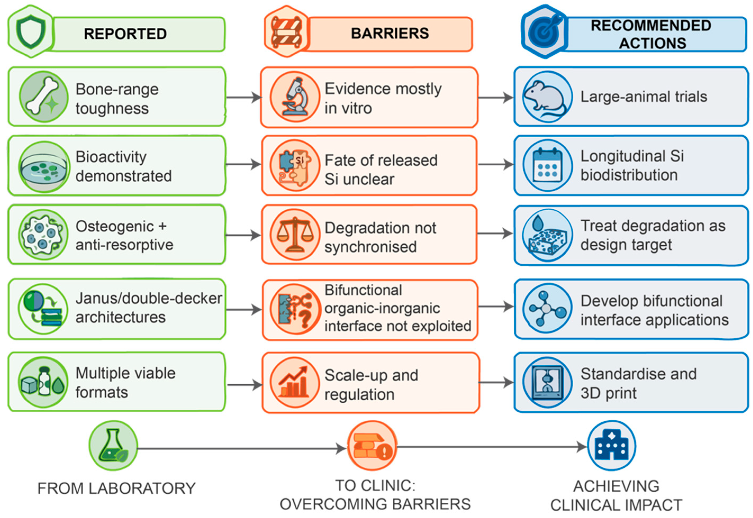
Table 7.
Research priorities, in the authors’ order of importance.
8.7. Biological Evaluation and Reporting Beyond ALP and Osteocalcin
If translation is a central aim, biological evaluation should extend beyond alkaline phosphatase activity and osteocalcin secretion. A more complete programme would include (i) osteoblast differentiation and matrix mineralisation, (ii) osteoclast formation and resorption, (iii) macrophage and foreign-body responses and osteoimmunomodulation, and (iv) angiogenesis and perfusion. It would also include (v) inflammatory cytokines, (vi) cytotoxicity of degradation products, (vii) the local and systemic distribution of silicon-containing species, with renal or hepatic clearance where relevant, and (viii) chronic inflammation. In vivo, micro-CT should be accompanied by quantitative morphometry and histomorphometry, by mechanical assessment of bone–implant integration (push-out, pull-out or torsion tests where appropriate), and by follow-up long enough to observe remodelling.
Future animal studies should follow the ARRIVE 2.0 guidelines [122], including randomisation, blinding, sample-size justification, reporting of exclusions and transparent outcome reporting. For eventual translation, biological safety would be assessed within the risk-based framework of ISO 10993-1:2025 [123], with chemical characterisation of the material and its degradation products according to ISO 10993-18:2020 [124].
9. Conclusions
Hydroxyapatite and silsesquioxanes offer complementary properties and a reasonable basis for hybrid design. The evidence that the hybrids work, however, is thinner and more conditional than the tone of the literature conveys. Three conclusions appear defensible. First, silsesquioxane reinforcement is conditional and not general. In dental-composite and related polymer systems, the mechanical effect of POSS depends on loading, functionality, dispersion, and interfacial integration, and in at least one systematic study no POSS-loaded composite bettered a POSS-free control. Whether the same relationships hold in HA–POSS bone materials remains unknown. Second, the silicon-release mechanism has not been demonstrated directly in HA–POSS bone materials. Silicon release has been measured for silicate-substituted hydroxyapatite, but adjacent Si-HA systems do not consistently show superior biological performance. Third, the available cross-study data do not permit a ranking of silsesquioxane systems against competing reinforcements. The most defensible rationale is the combination of properties reported for one HA–POSS–poly(lactide) platform, namely covalent integration, tunable degradation, and shape-memory behaviour, rather than superior reinforcement. Because the direct HA–POSS evidence base is small, several of these conclusions rest on POSS systems without pre-incorporated HA, on dental composites, or on Si-HA, and they are stated as such.
This review does not conclude that HA–silsesquioxane hybrids are unpromising. Several show promising combinations of properties for bone tissue engineering, and control over cage architecture offers interfacial design opportunities that conventional fillers do not. Further progress will depend on more rigorous experimental design, including POSS-free controls, systematic loading series, quantitative benchmarking against competing materials, direct measurement of the mechanism now assumed, and load-bearing large-animal evidence before any clinical use.
Author Contributions
Conceptualization, R.K. and M.U.; methodology, R.K. and P.M.S.; investigation, R.K. and P.M.S.; formal analysis, R.K.; writing—original draft preparation, R.K.; writing—review and editing, R.K., P.M.S. and M.U.; visualization, P.M.S., U.B. and S.L.; project administration, R.K. and M.U.; funding acquisition, R.K. All authors have read and agreed to the published version of the manuscript.
Funding
This research was funded by Walailak University, New Researcher Development Scheme, grant number WU68206, and the Walailak University International Research Collaboration Scheme, grant number WU-CIA-03504/2025. The APC was fully waived by the journal.
Institutional Review Board Statement
Not applicable.
Informed Consent Statement
Not applicable.
Data Availability Statement
No new data were created or analysed in this study. Data sharing is not applicable to this article.
Acknowledgments
The authors thank colleagues at Walailak University, Chulalongkorn University, and Gunma University for helpful discussions.
Conflicts of Interest
The authors declare no conflicts of interest.
Abbreviations
| AF4-MALS | Asymmetric-flow field-flow fractionation coupled to multi-angle light scattering |
| AFM | Atomic force microscopy |
| ALP | Alkaline phosphatase |
| ARRIVE | Animal Research: Reporting of In Vivo Experiments |
| ASC | Apoptosis-associated speck-like protein containing a CARD |
| ASTM | ASTM International (formerly the American Society for Testing and Materials) |
| BMP-2 | Bone morphogenetic protein-2 |
| β-TCP | β-Tricalcium phosphate |
| caspase-1 | Cysteine-aspartic protease 1 |
| DDSQ | Double-decker silsesquioxane |
| DLS | Dynamic light scattering |
| DMA | Dynamic mechanical analysis |
| EDX | Energy-dispersive X-ray spectroscopy |
| GMP | Good manufacturing practice |
| HA | Hydroxyapatite |
| ICP-OES/MS | Inductively coupled plasma optical emission spectrometry/mass spectrometry |
| IL-1β | Interleukin-1β |
| IL-8 | Interleukin-8 |
| ISO | International Organization for Standardization |
| KIC | Fracture toughness (mode I critical stress-intensity factor) |
| MBG | Mesoporous bioactive glass |
| micro-CT | Micro-computed tomography |
| M-POSS | Methacrylate-functionalised POSS (MA-POSS and MI-POSS denote the multi- and monofunctional variants) |
| MPS | 3-(Trimethoxysilyl)propyl methacrylate |
| MSC | Mesenchymal stem cell |
| Na-CMC | Sodium carboxymethyl cellulose |
| NLRP3 | NLR family pyrin domain containing 3 (inflammasome) |
| NMR | Nuclear magnetic resonance |
| NTA | Nanoparticle-tracking analysis |
| OEG | Oligo(ethylene glycol) |
| OVX | Ovariectomised (ovariectomy-induced) |
| PBS | Phosphate-buffered saline |
| PCL | Poly(ε-caprolactone) |
| PEG | Poly(ethylene glycol) |
| PEGda | Poly(ethylene glycol) diacrylate |
| PGA | Poly(glycolic acid) |
| PLA | Poly(lactic acid) |
| PLGA | Poly(lactic-co-glycolic acid) |
| PMMA | Poly(methyl methacrylate) |
| POSS | Polyhedral oligomeric silsesquioxane |
| POSS-PCLU | POSS-modified poly(caprolactone-urea) urethane |
| POSS/PCL | Composites or nanofibres of POSS with poly(ε-caprolactone) |
| POSS-PCU | POSS-modified poly(carbonate-urea) urethane |
| PVA | Poly(vinyl alcohol) |
| RANKL | Receptor activator of nuclear factor κB ligand |
| RGD | Arginine–glycine–aspartate peptide sequence |
| rGO | Reduced graphite oxide |
| ROBINS-I | Risk Of Bias In Non-randomised Studies of Interventions |
| RUNX2 | Runt-related transcription factor 2 |
| SAXS/WAXS | Small-/wide-angle X-ray scattering |
| SBF | Simulated body fluid |
| SEM | Scanning electron microscopy |
| Si-HA | Silicate-substituted hydroxyapatite |
| Sr-MBG | Strontium-substituted mesoporous bioactive glass |
| SYRCLE | Systematic Review Centre for Laboratory Animal Experimentation |
| TEM | Transmission electron microscopy |
| TRAP | Tartrate-resistant acid phosphatase |
References
- Baldwin, P.; Li, D.J.; Auston, D.A.; Mir, H.S.; Yoon, R.S.; Koval, K.J. Autograft, allograft, and bone graft substitutes: Clinical evidence and indications for use in the setting of orthopaedic trauma surgery. J. Orthop. Trauma 2019, 33, 203–213. [Google Scholar] [CrossRef] [Scilit] [PubMed]
- Bucholz, R.W. Nonallograft osteoconductive bone graft substitutes. Clin. Orthop. Relat. Res. 2002, 395, 44–52. [Google Scholar] [CrossRef] [Scilit] [PubMed]
- Gupta, A.; Kukkar, N.; Sharif, K.; Main, B.J.; Albers, C.E.; El-Amin, S.F., III. Bone graft substitutes for spine fusion: A brief review. World J. Orthop. 2015, 6, 449–456. [Google Scholar] [CrossRef] [Scilit] [PubMed]
- Viateau, V.; Guillemin, G.; Bousson, V.; Oudina, K.; Hannouche, D.; Sedel, L.; Logeart-Avramoglou, D.; Petite, H. Long-bone critical-size defects treated with tissue-engineered grafts: A study on sheep. J. Orthop. Res. 2007, 25, 741–749. [Google Scholar] [CrossRef] [Scilit] [PubMed]
- Dimitriou, R.; Jones, E.; McGonagle, D.; Giannoudis, P.V. Bone regeneration: Current concepts and future directions. BMC Med. 2011, 9, 66. [Google Scholar] [CrossRef] [Scilit] [PubMed]
- Zimmermann, E.A.; Ritchie, R.O. Bone as a structural material. Adv. Healthc. Mater. 2015, 4, 1287–1304. [Google Scholar] [CrossRef] [Scilit] [PubMed]
- Liu, Y.; Luo, D.; Wang, T. Hierarchical structures of bone and bioinspired bone tissue engineering. Small 2016, 12, 4611–4632. [Google Scholar] [CrossRef] [Scilit] [PubMed]
- Gupta, H.S.; Seto, J.; Wagermaier, W.; Zaslansky, P.; Boesecke, P.; Fratzl, P. Cooperative deformation of mineral and collagen in bone at the nanoscale. Proc. Natl. Acad. Sci. USA 2006, 103, 17741–17746. [Google Scholar] [CrossRef] [Scilit] [PubMed]
- Aldegaither, N.; Sernicola, G.; Mesgarnejad, A.; Karma, A.; Balint, D.; Wang, J.; Saiz, E.; Shefelbine, S.J.; Porter, A.E.; Giuliani, F. Fracture toughness of bone at the microscale. Acta Biomater. 2021, 121, 475–483. [Google Scholar] [CrossRef] [Scilit] [PubMed]
- Owen, G.R.; Dard, M.; Larjava, H. Hydroxyapatite/beta-tricalcium phosphate biphasic ceramics as regenerative material for the repair of complex bone defects. J. Biomed. Mater. Res. B Appl. Biomater. 2018, 106, 2493–2512. [Google Scholar] [CrossRef] [Scilit] [PubMed]
- Rezwan, K.; Chen, Q.Z.; Blaker, J.J.; Boccaccini, A.R. Biodegradable and bioactive porous polymer/inorganic composite scaffolds for bone tissue engineering. Biomaterials 2006, 27, 3413–3431. [Google Scholar] [CrossRef] [Scilit] [PubMed]
- Arcos, D.; Vallet-Regí, M. Sol–gel silica-based biomaterials and bone tissue regeneration. Acta Biomater. 2010, 6, 2874–2888. [Google Scholar] [CrossRef] [Scilit] [PubMed]
- Bagheri, L.; Jafari-Gharabaghlou, D.; Dashti, M.-R.; Zarghami, N. An update on implication of POSS-based nanocomposites in bone tissue engineering: A review. J. Biomater. Sci. Polym. Ed. 2025, 36, 1587–1610. [Google Scholar] [CrossRef] [Scilit] [PubMed]
- Ghanbari, H.; Cousins, B.G.; Seifalian, A.M. A nanocage for nanomedicine: Polyhedral oligomeric silsesquioxane (POSS). Macromol. Rapid Commun. 2011, 32, 1032–1046. [Google Scholar] [CrossRef] [Scilit] [PubMed]
- Chen, M.; Wang, Y.; Wang, M.; Zhou, W.; Yu, S.; Lei, B. POSS-based hybrid biomaterials for tissue engineering and regenerative medicine. Mater. Today Bio 2025, 32, 101837. [Google Scholar] [CrossRef] [Scilit] [PubMed]
- Chang, Y.-K.; Hao, S.-J.; Wu, F.-G. Recent biomedical applications of functional materials based on polyhedral oligomeric silsesquioxane (POSS). Small 2024, 20, e2401762. [Google Scholar] [CrossRef] [Scilit] [PubMed]
- Loman-Cortes, P.; Binte Huq, T.; Vivero-Escoto, J.L. Use of polyhedral oligomeric silsesquioxane (POSS) in drug delivery, photodynamic therapy and bioimaging. Molecules 2021, 26, 6453. [Google Scholar] [CrossRef] [Scilit] [PubMed]
- Zheng, K.; Boccaccini, A.R. Sol-gel processing of bioactive glass nanoparticles: A review. Adv. Colloid Interface Sci. 2017, 249, 363–373. [Google Scholar] [CrossRef] [Scilit] [PubMed]
- Arcos, D.; Portolés, M.T. Mesoporous bioactive nanoparticles for bone tissue applications. Int. J. Mol. Sci. 2023, 24, 3249. [Google Scholar] [CrossRef] [Scilit] [PubMed]
- Reffitt, D.M.; Ogston, N.; Jugdaohsingh, R.; Cheung, H.F.J.; Evans, B.A.J.; Thompson, R.P.H.; Powell, J.J.; Hampson, G.N. Orthosilicic acid stimulates collagen type 1 synthesis and osteoblastic differentiation in human osteoblast-like cells in vitro. Bone 2003, 32, 127–135. [Google Scholar] [CrossRef] [Scilit] [PubMed]
- Bohner, M. Silicon-substituted calcium phosphates—A critical view. Biomaterials 2009, 30, 6403–6406. [Google Scholar] [CrossRef] [Scilit] [PubMed]
- Chen, M.; Zhang, Y.; Xie, Q.; Zhang, W.; Pan, X.; Gu, P.; Zhou, H.; Gao, Y.; Walther, A.; Fan, X. Long-term bone regeneration enabled by a polyhedral oligomeric silsesquioxane (POSS)-enhanced biodegradable hydrogel. ACS Biomater. Sci. Eng. 2019, 5, 4612–4623. [Google Scholar] [CrossRef] [Scilit] [PubMed]
- Liu, L.; Ma, S.; Xiao, Z.; Li, J.; Wang, Y.; Lu, Z.; Zhao, Y.; Guo, J. Hydrophilic silsesquioxane nanocages toughened extracellular matrix biomimetic poly(γ-glutamic acid) multidimensional self-polymerizable and osteogenic hybrid hydrogel for osteoporotic bone regeneration. Compos. Part B Eng. 2024, 284, 111713. [Google Scholar] [CrossRef] [Scilit]
- Buchwald, Z.; Buchwald, T.; Voelkel, A. Hydroxyapatite modified with polyhedral oligomeric silsesquioxane as a filler for dental resin-based composites. J. Mater. Res. 2024, 39, 2286–2294. [Google Scholar] [CrossRef] [Scilit]
- Porter, A.E.; Patel, N.; Skepper, J.N.; Best, S.M.; Bonfield, W. Comparison of in vivo dissolution processes in hydroxyapatite and silicon-substituted hydroxyapatite bioceramics. Biomaterials 2003, 24, 4609–4620. [Google Scholar] [CrossRef] [Scilit] [PubMed]
- Porter, A.E.; Botelho, C.M.; Lopes, M.A.; Santos, J.D.; Best, S.M.; Bonfield, W. Ultrastructural comparison of dissolution and apatite precipitation on hydroxyapatite and silicon-substituted hydroxyapatite in vitro and in vivo. J. Biomed. Mater. Res. A 2004, 69, 670–679. [Google Scholar] [CrossRef] [Scilit] [PubMed]
- Guth, K.; Buckland, T.; Hing, K.A. Silicon dissolution from microporous silicon substituted hydroxyapatite and its effect on osteoblast behaviour. Key Eng. Mater. 2006, 309–311, 117–120. [Google Scholar] [CrossRef] [Scilit]
- Zhou, X.; Sahai, N.; Qi, L.; Mankoci, S.; Zhao, W. Biomimetic and nanostructured hybrid bioactive glass. Biomaterials 2015, 50, 1–9. [Google Scholar] [CrossRef] [Scilit] [PubMed]
- Lu, N.; Lu, Y.; Liu, S.; Jin, C.; Fang, S.; Zhou, X.; Li, Z. Tailor-engineered POSS-based hybrid gels for bone regeneration. Biomacromolecules 2019, 20, 3485–3493. [Google Scholar] [CrossRef] [Scilit] [PubMed]
- Ai, X.; Liu, Z.; Wang, T.; Xie, Q.; Xie, W. POSS hybrid bioactive glass dental composite resin materials: Synthesis and analysis. J. Dent. 2024, 142, 104860. [Google Scholar] [CrossRef] [Scilit] [PubMed]
- Wang, W.; Sun, X.; Huang, L.; Gao, Y.; Ban, J.; Shen, L.; Chen, J. Structure–property relationships in hybrid dental nanocomposite resins containing monofunctional and multifunctional polyhedral oligomeric silsesquioxanes. Int. J. Nanomed. 2014, 9, 841–852. [Google Scholar] [CrossRef] [Scilit] [PubMed]
- Gasquères, G.; Bonhomme, C.; Maquet, J.; Babonneau, F.; Hayakawa, S.; Kanaya, T.; Osaka, A. Revisiting silicate substituted hydroxyapatite by solid-state NMR. Magn. Reson. Chem. 2008, 46, 342–346. [Google Scholar] [CrossRef] [Scilit] [PubMed]
- Fredericks, D.; Petersen, E.B.; Watson, N.; Grosland, N.; Gibson-Corley, K.; Smucker, J. Comparison of two synthetic bone graft products in a rabbit posterolateral fusion model. Iowa Orthop. J. 2016, 36, 167–173. [Google Scholar] [PubMed]
- Seebach, C.; Schultheiss, J.; Wilhelm, K.; Frank, J.; Henrich, D. Comparison of six bone-graft substitutes regarding cell seeding efficiency, metabolism and growth behaviour of human mesenchymal stem cells (MSC) in vitro. Injury 2010, 41, 731–738. [Google Scholar] [CrossRef] [Scilit] [PubMed]
- Nakayama, M. Macrophage recognition of crystals and nanoparticles. Front. Immunol. 2018, 9, 103. [Google Scholar] [CrossRef] [Scilit] [PubMed]
- Pavlin, M.; Lojk, J.; Strojan, K.; Hafner-Bratkovič, I.; Jerala, R.; Leonardi, A.; Križaj, I.; Drnovšek, N.; Novak, S.; Veranič, P.; et al. The relevance of physico-chemical properties and protein corona for evaluation of nanoparticles immunotoxicity—In vitro correlation analysis on THP-1 macrophages. Int. J. Mol. Sci. 2022, 23, 6197. [Google Scholar] [CrossRef] [Scilit] [PubMed]
- Crowley, C.; Klanrit, P.; Butler, C.R.; Varanou, A.; Platé, M.; Hynds, R.E.; Chambers, R.C.; Seifalian, A.M.; Birchall, M.A.; Janes, S.M. Surface modification of a POSS-nanocomposite material to enhance cellular integration of a synthetic bioscaffold. Biomaterials 2016, 83, 283–293. [Google Scholar] [CrossRef] [Scilit] [PubMed]
- Delaere, P.; Van Raemdonck, D. Tracheal replacement. J. Thorac. Dis. 2016, 8, S186–S196. [Google Scholar] [CrossRef] [Scilit] [PubMed]
- Yotsova, R.; Peev, S. Biological properties and medical applications of carbonate apatite: A systematic review. Pharmaceutics 2024, 16, 291. [Google Scholar] [CrossRef] [Scilit] [PubMed]
- DileepKumar, V.G.; Sridhar, M.S.; Aramwit, P.; Krut’ko, V.K.; Musskaya, O.N.; Glazov, I.E.; Reddy, N. A review on the synthesis and properties of hydroxyapatite for biomedical applications. J. Biomater. Sci. Polym. Ed. 2022, 33, 229–261. [Google Scholar] [CrossRef] [Scilit] [PubMed]
- Szcześ, A.; Hołysz, L.; Chibowski, E. Synthesis of hydroxyapatite for biomedical applications. Adv. Colloid Interface Sci. 2017, 249, 321–330. [Google Scholar] [CrossRef] [Scilit] [PubMed]
- Radulescu, D.-E.; Vasile, O.R.; Andronescu, E.; Ficai, A. Latest research of doped hydroxyapatite for bone tissue engineering. Int. J. Mol. Sci. 2023, 24, 13157. [Google Scholar] [CrossRef] [Scilit] [PubMed]
- Boonyang, U.; Chaopanich, P.; Wongchaisuwat, A.; Senthongkaew, P.; Siripaisarnpipat, S. Effect of phosphate precursor on the production of hydroxyapatite from crocodile eggshells. J. Biomim. Biomater. Tissue Eng. 2010, 5, 31–37. [Google Scholar] [CrossRef] [Scilit]
- Gong, T.; Xie, J.; Liao, J.; Zhang, T.; Lin, S.; Lin, Y. Nanomaterials and bone regeneration. Bone Res. 2015, 3, 15029. [Google Scholar] [CrossRef] [Scilit] [PubMed]
- Venkatesan, J.; Kim, S.-K. Nano-hydroxyapatite composite biomaterials for bone tissue engineering—A review. J. Biomed. Nanotechnol. 2014, 10, 3124–3140. [Google Scholar] [CrossRef] [Scilit] [PubMed]
- Lowe, B.; Hardy, J.G.; Walsh, L.J. Optimizing nanohydroxyapatite nanocomposites for bone tissue engineering. ACS Omega 2020, 5, 1–9. [Google Scholar] [CrossRef] [Scilit] [PubMed]
- Yamada, S.; Heymann, D.; Bouler, J.M.; Daculsi, G. Osteoclastic resorption of calcium phosphate ceramics with different hydroxyapatite/beta-tricalcium phosphate ratios. Biomaterials 1997, 18, 1037–1041. [Google Scholar] [CrossRef] [Scilit] [PubMed]
- Fellah, B.H.; Gauthier, O.; Weiss, P.; Chappard, D.; Layrolle, P. Osteogenicity of biphasic calcium phosphate ceramics and bone autograft in a goat model. Biomaterials 2008, 29, 1177–1188. [Google Scholar] [CrossRef] [Scilit] [PubMed]
- Bouler, J.M.; Pilet, P.; Gauthier, O.; Verron, E. Biphasic calcium phosphate ceramics for bone reconstruction: A review of biological response. Acta Biomater. 2017, 53, 1–12. [Google Scholar] [CrossRef] [Scilit] [PubMed]
- Blázquez-Carmona, P.; Mora-Macías, J.; Martínez-Vázquez, F.J.; Morgaz, J.; Domínguez, J.; Reina-Romo, E. Mechanics predicts effective critical-size bone regeneration using 3D-printed bioceramic scaffolds. Tissue Eng. Regen. Med. 2023, 20, 893–904. [Google Scholar] [CrossRef] [Scilit] [PubMed]
- Awasthi, S.; Pandey, S.K.; Arunan, E.; Srivastava, C. A review on hydroxyapatite coatings for the biomedical applications: Experimental and theoretical perspectives. J. Mater. Chem. B 2021, 9, 228–249. [Google Scholar] [CrossRef] [Scilit] [PubMed]
- Vahabzadeh, S.; Roy, M.; Bandyopadhyay, A.; Bose, S. Phase stability and biological property evaluation of plasma sprayed hydroxyapatite coatings for orthopedic and dental applications. Acta Biomater. 2015, 17, 47–55. [Google Scholar] [CrossRef] [Scilit] [PubMed]
- Kavitha Sri, A.; Arthi, C.; Neya, N.R.; Hikku, G.S. Nano-hydroxyapatite/collagen composite as scaffold material for bone regeneration. Biomed. Mater. 2023, 18, 032002. [Google Scholar] [CrossRef] [Scilit] [PubMed]
- Lickorish, D.; Ramshaw, J.A.M.; Werkmeister, J.A.; Glattauer, V.; Howlett, C.R. Collagen–hydroxyapatite composite prepared by biomimetic process. J. Biomed. Mater. Res. A 2004, 68, 19–27. [Google Scholar] [CrossRef] [Scilit] [PubMed]
- Mondal, S.; Dorozhkin, S.V.; Pal, U. Recent progress on fabrication and drug delivery applications of nanostructured hydroxyapatite. WIREs Nanomed. Nanobiotechnol. 2018, 10, e1504. [Google Scholar] [CrossRef] [Scilit] [PubMed]
- Mohd Zaffarin, A.S.; Ng, S.-F.; Ng, M.H.; Hassan, H.; Alias, E. Nano-hydroxyapatite as a delivery system for promoting bone regeneration in vivo: A systematic review. Nanomaterials 2021, 11, 2569. [Google Scholar] [CrossRef] [Scilit] [PubMed]
- Unno, M.; Suto, A.; Matsumoto, T. Laddersiloxanes—Silsesquioxanes with defined ladder structure. Russ. Chem. Rev. 2013, 82, 289–302. [Google Scholar] [CrossRef] [Scilit]
- Liu, Y.; Chaiprasert, T.; Ouali, A.; Unno, M. Well-defined cyclic silanol derivatives. Dalton Trans. 2022, 51, 4227–4245. [Google Scholar] [CrossRef] [Scilit] [PubMed]
- Unno, M.; Suto, A.; Takada, K.; Matsumoto, H. Synthesis of ladder and cage silsesquioxanes from 1,2,3,4-tetrahydroxycyclotetrasiloxane. Bull. Chem. Soc. Jpn. 2000, 73, 215–220. [Google Scholar] [CrossRef] [Scilit]
- Cordes, D.B.; Lyon, P.D.; Lichtenhan, J.D. Recent developments in the chemistry of cubic polyhedral oligosilsesquioxanes. Chem. Rev. 2010, 110, 2081–2173. [Google Scholar] [CrossRef] [Scilit] [PubMed]
- Chimjarn, S.; Kunthom, R.; Chancharone, P.; Sodkhomkhum, R.; Sangtrirutnugul, P.; Ervithayasuporn, V. Synthesis of aromatic functionalized cage-rearranged silsesquioxanes (T8, T10, and T12) via nucleophilic substitution reactions. Dalton Trans. 2015, 44, 916–919. [Google Scholar] [CrossRef] [Scilit] [PubMed]
- Laird, M.; Herrmann, N.; Ramsahye, N.; Totée, C.; Carcel, C.; Unno, M.; Bartlett, J.R.; Wong Chi Man, M. Large polyhedral oligomeric silsesquioxane cages: The isolation of functionalized POSS with an unprecedented Si18O27 core. Angew. Chem. Int. Ed. 2021, 60, 3022–3027. [Google Scholar] [CrossRef] [Scilit] [PubMed]
- Liu, Y.; Takeda, N.; Ouali, A.; Unno, M. Synthesis, characterization, and functionalization of tetrafunctional double-decker siloxanes. Inorg. Chem. 2019, 58, 4093–4098. [Google Scholar] [CrossRef] [Scilit] [PubMed]
- Oguri, N.; Egawa, Y.; Takeda, N.; Unno, M. Janus-cube octasilsesquioxane: Facile synthesis and structure elucidation. Angew. Chem. Int. Ed. 2016, 55, 9336–9339. [Google Scholar] [CrossRef] [Scilit] [PubMed]
- Chaiprasert, T.; Liu, Y.; Takeda, N.; Unno, M. Janus ring siloxane: A versatile precursor of the extended Janus ring and tricyclic laddersiloxanes. Dalton Trans. 2020, 49, 13533–13537. [Google Scholar] [CrossRef] [Scilit] [PubMed]
- John, Ł. Selected developments and medical applications of organic–inorganic hybrid biomaterials based on functionalized spherosilicates. Mater. Sci. Eng. C 2018, 88, 172–181. [Google Scholar] [CrossRef] [Scilit] [PubMed]
- Ghanbari, H.; de Mel, A.; Seifalian, A.M. Cardiovascular application of polyhedral oligomeric silsesquioxane nanomaterials: A glimpse into prospective horizons. Int. J. Nanomed. 2011, 6, 775–786. [Google Scholar] [CrossRef] [Scilit] [PubMed]
- Raghunath, J.; Georgiou, G.; Armitage, D.; Nazhat, S.N.; Sales, K.M.; Butler, P.E.; Seifalian, A.M. Degradation studies on biodegradable nanocomposite based on polycaprolactone/polycarbonate (80:20%) polyhedral oligomeric silsesquioxane. J. Biomed. Mater. Res. A 2009, 91, 834–844. [Google Scholar] [CrossRef] [Scilit] [PubMed]
- Ahmed, M.; Punshon, G.; Darbyshire, A.; Seifalian, A.M. Effects of sterilization treatments on bulk and surface properties of nanocomposite biomaterials. J. Biomed. Mater. Res. B Appl. Biomater. 2013, 101, 1182–1190. [Google Scholar] [CrossRef] [PubMed]
- Buchwald, Z.; Szołyga, M.; Zwolińska, J.; Marciniec, B.; Voelkel, A. Surface modification of hydroxyapatite with polyhedral oligomeric silsesquioxane. React. Funct. Polym. 2022, 170, 105131. [Google Scholar] [CrossRef] [Scilit]
- Pang, L.; Shen, Y.; Hu, H.; Zeng, X.; Huang, W.; Gao, H.; Wang, H.; Wang, D. Chemically and physically cross-linked polyvinyl alcohol–borosilicate gel hybrid scaffolds for bone regeneration. Mater. Sci. Eng. C 2019, 105, 110076. [Google Scholar] [CrossRef] [Scilit] [PubMed]
- Mahony, O.; Tsigkou, O.; Ionescu, C.; Minelli, C.; Ling, L.; Hanly, R.; Smith, M.E.; Stevens, M.M.; Jones, J.R. Silica-gelatin hybrids with tailorable degradation and mechanical properties for tissue regeneration. Adv. Funct. Mater. 2010, 20, 3835–3845. [Google Scholar] [CrossRef] [Scilit]
- Demeyer, S.; Athipornchai, A.; Pabunrueang, P.; Trakulsujaritchok, T. Development of mangiferin loaded chitosan–silica hybrid scaffolds: Physicochemical and bioactivity characterization. Carbohydr. Polym. 2021, 261, 117905. [Google Scholar] [CrossRef] [Scilit] [PubMed]
- Kaur, G.; Pickrell, G.; Sriranganathan, N.; Kumar, V.; Homa, D. Review and the state of the art: Sol–gel and melt quenched bioactive glasses for tissue engineering. J. Biomed. Mater. Res. B Appl. Biomater. 2016, 104, 1248–1275. [Google Scholar] [CrossRef] [Scilit] [PubMed]
- Karageorgiou, V.; Kaplan, D. Porosity of 3D biomaterial scaffolds and osteogenesis. Biomaterials 2005, 26, 5474–5491. [Google Scholar] [CrossRef] [Scilit] [PubMed]
- Mukasheva, F.; Adilova, L.; Dyussenbinov, A.; Yernaimanova, B.; Abilev, M.; Akilbekova, D. Optimizing scaffold pore size for tissue engineering: Insights across various tissue types. Front. Bioeng. Biotechnol. 2024, 12, 1444986. [Google Scholar] [CrossRef] [Scilit] [PubMed]
- Auniq, R.B.Z.; Pakasri, N.; Boonyang, U. Synthesis and in vitro bioactivity of three-dimensionally ordered macroporous–mesoporous bioactive glasses; 45S5 and S53P4. J. Korean Ceram. Soc. 2020, 57, 305–313. [Google Scholar] [CrossRef] [Scilit]
- Shi, X.; Wang, Y.; Ren, L.; Zhao, N.; Gong, Y.; Wang, D.-A. Novel mesoporous silica-based antibiotic releasing scaffold for bone repair. Acta Biomater. 2009, 5, 1697–1707. [Google Scholar] [CrossRef] [Scilit] [PubMed]
- Tamburaci, S.; Kimna, C.; Tihminlioglu, F. Bioactive diatomite and POSS silica cage reinforced chitosan/Na-carboxymethyl cellulose polyelectrolyte scaffolds for hard tissue regeneration. Mater. Sci. Eng. C 2019, 100, 196–208. [Google Scholar] [CrossRef] [Scilit] [PubMed]
- Tamburaci, S.; Cecen, B.; Ustun, O.; Ergur, B.U.; Havitcioglu, H.; Tihminlioglu, F. Production and characterization of a novel bilayer nanocomposite scaffold composed of chitosan/Si-nHap and zein/POSS structures for osteochondral tissue regeneration. ACS Appl. Bio Mater. 2019, 2, 1440–1455. [Google Scholar] [CrossRef] [Scilit] [PubMed]
- Xu, X.; Skelly, J.D.; Song, J. Chemically crosslinked amphiphilic degradable shape memory polymer nanocomposites with readily tuned physical, mechanical, and biological properties. ACS Appl. Mater. Interfaces 2023, 15, 2693–2704. [Google Scholar] [CrossRef] [Scilit] [PubMed]
- Bagheri, L.; Valizadeh, H.; Dindar-Safa, K.; Zarghami, N. Fabricating a robust POSS-PCL nanofiber scaffold for nesting of mesenchymal stem cells: Potential application in bone tissue regeneration. J. Biol. Eng. 2022, 16, 35. [Google Scholar] [CrossRef] [Scilit] [PubMed]
- Yan, X.; Yao, H.; Luo, J.; Li, Z.; Wei, J. Functionalization of electrospun nanofiber for bone tissue engineering. Polymers 2022, 14, 2940. [Google Scholar] [CrossRef] [Scilit] [PubMed]
- Engstrand, J.; López, A.; Engqvist, H.; Persson, C. Polyhedral oligomeric silsesquioxane (POSS)–poly(ethylene glycol) (PEG) hybrids as injectable biomaterials. Biomed. Mater. 2012, 7, 035013. [Google Scholar] [CrossRef] [Scilit] [PubMed]
- Bociong, K.; Kosior, B.; Soboń, N.; Domarecka, M.; Sokołowski, J.; Zimon, A.; Krasowski, M.; Szczesio-Wlodarczyk, A. Impact of M-POSS on selected properties of experimental methacrylate matrices and composites. Materials 2026, 19, 1261. [Google Scholar] [CrossRef] [Scilit] [PubMed]
- Tamburaci, S.; Tihminlioglu, F. Chitosan-hybrid POSS nanocomposites for bone regeneration: The effect of POSS nanocage on surface, morphology, structure and in vitro bioactivity. Int. J. Biol. Macromol. 2020, 142, 643–657. [Google Scholar] [CrossRef] [Scilit] [PubMed]
- Huang, L.; Tan, J.; Li, W.; Zhou, L.; Liu, Z.; Luo, B.; Lu, L.; Zhou, C. Functional polyhedral oligomeric silsesquioxane reinforced poly(lactic acid) nanocomposites for biomedical applications. J. Mech. Behav. Biomed. Mater. 2019, 90, 604–614. [Google Scholar] [CrossRef] [Scilit] [PubMed]
- Celesti, C.; Iannazzo, D.; Espro, C.; Visco, A.; Legnani, L.; Veltri, L.; Visalli, G.; Di Pietro, A.; Bottino, P.; Chiacchio, M.A. Chitosan/POSS hybrid hydrogels for bone tissue engineering. Materials 2022, 15, 8208. [Google Scholar] [CrossRef] [Scilit] [PubMed]
- Chen, M.; Zhang, Y.; Zhang, W.; Li, J. Polyhedral oligomeric silsesquioxane-incorporated gelatin hydrogel promotes angiogenesis during vascularized bone regeneration. ACS Appl. Mater. Interfaces 2020, 12, 22410–22425. [Google Scholar] [CrossRef] [Scilit] [PubMed]
- ISO 23317:2025; Implants for Surgery—Materials—Simulated Body Fluid (SBF) Preparation Procedure and Test Method to Detect Apatite Formation in SBF for Initial Screening of Bone-Contacting Implant Materials. International Organization for Standardization: Geneva, Switzerland, 2025.
- Belton, D.J.; Deschaume, O.; Perry, C.C. An overview of the fundamentals of the chemistry of silica with relevance to biosilicification and technological advances. FEBS J. 2012, 279, 1710–1720. [Google Scholar] [CrossRef] [Scilit] [PubMed]
- Zhou, H.; Jiao, G.; Dong, M.; Chi, H.; Wang, H.; Wu, W.; Liu, H.; Ren, S.; Kong, M.; Li, C.; et al. Orthosilicic acid accelerates bone formation in human osteoblast-like cells through the PI3K-Akt-mTOR pathway. Biol. Trace Elem. Res. 2019, 190, 327–335. [Google Scholar] [CrossRef] [Scilit] [PubMed]
- Dong, M.; Jiao, G.; Liu, H.; Wu, W.; Li, S.; Wang, Q.; Xu, D.; Li, X.; Liu, H.; Chen, Y. Biological silicon stimulates collagen type 1 and osteocalcin synthesis in human osteoblast-like cells through the BMP-2/Smad/RUNX2 signaling pathway. Biol. Trace Elem. Res. 2016, 173, 306–315. [Google Scholar] [CrossRef] [Scilit] [PubMed]
- Magnusson, C.; Uribe, P.; Jugdaohsingh, R.; Powell, J.J.; Johansson, A.; Ransjö, M. Inhibitory effects of orthosilicic acid on osteoclastogenesis in RANKL-stimulated RAW264.7 cells. J. Biomed. Mater. Res. A 2021, 109, 1967–1978. [Google Scholar] [CrossRef] [Scilit] [PubMed]
- Ma, W.; Wang, F.; You, Y.; Wu, W.; Chi, H.; Jiao, G.; Zhang, L.; Zhou, H.; Wang, H.; Chen, Y. Ortho-silicic acid inhibits RANKL-induced osteoclastogenesis and reverses ovariectomy-induced bone loss in vivo. Biol. Trace Elem. Res. 2021, 199, 1864–1876. [Google Scholar] [CrossRef] [Scilit] [PubMed]
- Magnusson, C.; Ransjö, M. Orthosilicic acid inhibits human osteoclast differentiation and bone resorption. PLoS ONE 2024, 19, e0312169. [Google Scholar] [CrossRef] [Scilit] [PubMed]
- Porter, A.E. Nanoscale characterization of the interface between bone and hydroxyapatite implants and the effect of silicon on bone apposition. Micron 2006, 37, 681–688. [Google Scholar] [CrossRef] [Scilit] [PubMed]
- Patel, N.; Brooks, R.A.; Clarke, M.T.; Lee, P.M.T.; Rushton, N.; Gibson, I.R.; Best, S.M.; Bonfield, W. In vivo assessment of hydroxyapatite and silicate-substituted hydroxyapatite granules using an ovine defect model. J. Mater. Sci. Mater. Med. 2005, 16, 429–440. [Google Scholar] [CrossRef] [Scilit] [PubMed]
- Zhao, D.; Zhu, T.; Li, J.; Cui, L.; Zhang, Z.; Zhuang, X.; Ding, J. Poly(lactic-co-glycolic acid)-based composite bone-substitute materials. Bioact. Mater. 2021, 6, 346–360. [Google Scholar] [CrossRef] [Scilit] [PubMed]
- Gentile, P.; Chiono, V.; Carmagnola, I.; Hatton, P.V. An overview of poly(lactic-co-glycolic acid) (PLGA)-based biomaterials for bone tissue engineering. Int. J. Mol. Sci. 2014, 15, 3640–3659. [Google Scholar] [CrossRef] [Scilit] [PubMed]
- Dai, Z.; Ronholm, J.; Tian, Y.; Sethi, B.; Cao, X. Sterilization techniques for biodegradable scaffolds in tissue engineering applications. J. Tissue Eng. 2016, 7, 2041731416648810. [Google Scholar] [CrossRef] [Scilit] [PubMed]
- Ressler, A. Chitosan-based biomaterials for bone tissue engineering applications: A short review. Polymers 2022, 14, 3430. [Google Scholar] [CrossRef] [Scilit] [PubMed]
- Guo, C.; Qi, J.; Liu, J.; Wang, H.; Liu, Y.; Feng, Y.; Xu, G. The ability of biodegradable thermosensitive hydrogel composite calcium–silicon-based bioactive bone cement in promoting osteogenesis and repairing rabbit distal femoral defects. Polymers 2022, 14, 3852. [Google Scholar] [CrossRef] [Scilit] [PubMed]
- Krishnan, L.; Priddy, L.B.; Esancy, C.; Klosterhoff, B.S.; Stevens, H.Y.; Tran, L.; Guldberg, R.E. Delivery vehicle effects on bone regeneration and heterotopic ossification induced by high dose BMP-2. Acta Biomater. 2017, 49, 101–112. [Google Scholar] [CrossRef] [Scilit] [PubMed]
- Rezania, N.; Asadi-Eydivand, M.; Abolfathi, N.; Bonakdar, S.; Mehrjoo, M.; Solati-Hashjin, M. Three-dimensional printing of polycaprolactone/hydroxyapatite bone tissue engineering scaffolds: Mechanical properties and biological behavior. J. Mater. Sci. Mater. Med. 2022, 33, 31. [Google Scholar] [CrossRef] [Scilit] [PubMed]
- Zhao, S.; Zhang, J.; Zhu, M.; Zhang, Y.; Liu, Z.; Tao, C.; Zhu, Y.; Zhang, C. Three-dimensional printed strontium-containing mesoporous bioactive glass scaffolds for repairing rat critical-sized calvarial defects. Acta Biomater. 2015, 12, 270–280, Erratum in Acta Biomater. 2021, 134, 819–820. https://doi.org/10.1016/j.actbio.2021.09.044. [Google Scholar] [CrossRef] [Scilit] [PubMed]
- Reyes-Peces, M.V.; Félix, E.; Martínez-Vázquez, F.J.; Fernández-Montesinos, R.; Bomati-Miguel, Ó.; Mesa-Díaz, M.d.M.; Alcántara, R.; Vilches-Pérez, J.I.; Salido, M.; De la Rosa-Fox, N.; et al. Robocasting and laser micromachining of sol-gel derived 3D silica/gelatin/β-TCP scaffolds for bone tissue regeneration. Gels 2022, 8, 634. [Google Scholar] [CrossRef] [Scilit] [PubMed]
- Bagheri, L.; Jafari-Gharabaghlou, D.; Valizadeh, H.; Barzegari, A.; Zarghami, N. Design and development of biodegradable POSS-PCL-Zeolite (β) nano-scaffold for potential applications in bone regeneration. J. Biomater. Sci. Polym. Ed. 2023, 34, 1559–1578. [Google Scholar] [CrossRef] [Scilit] [PubMed]
- Liu, Y.; Huang, J.; Li, H. Synthesis of hydroxyapatite–reduced graphite oxide nanocomposites for biomedical applications: Oriented nucleation and epitaxial growth of hydroxyapatite. J. Mater. Chem. B 2013, 1, 1826–1834. [Google Scholar] [CrossRef] [Scilit] [PubMed]
- Yao, Y.; Guan, D.; Zhang, C.; Liu, J.; Zhu, X.; Huang, T.; Liu, J.; Cui, H.; Tang, K.-L.; Lin, J.; et al. Silkworm spinning inspired 3D printing toward a high strength scaffold for bone regeneration. J. Mater. Chem. B 2022, 10, 6946–6957. [Google Scholar] [CrossRef] [Scilit] [PubMed]
- Li, Z.; Zhu, W.; Bi, S.; Li, R.; Hu, H.; Lin, H.; Tuan, R.S.; Khor, K.A. Incorporating silica-coated graphene in bioceramic nanocomposites to simultaneously enhance mechanical and biological performance. J. Biomed. Mater. Res. A 2020, 108, 1016–1027. [Google Scholar] [CrossRef] [Scilit] [PubMed]
- Li, M.; Xiong, P.; Yan, F.; Li, S.; Ren, C.; Yin, Z.; Li, A.; Li, H.; Ji, X.; Zheng, Y.; et al. An overview of graphene-based hydroxyapatite composites for orthopedic applications. Bioact. Mater. 2018, 3, 1–18. [Google Scholar] [CrossRef] [Scilit] [PubMed]
- Boccardi, E.; Melli, V.; Catignoli, G.; Altomare, L.; Tavafoghi Jahromi, M.; Cerruti, M.; Lefebvre, L.-P.; De Nardo, L. Study of the mechanical stability and bioactivity of Bioglass-based glass-ceramic scaffolds produced via powder metallurgy-inspired technology. Biomed. Mater. 2016, 11, 015005. [Google Scholar] [CrossRef] [Scilit] [PubMed]
- Teimouri, R.; Hamed Akbari Toosi, S.; Mohammadi, M.; Nekooei, S.; Vahidi Toorchi, S.M.; Ramezani, M.; Alibolandi, M. Effect of berberine-loaded mesoporous silica nanoparticles and their combination with collagen/hydroxyapatite sponge scaffold on the regeneration of rabbit skull defects. Int. J. Biol. Macromol. 2026, 338, 149786. [Google Scholar] [CrossRef] [Scilit] [PubMed]
- Kane, R.J.; Weiss-Bilka, H.E.; Meagher, M.J.; Liu, Y.; Gargac, J.A.; Niebur, G.L.; Wagner, D.R.; Roeder, R.K. Hydroxyapatite reinforced collagen scaffolds with improved architecture and mechanical properties. Acta Biomater. 2015, 17, 16–25. [Google Scholar] [CrossRef] [Scilit] [PubMed]
- Xie, R.; Hu, J.; Hoffmann, O.; Zhang, Y.; Ng, F.; Qin, T.; Guo, X. Self-fitting shape memory polymer foam inducing bone regeneration: A rabbit femoral defect study. Biochim. Biophys. Acta Gen. Subj. 2018, 1862, 936–945. [Google Scholar] [CrossRef] [Scilit] [PubMed]
- Jungebluth, P.; Alici, E.; Baiguera, S.; Blomberg, P.; Bozóky, B.; Crowley, C.; Einarsson, O.; Gudbjartsson, T.; Le Guyader, S.; Henriksson, G.; et al. Tracheobronchial transplantation with a stem-cell-seeded bioartificial nanocomposite: A proof-of-concept study. Lancet 2011, 378, 1997–2004, Retraction in Lancet 2018, 392, 11. https://doi.org/10.1016/S0140-6736(18)31558-7. [Google Scholar] [CrossRef] [Scilit] [PubMed]
- Pepper, V.; Best, C.A.; Buckley, K.; Schwartz, C.; Onwuka, E.; King, N.; White, A.; Dharmadhikari, S.; Reynolds, S.D.; Johnson, J.; et al. Factors influencing poor outcomes in synthetic tissue-engineered tracheal replacement. Otolaryngol. Head Neck Surg. 2019, 161, 458–467. [Google Scholar] [CrossRef] [Scilit] [PubMed]
- Vranckx, J.J.; Delaere, P. The current status and outlook of trachea transplantation. Curr. Opin. Organ Transplant. 2020, 25, 601–608. [Google Scholar] [CrossRef] [Scilit] [PubMed]
- Teoh, G.Z.; Crowley, C.; Birchall, M.A.; Seifalian, A.M. Development of resorbable nanocomposite tracheal and bronchial scaffolds for paediatric applications. Br. J. Surg. 2015, 102, e140–e150. [Google Scholar] [CrossRef] [Scilit] [PubMed]
- Lakhani, H.A.; de Mel, A.; Seifalian, A.M. The effect of TGF-β1 and BMP-4 on bone marrow-derived stem cell morphology on a novel bioabsorbable nanocomposite material. Artif. Cells Nanomed. Biotechnol. 2015, 43, 230–234. [Google Scholar] [CrossRef] [Scilit] [PubMed]
- Percie du Sert, N.; Hurst, V.; Ahluwalia, A.; Alam, S.; Avey, M.T.; Baker, M.; Browne, W.J.; Clark, A.; Cuthill, I.C.; Dirnagl, U.; et al. The ARRIVE guidelines 2.0: Updated guidelines for reporting animal research. PLoS Biol. 2020, 18, e3000410. [Google Scholar] [CrossRef] [Scilit] [PubMed]
- ISO 10993-1:2025; Biological Evaluation of Medical Devices—Part 1: Requirements and General Principles for the Evaluation of Biological Safety within a Risk Management Process. International Organization for Standardization: Geneva, Switzerland, 2025.
- ISO 10993-18:2020; Biological Evaluation of Medical Devices—Part 18: Chemical Characterization of Medical Device Materials within a Risk Management Process. International Organization for Standardization: Geneva, Switzerland, 2020.
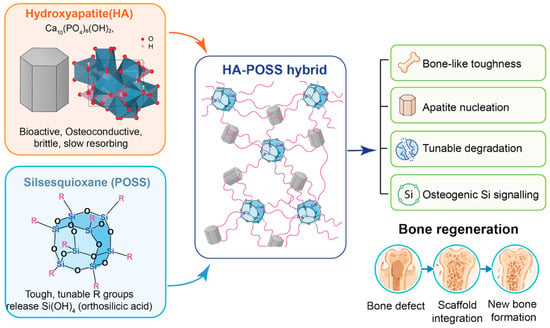
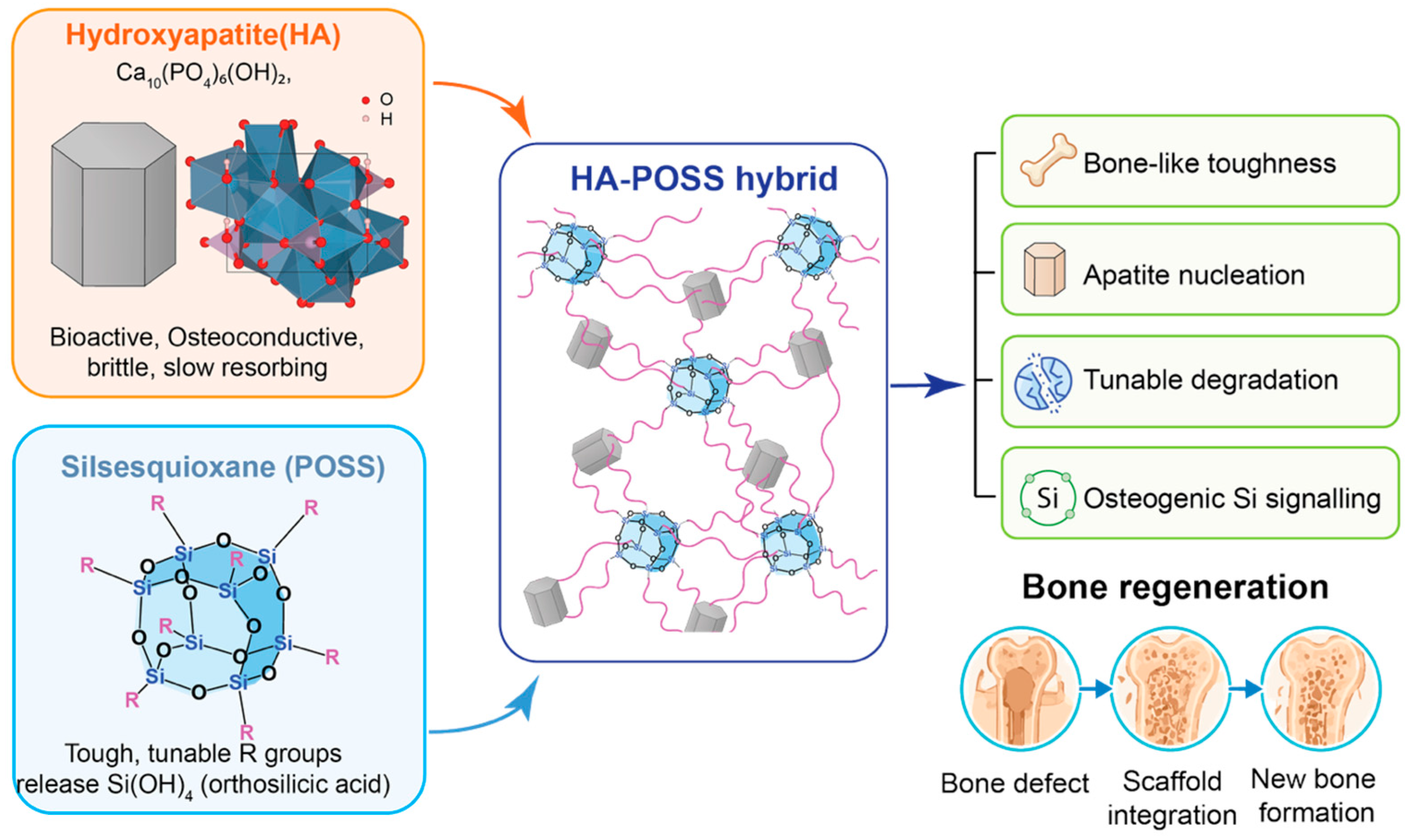
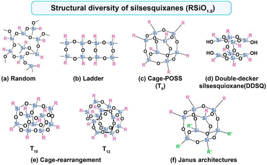
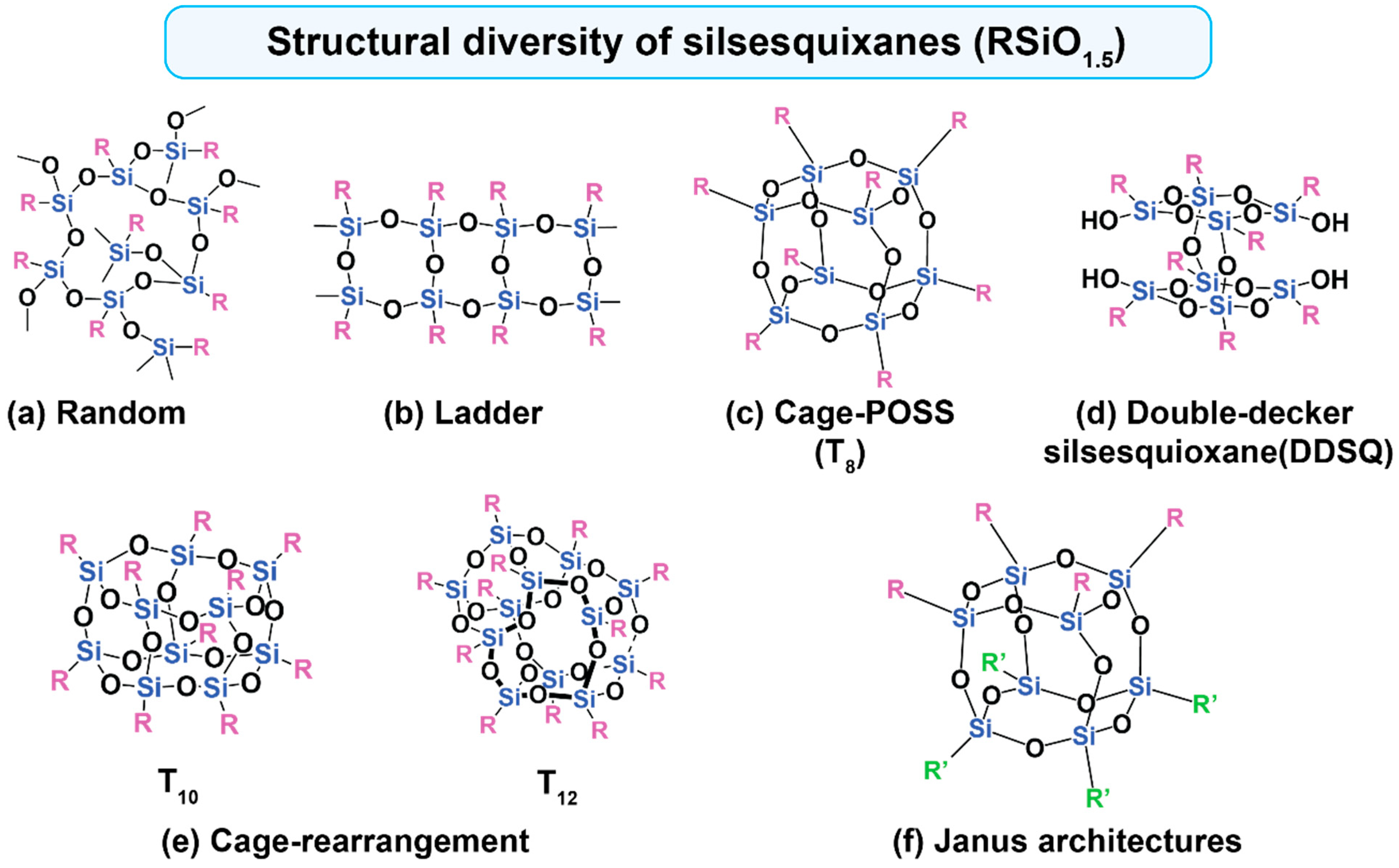
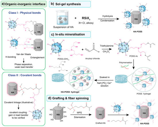
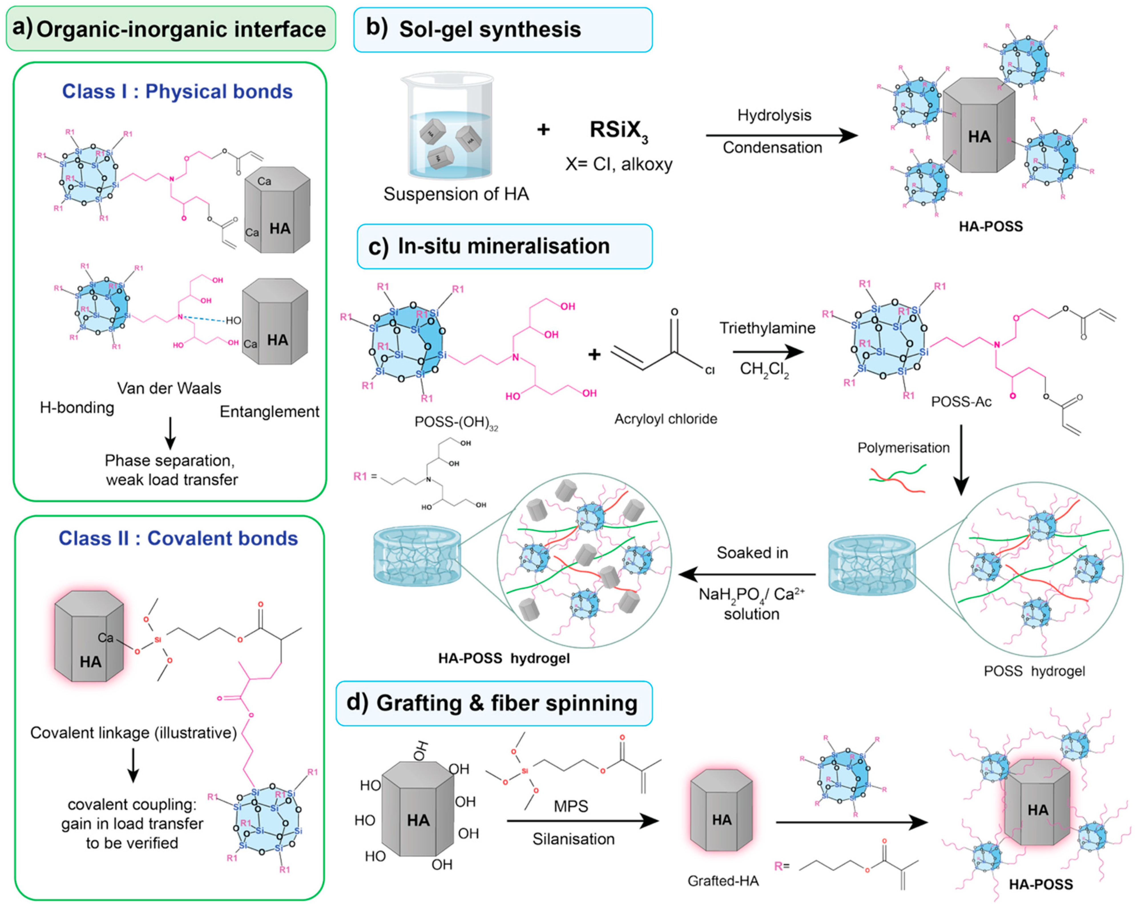
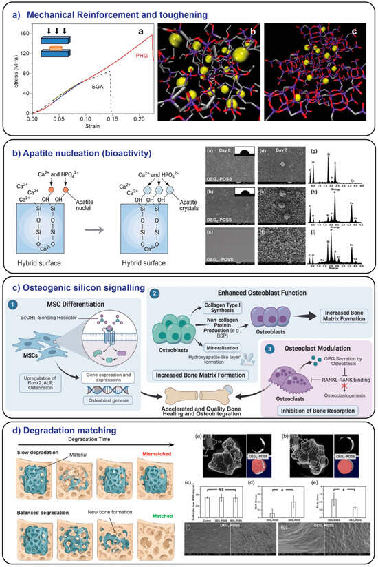
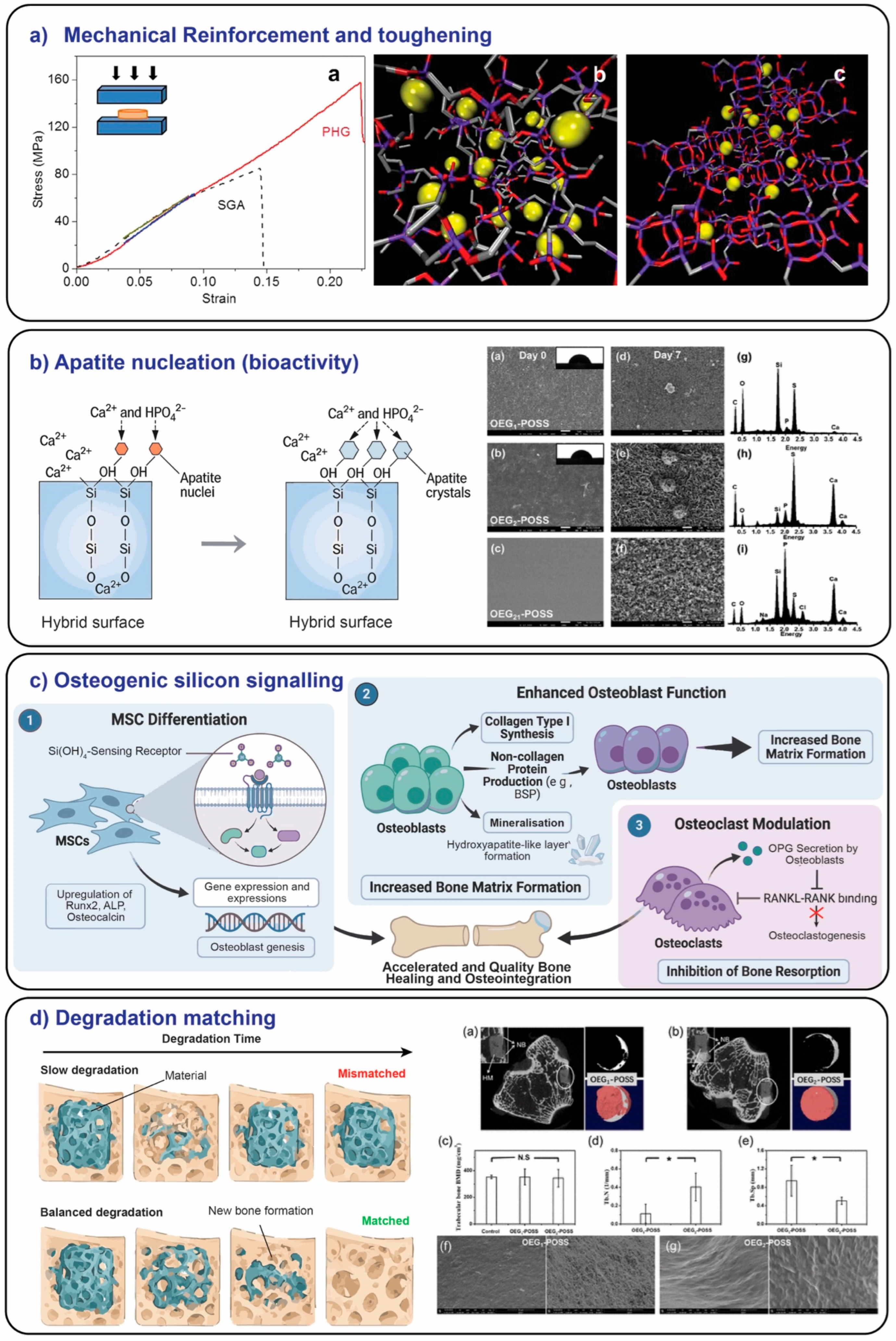

Disclaimer/Publisher’s Note: The statements, opinions and data contained in all publications are solely those of the individual author(s) and contributor(s) and not of MDPI and/or the editor(s). MDPI and/or the editor(s) disclaim responsibility for any injury to people or property resulting from any ideas, methods, instructions or products referred to in the content. |
© 2026 by the authors. Licensee MDPI, Basel, Switzerland. This article is an open access article distributed under the terms and conditions of the Creative Commons Attribution (CC BY) license.