


Abstract
Toll-like receptors (TLRs) play an important role in innate immunity, mediating viral recognition, antiviral responses, and cytokine production. This study evaluated TLR7 and TLR9 expression levels in COVID-19 patients and their associations with COVID-19 infection and disease severity. A total of 134 adults were included: 94 COVID-19 patients and 40 controls. RT-PCR confirmed SARS-CoV-2 infection; TLR7 and TLR9 expression levels were determined by quantitative real-time PCR. Participants’ clinical characteristics and laboratory findings were evaluated. TLR7 and TLR9 expression levels were significantly higher in COVID-19 patients than in controls (both p < 0.001). Both markers were independently associated with COVID-19 infection by multivariate analysis (OR: 6996.516, p < 0.001; OR: 7.365, p = 0.004) and showed excellent discriminatory performance by ROC analysis (AUC: 0.973 and 0.937, both p < 0.001). Among COVID-19 patients, TLR9 expression and ALT were independently associated with moderate-to-critical disease (OR: 1.448, p = 0.030; OR: 1.092, p = 0.032). TLR9 expression and ALT showed modest discriminatory performance for distinguishing moderate-to-critical COVID-19 from mild disease (AUC: 0.623, p = 0.042; AUC: 0.641, p = 0.020, respectively), while the combined model demonstrated a slight improvement (AUC: 0.659, p = 0.008). TLR7 and TLR9 may represent potential biomarkers for distinguishing COVID-19 patients from controls, and TLR9 may provide additional information on disease severity. These findings need validation in larger, multicenter studies before clinical application.
1. Introduction
The coronavirus disease 2019 (COVID-19) pandemic emerged at the end of 2019. It is caused by severe acute respiratory syndrome coronavirus 2 (SARS-CoV-2) [1,2]. Variable clinical manifestations characterize this infection, ranging from mild respiratory disease to acute respiratory distress syndrome (ARDS) and severe pneumonia, and may even cause mortality. Although the announcement of the end of the COVID-19 as a worldwide public emergency by the World Health Organization (WHO), it stated the potential for SARS-CoV-2 variants to evolve, leading to future increases in cases and mortality [3,4]. Many risk factors have been significantly associated with COVID-19 severity and mortality, including older age, male sex, and comorbidities [5,6].
A set of unique transmembrane receptors known as toll-like receptors (TLRs) is a central component of both innate and adaptive immunity [7]. They are responsible for pathogen recognition, cytokine synthesis, and initiation of antiviral responses through the recognition of pathogen-associated molecular patterns (PAMPs) [8,9]. TLR7 and TLR9 are principal regulators in the detection and combat of viral infections, and they are primarily located in intracellular endosomal compartments [7,10]. TLR7 recognizes single-stranded viral RNA and plays a critical role in antiviral innate immune responses, whereas TLR9 recognizes unmethylated cytosine–phosphate–guanine (CpG)-rich nucleic acid motifs and contributes to inflammatory responses during viral infection [7,11].
It has been suggested that dysregulated TLR7 and TLR9 signaling may affect the host immune reaction to SARS-CoV-2 and contribute to differences in illness severity and patient outcomes [7]. Increased expression of TLR mRNAs, such as TLR3, TLR7, TLR8, and TLR9, is significantly more common in patients with COVID-19 than in healthy controls and is likely associated with disease severity [9]. However, the expression patterns and clinical relevance of TLR7 and TLR9 in COVID-19 remain poorly characterized, particularly in Egyptian patients. Therefore, this study determined TLR7 and TLR9 expression levels in patients with confirmed COVID-19 compared with controls and evaluated their associations with COVID-19 infection and disease severity, as well as their discriminatory performance in Egyptian individuals.
2. Materials and Methods
2.1. Design and Laboratory Evaluation
A total of 134 adult participants (aged > 18 years) were enrolled in this study, comprising 94 COVID-19 patients and 40 healthy controls (from July 2022 to June 2023). SARS-CoV-2 infection was proven by clinical assessment and reverse transcription polymerase chain reaction (RT-PCR) (Cobas® 6800, Roche Diagnostics, Pleasanton, CA, USA) of a nasopharyngeal swab specimen. The severity of infections in the COVID-19 group was assessed according to the World Health Organization (WHO) guidelines [12,13]. Participants’ vaccination status was not confirmed. The control group included those who appeared healthy, had no active respiratory disease, and had no previous history of COVID-19. They were additionally confirmed to be COVID-19-negative clinically and by RT-PCR. Pregnant women, smokers, patients with chronic respiratory, kidney, or cardiovascular diseases, cancer, hypertension, or diabetes mellitus, or past COVID-19 infection, recipients of transplants or immunosuppressive treatment, and patients undergoing end-of-life protocols were excluded from the study.
Participants’ demographic data were collected, and body mass index (BMI) was determined [3]. Peripheral blood samples for laboratory testing and TLR gene expression analysis were collected during the initial clinical evaluation, at the time of COVID-19 diagnosis, and before the initiation of COVID-19 therapy. Laboratory assessments included fasting blood sugar (FBS), 2-h postprandial blood sugar (PPBS), erythrocyte sedimentation rate (ESR), complete blood count (CBC), liver function tests (aspartate aminotransferase, AST; and alanine aminotransferase, ALT), and lactate dehydrogenase (LDH). Platelet-to-lymphocyte ratio (PLR), monocyte-to-lymphocyte ratio (MLR), and neutrophil-to-lymphocyte ratio (NLR) were calculated [14].
2.2. Toll-like Receptor Gene Expression
Quantitative real-time PCR was performed to determine the mRNA expression profiles of TLR7 and TLR9. Total RNA was extracted from whole blood using GeneJETTM Whole Blood RNA Purification Mini Kit (Thermo Fisher Scientific, Vilnius, Lithuania; #K0761). The NanoDrop 2000 spectrophotometer (Thermo Scientific, Wilmington, DE, USA) was used to assess RNA concentration and purity at A260/A280 and A260/A230 ratios. RNA integrity was additionally assessed by agarose gel electrophoresis, visualizing the 28S and 18S rRNA bands. For expression analysis, 1000 ng of RNA was used for reverse transcription. High-Capacity cDNA Reverse Transcription Kit (Applied Biosystems, Carlsbad, CA, USA; Part Number: 4375575) was used to synthesize complementary DNA (cDNA).
Amplifications were performed using the TaqMan® Gene Expression Assays (Applied Biosystems, Carlsbad, CA, USA; Part Number: 4333458), with each sample analyzed in duplicate. Human TaqMan Gene Expression Assays were used for TLR7 (assay ID: Hs01933259_s1) and TLR9 (assay ID: Hs00370913_s1). The internal reference gene was β-actin. The cycling conditions were adapted from those described by El-Hefnawy et al. [15] for gene amplification. The Applied Biosystems StepOneTM/StepOnePlusTM Real-Time PCR System (Applied Biosystems, Carlsbad, CA, USA) was used to perform fluorescence detection and data analysis. The relative expression levels of TLR7 and TLR9 mRNA were calculated according to the comparative threshold cycle (2−∆∆Ct) method [15]. β-actin was the endogenous reference gene, and the healthy control group served as the calibrator.
2.3. Statistical Analysis
Sample size estimation was performed using OpenEpi (version 3.01) with 80% statistical power and a 95% confidence level. The minimum number of participants required was estimated at 66 before study initiation. The final analysis included 134 participants: 94 COVID-19 patients and 40 healthy controls. Categorical data were presented as frequencies and percentages, and comparisons between two groups were performed by Chi-square or Fisher’s exact test. The Kolmogorov–Smirnov and Shapiro–Wilk tests evaluated the normality of the continuous data and indicated that the data distributions were non-normal. Data were reported as medians and interquartile ranges (IQRs), and comparisons between two groups were made using the Mann–Whitney test. Multivariate logistic regression analyses were performed to evaluate factors independently associated with COVID-19 infection and disease severity, with age, sex, and BMI used as covariates. Firth-penalized logistic regression analysis for factors associated with COVID-19 infection was also performed. The receiver operating characteristic (ROC) curves were used to evaluate the discriminatory performance of significant markers. The Youden index was used to determine optimal cutoffs, and the cutoffs for the combined model were based on predicted probabilities derived from binary logistic regression. Bootstrap optimism correction was performed to obtain optimism-corrected AUC estimates for the combined ROC models. For the combined TLR7 + TLR9 model, predicted probabilities were derived from a Firth-penalized logistic regression model. Spearman correlation analysis was performed to explore the relationships between TLR7 and TLR9 expression levels and selected relevant parameters in COVID-19 patients. Statistical significance was considered when the p-value was ≤0.05. IBM SPSS Statistics version 25 was used for data analysis. Firth-penalized logistic regression and bootstrap optimism correction were performed using R software version 4.5.3 (R Foundation for Statistical Computing).
3. Results
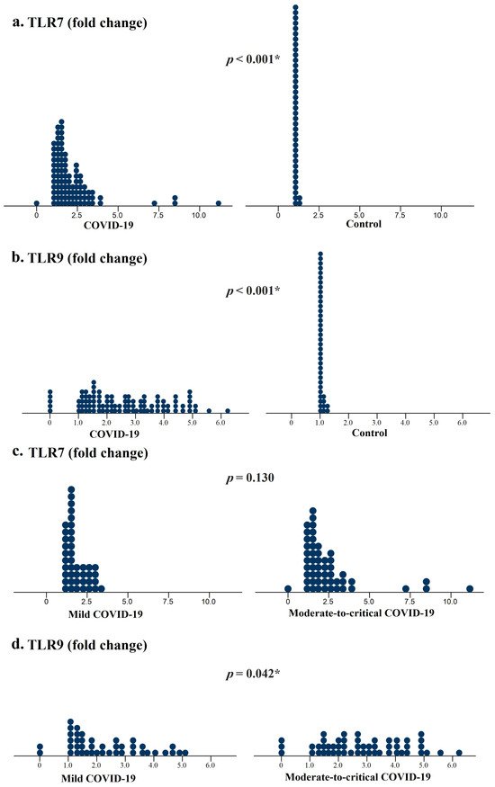
Table 1 summarizes the clinical characteristics and laboratory findings of the participants, including the COVID-19 group (n = 94) and healthy control group (n = 40). The median participant age was 42.5 years (IQR: 35–56), and males accounted for 58.2% of participants. Sex and BMI were comparable between COVID-19 patients and controls (p = 0.623 and 0.351, respectively); however, age was significantly higher in patients with COVID-19 (p = 0.006). The distribution of TLR7 and TLR9 expression levels in COVID-19 patients and controls is shown in Figure 1a and Figure 1b, respectively. Median values of TLR7 and TLR9 relative expression were significantly higher in COVID-19 patients (1.68, IQR: 1.39–2.49 vs. 1.00, IQR: 0.99–1.00 fold-change for TLR7; 2.44, IQR: 1.50–3.75 vs. 1.00, IQR: 0.99–1.00 fold-change for TLR9; both p < 0.001). Median values of platelet count, FBS, ALT, AST, ESR, MLR, PLR, and NLR were significantly higher in COVID-19 cases, whereas median values of absolute lymphocyte count (ALC), absolute neutrophil count (ANC), absolute monocyte count (AMC), white blood cell count, red blood cell count, and hematocrit were significantly lower (all p < 0.05).
Table 1.
Clinical characteristics and laboratory findings of participants.
Figure 1.
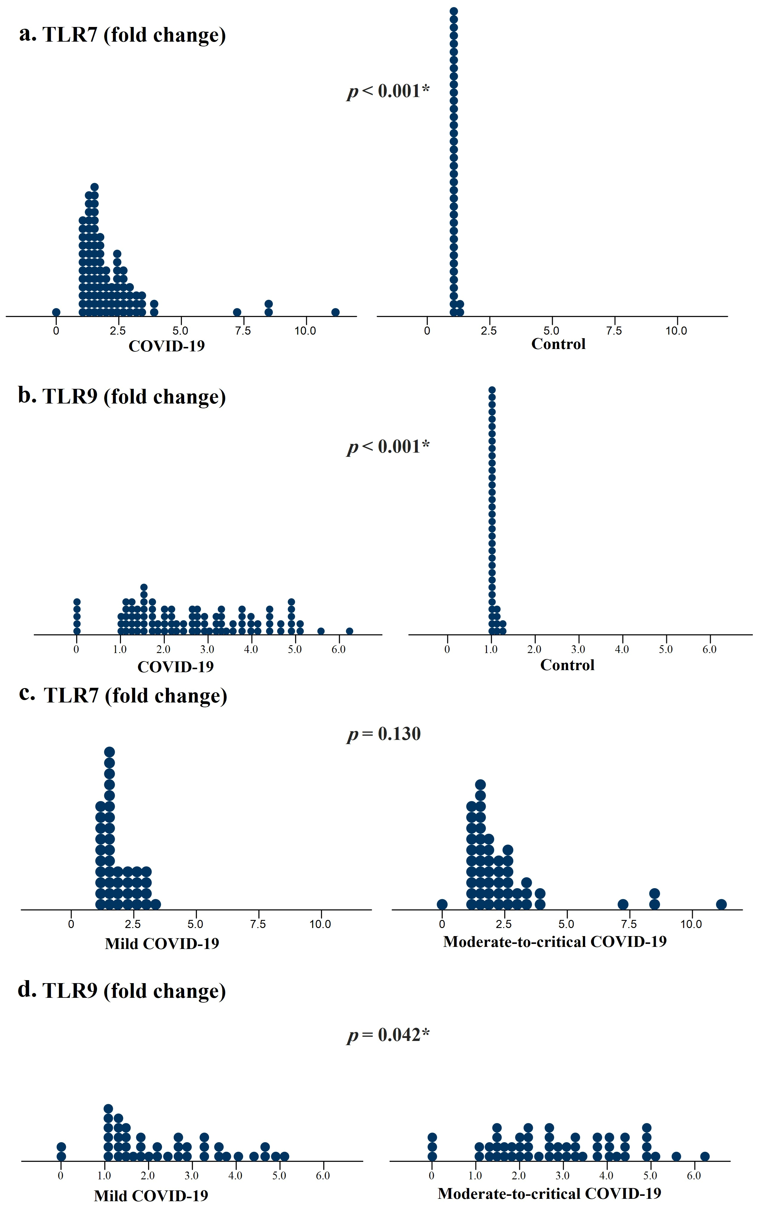
Distribution of TLR7 and TLR9 expression levels in COVID-19 patients vs. healthy controls and according to disease severity. (a) TLR7 and (b) TLR9 among COVID-19 patients vs. controls; (c) TLR7 and (d) TLR9 among patients with mild vs. moderate-to-critical COVID-19. Each dot represents an individual participant. p-values represent between-group differences; statistically significant results are indicated by an asterisk (*). COVID-19, coronavirus disease 2019; TLR, toll-like receptor.
Multivariate logistic regression was performed to identify factors independently associated with COVID-19 infection, adjusting for age, sex, and BMI (Table 2). Analysis revealed that both TLR7 and TLR9 expression levels were independently associated with COVID-19 infection (OR: 6996.516, 95% CI: 91.312–536,086.388, p < 0.001; OR: 7.365, 95% CI: 1.882–28.829, p = 0.004, respectively). Firth-penalized logistic regression sensitivity analysis showed a substantially lower OR of TLR7; however, it also confirmed the independent association of both TLR7 and TLR9 with COVID-19 infection (OR: 110.603, 95% Wald CI: 18.856–648.762, p < 0.001; OR: 3.914, 95% Wald CI: 1.849–8.284, p < 0.001, respectively) (Table S1).
Table 2.
Multivariate logistic regression analysis of factors associated with COVID-19 infection.
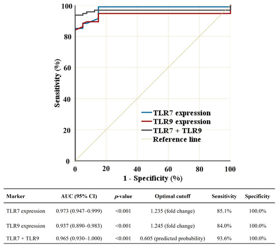
ROC curve analysis evaluated the discriminatory performance of TLR7 and TLR9 expression in distinguishing COVID-19 patients from controls, as illustrated in Figure 2 and Table S2. Both TLR7 and TLR9 expression levels showed excellent performance, with area under the curve (AUC) values of 0.973 and 0.937 (95% CI: 0.947–0.999 and 0.890–0.983, respectively; both p < 0.001). In addition, the combined model (TLR7 + TLR9) showed excellent discrimination (AUC: 0.965, 95% CI: 0.930–1.000, p < 0.001). Using the Firth-penalized model, the apparent AUC of the combined model was 0.964. However, the mean bootstrap optimism from 2000 resamples was 0.004, yielding an optimism-corrected AUC of 0.960.
Figure 2.
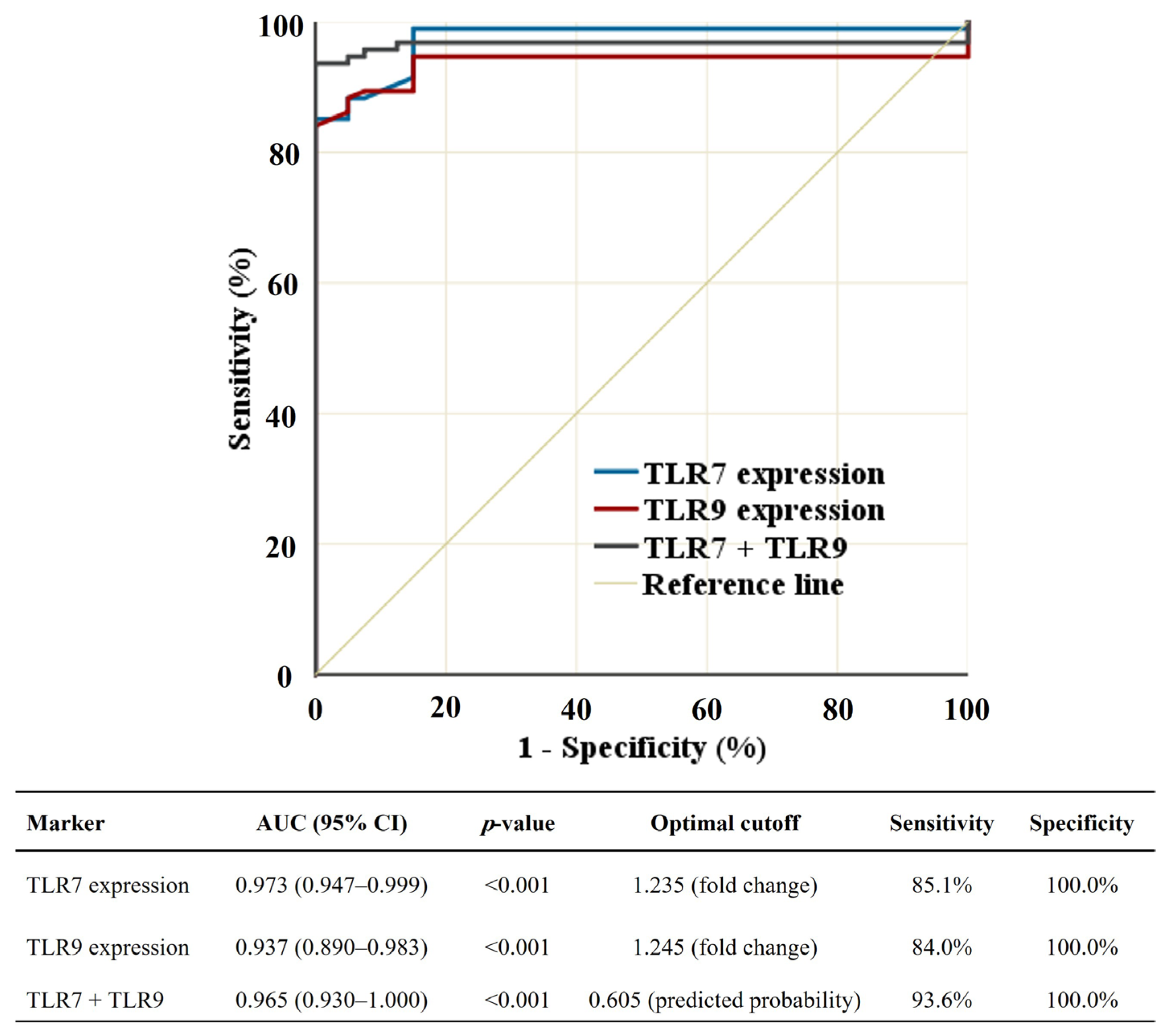
Receiver operating characteristic (ROC) curves showing the discriminatory performance of significant markers for distinguishing COVID-19 patients from healthy controls. COVID-19, coronavirus disease 2019; AUC, area under the curve; CI, confidence interval; TLR, toll-like receptor.
Regarding COVID-19 severity, most patients had no to mild symptoms (n = 42, 44.7%), while 28 (29.8%), 13 (13.8%), and 11 (11.7%) patients had moderate, severe, and critical disease, respectively. A comparison between COVID-19 patients with mild infection (n = 42) and those with moderate-to-critical infection (n = 52) was also performed. Figure 1c and Figure 1d illustrate the distribution of TLR7 and TLR9 expression levels across the two groups, respectively. Clinical and laboratory characteristics were compared between the two severity groups (Table 3). TLR9 expression and ALT median values were significantly elevated in moderate-to-critical than in mild COVID-19 (p = 0.042 and 0.019, respectively). However, absolute monocyte and white blood cell counts were significantly lower in moderate-to-critical COVID-19 (p = 0.032 and 0.034, respectively).
Table 3.
Clinical characteristics and laboratory findings of COVID-19 patients according to disease severity.
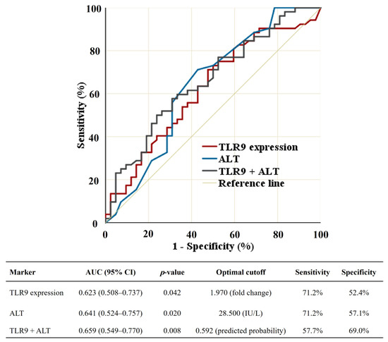
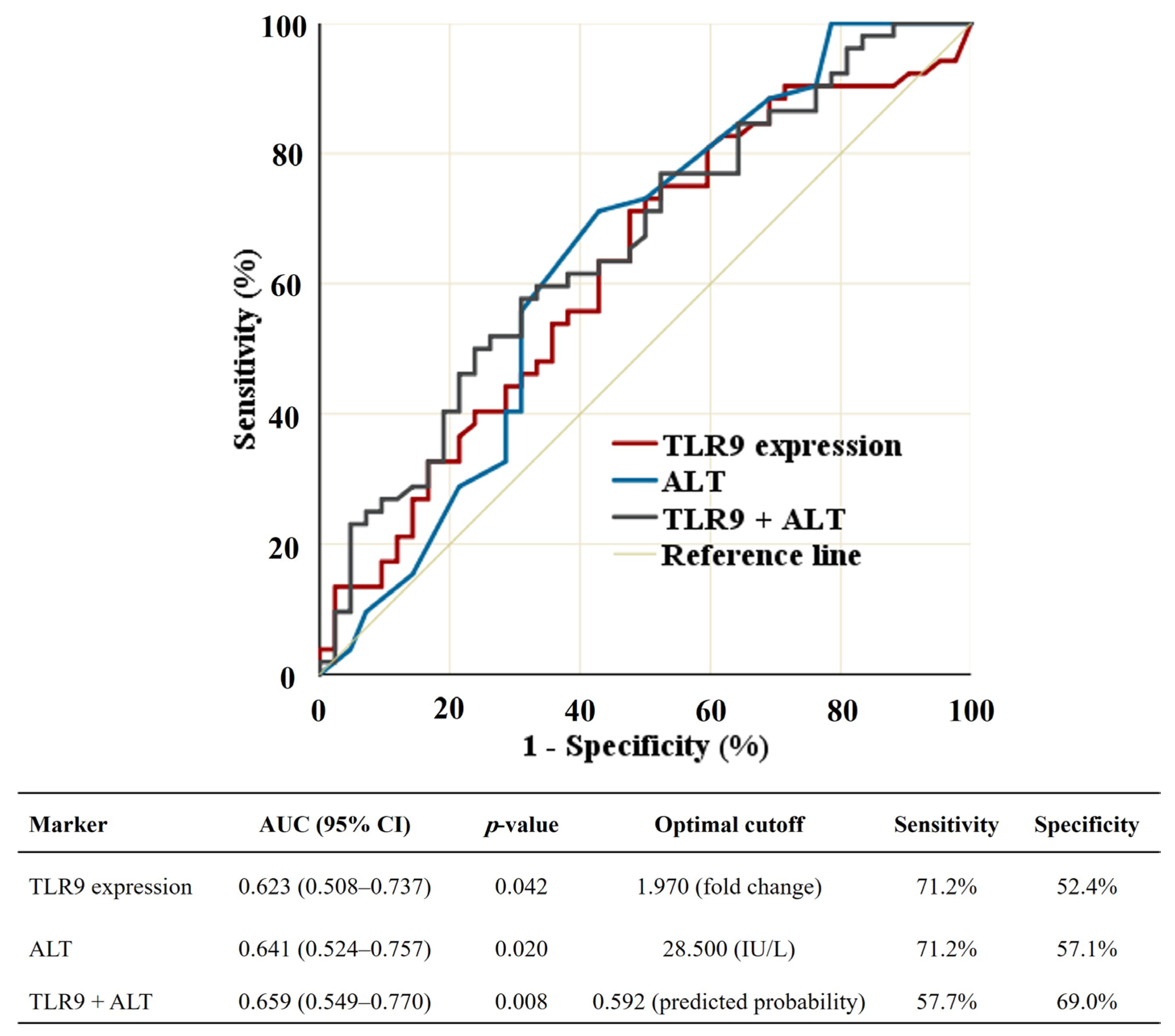
Increased TLR9 expression and ALT levels were independently associated with moderate-to-critical COVID-19 severity after adjustment for BMI, sex, and age (OR: 1.448, 95% CI: 1.037–2.022, p = 0.030; OR: 1.092, 95% CI: 1.007–1.184, p = 0.032, respectively), as shown in Table 4. Sensitivity analysis, additionally adjusting for ALC and AMC, revealed that both TLR9 expression and ALT remained independently associated with disease severity (p = 0.034 and 0.032, respectively) (Table S3). Both TLR9 expression and ALT showed modest ability to discriminate moderate-to-critical COVID-19 from mild COVID-19 cases (AUC: 0.623, 95% CI: 0.508–0.737, p = 0.042; AUC: 0.641, 95% CI: 0.524–0.757, p = 0.020, respectively). The combined model (TLR9 + ALT) demonstrated a slight improvement in discriminatory performance with an AUC of 0.659 (95% CI: 0.549–0.770, p = 0.008) (Figure 3 and Table S4). After applying a bootstrap optimism correction with 2000 resamples, the combined model showed modest discriminatory performance, with a mean optimism of 0.021 and an AUC decreased to 0.638.
Table 4.
Multivariate logistic regression analysis of factors associated with moderate-to-critical COVID-19.
Figure 3.
Receiver operating characteristic (ROC) curves showing the discriminatory performance of significant markers for distinguishing moderate-to-critical from mild COVID-19 cases. COVID-19, coronavirus disease 2019; AUC, area under the curve; CI, confidence interval; TLR, toll-like receptor; ALT, alanine aminotransferase.
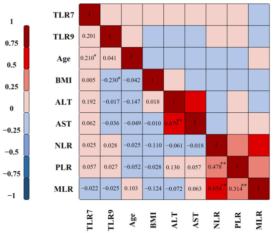
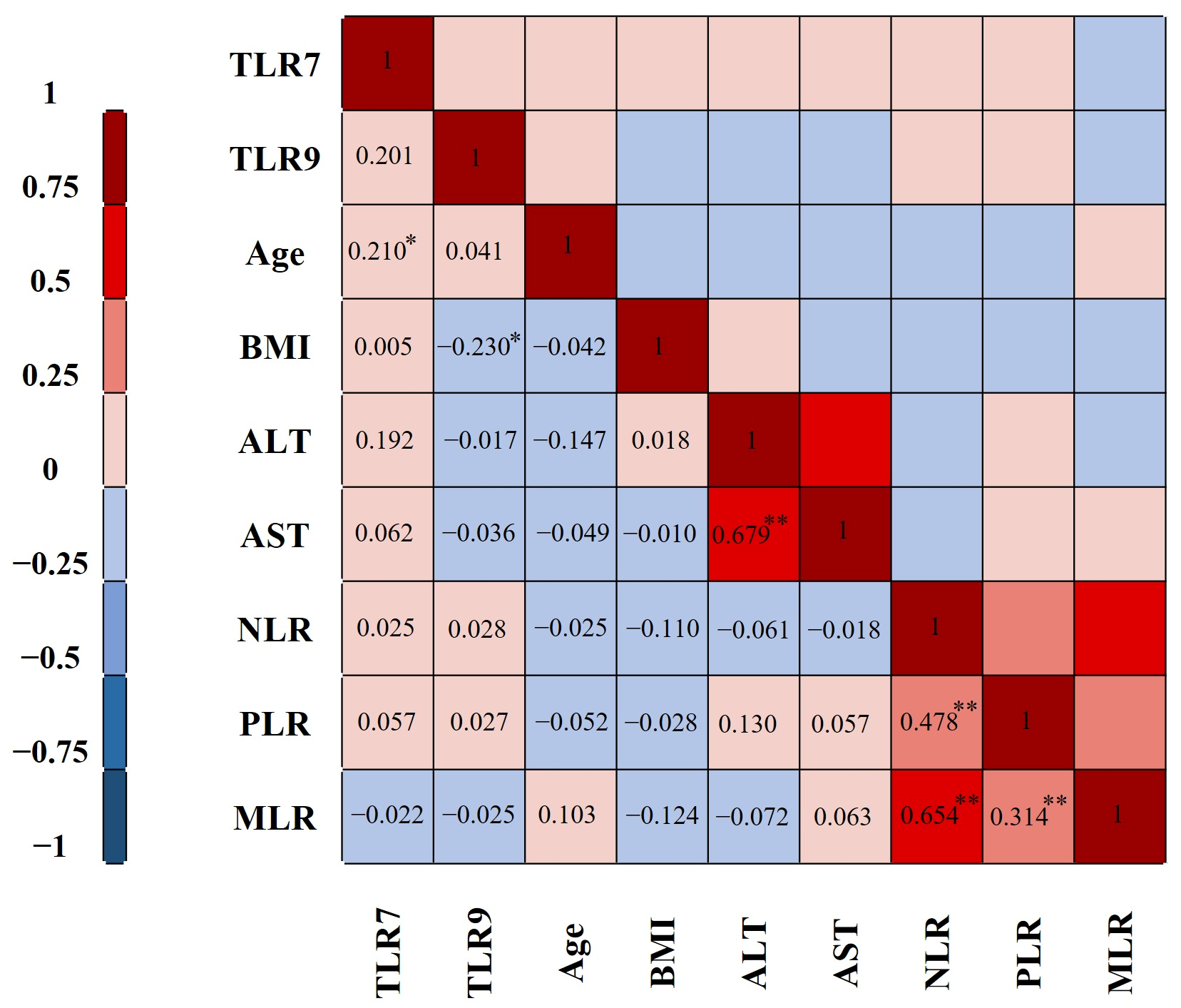
Among 94 COVID-19 patients, an exploratory correlation analysis showed a significant weak positive correlation between TLR7 expression and age (r = 0.210, p = 0.042), whereas TLR9 expression showed a significant weak negative correlation with BMI (r = −0.230, p = 0.026) (Figure 4). A significant moderate positive correlation was observed between ALT and AST (r = 0.679, p < 0.001), between NLR and both PLR and MLR (r = 0.478, p < 0.001; r = 0.654, p < 0.001, respectively), and between PLR and MLR (r = 0.314, p = 0.002). No significant correlations were found between TLR7 and TLR9 expression or between TLR7 or TLR9 expression and any of the remaining tested markers (all p > 0.05).
Figure 4.
Correlation heatmap showing the relationships among TLR7 expression, TLR9 expression, age, BMI, ALT, AST, NLR, PLR, and MLR in 94 COVID-19 patients. Cell values represent Spearman’s correlation coefficients, and color intensity reflects the strength and direction of correlations. Statistically significant correlations are indicated by asterisks (*: p < 0.05 and **: p < 0.01). TLR, toll-like receptor; BMI, body mass index; ALT, alanine aminotransferase; AST, aspartate aminotransferase; NLR, neutrophil-to-lymphocyte ratio; PLR, platelet-to-lymphocyte ratio; MLR, monocyte-to-lymphocyte ratio.
4. Discussion
Despite the end of the global COVID-19 emergency, this pandemic continues to pose a significant public health problem [3,16]. Previous studies have identified many factors and comorbidities associated with increased risk of COVID-19 infection, including male sex, older age, cancer, and diabetes [13,17]. In our study, age was significantly higher in patients with COVID-19 than in controls (p = 0.006). A study by Clarfield and Dwolatzky [18] found that older individuals are at higher risk of coronavirus infections and are more susceptible to post-COVID complications and mortality. However, age did not differ significantly based on clinical severity in our cohort (p = 0.775). In addition, both BMI and sex were comparable between cases and controls, and between mild and moderate-to-critical COVID-19.
Many laboratory abnormalities have been observed in COVID-19 patients, including lymphopenia, elevated platelet counts, and elevated inflammatory markers, with lymphopenia and systemic inflammation being the disease’s characteristic findings [3]. In this study, ALC was significantly lower, whereas MLR, PLR, NLR, and ESR were significantly higher in COVID-19 cases compared to controls (all p < 0.05). These findings are consistent with the inflammatory and immune dysregulation associated with SARS-CoV-2 infection.
TLRs are expressed by many immune cells and are key components of immune responses against viral infections, contributing to viral recognition, initiation of antiviral responses, and cytokine production [9]. Some studies observed multiple single-stranded RNA fragments within the SARS-CoV-2 genome that TLR7 could probably recognize. TLR9 also has an important role in inflammatory responses to viruses, including coronavirus infections, particularly through interferon (IFN) production [19]. However, these biological functions cannot be directly inferred from mRNA expression measurements alone. Interestingly, in our work, patients with COVID-19 showed significantly higher expression of both TLR7 and TLR9 than the control group (both p < 0.001). Bagheri-Hosseinabadi et al. also observed significant overexpression of TLR7 and TLR9 in cases compared to controls (p = 0.003 and 0.009, respectively) [9]. In addition, two previous studies reported separately that TLR7 [15] and TLR9 [20] were significantly more highly expressed in COVID-19 patients compared with the control group (p = 0.001 and 0.009, respectively). Overall, our findings are consistent with previous reports showing increased TLR7 and TLR9 expression levels in COVID-19 patients, providing additional evidence from an independent cohort. Moreover, our study extends previous work by including multivariate logistic regression and ROC curve analyses.
Multivariate analysis showed that TLR7 and TLR9 overexpression were independently associated with COVID-19 infection (p < 0.001 and p = 0.004, respectively). Moreover, TLR7, TLR9, and the combined TLR7 + TLR9 model showed excellent discriminatory performance in distinguishing COVID-19 patients from controls (AUCs: 0.973, 0.937, and 0.965, respectively; all p < 0.001). The combined model did not improve discrimination over TLR7 alone, highlighting TLR7′s strong discriminatory performance in our cohort. Consistent with our findings, TLR9 demonstrated excellent diagnostic performance for COVID-19, with an AUC of 0.94 [21].
In our study, although TLR7 demonstrated the highest discriminatory performance, the very large OR obtained from the conventional logistic regression should be interpreted with caution, particularly with the relatively small sample size and potential quasi-complete separation. Importantly, Firth-penalized logistic regression sensitivity analysis substantially attenuated the TLR7 OR while confirming TLR7′s independent association with COVID-19 infection (p < 0.001). These findings suggest the possibility of effect size inflation in the conventional OR but also confirm the direction and statistical significance of the observed associations. Furthermore, bootstrap optimism correction resulted in only a small reduction in the AUC of the combined TLR7 + TLR9 model (optimism-corrected AUC: 0.960), suggesting that its apparent discrimination was only minimally affected by optimism in our cohort. Therefore, TLR7 and TLR9 may have potential as biomarkers for distinguishing COVID-19 cases from controls in our cohort. Nevertheless, these findings require confirmation in larger cohorts with external validation before clinical application.
Regarding COVID-19 severity, we found that TLR9 overexpression and elevated ALT were significantly associated with moderate-to-critical disease compared to mild disease (p = 0.042 and 0.019, respectively). Both biomarkers remained independently associated with disease severity after multivariate logistic regression analysis (p = 0.030 and p = 0.032, respectively). Previous studies reported that TLR activation is implicated in severe and complicated COVID-19 by inducing the production of inflammatory biomarkers, particularly interleukin-6 (IL-6), tumor necrosis factor (TNF-α), and type 1 interferons (IFN-α and IFN-β) [22,23]. However, because our study measured only TLR7 and TLR9 mRNA expression, these findings should not be interpreted as evidence of increased receptor activation or downstream cytokine signaling. Agmy et al. [24] also reported that overexpression of TLR7 and TLR9 was a predictor of clinical COVID-19 severity, with higher expression associated with worse outcomes and deaths. Future studies with a larger sample size, using comparison and ordinal regression across the four WHO categories, would provide insight into COVID-19 severity trends.
TLRs are expressed on different circulating immune cells, including monocytes, B cells, and dendritic cells [7,25]. Thus, changes in whole-blood leukocyte composition may affect measured TLR7 and TLR9 transcript levels. To address this potential confounding, a sensitivity analysis using logistic regression, adjusted for ALC and AMC, was performed. The independent associations of TLR9 expression and ALT with COVID-19 severity remained essentially unchanged after this additional adjustment (p = 0.034 and 0.032, respectively), supporting the robustness of these associations despite differences in leukocyte composition. Nevertheless, we cannot completely rule out the contribution of differences in immune cell composition to the observed TLR expression levels. Further studies using cell-specific expression analyses, protein-level measurements, and functional assessment of TLR signaling are warranted to clarify these issues and confirm our findings.
Costa et al. [26] concluded that TLR9 stimulates inflammatory immune responses, leading to endothelial cell dysfunction and severe disease. However, these mechanisms were not evaluated in our study. Higher ALT levels indicate liver injury and are usually associated with disease severity, poor prognosis, and increased mortality [27]. However, in this study, the ROC curve analysis of both TLR9 and ALT showed modest discriminatory performance with AUCs of 0.623 and 0.641, respectively. Only a slight improvement in COVID-19 severity was observed with combined TLR9 + ALT (AUC: 0.659). Thus, TLR9 and ALT may provide complementary information regarding COVID-19 severity. However, their discriminatory performance was modest in our cohort and therefore requires validation in multicenter studies with larger sample sizes.
In our study, although both TLR7 and TLR9 expression levels were significantly elevated in patients with COVID-19, they revealed no significant correlation. Additionally, neither TLR7 nor TLR9 showed significant correlations with the hematological, biochemical, or inflammatory parameters tested. These findings indicate that TLR7 and TLR9 expression levels did not show consistent correlations with the measured clinical and laboratory parameters in our cohort. Conversely, TLR7 and TLR9 were significantly correlated in the study by Agmy et al. [24]. This difference in results may be due to differences in populations, disease severity, or related sampling variations. Furthermore, inflammatory markers more likely linked to TLR activation were not evaluated in our study and may have shown significant correlations with TLR expression [22]. Therefore, further studies incorporating these biomarkers are warranted to better clarify the relationship between TLR7 and TLR9 expression levels and immune responses to SARS-CoV-2 infection.
Although the relatively small control sample size may be a limitation of our study, we provide a comprehensive evaluation of both TLR7 and TLR9 by combining expression analysis with multivariate regression, ROC curve analysis, and disease severity assessment in the same cohort. Unlike most previous studies, which primarily reported univariate comparisons of a single TLR, our study evaluated the independent associations and discriminatory performance of both TLR7 and TLR9. Although TLR7 demonstrated excellent discriminatory performance, the very large adjusted OR should be interpreted with caution, given the relatively small sample size and potential model instability. Firth-penalized logistic regression sensitivity analysis substantially attenuated TLR7 OR while confirming the independent association of both TLR7 and TLR9 with COVID-19 infection. In addition, the bootstrap optimism correction provided internal validation of the ROC performance of the combined models. However, external validation was not performed. Additionally, viral load was not evaluated, which may limit exploration of its relationship with TLR expression. Moreover, TLR7 and TLR9 mRNA expression were measured in whole blood, and protein expression, receptor activation, or downstream signaling were not assessed. Therefore, the observed TLR expression level differences cannot be assumed to correspond to changes in protein expression or receptor activity, and the potential contribution of differences in leukocyte composition to transcript levels cannot be completely excluded, despite the robustness of the TLR9 association with disease severity in sensitivity analysis, additionally adjusted for ALC and AMC. Future studies—including larger, multicenter cohorts that specifically investigate the relationships between TLR7 and TLR9 expression levels and the inflammatory biomarkers involved in their cytokine stimulation, viral load, and their diagnostic and severity-associated performance with external validation—are needed to confirm our findings.
5. Conclusions
High expression levels of TLR7 and TLR9 are independently associated with COVID-19 infection. Additionally, increased TLR9 expression and elevated ALT are independently associated with greater disease severity. These findings support an association between TLR7 and TLR9 mRNA expression and COVID-19. However, future multicenter studies with larger sample sizes are required to validate these findings, evaluate their performance for discriminating COVID-19 infection and disease severity, and determine whether increased transcript abundance is accompanied by changes in protein expression or TLR signaling activity.
Supplementary Materials
The following supporting information can be downloaded at https://www.mdpi.com/article/10.3390/covid6090156/s1. Table S1. Firth-penalized logistic regression sensitivity analysis of factors associated with COVID-19 infection. Statistically significant p-values are identified by an asterisk (*). # Wald 95% CIs derived from the Firth estimates because profile likelihood confidence limits did not converge. CI, confidence interval; COVID-19, coronavirus disease 2019; BMI, body mass index; TLR, toll-like receptor. Table S2. ROC analysis of the discriminatory performance of TLR7 and TLR9 expression and their combined model for distinguishing COVID-19 patients from healthy controls. 95% confidence intervals for sensitivity and specificity were estimated using 2000 stratified bootstrap replicates. ROC, receiver operating characteristic; COVID-19, coronavirus disease 2019; AUC, area under the curve; CI, confidence interval; TLR, toll-like receptor. Table S3. Sensitivity multivariate logistic regression analysis of factors associated with moderate-to-critical COVID-19 severity. Statistically significant p-values are identified by an asterisk (*). COVID-19, coronavirus disease 2019; BMI, body mass index; ALT, alanine aminotransferase; TLR, toll-like receptor; ALC, absolute lymphocyte count; AMC, absolute monocyte count. Table S4. ROC analysis of the discriminatory performance of TLR9 expression, ALT, and their combined model for distinguishing moderate-to-critical from mild COVID-19 cases. 95% confidence intervals for sensitivity and specificity were estimated using 2000 stratified bootstrap replicates. ROC, receiver operating characteristic; COVID-19, coronavirus disease 2019; AUC, area under the curve; CIs, confidence intervals; TLR, toll-like receptor; ALT, alanine aminotransferase.
Author Contributions
Conceptualization, R.M.A.-H., T.A.S., G.H.A.-M., K.R., G.H., R.I.S., O.M.A. and A.A.M.; Methodology, R.M.A.-H.; Validation, R.M.A.-H.; Formal Analysis, R.M.A.-H.; Investigation, R.M.A.-H., N.M.M., S.A.F., E.S.S.A., M.M.S.S., E.A.A.G. and A.A.M.; Resources, N.M.M. and A.A.M.; Data Curation, N.M.M., S.A.F., E.S.S.A., B.E.E., R.M. and S.A.R.; Writing—Original Draft Preparation, R.M.A.-H. and N.M.M.; Writing—Review and Editing, B.E.E., T.A.S., R.M., S.A.R., G.H.A.-M., K.R., G.H., M.M.S.S., E.A.A.G., R.I.S. and O.M.A.; Visualization, R.M.A.-H.; Supervision, R.M.A.-H.; Project Administration, A.A.M.; Funding Acquisition, none. All authors have read and agreed to the published version of the manuscript.
Funding
This research received no external funding.
Institutional Review Board Statement
The Research Ethics Committee of the General Organization for Teaching Hospitals and Institutes authorized the study protocol with approval number ITH00137 (22 June 2022).
Informed Consent Statement
The study was conducted in accordance with the principles of the Helsinki Declaration, and informed consent was obtained from all study participants.
Data Availability Statement
Any required data will be provided by the corresponding author upon reasonable request.
Conflicts of Interest
The authors declare no conflicts of interest.
Abbreviations
| ALC | Absolute Lymphocyte Count |
| ALT | Alanine Aminotransferase |
| AMC | Absolute Monocyte Count |
| ANC | Absolute Neutrophil Count |
| ARDS | Acute Respiratory Distress Syndrome |
| AST | Aspartate Aminotransferase |
| AUC | Area Under the Curve |
| BMI | Body Mass Index |
| cDNA | Complementary DNA |
| CI | Confidence Interval |
| COVID-19 | Coronavirus Disease 2019 |
| CpG | Cytosine–Phosphate–Guanine |
| ESR | Erythrocyte Sedimentation Rate |
| FBS | Fasting Blood Sugar |
| Hb | Hemoglobin |
| HCT | Hematocrit |
| IFN | Interferon |
| IL-6 | Interleukin 6 |
| IQR | Interquartile Range |
| LDH | Lactate Dehydrogenase |
| MCH | Mean Corpuscular Hemoglobin |
| MCHC | Mean Corpuscular Hemoglobin Concentration |
| MCV | Mean Corpuscular Volume |
| MLR | Monocyte-to-Lymphocyte Ratio |
| NLR | Neutrophil-to-Lymphocyte Ratio |
| PAMPs | Pathogen-Associated Molecular Patterns |
| PCR | Polymerase Chain Reaction |
| PLR | Platelet-to-Lymphocyte Ratio |
| PLT | Platelet Count |
| PPBS | Postprandial Blood Sugar |
| RBC | Red Blood Cell Count |
| ROC | Receiver Operating Characteristic |
| RT-PCR | Reverse Transcription Polymerase Chain Reaction |
| SARS-CoV-2 | Severe Acute Respiratory Syndrome Coronavirus 2 |
| TLR | Toll-Like Receptor |
| TNF | Tumor Necrosis Factor |
| WBC | White Blood Cell Count |
| WHO | World Health Organization |
References
- Abdel-Hamid, R.M.; El-Mahallawy, H.A.; Allam, R.M.; Zafer, M.M.; Elswify, M. Changing Patterns of Bacterial Profile and Antimicrobial Resistance in High-Risk Patients during the COVID-19 Pandemic at a Tertiary Oncology Hospital. Arch. Microbiol. 2024, 206, 250. [Google Scholar] [CrossRef] [Scilit]
- Sakr, A.A.; Ahmed, A.E.; Hasona, N.A.; Abdel-Hamid, R.M.; Faisal, A.S.; Shaaban, E.E.; Mohamed, S.; Abbas, A.M.; Mohamed, A.A. Diagnostic and Immunological Roles of Leptin Gene Rs7799039 Polymorphism and Cytokines in COVID-19, HCV, and Dual Infection. Sci. Rep. 2026, 16, 4361. [Google Scholar] [CrossRef] [Scilit]
- Mohamed, A.A.; Alanazi, A.T.; Ahmed, H.H.; Elfiky, S.; Abdel Ghafar, M.T.; Maher, I.; Taha, S.A.; AbuRahma, M.Z.A.; Elagawy, W.; Mohareb, D.A.; et al. FokI Polymorphism of the Vitamin D Receptor Gene: Linking COVID-19 Risk to Genetic Susceptibility in Children. Cytokine 2025, 191, 156958. [Google Scholar] [CrossRef] [Scilit]
- World Health Organization. Statement on the Fifteenth Meeting of the IHR (2005) Emergency Committee on the COVID-19 Pandemic. WHO. 2023. Available online: https://www.who.int/news/item/05-05-2023-statement-on-the-fifteenth-meeting-of-the-international-health-regulations-(2005)-emergency-committee-regarding-the-coronavirus-disease-(covid-19)-pandemic (accessed on 24 August 2026).
- Zheng, Z.; Peng, F.; Xu, B.; Zhao, J.; Liu, H.; Peng, J.; Li, Q.; Jiang, C.; Zhou, Y.; Liu, S.; et al. Risk Factors of Critical & Mortal COVID-19 Cases: A Systematic Literature Review and Meta-Analysis. J. Infect. 2020, 81, e16–e25. [Google Scholar] [CrossRef] [Scilit]
- Wolff, D.; Nee, S.; Hickey, N.S.; Marschollek, M. Risk Factors for Covid-19 Severity and Fatality: A Structured Literature Review. Infection 2021, 49, 15–28. [Google Scholar] [CrossRef] [Scilit]
- Bezemer, G.F.G.; Garssen, J. TLR9 and COVID-19: A Multidisciplinary Theory of a Multifaceted Therapeutic Target. Front. Pharmacol. 2021, 11, 601685. [Google Scholar] [CrossRef] [Scilit]
- Satyanarayanan, S.K.; Yip, T.F.; Han, Z.; Zhu, H.; Qin, D.; Lee, S.M.Y. Role of Toll-like Receptors in Post-COVID-19 Associated Neurodegenerative Disorders? Front. Med. 2025, 12, 1458281. [Google Scholar] [CrossRef] [Scilit]
- Bagheri-Hosseinabadi, Z.; Rezazadeh Zarandi, E.; Mirabzadeh, M.; Amiri, A.; Abbasifard, M. MRNA Expression of Toll-like Receptors 3, 7, 8, and 9 in the Nasopharyngeal Epithelial Cells of Coronavirus Disease 2019 Patients. BMC Infect. Dis. 2022, 22, 448. [Google Scholar] [CrossRef] [Scilit]
- Alturaiki, W.; Alkadi, H.; Alamri, S.; Awadalla, M.E.; Alfaez, A.; Mubarak, A.; Alanazi, M.A.; Alenzi, F.Q.; Flanagan, B.F.; Alosaimi, B. Association between the Expression of Toll-like Receptors, Cytokines, and Homeostatic Chemokines in SARS-CoV-2 Infection and COVID-19 Severity. Heliyon 2023, 9, e12653. [Google Scholar] [CrossRef] [Scilit]
- Huang, Z.; Gao, Y.; Han, Y.; Yang, J.; Yang, C.; Li, S.; Zhou, D.; Huang, Q.; Yang, J. Revealing the Roles of TLR7, a Nucleic Acid Sensor for COVID-19 in Pan-Cancer. Biosaf. Health 2023, 5, 211–226. [Google Scholar] [CrossRef] [Scilit]
- World Health Organization. Clinical Management of Severe Acute Respiratory Infection (SARI) When COVID-19 Disease Is Suspected: Interim Guidance. World Health Organization. 2020. Available online: https://apps.who.int/iris/handle/10665/331446 (accessed on 24 August 2026).
- Abdel-Hamid, R.M.; Bayoumi, A.; Abdellateif, M.S.; Nooh, H.A.; Refaat, L.; Kandeel, E.Z.; Hassan, S.S. Bacterial Co-Infections in Cancer Patients with COVID-19: Predictors and Antimicrobial Resistance Trends. J. Infect. Dev. Ctries. 2024, 18, 1185–1195. [Google Scholar] [CrossRef] [Scilit]
- Abdel-Hamid, R.M.; Allam, R.M.; Refaat, L.; Nooh, H.A.; Mahmoud, F.M.; Bayoumi, A.; Hassan, S.S. Unraveling Mortality Risks in Pediatric Oncology: Exploring Bloodstream Coinfections and Inflammatory Biomarkers in COVID-19. J. Infect. Chemother. 2025, 31, 102741. [Google Scholar] [CrossRef] [Scilit]
- El-Hefnawy, S.M.; Eid, H.A.; Mostafa, R.G.; Soliman, S.S.; Omar, T.A.; Azmy, R.M. COVID-19 Susceptibility, Severity, Clinical Outcome and Toll-like Receptor (7) MRNA Expression Driven by TLR7 Gene Polymorphism (Rs3853839) in Middle-Aged Individuals without Previous Comorbidities. Gene Rep. 2022, 27, 101612. [Google Scholar] [CrossRef] [Scilit]
- Ashraf, A.M.; Al-Maqtoofi, M.Y.; Burghal, A.A. Cytokine Production and TLR 7/8 Gene Expression Following BBIBP-CorV COVID-19 Vaccination. Vacunas 2025, 26, 500459. [Google Scholar] [CrossRef] [Scilit]
- Weidmann, M.D.; Berry, G.J.; Zucker, J.E.; Huang, S.; Sobieszczyk, M.E.; Green, D.A. Bacterial Pneumonia and Respiratory Culture Utilization among Hospitalized Patients with and without COVID-19 in a New York City Hospital. J. Clin. Microbiol. 2022, 60, e0017422. [Google Scholar] [CrossRef] [Scilit]
- Clarfield, A.M.; Dwolatzky, T. Age and Ageing During the COVID-19 Pandemic; Challenges to Public Health and to the Health of the Public. Front. Public Health 2021, 9, 655831. [Google Scholar] [CrossRef] [Scilit]
- Lee, N.; Ko, R.; Lee, S.Y. Differential Expression Patterns of Toll-like Receptors in COVID-19 Patients. Front. Biosci.-Landmark 2023, 28, 307. [Google Scholar] [CrossRef] [Scilit]
- Kamiab, Z.; Kazemi Arababadi, M.; Bahrehmand, F.; Bazmandegan, G.; Sayadi, A.; Abbasifard, M. Toll-Like Receptor Dysregulation in The Hospitalized COVID-19 Patients. Adv. Biomed. Res. 2025, 14, 78. [Google Scholar] [CrossRef] [Scilit]
- Mohamed, A.K.; Mohamed, A.A.; Foda, M.S.; Elamir, A.Y.; Ezz AL Arab, M.; Abdulmohsen, M.A.; Labib, G.H.; Omran, M.M. Evaluation of the Diagnostic Performance of Toll-Like Receptors 4 and 9 as Reliable Markers for Corona Virus Disease-19. Egypt. Acad. J. Biol. Sci. C Physiol. Mol. Biol. 2024, 16, 195–210. [Google Scholar] [CrossRef] [Scilit]
- Onofrio, L.; Caraglia, M.; Facchini, G.; Margherita, V.; Placido, S.D.; Buonerba, C. Toll-Like Receptors and COVID-19: A Two-Faced Story with an Exciting Ending. Future Sci. OA 2020, 6, FSO605. [Google Scholar] [CrossRef] [Scilit]
- Islamuddin, M.; Mustfa, S.A.; Ullah, S.N.M.N.; Omer, U.; Kato, K.; Parveen, S. Innate Immune Response and Inflammasome Activation During SARS-CoV-2 Infection. Inflammation 2022, 45, 1849–1863. [Google Scholar] [CrossRef] [Scilit]
- Agmy, G.; Khalifa, F.; Adam, M.; Mokhtar, M.; Mahmoud, M.A. Expression of Toll-like Receptors 3, 7, and 9 in Peripheral Blood Mononuclear Cells and Prognosis of Pneumonia in Hospital-Admitted Coronavirus Disease 2019 Patients: Is There an Association? Egypt. J. Chest Dis. Tuberc. 2026, 75, 180–189. [Google Scholar] [CrossRef] [Scilit]
- Fong, F.L.Y.; Kirjavainen, P.V.; El-Nezami, H. Immunomodulation of Lactobacillus Rhamnosus GG (LGG)-Derived Soluble Factors on Antigen-Presenting Cells of Healthy Blood Donors. Sci. Rep. 2016, 6, 22845. [Google Scholar] [CrossRef] [Scilit]
- Costa, T.J.; Potje, S.R.; Fraga-Silva, T.F.C.; da Silva-Neto, J.A.; Barros, P.R.; Rodrigues, D.; Machado, M.R.; Martins, R.B.; Santos-Eichler, R.A.; Benatti, M.N.; et al. Mitochondrial DNA and TLR9 Activation Contribute to SARS-CoV-2-Induced Endothelial Cell Damage. Vasc. Pharmacol. 2022, 142, 106946. [Google Scholar] [CrossRef] [Scilit]
- Chew, W.D.; Kuang, J.; Lin, H.; Ang, L.W.; Yang, W.L.; Lye, D.C.; Young, B.E. Clinical Predictors for Abnormal ALT in Patients Infected with COVID-19—A Retrospective Single Centre Study. Pathogens 2023, 12, 473. [Google Scholar] [CrossRef] [Scilit]

Disclaimer/Publisher’s Note: The statements, opinions and data contained in all publications are solely those of the individual author(s) and contributor(s) and not of MDPI and/or the editor(s). MDPI and/or the editor(s) disclaim responsibility for any injury to people or property resulting from any ideas, methods, instructions or products referred to in the content. |
© 2026 by the authors. Licensee MDPI, Basel, Switzerland. This article is an open access article distributed under the terms and conditions of the Creative Commons Attribution (CC BY) license.