


Abstract
The production of polymeric nanoparticles using microfluidic systems holds great potential for controlled drug delivery applications. In this study, the effects of flow parameters and surfactant properties on the characteristics of PLGA (Poly (lactic-co-glycolic acid)) nanoparticles were systematically investigated. First, the total flow rate (TFR) and flow rate ratio (FRR) were optimized to ensure stable droplet formation. Subsequently, the effects of different surfactant types (anionic, cationic, and nonionic) and their varying concentrations were evaluated. Using the selected parameters, capsaicin-loaded PLGA nanoparticles were successfully produced. The particles were prepared using a microfluidic platform, and the organic phase was subsequently removed via solvent evaporation. The resulting formulations were comprehensively characterized in terms of particle size, polydispersity index (PDI), zeta potential, and encapsulation efficiency (%EE). Additionally, the in vitro release profiles and cytotoxicity of capsaicin-loaded nanoparticles were evaluated. This study aimed to elucidate the decisive role of surfactant parameters in the microfluidic production of PLGA nanoparticles and to contribute to the development of optimized and reproducible formulations.
Keywords:
microfluidic system; surfactants; polymeric nanoparticles; PLGA; capsaicin; Kolliphor EL; CTAC; SDS 1. Introduction
Microfluidic technology provides a highly controlled environment for the synthesis of nanoparticles (NPs), overcoming the limitations of traditional bulk methods by offering precise manipulation of fluid dynamics. While traditional synthesis often struggles with high energy consumption, excessive heat loss, and slow processing, microfluidic technology provides a compact and controllable alternative [1]. This technology enables the reproducible production of nanomaterials with adjustable size, desired morphology, and high monodispersity by allowing precise manipulation of fluid dynamics and mixing parameters at the micro-scale [2,3].
PLGA is a clinically approved, highly biocompatible polymer widely used in targeted drug delivery [4]. However, the therapeutic efficacy of PLGA-based systems depends directly on the colloidal stability achieved during synthesis. Surfactants are incorporated into the formulation components to reduce the system’s free energy during nanoparticle formation and prevent the particles from aggregating by adhering to one another. Surfactants play a central role in controlling the critical physicochemical parameters of the synthesized nanoparticles, such as particle size, zeta potential, and polydispersity, by reducing the interfacial tension between the polymeric matrix and the aqueous phase. Therefore, the selection and concentration of surfactants constitute some of the most critical steps in the formulation development process for achieving the desired stability and drug-loading capacity of PLGA NPs [5,6,7,8]. To evaluate these optimized nanocarriers, capsaicin (CPS) was chosen as an ideal model drug, as its clinical application is severely hindered by poor water solubility and low bioavailability despite its significant therapeutic benefits [9,10,11].
This study aims to systematically elucidate the decisive role of surfactant properties, such as chemical structure, ionic charge, and concentration, in the production of microfluidic-based PLGA NPs. Although microfluidic platforms provide superior control over nanoparticle synthesis, the complex interaction between fluid dynamics and surfactant-mediated interfacial stabilization remains a critical factor in achieving reproducible formulations. In the first phase of the study, basic flow parameters, TFR and FRR, were optimized using polyvinyl alcohol (PVA), which is identified in the literature as the most commonly used stabilizer for PLGA-based systems. Subsequently, comprehensive screening was conducted to evaluate the effect of various surfactants on particle size, PDI, and zeta potential. The study examined nonionic surfactants (PVA, Pluronic F-127, Pluronic F-68, Tween 80, Tween 20, Kolliphor EL, Brij 35, and PVP K90), cationic surfactants (cetyl trimethylammonium chloride [CTAC], cetyl trimethylammonium bromide [CTAB], benzalkonium chloride, polyethylenimine [PEI], and cetylpyridinium chloride), and anionic surfactants (sodium dodecyl sulfate [SDS], sodium cholate, and sodium oleate). Rather than confining the study to a few model stabilizers, this comprehensive array of surfactants was strategically utilized to systematically unravel how the interaction between a surfactant’s molecular architecture and its diverse physicochemical attributes, such as ionic charge, molecular size, HLB, CMC, and pH responsiveness, influences the rapid nucleation and growth kinetics during microfluidic assembly. As a key point, surfactant-free aqueous phases, PBS (pH 7.4) and ultrapure water were included to critically investigate whether the addition of external stabilizers is fundamentally necessary to ensure stable nanoparticle formation in a microfluidic environment or whether the process itself provides sufficient intrinsic stabilization. Following the screening process, representative surfactants from each ionic class were selected to evaluate concentration-dependent effects. Finally, capsaicin was used as a model active ingredient to validate the performance of optimized surfactant systems in a practical drug delivery context. To provide a robust and optimized framework for the development of highly controlled and reproducible polymeric nanocarriers, the encapsulation efficiency, and in vitro release profiles of these CPS-loaded nanoparticles were evaluated, along with their biological validation through cytotoxicity tests.
2. Materials and Methods
2.1. Materials
The BM5-Z500 Bifurcating Mixing Microfluidic Chip (Nehir Biotechnology, Ankara, Turkiye) was used for nanoparticle synthesis. PLGA (50:50 lactide:glycolide, Mw 40,000–75,000 g/mol), PVA (Mw 13,000–23,000), Pluronic F-68, Pluronic F-127, Kolliphor EL, CTAC, CTAB, PEI, cetylpyridinium chloride, SDS, sodium cholate, and acetone were purchased from Sigma-Aldrich (Merck KGaA, Darmstadt, Germany). Tween 80 and Tween 20 were obtained from Merck KGaA (Darmstadt, Germany). PVP K90 and sodium oleate were purchased from Fluka (Buchs, Switzerland). Brij 35 was purchased from Acros Organics (Geel, Belgium) and benzalkonium chloride was purchased from Central Drug House Ltd. (New Delhi, India). For cell culture studies, Dulbecco’s Modified Eagle’s Medium (DMEM) supplemented with L-glutamine, fetal bovine serum, penicillin, and streptomycin (all purchased from Gibco, Grand Island, NY, USA) was used. Human prostate cancer (PC3) and breast cancer (MCF-7) cell lines were obtained from the American Type Culture Collection (ATCC, Manassas, VA, USA).
2.2. Methods
2.2.1. Microfluidic Device and Setup
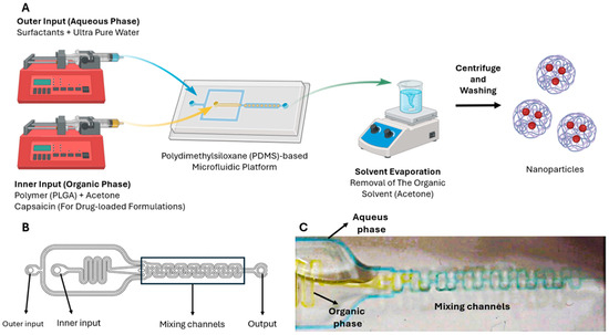
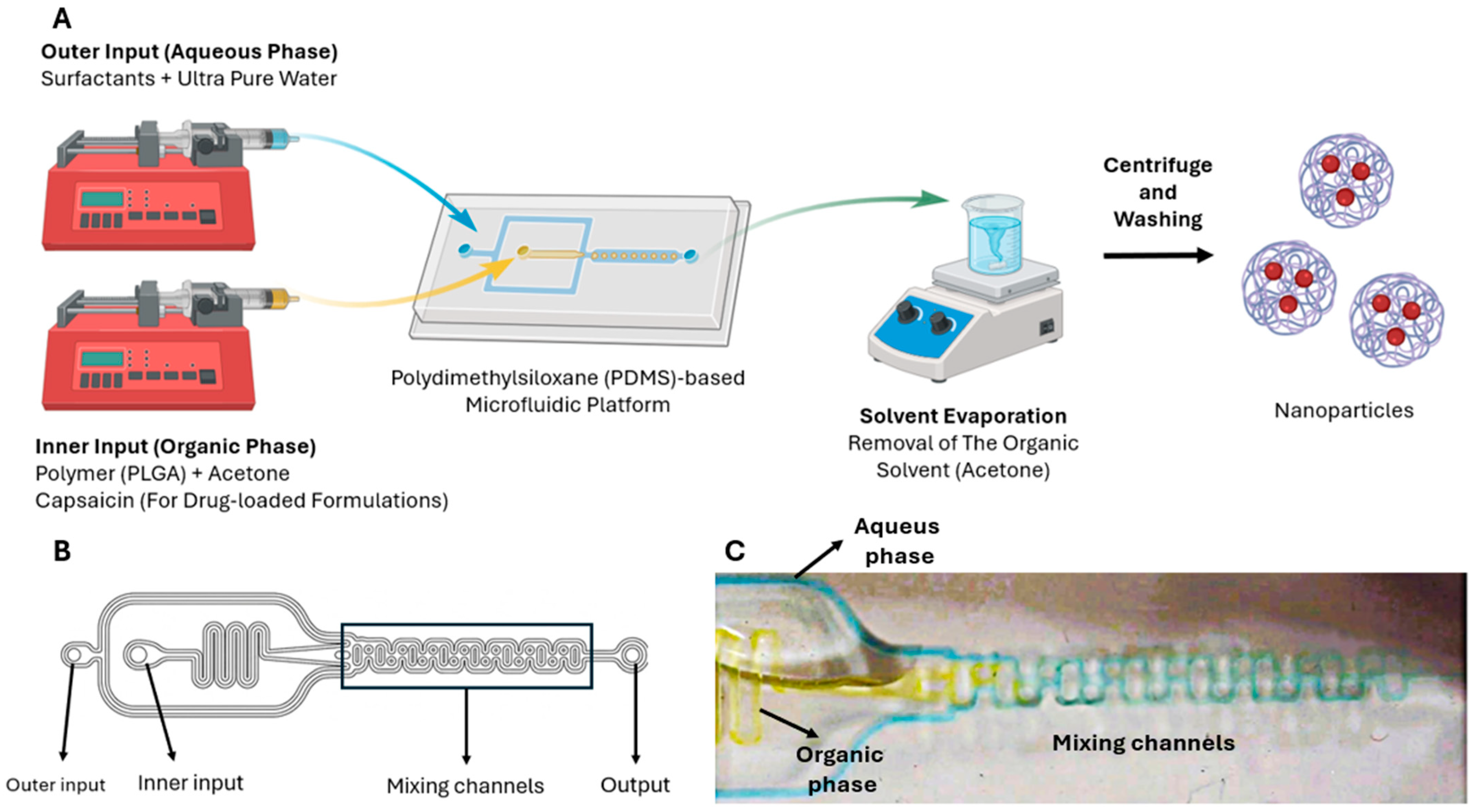
The nanoparticles were produced using a polydimethylsiloxane (PDMS)-based microfluidic platform (Figure 1). During the preparation process, the organic phase (acetone) containing the polymer was pumped through the microfluidic chip’s “inner” input via a syringe pump, while the aqueous phase (ultra pure water) consisting of various surfactants was pumped through the chip’s “outer” input. The nanoparticles were collected at the system’s output. Following the collection step, a solvent evaporation method was applied to the formulations to remove the organic solvent [12]. For drug-loaded formulations, capsaicin was dissolved in acetone together with PLGA to form the organic phase.
Figure 1.
(A) General Schematic Flow Diagram for the Microfluidic-Based Synthesis and Processing of PLGA Nanoparticles. (B) Detailed microchannel design of the BM5-Z500 microfluidic chip. (C) Real-time optical photograph captured using a digital microscope (Celestron Labs Digital, 5MP; Celestron, Torrance, CA, USA) showcasing the stable fluid flow profile and dynamic phase interactions (aqueous and organic phases) within the mixing channels during device operation.
2.2.2. Optimization of Flow Parameters
Prior to investigating the effect of surfactants on NP production via a microfluidic system, a comprehensive preliminary study was conducted to determine the optimal operating parameters. Since PVA is the most widely utilized surfactant for PLGA NPs in the literature [13,14], it was selected as the model surfactant for the flow rate optimization phase. At this stage, while maintaining constant compositions for the inner phase (PLGA 1 mg/mL) and outer phase (0.5% PVA), the effects of FRR (Inner:Outer) and TFR on particle size, PDI, and zeta potential were systematically evaluated. The flow rate was set at base levels of 1.0, 0.5, and 0.1 mL/min; the FRR values of 1:5, 1:2, 1:1, and 2:1 (Inner:Outer) for each base value. This approach allowed for the systematic observation of the effects of both different FRR values and variations in TFR within each FRR cohort on particle characteristics. Based on the obtained data, representative FRR and TFR parameters were selected for use in subsequent surfactant studies.
2.2.3. Influence of Surfactant Type and Concentration
Following the optimization of the microfluidic system parameters, the effect of various types of surfactants on nanoparticle properties was systematically investigated. At this stage, based on the preliminary study described in Section 2.2.2, a FRR of 1:5 and a TFR of 6 mL/min were fixed for all formulations. While the composition of the internal phase (PLGA 1 mg/mL) and the surfactant concentration in the external phase (0.5% w/v) were kept constant, a total of 18 different formulations (B1–B18) were prepared based on the chemical structure and charge of the surfactants.
The nonionic surfactants and stabilizers used in the study consisted of PVA, Pluronic F-127, Pluronic F-68, Tween 80, Tween 20, Kolliphor EL, Brij 35, and PVP K90. CTAC, CTAB, benzalkonium chloride, PEI, and cetylpyridinium chloride were selected as cationic surfactants, while SDS, sodium cholate, and sodium oleate were used in the anionic group. Fundamental information regarding the selected surfactants is provided in Table 1. Additionally, surfactant-free control groups containing PBS (pH 7.4) and ultrapure water were prepared to observe the system’s intrinsic nanoparticle formation capacity and internal stability in the absence of any surfactant. All prepared NPs were characterized in terms of particle size, PDI, and zeta potential.
Table 1.
Physicochemical properties of the surfactants and stabilizers used in the study.
Following the evaluation of surfactant types, one representative from each main category (nonionic, cationic, and anionic) that yielded the smallest particle size was selected. This selection was intended to specifically investigate the impact of surfactant concentration on nanoparticle properties. Accordingly, Kolliphor EL, CTAC, and SDS were chosen for the subsequent experimental phase. At this stage, five different concentration levels (0.1%, 0.25%, 0.5%, 1%, and 2% w/v) were determined for each surfactant, resulting in a total of 15 formulations (C1–C15). All other parameters, including a FRR of 1:5, a TFR of 6 mL/min, and a PLGA concentration of 1 mg/mL, were held constant to ensure consistency with the previous experiments.
2.2.4. Preparation of Capsaicin-Loaded PLGA NPs
Following the optimization of surfactant concentrations, the 2% (w/v) level, which yielded the minimum particle size across all three surfactant categories, was selected for the preparation of drug-loaded nanoparticles. In this phase, CPS-loaded formulations (D1–D3) were developed. The internal phase consisted of 1 mg/mL PLGA, and 0.25 mg/mL CPS dissolved in acetone. Microfluidic process parameters, including an FRR of 1:5 and a TFR of 6 mL/min, were maintained to ensure consistency with the previous experimental stages. In addition to particle size, PDI, and zeta potential, the prepared nanoparticles (NPs) were characterized in terms of encapsulation efficiency (%EE) and in vitro drug release profiles.
2.2.5. Particle Size, PDI, Zeta Potential
The mean particle size, PDI, and zeta potential of the nanoparticles were measured using a Zetasizer Nano ZS (Malvern Instruments, Worcestershire, UK). Prior to analysis, the nanoparticle dispersions were diluted 1:100 with ultrapure water, and measurements were conducted at 25 °C [47]. To ensure the accuracy and reproducibility of the results, each sample was analyzed in triplicate (n = 3).
2.2.6. Encapsulation Efficiency and Drug Loading
The drug encapsulation efficiency was determined indirectly by quantifying the amount of unloaded drug in the supernatant. The formulations were subjected to ultracentrifugation at 30,000 rpm for 1 h at 5 °C. Following centrifugation, the unencapsulated amount of CPS in the supernatant was measured spectrophotometrically at 280 nm (ThermoScientific, Waltham, MA, USA), and the %EE was calculated using the following equation [48]:
Drug encapsulation efficiency (%) = (Amount of total drug − Amount of unloaded drug)∕Amount of total drug × 100.
2.2.7. pH Measurement
The pH values of the surfactant solutions were measured to evaluate their potential influence on the zeta potential of the formulated nanoparticles. For this purpose, 0.5% (w/v) aqueous solutions of each surfactant were prepared using ultrapure water. Measurements were conducted using a digital pH meter (FiveEasy, Mettler Toledo, Greifensee, Switzerland), at room temperature (25 ± 1 °C). All measurements were performed in triplicate to ensure accuracy, and the results are presented as mean values.
2.2.8. In Vitro Release
In Vitro, drug release studies were performed on CPS-loaded nanoparticle formulations using the dialysis membrane method. PBS buffer (pH 7.4) containing 30% methanol was utilized as the dissolution medium to ensure sink conditions. The nanoparticles were dispersed in 2 mL of the release medium and placed into a dialysis membrane (14 kDa MWCO, Sigma). The membranes were then immersed in 25 mL of dissolution medium and horizontally shaken at 50 rpm in an incubator maintained at 37 °C ± 0.5 °C. At predetermined time intervals, 1 mL of sample were withdrawn and replaced with an equal volume of fresh medium to maintain a constant volume [49]. The concentration of CPS in the samples was measured spectrophotometrically at 280 nm. All experiments were conducted in triplicate (n = 3), and the in vitro release profiles were obtained by plotting the cumulative percentage of drug released against time.
2.2.9. Cell Viability Assay
The (3-(4,5-dimethylthiazol-2-yl)-5-(3-carboxymethoxyphenyl)-2-(4-sulfophenyl)-2H-tetrazolium (MTS) assay, a colorimetric method used to assess the effects of substances on cell viability, was employed [50]. PC3 and MCF-7 cells were utilized in cell culture. Cells were added to Falcon tubes containing 5 mL of DMEM within a laminar flow cabinet, followed by centrifugation at 1300 rpm for 5 min. Subsequently, the supernatant was carefully removed using a sterile serological pipette, leaving the cell pellet intact. D1, C5 and CPS were administered to the cells for 24 h. The compounds were applied at 7.81 μM, 16.63 μM, 31.25 μM, 62.5 μM, 125 μM, and 250 μM concentrations. Cell viability was evaluated by measuring optical density at a wavelength of 450 nm using an ELISA plate reader (BioTek Instruments, Inc., Winooski, VT, USA) (n = 4).
3. Results and Discussion
3.1. Effect of Total Flow Rate and Flow Rate Ratio on Particle Formation
The formulation parameters and characterization results of the formulations produced within the scope of the study conducted to determine the optimal operating parameters are shared in Table 2.
Table 2.
Influence of FRR and TFR on the physicochemical characteristics of PLGA nanoparticles.
In all developed formulations, it was observed that increasing the flow rate of the outer phase (surfactant-containing phase) led to a significant decrease in particle size; conversely, increasing the flow rate of the inner phase (polymer-containing phase) relative to the outer phase resulted in the formation of larger particle sizes. This situation proves that the high shear stress created by the high speed of the outer phase supports the formation of smaller droplets [51,52]. High external phase flow rates increase the concentration gradient between the internal and external phases, accelerating the diffusion rate of the organic solvent into the external phase. This rapid solvent exchange causes the polymer to settle much faster, limiting the time required for particle growth. Rapid mixing and high energy input increases the homogeneous nucleation rate in the system, triggering the formation of numerous small nuclei [53,54,55]. Since the amount of polymer in the medium is shared among these numerous nuclei, the final size of each particle remains limited. The surfactant (PVA) is in the outer phase and coats the surface of the newly formed droplets, preventing coalescence [56]. A high outer phase flow rate can accelerate the mass transfer of surfactant molecules to the newly formed interfaces. This allows the nano-droplets to stabilize rapidly before they have a chance to coalesce and grow. Consequently, in addition to shear stress, the increase in outer phase flow focuses the inner phase in a thinner line, shortening the diffusion distance and increasing the solvent exchange rate, resulting in faster and more homogeneous precipitation of the polymer, forming smaller particles [57,58].
To examine the TFR effect, evaluating formulations with the same FRR values reveals that a decrease in total flow rate leads to a systematic increase in particle size. This situation can be explained by the mixing kinetics and solvent exchange rate within the microfluidic channels. High TFR values accelerate interphase interaction, ensuring homogeneous mixing in a shorter time and triggering the polymer to rapidly reach “supersaturation,” promoting the formation of numerous small nuclei (nucleations) [59,60]. Conversely, when the TFR decreases, the mixing efficiency decreases, and the time required for solvent diffusion increases. This slows down the nucleation rate, allowing more time for the growth or aggregation of existing nuclei, ultimately leading to the formation of larger nanoparticles [61]. At low TFR values, the local polymer concentration remains high for longer, increasing the probability of collisions between particles. High TFR, on the other hand, accelerates stabilization by rapidly separating newly formed particles and bringing them into contact with fresh surfactant molecules more quickly. This minimizes aggregation, resulting in smaller and more tightly packed particles [62]. In summary, TFR holds kinetic control of the system; as TFR increases, the mixing time shortens and the nucleation rate dominates the growth rate, which can be considered the primary driving force behind size reduction.
The results are strongly supported by existing literature, where increasing the outer aqueous phase flow rate (or FRR) is consistently shown to reduce PLGA nanoparticle size by accelerating solvent extraction and polymer precipitation [63,64,65]. However, an interesting exception to this trend was reported by Kozalak et al., where increasing the FRR resulted in larger particles and a loss of monodispersity. This divergence was attributed to the specific geometric limitations of the staggered herringbone channels used in their study, which failed to maintain mixing efficiency and shear force control at high flow ratios. This contrast clearly highlights that optimizing flow parameters alone is insufficient; the dynamic interaction between fluid kinetics and specific microfluidic channel architecture ultimately dictates the final particle properties [66].
The PDI indicates how narrow (homogeneous) the particle size distribution is. According to the generally accepted rule, values below 0.3 for polymeric nanoparticles are considered “narrowly distributed” (monodisperse) and successful [67]. Upon examining the characterization results, it was observed that all formulations had PDI values below 0.3. The fact that all values in the table range from 0.024 to 0.235 demonstrates that the microfluidic system produces highly controlled and homogeneous particles.
Zeta potential indicates the surface charge of the particles and, consequently, their physical stability. The fact that all values are negative (ranging from −12.8 to −21.7 mV) is attributed to the terminal carboxyl groups in the structure of the polymeric matrix, PLGA. This serves as chemical evidence that the particles were successfully formed. Generally, values above ±30 mV are considered to indicate high stability [68]. In the A4, A8, and A12 formulations, where the FRR is 2:1 (indicating a more dominant internal phase), the zeta potential remains stable around −21 mV. This suggests that as the internal phase ratio increases, the PLGA concentration or the arrangement of polymer chains results in a more stable distribution of charge. In addition, non-ionic surfactants provide stability through steric hindrance (physical barrier) because they do not carry an electrical charge; therefore, even at low zeta potential values, the long polar chains of the molecules can prevent droplets from approaching and merging [69].
This study was conducted to determine a representative flow rate during the formulation production stage and to systematically investigate the effects of flow parameters on NPs characterization. The findings indicate that different FRR and TFR values can be selected based on the targeted NP properties, and that precise control over NP characterization can be achieved through the optimization of these parameters. In our study, the FRR and TFR parameters of the A1 formulation, which yielded the smallest particle size of 82.3 ± 2.5 nm, were selected as representative values for use in subsequent stages where the effect of surfactants would be investigated, and these values were kept constant throughout all subsequent experiments.
3.2. Impact of Surfactant Type and Concentration on NP Characteristics
The formulation parameters and characterization results of the formulations produced as part of the study conducted to determine the effect of various types of surfactants on NP properties are presented in Table 3.
Table 3.
Impact of surfactant types.
In experiments conducted under constant flow conditions (A1 conditions: FRR 1:5, TFR 6 mL/min), it was observed that the type of surfactant caused significant differences in particle size, PDI, and zeta potential. Nonionic surfactants play a critical role in the development of PLGA NP formulations due to their high bioavailability and low toxicity profiles [15]. Comparing Tween variants, Tween 80’s 18-carbon unsaturated oleic acid chain provides stronger, broader hydrophobic interactions with the PLGA matrix than Tween 20’s 12-carbon saturated chain, more effectively inhibiting droplet growth during nucleation [70,71]. Furthermore, Tween 80’s lower HLB value grants it a higher interfacial affinity, which rapidly reduces interfacial tension and stabilizes the Marangoni effect to yield smaller particles [57,72,73,74,75]. Additionally, the approximately three-fold lower critical micelle concentration of Tween 80 ensures rapid surface coating during the millisecond-scale microfluidic mixing process. In contrast, Tween 20’s higher CMC results in slower adsorption kinetics that fail to outpace the rapid nucleation rate, leading to particle instability and aggregation [76,77]. Ultimately, the excellent PDI value of 0.095 obtained with Tween 80 demonstrates that its rapid stabilization capacity perfectly complements the precise fluidic control of the system.
Pluronic F-68 (Poloxamer 188) and F-127 (Poloxamer 407), which are FDA-approved and biocompatible triblock copolymers, play a critical role in the stabilization of PLGA NPs due to their PEO-PPO-PEO structures [17,78]. Theoretically, Pluronic F-127’s low critical micelle concentration (CMC) and low HLB value (12–14), which reflect its lipophilic nature, might suggest that it would bind more effectively to the PLGA surface and form smaller NP sizes; however, experimental data reveal that formulations prepared with F-127 (B2) and F-68 (B3) exhibit similar characterization results. This situation can be explained by the balancing mechanisms between the dynamic nature of microfluidic systems and the physicochemical properties of surfactants; since the rate at which molecules reach the newly formed interface during the millisecond-long mixing time in microfluidic channels is decisive [79,80,81]. Due to its lower molecular weight compared to F-127, F-68 achieves a higher diffusion coefficient and, through this “kinetic advantage,” can halt particle growth at an earlier stage during the nucleation phase [82,83]. Moreover, F-68’s higher PEO ratio forms a denser hydrophilic corona on the surface, establishing a rigid hydration shield that physically inhibits further growth [84,85]. Since DLS hydrodynamic diameter measurements are influenced by surface chain length, F-127’s longer block structure extending into the aqueous phase likely artificially increases its measured size [86,87]. Consequently, while F-127 offers stronger PPO-mediated binding, F-68’s rapid diffusion and dense steric shielding compensate for the final particle size, demonstrating that kinetic molecular dynamics must be prioritized alongside classical HLB/CMC parameters in microfluidic systems.
Although comparative analysis of the heterogeneous physicochemical structures of other nonionic surfactants complicates the matter, it is observed that Kolliphor EL, which has the lowest HLB value within this group, enables the production of the smallest-sized (56.4 nm) NPs. While the size advantage provided by Kolliphor EL stands out for applications requiring rapid cellular penetration [88], Pluronic F-68 emerges as the preferred choice when long-term stability and resistance to opsonization (protein adsorption) in biological environments are targeted, thanks to its high negative charge (−42.2 mV) and superior stability profile [89]. On the other hand, the use of PVP K90 (B8), which has the highest molecular weight (approximately 360,000 g/mol), resulted in the highest particle size in the group at 440.9 nm. In terms of zeta potential, although all surfactants used were nonionic in nature, the fact that the prepared NPs exhibited distinct negative charges ranging from −12 mV (PVA) to −42 mV (P68) demonstrates that the formulation components modulate surface properties through different mechanisms.
With the transition to the cationic group, we observe a dramatic change in the table: the zeta potential has shifted strongly from negative (the natural charge of PLGA) to positive. This is a critical advantage, particularly for drug delivery systems expected to interact with the cell membrane (which is negatively charged) [90]. Cationic surfactants coat the PLGA NP surface, providing electrostatic stabilization [91]. The negative charges observed in the nonionic group have been replaced by very high positive values ranging from +39.2 mV to +56.6 mV. The fact that these values are well above the +30 mV threshold indicates that the particles repel each other very strongly and possess “perfect” physical stability from a colloidal perspective. PEI, with its polycationic structure and enormous molecular weight (Mw: 750,000 g/mol), exhibits the highest charge at +56.6 mV. This proves that the numerous amine groups (polycationic) on the PEI chains completely dominate the surface. In addition, just like PVP K90 in the non-ionic group, PEI also exhibited the largest particle size in its group at 290.3 nm. PEI’s polycationic nature may have triggered aggregation by bridging between nuclei during nucleation. CTAC, benzalkonium chloride, and cetylpyridinium chloride yielded similar particle sizes (112–132 nm). When the CTAC (B9), CTAB (B10), and benzalkonium chloride (B11) groups are evaluated together, the group’s common physicochemical properties are very similar: low molecular weights (320–365 g/mol), high HLB values (20–21), and low CMC levels (0.94 mM–1.3 mM). We observe this similarity in the PLGA nanoparticles produced using a microfluidic system, yielding highly consistent results in terms of both size (112–133 nm) and homogeneity (PDI < 0.13), as well as high zeta potential values.
With our shift to the anionic surfactant group, anionic surfactants maximize electrostatic repulsion by adding an additional negative layer to PLGA rather than masking its natural negative charge.
Comparison of surfactant-free control groups (B17 and B18) with surfactant-containing formulations reveals that in surfactant-free systems, particle size increases to 562.7 nm (B17) and PDI values reach highly heterogeneous levels such as 0.535. This indicates that the rapid mixing advantage provided by the microfluidic system alone is insufficient, and a stabilizer layer is necessary to reduce interfacial tension at the nucleation stage and prevent droplet growth.
The pH of the dispersion medium significantly influenced the zeta potential, particularly in nonionic and surfactant-free formulations. Higher pH values (e.g., Pluronics and PBS) promoted the deprotonation of PLGA’s terminal carboxyl groups, yielding significantly more negative zeta potentials compared to more acidic environments (e.g., Brij 35 and Tween 80). Conversely, in formulations stabilized by ionic surfactants, the inherent strong electrical charge of the surfactant molecules dominated the surface properties, effectively overshadowing the pH-dependent variations.
The study continued by evaluating the effect of different surfactant concentrations, selecting the surfactant that yielded the smallest particle size from each group of nonionic, cationic, and anionic surfactants. In this context, Kolliphor EL was selected as the nonionic surfactant, CTAC as the cationic surfactant, and SDS as the anionic surfactant. The formulation parameters and characterization results of the formulations produced as part of the study conducted to determine the effect of various concentrations of surfactants on NP properties are presented in Table 4.
Table 4.
Impact of surfactant concentration.
As a general trend, it is observed that an increase in concentration leads to a decrease in particle size in all three surfactant groups (Kolliphor EL, CTAC, and SDS). For the ionic surfactants CTAC (C6–C10) and SDS (C11–C15), it was found that particle size decreased as the concentration increased from 0.1% to 2%. This phenomenon can be explained by the increased number of surfactant molecules more effectively reducing interfacial tension, thereby promoting the formation of smaller droplets during nucleation and rapidly covering the newly formed surface to prevent particle coalescence [92,93]. In the Kolliphor EL group, a nonionic surfactant, one of the smallest sizes (51.0 nm) was achieved at a 0.25% concentration; however, a slight tendency for the size to increase was observed at higher concentrations (C4: 66.6 nm). This situation may be due to the thickening of the coating layer on the particle surface or changes in the viscosity of the medium in the presence of excessive surfactant. The electrical charges of surfactants directly dominate the surface properties of NPs. NPs prepared with the cationic surfactant CTAC exhibited high positive charges ranging from +39 mV to +48.6 mV, while those prepared with the anionic surfactant SDS exhibited strong negative charges ranging from −39.1 mV to −54.1 mV. These high absolute zeta potential values create a strong electrostatic repulsive force between particles, thereby enhancing the system’s physical stability. In the nonionic Kolliphor EL series, the zeta potential was observed to range from −14.4 mV to −30.9 mV. Despite its nonionic character, this observed negative charge may originate from the terminal carboxyl groups of the polymer (PLGA) or from the electrical double-layer effect formed during the surfactant’s deposition on the surface. It is noteworthy that ionic surfactants (particularly SDS and CTAC) yield significantly lower and more stable PDI values (typically <0.13) compared to Kolliphor EL. This demonstrates that strong electrostatic repulsion, combined with the rapid mixing dynamics in the microfluidic system, results in a highly homogeneous (monodisperse) distribution. While CTAC and SDS have a linear structure, Kolliphor EL has a branched structure and contains fatty acids. In the Kolliphor EL series, however, the higher and more variable PDI values (0.11–0.31) suggest that this surfactant relies primarily on steric hindrance for stabilization, and that this mechanism cannot produce a distribution as narrow as that achieved by electrostatic stabilization under the high shear forces present in the microfluidic system.
The results regarding concentration-dependent size reduction and the varied stabilization capacities of different surfactant types align robustly with dynamic interfacial tension frameworks reported in the literature. In the millisecond-scale mixing environment of microfluidics, rapid adsorption kinetics are paramount. Achieving effective interfacial stabilization in such highly dynamic systems often requires surfactant concentrations well above their static CMC. The structural differences among surfactants fundamentally dictate their transport mechanisms to the newly formed interfaces. For instance, the rapid convection-based transport of micromolecular surfactants like SDS is often more kinetically efficient than the micelle-breakup-limited transport of cationic alternatives like CTAB [94,95,96]. Similarly, lower molecular weight surfactants inherently exhibit faster adsorption kinetics compared to bulky macromolecular stabilizers (e.g., PVA), thereby halting nucleation earlier and yielding smaller, more monodisperse particles [97].
3.3. Physicochemical Characterization of Capsaicin-Loaded PLGA NPs
Formulations containing 2% surfactant, which exhibited smaller particle sizes, were selected for capsaicin loading and further evaluation. The formulation parameters and physicochemical properties of capsaicin-loaded PLGA nanoparticles are presented in Table 5. A slight decrease in particle size was observed after capsaicin was added to the formulations. A similar result has been seen in other studies with capsaicin [98,99]. The greatest reduction in particle size is observed in the formulation prepared with Kolliphor EL. It is known that the hydrophobic structural parts of the capsaicin molecule are generally found in the lipid region of nanostructures [100]. This phenomenon is attributed to the use of Kolliphor EL, which has a lower HLB value than CTAC and SDS and is more hydrophobic. Under these conditions, capsaicin preferentially partitions into the hydrophobic core of the nanoparticle, enhancing hydrophobic interactions with the polymer chains and resulting in a more compact core structure.
Table 5.
Physicochemical characterization of capsaicin-loaded nanoparticles.
3.4. In Vitro Release Profiles
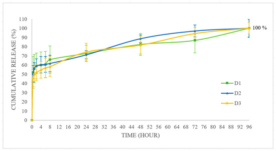
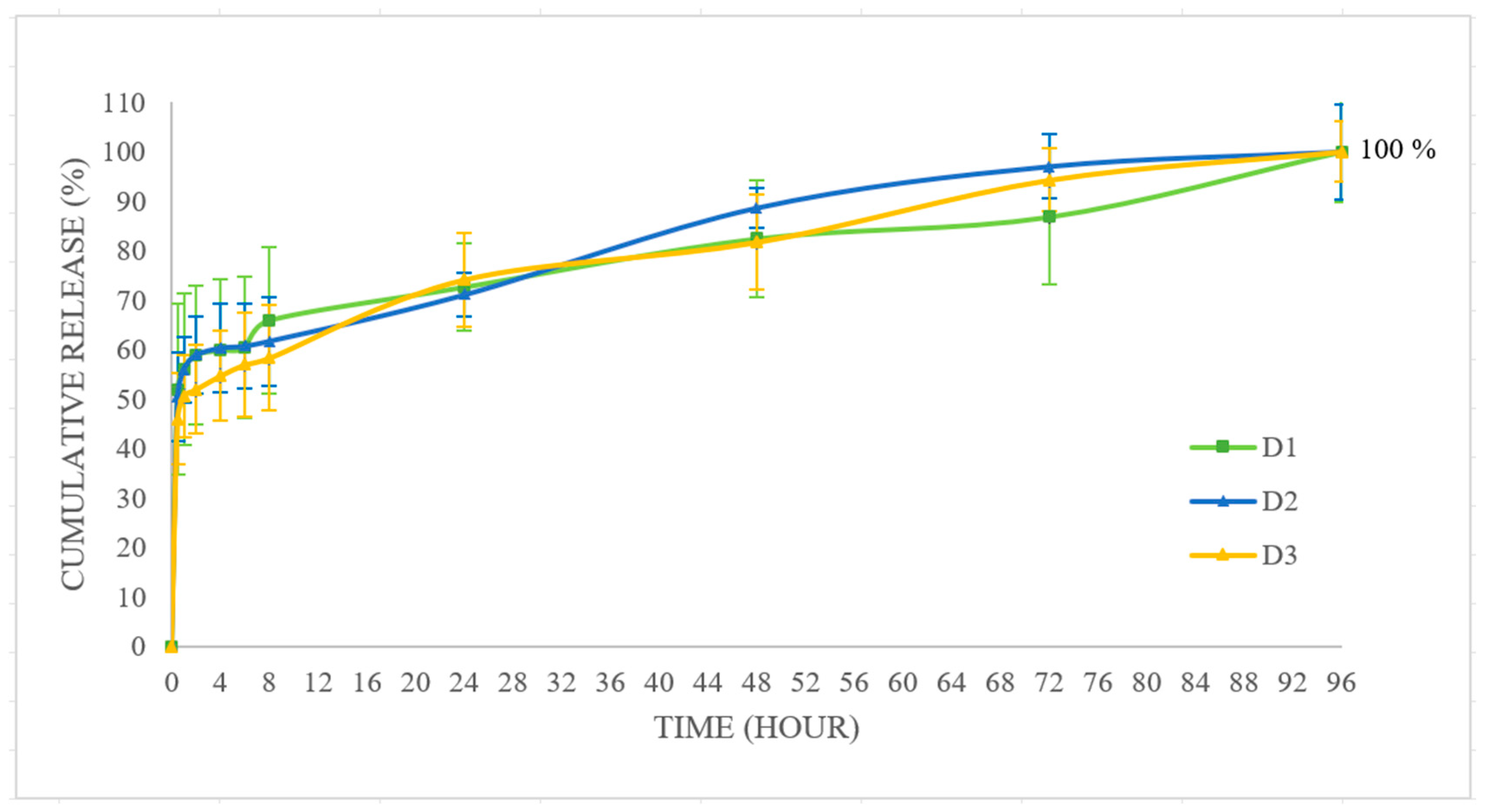
All three formulations exhibit a biphasic release profile (Figure 2). A pronounced initial burst release occurs very rapidly, with the primary turning point observed within the first 1 to 2 h, where approximately 45–55% of the drug load is released. This rapid phase then transitions into a more gradual release, reaching 55–65% by the 8th hour. While this initial burst is partially attributed to the active substance loosely attached to or trapped near the nanoparticle surface, such a high percentage is primarily driven by additional physicochemical factors inherent to the system [101]. Specifically, the exceptionally high surface-area-to-volume ratio of the nanoparticles drastically shortens the diffusion path for the encapsulated drug. Furthermore, the rapid solvent exchange and precipitation kinetics during microfluidic mixing often result in a drug-enriched outer polymeric shell and initial rapid hydration of the PLGA matrix, accelerating early drug diffusion. Following this burst phase, from the 8th to the 96th hour, a slower and highly controlled release rate is observed, ultimately reaching 100% release.
Figure 2.
In vitro release profiles of CPS-loaded nanoparticles.
Despite having different physicochemical properties (size and zeta potential), the in vitro release profiles of all three formulations (D1, D2, and D3) are quite similar. This indicates that the release kinetics in a buffer medium are primarily governed by the erosion of the PLGA matrix and drug diffusion, rather than the surfactant type or specific surface coating. However, this similarity does not render the optimization process redundant. It is well established in the literature that the ultimate therapeutic efficacy of nanocarriers extends beyond their in vitro release profiles and is fundamentally dictated by their interactions with biological barriers. Particle size and surface charge are critical physicochemical parameters that govern targeting capabilities and cellular uptake mechanisms. Smaller nanoparticles typically demonstrate enhanced cellular internalization via various endocytosis pathways, allowing for more efficient intracellular drug accumulation. Furthermore, the surface charge plays a decisive role in membrane interactions; positively charged nanoparticles exhibit a strong electrostatic affinity for inherently negative cell membranes, thereby facilitating rapid binding and subsequent cellular entry [102]. Therefore, the meticulous optimization of surfactants and microfluidic parameters is an indispensable step. Even if these parameters do not drastically alter the drug release rate from the PLGA matrix in a buffer medium, they are essential for tuning the precise size and charge required to ensure optimal colloidal stability and successful biological performance of the formulation.
3.5. Cell Viability
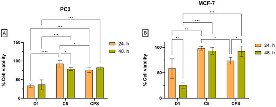
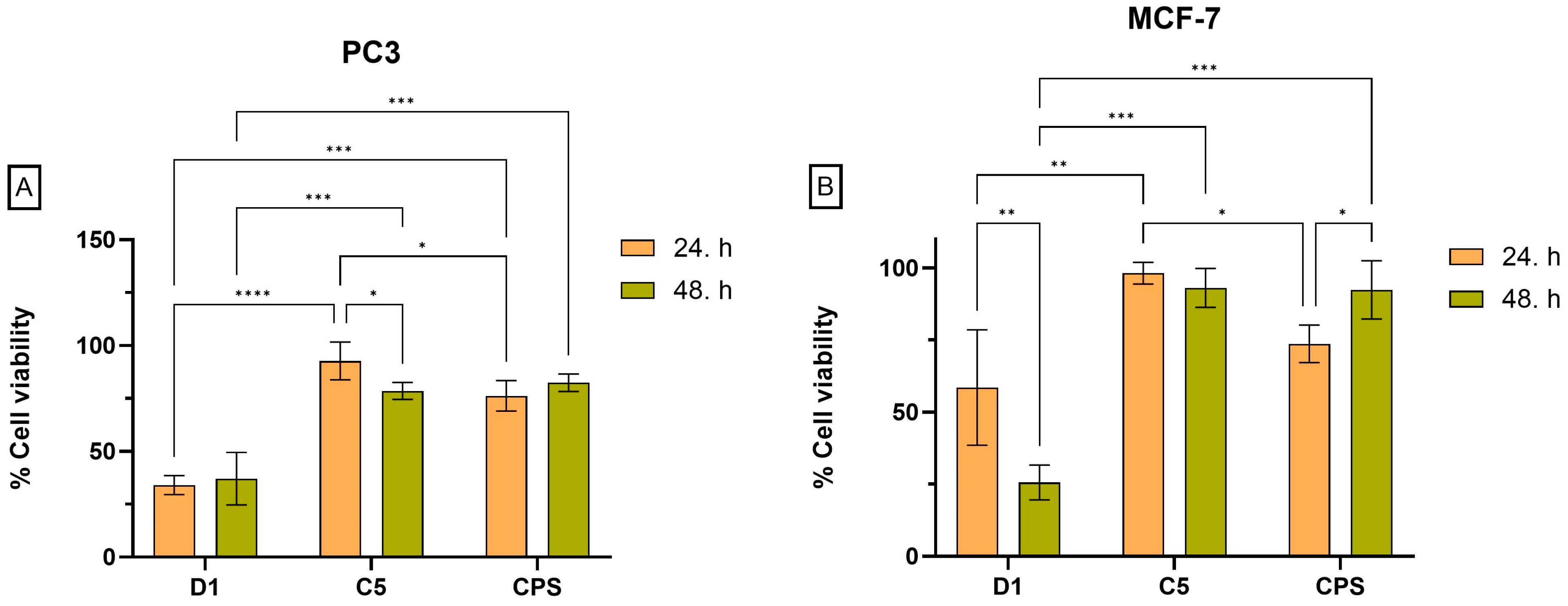
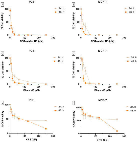
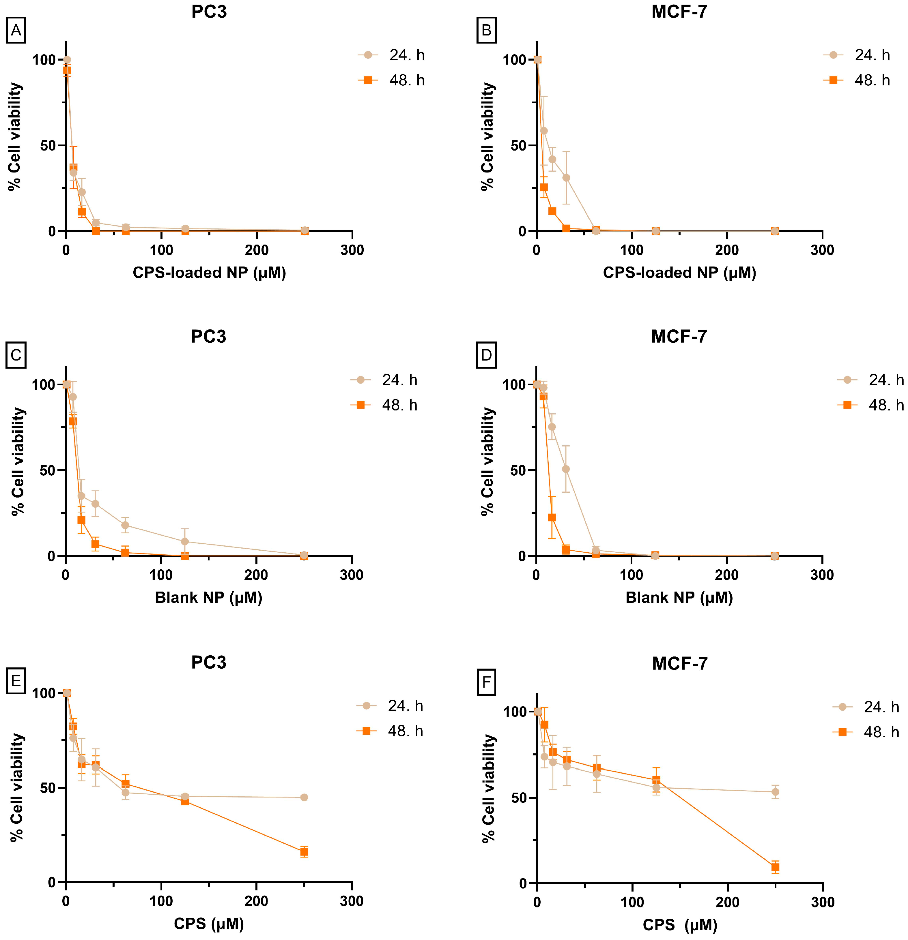
Cell viability was ultimately observed to be significantly enhanced by the capsaicin-containing nanoparticle formulation (D1) compared to capsaicin alone in both PC3 and MCF-7 cell lines (Figure 3). Drug-free nanoparticles (C5) containing Kollifor EL also showed increased cytotoxic effects, exceeding 31.25 μM concentration in PC3 cells and 62.5 μM concentration in MCF-7 cells (Figure 4). This could be attributed to the fact that a part of Kolliphor EL was adsorbed onto the nanoparticle surface and not removed by washing, disrupting cell membrane integrity, affecting mitochondrial function, and causing oxidative stress [103,104].
Figure 3.
Effects of free capsaicin (CPS), capsaicin-loaded formulation (D1; containing 7.81 µM CPS), and blank carrier system (C5; containing the same amount of carrier without CPS) on cell viability in PC3 (A) and MCF-7 (B) cells following 24 h and 48 h treatments. Cell viability was expressed as percentage relative to the untreated control group. Data is presented as mean ± SD. Statistical significance between the indicated groups is represented by brackets. p values are indicated as follows: p < 0.05 (*), p < 0.01 (**), p < 0.001 (***), and p < 0.0001 (****).
Figure 4.
The cytotoxic effect of compound D1, C5, and CPS was evaluated in MCF-7 and PC3 cell lines. The percentages of cell viability for MCF-7 (B,D,F) and PC3 (A,C,E) are presented. The data represents the mean viability percentages obtained from at least three independent experiments performed in triplicate, across a concentration range of 7.81–250 μM and at multiple time points. Cell viability was calculated by normalizing the absorbance values to those of untreated control cells, which were defined as 100% viability for each time point.
4. Conclusions and Future Perspectives
This study successfully performed the systematic optimization and fabrication of capsaicin-loaded PLGA nanoparticles using a microfluidic platform. Our findings revealed that flow parameters are the key determinants of initial droplet formation; high total flow rates (TFRs) and increasing flow rate ratios (FRRs) favoring the surfactant-containing aqueous phase significantly reduce particle size through increased shear stress. Furthermore, the physicochemical nature of the surfactants (molecular weight, HLB, and ionic charge) was found to be a decisive factor in modulating the final nanoparticle properties. In particular, Kolliphor EL provided the smallest particle sizes, while Pluronic F-68 offered superior surface stabilization with a high negative zeta potential. A critical finding of this research is that, despite differences in particle size and surface charge, the in vitro release kinetics of capsaicin remained remarkably similar across different formulations. This indicates that the drug release rate is primarily controlled by the erosion and diffusion mechanisms of the PLGA polymeric matrix, rather than by the surface properties provided by the surfactants. Consequently, the data obtained from this study offer a rational framework for the reproducible production of polymeric nanoparticles and make a significant contribution to the development of effective microfluidic-based drug delivery systems.
Although this study focuses on the effects of water-soluble surfactants, future research could investigate the synergistic effects of using lipid-soluble surfactants (such as Span 80, lecithin, or sorbitan monostearate) alone or in combination with hydrophilic surfactants on nanoparticle stability and characterization. Optimizing surfactant type, concentration, and ratios in binary or multiple surfactant systems can allow for much more precise control of NP properties. Furthermore, how to minimize precipitation and clogging problems frequently encountered in microfluidic channels through surfactant selection is an important research topic. Methodologically, incorporating experimental design (DoE) methods into the process and supporting the obtained data with computational chemistry and physics simulations will transform NP production from a ‘trial-and-error’ approach into a rational design paradigm. Although PLGA was used as the model polymer in our study, the interaction mechanisms of the physicochemical properties of different polymer types with surfactants and the applicability of these findings to other drug delivery systems such as liposomes should be investigated. As a result, through integrated control of chip architecture, flow parameters, and formulation components, it will be possible to produce NPs with optimum properties specific to the disease type, stage, and targeted tissue.
Author Contributions
Conceptualization, A.B. and B.K.; methodology, A.B., B.K. and H.Y.; formal analysis A.B., B.K. and H.Y.; investigation, A.B., B.K. and H.Y.; resources, A.B., B.K. and H.Y.; data curation, A.B., B.K. and H.Y.; writing—original draft preparation, A.B., B.K. and H.Y.; writing—review and editing, A.B. and B.K.; visualization, A.B., B.K. and H.Y.; supervision, B.K. All authors have read and agreed to the published version of the manuscript.
Funding
This research received no external funding.
Institutional Review Board Statement
Not applicable.
Informed Consent Statement
Not applicable.
Data Availability Statement
Data is contained within the article.
Acknowledgments
The authors would like to express their gratitude to the Pharmaceutical Nanobiotechnology Research Laboratory, Faculty of Pharmacy, Ankara University, for their valuable support during this study. Additionally, Figure 1A was created with BioRender.com, and Figure 1B was generated using generative artificial intelligence.
Conflicts of Interest
The authors declare no conflicts of interest.
Abbreviations
The following abbreviations are used in this manuscript:
| PLGA | Poly(lactic-co-glycolic acid) |
| TFR | Total flow rate |
| FRR | Flow rate ratio |
| PDI | Polydispersity index |
| %EE | Encapsulation efficiency |
| NP | Nanoparticle |
| CPS | Capsaicin |
| PVA | polyvinyl alcohol |
| CTAC | cetyl trimethylammonium chloride |
| CTAB | cetyl trimethylammonium bromide |
| PEI | Polyethylenimine |
| SDS | sodium dodecyl sulfate |
| PVP | Polyvinylpyrrolidone |
| Mw | Molecular Weight |
| nm | Nanometer |
| PDMS | polydimethylsiloxane |
| PBS | Phosphate-Buffered Saline |
| mM | millimolar |
| rpm | revolutions per minute |
| MWCO | Molecular Weight Cut-Off |
| kDa | kilodalton |
| mV | millivolt |
References
- Gimondi, S.; Ferreira, H.; Reis, R.L.; Neves, N.M. Microfluidic devices: A tool for nanoparticle synthesis and performance evaluation. ACS Nano 2023, 17, 14205–14228. [Google Scholar] [CrossRef] [Scilit] [PubMed]
- Küçüktürkmen, B.; Inam, W.; Howaili, F.; Gouda, M.; Prabhakar, N.; Zhang, H.; Rosenholm, J.M. Microfluidic-assisted fabrication of dual-coated pH-sensitive mesoporous silica nanoparticles for protein delivery. Biosensors 2022, 12, 181. [Google Scholar] [CrossRef] [Scilit] [PubMed]
- Bezelya, A.; Küçüktürkmen, B.; Bozkır, A. Microfluidic devices for precision nanoparticle production. Micro 2023, 3, 822–866. [Google Scholar] [CrossRef] [Scilit]
- Makadia, H.K.; Siegel, S.J. Poly lactic-co-glycolic acid (PLGA) as biodegradable controlled drug delivery carrier. Polymers 2011, 3, 1377–1397. [Google Scholar] [CrossRef] [Scilit] [PubMed]
- Filippousi, M.; Angelakeris, M.; Katsikini, M.; Paloura, E.; Efthimiopoulos, I.; Wang, Y.; Zamboulis, D.; Van Tendeloo, G. Surfactant effects on the structural and magnetic properties of iron oxide nanoparticles. J. Phys. Chem. C 2014, 118, 16209–16217. [Google Scholar] [CrossRef] [Scilit]
- Harikrishnan, A.; Dhar, P.; Agnihotri, P.K.; Gedupudi, S.; Das, S.K. Effects of interplay of nanoparticles, surfactants and base fluid on the surface tension of nanocolloids. Eur. Phys. J. E 2017, 40, 53. [Google Scholar] [CrossRef] [Scilit] [PubMed]
- Lugo, D.M.; Oberdisse, J.; Lapp, A.; Findenegg, G.H. Effect of nanoparticle size on the morphology of adsorbed surfactant layers. J. Phys. Chem. B 2010, 114, 4183–4191. [Google Scholar] [CrossRef] [Scilit] [PubMed]
- Shkodra, B.; Grune, C.; Traeger, A.; Vollrath, A.; Schubert, S.; Fischer, D.; Schubert, U. Effect of surfactant on the size and stability of PLGA nanoparticles encapsulating a protein kinase C inhibitor. Int. J. Pharm. 2019, 566, 756–764. [Google Scholar] [CrossRef] [Scilit] [PubMed]
- Arul, B.; Kothai, R. Anticancer effect of capsaicin and its analogues. In Capsicum; IntechOpen: London, UK, 2020. [Google Scholar]
- Clark, R.; Lee, S.-H. Anticancer properties of capsaicin against human cancer. Anticancer Res. 2016, 36, 837–843. [Google Scholar] [PubMed]
- Küçüktürkmen, B.; Öz, U.C.; Er, E.; Gómez, I.J.; Tekneci, S.I.; Eşim, Ö.; Özköse, U.U.; Gülyüz, S.; Üstündağ, A.; Yılmaz, Ö. Design of polymeric nanoparticles for theranostic delivery of capsaicin as anti-cancer drug and fluorescent nitrogen-doped graphene quantum dots. Macromol. Biosci. 2024, 24, 2400149. [Google Scholar] [CrossRef] [Scilit]
- Bezelya, A.; Küçüktürkmen, B.; Böncü, T.E.; Bozkır, A. Microfluidics-Based Nanoparticle Formulations: Preparation and Evaluation of Protein Delivery Systems. Polym. Adv. Technol. 2025, 36, e70250. [Google Scholar] [CrossRef] [Scilit]
- Menon, J.U.; Kona, S.; Wadajkar, A.S.; Desai, F.; Vadla, A.; Nguyen, K.T. Effects of surfactants on the properties of PLGA nanoparticles. J. Biomed. Mater. Res. Part A 2012, 100, 1998–2005. [Google Scholar] [CrossRef] [Scilit]
- Kennedy, P.J.; Perreira, I.; Ferreira, D.; Nestor, M.; Oliveira, C.; Granja, P.L.; Sarmento, B. Impact of surfactants on the target recognition of Fab-conjugated PLGA nanoparticles. Eur. J. Pharm. Biopharm. 2018, 127, 366–370. [Google Scholar] [CrossRef] [Scilit] [PubMed]
- Cortés, H.; Hernández-Parra, H.; Bernal-Chávez, S.A.; Prado-Audelo, M.L.D.; Caballero-Florán, I.H.; Borbolla-Jiménez, F.V.; González-Torres, M.; Magaña, J.J.; Leyva-Gómez, G. Non-ionic surfactants for stabilization of polymeric nanoparticles for biomedical uses. Materials 2021, 14, 3197. [Google Scholar] [CrossRef] [Scilit] [PubMed]
- Turk, C.T.S.; Oz, U.C.; Serim, T.M.; Hascicek, C. Formulation and optimization of nonionic surfactants emulsified nimesulide-loaded PLGA-based nanoparticles by design of experiments. Aaps Pharmscitech 2014, 15, 161–176. [Google Scholar] [PubMed]
- Khaliq, N.U.; Lee, J.; Kim, S.; Sung, D.; Kim, H. Pluronic F-68 and F-127 based nanomedicines for advancing combination cancer therapy. Pharmaceutics 2023, 15, 2102. [Google Scholar] [CrossRef] [Scilit] [PubMed]
- Bollenbach, L.; Buske, J.; Mäder, K.; Garidel, P. Poloxamer 188 as surfactant in biological formulations–An alternative for polysorbate 20/80? Int. J. Pharm. 2022, 620, 121706. [Google Scholar] [PubMed]
- Ćirin, D.M.; Poša, M.M.; Krstonošić, V.S.; Milanović, M.L. Conductometric study of sodium dodecyl sulfate-nonionic surfactant (Triton X-100, Tween 20, Tween 60, Tween 80 or Tween 85) mixed micelles in aqueous solution. Hem. Ind. 2012, 66, 21–28. [Google Scholar]
- Mahmood, M.E.; Al-Koofee, D.A. Effect of temperature changes on critical micelle concentration for tween series surfactant. Glob. J. Sci. Front. Res. Chem. 2013, 13, 4. [Google Scholar]
- Belo, I.; García-Abuín, A.; Gómez-Díaz, D.; Navaza, J.M.; Vidal-Tato, I. Effect of Tween 80 on bubble size and mass transfer in a bubble contactor. Chem. Eng. Technol. 2011, 34, 1790–1796. [Google Scholar] [CrossRef] [Scilit]
- Bolten, D.; Lietzow, R.; Türk, M. Solubility of ibuprofen, phytosterol, salicylic acid, and naproxen in aqueous solutions. Chem. Eng. Technol. 2013, 36, 426–434. [Google Scholar] [CrossRef] [Scilit]
- Rowe, R.C.; Sheskey, P.J.; Quinn, M.E. Handbook of Pharmaceutical Excipients; Pharmaceutical Press: London, UK, 2006; Volume 6. [Google Scholar]
- Monteiro-Riviere, N.A.; Inman, A.O.; Wang, Y.Y.; Nemanich, R.J. Surfactant effects on carbon nanotube interactions with human keratinocytes. Nanomed. Nanotechnol. Biol. Med. 2005, 1, 293–299. [Google Scholar] [CrossRef] [Scilit]
- BASF SE. Technical Information Kolliphor EL; BASF SE: Ludwigshafen, Germany, 2012. [Google Scholar]
- Tanhaei, B.; Saghatoleslami, N.; Chenar, M.P.; Ayati, A.; Hesampour, M.; Mänttäri, M. Experimental study of CMC evaluation in single and mixed surfactant systems, using the UV–Vis spectroscopic method. J. Surfactants Deterg. 2013, 16, 357–362. [Google Scholar]
- Mabrouk, M.M.; Hamed, N.A.; Mansour, F.R. Spectroscopic methods for determination of critical micelle concentrations of surfactants; a comprehensive review. Appl. Spectrosc. Rev. 2023, 58, 206–234. [Google Scholar]
- Tang, J.; Wang, Y.; Wang, D.; Wang, Y.; Xu, Z.; Racette, K.; Liu, F. Key structure of brij for overcoming multidrug resistance in cancer. Biomacromolecules 2013, 14, 424–430. [Google Scholar] [CrossRef] [Scilit] [PubMed]
- Lee, J.-M.; Jun, Y.-D.; Kim, D.-W.; Lee, Y.-H.; Oh, S.-G. Effects of PVP on the formation of silver–polystyrene heterogeneous nanocomposite particles in novel preparation route involving polyol process: Molecular weight and concentration of PVP. Mater. Chem. Phys. 2009, 114, 549–555. [Google Scholar] [CrossRef] [Scilit]
- Zhang, J.; Bai, Y.; Du, W.; Wu, Y.; Gu, X.; Li, H.; Ma, Y.; Qu, C.; Chen, G. The effect of anion on cationic surfactants and a structure-efficiency relationship study. Desalin. Water Treat. 2019, 140, 207–211. [Google Scholar] [CrossRef] [Scilit]
- Rosen, M.J.; Kunjappu, J.T. Surfactants and Interfacial Phenomena; John Wiley & Sons: New York, NY, USA, 2012. [Google Scholar]
- ABDOLLA, N.; Gomaa, E.A. Measurements and modelling of the micellization of alkyl benzyl dimethyl ammonium chloride and cetyl trimethyl ammonium chloride in various aqueous media at 298.15 K. Egypt. J. Chem. 2023, 66, 1415–1431. [Google Scholar] [CrossRef] [Scilit]
- Irfan, M.; Usman, M.; Mansha, A.; Rasool, N.; Ibrahim, M.; Rana, U.A.; Siddiq, M.; Zia-Ul-Haq, M.; Jaafar, H.Z.; Khan, S.U.-D. Thermodynamic and spectroscopic investigation of interactions between reactive red 223 and reactive orange 122 anionic dyes and cetyltrimethyl ammonium bromide (CTAB) cationic surfactant in aqueous solution. Sci. World J. 2014, 2014, 540975. [Google Scholar] [CrossRef] [Scilit]
- Wen, X.; Lerch, S.; Wang, Z.; Aboudiab, B.; Tehrani-Bagha, A.R.; Olsson, E.; Moth-Poulsen, K. Synthesis of palladium nanodendrites using a mixture of cationic and anionic surfactants. Langmuir 2020, 36, 1745–1753. [Google Scholar] [CrossRef] [Scilit] [PubMed]
- Miraglia, D.B.; Rodríguez, J.L.; Minardi, R.M.; Schulz, P.C. Critical micelle concentration and HLB of the sodium oleate–hexadecyltrimethylammonium bromide mixed system. J. Surfactants Deterg. 2011, 14, 401–408. [Google Scholar]
- Brycki, B.; Małecka, I.; Koziróg, A.; Otlewska, A. Synthesis, structure and antimicrobial properties of novel benzalkonium chloride analogues with pyridine rings. Molecules 2017, 22, 130. [Google Scholar] [CrossRef] [Scilit] [PubMed]
- Tyczyńska, M.; Wasiak, M. Micellar properties of selected benzalkonium surfactants by calorimetric analysis. J. Mol. Liq. 2022, 360, 119382. [Google Scholar] [CrossRef] [Scilit]
- Demeneix, B.; Behr, J.P. Polyethylenimine (pei). Adv. Genet. 2005, 53, 215–230. [Google Scholar] [CrossRef] [Scilit]
- Varade, D.; Joshi, T.; Aswal, V.; Goyal, P.; Hassan, P.; Bahadur, P. Effect of salt on the micelles of cetyl pyridinium chloride. Colloids Surf. A Physicochem. Eng. Asp. 2005, 259, 95–101. [Google Scholar] [CrossRef] [Scilit]
- Mateos-Moreno, M.V.; Mira, A.; Ausina-Márquez, V.; Ferrer, M. Oral antiseptics against coronavirus: In-vitro and clinical evidence. J. Hosp. Infect. 2021, 113, 30–43. [Google Scholar] [CrossRef] [Scilit] [PubMed]
- Fuguet, E.; Ràfols, C.; Rosés, M.; Bosch, E. Critical micelle concentration of surfactants in aqueous buffered and unbuffered systems. Anal. Chim. Acta 2005, 548, 95–100. [Google Scholar] [CrossRef] [Scilit]
- Wołowicz, A.; Staszak, K. Study of surface properties of aqueous solutions of sodium dodecyl sulfate in the presence of hydrochloric acid and heavy metal ions. J. Mol. Liq. 2020, 299, 112170. [Google Scholar] [CrossRef] [Scilit]
- Maslova, V.; Kiselev, M. Structure of sodium cholate micelles. Crystallogr. Rep. 2018, 63, 472–475. [Google Scholar] [CrossRef] [Scilit]
- Egan, R.W. Hydrophile-lipophile balance and critical micelle concentration as key factors influencing surfactant disruption of mitochondrial membranes. J. Biol. Chem. 1976, 251, 4442–4447. [Google Scholar] [CrossRef] [Scilit] [PubMed]
- Varade, D.; Patel, V.; Bahadur, A.; Bahadur, P.; Vethamuthu, M.S. Mixed micelles of cationic surfactants and sodium cholate in water. Indian J. Biochem. Biophys. 2004, 41, 107–112. [Google Scholar] [PubMed]
- Wu, Y.; Zhang, Y.; Zheng, H.; Dai, S.; Ye, G.; Miao, Y.; Yu, S.; Zhang, G. Self-assembly behavior of sodium oleate and its impact on smithsonite surface wettability: A new perspective on adsorption layer structure. Surf. Interfaces 2025, 74, 107673. [Google Scholar] [CrossRef] [Scilit]
- Saka, O.M.; Öz, U.C.; Küçüktürkmen, B.; Devrim, B.; Bozkır, A. Central composite design for optimization of zoledronic acid loaded PLGA nanoparticles. J. Pharm. Innov. 2020, 15, 3–14. [Google Scholar]
- Tonbul, H.; Şahin, A.; Öztürk, S.C.; Ultav, G.; Tavukçuoğlu, E.; Akbaş, S.; Aktaş, Y.; Esendağlı, G.; Çapan, Y. An all-in-one nanoparticle for overcoming drug resistance: Doxorubicin and elacridar co-loaded folate receptor targeted PLGA/MSN hybrid nanoparticles. J. Drug Target. 2024, 32, 1101–1110. [Google Scholar] [CrossRef] [Scilit] [PubMed]
- Amatya, S.; Park, E.J.; Park, J.H.; Kim, J.S.; Seol, E.; Lee, H.; Choi, H.; Shin, Y.-H.; Na, D.H. Drug release testing methods of polymeric particulate drug formulations. J. Pharm. Investig. 2013, 43, 259–266. [Google Scholar] [CrossRef] [Scilit]
- Yüce, H.; Şahin, Y.; Türkmen, N.B.; Özek, D.A.; Ünüvar, S.; Çiftçi, O. Apoptotic, cytotoxic and antimigratory activities of phenolic compounds. J. Evol. Biochem. Physiol. 2022, 58, 1819–1833. [Google Scholar] [CrossRef] [Scilit]
- Forigua, A.; Dalili, A.; Kirsch, R.; Willerth, S.M.; Elvira, K.S. Microfluidic generation of therapeutically relevant polycaprolactone (PCL) microparticles: Computational and experimental approaches. ACS Appl. Polym. Mater. 2022, 4, 7004–7013. [Google Scholar] [CrossRef] [Scilit]
- Zhigaltsev, I.V.; Belliveau, N.; Hafez, I.; Leung, A.K.; Huft, J.; Hansen, C.; Cullis, P.R. Bottom-up design and synthesis of limit size lipid nanoparticle systems with aqueous and triglyceride cores using millisecond microfluidic mixing. Langmuir 2012, 28, 3633–3640. [Google Scholar] [CrossRef] [Scilit] [PubMed]
- Hyden-Shepherd, T.; Tiboni, M.; Casettari, L.; Bolognesi, G.; Vladisavljevic, G.; Davies, O. Flow rate ratio as a key parameter in the microfluidic synthesis of doxorubicin-loaded liposomes. Int. J. Pharm. 2026, 694, 126728. [Google Scholar] [CrossRef] [Scilit] [PubMed]
- Truong, N.; Black, S.K.; Shaw, J.; Scotland, B.L.; Pearson, R.M. Microfluidic-Generated Immunomodulatory Nanoparticles and Formulation-Dependent Effects on Lipopolysaccharide-Induced Macrophage Inflammation. AAPS J. 2021, 24, 6. [Google Scholar] [CrossRef] [Scilit] [PubMed]
- Naveira-Souto, I.; Fabrega Alsina, R.; Rosell-Vives, E.; Pena-Rodríguez, E.; Fernandez-Campos, F.; Malavia, J.; Julia Camprodon, X.; Schelden, M.; Günday-Türeli, N.; Cruz-Conesa, A. Comparative Analysis of Sonication, Microfluidics, and High-Turbulence Microreactors for the Fabrication and Scaling-Up of Diclofenac-Loaded Liposomes. Pharmaceutics 2026, 18, 105. [Google Scholar] [CrossRef] [Scilit] [PubMed]
- Jin, Y.; Liu, D.; Hu, J. Effect of surfactant molecular structure on emulsion stability investigated by interfacial dilatational rheology. Polymers 2021, 13, 1127. [Google Scholar] [CrossRef] [Scilit] [PubMed]
- Kalli, M.; Pico, P.; Chagot, L.; Kahouadji, L.; Shin, S.; Chergui, J.; Juric, D.; Matar, O.; Angeli, P. Effect of surfactants during drop formation in a microfluidic channel: A combined experimental and computational fluid dynamics approach. J. Fluid Mech. 2023, 961, A15. [Google Scholar] [CrossRef] [Scilit]
- Martin, J.D.; Hudson, S.D. Mass transfer and interfacial properties in two-phase microchannel flows. New J. Phys. 2009, 11, 115005. [Google Scholar] [CrossRef] [Scilit]
- van Ballegooie, C.; Man, A.; Andreu, I.; Gates, B.D.; Yapp, D. Using a microfluidics system to reproducibly synthesize protein nanoparticles: Factors contributing to size, homogeneity, and stability. Processes 2019, 7, 290. [Google Scholar] [CrossRef] [Scilit]
- Lee, C.-Y.; Tsai, T.; Peng, P.-C.; Chen, C.-T. Fabrication of doxorubicin-loaded lipid-based nanocarriers by microfluidic rapid mixing. Biomedicines 2022, 10, 1259. [Google Scholar] [PubMed]
- Ahl, P.L. Microfluidic and Turbulent Mixing Fundamentals for Formulating Lipid Nanoparticle Vaccines. Preprints 2025. [Google Scholar] [CrossRef]
- Javid-Naderi, M.J.; Shaegh, S.A.M. Advanced microfluidic techniques for the preparation of solid lipid nanoparticles: Innovations and biomedical applications. Int. J. Pharm. X 2025, 10, 100399. [Google Scholar] [CrossRef] [Scilit] [PubMed]
- Varani, M.; Campagna, G.; Bentivoglio, V.; Serafinelli, M.; Martini, M.L.; Galli, F.; Signore, A. Synthesis and biodistribution of 99mTc-labeled PLGA nanoparticles by microfluidic technique. Pharmaceutics 2021, 13, 1769. [Google Scholar] [CrossRef] [Scilit] [PubMed]
- He, C.; Zeng, W.; Su, Y.; Sun, R.; Xiao, Y.; Zhang, B.; Liu, W.; Wang, R.; Zhang, X.; Chen, C. Microfluidic-based fabrication and characterization of drug-loaded PLGA magnetic microspheres with tunable shell thickness. Drug Deliv. 2021, 28, 692–699. [Google Scholar] [PubMed]
- Hung, L.-H.; Teh, S.-Y.; Jester, J.; Lee, A.P. PLGA micro/nanosphere synthesis by droplet microfluidic solvent evaporation and extraction approaches. Lab Chip 2010, 10, 1820–1825. [Google Scholar] [CrossRef] [Scilit] [PubMed]
- Kozalak, G.; Heyat Davoudian, S.; Natsaridis, E.; Gogniat, N.; Koşar, A.; Tagit, O. Optimization of PLGA nanoparticle formulation via microfluidic and batch nanoprecipitation techniques. Micromachines 2025, 16, 972. [Google Scholar] [CrossRef] [Scilit] [PubMed]
- Weng, J.; Tong, H.H.; Chow, S.F. In vitro release study of the polymeric drug nanoparticles: Development and validation of a novel method. Pharmaceutics 2020, 12, 732. [Google Scholar] [CrossRef] [Scilit] [PubMed]
- Németh, Z.; Csóka, I.; Semnani Jazani, R.; Sipos, B.; Haspel, H.; Kozma, G.; Kónya, Z.; Dobó, D.G. Quality by design-driven zeta potential optimisation study of liposomes with charge imparting membrane additives. Pharmaceutics 2022, 14, 1798. [Google Scholar] [CrossRef] [Scilit] [PubMed]
- Souza, I.D.; Saez, V.; Mansur, C.R. Lipid nanoparticles containing coenzyme Q10 for topical applications: An overview of their characterization. Colloids Surf. B Biointerfaces 2023, 230, 113491. [Google Scholar] [CrossRef] [Scilit] [PubMed]
- Kerwin, B.A. Polysorbates 20 and 80 used in the formulation of protein biotherapeutics: Structure and degradation pathways. J. Pharm. Sci. 2008, 97, 2924–2935. [Google Scholar] [CrossRef] [Scilit] [PubMed]
- Kothekar, S.C.; Ware, A.M.; Waghmare, J.T.; Momin, S. Comparative analysis of the properties of Tween-20, Tween-60, Tween-80, Arlacel-60, and Arlacel-80. J. Dispers. Sci. Technol. 2007, 28, 477–484. [Google Scholar]
- Ho, T.M.; Razzaghi, A.; Ramachandran, A.; Mikkonen, K.S. Emulsion characterization via microfluidic devices: A review on interfacial tension and stability to coalescence. Adv. Colloid Interface Sci. 2022, 299, 102541. [Google Scholar] [CrossRef] [Scilit] [PubMed]
- Chen, Y. New Approaches for the Construction of Ternary Solute/Solvent/Non-Solvent Phase Diagrams and Applications in the Field of Nanoprecipitation. Doctoral Dissertation, Université de Lyon, Lyon, France, 2024. [Google Scholar]
- Kovalchuk, N.M.; Simmons, M.J. Effect of surfactant dynamics on flow patterns inside drops moving in rectangular microfluidic channels. Colloids Interfaces 2021, 5, 40. [Google Scholar] [CrossRef] [Scilit]
- Annisa, R.; Yuwono, M.; Hendradi, E. Design and optimization of Eleutherine palmifolia extractloaded SNEDDS using HLB approach. J. Res. Pharm. 2020, 24, 943–951. [Google Scholar] [CrossRef] [Scilit]
- Bnyan, R.; Khan, I.; Ehtezazi, T.; Saleem, I.; Gordon, S.; O’Neill, F.; Roberts, M. Surfactant effects on lipid-based vesicles properties. J. Pharm. Sci. 2018, 107, 1237–1246. [Google Scholar] [CrossRef] [Scilit] [PubMed]
- Vaidya, S.; Ganguli, A.K. Microemulsion Methods for Synthesis of Nanostructured Materials; Academic Press: New York, NY, USA, 2019. [Google Scholar]
- Pourbakhsh, M.; Jabraili, M.; Akbari, M.; Jaymand, M.; Jahanban Esfahlan, R. Poloxamer-based drug delivery systems: Frontiers for treatment of solid tumors. Mater. Today Bio 2025, 32, 101727. [Google Scholar] [CrossRef] [Scilit] [PubMed]
- Tomeh, M.A.; Zhao, X. Recent advances in microfluidics for the preparation of drug and gene delivery systems. Mol. Pharm. 2020, 17, 4421–4434. [Google Scholar] [CrossRef] [Scilit] [PubMed]
- Bringer, M.R.; Gerdts, C.J.; Song, H.; Tice, J.D.; Ismagilov, R.F. Microfluidic systems for chemical kinetics that rely on chaotic mixing in droplets. Philos. Trans. R. Soc. Lond. Ser. A Math. Phys. Eng. Sci. 2004, 362, 1087–1104. [Google Scholar] [CrossRef] [Scilit]
- Lee, C.-Y.; Chang, C.-L.; Wang, Y.-N.; Fu, L.-M. Microfluidic mixing: A review. Int. J. Mol. Sci. 2011, 12, 3263–3287. [Google Scholar] [CrossRef] [Scilit] [PubMed]
- Ozisik, R.; von Meerwall, E.D.; Mattice, W.L. Comparison of the diffusion coefficients of linear and cyclic alkanes. Polymer 2002, 43, 629–635. [Google Scholar] [CrossRef] [Scilit]
- Valencia, D.P.; González, F.J. Estimation of diffusion coefficients by using a linear correlation between the diffusion coefficient and molecular weight. J. Electroanal. Chem. 2012, 681, 121–126. [Google Scholar] [CrossRef] [Scilit]
- Zhang, M.; Djabourov, M.; Bourgaux, C.; Bouchemal, K. Nanostructured fluids from pluronic® mixtures. Int. J. Pharm. 2013, 454, 599–610. [Google Scholar] [CrossRef] [Scilit] [PubMed]
- Dalgakiran, E.A.; Ergin, A.D.; Kacar, G. Properties of Pluronic F68 and F127 micelles interacting furosemide from coarse-grained molecular simulations as validated by experiments. Colloids Surf. A Physicochem. Eng. Asp. 2023, 666, 131352. [Google Scholar] [CrossRef] [Scilit]
- Jia, Z.; Li, J.; Gao, L.; Yang, D.; Kanaev, A. Dynamic light scattering: A powerful tool for in situ nanoparticle sizing. Colloids Interfaces 2023, 7, 15. [Google Scholar] [CrossRef] [Scilit]
- Kroll, P.; Benke, J.; Enders, S.; Brandenbusch, C.; Sadowski, G. Influence of temperature and concentration on the self-assembly of nonionic CiEj surfactants: A light scattering study. ACS Omega 2022, 7, 7057–7065. [Google Scholar] [CrossRef] [Scilit] [PubMed]
- Yu, M.; Xu, L.; Tian, F.; Su, Q.; Zheng, N.; Yang, Y.; Wang, J.; Wang, A.; Zhu, C.; Guo, S. Rapid transport of deformation-tuned nanoparticles across biological hydrogels and cellular barriers. Nat. Commun. 2018, 9, 2607. [Google Scholar] [CrossRef] [Scilit] [PubMed]
- Honary, S.; Zahir, F. Effect of Zeta Potential on the Properties of Nano-Drug Delivery Systems—A Review (Part 1); University of Benin: Benin City, Nigeria, 2013. [Google Scholar]
- Behzadi, S.; Serpooshan, V.; Tao, W.; Hamaly, M.A.; Alkawareek, M.Y.; Dreaden, E.C.; Brown, D.; Alkilany, A.M.; Farokhzad, O.C.; Mahmoudi, M. Cellular uptake of nanoparticles: Journey inside the cell. Chem. Soc. Rev. 2017, 46, 4218–4244. [Google Scholar] [CrossRef] [Scilit] [PubMed]
- Chiappisi, L.; Yalcinkaya, H.; Gopalakrishnan, V.K.; Gradzielski, M.; Zemb, T. Catanionic surfactant systems—Thermodynamic and structural conditions revisited. Colloid Polym. Sci. 2015, 293, 3131–3143. [Google Scholar] [CrossRef] [Scilit]
- Li, S.; Cheng, T.; Mu, M.; Yu, H.; Dong, K.; Wang, B. Effect of surfactant concentration on fine particle removal enhanced by atomization agglomeration: An experimental and molecular dynamics simulated study. Sep. Purif. Technol. 2025, 362, 131960. [Google Scholar] [CrossRef] [Scilit]
- Nasef, A.M.; Gardouh, A.R.; Ghorab, M.M. Polymeric nanoparticles: Influence of polymer, surfactant and composition of manufacturing vehicle on particle size. World J. Pharm. Sci. 2015, 3, 2308–2322. [Google Scholar]
- Xu, J.; Dong, P.; Zhao, H.; Tostado, C.; Luo, G. The dynamic effects of surfactants on droplet formation in coaxial microfluidic devices. Langmuir 2012, 28, 9250–9258. [Google Scholar] [CrossRef] [Scilit] [PubMed]
- Chen, Y.; Xu, J.-H.; Luo, G.-S. The dynamic adsorption of different surfactants on droplet formation in coaxial microfluidic devices. Chem. Eng. Sci. 2015, 138, 655–662. [Google Scholar] [CrossRef] [Scilit]
- Baret, J.-C. Surfactants in droplet-based microfluidics. Lab Chip 2012, 12, 422–433. [Google Scholar] [CrossRef] [Scilit] [PubMed]
- Kim, S.T.; Lee, H.M.; Jung, J.H.; Kook, J.-W. Microfluidic synthesis of stable and uniform curcumin-loaded solid lipid nanoparticles with high encapsulation efficiency. RSC Adv. 2025, 15, 10547–10556. [Google Scholar] [CrossRef] [Scilit] [PubMed]
- Ahmady, A.R.; Solouk, A.; Saber-Samandari, S.; Akbari, S.; Ghanbari, H.; Brycki, B.E. Capsaicin-loaded alginate nanoparticles embedded polycaprolactone-chitosan nanofibers as a controlled drug delivery nanoplatform for anticancer activity. J. Colloid Interface Sci. 2023, 638, 616–628. [Google Scholar] [CrossRef] [Scilit] [PubMed]
- Mudhol, S.; Peddha, M.S. Development of capsaicin loaded nanoparticles based microneedle patch for transdermal drug delivery. J. Drug Deliv. Sci. Technol. 2023, 80, 104120. [Google Scholar] [CrossRef] [Scilit]
- Swain, J.; Kumar Mishra, A. Location, partitioning behavior, and interaction of capsaicin with lipid bilayer membrane: Study using its intrinsic fluorescence. J. Phys. Chem. B 2015, 119, 12086–12093. [Google Scholar] [CrossRef] [Scilit] [PubMed]
- Yoo, J.; Won, Y.-Y. Phenomenology of the initial burst release of drugs from PLGA microparticles. ACS Biomater. Sci. Eng. 2020, 6, 6053–6062. [Google Scholar] [CrossRef] [Scilit] [PubMed]
- He, C.; Hu, Y.; Yin, L.; Tang, C.; Yin, C. Effects of particle size and surface charge on cellular uptake and biodistribution of polymeric nanoparticles. Biomaterials 2010, 31, 3657–3666. [Google Scholar] [CrossRef] [Scilit] [PubMed]
- Bu, P.; Narayanan, S.; Dalrymple, D.; Cheng, X.; Serajuddin, A.T. Cytotoxicity assessment of lipid-based self-emulsifying drug delivery system with Caco-2 cell model: Cremophor EL as the surfactant. Eur. J. Pharm. Sci. 2016, 91, 162–171. [Google Scholar] [CrossRef] [Scilit] [PubMed]
- Ilinskaya, A.N.; Clogston, J.D.; McNeil, S.E.; Dobrovolskaia, M.A. Induction of oxidative stress by Taxol® vehicle Cremophor-EL triggers production of interleukin-8 by peripheral blood mononuclear cells through the mechanism not requiring de novo synthesis of mRNA. Nanomed. Nanotechnol. Biol. Med. 2015, 11, 1925–1938. [Google Scholar] [CrossRef] [Scilit]

Disclaimer/Publisher’s Note: The statements, opinions and data contained in all publications are solely those of the individual author(s) and contributor(s) and not of MDPI and/or the editor(s). MDPI and/or the editor(s) disclaim responsibility for any injury to people or property resulting from any ideas, methods, instructions or products referred to in the content. |
© 2026 by the authors. Licensee MDPI, Basel, Switzerland. This article is an open access article distributed under the terms and conditions of the Creative Commons Attribution (CC BY) license.