


Abstract
Hydrogen sulfide (H2S) is a contaminant for water quality, which can affect the eyes, respiratory system, and central nervous system, and may also cause damage to multiple organs such as the heart. Therefore, rapid and sensitive detection of trace H2S is of great importance. In this work, a novel gold nanoparticle/2D porphyrin metal–organic framework nanocomposite (Au NPs/2D Cu-TCPP MOF) was prepared, and a novel electrochemical sensing method was established for the rapid determination of H2S by differential pulse voltammetry (DPV). In 0.1 M PBS (pH 7.0), the detection limit of H2S is as low as 0.03 μM, the linear range is 0.1–10 μM, and the response time is about 7 s. In addition, this method exhibits good stability and reproducibility, which can be applied to the rapid detection of H2S in mine water samples. This study provides a reference for the development of new detection methods for H2S in various complex environments.
1. Introduction
Hydrogen sulfide (H2S) is a colorless, flammable, acidic gas with an odor of rotten eggs [1,2,3]. It can be generated through various chemical reactions or the natural decomposition of proteins and activities such as the excavation and remediation of swamps, ditches, dyeing, sewer systems, and tunnels, as well as waste and sewage disposal [4,5]. Many studies have shown that H2S plays important roles in the digestive, urinary, and blood systems [6,7,8]. It has significant physiological effects in the nervous, digestive, and cardiovascular systems [9,10]. Inhalation of small amounts of high-concentration H2S can be fatal within a short period, as high concentrations can paralyze the olfactory nerve [11]. Low concentrations of hydrogen sulfide can affect the eyes, respiratory system, and central nervous system, and may also cause damage to multiple organs such as the heart [12]. Therefore, the rapid detection of hydrogen sulfide is of great importance due to the unique characteristics and its impact across various fields.
There are various methods for H2S detection: Traditional chemical methods [13,14]: Iodometric titration for hydrogen sulfide is the most representative traditional chemical method. It has a wide measurement range, is simple and convenient to operate, and does not require expensive instruments, making it highly valuable for calibrating standard samples. However, it has significant limitations in terms of sensitivity and selectivity when processing real-world samples. Spectroscopic detection [14]: Spectroscopic methods offer a wide detection range, are rarely affected by impurities, and provide precise analytical results. However, the instruments required for spectroscopy are expensive and require specialized operational expertise. Consequently, spectroscopic methods are primarily used in professional research and testing environments. Chromatographic methods [15,16]: Chromatographic methods for detecting hydrogen sulfide are characterized by their speed, minimal interference, high sensitivity, and accurate results. They are generally very effective for detecting organic sulfur compounds. However, like spectroscopic methods, chromatography requires expensive instrumentation and specialized operational skills, which significantly limits its application scope. In contrast, electrochemical sensing offers numerous advantages such as high sensitivity, fast detection speed, high efficiency, small sample volume requirements, simple operation, and low cost [17,18,19]. It holds broad application prospects for the rapid determination of hydrogen sulfide. Wang et al. proposed the use of a solid polymer platinum electrode for the quantitative detection of H2S gas in laboratory environments. This electrode demonstrated a low detection limit, good reproducibility (±1 ppm), a very fast response rate (10 s), and a favorable linear range (0–100 ppm) [18]. Gu et al. constructed a TWME/rGO/Ti3C2Tx/Pd@Ni modified electrode for detecting H2S in natural waters, achieving a detection limit as low as 10 nM. This electrode also enables the simultaneous determination of H2S and dissolved oxygen in natural waters [20]. However, several issues remain in current research on electrochemical H2S sensing. First, the limit of detection (LOD) and sensitivity need further improvement, as most sensors still exhibit LODs at the micromolar level, making it difficult to meet the requirements for trace analysis. Second, in order to enhance the performance of electrochemical H2S sensors, complex nanocomposite materials with multiple components are often used to modify electrodes, which involves tedious preparation steps and poor controllability. Therefore, how to construct high-performance electrochemical H2S sensors based on simple-component nanomaterials remains a critical challenge in current research.
In recent years, the development of functional nanomaterials has provided tremendous opportunities for the construction of high-performance electrochemical sensors. Among them, metal–organic framework (MOF) nanomaterials, formed by the self-assembly of central metal ions or metal–oxo clusters and organic ligands, are a class of porous materials with periodic network frameworks. Benefiting from high specific surface area, high porosity, abundant pore structures, and confined metal sites, MOFs have become ideal precursors for preparing various functional nanomaterials with complex compositions and structures (i.e., metal/oxide/carbide/nitride porous carbons). These functional nanomaterials have been proven to possess high electrochemical activity and conductivity and have garnered widespread attention in electrochemical sensors [21,22]. Furthermore, it is noteworthy that 2D MOF-derived functional nanomaterials with an ultrathin nanosheet morphology can not only maximize the exposure of active metal sites on the surface but also facilitate rapid mass transfer processes, showing great potential in electrochemical sensing applications. In our previous research, we used tetrakis (4-carboxyphenyl) porphyrin (TCPP) as the organic ligand and transition metals as complexes to controllably prepare various ultrathin 2D MOF nanomaterials [23]. These materials were successfully used for electrochemical sensing of small molecules such as hydrogen peroxide, dopamine, and nitrite, which exhibited low LOD and high sensitivity.
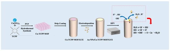
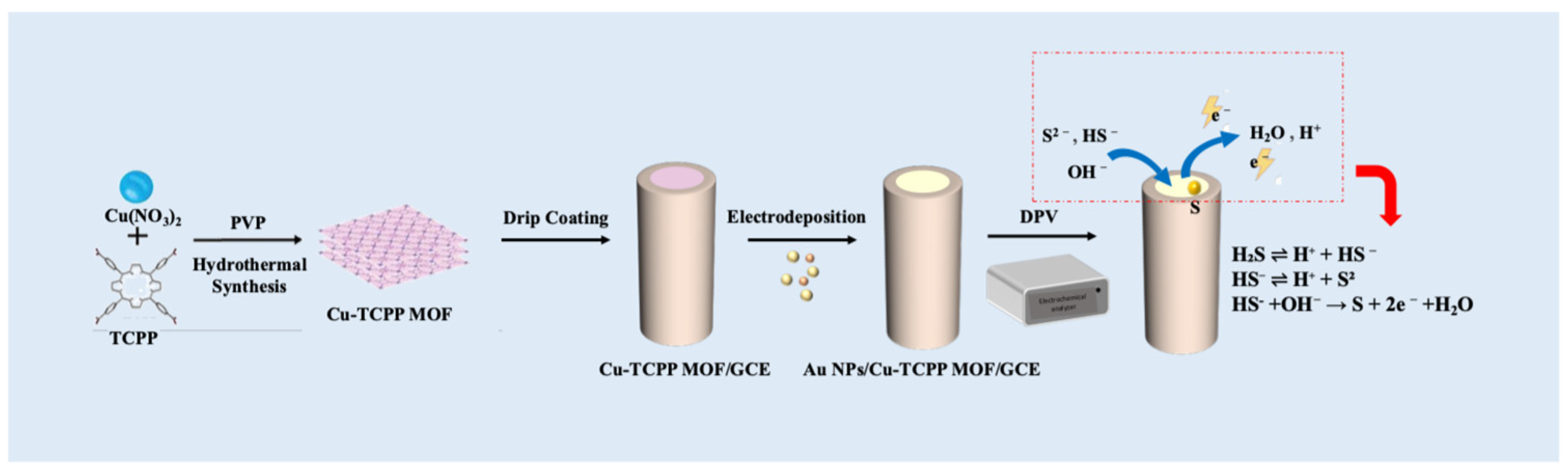
Based on these, in this work, the ultrathin 2D Cu-TCPP MOF was used as a precursor, and the gold nanoparticles (Au NPs)/Cu-TCPP MOF nanocomposites were prepared through simple electrodeposition. Then, the obtained Au NP/Cu-TCPP MOF nanocomposites were used for the fabrication of an H2S electrochemical sensor, which was successfully applied for H2S detection in mine water (Scheme 1). On the one hand, the sensor exhibits lower LOD and wider linear range; on the other hand, the modified electrode material features relatively simple components, straightforward preparation steps, and better controllability. This research provides a simple electrochemical sensing strategy, offering a promising and reliable approach for the detection of trace H2S in complex samples.
Scheme 1.
Schematic illustration of sensor preparation and detection of H2S.
2. Materials and Methods
2.1. Materials
Copper nitrate trihydrate (Cu(NO3)2·3H2O), 4, 4′, 4″, 4‴-(porphine-5,10,15,20-tetrayl) tetrakis (benzoic acid) (TCPP), N, N-dimethylformamide (DMF, 99.5%), hydrogen sulfide standard solution (from Na2S; Na2S is commonly used as a source of H2S [24]), chloroauric acid from Aladdin Biochemical Co., Ltd. (Shanghai, China). Polyvinylpyrrolidone (PVP, MW = 40,000 g/mol) from Yuanye Biological Technology Co., Ltd. (Shanghai, China), and ethanol absolute (C2H5OH, 99.7%) from Shanghai Titan Scientific Co., Ltd. (Shanghai, China). Other reagents were of analytical grade.
2.2. Synthesis of Cu-TCPP MOF Nanofilm
Cu-TCPP MOF nanofilm was prepared according to our previous work using a surfactant-assisted method [25]. Specifically, 2.9 mg of Cu (NO3)2·3H2O and 20.0 mg of PVP were dispersed in a mixture of 12 mL DMF and ethanol (V:V = 3:1) in a 25 mL beaker. Subsequently, 12 mg of TCPP was added into the beaker and further ultrasonicated for 10 min. The mixture was transferred into a 20 mL Teflon-lined autoclave and maintained at 80 °C for 12 h, followed by cooling to room temperature. The precipitates were centrifuged and washed by ethanol three times. In this step, PVP is a surface active agent; it can be selectively attached on the surface of MOFs, which plays a key role in the anisotropic growth of Cu-TCPP, resulting in the formation of ultrathin 2D Cu-TCPP nanofilms [25].
2.3. Preparation of Au NP/Cu-TCPP MOF Sensors
The surface of the glassy carbon electrode (GCE) was sequentially polished with 1.0 μm and 0.3 μm aluminum oxide (Al2O3) powders until a mirror-like finish was achieved. The electrode was sonicated in ethanol and ultrapure water for 30 s each, and then dried at room temperature. Then, 5.0 mg of Cu-TCPP was dispersed in 10.0 mL of ultrapure water to obtain 0.5 mg/mL suspensions, respectively. A 7.5 μL aliquot of the prepared suspension was drop-cast onto the pretreated GCE surface. The modified electrode was then immersed in 10 mL 0.1 wt% HAuCl4 solution containing polyvinylpyrrolidone (PVP), followed by electrochemical deposition at a constant potential of −0.1 V for 150 s to form the AuNPs/Cu-TCPP/GCE modified electrode (Scheme 1). Finally, the fabricated sensor was air-dried at room temperature for further use.
2.4. Electrochemical Measurement
All electrochemical tests were performed on a CHI 660 electrochemical workstation (Shanghai CH Instruments, Shanghai, China) using a standard three-electrode system. The working electrode, reference electrode, and counter electrode were the modified electrode, an Ag/AgCl electrode, and a platinum electrode, respectively. The electrochemical characterization of the bare and modified GCE was performed by cyclic voltammetry (CV) and electrochemical impedance spectroscopy (EIS) in 5.0 mM K3[Fe(CN)6]/K4[Fe(CN)6] containing 0.1 M KCl solution (the parameters were set as follows: an applied potential of 0.225 V, a frequency range from 0.01 Hz to 100 kHz, and an alternating current amplitude of 5 mV). For EIS measurements, the parameters were set as follows: an applied potential of 0.225 V, a frequency range from 0.01 Hz to 100 kHz, and an alternating current amplitude of 5 mV. Na2S is commonly used as a source of H2S; therefore, different concentrations of Na2S were used to represent dissolved H2S [24]. The electrochemical behaviors of dissolved H2S on the modified electrodes were investigated by CV in 0.1 M PBS (pH 7.0) at a scan rate of 100 mV/s [25,26]. Furthermore, the reaction kinetics were studied by varying the scan rate from 20 to 200 mV/s. Differential pulse voltammetry (DPV) in the same buffer system was employed to systematically evaluate the sensor’s performance and its capability for dissolved H2S detection in real samples. The mine water samples were diluted with 10× PBS (pH 7.0) at a volume ratio of 1: 9 (v/v), and directly used for electrochemical analysis. All the electrochemical experiments were performed under air conditions.
2.5. Characterization Techniques
The morphology and elemental composition of the synthesized materials were characterized by scanning electron microscopy (SEM, JSM-6700F, JEOL, Tokyo, Japan) and transmission electron microscopy (TEM, Talos F200X, FEI, Washington, DC, USA); X-ray diffraction (XRD) patterns were recorded with a D8 ADVANCE diffractometer (Bruker, Berlin, Germany) and CHI 660 electrochemical workstation (Shanghai CH Instruments, Shanghai, China).
3. Results and Discussion
3.1. Characterization of Cu-TCPP MOF Nanofilms and Au NP/Cu-TCPP Nanocomposites
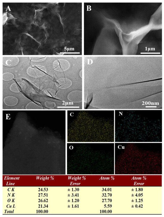
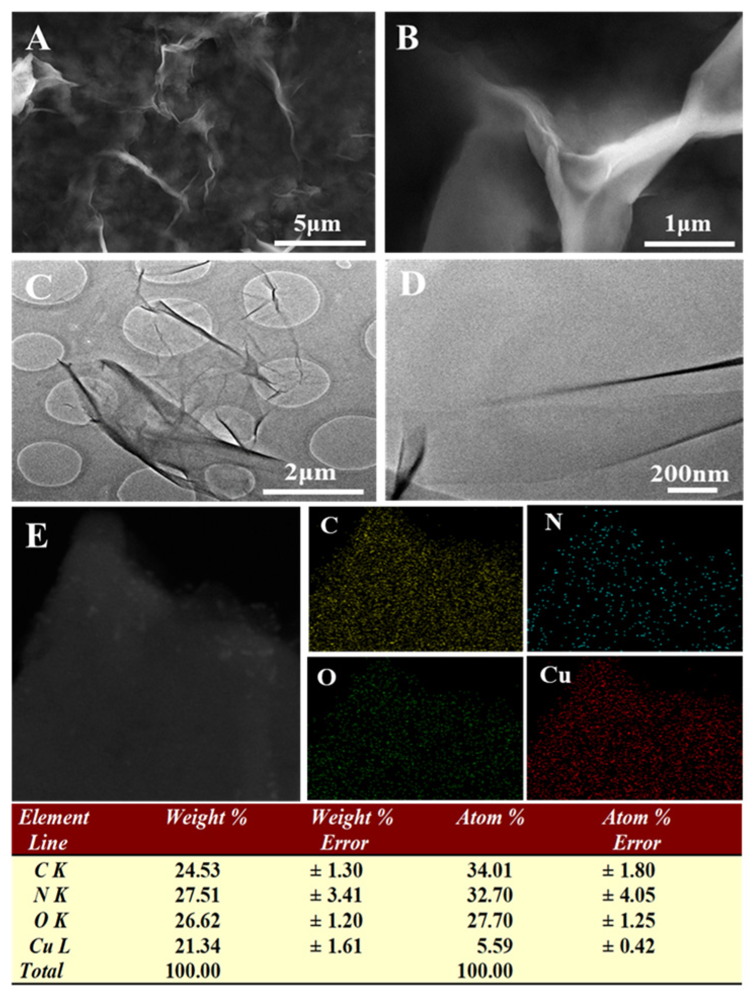
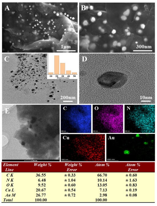
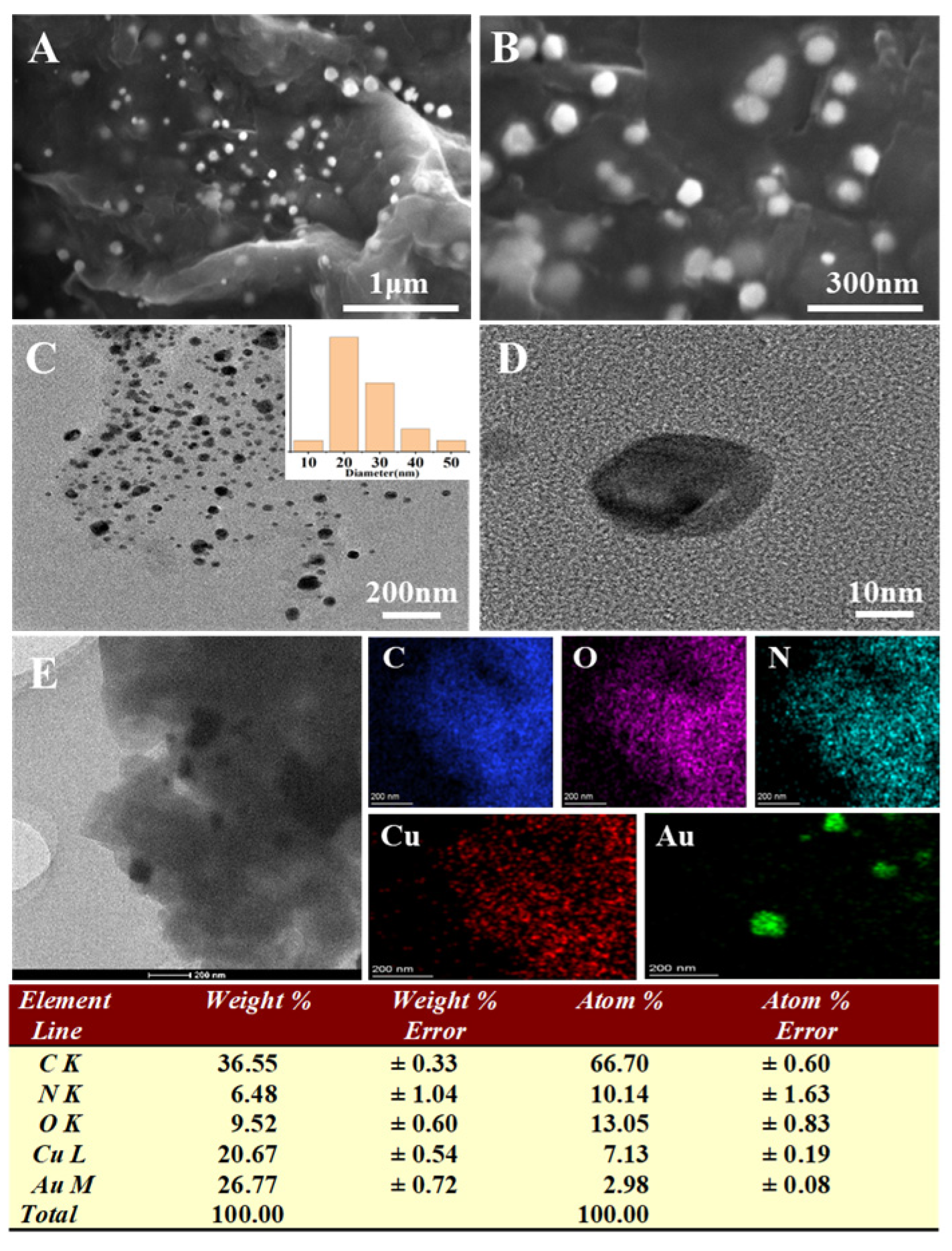
Figure 1 shows the morphology, structure, and composition of Cu-TCPP MOF nanofilms. The SEM (A, B) and TEM (C, D) results reveal that the Cu-TCPP MOF exhibits a wrinkled, film-like morphology. The element mapping images (E) confirm the homogeneous distribution of C, N, O, and Cu elements within the material. After Au deposition (Figure 2), Au nanoparticles are uniformly dispersed on the surface of the Cu-TCPP MOF, and the diameter of the Au nanoparticles is about 20 nm (Figure 2A–D). The element mapping images confirm the homogeneous distribution of C, N, O, Cu and Au elements within the material (Figure 2E), signifying the successful preparation of the Au NP/Cu-TCPP MOF nanocomposites.
Figure 1.
SEM (A,B), TEM (C,D) and corresponding element mapping images (E) of Cu-TCPP MOF nanomaterials.
Figure 2.
SEM (A,B), TEM (C,D) and corresponding element mapping images (E) of AuNPs/Cu-TCPP MOF nanocomposites.
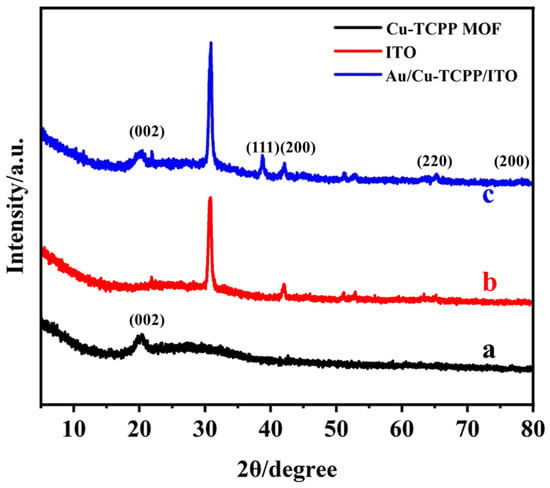
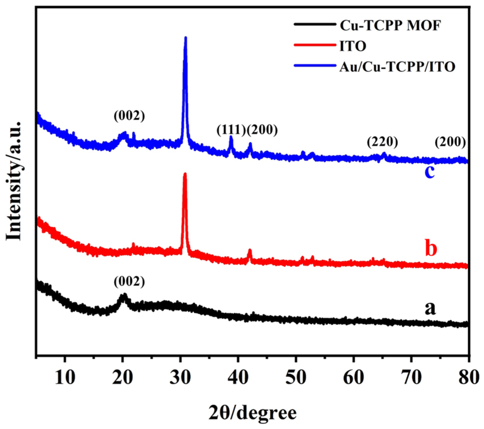
Figure 3 presents the XRD characterization results for various materials (a: Cu-TCPP MOF, b: ITO, c: Au NPs/Cu-TCPP MOF). As shown, the Cu-TCPP MOF exhibits a distinct characteristic diffraction peak at 20° (Figure 3a), corresponding to the (002) crystal plane, which demonstrated that prepared 2D Cu-TCPP MOF was dominantly c-oriented. This aligns with the references and our previous research [27,28], confirming that the synthesized MOF material successfully retains the layered structural characteristics of TCPP. For the Au NPs/Cu-TCPP MOF, please ignore the inherent peaks from ITO itself, which is characterized by peaks at 30.8°, 42.1°, 51.1°, 52.8°, and 65° (Figure 3b); the new diffraction peaks appear at 38.5°, 45.0°, 65.3°, and 77.9° (Figure 3c). The results are compared to the standard diffraction data for Au [29,30] (PDF#04-0784, standard peak positions: 38.18°, 44.39°, 64.58°, 77.55°, corresponding to crystal planes (111), (200), (220), (311)).
Figure 3.
XRD spectra of Cu-TCPP MOF (a), ITO (b), Au NPs/Cu-TCPP MOF (c).
In summary, this study successfully prepared both Cu-TCPP MOF and Au NP/Cu-TCPP MOF nanocomposites.
3.2. Electrochemical Characterization of the Modified Electrode
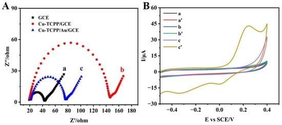
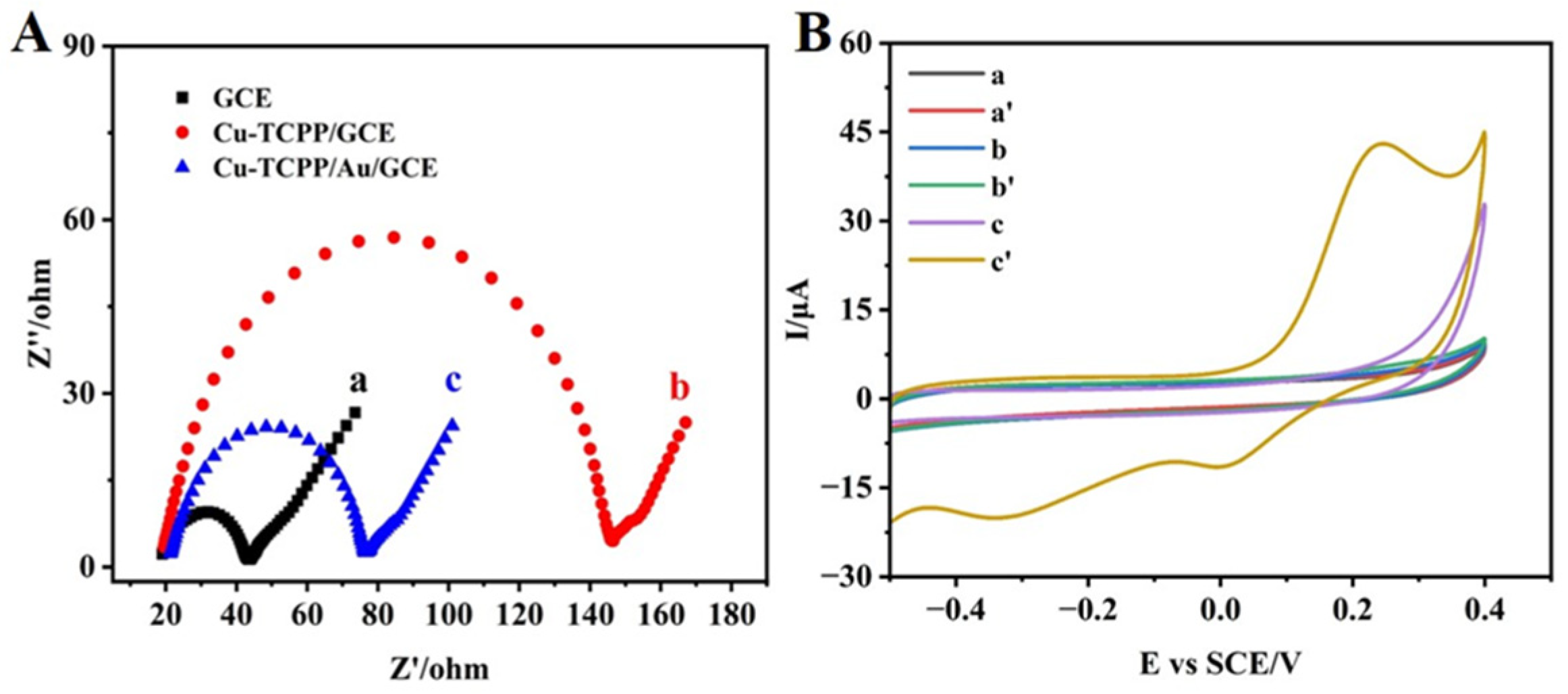
EIS was employed to probe the surface properties and charge-transfer behavior of the modified electrodes [30]. In Nyquist plots, the semicircle diameter corresponds to the charge-transfer resistance (Rct). As shown in Figure 4A, the bare GCE exhibits the smallest Rct, indicative of rapid electron transfer, whereas Cu-TCPP MOF/GCE shows a larger Rct due to its low conductivity. Following Au NP electrodeposition, the Rct values of Au NPs/Cu-TCPP MOF/GCE decrease markedly, as the Au NPs increase the effective electroactive surface area and facilitate electron transfer, thereby enhancing overall electrochemical performance. The electrochemical responses of various modified electrodes toward 10 μM H2S were comparatively investigated using CV at a scan rate of 100 mV s−1. As shown in Figure 4B, in the absence of H2S, the bare GCE and electrodes modified with Cu-TCPP MOF and Au NPs/Cu-TCPP exhibited negligible electrochemical responses. However, upon the addition of 10 μM H2S, all electrodes displayed different electrochemical responses. GCE and Cu-TCPP MOF/GCE showed only a weak response, whereas the Au NP/Cu-TCPP MOF-based modified electrodes exhibited a pronounced electrochemical response; a prominent oxidation current peak appeared near 0.25 V, which was due to the good electro-catalytic activity of the Au NP/Cu-TCPP MOF nanocomposite. It should be noted that Na2S is a strong electrolyte, which will completely dissociate into Na+ and S2−. However, most S2− will be hydrolyzed to HS−. Therefore, there is little S2− and more HS− in the solution [31,32]. The sensing mechanism on the electrode surface is shown in Scheme 1.
Figure 4.
(A) EIS diagram of (a) GCE, (b) Cu-TCPP MOF/GCE, (c) Au NPs/Cu-TCPP MOF/GCE in 0.1M K3[Fe(CN)6] containing 0.1 M KCl. CVs (B) of bare GCE (a, a’), Cu-TCPP MOF/GCE (b, b’) and Au NPs/Cu-TCPP MOF/GCE (c, c’) in the absence (a, b, c) and presence (a’, b’, c’) of 10 μM H2S in 0.1 M PBS (pH 7.0) solution.
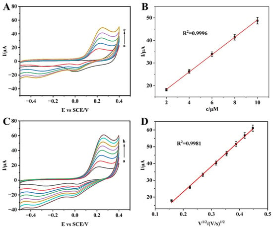
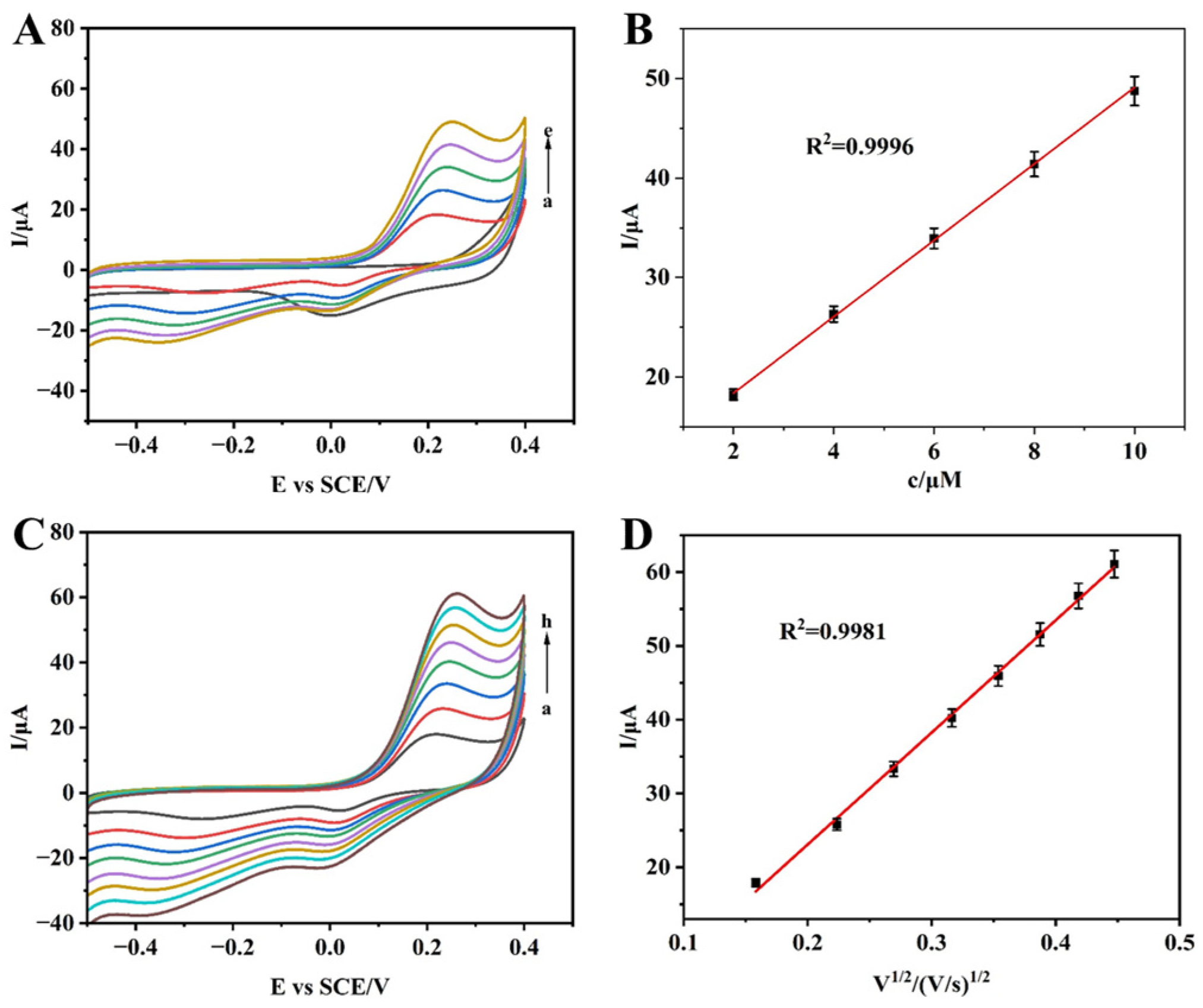
Then, CV was employed to investigate the effects of analyte concentration and scan rate on the electrochemical behavior of three sensors. As shown in Figure 5A,B, within the H2S concentration range of 2–10 μM, the oxidation peak currents recorded on Au NPs/Cu-TCPP MOF/GCE exhibited strong linear relationships with the concentrations of H2S, with excellent correlation coefficients (R2). In order to investigate the kinetics of H2S on the surface of the modified electrode, the CV responses of Au NPs/Cu-TCPP MOF/GCE towards H2S were further evaluated at scan rates ranging from 20 to 200 mV s−1. As shown in Figure 5C,D, in the presence of 10 μM H2S, the oxidation peak current exhibited a linear relationship with the square root of the scan rate (v1/2), with a correlation coefficient of R2 = 0.9981, indicating that the oxidation process of H2S is diffusion-controlled.
Figure 5.
(A) CVs of Au NPs/Cu-TCPP MOF/GCE in 0.1M PBS (pH 7.0) in the presence of H2S with different concentrations (from a to e: 0, 2, 4, 6, 8 and 10 μM) at a scan rate of 50 mV/s. (B) Linear fitting program of the oxidation peak currents with the H2S concentrations. (C) CVs of Au NPs/Cu-TCPP MOF/GCE in 0.1M PBS (pH 7.0) containing 10 μM H2S at different scan rates (from a to h: 20, 50, 100, 120, 140, 160, 180, and 200 mV/s). (D) Linear fitting program of the oxidation peak currents with the square root of scan rate in 0.1 M PBS (pH 7.0) solution.
3.3. Electrochemical Study of the Constructed Sensors
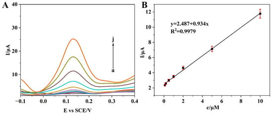
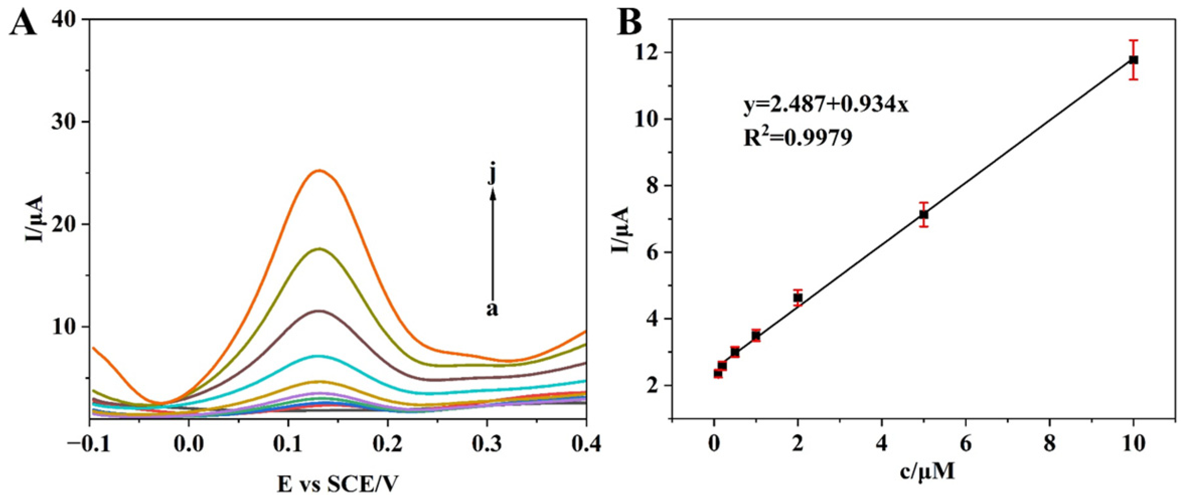
The Au NP/Cu-TCPP MOF/GCE was further utilized as an electrochemical sensing platform for the quantitative detection of H2S. As illustrated in Figure 6A, H2S at a trace concentration of 0.1 μM exhibited a distinct reduction peak at 0.15 V (vs. SCE) with a response time of about 7 s. With increasing H2S concentration, the peak intensity progressively enhanced. As shown in Figure 6B, the sensor displayed a linear response range of 0.1–10 μM, with a limit of detection (LOD) as low as 0.03 μM (S/N = 3), which was derived from the equation described below:
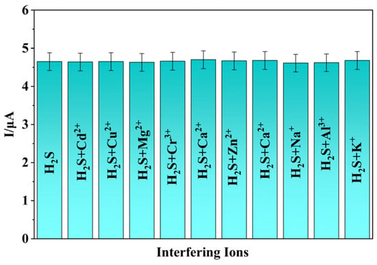
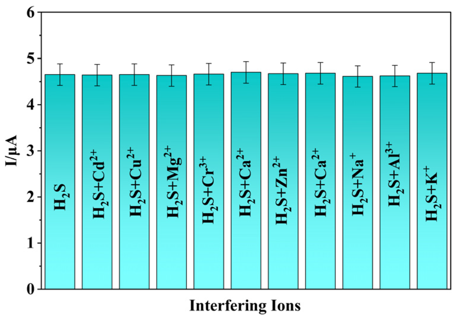
where SDB is standard deviation of blank. Compared with previously reported sensors, the sensor exhibits lower LOD, which is due to the good electro-catalytic property of Au NP/Cu-TCPP MOF nanocomposites (Table 1). In addition, as shown in Figure 7, it can been seen that, in the absence and presence of various interfering substances, the electrochemical signals did not show any significant changes, which indicates that the sensor exhibits good anti-interference.
Figure 6.
(A) DPV curves of the electrochemical sensing method in the presence of H2S at different concentrations (from a to j: 0, 0.1, 0.2, 0.5, 1, 2, 5, 10, 20, 50 μM). (B) Calibration curve of the DPV peak current intensity against the concentration of H2S.
Table 1.
Comparison of the electrochemical sensors described in this work with other H2S sensors.
Figure 7.
The DPV peak current in the presence of 2 μM H2S and different interfering substances (all the interfering substances are 20 μM).
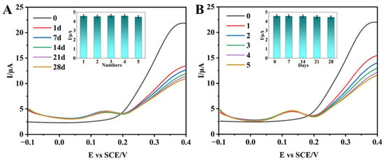
For stability (Figure 8A), one identical Au NP/Cu-TCPP MOF/GCE was used for H2S detection over a period of 28 days; according to the experiment results, it can be seen that the modified electrode exhibits good stability. In addition, five Au NPs/Cu-TCPP MOF/GCEs were measured for H2S (Figure 8B). In reproducibility research, it can be seen that the five modified electrodes exhibit similar electrochemical responses and peak currents, which indicates good reproducibility. These may be due to the good catalytic stability of Au NP/Cu-TCPP MOF nanocomposites.
Figure 8.
(A) Stability investigation. DPV responses of the same Au NPs/Cu-TCPP MOF/GCE over a period of 28 days in the presence of 2 μM H2S. (B) Reproducibility investigation. DPV responses of five Au NPs/Cu-TCPP MOF/GCEs in the presence of 2 μM H2S.
Finally, the Au NP/Cu-TCPP MOF/GCE sensor was used for real-sample analysis, and mine water was selected as representative sample matrices. The mine water was directly used after simple filtration to remove solid impurities. Recovery tests, as presented in Table 2, were conducted using the standard addition method with concentrations of 1 μM, 5 μM, and 10 μM. The results revealed recovery rates of 105–120% for H2S, with RSD values below 3.6%. These results fully demonstrate the high accuracy and stability of the sensor system in real-sample analysis.
Table 2.
Determination of H2S in mine water (n = 5).
4. Conclusions
In conclusion, novel Au NP/Cu-TCPP MOF nanocomposites were prepared based on 2D TCPP MOF nanofilms, which were used to fabricate electrochemical sensors for determination of H2S. The sensor exhibited an LOD of 0.03 μM and a linear range of 0.1–10 μM. Additionally, the fabricated sensor exhibited good stability, reproducibility and anti-interference (Figure 7 and Figure 8), which were used for the determination of H2S in real mine water samples. Compared with reported sensors, the sensor exhibits the following two advantages: First, lower LOD, which may be due to the good electro-catalytic property of Au NP/Cu-TCPP MOF nanocomposites. Second, in order to enhance the performance of sensors, complex nanocomposite materials with multiple components were often used to modify electrodes in previous research, which involves tedious preparation steps and poor controllability. In contrast, due to the good stability of electrodeposition, the materials used in this research were facile to prepare and exhibit good controllability. However, there are still some limitations in this research, such as the feasibility of on-site measurement of samples and the miniaturization of sensors. Certainly, these will also be the focus of our future research.
Author Contributions
Methodology, software, validation, writing—original draft preparation, writing—review and editing, B.P. and Y.L. (Yang Liu); formal analysis, investigation, resources, F.C. and L.L.; data curation, visualization, supervision, H.L., Y.L. (Yongyuan Li) and H.Z.; project administration, funding acquisition, Y.C. and W.B. All authors have read and agreed to the published version of the manuscript.
Funding
We gratefully acknowledge the financial support from the Huaning Group Headquarters Science and Technology Project: Research and Application of H2S Toxic Gas Migration Law and Prevention and Control Technology in Typical Coal–Oil–Gas Coexisting Mines of the Ningzheng Mining Area (HNKJ23-H9). We also thank the Natural Science Foundation of Shaanxi Province (2024JC-YBQN-0156) and the Science and Technology Program Project of Yulin City (2024-CXY-174).
Data Availability Statement
The data that support the findings of this study are available from the corresponding author upon reasonable request.
Conflicts of Interest
Authors Bo Pan, Lei Li and Yongyuan Li are employed by the company Huaneng Coal Technology Research Co., Ltd. Author Furong Cao is employed by the company Huaneng Coal Industry Co., Ltd. The remaining authors declare that the research was conducted in the absence of any commercial or financial relationships that could be construed as a potential conflict of interest.
References
- Ahmed, I.; Iqbal, S.; Khan, A.; Fatima, F.; Mahar, H.; Bhutto, R.A. Advances in Fluorescent Probes for Hydrogen Sulfide Detection: Applications in Food Safety, Environmental Monitoring, and Biological Systems. Microchem. J. 2025, 215, 114188. [Google Scholar] [CrossRef] [Scilit]
- Qin, G.; Wu, X.; Wei, C.; Zhao, P.; Li, J.; Chang, J.; Jia, Z.; Yu, Z.; Zhang, Y. Forensic Analysis of Hydrogen Sulfide Poisoning Cases: Innovative LC-MS/MS Methodology for Accurate Detection in Human Blood. Microchem. J. 2024, 204, 111140. [Google Scholar] [CrossRef] [Scilit]
- Abdellaoui, B.; Benali, A.; Bejar, M.; Dhahri, E.; Wu, J. Effect of the Barium Substitution by Strontium on the H2S Detection Properties of Spinel Nanoferrites. Sens. Actuators B Chem. 2026, 446, 138614. [Google Scholar] [CrossRef] [Scilit]
- Iddrisu, M.; Drmosh, Q.A.; Onaizi, S.A. A Novel Approach for the Enhancement of H2S Removal and Rheological Properties of Water-Based Drilling Fluids through the Inclusion of Layered Triple Hydroxides. Geoenergy Sci. Eng. 2026, 256, 214150. [Google Scholar] [CrossRef] [Scilit]
- Cai, S.; Xiao, Q.; Yang, X. Geochemical Constraints on the Genetic Pathways of Hydrogen Sulfide in the Bamianhe Oilfield of Bohai Bay Basin, China. Mar. Pet. Geol. 2025, 178, 107422. [Google Scholar] [CrossRef] [Scilit]
- Baek, S.-H.; Lee, S.Y.; Seo, S.W.; Yun, H.-Y.; Kim, S.Y.; Lee, G.H.; Jeong, H.G. Effects of G Protein-Coupled Estrogen Receptor on Hydrogen Sulfide Production and Cystathionine γ-Lyase Expression in Human Endothelial Cells. Biochem. Pharmacol. 2025, 242, 117354. [Google Scholar] [CrossRef] [Scilit]
- Lin, J.; Ding, L.; Wu, Y.; Li, H.; Wu, Y.; Li, C.; Zhu, Q.; Chen, Z.; Cui, X.; Wang, T. Photoactivatable Hydrogen Sulfide Donors for Visualizing H2S Controlled Release in Accelerated Chronic Wound Healing. Chin. Chem. Lett. 2025, 112100. [Google Scholar] [CrossRef] [Scilit]
- Fang, F.; Guan, Y.-N.; Zhong, M.-J.; Wen, J.-Y.; Chen, Z.-W. H2S Protects Rat Cerebral Ischemia-Reperfusion Injury by Inhibiting Expression and Activation of Hippocampal ROCK2 at the Thr436 and Ser575 Sites. Eur. J. Pharmacol. 2024, 985, 177079. [Google Scholar] [CrossRef] [Scilit]
- Feng, J.; Zhuo, S.; Liu, D.; Peng, H.; Guo, D.; Li, N.; Sun, H.; Zhang, C.; Zhao, J. H2S Inhibits LiCl/Pilocarpine-Induced Seizures and Promotes Neuroprotection by Regulating TRPV2 Expression via the AC3/CAMP/PKA Pathway. Neurochem. Int. 2024, 174, 105677. [Google Scholar] [CrossRef] [Scilit] [PubMed]
- Yang, J.; Ge, S.; Tan, S.; Liu, H.; Yang, M.; Liu, W.; Zhang, K.; Zhang, Z.; Liu, J.; Shi, J.; et al. Modified Montmorillonite Armed Probiotics with Enhanced On-Site Mucus-Depleted Intestinal Colonization and H2S Scavenging for Colitis Treatment. J. Control. Release 2024, 374, 140–153. [Google Scholar] [CrossRef] [Scilit]
- Godwin, M.; Hine, C. Targeting Macro- and Micro-Nutrient Regulation of H2S Signaling for the Aging Brain. Neurotherapeutics 2025, 22, e00638. [Google Scholar] [CrossRef] [Scilit] [PubMed]
- Resiere, D.; Florentin, J.; Névière, R. Evaluating the Ten-Year Health Impact of Hydrogen Sulfide (H2S) and Ammonia (NH3) Exposure from Sargassum Seaweed Invasions in the Caribbean: Public Health Implications. Harmful Algae 2025, 152, 103027. [Google Scholar] [CrossRef] [Scilit] [PubMed]
- Yu, H.; Hu, J.; Jiang, X.; Hou, X.; Tian, Y. Point Discharge Microplasma Reactor for High Efficiency Conversion of H2S to SO2 for Speciation Analysis of Sulfide and Sulfite Using Molecular Fluorescence Spectrometry. Anal. Chim. Acta. 2018, 1042, 79–85. [Google Scholar] [CrossRef] [Scilit]
- Kumar, N.; Kumar Pal, A.; Kshirsagar, R.J. Near-Infrared Detection and Line Intensity Measurements of H2S with ECDL Source Based off-Axis Integrated Cavity Output Spectroscopy. Chem. Phys. 2023, 567, 111804. [Google Scholar] [CrossRef] [Scilit]
- Arif, H.M.; Fu, M.; Amanpour, T.; Wang, R. A HPLC-Based Methylene Blue/Methylene Green Method for Accurate Measurement of Hydrogen Sulfide in Plasma. Biochem. Biophys. Res. Commun. 2025, 782, 152557. [Google Scholar] [CrossRef] [Scilit]
- Li, S.; Zhu, R.; Li, Y.; Shi, S.; Li, J.; Zhang, K. Lanthanide Schiff-Base Paper-Based Composites as Fluorescence-Based Gas Sensors for Selective, Quantitative and Visual Detection of H2S Gas. Sens. Actuators B Chem. 2026, 448, 139039. [Google Scholar] [CrossRef] [Scilit]
- Jeong, S.; Song, C.; Kim, J.; Lee, Y.; Kim, M.H. Rational Synthesis of Phosphorous Rich Iridium Diphosphide (IrP2) Nanostructures for Electrochemical H2S Detection. J. Alloys Compd. 2023, 947, 169649. [Google Scholar] [CrossRef] [Scilit]
- Chen, Z.; Chen, S.; Wang, W.; Liu, W.; Gu, H.; Zhang, P.; Zeng, R.; Hao, Y. Electrochemical in Situ Transformation of Bi-Based Photoactive Materials for Photoelectrochemical Detection of H2S in the Living Brain. Microchem. J. 2025, 211, 113164. [Google Scholar] [CrossRef] [Scilit]
- Shang, H.; Li, J.; Zhang, X.; Zhang, B.; Li, Y.; Zhang, W.; Zhang, R. Target-Triggered Interaction Reaction Adjusted Nanozyme-Based Electrochemical Sensing for Fast Detection of H2S. Chem. Eng. J. 2025, 524, 169486. [Google Scholar] [CrossRef] [Scilit]
- Zuo, Y.; Wang, S.; Lin, R.; Xiao, G.; Chen, S.; Zeng, R.; Gu, H. A Portable Electrochemical Microsensor for In-Site Measurement of Dissolved Oxygen and Hydrogen Sulfide in Natural Water. Talanta 2023, 256, 124269. [Google Scholar] [CrossRef] [Scilit]
- Qu, L.; Xu, Y.; Cui, W.; Wu, L.; Feng, Y.; Gu, Y.; Pan, H. Trends in Conductive MOFs for Sensing: A Review. Anal. Chim. Acta. 2025, 1336, 343307. [Google Scholar] [CrossRef] [Scilit] [PubMed]
- Chaudhuri, H.; Cho, C.-W.; Yun, Y.-S. Advantages of MOFs, COFs, and POPs over Other 2D/3D Materials and Hybrids as Heterogeneous Catalysts for C H Bond Activation and Functionalization. Coord. Chem. Rev. 2026, 549, 217275. [Google Scholar] [CrossRef] [Scilit]
- Zhu, Y.; Tang, Z. Conductive 2D MOF Coupled with Superprotonic Conduction and Interfacial Pseudo-Capacitance. Matter 2020, 2, 798–800. [Google Scholar] [CrossRef] [Scilit]
- Zhao, H.; Li, X.; Yu, K.; Xu, H.; Zhang, Z.; Chai, H.; Tian, M.; Qu, L.; Zhang, X.; Zhang, G. A MOF-on-MOF based photoelectrochemical sensor for hydrogen sulfide detection in food quality assessment. Sens. Actuators B Chem. 2025, 433, 137525. [Google Scholar] [CrossRef] [Scilit]
- Liu, Q.; Chen, Y.; Zhang, C.; Zhang, J.; Yu, L.; Wang, Y. Fe-N5 single-atom sites on C3N4 enable ultrasensitive electrochemical sensing of hydrogen sulfide in bone metastatic microenvironments. Biosens. Bioelectron. 2026, 305, 118688. [Google Scholar] [CrossRef] [Scilit]
- Brown, M.; Hall, G.; Schoenfisch, M. A direct and selective electrochemical hydrogen sulfide sensor. Anal. Chim. Acta. 2019, 1045, 67. [Google Scholar] [CrossRef] [Scilit]
- Ma, J.; Bai, W.; Liu, X.; Zheng, J. Electrochemical Dopamine Sensor Based on Bi-Metallic Co/Zn Porphyrin Metal–Organic Framework. Microchim. Acta 2022, 189, 20. [Google Scholar] [CrossRef] [Scilit]
- Bai, W.; Li, S.; Ma, J.; Cao, W.; Zheng, J. Ultrathin 2D metal–organic framework (nanosheets and nanofilms)-based xD–2D hybrid nanostructures as biomimetic enzymes and supercapacitors. J. Mater. Chem. A 2019, 7, 9086. [Google Scholar] [CrossRef] [Scilit]
- Chen, X.; Li, X.; Peng, Y.; Yang, H.; Tong, Y.; Balogun, M.-S.; Huang, Y. Tailoring Carrier Dynamics of BiVO4 Photoanode via Dual Incorporation of Au and Co(OH) x Cooperative Modification for Photoelectrochemical Water Splitting. Adv. Funct. Mater. 2025, 35, 2416091. [Google Scholar] [CrossRef] [Scilit]
- Bai, W.; Li, Y.; Liang, P.; Zhang, Z.; Xu, Y.; Yang, Z.; Yang, K.; Lv, J.; Xie, H.; Cao, W.; et al. Density Functional Theory–Guided Electrochemical Determination of Nitrite in Foods Based on 2D Metal-Organic Frameworks. J. Food Compos. Anal. 2023, 121, 105385. [Google Scholar] [CrossRef] [Scilit]
- Wang, S.; Liu, X.; Zhang, M. Reduction of Ammineruthenium (III) by Sulfide Enables In Vivo Electrochemical Monitoring of Free Endogenous Hydrogen Sulfide. Anal. Chem. 2017, 89, 5382. [Google Scholar] [CrossRef] [Scilit] [PubMed]
- Kim, Y.; Choi, S.; Lee, J.; Lee, Y.; Kim, M. Facile formation of highly single crystalline CuV2O6 nanorods: Utilization for electrochemical H2S sensing. J. Alloys Compd. 2025, 1037, 182204. [Google Scholar] [CrossRef] [Scilit]
- Cao, X.; Xu, H.; Ding, S.; Ye, Y.; Ge, X.; Yu, L. Electrochemical Determination of Sulfide in Fruits Using Alizarin–Reduced Graphene Oxide Nanosheets Modified Electrode. Food Chem. 2016, 194, 1224–1229. [Google Scholar] [CrossRef] [Scilit]
- Liu, Z.; Ma, H.; Sun, H.; Gao, R.; Liu, H.; Wang, X.; Xu, P.; Xun, L. Nanoporous Gold-Based Microbial Biosensor for Direct Determination of Sulfide. Biosens. Bioelectron. 2017, 98, 29–35. [Google Scholar] [CrossRef] [Scilit] [PubMed]
- Giovanelli, D.; Lawrence, N.S.; Jiang, L.; Jones, T.G.J.; Compton, R.G. Electrochemical Determination of Sulphide at Nickel Electrodes in Alkaline Media: A New Electrochemical Sensor. Sens. Actuators B Chem. 2003, 88, 320–328. [Google Scholar] [CrossRef] [Scilit]
- Paim, L.L.; Hammer, P.; Stradiotto, N.R. Electrochemical Behavior of a Glassy Carbon Electrode Chemically Modified with Nickel Pentacyanonitrosylferrate in Presence of Sulfur Compounds. Electroanalysis 2011, 23, 1488–1496. [Google Scholar] [CrossRef] [Scilit]

Disclaimer/Publisher’s Note: The statements, opinions and data contained in all publications are solely those of the individual author(s) and contributor(s) and not of MDPI and/or the editor(s). MDPI and/or the editor(s) disclaim responsibility for any injury to people or property resulting from any ideas, methods, instructions or products referred to in the content. |
© 2026 by the authors. Licensee MDPI, Basel, Switzerland. This article is an open access article distributed under the terms and conditions of the Creative Commons Attribution (CC BY) license.