


Abstract
Celiac disease (CD) is an autoimmune disorder triggered by immunogenic gluten peptides that resist gastrointestinal digestion. The only current treatment is a strict gluten-free diet, which is challenging to maintain. Lactic acid bacteria (LAB) with specific proteolytic systems offer a promising strategy for gluten hydrolysis and potential reduction of immunogenicity. This study aims to isolate and characterize gluten-degrading LAB from traditional Spanish and Algerian dairy products. A total of 27 artisanal dairy samples were collected. LAB were isolated on MRS and Elliker agar. Gluten-degrading activity was screened using a well diffusion assay with cell-free supernatants and a spot assay with live cultures. Active isolates were identified by 16S rRNA gene sequencing. Out of 123 isolates, 40 (32.5%) were positive in the well assay, while 67 (54.5%) were positive in the spot assay, indicating the latter’s higher sensitivity for detecting cell-associated proteases. Halo diameters ranged from 6 to 16 mm. Algerian isolates exhibited significantly stronger activity (mean halo: 12.6 ± 2.1 mm) compared to Spanish isolates (10.2 ± 2.0 mm; p < 0.001). Molecular identification of the 32 most active isolates revealed the following dominant species: Lactiplantibacillus plantarum, L. pentosus, Levilactobacillus brevis, and Enterococcus faecium. This study confirms that artisanal dairy fermentations are rich sources of LAB with robust gluten-degrading potential. The superior activity of Lactiplantibacillus spp. aligns with their complex peptidase systems. The geographical variation highlights the influence of local fermentation practices. Selected strains represent excellent candidates for developing adjunct cultures to produce gluten-reduced foods and warrant further investigation as potential probiotics, pending safety and efficacy validation in vivo and in clinical studies.
1. Introduction
Celiac disease (CD) is a chronic immune-mediated enteropathy affecting approximately 0.7–1% of the global population [1]. It is triggered in genetically susceptible individuals by the ingestion of gluten proteins from wheat, barley, and rye [2]. Gluten’s high proline and glutamine contents render it resistant to complete proteolysis by human digestive enzymes, leading to the persistence of immunogenic peptides in the gastrointestinal tract [3]. Key peptides like gliadin’s 33-mer are deamidated by tissue transglutaminase, increasing their binding to human leukocyte antigen (HLA) molecules. This triggers a T-helper 1 inflammatory response, leading to villous atrophy, malabsorption, and systemic symptoms [4,5]. The cornerstone of CD management remains a strict, lifelong gluten-free diet (GFD). However, the GFD presents significant challenges, including limited food choices, higher costs, potential nutritional deficiencies, and the constant risk of cross-contamination [6,7]. Consequently, there is a pressing need for complementary or alternative therapeutic strategies [8]. Among these, the enzymatic degradation of gluten peptides before they reach the small intestine has emerged as a highly promising approach [9,10].
Lactic acid bacteria (LAB), widely used in food fermentations and recognized for their probiotic properties, possess sophisticated proteolytic systems [11]. These systems include cell-envelope proteinases that initiate the breakdown of proteins into oligopeptides, which are subsequently internalized and hydrolyzed by a broad array of intracellular peptidases, including proline-specific peptidases essential for cleaving gluten [12]. Several LAB strains, particularly from the genera Lactobacillus (recently reclassified into Lactiplantibacillus and Levilactobacillus), have demonstrated the ability to hydrolyze immunogenic gluten epitopes [13,14,15].
Sourdough ecosystems have been extensively explored in search of gluten-degrading LAB [16]. Several studies have isolated LAB with proteolytic activity from cheeses and fermented milks [11,17]; however, few studies have directly compared the gluten-degrading potential of LAB from traditional fermented dairy products from different Mediterranean regions, specifically Spain and Algeria, representing a significant gap in knowledge.
Artisanal dairy products, produced through spontaneous fermentation, typically host a more diverse and complex microbiota compared to products made with defined starter cultures. This diversity arises from variations in raw milk microbiota, animal diet, local environmental conditions, and traditional manufacturing practices [18,19]. This makes the traditional fermented milk products particularly interesting as sources of novel LAB with functional properties, including enhanced proteolytic activity. The protein-rich environment of dairy matrices may specifically select for LAB with robust proteolytic systems adapted to degrade complex proteins like casein—systems that may also be effective against gluten proteins [20]. In the Mediterranean region, Spain and Algeria have rich traditions of artisanal dairy production but differ in climate, animal husbandry practices, and fermentation techniques. These differences likely shape distinct LAB communities with potentially varied proteolytic capabilities. Understanding these geographical variations could reveal novel strains with superior gluten-degrading activity and provide insights into how fermentation ecosystems select for specific enzymatic functions [21].
This study aimed to: (i) isolate LAB from a diverse collection of traditional Spanish and Algerian dairy products; (ii) screen their gluten-degrading potential using complementary agar-based assays; (iii) molecularly identify the most active isolates; and (iv) compare the functional and taxonomic profiles of isolates from the two geographical origins.
2. Materials and Methods
2.1. Sample Collection
A total of 27 artisanal dairy products were aseptically collected between May and August 2023 from small-scale producers in Spain (n = 14) and Algeria (n = 13). The Spanish samples included various cheeses from cow, goat, and sheep milk (Table 1). The Algerian samples encompassed traditional products such as Jben, Bouhaza cheese, Kefir, Klila, and Rayeb. Samples were transported to the laboratory under refrigerated conditions (4 °C) and processed within 24 h.
Table 1.
Nomenclature of collected samples.
2.2. Isolation and Preservation of Lactic Acid Bacteria
From each sample, 5 g were homogenized with 45 mL of sterile saline solution (0.90% NaCl). Serial decimal dilutions were prepared and spread-plated on de Man, Rogosa, and Sharpe (MRS) agar and Elliker agar. For each sample, isolations were performed in triplicate. Plates were incubated at 30 °C for 24–48 h. Colonies with distinct morphologies were selected, purified and stored at −20 °C in MRS broth with 20% (v/v) glycerol.
2.3. Screening for Gluten-Degrading Activity
Gluten degradation activity was analyzed using two complementary methods: a diffusion assay in wells using cell-free supernatants (CFS) to detect extracellular proteolytic enzymes, and a spot assay using live bacterial cultures to detect both extracellular and cell surface-associated proteolytic activities. This dual approach allows differentiation between enzymes secreted into the medium and those bound to the bacterial cell envelope, which are characteristic of the proteolytic system of LAB [11].
2.3.1. Preparation of Gluten Agar
The basal medium for gluten degradation assays contained (per liter): 10 g peptone, 5 g NaCl, and 10 g bacteriological agar. After autoclaving, the medium was cooled to 50 °C and supplemented with 8 g/L (0.8% w/v) of vital wheat gluten (Sigma-Aldrich, St. Louis, MO, USA; G5004) [22]. This concentration is standard for agar-based protease screening assays, providing an opaque matrix suitable for the clear visualization of proteolytic halos. The pH of the medium was adjusted to 6.5 ± 0.1 using 1 M NaOH prior to autoclaving.
2.3.2. Well Diffusion Assay
LAB isolates were cultured in MRS broth at 30 °C for 24 h without agitation. Culture age was standardized to ensure consistent protease production across all isolates. LAB cultures were centrifuged (10,000× g, 10 min, 4 °C) to obtain cell-free supernatants (CFS). The (CFS) was carefully collected, and its pH was measured. To exclude the effect of organic acids on protease activity, supernatants were neutralized to pH 6.8 using sterile 1 M NaOH before testing. Sterile stainless-steel cylinders were placed on gluten agar, covered with a tempered gluten agar overlay, and removed after solidification. Wells were filled with 75 µL of CFS, and plates were incubated at 30 °C for 24 h. For each assay plate, a negative control (sterile MRS broth, adjusted to pH 6.8) was included. Each isolate was tested in triplicate on separate plates to ensure reproducibility.
2.3.3. Spot Assay
Five microliters of active overnight MRS cultures were spotted onto gluten agar plates, air-dried, and incubated at 30 °C for 24 h. A negative control was included on each plate. Each isolate was tested in triplicate on separate plates.
2.3.4. Visualization and Measurement
After incubation, the plates were flooded with Coomassie Brilliant Blue G-250 staining solution (1 g/L in a mixture of methanol: acetic acid: water, 4:1:5) for 30 min. The plates were then destained overnight with a solution of methanol: acetic acid: water (5:1:4). Clear, transparent zones (halos) around the wells or spots indicated proteolysis of gluten. The diameters of the halos were measured using a caliper. Measurements from technical replicates were averaged, and the mean ± standard deviation (SD) was calculated for each isolate. Only halos distinct from the negative control were considered positive.
2.4. Molecular Identification of Active Isolates
The thirty-two most promising isolates, selected based on halo diameter of ≥10 mm in a well-diffusion assay, were subjected to molecular identification. Bacterial genomic DNA was extracted using the GenElute Bacterial Genomic DNA Kit (Sigma-Aldrich, St. Louis, MO, USA) according to the manufacturer’s instructions. The 16S rRNA gene was amplified via PCR with the universal bacterial primers 27F (5′-AGAGTTTGATCCTGGCTCAG-3′) and 1492R (5′-GGTTACCTTGTTACGACTT-3′). Successful amplification was verified by electrophoresis on a 1% agarose gel. Amplicons were purified and sent for Sanger sequencing (Stab Vida, Caparica, Portugal) using the 27F primer. The resulting sequences, covering the hypervariable domains V1-V4, were queried against the NCBI GenBank database using the BLASTN algorithm, and species-level identification was assigned to isolates with ≥98.5% sequence similarity to a reference strain. While this methodology provided reliable taxonomic assignment for the scope of this functional screening, these identifications were considered as best-fit, recognizing that more detailed genomic characterization would be needed for definitive species-level differentiation within closely related groups.
2.5. Statistical Analysis
Data are presented as mean ± standard deviation (SD). Differences in halo diameters between geographical origins were analyzed using an unpaired two-sample t-test. Differences across bacterial species were analyzed using a one-way ANOVA followed by Tukey’s post hoc test. A p-value of < 0.05 was considered significant. Analyses were performed using R software (v4.3.0).
3. Results
3.1. Isolation and Screening for Gluten-Degrading Activity
A total of 123 presumptive LAB isolates were obtained, 84 from Spain and 39 from Algeria (Table 2 and Table 3, respectively). Screening revealed that 40 isolates (32.5%) were positive in the well diffusion assay, which detects extracellular proteases. In contrast, the spot assay, which detects both extracellular and cell-associated activity, identified a higher number of positive isolates; 67 isolates (54.5%) were positive in the spot assay (Table 4), indicating a significant contribution of cell-bound enzymes.
Table 2.
Results of Spanish Samples (ES).
Table 3.
Results of Algerian Samples (DZ).
Table 4.
Summary of gluten-degrading activity among lactic acid bacteria (LAB) isolates.
This comprehensive analysis of 123 LAB isolates from traditional Spanish (n = 84) and Algerian (n = 39) dairy products revealed significant gluten-degrading capabilities, with distinct geographical patterns emerging. The spot assay proved more sensitive than the well diffusion assay, detecting 54.5% positive isolates overall compared to 32.5% in the well assay, indicating a substantial contribution of cell-associated proteases alongside extracellular enzymes. As shown in Table 4, Algerian samples demonstrated superior proteolytic activity, particularly in extracellular enzyme production (38.5% positivity in well assays versus 29.8% for Spanish samples), while both regions showed comparable effectiveness in spot assays (56.4% Algeria vs. 53.6% Spain). Notably, several Algerian products including Kefir (KIF), spicy Bouhaza (BP), and Klila (KL) showed exceptional performance with 100% positivity in well diffusion assays, while strong activity (≥13 mm halo diameter) was observed in isolates from both Spanish (QF2, OBII series, EM3) and Algerian (KIF5, OB1, BP2) samples. These findings highlight traditional dairy fermentations, particularly Algerian products, as valuable reservoirs of LAB with robust gluten-degrading potential, offering promising candidates for developing adjunct cultures and novel probiotic therapeutics for celiac disease management.
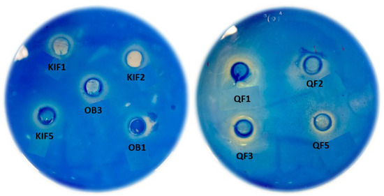
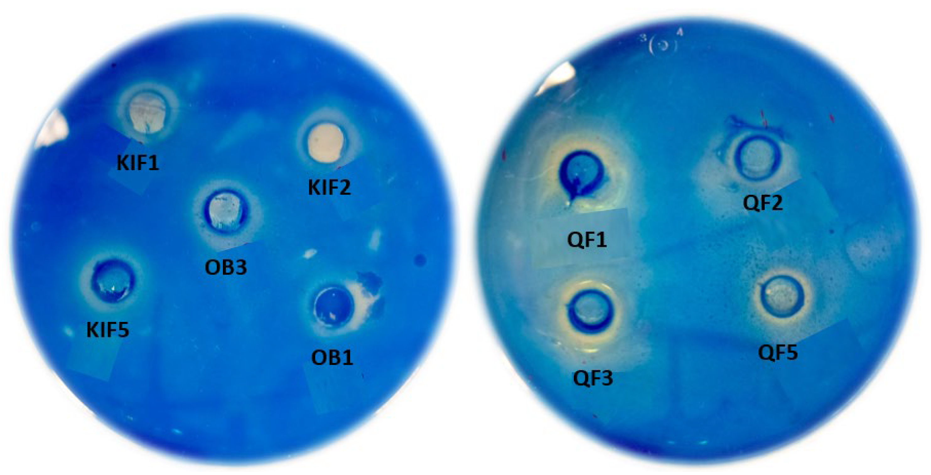
Halo diameters ranged from 6 to 16 mm. Isolates from Algerian products produced significantly larger halos than those from Spanish products (p < 0.001). The most potent activity was associated with isolates from Algerian Kefir (KIF1, KIF2, KIF5) and Klila (KL3, KL4).
3.2. Molecular Identification of Gluten-Degrading Isolates
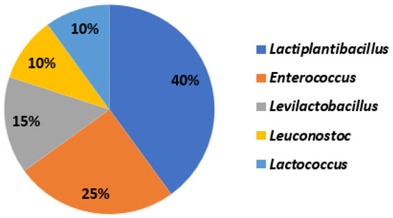
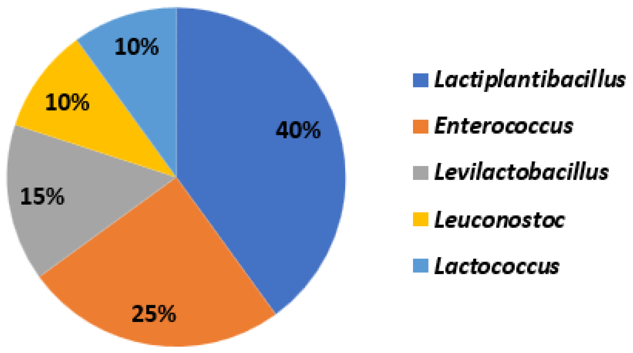
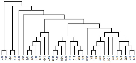
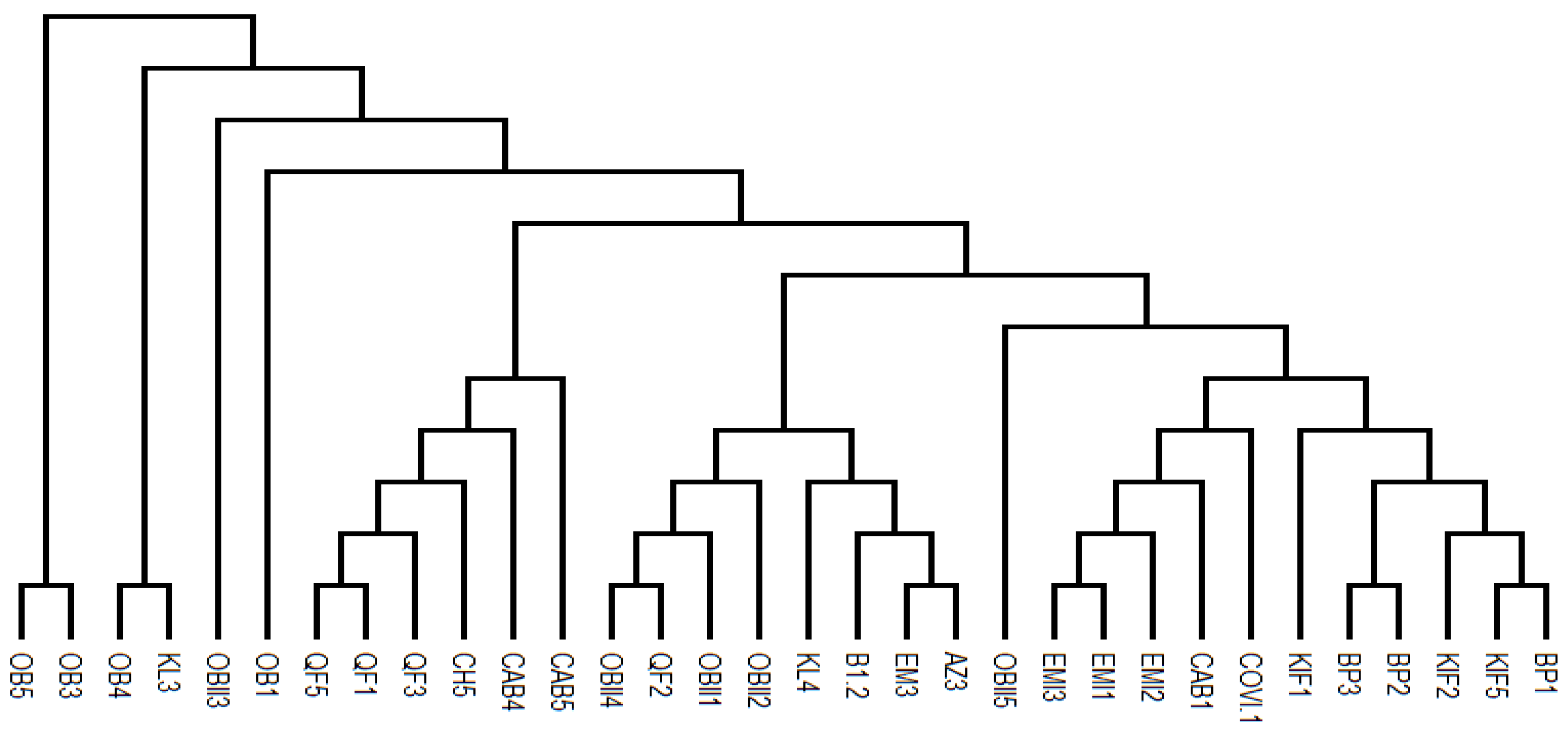
Molecular identification of the 32 most active isolates revealed a diverse taxonomy, with Lactiplantibacillus being the most prominent among highly active strains (Table 5). 40% were identified as Lactiplantibacillus, 25% as Enterococcus, 15% as Levilactobacillus, 10% as Leuconostoc, and the remaining 10% as Lactococcus (Figure 1). The phylogenetic relationships of these isolates are depicted in Figure 2, highlighting their clustering with closely related species.
Table 5.
Taxonomic identification and activity of selected gluten-degrading LAB isolates.
Figure 1.
Taxonomic Distribution of the Most Active Isolates.
Figure 2.
Phylogenetic Tree of Selected Strains.
The analysis of the proteolytic strains reveals distinct microbial profiles between the two regions. The Spanish samples exhibit exceptional biodiversity with remarkable performances: strains of Lactococcus lactis (QF2, OBII1, OBII2, OBII4) from fresh and sheep’s milk cheeses show the highest proteolytic activity (14 mm), while Leuconostoc mesenteroides (EM3, AZ3) from blue cheese and Emmental, and Levilactobacillus brevis (EMI1, EMI2) from mixed goat-sheep milk cheeses, demonstrate activities between 12 and 13 mm, complemented by various Enterococcus strains (OBII3, CH5, QF3) with halo diameters of 12–13 mm. The Algerian samples show marked specialization around the Lactiplantibacillus genus, with strains KIF5 (13 mm), BP2 (12 mm), BP3 (11 mm) and KIF1-KIF2 (12 mm) demonstrating consistent activity, particularly in traditional fermented products like Kefir and spicy Bouhaza, accompanied by Enterococcus faecalis (OB1, OB4, OB5) and Leuconostoc mesenteroides (KL4) mainly isolated from sheep’s milk cheeses and Klila, with halo diameters ranging between 11 and 13 mm. This distribution reflects both the influence of terroirs and traditional manufacturing methods on the selection of microbial communities with differentiated proteolytic capabilities. Figure 3 shows some of the inhibition halos resulting from the proteolytic activity of some of the selected strains.
Figure 3.
Proteolytic Activity of Selected Strains.
4. Discussion
This study provides evidence that traditional dairy fermentations, particularly from Algeria, are a rich source of LAB with significant gluten-degrading potential. The higher sensitivity of the spot assay (56.4% positive) compared to the well diffusion assay (38.5%) strongly suggests a significant contribution from the proteolytic system of these isolates. This difference could be attributed to the activity of cell-wall-associated proteinases, which are in direct contact with the substrate in the spot assay, consistent with the known proteolytic system of LAB [11,23]. However, since live cultures were used, it is also possible that the higher detection rate in the spot assay is due to the continuous production and accumulation of extracellular enzymes during active bacterial growth and incubation. Further studies with isolated cell wall fractions would be needed to accurately compartmentalize this activity.
The most potent activity was predominantly associated with Lactiplantibacillus plantarum and L. pentosus. This agrees with previous studies highlighting their robust proteolytic systems, including proline-specific peptidases essential for hydrolyzing proline-rich gluten peptides [13,15,24]. Our results corroborate recent studies demonstrating the efficacy of L. plantarum in degrading gliadin and reducing immunogenicity [19,25].
The significantly stronger activity in Algerian isolates may be attributed to several factors. Traditional Algerian products like Kefir and Klila are complex ecosystems involving symbiotic communities of LAB and yeasts, which may drive evolution towards enhanced proteolytic capabilities [26]. Differences in raw milk microbiota, animal diet, and spontaneous fermentation processes unique to Algerian artisanship likely contribute to selecting a more proteolytically active microbiome [17,27].
Beyond Lactiplantibacillus, we identified moderate activity in Levilactobacillus brevis and Leuconostoc mesenteroides, which have been reported to contribute to gluten hydrolysis [28]. The activity of some Enterococcus strains, while notable, necessitates careful safety evaluation due to potential virulence factors in this genus [29,30]. The variable activity observed among Lactococcus lactis strains suggests strain-specific proteolytic capabilities, though this species is generally recognized for its primary focus on lactose fermentation [11].
From an application perspective, the selected Lactiplantibacillus strains are prime candidates for development as adjunct cultures for “gluten-reduced” fermented foods [31]. Furthermore, their probiotic potential could offer dual benefits by detoxifying gluten and modulating the gut immune response [32,33]. Future work must validate efficacy under physiologically relevant conditions, characterizing hydrolysis products and assessing residual immunogenicity [10,14]. However, such probiotic applications require rigorous in vivo safety assessment and clinical validation, which are beyond the scope of the present screening study.
Limitations and Future Perspectives
It is important to recognize the limitations of the methodological approach used in this study. Agar diffusion assays served as an effective initial test for detecting general proteolytic activity against gluten; however, the hydrolysis zones indicate broad protease action but do not confirm the degradation of specific proline- and glutamine-rich immunogenic epitopes (e.g., the 33-mer peptide) responsible for coeliac disease.
Consequently, promising strains identified, particularly those of Lactiplantibacillus spp., require rigorous quantitative verification. Future work should include: (i) quantification of proteolytic activity in enzyme units (U/mg protein) using standardized substrates; (ii) incubation with pure immunogenic peptides (e.g., 33-mer) followed by HPLC/MS analysis to determine degradation percentages and cleavage sites; (iii) evaluation of residual immunogenicity by ELISA or T-cell assays; (iv) comparison with reference strains or enzymes to compare activity levels with published studies [14]; and (v) the assessment of strain viability and the stability of their gluten-degrading enzymes under simulated gastrointestinal conditions (digestive enzymes, low pH and bile salts). These steps are essential to determine the suitability of the selected isolates for development into functional probiotic products or adjunct cultures in the food industry.
5. Conclusions
This study successfully identified a diverse collection of LAB from Spanish and Algerian artisanal dairy products with the ability to degrade gluten. The spot assay proved more sensitive for detecting this activity, highlighting the importance of cell-associated enzymes. Strains of Lactiplantibacillus plantarum and L. pentosus, particularly from Algerian Kefir and Klila, exhibited the highest degradation potential. These findings highlight the value of traditional food ecosystems as a source of microbes with unique functional properties. The selected strains represent promising biological tools for developing novel dietary strategies for individuals with celiac disease and other gluten-related disorders, although further studies are essential to validate their efficacy in the hydrolysis of immunogenic epitopes (e.g., 33-mer) and to evaluate their safety and activity under gastrointestinal conditions.
Author Contributions
Conceptualization, A.M., R.P.P. and M.J.G.; methodology, A.M. and R.P.P.; formal analysis, A.M., R.P.P. and M.J.G.; investigation, A.M.; resources, R.L. and A.G.; writing—original draft preparation, A.M., R.P.P. and M.J.G.; writing—review and editing, J.R.L. and A.G.; supervision, R.L.; funding acquisition, A.G. All authors have read and agreed to the published version of the manuscript.
Funding
This research was funded by the University of Jaén, grant number AGR230. The APC was funded by the University of Jaén.
Institutional Review Board Statement
Not applicable. This study did not involve humans or animals.
Informed Consent Statement
Not applicable.
Data Availability Statement
The data presented in this study are available on request from the corresponding author. The data are not publicly available due to their use as preliminary screening and orientation tools within the experimental design.
Acknowledgments
The authors acknowledge the University of Jaén (Jaén, Spain) for the support provided through the “Plan de apoyo a la investigación”.
Conflicts of Interest
The authors declare no conflicts of interest. The funders had no role in the design of the study; in the collection, analyses, or interpretation of data; in the writing of the manuscript, or in the decision to publish the results.
References
- Iversen, R.; Sollid, L.M. The immunobiology and pathogenesis of celiac disease. Annu. Rev. Pathol. Mech. Dis. 2023, 18, 47–70. [Google Scholar] [CrossRef] [Scilit]
- Balakireva, A.V.; Zamyatnin, A.A. Properties of gluten intolerance: Gluten structure, evolution, pathogenicity and detoxification capabilities. Nutrients 2016, 8, 644. [Google Scholar] [CrossRef] [Scilit]
- Shan, L.; Molberg, Ø.; Parrot, I.; Hausch, F.; Filiz, F.; Gray, G.M.; Khosla, C. Structural basis for gluten intolerance in celiac sprue. Science 2002, 297, 2275–2279. [Google Scholar] [CrossRef] [Scilit]
- Sollid, L.M.; Jabri, B. Triggers and drivers of autoimmunity: Lessons from coeliac disease. Nat. Rev. Immunol. 2013, 13, 294–302. [Google Scholar] [CrossRef] [Scilit] [PubMed]
- Lebwohl, B.; Sanders, D.S.; Green, P.H. Coeliac disease. Lancet 2018, 391, 70–81. [Google Scholar] [CrossRef] [Scilit]
- Lamacchia, C.; Camarca, A.; Picascia, S.; Di Luccia, A.; Gianfrani, C. Cereal-based gluten-free food: How to reconcile nutritional and technological properties of wheat proteins with safety for celiac disease patients. Nutrients 2014, 6, 575–590. [Google Scholar] [CrossRef] [Scilit]
- Vargas, F.M.; Cardoso, L.T.; Didoné, A.; Lima, J.P.M.; Venzke, J.G.; de Oliveira, V.R. Celiac disease: Risks of cross-contamination and strategies for gluten removal in food environments. Int. J. Environ. Res. Public Health 2024, 21, 124. [Google Scholar] [CrossRef] [Scilit]
- Yoosuf, S.; Makharia, G.K. Evolving therapy for celiac disease. Front. Pediatr. 2019, 7, 193. [Google Scholar] [CrossRef] [Scilit]
- Gänzle, M.G.; Loponen, J.; Gobbetti, M. Proteolysis in sourdough fermentations: Mechanisms and potential for improved bread quality. Trends Food Sci. Technol. 2008, 19, 513–521. [Google Scholar] [CrossRef] [Scilit]
- Kõiv, V.; Tenson, T. Gluten-degrading bacteria: Availability and applications. Appl. Microbiol. Biotechnol. 2021, 105, 3045–3059. [Google Scholar] [CrossRef] [Scilit] [PubMed]
- Savijoki, K.; Ingmer, H.; Varmanen, P. Proteolytic systems of lactic acid bacteria. Appl. Microbiol. Biotechnol. 2006, 71, 394–406. [Google Scholar] [CrossRef] [Scilit]
- Brzozowski, B.; Stasiewicz, K.; Ostolski, M.; Adamczak, M. Reducing Immunoreactivity of Gliadins and Coeliac-Toxic Peptides Using Peptidases from L. acidophilus 5e2 and A. niger. Catalysts 2020, 10, 923. [Google Scholar] [CrossRef] [Scilit]
- De Angelis, M.; Rizzello, C.G.; Fasano, A.; Clemente, M.G.; De Simone, C.; Silano, M.; Gobbetti, M. VSL#3 probiotic preparation has the capacity to hydrolyze gliadin polypeptides responsible for celiac disease. Appl. Environ. Microbiol. 2006, 72, 4490–4495. [Google Scholar] [CrossRef] [Scilit]
- Rizzello, C.G.; Curiel, J.A.; Nionelli, L.; Vincentini, O.; Di Cagno, R.; Silano, M.; Gobbetti, M.; Coda, R. Use of fungal proteases and selected sourdough lactic acid bacteria for making wheat bread with an intermediate content of gluten. Food Microbiol. 2014, 37, 59–68. [Google Scholar] [CrossRef] [Scilit]
- Álvarez-Sieiro, P.; Redruello, B.; Ladero, V.; Martín, M.C.; Fernández, M.; Alvarez, M.A. Screening sourdough samples for gliadin-degrading activity revealed Lactobacillus casei strains able to individually metabolize the coeliac-disease-related 33-mer peptide. Can. J. Microbiol. 2016, 62, 422–430. [Google Scholar] [CrossRef] [Scilit]
- Gänzle, M.G. Enzymatic and bacterial conversions during sourdough fermentation. Food Microbiol. 2014, 37, 2–10. [Google Scholar] [CrossRef] [Scilit] [PubMed]
- Yu, J.; Wang, W.H.; Menghe, B.L.; Jiri, M.T.; Wang, H.M.; Liu, W.J.; Bao, Q.H.; Lu, Q.; Zhang, J.C.; Wang, F.; et al. Diversity of lactic acid bacteria associated with traditional fermented dairy products in Mongolia. J. Dairy Sci. 2011, 94, 3229–3241. [Google Scholar] [CrossRef] [Scilit] [PubMed]
- Mefleh, M.; Darwish, A.M.G.; Mudgil, P.; Maqsood, S.; Boukid, F. Traditional Fermented Dairy Products in Southern Mediterranean Countries: From Tradition to Innovation. Fermentation 2022, 8, 743. [Google Scholar] [CrossRef] [Scilit]
- Leszczyńska, J.; Szczepankowska, A.K.; Majak, I.; Mańkowska, D.; Smolińska, B.; Ścieszka, S.; Diowksz, A.; Cukrowska, B.; Aleksandrzak-Piekarczyk, T. Reducing Immunoreactivity of Gluten Peptides by Probiotic Lactic Acid Bacteria for Dietary Management of Gluten-Related Diseases. Nutrients 2024, 16, 976. [Google Scholar] [CrossRef] [Scilit] [PubMed]
- Wouters, J.T.; Ayad, E.H.; Hugenholtz, J.; Smit, G. Microbes from raw milk to fermented dairy products. Int. Dairy. J. 2002, 12, 91–109. [Google Scholar] [CrossRef] [Scilit]
- Indio, V.; Gonzales-Barron, U.; Oliveri, C.; Lucchi, A.; Valero, A.; Achemchem, F.; Manfreda, G.; Savini, F.; Serraino, A.; De Cesare, A. Comparative analysis of the microbiome composition of artisanal cheeses produced in the Mediterranean area. Ital. J. Food Saf. 2024, 13, 12818. [Google Scholar] [CrossRef] [Scilit] [PubMed]
- Rashmi, B.S.; Gayathri, D.; Vasudha, M.; Prashantkumar, C.S.; Kumbhar, Y.B. Gluten hydrolyzing activity of Bacillus spp. isolated from sourdough. Microb. Cell Fact. 2020, 19, 130. [Google Scholar] [CrossRef] [Scilit] [PubMed]
- Liang, J.; Chai, K.; Wang, X.; Li, J.; Zhu, L.; Zhang, G. From gluten structure to immunogenicity: Investigating the effects of lactic acid bacteria and yeast co-fermentation on wheat allergenicity in steamed buns. Food Chem. 2025, 492, 145356. [Google Scholar] [CrossRef] [Scilit] [PubMed]
- Rizzello, C.G.; De Angelis, M.; Di Cagno, R.; Camarca, A.; Silano, M.; Losito, I.; Gobbetti, M. Highly efficient gluten degradation by lactobacilli and fungal proteases during food processing: New perspectives for celiac disease. Appl. Environ. Microbiol. 2007, 73, 4499–4507. [Google Scholar] [CrossRef] [Scilit]
- Fu, W.; Jia, X.; Liu, C.; Meng, X.; Zhang, K.; Tao, S.; Xue, W. Sourdough yeast-bacteria interactions results in reduced immunogenicity by increasing depolymerization and hydrolysis of gluten. Innov. Food Sci. Emerg. Technol. 2023, 84, 103281. [Google Scholar] [CrossRef] [Scilit]
- Usal, M.; Özgölet, M.; Arici, M.; Törnük, F. Enzymatic and antimicrobial activities of lactic acid bacteria and yeasts isolated from boza, a traditional fermented grain based beverage. Food Biosci. 2024, 61, 104681. [Google Scholar] [CrossRef] [Scilit]
- Zolnikova, O.; Dzhakhaya, N.; Bueverova, E.; Sedova, A.; Kurbatova, A.; Kryuchkova, K.; Butkova, T.; Izotov, A.; Kulikova, L.; Yurku, K.; et al. The Contribution of the Intestinal Microbiota to the Celiac Disease Pathogenesis along with the Effectiveness of Probiotic Therapy. Microorganisms 2023, 11, 2848. [Google Scholar] [CrossRef] [Scilit]
- Mickowska, B.; Romanova, K.; Urminská, D. Reduction of immunoreactivity of rye and wheat prolamins by lactobacilli and flavourzyme proteolysis during sourdough fermentation—A way to obtain low-gluten bread. J. Food Nutr. Res. 2019, 58, 153–166. Available online: https://www.vup.sk/en/index.php?mainID=2&navID=34&version=2&volume=58&article=2142 (accessed on 12 March 2026).
- Hanchi, H.; Mottawea, W.; Sebei, K.; Hammami, R. The genus Enterococcus: Between probiotic potential and safety concerns—An update. Front. Microbiol. 2018, 9, 1791. [Google Scholar] [CrossRef] [Scilit]
- Zheng, J.; Wittouck, S.; Salvetti, E.; Franz, C.M.; Harris, H.M.; Mattarelli, P.; Lebeer, S. A taxonomic note on the genus Lactobacillus: Description of 23 novel genera, emended description of the genus Lactobacillus Beijerinck 1901, and union of Lactobacillaceae and Leuconostocaceae. Int. J. Syst. Evol. Microbiol. 2020, 70, 2782–2858. [Google Scholar] [CrossRef] [Scilit]
- Francavilla, R.; Cristofori, F.; Vacca, M.; Barone, M.; De Angelis, M. Advances in understanding the potential therapeutic applications of gut microbiota and probiotic mediated therapies in celiac disease. Expert Rev. Gastroenterol. Hepatol. 2020, 14, 323–333. [Google Scholar] [CrossRef] [Scilit] [PubMed]
- Cristofori, F.; Dargenio, V.N.; Dargenio, C.; Miniello, V.L.; Barone, M.; Francavilla, R. Anti-inflammatory and immunomodulatory effects of probiotics in gut inflammation: A door to the body. Front. Immunol. 2021, 12, 578386. [Google Scholar] [CrossRef] [Scilit] [PubMed]
- Salvi, P.S.; Cowles, R.A. Butyrate and the Intestinal Epithelium: Modulation of Proliferation and Inflammation in Homeostasis and Disease. Cells 2021, 10, 1775. [Google Scholar] [CrossRef] [Scilit] [PubMed]

Disclaimer/Publisher’s Note: The statements, opinions and data contained in all publications are solely those of the individual author(s) and contributor(s) and not of MDPI and/or the editor(s). MDPI and/or the editor(s) disclaim responsibility for any injury to people or property resulting from any ideas, methods, instructions or products referred to in the content. |
© 2026 by the authors. Licensee MDPI, Basel, Switzerland. This article is an open access article distributed under the terms and conditions of the Creative Commons Attribution (CC BY) license.