


Simple Summary
Small cell carcinoma of the bladder is a rare and aggressive cancer with limited treatment options and generally poor prognosis once metastatic. We report an unusual case of a patient with metastatic disease who achieved long-term disease control following chemotherapy, radiotherapy, and immunotherapy. Upon disease progression, molecular testing of the tumour helped guide the decision to initiate immunotherapy, which resulted in sustained remission despite treatment discontinuation due to serious treatment-related side effects. Nearly seven years after initial diagnosis, a single brain metastasis was detected and successfully treated with surgery and focused radiotherapy. This case demonstrates that prolonged survival can occur in selected patients with metastatic small cell carcinoma of the bladder and highlights the potential value of individualized treatment.
Abstract
Background: Small cell carcinoma of the bladder (SCCB) is a rare, aggressive malignancy with no established standard of care for metastatic disease. The prognosis is dismal; median survival ranges from 12 to 20 months, and long-term survival is exceptional. Case Presentation: We present a 67-year-old man diagnosed in 2018 with metastatic SCCB involving the liver, bone, and ureter. He achieved a complete metabolic response following six cycles of cisplatin–etoposide, followed by consolidative pelvic radiotherapy with a bladder boost. Upon disease progression in 2019, molecular profiling identified a high tumour mutational burden (19.9 mut/Mb), PD-L1 CPS > 1%, and multiple genomic alterations (including MDM2, RAD51D, and TERT). Dual immune checkpoint inhibition (ICI) with ipilimumab and nivolumab was initiated. Although treatment was interrupted and subsequently discontinued due to grade 3 immune-related adverse events, including hepatitis and pneumonitis, the patient maintained a sustained remission off therapy. In January 2025, nearly seven years post-diagnosis, he developed a solitary frontal brain metastasis. He underwent resection and adjuvant stereotactic radiosurgery, achieving local control. He remains under surveillance with no evidence of extracranial recurrence. Conclusions: This case illustrates one of the longest reported survivals in metastatic SCCB. It highlights the utility of molecular profiling in guiding salvage therapy and the potential for durable ICI responses even after discontinuation due to toxicity. Furthermore, late recurrence in the brain emphasizes the central nervous system’s role as a sanctuary site, necessitating prolonged vigilance.
1. Introduction
Small cell carcinoma of the bladder (SCCB) is an exceptionally rare and aggressive neuroendocrine malignancy, accounting for less than 1% of all bladder tumours. Histologically, it presents a diagnostic challenge; approximately 50% of cases exhibit mixed features, co-existing with high-grade urothelial carcinoma [1]. Its clinical presentation is similar to conventional urothelial carcinoma, typically involving painless gross hematuria. However, SCCB is usually diagnosed at an advanced stage due to rapid progression. Its prognosis is poor; even with treatment, the five-year survival rate is approximately 10% [2]. Long-term survivors are rare. Only one prior case describes survival exceeding 120 months in metastatic disease, making reports of prolonged remission clinically valuable [3].
Given the rarity of SCCB, prospective randomized trials are absent. Treatment protocols are therefore adapted from small cell lung carcinoma (SCLC) guidelines [4]. For localized disease, the recommended treatment includes platinum-based chemotherapy combined with radical cystectomy or radiotherapy. In metastatic disease, systemic chemotherapy is the standard of care [5,6]. However, despite aggressive therapy, the prognosis remains poor. This emphasizes the need to define the molecular biology of SCCB and identify new therapeutic targets [7].
The IMpower133 and CASPIAN trials validated the use of atezolizumab and durvalumab with platinum chemotherapy for SCLC [8,9]. These results support the use of ICIs in extrapulmonary small cell carcinomas. However, no standard second-line therapy has been established for SCCB, and clinical management after progression on platinum-based chemotherapy remains highly challenging [10]. Treatment decisions in this setting are largely extrapolated from data in SCLC or from limited experiences in other genitourinary (GU) malignancies, with generally poor outcomes.
Evidence supporting the use of an ICI combination in the second-line setting for GU malignancies is emerging from phase II studies. In metastatic urothelial carcinoma, the TITAN-TCC trial evaluated a tailored approach in which patients who failed to respond to nivolumab induction after platinum-based chemotherapy were escalated to ipilimumab plus nivolumab. This strategy improved objective response rates compared with historical controls for nivolumab monotherapy, with toxicity considered manageable [11,12]. Similarly, the CheckMate 032 trial included a platinum-pretreated urothelial carcinoma cohort and demonstrated the highest response rates with the regimen of nivolumab 1 mg/kg plus ipilimumab 3 mg/kg [13].
We report a case of metastatic SCCB initially managed with cisplatin and etoposide chemotherapy, followed by consolidative pelvic radiotherapy. Upon disease progression, the patient switched to a combination of immunotherapy with ipilimumab and nivolumab. While the tumour showed a significant response, therapy was ultimately halted due to severe irAEs. Following a period of remission, the patient developed a solitary brain metastasis which was treated with surgical resection and adjuvant radiotherapy. This case demonstrates the potential efficacy of ICIs in refractory disease, weighed against the risks of adverse events.
2. Case Presentation
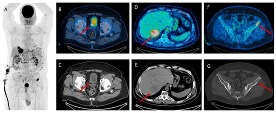
A 67-year-old male former smoker (5 pack-years, quit in 1998) had a past medical history significant for a sleeve gastrectomy performed in 2015. His ECOG performance status was one. His family history included a diagnosis of lymphoma in his father at age 67. The patient initially presented in 2017 with intermittent hematuria. A subsequent evaluation in January 2018 led to a cystoscopy and transurethral resection of bladder tumour (TURBT). Pathology revealed small cell carcinoma of the bladder (SCCB) with muscle invasion (T2) and necrosis. Immunohistochemical staining was positive for synaptophysin, chromogranin, CD56, and keratin, with a Ki-67 proliferation index of 85–90%. No coexisting urothelial carcinoma component was identified on histopathological review of the resected specimen. No baseline urine cytology, serum tumour markers, or endocrine evaluation were performed. A staging PET-CT performed in February 2018 revealed a hypermetabolic bladder mass with metastases to the liver, bone, and ureter (Figure 1). The primary bladder tumour and, later, the brain metastasis were histologically confirmed; the liver, bone, and ureteral lesions were identified and followed radiologically, without additional tissue sampling.
Figure 1.
(A) Whole-body PET-CT demonstrating multi-site hypermetabolic disease, including a dominant pelvic lesion adjacent to the prostate (B,C), a metabolically active hepatic metastasis (D,E), and an FDG-avid lytic lesion in the left iliac bone (F,G).
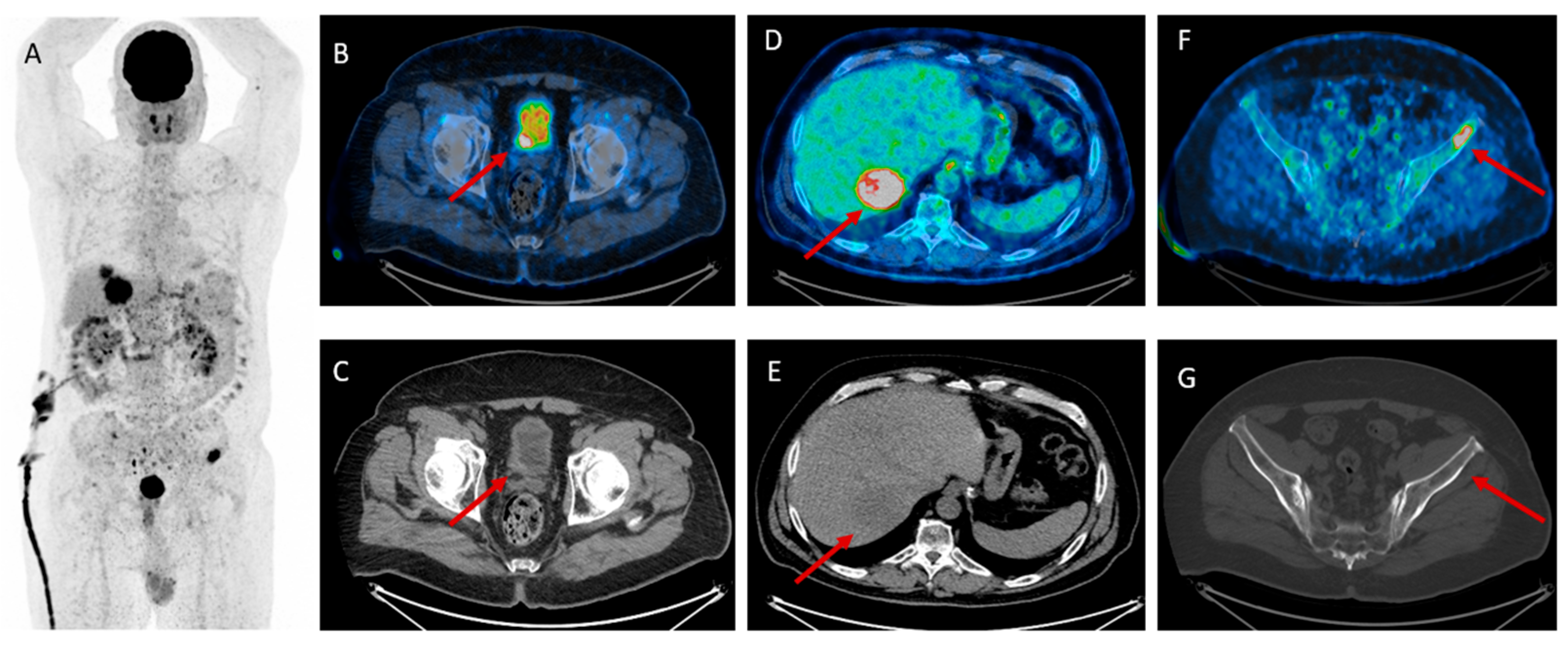
The patient completed six cycles of cisplatin and etoposide between February and July 2018. An interim PET-CT in May 2018 showed a complete metabolic response (Figure 2). By August 2018, however, imaging raised suspicion for bone and liver recurrence. Because these findings were equivocal, he underwent consolidative pelvic radiotherapy with a bladder boost (54 Gy) in October and November 2018.
Figure 2.
PET-CT demonstrating complete metabolic response. Whole-body PET (A) and axial PET-CT fusion (B) show complete resolution of FDG uptake in the pelvic bones. Corresponding CT (C) shows stable sclerotic bone changes without residual metabolic activity, consistent with treated osseous metastases.
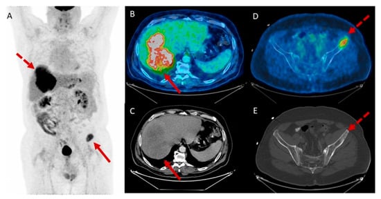
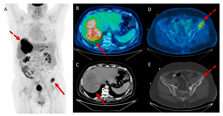
In January 2019, PET-CT revealed disease recurrence in the liver and bone (Figure 3).
Figure 3.
(A) Whole-body PET image demonstrating two prominent FDG-avid foci in the upper abdomen and left pelvis. (B,C) Axial PET/CT and corresponding CT confirming an intensely hypermetabolic lesion in the right hepatic lobe, consistent with hepatic metastasis. (D,E) Axial PET/CT and corresponding CT demonstrating an FDG-avid destructive lesion in the left iliac bone, compatible with osseous metastasis.
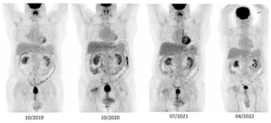
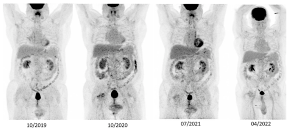
Genomic profiling (FoundationOne) identified multiple alterations, including MDM2 amplification, RAD51D, CDKN1C, KEL, MLL2, and TERT. The tumour was microsatellite stable (MSS) with a tumour mutational burden (TMB) of 19.9 mut/Mb and a PD-L1 Combined Positive Score (CPS) > 1%. Following multidisciplinary tumour board (MDT) review, the patient started second-line immunotherapy with ipilimumab (1 mg/kg) plus nivolumab (3 mg/kg) every three weeks on 30 January 2019. In May 2019, treatment was interrupted due to grade 3 immune-mediated hepatitis (graded per CTCAE v4.0). The patient discontinued ICI therapy and was managed with a prednisone taper starting at 1 mg/kg for a duration of four months with slow tapering, resulting in a full resolution of symptoms. During this treatment-free interval, restaging PET-CT scans in July and October 2019 showed no evidence of disease (NED) (Figure 4). He was re-challenged with nivolumab 3 mg/kg monotherapy in October 2019, which he tolerated well.
Figure 4.
Serial whole-body FDG PET scans demonstrating sustained absence of metabolically active disease over time.
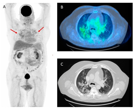
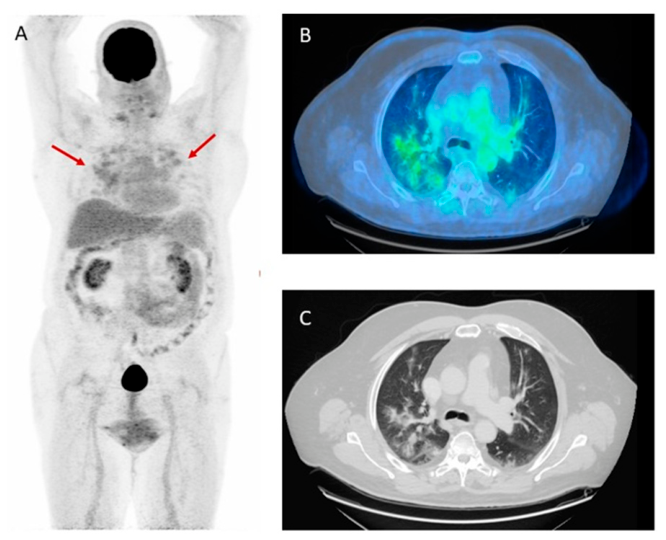
In September 2022, surveillance PET-CT identified inflammatory pulmonary changes consistent with toxicity (Figure 5). Consequently, nivolumab was permanently discontinued due to grade 3 immune-related pneumonitis. Since then, the patient has remained off systemic therapy, maintaining prolonged disease control.
Figure 5.
(A) Whole-body FDG PET demonstrating diffuse bilateral pulmonary hypermetabolism (arrows), suggestive of an acute inflammatory process. (B) Axial PET/CT revealing extensive FDG-avid patchy consolidations throughout both lungs. (C) Corresponding CT confirming widespread ground-glass opacities and consolidations consistent with pneumonitis.
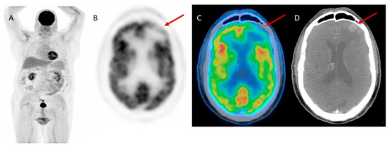
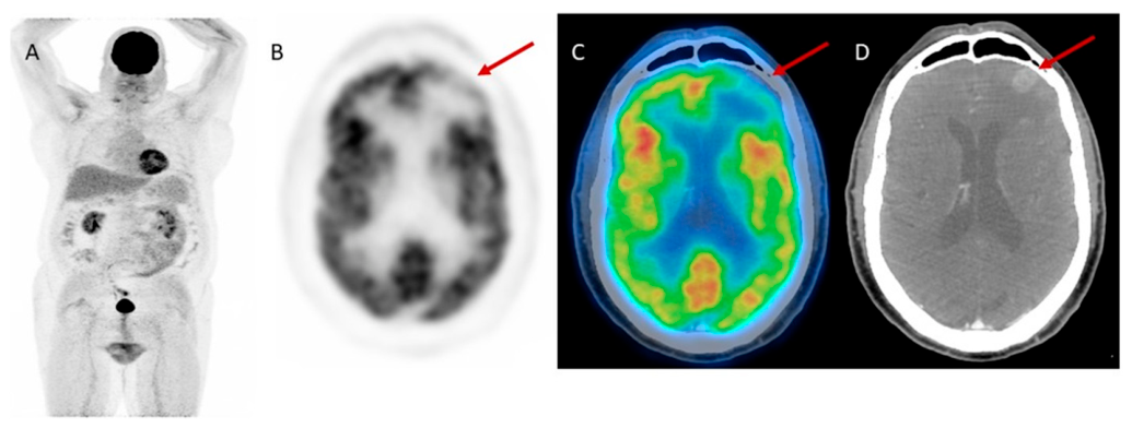
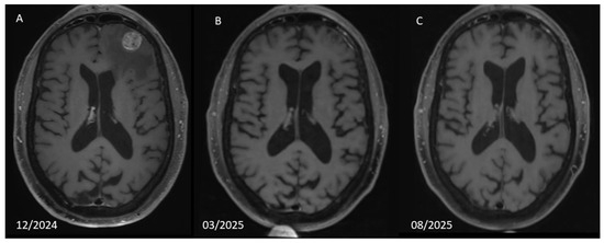
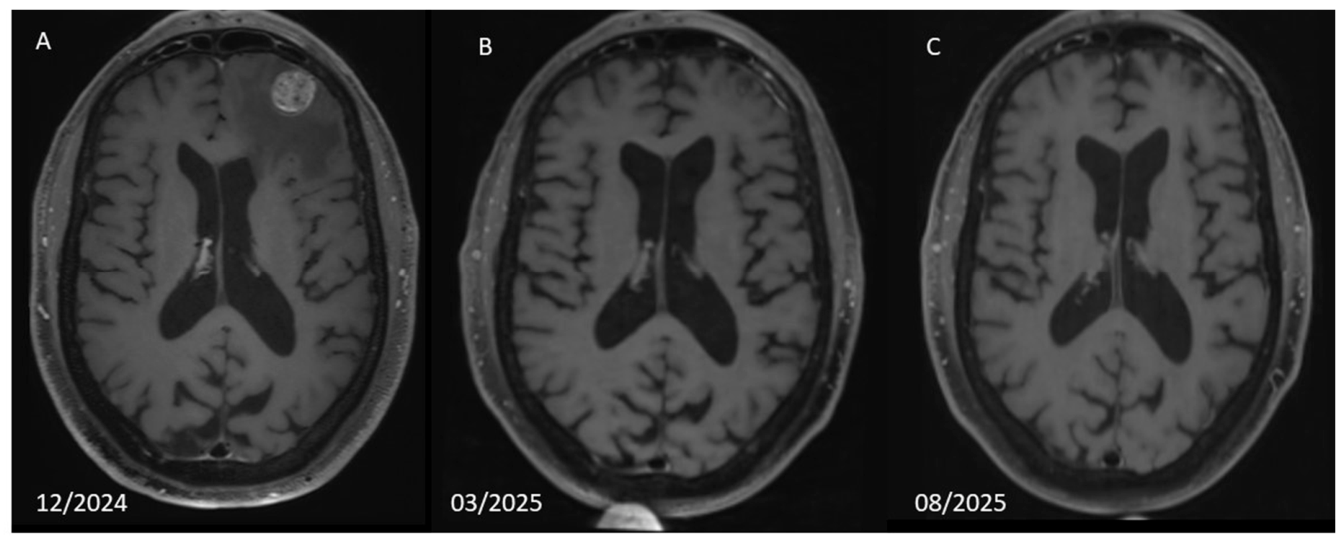
In December 2024, the patient presented with new-onset headaches. Brain imaging had not been performed on a routine surveillance basis prior to this presentation; this intracranial relapse was identified after the onset of new neurological symptoms. A brain CT identified a solitary left frontal lesion. Restaging FDG PET-CT demonstrated increased uptake corresponding to a hyperdense left frontal lesion, with no evidence of extracranial disease (Figure 6). Subsequent brain MRI confirmed a single left frontal lobe lesion highly suspicious for malignancy (Figure 7A). Following MDT discussion, the patient underwent a craniotomy in February 2025. Histopathological analysis confirmed metastatic SCCB. On histopathological review, the resected lesion demonstrated pure small cell carcinoma with no coexisting urothelial component, consistent with the histology of the primary tumour. This intracranial relapse occurred nearly seven years after the initial diagnosis.
Figure 6.
PET/CT Demonstrating No Systemic Disease but a Suspected Brain Metastasis (A) Whole-body FDG PET showing no evidence of systemic metabolically active extracranial disease. (B) Axial brain PET image revealing increased uptake in the left frontal region (arrow). (C,D) Fused PET/CT and corresponding non-contrast CT demonstrating increased FDG uptake associated with a hyperdense left frontal cortical lesion (arrow), suspicious for metastatic involvement.
Figure 7.
Brain Metastasis Identified on MRI and Complete Resolution Following Surgical Resection (A) Axial contrast-enhanced MRI from December 2024 demonstrating a well-defined enhancing lesion in the left frontal lobe, consistent with brain metastasis. (B) Follow-up MRI from March 2025 showing postoperative changes with no residual or recurrent enhancing disease. (C) MRI from August 2025 confirming sustained absence of intracranial metastatic disease.
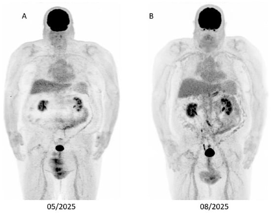
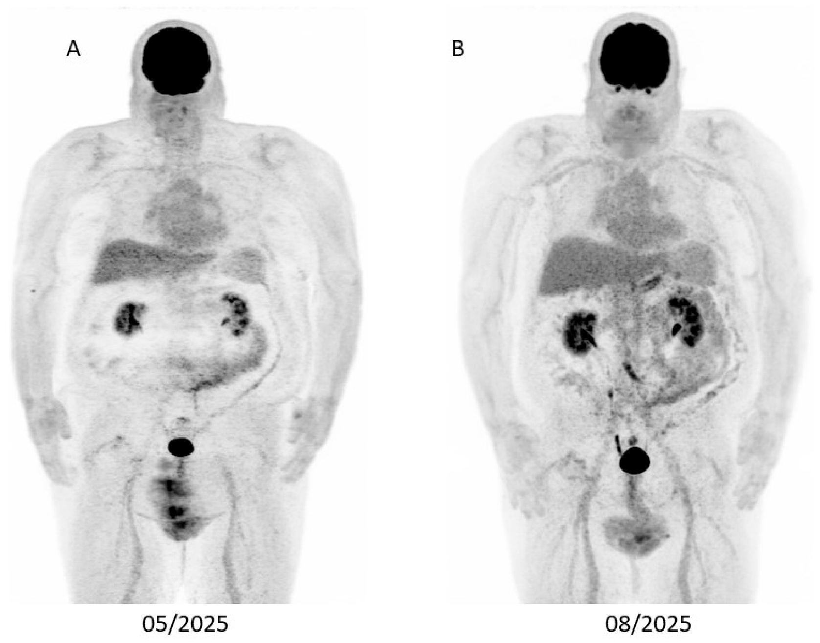
Postoperative MRI confirmed no residual disease (Figure 7B). The patient subsequently received adjuvant stereotactic radiosurgery (SRS) to the surgical cavity. His follow-up PET-CT showed no evidence of systemic disease. Considering the isolated nature of the CNS relapse, the absence of extracranial progression, and the history of severe irAEs, the MDT opted for active surveillance rather than resuming systemic therapy. Serial follow-up with brain MRI (Figure 7B,C) and PET-CT (Figure 8) has shown no evidence of recurrence. As of the most recent follow-up in July 2026, the patient remains alive with no evidence of disease.
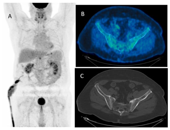
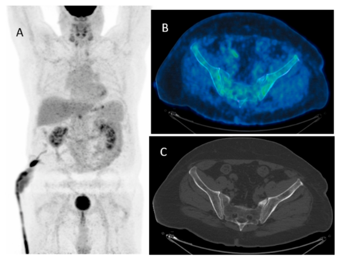
Figure 8.
Serial PET/CT Scans Demonstrating Sustained No Evidence of Disease (A) Whole-body FDG PET from May 2025 showing no metabolically active lesions. (B) Whole-body FDG PET from August 2025 also demonstrating complete metabolic response with no evidence of disease.
3. Discussion
SCCB is an exceptionally rare and aggressive malignancy. Its biological behaviour closely parallels that of SCLC, characterized by rapid proliferation, early dissemination, and poor long-term survival outcomes [10]. Retrospective series consistently report a median overall survival of approximately 12–20 months in metastatic disease, with a five-year survival rate below 10% [6]. Published reports of prolonged survival in metastatic SCCB are summarized in Table 1. Against this backdrop, the present case of a patient surviving nearly seven years with durable disease control on immunotherapy despite treatment-limiting toxicities represents an exceptional clinical outcome. First-line platinum-based chemotherapy remains the standard of care for metastatic SCCB, mirroring protocols used for SCLC [10].
Table 1.
Reported cases and series of long-term survival in metastatic small cell carcinoma of the bladder (SCCB).
The role of consolidative pelvic radiotherapy in this setting remains uncertain. Current NCCN guidelines suggest that local radiotherapy in metastatic disease should primarily be considered for palliation rather than disease control [4,10]. This contrasts with limited-stage SCCB, where chemoradiation is offered for curative intent in selected patients. In our case, the decision to deliver consolidative pelvic radiotherapy with a bladder boost was driven by equivocal PET-CT findings post-chemotherapy, which raised concern for local persistence. The MDT elected to proceed with pelvic irradiation to maximize locoregional control even in the presence of metastatic disease, reflecting an individualized, aggressive strategy to extend progression-free survival.
Upon disease progression, therapeutic options were limited by the absence of an established second-line regimen for SCCB [14]. Management decisions in this setting are generally extrapolated from SCLC or urothelial carcinoma protocols, though historical outcomes are poor [15].
Given these limitations, comprehensive molecular profiling was undertaken. High tumour mutational burden (TMB: 19.9 mut/Mb) and PD-L1 expression (CPS > 1%) are associated with an increased likelihood of response to immune checkpoint inhibitors (ICI), though neither is a definitive predictive biomarker [16,17,18]. The presence of additional alterations, such as MDM2 amplification and mutations in RAD51D, CDKN1C, KEL, MLL2, and TERT, further supported the rationale for immunotherapy, as these features are linked to genomic instability and increased neoantigen load [16,17,18]. However, as a single exceptional responder, this case cannot establish the predictive utility of TMB, PD-L1 CPS, or these specific molecular alterations for immunotherapy response in SCCB.
The increasing integration of molecular profiling into genitourinary oncology has highlighted the biological heterogeneity of urothelial malignancies and the potential role of genomic and immune biomarkers in treatment selection [19]. Nevertheless, the predictive value of individual biomarkers remains context-dependent, and their clinical interpretation requires caution, particularly in rare histological subtypes such as small cell carcinoma of the bladder. Although tumour mutational burden and PD-L1 expression have been investigated as potential markers of response to immune checkpoint inhibition across urothelial and other malignancies, neither has been prospectively validated as a treatment-selection biomarker specifically in SCCB. Furthermore, the complex tumour immune microenvironment and molecular heterogeneity of urothelial malignancies may influence response to immunotherapy and should be considered when interpreting exceptional clinical responses [20]. In this context, the prolonged disease control observed in our patient, together with the molecular findings, should be regarded as hypothesis-generating rather than evidence of a predictive relationship between a specific biomarker or genomic alteration and response to immune checkpoint blockade.
Based on MDT consensus, supported by emerging evidence from trials such as CheckMate 032 and TITAN-TCC, the patient commenced ipilimumab (1 mg/kg) plus nivolumab (3 mg/kg) every three weeks [12,13]. The deep and durable response achieved, even with significant immune-related adverse events (irAEs), underscores both the therapeutic potential and the challenges of immunotherapy in this rare disease. Durable remission following discontinuation of ICI due to irAEs has been well-documented in metastatic urothelial carcinoma, melanoma, and renal cell carcinoma [21,22,23]. In our patient, treatment was complicated by grade 3 hepatitis and subsequent grade 3 pneumonitis, necessitating permanent discontinuation. Following this, he maintained remission for several years off treatment.
Central nervous system involvement is less common in SCCB than in SCLC, and isolated late brain metastasis is a particularly rare event [6,24]. The occurrence of a solitary frontal metastasis nearly seven years after diagnosis represents an extraordinary clinical course. Given the absence of extracranial progression and the patient’s preserved performance status, the MDT pursued an aggressive local approach consisting of surgical resection followed by SRS. This strategy was extrapolated from SCLC and urothelial carcinoma data, where SRS has demonstrated improved local control and neurocognitive preservation compared to whole-brain radiotherapy [25,26]. The choice of local therapy alone, without resuming systemic treatment, was guided by the patient’s history of life-threatening immune toxicities and the absence of systemic relapse on imaging. The concept of the CNS as an immune “sanctuary site,” well described in SCLC and melanoma, offers one plausible interpretation for why intracranial relapse emerged while systemic immune control persisted, although this remains an interpretation rather than a mechanism demonstrated by a single case [27,28]. Thus, definitive local treatment was considered the most appropriate course, balancing effective disease control with the avoidance of further systemic toxicity.
4. Conclusions
This case illustrates the potential for long-term survival in metastatic SCCB through a multimodal approach integrating chemotherapy, consolidative radiotherapy, and immunotherapy. It highlights the utility of genomic profiling, specifically tumour mutational burden, in identifying candidates for ICI treatment even in the absence of standard guidelines. Of note, the development of an isolated brain metastasis after years of remission highlights the need for vigilance regarding CNS relapse, which may serve as a sanctuary site even in excellent responders. In the end, this report supports an aggressive, multidisciplinary management strategy for fit patients with this rare and aggressive malignancy.
Author Contributions
Conceptualization, J.B. and A.P.; methodology, J.B., A.P. and T.T.; data collection, J.B., M.B. and B.T.; writing—original draft preparation, J.B.; writing—review and editing, J.B., M.B., B.T., T.T. and A.P.; supervision, A.P. and T.T. All authors have read and agreed to the published version of the manuscript.
Funding
This research received no external funding.
Institutional Review Board Statement
This manuscript is a single-patient case report. Formal ethics committee approval was not obtained, as ethical review and approval are not required for single-patient case reports according to our institutional practice and local requirements, provided that patient confidentiality is maintained and written informed consent for publication is obtained.
Informed Consent Statement
Written informed consent for publication was obtained from the patient prior to publication.
Data Availability Statement
The data presented in this study are available on request from the corresponding author due to patient privacy and confidentiality restrictions inherent to identifiable clinical information in the single-patient case report.
Conflicts of Interest
The authors declare no conflicts of interest.
References
- Park, S.; Reuter, V.E.; Hansel, D.E. Non-urothelial carcinomas of the bladder. Histopathology 2019, 74, 97–111. [Google Scholar] [CrossRef] [Scilit] [PubMed]
- Ismaili, N. A rare bladder cancer—Small cell carcinoma: Review and update. Orphanet J. Rare Dis. 2011, 6, 75. [Google Scholar] [CrossRef] [Scilit] [PubMed]
- García Rico, E.J.; Ortega Polledo, L.E.; Sánchez Pellejero, A.; Vázquez Valdés, S.; Montuenga Fernandez, I. Small-cell neuroendocrine tumor of the bladder: Unexpected long-term survival with carboplatin-etoposide therapy in a patient with metastatic stage disease. Urol. Ann. 2023, 15, 331–333. [Google Scholar] [CrossRef] [Scilit] [PubMed]
- Kouba, E.J.; Cheng, L. Understanding the Genetic Landscape of Small Cell Carcinoma of the Urinary Bladder and Implications for Diagnosis, Prognosis, and Treatment: A Review. JAMA Oncol. 2017, 3, 1570–1578. [Google Scholar] [CrossRef] [Scilit] [PubMed]
- Patel, S.G.; Stimson, C.J.; Zaid, H.B.; Resnick, M.J.; Cookson, M.S.; Barocas, D.A.; Chang, S.S. Locoregional small cell carcinoma of the bladder: Clinical characteristics and treatment patterns. J. Urol. 2014, 191, 329–334. [Google Scholar] [CrossRef] [Scilit] [PubMed]
- Chau, C.; Rimmer, Y.; Choudhury, A.; Leaning, D.; Law, A.; Enting, D.; Lim, J.H.; Hafeez, S.; Khoo, V.; Huddart, R.; et al. Treatment outcomes for small cell carcinoma of the bladder: Results from a UK patient retrospective cohort study. Int. J. Radiat. Oncol. Biol. Phys. 2021, 110, 1143–1150. [Google Scholar] [CrossRef] [Scilit] [PubMed]
- Church, D.N.; Bahl, A. Clinical review—Small cell carcinoma of the bladder. Cancer Treat. Rev. 2006, 32, 588–593. [Google Scholar] [CrossRef] [Scilit] [PubMed]
- Horn, L.; Mansfield, A.S.; Szczęsna, A.; Havel, L.; Krzakowski, M.; Hochmair, M.J.; Huemer, F.; Losonczy, G.; Johnson, M.L.; Nishio, M.; et al. First-Line Atezolizumab plus Chemotherapy in Extensive-Stage Small-Cell Lung Cancer. N. Engl. J. Med. 2018, 379, 2220–2229. [Google Scholar] [CrossRef] [Scilit] [PubMed]
- Paz-Ares, L.; Dvorkin, M.; Chen, Y.; Reinmuth, N.; Hotta, K.; Trukhin, D.; Statsenko, G.; Hochmair, M.J.; Özgüroğlu, M.; Ji, J.H.; et al. Durvalumab plus platinum-etoposide versus platinum-etoposide in first-line treatment of extensive-stage small-cell lung cancer (CASPIAN): A randomised, controlled, open-label, phase 3 trial. Lancet 2019, 394, 1929–1939. [Google Scholar] [CrossRef] [Scilit] [PubMed]
- Stelwagen, J.; de Vries, E.G.E.; Walenkamp, A.M.E. Current Treatment Strategies and Future Directions for Extrapulmonary Neuroendocrine Carcinomas: A Review. JAMA Oncol. 2021, 7, 759–770. [Google Scholar] [CrossRef] [Scilit] [PubMed]
- Grimm, M.O.; Grün, C.B.; Niegisch, G.; Pichler, M.; Roghmann, F.; Schmitz-Dräger, B.; Baretton, G.; Schmitz, M.; Bolenz, C.; Foller, S.; et al. Tailored immunotherapy approach with nivolumab with or without ipilimumab in patients with advanced transitional cell carcinoma after platinum-based chemotherapy (TITAN-TCC): A multicentre, single-arm, phase 2 trial. Lancet Oncol. 2023, 24, 347–359. [Google Scholar] [CrossRef] [Scilit] [PubMed]
- Grimm, M.O.; Schostak, M.; Grün, C.B.; Loidl, W.; Pichler, M.; Zimmermann, U.; Schmitz-Dräger, B.; Steiner, T.; Roghmann, F.; Niegisch, G.; et al. Nivolumab + Ipilimumab as Immunotherapeutic Boost in Metastatic Urothelial Carcinoma: A Nonrandomized Clinical Trial. JAMA Oncol. 2024, 10, 755–764. [Google Scholar] [CrossRef] [Scilit] [PubMed]
- Sharma, P.; Siefker-Radtke, A.; de Braud, F.; Basso, U.; Calvo, E.; Bono, P.; Morse, M.A.; Ascierto, P.A.; Lopez-Martin, J.; Brossart, P.; et al. Nivolumab Alone and With Ipilimumab in Previously Treated Metastatic Urothelial Carcinoma: CheckMate 032 Nivolumab 1 mg/kg Plus Ipilimumab 3 mg/kg Expansion Cohort Results. J. Clin. Oncol. 2019, 37, 1608–1616, Erratum in J. Clin. Oncol. 2019, 37, 2094. https://doi.org/10.1200/JCO.19.01700. [Google Scholar] [CrossRef] [Scilit] [PubMed]
- Trigo, J.; Subbiah, V.; Besse, B.; Moreno, V.; López, R.; Sala, M.A.; Peters, S.; Ponce, S.; Fernández, C.; Alfaro, V.; et al. Lurbinectedin as second-line treatment for patients with small-cell lung cancer: A single-arm, open-label, phase 2 basket trial. Lancet Oncol. 2020, 21, 645–654, Correction in Lancet Oncol. 2020, 21, E553. https://doi.org/10.1016/S1470-2045(20)30676-8. [Google Scholar] [CrossRef] [Scilit] [PubMed]
- Petty, W.J.; Paz-Ares, L. Emerging Strategies for the Treatment of Small Cell Lung Cancer: A Review. JAMA Oncol. 2023, 9, 419–429. [Google Scholar] [CrossRef] [Scilit] [PubMed]
- Khurshid, H.; Ismaila, N.; Bian, J.; Dabney, R.; Das, M.; Ellis, P.; Feldman, J.; Hann, C.; Kulkarni, S.; Laskin, J.; et al. Systemic Therapy for Small-Cell Lung Cancer: ASCO-Ontario Health (Cancer Care Ontario) Guideline. J. Clin. Oncol. 2023, 41, 5448–5472. [Google Scholar] [CrossRef] [Scilit] [PubMed]
- Lenis, A.T.; Lec, P.M.; Chamie, K. Bladder Cancer: A Review. JAMA 2020, 324, 1980–1991. [Google Scholar] [CrossRef] [Scilit] [PubMed]
- Mancini, M.; Righetto, M.; Noessner, E. Checkpoint Inhibition in Bladder Cancer: Clinical Expectations, Current Evidence, and Proposal of Future Strategies Based on a Tumor-Specific Immunobiological Approach. Cancers 2021, 13, 6016. [Google Scholar] [CrossRef] [Scilit] [PubMed]
- Coca Membribes, S.; Szabados, B.; Powles, T. Towards biomarker-driven therapies for urothelial carcinoma. Nat. Rev. Clin. Oncol. 2026, 23, 92–106. [Google Scholar] [CrossRef] [Scilit] [PubMed]
- Germana, E.; Pepe, L.; Pizzimenti, C.; Ballato, M.; Pierconti, F.; Tuccari, G.; Ieni, A.; Giuffrè, G.; Fadda, G.; Fiorentino, V.; et al. Programmed Cell Death Ligand 1 (PD-L1) Immunohistochemical Expression in Advanced Urothelial Bladder Carcinoma: An Updated Review with Clinical and Pathological Implications. Int. J. Mol. Sci. 2024, 25, 6750. [Google Scholar] [CrossRef] [Scilit] [PubMed]
- Tzeng, A.; Tzeng, T.H.; Ornstein, M.C. Treatment-free survival after discontinuation of immune checkpoint inhibitors in metastatic renal cell carcinoma: A systematic review and meta-analysis. J. Immunother. Cancer 2021, 9, e003473. [Google Scholar] [CrossRef] [Scilit] [PubMed]
- Lallas, K.; Chatziioannou, E.; Durak, D.; Frey, G.; Serna-Higuita, L.M.; Rasch, M.L.; Kyrgidis, A.; Timotheadou, E.; Apalla, Z.; Leiter, U.; et al. Discontinuation of immune checkpoint inhibitors for reasons other than disease progression and the impact on relapse and survival of advanced melanoma patients. A systematic review and meta-analysis. Front. Immunol. 2025, 16, 1524945. [Google Scholar] [CrossRef] [Scilit] [PubMed]
- Robert, C.; Marabelle, A.; Herrscher, H.; Caramella, C.; Rouby, P.; Fizazi, K.; Besse, B. Immunotherapy discontinuation—How, and when? Data from melanoma as a paradigm. Nat. Rev. Clin. Oncol. 2020, 17, 707–715. [Google Scholar] [CrossRef] [Scilit] [PubMed]
- Moussa, M.J.; Tabet, G.C.; Siefker-Radtke, A.O.; Xiao, L.; Wilson, N.R.; Gao, J.; Logothetis, C.J.; Grivas, P.; Lee, B.; Shah, A.Y.; et al. Histopathologic Progression and Metastatic Relapse Outcomes in Small Cell Neuroendocrine Carcinomas of the Urinary Tract. Cancer Med. 2025, 14, e70594. [Google Scholar] [CrossRef] [Scilit] [PubMed]
- Robinson, S.D.; Wong, C.D.; Mazarakis, N.K.; Lorimer, C.; Critchley, G.; Chandy, E. A Review of Focal Therapies for Small Cell Lung Cancer Brain Metastases. Clin. Lung Cancer 2025, 26, 441–457. [Google Scholar] [CrossRef] [Scilit] [PubMed]
- Perron, R.; Iorio-Morin, C.; Chytka, T.; Simonova, G.; Chiang, V.; Singh, C.; Niranjan, A.; Wei, Z.; Lunsford, L.D.; Peker, S.; et al. International multicenter study of stereotactic radiosurgery for bladder cancer brain metastases. J. Neuro-Oncol. 2025, 174, 235–241. [Google Scholar] [CrossRef] [Scilit] [PubMed]
- Bander, E.D.; Yuan, M.; Carnevale, J.A.; Reiner, A.S.; Panageas, K.S.; Postow, M.A.; Tabar, V.; Moss, N.S. Melanoma brain metastasis presentation, treatment, and outcomes in the age of targeted and immunotherapies. Cancer 2021, 127, 2062–2073. [Google Scholar] [CrossRef] [Scilit] [PubMed]
- Buckley, M.W.; McGavern, D.B. Immune dynamics in the CNS and its barriers during homeostasis and disease. Immunol. Rev. 2022, 306, 58–75. [Google Scholar] [CrossRef] [Scilit] [PubMed]

Disclaimer/Publisher’s Note: The statements, opinions and data contained in all publications are solely those of the individual author(s) and contributor(s) and not of MDPI and/or the editor(s). MDPI and/or the editor(s) disclaim responsibility for any injury to people or property resulting from any ideas, methods, instructions or products referred to in the content. |
© 2026 by the authors. Licensee MDPI, Basel, Switzerland. This article is an open access article distributed under the terms and conditions of the Creative Commons Attribution (CC BY) license.