


Simple Summary
Proteolysis-targeting chimeras (PROTACs) are examined in this review as a novel approach to cancer therapy. A target-protein ligand, an E3 ubiquitin-ligase ligand, and a connecting linker make up heterobifunctional molecules known as PROTACs. Instead of just blocking the activity of disease-associated proteins, they enlist the ubiquitin–proteasome system to identify and eliminate them by generating a ternary complex. This catalytic, event-driven approach can overcome resistance, eradicate enzymatic and non-enzymatic functions, provide long-lasting effects at lower exposure, and target proteins that were previously thought to be undruggable. The development from early peptide-based constructs to small-molecule degraders employing ligases like cereblon and VHL is tracked in the study. It emphasizes uses against key carcinogenic targets, including as kinases, transcriptional regulators, androgen and estrogen receptors, as well as proteins linked to apoptosis. Clinical candidates that promote therapeutic translation include vepdegestrant and bavdegalutamide, which have shown good efficacy in breast and prostate malignancies. High molecular weight, restricted membrane permeability, poor oral bioavailability, complex pharmacokinetics, off-target degradation, toxicity, and potential resistance due to changes in ligases or proteasomal machinery continue to limit PROTAC development. Therefore, rational ligand selection, tissue-specific ligase recruitment, ternary-complex stability, linker modification, and enhanced delivery are crucial. All things considered, PROTACs provide a flexible platform for accurate, customized oncology; nevertheless, prior to widespread adoption, further extensive clinical validation, safety evaluation, and manufacturing optimization are needed.
Abstract
PROTACs represent a cutting-edge approach to the discovery of innovative cancer therapies. By exploiting the cell’s ubiquitin–proteasome system, PROTACs induce the selective degradation of target proteins, providing a distinct advantage over conventional inhibitors that only block protein function. This review delves into the design principles and mechanisms underlying PROTACs, emphasizing their potential to circumvent drug resistance and achieve prolonged therapeutic effects. Recent advancements have focused on enhancing the specificity, bioavailability, and overall efficacy of PROTACs, demonstrating promising results in preclinical cancer models. The ability of PROTACs to target previously “undruggable” proteins opens new avenues for cancer treatment, offering the potential for more effective and personalized therapeutic strategies. As research continues to evolve, the optimization and clinical translation of PROTACs could declare a new era in oncology, revolutionizing the landscape of cancer drug discovery and personalized medicine.
1. Introduction
The idea of targeted protein degradation (TPD) arose from the realization that disease-associated proteins might be removed instead of just inhibited. PROTACs, a unique class of heterobifunctional molecules created to take advantage of the ubiquitin–proteasome system for targeted protein degradation, are the result of this idea [1]. A proof-of-concept that a target protein might be drawn to an E3 ubiquitin ligase and then exposed to ubiquitination and proteasomal destruction was presented by Sakamoto et al. in their 2001 publication on the first PROTAC [2]. Early PROTACs showed limits in cellular permeability and drug-like qualities, and they were primarily peptide-based.
Small-molecule PROTACs with ligands for E3 ligases like von Hippel–Lindau (VHL) and cereblon (CRBN) were created as a result of later developments in medicinal chemistry, significantly increasing their cellular activity and therapeutic potential [3]. PROTACs are catalytic, event-driven pharmacological agents that can effectively and persistently deplete target proteins, as demonstrated by the emergence of these specialized degraders. PROTAC technology has developed over the last 20 years from an experimental proof-of-concept to a quickly growing therapeutic platform, with many degraders being studied in preclinical and clinical trials, especially for the treatment of cancer [4]. Applying PROTACs to oncogenic and otherwise challenging-to-drug proteins has attracted a lot of attention due to this evolution, offering a promising approach for the creation of next-generation treatments, such as those that target gastrointestinal malignancies.
A PROTAC is a bifunctional molecule designed to induce selective protein degradation. It consists of two ligands connected by a linker, which facilitates the formation of a functional complex between the target protein and an E3 ubiquitin ligase [5]. A PROTAC simultaneously binds to a protein of interest (POI) through one ligand and an E3 ubiquitin ligase through the other, thereby promoting the formation of a ternary complex. This complex facilitates the ubiquitination of the POI, which is subsequently recognized and degraded by the 26S proteasome through the ubiquitin–proteasome system (UPS) [4,6]. Unlike conventional inhibitors, which generally modulate the activity of their target proteins, PROTACs can eliminate the target protein itself from the cell. This approach enables the targeting of a broad range of proteins, including enzymes, transcription factors, structural proteins, and regulatory proteins [7,8]. The rapid development of PROTAC technology has generated considerable interest in diverse therapeutic areas, particularly oncology and neurological disorders. These heterobifunctional molecules exploit the endogenous cellular protein quality-control machinery by recruiting it to target proteins, thereby inducing their selective degradation [9,10,11]. This approach is based on the ability of rationally designed PROTAC molecules to promote targeted protein degradation. Numerous studies have demonstrated that PROTAC-mediated targeted protein degradation offers several potential advantages over conventional protein inhibition, as it eliminates the target protein rather than merely suppressing its activity. PROTACs must enter cells to access intracellular target proteins and E3 ubiquitin ligases. Although PROTACs can independently interact with either the POI or the E3 ligase to form binary complexes, their primary function is to facilitate the simultaneous engagement of both proteins, resulting in the formation of a productive ternary complex [12,13].
PROTACs represent a promising targeted protein degradation technology. By inducing the ubiquitination and subsequent proteasomal degradation of target proteins, PROTACs can eliminate the entire protein rather than simply inhibit a specific functional site. This feature may also suppress non-catalytic functions of target proteins, including scaffolding and structural activities, which may persist following conventional inhibition of an enzymatic or binding domain. Furthermore, a substantial proportion of the human proteome remains challenging to target using conventional small-molecule inhibitors because many proteins lack suitable ligand-binding pockets or possess structurally inaccessible or poorly defined binding sites. In addition, prolonged or high-dose administration of conventional inhibitors may result in off-target effects and dose-limiting toxicities [14,15].
PROTACs exploit the ubiquitin–proteasome system (UPS) by recruiting an E3 ubiquitin ligase to the target protein, thereby facilitating its ubiquitination and subsequent recognition and degradation by the 26S proteasome. Several E3 ligases have been utilized in PROTAC development, including cereblon (CRBN), von Hippel–Lindau (VHL), and components of the Skp1–Cullin–F-box (SCF) complex. The choice of E3 ligase can influence PROTAC activity, selectivity, cellular distribution, and degradation efficiency. The availability and tissue-specific expression of E3 ligases are therefore important considerations in the design and optimization of PROTACs [16,17,18].
2. Role of Protac in Drug Discovery and Design
In 2001, Craig M. Crews introduced the idea of PROTACs, a revolutionary therapeutic approach that uses the ubiquitin–proteasome system to selectively degrade proteins linked to illness [19]. PROTAC technology has evolved from a proof-of-concept strategy to a therapeutic platform with clinical validation during the past 20 years. The clinical development of PROTAC-based treatments has been expedited by a number of pharmaceutical and biotechnology firms, such as Arvinas, Nurix Therapeutics, Kymera Therapeutics, Bristol Myers Squibb, and Novartis. More than 15 PROTAC candidates had started clinical trials by the end of 2021, and as targeted protein degradation programmes have grown, this number has kept rising [20,21].
ARV-110 (bavdegalutamide) and ARV-471 (vepdegestrant) produced the first clinical proof-of-concept for PROTACs. In patients with metastatic castration-resistant prostate cancer, ARV-110 demonstrated promising anti-tumour activity in Phase I/II clinical trials, especially in tumours containing AR T878A/S and H875Y mutations. Approximately 46% of patients achieved a ≥50% reduction in prostate-specific antigen (PSA) while maintaining an acceptable safety profile [22]. In a similar vein, ARV-471 showed encouraging efficacy in patients with ER-positive/HER2-negative metastatic breast cancer, attaining durable responses, favourable tolerability, and a clinical benefit rate of roughly 40%. This validated oestrogen receptor degradation as a promising therapeutic approach [23].
There are still several translational issues in spite of these positive clinical results. When compared to traditional small-molecule inhibitors, PROTACs’ comparatively high molecular weight and structural complexity may impair their oral bioavailability, membrane permeability, and pharmacokinetic characteristics. The oral exposure, systemic stability, and tissue distribution of next-generation degraders have significantly improved due to recent developments in medicinal chemistry, including linker optimisation, ligand engineering, conformational design, and optimisation of physicochemical parameters. Furthermore, possible difficulties such as off-target protein degradation, on-target toxicity in normal tissues, and long-term safety still need to be carefully considered during clinical development, even though clinical trials have often found tolerable toxicity profiles.
PROTAC technology has spread to several treatment domains outside of oncology. N. S. Grey’s and E. S. Fischer’s research created the notion of the “degradable kinome”, finding over 170 kinase targets that might be broken down and speeding up the creation of kinase-directed degraders [24,25]. Additionally, PROTACs have demonstrated therapeutic potential in viral infections by targeting the hepatitis C virus (HCV) NS3/4A protease and in neurodegenerative illnesses by degrading abnormal proteins, including tau and α-synuclein. Furthermore, degraders that target PCAF, sirtuins, and IRAK4 have shown promise in immune-mediated and inflammatory disorders, demonstrating the wide therapeutic relevance of targeted protein degradation outside of cancer [26,27,28].
3. Mechanistic Role of Protac in Cancer Management
In wealthy countries, cancer ranks second in terms of mortality, but it is the top cause of death in nations that are developing. Additionally, the incidence of cancer is rising at an incredible rate [29,30]. Innovative therapeutic approaches are being meticulously developed to tackle these challenges, with PROTAC emerging as a particularly promising technique. PROTACs consist of linkers, protein-targeting moieties, and E3 ligase-activating components. This structure allows the proteasome to recognize and degrade the cellular protein by sensing the attached ubiquitin [31]. Numerous proteins, including cycle-dependent kinases (CDKs), have been examined with various PROTACs. Small interfering RNA (SiRNA). It frequently exhibits off-target effects due to imprecise matching to short RNA or the transfer of endogenous microRNA activities [32]. In androgen-insensitive cells, PROTAC showed no effect on either cell growth or levels of the androgen receptor (AR). Similarly, hypoxia-inducible factor (HIF) in PROTAC treatment inhibited cell growth and reduced levels in estrogen-dependent (Michigan Cancer Foundation-7) MCF-7 cells [33].
4. Recent Advances in Protac as Anticancer Therapeutics
After being injected and absorbed into cells, PROTACs simultaneously bind to the POI and an E3 ubiquitin ligase, bringing them into proximity [34]. Figure 1 shows that the interaction between the E3 ligase and the POI leads to polyubiquitination, which is the covalent attachment of multiple ubiquitin molecules to the POI.
Figure 1.
General mechanism of PROTAC-mediated targeted protein degradation through E3 ligase recruitment, POI polyubiquitination, and proteasomal degradation. Created by the authors using software ChemDraw (ChemBio Draw Ultra 14.9).
In PROTAC-mediated protein degradation, an E3 ubiquitin ligase is typically attracted to a protein of interest (POI) via a heterobifunctional molecule made up of a linker, an E3 ligase-recruiting ligand, and a POI-binding ligand. The PROTAC facilitates the development of a ternary complex by binding the POI and E3 ligase simultaneously, as seen in Figure 1. Ubiquitin is initially activated by ATP-dependent E1 ubiquitin-activating enzymes within the ubiquitin–proteasome system (UPS), and then it is transferred to E2 ubiquitin-conjugating enzymes. The ubiquitin is subsequently transferred from E2 to the POI by the recruited E3 ubiquitin ligase.
A polyubiquitin chain is created by repeated ubiquitination, and this chain acts as a signal for the 26S proteasome to recognize and degrade the POI. In order to contribute to its catalytic or event-driven mode of action, the PROTAC may be released after proteasomal degradation and take part in further degradation cycles. Crucially, Figure 1 depicts a broad paradigm of PROTAC-mediated degradation, and the molecular processes of individual PROTACs may vary based on the target protein, ternary-complex architecture, recruited E3 ligase, and cellular environment.
4.1. Role of PROTAC in Gastrointestinal Cancer
According to the American Cancer Society’s calculation, 338,090 additional instances of gastrointestinal cancer were recorded in the United States in 2016. Whereas in 2021, 169,280 deaths were predicted for that year [35]. Normal adult tissues comprise differentiated cells, progenitor cells, and stem cells [36]. Cancer stem cells (CSCs) share several signalling pathways with normal stem cells, and these pathways are essential for CSC self-renewal and maintenance. These pathways include those associated with prominin-1 (CD133)+ transmembrane glycoprotein (CD44) and the CD44+ aldehyde dehydrogenase fraction, both observed in colorectal cancer (CRC) as initial indicators of gastrointestinal CSCs [37]. The theory suggests that CSCs originate from polycomb group proteins and are influenced by Wnt, Hedgehog, and Notch signalling pathways. Additionally, hypoxia plays a crucial role in regulating the self-renewal of both normal cells and CSCs, with hypoxia-inducible factors (HIFs) being particularly important. Furthermore, CSCs produce various aberrant miRNAs, which may disrupt signalling pathways [38,39].
4.2. Strategy of PROTAC Mechanism in Gastrointestinal Cancer
In this mechanism, the E1 enzyme first activates and transfers ubiquitin to the E2 enzyme. The E3 ligase then binds to both the POI and the E2 enzyme, facilitating the transfer of ubiquitin to the POI. The ubiquitinated POI is subsequently recognized and degraded by the 26S proteasome into peptide fragments. Following cellular uptake, the PROTAC molecule binds simultaneously to the intracellular POI and an E3 ubiquitin ligase, promoting ternary complex formation and subsequent ubiquitination of the POI. This forms a complex involving the POI, a PROTAC, and the E3 ligase. The E3 ligase, assisted by the E2 enzyme, ubiquitinates the POI. The ubiquitin-tagged POI is then selectively recognized and degraded by the proteasome, resulting in the disassembly of the ternary complex [40,41,42]. PROTACs selectively recruit an E3 ubiquitin ligase (e.g., VHL or CRBN) to gastrointestinal cancer-associated oncoproteins, such as EGFR, HER2, KRAS, BCL-XL, or BRD4, leading to ubiquitination, proteasomal degradation, and suppression of tumor growth and drug resistance Figure 2. Some examples of PROTACs used in gastrointestinal cancer are expressed in Figure 3.
Figure 2.
The strategy of the PROTAC Mechanism in Gastrointestinal Cancer. Created by the authors using MS PowerPoint software.
Figure 3.
Examples of PROTAC in the gastrointestinal tract. Created by the authors using software ChemDraw.
4.3. PROTAC Therapy in Lung Cancer
Smoking cessation, early detection, and treatment advancements have resulted in fewer lung cancer diagnoses and deaths [43]. As a result, it is now more widely acknowledged that thorough genetics as well as tumor, node, and metastasis staging are crucial to determining how best to treat those suffering from lung carcinoma. Lung cancer, which accounts for about 20% of all cancer deaths, is still the most common type of fatal cancer [44]. However, only 10% to 20% of individuals with lung carcinoma are smokers. As a result, it is now more widely acknowledged that both thorough genetics and tumor, node, and metastasis staging are crucial to deciding how best to treat those suffering from lung cancer [45]. Patients diagnosed with lung cancer undergo biopsy, tumour staging, and molecular profiling to identify actionable targets and guide personalized treatment. Depending on the disease stage and genetic alterations, management may include surgery, radiotherapy, chemotherapy, targeted therapy, immunotherapy, or emerging therapeutic strategies such as PROTAC-based targeted protein degradation. In recent years, PROTACs have been rapidly developed as innovative therapies for lung cancer and as novel approaches to combat drug resistance. Several PROTAC compounds have been created to target therapeutic markers in non-small cell lung cancer (NSCLC), including epidermal growth factor receptor (EGFR), Kirsten rat sarcoma viral oncogene homolog (KRAS), anaplastic lymphoma kinase (ALK), serine/threonine-protein kinase B-Raf (BRAF), and B-cell lymphoma extra-large (BCL-XL). These compounds have demonstrated promising anticancer efficacy in preclinical tumor models and cell cultures [46], as discussed in Table 1. Figure 4 presents representative EGFR tyrosine kinase inhibitors that serve as warheads for the development of EGFR-targeting PROTACs in lung cancer.
Table 1.
Types of PROTACs that have been studied at different preclinical stages for lung cancer to target primary cancer pathways.
Figure 4.
Representative EGFR tyrosine kinase inhibitors (Erlotinib, Gefitinib, Afatinib, and Dacomitinib) that serve as parent scaffolds or warheads for the design and development of EGFR-targeting PROTACs in lung cancer. Created by the authors using software ChemDraw.
4.4. Attempted Constructive PROTAC in Breast Cancer
According to this design strategy, they demonstrated the first nucleolin-targeting aptamer, PROTAC ZL216, which specifically decomposed nucleolin under laboratory conditions and in vivo. Additionally, they demonstrated that ZL216 could stop breast cancer cells that were hormone receptor (HR) positive from expanding and propagating [47,48,49,50,51,52,53,54,55,56,57]. Single-stranded nucleic acids called aptamers have high selectivity and affinity for their targets. Nowadays, aptamers are extensively employed in targeted drug delivery and tumor treatment because of their special advantages, which include low immunogenicity, wide cell accessibility, outstanding durability, and ease of production [58]. Specifically bound by AS1411, a short single-stranded deoxyribonucleic acid (DNA) oligonucleotide, nucleolin accumulates on certain cancer cells and tumor cells and may be associated with multiple mechanisms of carcinogen and angiogenesis. Clinical studies investigating antiproliferative properties on tumor cells are presently being examined, as per a previous study [59]. Some examples involved in breast cancer are represented in Figure 5.
Figure 5.
Examples of drugs used in breast cancer. Created by the authors using software ChemDraw.
4.5. PROTAC Effects on Lymphatic Cancer
PROTACs comprise three structural elements: a ligand that binds to the target protein, a ligand that binds to an E3 ubiquitin ligase, and a linker that connects the two ligands [60]. The principle of event-based protein degraders is that even transient interactions can lead to selective degradation of the targeted proteins via proteolysis [61]. Additionally, PROTACs can degrade the overexpressed or mutated target proteins with relatively low binding affinity. PROTACs may have the ability to overcome drug resistance [62]. We thus describe UBX-382, a highly potent Bruton’s tyrosine kinase (BTK) PROTAC that potently inhibits B-cell receptor (BCR) signalling and targets BTK in diffuse large B-cell lymphoma (DLBCL) [63]. UBX-382 showed inhibitory effects in tumor growth studies in vitro and in vivo with accumulation of wild-type (WT) and mutant BTK.
Additionally, Cereblon (CRBN), a component of immunomodulatory imide drugs (IMiDs), has been shown to mediate selective degradation of new substrates such as Ikaros/Aiolos and the translation termination factor G1 (GSPT1) through phase transition protein UBX-382 in a range of haematological malignancies. The ability of UBX-382 to inhibit tumor proliferation suggests a broad response to the treatment, as illustrated in Figure 6.
Figure 6.
Examples of various drugs in lymphatic cancer. Created by the authors using software ChemDraw.
5. Development of Protacs for Targeting Cancer-Associated Pathways and Phenotypes
Cancer occurrence has been addressed more selectively and noticeably, decreasing the cell’s ability to target, and far fewer adverse effects than conventional small molecule inhibitors, PROTACs offer a novel method [64]. Since Craig M. Crews created the first PROTAC in 2001, the field has advanced quickly throughout the past two decades [65].
5.1. Therapy Involvement in the Process of Cancer Criteria: Ubiquitin Proteasome System
The ubiquitin–proteasome process is a crucial mechanism that controls intrinsic protein levels and also maintains a precise equilibrium between protein formation and breakdown. The UPS regulates many important physiological activities in addition to the creation of proteins, including the development of the cell cycle, mortality, protein adaptation, growth communication channels, autophagy, and the restoration of DNA [66,67]. The 26S proteasome destroys multi-ubiquitinated proteins that contain at least 4 ubiquitins connected to a protein [68].
5.2. Cancer-Causing Proteins for Destruction
Our approach to curing human disease involves targeting the cancer-causing protein to an E3 ligase for subsequent ubiquitination and destruction. The PROTAC methodology is employed to counteract the cancer-causing protein by facilitating its degradation. Zhou and Howley initially proposed this concept, using a genetic treatment strategy to select specific amino acids for ubiquitination and subsequent destruction [69]. Cell division cycle protein 4 (Cdc4), SCF in yeast, and a binding companion of the phosphorylated Retinoblastoma protein (pRP) called E7N were incorporated to generate a fusion protein by Zhou et al. The PRP was attracted by the fusion protein to the SCF ubiquitin ligase, where it was ubiquitinated and degraded [70]. Unfortunately, this approach was restricted by poor conversion efficiency and the role of potent, dangerous lentiviruses, making it challenging to adapt and use in healthcare [71].
5.3. Protein Binding Covalently for Ubiquitination
We initially developed a PROTAC compound that interacts with the protein methionine aminopeptidase-2 (MetAP-2) to promote ubiquitination and destruction as a proof of concept. MetAP-2 is a target of the angiogenesis blockers fumagillin and ovarian and divides the N-terminal methionine from developing polypeptides [72].
5.4. Protein Binding of Noncovalent Interactions for Ubiquitination
The next test aimed to demonstrate that PROTACs could link cancer-causing proteins through noncovalent interactions. High-affinity interactions between ligands and steroid hormone receptors facilitate this association. Estradiol and testosterone are ligands for the estrogen and androgen receptors within the steroid hormone receptor complex, both of which have been extensively studied and linked to cancer growth. In particular, the estrogen receptor (ER) has been associated with the development of breast cancer [73,74].
5.5. Development of Target PROTAC in Cells
PROTAC medicinal purposes are dependent on the protein target being efficiently ubiquitinated and processed by cellular proteasomes and endogenous ubiquitin ligases. It was discovered that the initial generation of PROTAC was extremely polar and did not enter cells because it contained a phosphopeptide. This variant of PROTAC was effectively microinjected [75]. Because few small molecules bind to E3 ligase, a HIF1- dihydroxytestosterone (DHT) PROTAC was developed to increase accessibility. The sequence of 7 amino acids that make up ALAPYIP of HIF1, an innate precursor in Von Hippel-Lindau syndrome (VHL), was selected to make up the E3 recognition site in the HIF1-PROTAC [76].
5.6. Protein to Target Endogenously in Cells
Although the HIF1-testosterone PROTAC had excellent results in 293 human embryonic kidney cells that produced green fluorescent protein-androgen receptor (GFP-AR), this is not comparable to the natural AR found in prostate cancer cells. We, therefore, investigated PROTAC efficiency in AR-positive participants next. HIF1-testosterone PROTAC therapy was reported to drastically lower endogenous AR levels in expressing the ER malignant breast cells and limit proliferation in androgen-dependent prostate carcinoma [77].
6. Recent Advances in Targeted Protein Degradation Technologies Beyond PROTACs
The first and fastest-evolving decay technology (PDT) is introduced here, followed by a review of the development of destruction techniques by PROTAC action over the past 20 years, including careful consideration of the advantages and disadvantages of multiple decay techniques as well as several innovative and thought-provoking recommendations for future development. For example, numerous CRBN-based PROTACs have been effectively designed to degrade BRD4 [78], CDK9 [79], Sirt2 [80], Kirsten rat sarcoma viral oncogene homolog (KRASG12C) [81], and other immunomodulatory medications (IMiDs) such as thalidomide, lenalidomide, and pomalidomide. Similarly, VHL-based PROTACs have successfully degraded HaloTag7 fusion proteins [82], Estrogen-related receptor (ERR) [83], receptor-interacting serine protein kinase 2 (RIPK2) [84], BRD [85], small warm phenotype (SMAD) [86], BRD4 [87], breakpoint cluster region (BCR), and Abelson (ABL) [73,74]. Additionally, targets like receptor tyrosine kinase (RT) [88], ring finger protein (RNF114) based PROTAC Receptor-interacting serine/threonine kinase 2 (RIPK2), CDK4/6, Bcl-X [89], and other PROTAC targets have been degraded [90].
6.1. Light-Controllable PROTAC
In one case in 2019, Opto-PROTAC was created by the group of Jian Jin and Wenyi Wei [91]. In terms of precision medicine, light-controlled opto-PROTAC is probably a better option than PROTAC. Simultaneously, Xue et al. created photoactivatable PROTACs, also known as photo-caged PROTACs (pc-PROTACs), derived from the well-known BRD2-4 degrader bromodomain and extraterminal domain (DBet) [92]. The moieties on PROTACs were designed to enable the PROTAC to turn instantly either operational or inert by photochemically isomerizing in response to different wavelengths of light. For example, Pfaff et al. showed that photoswitchable PROTACs can be created by using ortho-F4-azobenzene intermediaries between the POI substrate and E3 ligase substrate, as discussed in Figure 7 [93]. ABL and BCR-ABL fusion proteins were also encouraged by Jin et al. to be degraded by azo-PROTACs in chronic myelogenous leukaemia (CML) cells [94].
Figure 7.
Various involvements of the compounds in the light-controllable PROTAC. Created by the authors using software ChemDraw.
6.2. Transcription Factor Proteolysis Target Chimera (TF-PROTAC)
Transcription Factor Proteolysis Target Chimera (TF-PROTAC) is believed to be an interesting strategy for cancer treatment because of its close connections to carcinogenesis and cell proliferation. TFs precisely recognize and attach to the region upstream of the gene, controlling the activity of many genes [95]. However, transcription factors (TFs) typically lack active sites or allosteric regulatory regions, unlike kinases or other enzymes. As a result, conventional therapeutics, including small-molecule inhibitors targeting TFs, have been largely inaccessible for decades [96], with some examples illustrated in Figure 8. Fortunately, most TFs have a distinctive DNA-binding motif, which has been identified and predicted by various research efforts. The specific DNA sequences that interact with TFs have also been elucidated. Leveraging these characteristics, the Wenyi Wei group developed a platform called TF-PROTACs to selectively degrade TFs [97].
Figure 8.
Linkage of TF-PROTAC at ligand. Created by the authors using software ChemDraw.
6.3. Phosphorylation Proteolysis Target Chimera (PhospoTAC)
One of the significant post-translational changes in proteins is phosphorylation. The incidence of certain disorders, including cancer and Alzheimer’s disease, is intimately matched with the abnormal phosphorylation of a few vital proteins in cells [98,99]. The Crews research team created phosphodependent proteolysis targeting chimera (phosphoproteins) based on the fundamentals of PROTAC [100] as discussed in Figure 9.
Figure 9.
Sequencing of the PhospoTAC. Created by the authors using software ChemDraw.
6.4. Lysosome Targeting Chimera (LYTAC)
While PROTAC has a lot of benefits, it also has certain drawbacks. For instance, PROTAC only controls internal proteins, leaving cell membranes and secreted proteins uncontrolled. Carolyn R. Bertozzi developed and verified this LYTAC approach in 2020 to break through this restriction by utilizing a lysosomal-degrading way to break down secretory as well as cell wall proteins [101]. On the cell membrane, there are lysosome targeting receptors (LTRs), which can facilitate protein movement to lysosomes for later destruction [102]. An example LTR that can deliver proteins containing N-glycans capped in the presence of mannose-6-phosphate (M6P) to lysosomes for destruction is the cation-independent mannose-6-phosphate receptor (CI-M6PR) [103], as described in Figure 10.
Figure 10.
Mechanism of action of lysosomal target chimera. Created by the authors using software ChemDraw, PowerPoint, and Bio Render.
6.5. Autophagosome-Tethering Compounds (ATTEC)
To break down specific proteins, Professor Lu’s group created an Autophagosome-Tethering Compound (ATTEC), or autophagosome-tethering chemical, in 2019 [104]. This material binds the pathogenic targeted protein to microtubule-associated protein 1 light chain 3α (LC3), encases it in an autophagosome, and transports it to the lysosome, where it is broken down and has been represented in Figure 11. In 2021, the group led by Professor Lu developed a new ATTEC [105].
Figure 11.
ATTEC degradation phenomena in PROTAC. Created by the authors using software ChemDraw.
6.6. CMA Chaperone Mediated Autophagy (CMA)
Protein degradation in cells can be mediated by a type of autophagy called CMA. The substrate protein has a KFERQ pentapeptide motif, which is the mechanism of CMA, as explained in Figure 12. It can be specifically identified and recognised when combined with the chaperone heat shock protein HSC70 (heat shock cognate protein of 70 kDa) in the cell membrane. This single-molecule LAMP2A is then multimerized as a result of the complex’s interaction with the lysosomal-associated membrane protein 2A (LAMP2A). The assembly can finally reach the lysosomal lumen for protein breakdown thanks to polymerized LAMP2A [106]. Wang’s team created a unique chimeric peptide technique responsible for the process of CMA, and it successfully destroys the specific proteins in cells [93].
Figure 12.
Mechanism of CMA-based degrader-mediated autophagy. The CMA-based degrader binds the target protein, which is recognized by Hsc70 and LAMP-2A, facilitating lysosomal degradation and protein recycling. Created by the authors using software ChemDraw, PowerPoint, and BioRender.
6.7. Ribonucleic Acid Proteolysis Target Chimera (RNA-PROTAC)
RNA-PROTAC, a tool to destroy RNA-binding proteins (RBPs), was created in 2019 [107]. A variety of illnesses are linked to irregularities of RBPs, a type of cellular protein that can attach to specific RNAs [108], and some examples are illustrated in Figure 13.
Figure 13.
RNA-PROTACS that degraded RBPs. Created by the authors using software ChemDraw.
6.8. Ribonuclease Targeting Chimera (RIBOTAC)
In 2018, Disney et al. developed the ribonuclease-targeting chimera (RIBOTAC). This small molecule works by recruiting the native ribonuclease RNase to a specific RNA site, utilizing the enzyme’s intrinsic nucleic acid cleavage mechanism to effectively degrade the RNA [109]. A recent update from the Disney team [110] reports that dovitinib, a receptor tyrosine kinase (RTK) inhibitor, was recently converted into a small molecule RIBOTAC to target and degrade pathological RNA with 2500-fold enhanced specificity and fewer side effects [110], as shown in Figure 14.
Figure 14.
Structure of various RIBOTACS. Created by the authors using software ChemDraw.
6.9. Clinical Progress
Due to the effective breakdown of pathogenic proteins that were previously believed to be “undruggable” by PROTAC, people have begun employing PROTACs in treatments in recent years to treat conditions like cancerous cells, autoimmune illnesses, dementia, loss of hair, breakouts, and bronchitis. There is research employing this technique to treat COVID-19 bronchitis symptoms and Alzheimer’s disorder worldwide [111,112,113]. Improvements in high-throughput screening (HTS), virtual screening, structure-based drug design (SBDD), and fragment-based drug design (FBDD) will probably lead to a faster and more comprehensive development of PROTAC. In 2021, a group proposed a systematic approach to assess the PROTAC tractability of peptide substrates [114].
7. Emerging Non-Oncological Applications of PROTACs
Although PROTAC technology has been extensively investigated for cancer therapy, recent studies have expanded its applications to several non-oncological diseases. By selectively degrading disease-associated proteins, PROTACs offer therapeutic opportunities for immune disorders, neurodegenerative diseases, cardiovascular diseases, and viral infections. The following sections summarize recent PROTAC-based advances beyond oncology.
7.1. PROTACs in Immune and Inflammatory Diseases
Immune-inflammatory disorders, including ulcerative colitis, inflammatory bowel disease, and systemic lupus erythematosus, remain major therapeutic challenges. PROTAC technology has emerged as a promising strategy by selectively degrading key regulators of inflammatory signalling. Among these, IRAK3 modulates pro-inflammatory responses in neonatal leukocytes, whereas IRAK4 is a critical kinase in Toll-like receptor (TLR) and interleukin-1 receptor (IL-1R) signalling pathways. Targeted degradation of IRAK4 using PROTACs has demonstrated potential for suppressing excessive inflammatory responses while offering therapeutic benefits in autoimmune diseases and inflammation-associated cancers. These findings highlight the ability of PROTACs to modulate immune signalling through selective protein degradation rather than conventional enzymatic inhibition [115,116,117,118,119,120,121,122,123,124]. Interleukin-1 receptor-associated kinase 4 (IRAK-4), a dual target for cancer and autoimmune diseases, is a crucial protein in the immune system’s response, regulated by toll-like and interleukin receptors [125,126,127,128].
7.2. PROTACs in Neurodegenerative Disorders
Neurodegenerative diseases are characterized by the accumulation of misfolded proteins that are difficult to target using conventional small-molecule inhibitors. PROTAC technology provides a novel therapeutic approach by promoting the degradation of pathogenic proteins. Tau protein, a microtubule-associated protein essential for neuronal stability, undergoes abnormal aggregation in Alzheimer’s disease and related tauopathies. Several PROTACs have demonstrated efficient tau degradation in preclinical studies, reducing pathological protein accumulation and representing a promising strategy for treating protein aggregation disorders [129,130,131,132,133,134].
7.3. PROTACs for Huntington’s Disease
Huntington’s disease is caused by expanded CAG repeats in the HTT gene, resulting in the accumulation of mutant huntingtin (mHTT) protein within neurons. Since mutant huntingtin is difficult to inhibit pharmacologically, PROTAC-mediated degradation has emerged as an attractive therapeutic strategy. Recently developed PROTACs employing mutant huntingtin ligands linked to IAP or CRBN E3 ligase recruiters have demonstrated enhanced affinity and selective degradation of mHTT, supporting their potential for disease-modifying therapy [135,136,137,138,139,140,141,142].
7.4. PROTACs as Cardiovascular Disease
3-Hydroxy-3-methylglutaryl-CoA reductase (HMGCR) is the rate-limiting enzyme in cholesterol biosynthesis and the primary target of statins. Beyond enzyme inhibition, PROTACs have been developed to induce selective degradation of HMGCR, thereby suppressing cholesterol synthesis more efficiently. For example, Xiang and colleagues designed VHL-recruiting PROTACs derived from lovastatin that effectively degraded HMGCR and activated sterol regulatory element-binding protein (SREBP)-mediated cholesterol homeostasis. These findings suggest that targeted protein degradation may provide an alternative therapeutic strategy for cardiovascular diseases [143,144,145,146,147,148].
7.5. PROTACs as Antiviral Therapeutics
PROTAC technology has also shown promise against viral infections by selectively degrading essential viral proteins. The hepatitis C virus (HCV) NS3/4A protease is an established antiviral target. PROTACs constructed by conjugating the NS3/4A inhibitor telaprevir (VX-950) with a CRBN-recruiting ligand have demonstrated selective degradation of NS3, thereby reducing viral replication; examples are illustrated in Figure 15. Yang, Priscilla L. et al. coupled VX-950 as well as a CRBN linker to the PROTAC to create numerous NS3-targeting PROTACs [149,150,151,152,153,154,155]. This work increases the chances of PROTACs serving as possible antiviral medications, as an approach that has also been employed to combat the emergence of Severe Acute Respiratory Syndrome Coronavirus (SARS-CoV-2).
Figure 15.
Various targets involved in the PROTAC phenomenon in various diseases. Created by the authors using software ChemDraw.
8. Conclusions and Future Perspectives
Through the natural ubiquitin–proteasome system of the cell, PROTACs selectively induce the degradation of disease-associated proteins, making them a possible therapeutic approach for cancer treatment. PROTACs offer prospective benefits in targeting previously challenging-to-treat targets and overcoming specific mechanisms of drug resistance because, in contrast to conventional inhibitors, they destroy target proteins rather than just blocking their function. A number of issues, such as high molecular weight, restricted cell permeability, poor oral bioavailability, complex pharmacokinetic properties, off-target degradation, and possible toxicity, continue to impede their wider clinical translation despite promising preclinical and emerging clinical findings.
Therefore, improving PROTAC structure, target and tissue selectivity, pharmacokinetic characteristics, and safety profiles should be the main goals of future research. The therapeutic potential of targeted protein degradation may be further expanded by novel strategies such as tumor-selective PROTACs, PROTAC prodrugs, targeted delivery systems, photoactivatable degraders, molecular glues, and AI-assisted degrader design. These developments may make it easier to create degraders against clinically significant oncogenic proteins in gastrointestinal malignancies and offer fresh approaches to overcoming treatment resistance. Overall, PROTACs may become a crucial part of next-generation precision cancer treatment if medicinal chemistry, computational design, targeted delivery, and clinical validation are further integrated.
Author Contributions
S. has conceptualized and written the original draft. R.S. and K.R. have supervised the writing of the manuscript. M.J.A., A.M., R.K. and C.M. have contributed to writing the original draft. All authors have read and agreed to the published version of the manuscript.
Funding
This research received no external funding.
Institutional Review Board Statement
Not applicable.
Informed Consent Statement
Not applicable.
Data Availability Statement
No new data were created or analyzed in this study. Data sharing is not applicable to this article.
Acknowledgments
The authors would like to express their gratitude to Noida Institute of Engineering and Technology (Pharmacy Institute), Greater Noida, Metro College of Health Science and Research, Greater Noida, Jahangirabad Institute of Technology, Barabanki, for providing the necessary facilities, research support, and institutional infrastructure to conduct this work.
Conflicts of Interest
The authors declare no conflicts of interest.
References
- Sakamoto, K.M.; Kim, K.B.; Verma, R.; Ransick, A.; Stein, B.; Crews, C.M.; Deshaies, R.J. Development of Protacs to target cancer-promoting proteins for ubiquitination and degradation. Mol. Cell. Proteom. 2003, 2, 1350–1358. [Google Scholar] [CrossRef] [Scilit]
- Sakamoto, K.M.; Kim, K.B.; Kumagai, A.; Mercurio, F.; Crews, C.M.; Deshaies, R.J. Protacs: Chimeric molecules that target proteins to the Skp1–Cullin–F box complex for ubiquitination and degradation. Proc. Natl. Acad. Sci. USA 2001, 98, 8554–8559. [Google Scholar] [CrossRef] [Scilit]
- Bondeson, D.P.; Mares, A.; Smith, I.E.D.; Ko, E.; Campos, S.; Miah, A.H.; Mulholland, K.E.; Routly, N.; Buckley, D.L.; Gustafson, J.L.; et al. Catalytic in vivo protein knockdown by small-molecule PROTACs. Nat. Chem. Biol. 2015, 11, 611–617. [Google Scholar] [CrossRef] [Scilit]
- Békés, M.; Langley, D.R.; Crews, C.M. PROTAC targeted protein degraders: The past is prologue. Nat. Rev. Drug Discov. 2022, 21, 181–200. [Google Scholar] [CrossRef] [Scilit]
- Li, B.; Wang, W.; Zhao, L.; Li, M.; Yan, D.; Li, X.; Zhang, J.; Gao, Q.; Feng, Y.; Zheng, J.; et al. Aggregation-induced emission-based macrophage-like nanoparticles for targeted photothermal therapy and virus transmission blockage in monkeypox. Adv. Mater. 2024, 36, 2305378. [Google Scholar] [CrossRef] [Scilit]
- Jin, J.; Wu, Y.; Chen, J.; Shen, Y.; Zhang, L.; Zhang, H.; Chen, L.; Yuan, H.; Chen, H.; Zhang, W.; et al. The peptide PROTAC modality: A novel strategy for targeted protein ubiquitination. Theranostics 2020, 10, 10141–10153. [Google Scholar] [CrossRef] [Scilit]
- Ishikawa, M.; Tomoshige, S.; Demizu, Y.; Naito, M. Selective Degradation of Target Proteins by Chimeric Small-Molecular Drugs, PROTACs and SNIPERs. Pharmaceuticals 2020, 13, 74. [Google Scholar] [CrossRef] [Scilit]
- Jobin, P.G.; Butler, G.S.; Overall, C.M. New intracellular activities of matrix metalloproteinases shine in the moonlight. Biochim. Biophys. Acta-Mol. Cell Res. 2017, 1864, 2043–2055. [Google Scholar] [CrossRef] [Scilit]
- Ottis, P.; Crews, C.M. Proteolysis-Targeting Chimeras: Induced protein degradation as a therapeutic strategy. ACS Chem. Biol. 2017, 12, 89298. [Google Scholar] [CrossRef] [Scilit]
- Saenz, D.T.; Fiskus, W.; Qian, Y.; Manshouri, T.; Rajapakshe, K.; Raina, K.; Coleman, K.G.; Crew, A.P.; Shen, A.; Mill, C.P.; et al. Novel bet protein proteolysis-targeting chimera exerts superior lethal activity than bromodomain inhibitor (beta) against post-myeloproliferative neoplasm secondary (s) AML cells. Leukemia 2017, 31, 1951–1961. [Google Scholar] [CrossRef] [Scilit]
- Ohoka, N.; Nagai, K.; Shibata, N.; Hattori, T.; Nara, H.; Cho, N.; Naito, M. SNIPER(TACC3) induces cytoplasmic vacuolization and sensitizes cancer cells to bortezomib. Cancer Sci. 2017, 108, 1032–1041. [Google Scholar] [CrossRef] [Scilit]
- Watt, G.F.; Scott-Stevens, P.; Gaohua, L. Targeted Protein Degradation In vivo with proteolysis targeting chimeras: Current status and future considerations. Drug Discov. Today Technol. 2019, 31, 69–80. [Google Scholar] [CrossRef] [Scilit]
- Daniels, D.L.; Riching, K.M.; Urh, M. Monitoring, and deciphering protein degradation pathways inside cells. Drug Discov. Today Technol. 2019, 31, 61–68. [Google Scholar] [CrossRef] [Scilit]
- Zhou, X.; Dong, R.; Zhang, J.; Zheng, X.; Sun, L. PROTAC: A promising technology for cancer treatment. Eur. J. Med. Chem. 2020, 203, 112539. [Google Scholar] [CrossRef] [Scilit]
- Li, X.; Pu, W.; Zheng, Q.; Ai, M.; Chen, S.; Peng, Y. Proteolysis-targeting chimeras (PROTACs) in cancer therapy. Mol. Cancer 2022, 21, 99. [Google Scholar] [CrossRef] [Scilit]
- Zheng, N.; Shabek, N. Ubiquitin ligases: Structure function and regulation. Annu. Rev. Biochem. 2017, 86, 129–157. [Google Scholar] [CrossRef] [Scilit]
- Han, X.; Zhao, L.; Xiang, W.; Qin, C.; Miao, B.; McEachern, D.; Wang, Y.; Metwally, H.; Wang, L.; Matvekas, A.; et al. Strategies toward discovery of potent and orally bioavailable proteolysis targeting chimera degraders of androgen receptor for the treatment of prostate cancer. J. Med. Chem. 2021, 264, 12831–12854. [Google Scholar] [CrossRef] [Scilit]
- Yu, H.; Lin, L.; Zhang, Z.; Zhang, H.; Hu, H. Targeting NF-κB pathway for the therapy of diseases: Mechanism and clinical study. Signal Transduct. Target. Ther. 2020, 5, 209. [Google Scholar] [CrossRef] [Scilit]
- Lai, A.C.; Crews, C.M. Induced protein degradation: An emerging drug discovery paradigm. Nat. Rev. Drug Discov. 2017, 16, 101–114. [Google Scholar] [CrossRef] [Scilit]
- Huang, X.; Dixit, V.M. Drugging the undruggables: Exploring the ubiquitin system for drug development. Cell Res. 2016, 26, 484–498. [Google Scholar] [CrossRef] [Scilit]
- Zhou, H.; Bai, L.; Xu, R.; Zhao, Y.; Chen, J.; McEachern, D.; Chinnaswamy, K.; Wen, B.; Dai, L.; Kumar, P.; et al. Structure-based discovery of sd-36 as a potent, selective, and efficacious PROTAC degrader of stat3 protein. J. Med. Chem. 2019, 62, 11280–11300. [Google Scholar] [CrossRef] [Scilit]
- Mostofian, B.; Martin, H.J.; Razavi, A.; Patel, S.; Allen, B.; Sherman, W.; Izaguirre, J.A. Targeted protein degradation: Advances, challenges, and prospects for computational methods. J. Chem. Inf. Model. 2023, 63, 5408–5432. [Google Scholar] [CrossRef] [Scilit]
- Gopinath, P.; Natarajan, A.; Sathyanarayanan, A.; Veluswami, S.; Gopisetty, G. The multifaceted role of matricellular proteins in health and cancer, as biomarkers and therapeutic targets. Gene 2022, 815, 146137. [Google Scholar] [CrossRef] [Scilit]
- Gerarduzzi, C.; Hartmann, U.; Leask, A.; Drobetsky, E. The matrix revolution: Matricellular proteins and restructuring of the cancer microenvironment. Cancer Res. 2020, 80, 2705–2717. [Google Scholar] [CrossRef] [Scilit]
- Kargbo, R.B. Treatment of Alzheimer’s by PROTAC-tau protein degradation. ACS Med. Chem. Lett. 2019, 10, 699–700. [Google Scholar] [CrossRef] [Scilit]
- Yang, K.; Song, Y.; Xie, H.; Wu, H.; Wu, Y.T.; Leisten, E.D.; Tang, W. Development of the first small molecule histone deacetylase 6 (HDAC6) degraders. Bioorg. Med. Chem. Lett. 2018, 28, 2493–2497. [Google Scholar] [CrossRef] [Scilit]
- Kalac, M. Targeting NF-κB in Oncology, an Untapped Therapeutic Potential. Precision Cancer Therapies, Targeting Oncogenic Drivers and Signaling Pathways in Lymphoid Malignancies: From Concept to Practice; John Wiley & Sons: Hoboken, NJ, USA, 2023; Volume 15, pp. 428–443. [Google Scholar]
- He, X.; Jiang, Z.; Akakuru, O.; Li, J.; Wu, A. Nanoscale covalent organic frameworks: From controlled synthesis to cancer therapy. Chem. Comm. 2021, 57, 12417–12435. [Google Scholar] [CrossRef] [Scilit]
- Valenzuela, C.; Chen, C.; Sun, M.; Ye, Z.; Zhang, J. Strategies and applications of covalent organic frameworks as promising nanoplatforms in cancer therapy. J. Mat. Chem. B 2021, 9, 3450–3483. [Google Scholar] [CrossRef] [Scilit]
- Franco, P.; Martini, S.; Di Muzio, J.; Cavallin, C.; Arcadipane, F.; Rampino, M.; Ostellino, O.; Pecorari, G.; Garzino Demo, P.; Fasolis, M.; et al. Prospective assessment of oral mucositis and its impact on quality of life and patient-reported outcomes during radiotherapy for head and neck cancer. Med. Oncol. 2017, 34, 81. [Google Scholar] [CrossRef] [Scilit]
- Kidd, S.L.; Osberger, T.J.; Mateu, N.; Sore, H.F.; Spring, D.R. Recent Applications of Diversity-Oriented Synthesis Toward Novel, 3-Dimensional Fragment Collections. Front. Chem. 2018, 6, 411464. [Google Scholar] [CrossRef] [Scilit]
- Varghese, E.; Samuel, S.M.; Líšková, A.; Samec, M.; Kubatka, P.; Büsselberg, D. Targeting glucose metabolism to overcome resistance to anticancer chemotherapy in breast cancer. Cancers 2020, 12, 2252. [Google Scholar] [CrossRef] [Scilit]
- Taniguchi, H.; Moriya, C.; Igarashi, H.; Saitoh, A.; Yamamoto, H.; Adachi, Y.; Imai, K. Cancer stem cells in human gastrointestinal cancer. Cancer Sci. 2016, 107, 1556–1562. [Google Scholar] [CrossRef] [Scilit]
- Lan, J.; Chen, L.; Li, Z.; Liu, L.; Zeng, R.; He, Y.; Shen, Y.; Zhang, T.; Ding, Y. Multifunctional Biomimetic Liposomes with Improved Tumor-Targeting for TNBC Treatment by Combination of Chemotherapy, Anti-Angiogenesis and Immunotherapy. Adv. Healthc. Mat. 2024, 20, 2400046. [Google Scholar]
- Barbato, L.; Bocchetti, M.; Di Biase, A.; Regad, T. Cancer stem cells and targeting strategies. Cells 2019, 8, 926. [Google Scholar] [CrossRef] [Scilit]
- Yoshida, K.; Yamamoto, Y.; Ochiya, T. miRNA signaling networks in cancer stem cells. Regen.Ther. 2021, 17, 1–7. [Google Scholar] [CrossRef] [Scilit]
- Khan, A.Q.; Ahmed, E.I.; Elareer, N.R.; Junejo, K.; Steinhoff, M.; Uddin, S. Role of miRNA-Regulated Cancer Stem Cells in the Pathogenesis of Human Malignancies. Cells 2019, 8, 840. [Google Scholar] [CrossRef] [Scilit]
- Yang, Q.; Chen, Y.; Guo, R.; Dai, Y.; Tang, L.; Zhao, Y.; Wu, X.; Li, M.; Du, F.; Shen, J.; et al. Interaction of epigenetic modifications in gastric cancer. Focus on histone modification. Front. Oncol. 2022, 11, 822745. [Google Scholar] [CrossRef] [Scilit]
- Sun, T.; Liu, Z.; Yang, Q. The role of ubiquitination and deubiquitination in cancer metabolism. Mol. Cancer 2020, 19, 146. [Google Scholar] [CrossRef] [Scilit]
- Tang, R.; Wang, Z.; Xiang, S.; Wang, L.; Yu, Y.; Wang, Q.; Deng, Q.; Hou, T.; Sun, H. Uncovering the kinetic characteristics and degradation preference of PROTAC systems with advanced theoretical analyses. JACS Au 2023, 3, 1775–1789. [Google Scholar] [CrossRef] [Scilit]
- Sung, H.; Ferlay, J.; Siegel, R.L.; Laversanne, M.; Soerjomataram, I.; Jemal, A.; Bray, F. Global cancer statistics 2020: Globocan estimates of incidence and mortality worldwide for 36 cancers in 185 countries. C.A. Cancer J. Clin. 2021, 71, 209–249. [Google Scholar] [CrossRef] [Scilit]
- Balata, H.; Fong, K.M.; Hendriks, L.E.; Lam, S.; Ostroff, J.S.; Peled, N.; Wu, N.; Aggarwal, C. Prevention and early detection for NSCLC: Advance in thoracic oncology. J. Thorac. Oncol. 2019, 14, 1513–1527. [Google Scholar] [CrossRef] [Scilit]
- Zheng, J.; Yue, R.; Yang, R.; Wu, Q.; Wu, Y.; Huang, M.; Chen, X.; Lin, W.; Huang, J.; Chen, X.; et al. Visualization of Zika virus infection via a light-initiated bio-orthogonal cycloaddition labeling strategy. Front. Bioeng. Biotech. 2022, 8, 940511. [Google Scholar]
- Rous, F.; Singhi, E.K.; Sridhar, A.; Faisal, M.S.; Desai, A. Lung cancer treatment advances in 2022. Cancer Investig. 2023, 41, 12–24. [Google Scholar] [CrossRef] [Scilit]
- Zhang, L.; Li, L.; Wang, X.; Liu, H.; Zhang, Y.; Xie, T.; Zhang, H.; Li, X.; Peng, T.; Sun, X.; et al. Development of a novel PROTAC using the nucleic acid aptamer as a targeting ligand for tumor-selective degradation of nucleolin. Mol. Ther. Nucleic Acids. 2022, 30, 66–79. [Google Scholar] [CrossRef] [Scilit]
- Cheng, M.; Yu, X.; Lu, K.; Xie, L.; Wang, L.; Meng, F.; Han, X.; Chen, X.; Liu, J.; Xiong, Y.; et al. Discovery of potent and selective epidermal growth factor receptor bifunctional small-molecule degraders. J. Med. Chem. 2020, 63, 1216–1232. [Google Scholar] [CrossRef] [Scilit]
- Yu, X.; Cheng, M.; Lu, K.; Shen, Y.; Zhong, Y.; Liu, J.; Xiong, Y.; Jin, J. Exploring degradation of mutant and wild-type epidermal growth factor receptors induced by a proteolysis-targeting chimera. J. Med. Chem. 2022, 65, 8416–8443. [Google Scholar] [CrossRef] [Scilit]
- Bond, M.J.; Chu, L.; Nalawansha, D.A.; Li, K.; Crews, C.M. Targeted degradation of oncogenic krasg12c by vhl-recruiting protacs. ACS Cent. Sci. 2020, 6, 1367–1375. [Google Scholar] [CrossRef] [Scilit]
- Tang, C.; Traquete, R.; Henderson, E.; Conde, J.; Picaud, S.; Filippakopoulos, P.; Bernardes, G. Enhancing selectivity and overcoming tissue-specific toxicity in drug discovery: A novel BET inhibitor with targeted delivery for prostate cancer treatment. Cancer Res. 2024, 84, 5779. [Google Scholar] [CrossRef] [Scilit]
- Martin-Vega, A.; Cobb, M.H. Navigating the ERK1/2 MAPK cascade. Biomolecules 2023, 13, 1555. [Google Scholar] [CrossRef] [Scilit]
- Gopalsamy, A. Selectivity through targeted protein degradation (TPD). J. Med. Chem. 2022, 65, 8113–8126. [Google Scholar] [CrossRef] [Scilit]
- Lim, S.K.; Peng, C.C.; Low, S.; Vijay, V.; Budiman, A.; Phang, B.H.; Lim, J.Q.; Jeyasekharan, A.D.; Lim, S.T.; Ong, C.K.; et al. Sustained activation of non-canonical NF-κb signaling drives glycolytic reprogramming in doxorubicin-resistant DLBCL. Leukemia 2023, 37, 441–452. [Google Scholar] [CrossRef] [Scilit]
- Liao, X.; Qian, X.; Zhang, Z.; Tao, Y.; Li, Z.; Zhang, Q.; Liang, H.; Li, X.; Xie, Y.; Zhuo, R.; et al. ARV-825 demonstrates antitumor activity in gastric cancer via MYC-targets and G2M-checkpoint signaling pathways. Front. Oncol. 2021, 11, 753119. [Google Scholar] [CrossRef] [Scilit]
- Zhang, H.T.; Peng, R.; Chen, S.; Shen, A.; Zhao, L.; Tang, W.; Wang, X.H.; Li, Z.Y.; Zha, Z.G.; Yi, M.; et al. Versatile nano-PROTAC-induced epigenetic reader degradation for efficient lung cancer therapy. Adv. Sci. 2022, 9, e2202039. [Google Scholar] [CrossRef] [Scilit]
- Qin, C.; Hu, Y.; Zhou, B.; Fernandez-Salas, E.; Yang, C.Y.; Liu, L.; McEachern, D.; Przybranowski, S.; Wang, M.; Stuckey, J.; et al. Discovery of QCA570 as exceptionally potent and efficacious proteolysis targeting chimera degrader of the bromodomain and extra-terminal proteins capable of inducing complete and durable tumor regression. J. Med. Chem. 2018, 61, 6685–6704. [Google Scholar] [CrossRef] [Scilit]
- Sun, D.; Nikonova, A.S.; Zhang, P.; Deneka, A.Y.; Fitzgerald, M.E.; Michael, R.E.; Lee, L.; Lilly, A.C.; Fisher, S.L.; Phillips, A.J.; et al. Evaluation of the small-molecule BRD4 degrader CFT-2718 in small-cell lung cancer and pancreatic cancer models. Mol. Cancer Ther. 2021, 20, 1367–1377. [Google Scholar] [CrossRef] [Scilit]
- Zhou, J.; Rossi, J. Aptamers as Targeted Therapeutics: Current potential and challenges. Nat. Rev. Drug Discov. 2017, 16, 181–202. [Google Scholar] [CrossRef] [Scilit]
- He, J.; Peng, T.; Peng, Y.; Ai, L.; Deng, Z.; Wang, X.Q.; Tan, W. Molecularly engineering triptolide with aptamers for high specificity and cytotoxicity for triple-negative breast cancer. J. Am. Chem. Soc. 2020, 142, 2699–26703. [Google Scholar] [CrossRef] [Scilit]
- Gadd, M.S.; Testa, A.; Lucas, X.; Chan, K.H.; Chen, W.; Lamont, D.J.; Zengerle, M.; Ciulli, A. Structural basis of PROTAC cooperative recognition for selective protein degradation. Nat. Chem. Biol. 2017, 13, 514–521. [Google Scholar] [CrossRef] [Scilit]
- Xia, L.; Liu, W.; Song, Y.; Zhu, H.; Duan, Y. The present and future of novel protein degradation technology. Curr. Top. Med. Chem. 2019, 19, 1784–1788. [Google Scholar] [CrossRef] [Scilit]
- Yao, H.; Hudson, R.; Wang, M. Progress and challenge in development of biotherapeutics targeting MET receptor for treatment of advanced cancer. Biochim. Biophys. Acta (BBA) –Rev. Cancer 2020, 1874, 188425. [Google Scholar] [CrossRef] [Scilit]
- Zeng, S.; Huang, W.; Zheng, X.; Zhang, Z.; Zhang, Z.; Wang, J.; Shen, Z. Proteolysis Targeting chimera (PROTAC) in drug discovery progress and future challenges. Eur. J. Med. Chem. 2021, 210, 112981. [Google Scholar] [CrossRef] [Scilit]
- Nguyen, K.M.; Busino, L. The biology of F-box proteins: The SCF family of E3 ubiquitin ligases. In Cullin-RING Ligases and Protein Neddylation; Springer: Singapore, 2020; pp. 111–122. [Google Scholar]
- Li, Q.; Zhang, W. Progress in Anticancer Drug Development Targeting Ubiquitination-Related Factors. Int. J. Mol. Sci. 2021, 23, 15104. [Google Scholar] [CrossRef] [Scilit]
- Dagar, G.; Kumar, R.; Yadav, K.K.; Singh, M.; Pandita, T.K. Ubiquitination and deubiquitination: Implications on cancer therapy. Biochim. Biophys. Acta (BBA) -Gene Reg. Mech. 2023, 1866, 194979. [Google Scholar] [CrossRef] [Scilit]
- Wang, Y.; Argiles-Castillo, D.; Kane, E.I.; Zhou, A.; Spratt, D.E. HECT E3 ubiquitin ligases–emerging insights into their biological roles and disease relevance. J. Cell Sci. 2020, 133, 228072. [Google Scholar] [CrossRef] [Scilit]
- Damgaard, R.B. The ubiquitin system: From cell signaling to disease biology and new therapeutic opportunities. Cell Death Differ. 2021, 28, 423–426. [Google Scholar] [CrossRef] [Scilit]
- Xu, N.; Lin, W.; Sun, J.; Sadahira, T.; Xu, A.; Watanabe, M.; Guo, K.; Araki, M.; Li, G.; Liu, C.; et al. Nitroxoline inhibits bladder cancer progression by reversing EMT process and enhancing anti-tumor immunity. J. Cancer 2020, 11, 6633–6641. [Google Scholar] [CrossRef] [Scilit]
- Anwar, Z.; Ali, M.S.; Galvano, A.; Perez, A.; La Mantia, M.; Bukhari, I.; Swiatczak, B. PROTACs: The Future of Leukemia Therapeutics. Front. Cell Dev. Biol. 2022, 10, 851087. [Google Scholar] [CrossRef] [Scilit]
- Jahan, N.; Jones, C.; Rahman, R.L. Endocrine prevention of breast cancer. Mol. Cell. Endocrinol. 2021, 530, 111284. [Google Scholar] [CrossRef] [Scilit]
- Qi, S.; Dong, J.; Xu, Z.; Cheng, X.; Zhang, W.; Qin, J. PROTAC: An Effective Targeted Protein Degradation Strategy for Cancer Therapy. Front. Pharmacol. 2021, 12, 692574. [Google Scholar] [CrossRef] [Scilit]
- Naito, M.; Ohoka, N.; Shibata, N.; Tsukumo, Y. Targeted Protein Degradation by Chimeric Small Molecules, PROTACs and SNIPERs. Front. Chem. 2019, 7, 499418. [Google Scholar] [CrossRef] [Scilit]
- Liu, L.; Shi, L.; Wang, Z.; Zeng, J.; Wang, Y.; Xiao, H.; Zhu, Y. Targeting Oncoproteins for Degradation by Small Molecule-Based Proteolysis-Targeting Chimeras (PROTACs) in Sex Hormone-Dependent Cancers. Front. Endocrinol. 2022, 13, 839857. [Google Scholar] [CrossRef] [Scilit]
- Lu, J.; Qian, Y.; Altieri, M.; Dong, H.; Wang, J.; Raina, K.; Hines, J.; Winkler, J.D.; Crew, A.P.; Coleman, K.; et al. Hijacking the E3 ubiquitin ligase cereblon to efficiently target BRD4. Chem. Biol. 2015, 22, 755–763. [Google Scholar] [CrossRef] [Scilit]
- Robb, C.M.; Contreras, J.I.; Kour, S.; Taylor, M.A.; Abid, M.; Sonawane, Y.A.; Zahid, M.; Murry, D.J.; Natarajan, A.; Rana, S. Chemically induced degradation of CDK9 by a proteolysis targeting chimera. Chem. Commun. 2017, 53, 7577–7580. [Google Scholar] [CrossRef] [Scilit]
- Schiedel, M.; Herp, D.; Hammelmann, S.; Swyter, S.; Lehotzky, A.; Robaa, D.; Oláh, J.; Ovádi, J.; Sippl, W.; Jung, M. Chemically induced degradation of sirtuin 2 (sirt2) by a proteolysis targeting chimera (PROTAC) based on sirtuin rearranging ligands. J. Med. Chem. 2018, 61, 482–491. [Google Scholar] [CrossRef] [Scilit]
- Troup, R.I.; Fallan, C.; Baud, J.M.G. Current strategies for the design of PROTAC linkers: A critical review. Explor. Target. Anti-Tumor Ther. 2020, 1, 273–312. [Google Scholar] [CrossRef] [Scilit]
- Su, S.; Yang, Z.; Gao, H.; Yang, H.; Zhu, S.; An, Z.; Wang, J.; Li, Q.; Chandarlapaty, S.; Deng, H.; et al. Potent and preferential degradation of CDK6 via proteolysis targeting chimera degraders. J. Med. Chem. 2019, 62, 7575–7582. [Google Scholar] [CrossRef] [Scilit]
- Hong, J.Y.; Jing, H.; Price, I.R.; Cao, J.; Bai, J.J.; Lin, H. Simultaneous inhibition of SIRT2 deacetylase and defatty-acylase activities via a PROTAC strategy. ACS Med. Chem. Lett. 2020, 21, 2305–2311. [Google Scholar] [CrossRef] [Scilit]
- Yang, F.; Wen, Y.; Wang, C.; Zhou, Y.; Zhou, Y.; Zhang, Z.; Liu, T.; Lu, X. Efficient targeted oncogenic KRASG12C degradation via first reversible-covalent PROTAC. Eur. J. Med. Chem. 2022, 230, 114088. [Google Scholar] [CrossRef] [Scilit]
- Zengerle, M.; Chan, K.H.; Ciulli, A. Selective small molecule induced degradation of the bet bromodomain protein BRD4. ACS Chem. Biol. 2015, 10, 1770–1777. [Google Scholar] [CrossRef] [Scilit]
- Wang, X.; Feng, S.; Fan, J.; Li, X.; Wen, Q.; Luo, N. New strategy for renal fibrosis: Targeting smad3 proteins for ubiquitination and degradation. Biochem. Pharmacol. 2016, 116, 200–209. [Google Scholar] [CrossRef] [Scilit]
- Hu, J.; Hu, B.; Wang, M.; Xu, F.; Miao, B.; Yang, C.Y.; Wang, M.; Liu, Z.; Hayes, D.F.; Chinnaswamy, K.; et al. Discovery of ERD-308 as a highly potent proteolysis targeting chimera (PROTAC) degrader of estrogen receptor (ER). J. Med. Chem. 2019, 62, 1420–1442. [Google Scholar] [CrossRef] [Scilit]
- Luo, M.; Spradlin, J.N.; Boike, L.; Tong, B.; Brittain, S.M.; McKenna, J.M.; Tallarico, J.A.; Schirle, M.; Maimone, T.J.; Nomura, D.K. Chemoproteomics-enabled discovery of covalent RNF114-based degraders that mimic natural product function. Cell Chem. Biol. 2021, 28, 559–566. [Google Scholar] [CrossRef] [Scilit]
- Tong, B.; Spradlin, J.N.; Novaes, L.F.T.; Zhang, E.; Hu, X.; Moeller, M.; Brittain, S.M.; McGregor, L.M.; McKenna, J.M.; Tallarico, J.A.; et al. A Nimbolide-based kinase degrader preferentially degrades oncogenic BCR-ABL. ACS Chem. Biol. 2020, 15, 1788–1794. [Google Scholar] [CrossRef] [Scilit]
- Burslem, G.M.; Smith, B.E.; Lai, A.C.; Jaime-Figueroa, S.; McQuaid, D.C.; Bondeson, D.P.; Toure, M.; Dong, H.; Qian, Y.; Wang, J.; et al. The advantages of targeted protein degradation over inhibition: An RTK case study. Cell Chem. Biol. 2018, 25, 67–77. [Google Scholar] [CrossRef] [Scilit]
- Steinebach, C.; Ng, Y.L.D.; Sosič, I.; Lee, C.S.; Chen, S.; Lindner, S.; Vu, L.P.; Bricelj, A.; Haschemi, R.; Monschke, M.; et al. Systematic Exploration of Different E3 Ubiquitin Ligases: An approach towards potent and selective CDK6 degraders. Chem. Sci. 2020, 11, 3474–3486. [Google Scholar] [CrossRef] [Scilit]
- Miah, A.H.; Smith, I.E.D.; Rackham, M.; Mares, A.; Thawani, A.R.; Nagilla, R.; Haile, P.A.; Votta, B.J.; Gordon, L.J.; Watt, G.; et al. Optimization of a series of RIPK2 PROTACs. J. Med. Chem. 2021, 64, 12978–13003. [Google Scholar] [CrossRef] [Scilit]
- Liu, J.; Chen, H.; Ma, L.; He, Z.; Wang, D.; Liu, Y.; Lin, Q.; Zhang, T.; Gray, N.; Kaniskan, H.Ü.; et al. Light-induced control of protein destruction by opto-PROTAC. Sci. Adv. 2020, 6, 5154–5179. [Google Scholar] [CrossRef] [Scilit]
- Xue, G.; Wang, K.; Zhou, D.; Zhong, H.; Pan, Z. Light-induced protein degradation with photocaged PROTACs. J. Am. Chem. Soc. 2019, 141, 18370–18374. [Google Scholar]
- Jin, Y.H.; Lu, M.C.; Wang, Y.; Shan, W.X.; Wang, X.Y.; You, Q.D.; Jiang, Z.Y. Azo-PROTAC novel light-controlled small-molecule tool for protein knockdown. J. Med. Chem. 2020, 63, 4644–4654. [Google Scholar] [CrossRef] [Scilit]
- Tsherniak, A.; Vazquez, F.; Montgomery, P.G.; Weir, B.A.; Kryukov, G.; Cowley, G.S.; Gill, S.; Harrington, W.F.; Pantel, S.; Krill-Burger, J.M.; et al. Defining a cancer dependency map. Cell 2017, 170, 564–576. [Google Scholar] [CrossRef] [Scilit]
- Lambert, S.A.; Jolma, A.; Campitelli, L.F.; Das, P.K.; Yin, Y.; Albu, M.; Chen, X.; Taipale, J.; Hughes, T.R.; Weirauch, M.T. The human transcription factors. Cell 2018, 172, 650–665. [Google Scholar] [CrossRef] [Scilit]
- Tapia-Rojas, C.; Cabezas-Opazo, F.; Deaton, C.A.; Vergara, E.H.; Johnson, G.V.; Quintanilla, R.A. It’s all about tau. Prog. Neurobiol. 2019, 175, 54–76. [Google Scholar] [CrossRef] [Scilit]
- Xu, J.; Lin, H.; Wu, G.; Zhu, M.; Li, M. IL-6/STAT3 Is a promising therapeutic target for hepatocellular carcinoma. Front. Oncol. 2021, 11, 760971. [Google Scholar] [CrossRef] [Scilit]
- Wan, Y.; Yan, C.; Gao, H.; Liu, T. Small-molecule PROTACs: Novel agents for cancer therapy. Future Med. Chem. 2020, 12, 915–938. [Google Scholar] [CrossRef] [Scilit]
- Banik, S.M.; Pedram, K.; Wisnovsky, S.; Ahn, G.; Riley, N.M.; Bertozzi, C.R. Lysosome-targeting chimeras for degradation of extracellular proteins. Nature 2020, 584, 291–297. [Google Scholar] [CrossRef] [Scilit]
- Čaval, T.; Zhu, J.; Tian, W.; Remmelzwaal, S.; Yang, Z.; Clausen, H.; Heck, A.J. Targeted Analysis of Lysosomal Directed Proteins and Their Sites of Mannose-6-phosphate Modification. Mol. Cell. Proteo. 2019, 18, 16–27. [Google Scholar] [CrossRef] [Scilit]
- Braulke, T.; Carette, J.E.; Palm, W. Lysosomal enzyme trafficking: From molecular mechanisms to human diseases. Trends Cell Bio 2024, 34, 198–210. [Google Scholar] [CrossRef] [Scilit]
- Li, Z.; Wang, C.; Wang, Z.; Zhu, C.; Li, J.; Sha, T.; Ma, L.; Gao, C.; Yang, Y.I.; Sun, Y.; et al. Allele-selective lowering of mutant HTT protein by HTT–LC3 linker compounds. Nature 2019, 575, 203–209. [Google Scholar] [CrossRef] [Scilit]
- Fu, Y.; Chen, N.; Wang, Z.; Luo, S.; Ding, Y.; Lu, B. Degradation of lipid droplets by chimeric autophagy-tethering compounds. Cell Res. 2021, 31, 965–979. [Google Scholar] [CrossRef] [Scilit]
- Kaushik, S.; Cuervo, A.M. The coming of age of chaperone-mediated autophagy. Nat. Rev. Mol. Cell Bio. 2018, 19, 365–381. [Google Scholar] [CrossRef] [Scilit]
- Xue, Y.; Bolinger, A.A.; Zhou, J. Novel approaches to targeted protein degradation technologies in drug discovery. Exp. Opin. Drug Disc. 2023, 18, 467–483. [Google Scholar] [CrossRef] [Scilit]
- Ghidini, A.; Cléry, A.; Halloy, F.; Allain, F.H.; Hall, J. RNA-PROTACs: Degraders of RNA-binding proteins. Angew. Chem. 2021, 133, 3200–3206. [Google Scholar] [CrossRef] [Scilit]
- Wang, E.T.; Taliaferro, J.M.; Lee, J.A.; Sudhakaran, I.P.; Rossoll, W.; Gross, C.; Moss, K.R.; Bassell, G.J. Dysregulation of mRNA localization and translation in genetic disease. J. Neurosci. 2016, 36, 11418–11426. [Google Scholar] [CrossRef] [Scilit]
- Costales, M.G.; Matsumoto, Y.; Velagapudi, S.P.; Disney, M.D. Small molecule targeted recruitment of a nuclease to RNA. J. Am. Chem. Soc. 2018, 140, 6741–6744. [Google Scholar] [CrossRef] [Scilit]
- Zhang, P.; Liu, X.; Abegg, D.; Tanaka, T.; Tong, Y.; Benhamou, R.I.; Baisden, J.; Crynen, G.; Meyer, S.M.; Cameron, M.D.; et al. Reprogramming of protein-targeted small-molecule medicines to RNA by ribonuclease recruitment. J. Am. Chem. Soc. 2021, 143, 13044–13055. [Google Scholar] [CrossRef] [Scilit]
- Su, X.; Ma, W.; Feng, D.; Cheng, B.; Wang, Q.; Guo, Z.; Zhou, D.; Tang, X. Efficient Inhibition of SARS-CoV-2 Using chimeric antisense oligonucleotides through RNase L activation. Angew. Chem. 2021, 133, 21830–21835. [Google Scholar] [CrossRef] [Scilit]
- Wang, W.; Zhou, Q.; Jiang, T.; Li, S.; Ye, J.; Zheng, J.; Wang, X.; Liu, Y.; Deng, M.; Ke, D.; et al. A Novel small-molecule protac selectively promotes tau clearance to improve cognitive functions in alzheimer-like models. Theranostics 2021, 11, 5279–5295. [Google Scholar] [CrossRef] [Scilit]
- Inuzuka, H.; Liu, J.; Wei, W.; Rezaeian, A.H. PROTACs technology for treatment of alzheimer’s disease. Advances and perspectives. Acta Mater. Med. 2022, 1, 24–41. [Google Scholar] [CrossRef] [Scilit]
- Xiao, M.; Zhao, J.; Wang, Q.; Liu, J.; Ma, L. Recent advances of degradation technologies based on PROTAC mechanism. Biomolecules 2022, 12, 1257. [Google Scholar] [CrossRef] [Scilit]
- Muñoz, J.; Sarosiek, S.; Castillo, J.J. Managing Ibrutinib-Intolerant Patients With B-Cell Malignancies. The Oncologist 2023, 28, 309–318. [Google Scholar] [CrossRef] [Scilit]
- Rhyasen, G.W.; Starczynowski, D.T. IRAK signaling in cancer. Br. J. Cancer 2015, 112, 232–237. [Google Scholar] [CrossRef] [Scilit]
- Trad, R.; Warda, W.; Alcazer, V.; Berceanu, A.; Nicod, C.; Haderbache, R.; Roussel, X.; Desbrosses, Y.; Daguindau, E.; Renosi, F.; et al. Original research: Chimeric antigen receptor T-cells targeting IL-1RAP: A promising new cellular immunotherapy to treat acute myeloid leukemia. J. Immunother. Cancer 2022, 10, e004222. [Google Scholar] [CrossRef] [Scilit]
- Daver, N.; Alotaibi, A.S.; Bücklein, V.; Subklewe, M. T-cell-based immunotherapy of acute myeloid leukemia. Current concepts and future developments. Leukemia 2021, 35, 1843–1863. [Google Scholar] [CrossRef] [Scilit]
- Mitchell, K.; Barreyro, L.; Todorova, T.I.; Taylor, S.J.; Antony-Debré, I.; Narayanagari, S.R.; Carvajal, L.A.; Leite, J.; Piperdi, Z.; Pendurti, G.; et al. IL1RAP potentiates multiple oncogenic signaling pathways in AML. J. Exp. Med. 2018, 215, 1709–1727. [Google Scholar] [CrossRef] [Scilit]
- Kelly, P.N.; Romero, D.L.; Yang, Y.; Shaffer, A.L., III; Chaudhary, D.; Robinson, S.; Miao, W.; Rui, L.; Westlin, W.F.; Kapeller, R.; et al. Selective interleukin-1 receptor–associated kinase 4 inhibitors for the treatment of autoimmune disorders and lymphoid malignancy. J. Exp. Med. 2015, 212, 2189–2201. [Google Scholar] [CrossRef] [Scilit]
- Booher, R.N.; Nowakowski, G.S.; Patel, K.; Lunning, M.A.; Samson, M.E.S.; Atoyan, R.; Ma, A.W.; Xu, G.; Dellarocca, S.; Modafferi, H.; et al. Preclinical Activity of IRAK4 Kinase Inhibitor CA-4948 Alone or in Combination with Targeted Therapies and Preliminary Phase 1 Clinical Results in Non-Hodgkin Lymphoma. Blood 2018, 132, 4168. [Google Scholar] [CrossRef] [Scilit]
- Cushing, L.; Winkler, A.; Jelinsky, S.A.; Lee, K.; Korver, W.; Hawtin, R.; Rao, V.R.; Fleming, M.; Lin, L.L. IRAK4 kinase activity controls Toll-like receptor–induced inflammation through the transcription factor IRF5 in primary human monocytes. J. Biol. Chem. 2017, 292, 18689–18698. [Google Scholar] [CrossRef] [Scilit]
- Çağdaş, D.; Erman, B.; Hanoğlu, D.; Tavil, B.; Kuşkonmaz, B.; Aydın, B.; Akyüz, C.; Uçkan, D.; Sanal, Ö.; Tezcan, İ. Course of IL-2-inducible T-cell kinase deficiency in a family: Lymphomatoid granulomatosis, lymphoma and allogeneic bone marrow transplantation in one sibling; and death in the other. Bone Marrow Transplant. 2016, 52, 126–129. [Google Scholar] [CrossRef] [Scilit]
- Kesselring, R.; Glaesner, J.; Hiergeist, A.; Naschberger, E.; Neumann, H.; Brunner, S.M.; Wege, A.K.; Seebauer, C.; Köhl, G.; Merkl, S.; et al. IRAK-M expression in tumor cells supports colorectal cancer progression through reduction of antimicrobial defense and stabilization of STAT3. Cancer Cell 2016, 29, 684–696. [Google Scholar] [CrossRef] [Scilit]
- Asami, J.; Shimizu, T. Structural and functional understanding of the toll-like receptors. Protein Sci. 2021, 30, 761–772. [Google Scholar] [CrossRef] [Scilit]
- Perrault, C. Essais de Physique, ou Recueil de Plusieurs Traités Touchant les Choses Naturelles; Hachette Bnf: Paris, France, 2017; p. 4. [Google Scholar]
- Hirata, Y.; Takahashi, M.; Morishita, T.; Noguchi, T.; Matsuzawa, A. Post-Translational Modifications of the TAK1-TAB Complex. Int. J. Mol. Sci. 2016, 18, 205. [Google Scholar] [CrossRef] [Scilit]
- Peixoto, B.; Baena-González, E. Management of plant central metabolism by SnRK1 protein kinases. J. Exp. Bot. 2022, 73, 7068–7082. [Google Scholar] [CrossRef] [Scilit]
- Malik, J.A.; Kaur, G.; Agrewala, J.N. Revolutionizing medicine with toll-like receptors: A path to strengthening cellular immunity. Inter. J. Bio. Macromol. 2023, 253, 127252. [Google Scholar] [CrossRef] [Scilit]
- Meftah, S.; Gan, J. Alzheimer’s disease as a synaptopathy: Evidence for dysfunction of synapses during disease progression. Front. Synap.Neurosci. 2023, 15, 1129036. [Google Scholar] [CrossRef] [Scilit]
- Radulescu, C.I.; Cerar, V.; Haslehurst, P.; Kopanitsa, M.; Barnes, S.J. The aging mouse brain: Cognition, connectivity and calcium. Cell Calcium 2021, 94, 102358. [Google Scholar] [CrossRef] [Scilit]
- Marquez-Valadez, B.; Rábano, A.; Llorens-Martin, M. Progression of Alzheimer’s disease parallels unusual structural plasticity of human dentate granule cells. Acta Neuropathol. Comm. 2022, 10, 125. [Google Scholar] [CrossRef] [Scilit]
- Upright, N.A.; Baxter, M.G. Prefrontal cortex and cognitive aging in macaque monkeys. Am. J. Primatol. 2021, 83, e23250. [Google Scholar] [CrossRef] [Scilit]
- Kumar, A.; Thinschmidt, J.S.; Foster, T.C. Subunit contribution to NMDA receptor hypofunction and redox sensitivity of hippocampal synaptic transmission during aging. Aging 2019, 11, 5140–5157. [Google Scholar] [CrossRef] [Scilit]
- Whone, A.; Luz, M.; Boca, M.; Woolley, M.; Mooney, L.; Dharia, S.; Broadfoot, J.; Cronin, D.; Schroers, C.; Barua, N.U.; et al. Randomized trial of intermittent intraputamenal glial cell line-derived neurotrophic factor in Parkinson’s disease. Brain 2019, 142, 512–525. [Google Scholar] [CrossRef] [Scilit]
- Reiner, A.; Deng, P. Disrupted striatal neuron inputs and outputs in Huntington’s disease. CNS Neurosci.Therapeu. 2018, 24, 250–280. [Google Scholar] [CrossRef] [Scilit]
- Ranganathan, M.; Kostyk, S.K.; Allain, D.C.; Race, J.A.; Daley, A.M. Age of onset and behavioral manifestations in Huntington’s disease: An Enroll-HD cohort analysis. Clin. Genet. 2020, 99, 133–142. [Google Scholar] [CrossRef] [Scilit]
- Purcell, N.L.; Goldman, J.G.; Ouyang, B.; Liu, Y.; Bernard, B. The effects of dual-task cognitive interference on gait and turning in Huntington’s disease. PLoS ONE 2020, 15, e0226827. [Google Scholar] [CrossRef] [Scilit]
- Desai, R.; Fritz, N.E.; Muratori, L.; Hausdorff, J.M.; Busse, M.; Quinn, L. Evaluation of gait initiation using inertial sensors in Huntington’s Disease: Insights into anticipatory postural adjustments and cognitive interference. Gait Posture 2021, 87, 117–122. [Google Scholar] [CrossRef] [Scilit]
- Cubo, E.; Martinez-Horta, S.I.; Santalo, F.S.; Descalls, A.M.; Calvo Gil-Polo, C.; Muñoz, I.; Llano, K.; Mariscal, N.; Diaz, D.; Gutierrez, A. Clinical manifestations of homozygote allele carriers in Huntington disease. Neurology 2019, 92, e2101–e2108. [Google Scholar] [CrossRef] [Scilit]
- Pyo, S.J.; Kim, H.; Kim, I.S.; Park, M.; Kim, J.; Lee, H.M.; Koh, B. Quantitative Gait Analysis in Patients with Huntington’s Disease. J. Mov. Disord. 2017, 10, 140–144. [Google Scholar] [CrossRef] [Scilit]
- Abeyasinghe, P.M.; Long, J.D.; Razi, A.; Pustina, D.; Paulsen, J.S.; Tabrizi, S.J.; Poudel, G.R.; Georgiou-Karistianis, N. Tracking Huntingtonʼs Disease Progression Using Motor, Functional, Cognitive, and Imaging Markers. Mov. Disord. 2021, 36, 2282–2292. [Google Scholar] [CrossRef] [Scilit]
- Johnson, E.B.; Ziegler, G.; Penny, W.; Rees, G.; Tabrizi, S.J.; Scahill, R.I.; Gregory, S. Dynamics of Cortical Degeneration Over a Decade in Huntington’s Disease. Bio. Psychiatry 2021, 89, 807–816. [Google Scholar] [CrossRef] [Scilit]
- Trialists, C.T. Efficacy and safety of LDL-lowering therapy among men and women: Meta-analysis of individual data from 174 000 participants in 27 randomised trials. The Lancet 2015, 385, 1397–1405. [Google Scholar] [CrossRef] [Scilit]
- Laakso, M.; Fernandes Silva, L. Statins and risk of type 2 diabetes: Mechanism and clinical implications. Front. Endocrinol. 2023, 14, 1239335. [Google Scholar] [CrossRef] [Scilit]
- Ennezat, P.V.; Guerbaai, R.A.; Maréchaux, S.; Le Jemtel, T.H.; François, P. Extent of low-density lipoprotein cholesterol reduction and all-cause and cardiovascular mortality benefit: A systematic review and meta-analysis. JCardiovasc. Pharmacol. 2023, 81, 35–44. [Google Scholar] [CrossRef] [Scilit]
- Cannon, C.P. Low-density lipoprotein cholesterol: Lower is totally better. J. Am. Coll. Cardiol. 2020, 75, 2119–2121. [Google Scholar]
- Averbukh, L.D.; Turshudzhyan, A.; Wu, D.C.; Wu, G.Y. Statin-induced Liver Injury Patterns: A Clinical Review. J. Clin. Transl. Hepatol. 2022, 10, 543–552. [Google Scholar] [CrossRef] [Scilit]
- Liu, G.; Li, Y.; Hu, Y.; Zong, G.; Li, S.; Rimm, E.B.; Hu, F.B.; Manson, J.E.; Rexrode, K.M.; Shin, H.J.; et al. Influence of lifestyle on incident cardiovascular disease and mortality in patients with diabetes mellitus. J. Am. Coll. Cardiol. 2018, 71, 2867–2876. [Google Scholar] [CrossRef] [Scilit]
- Leroy, V.; Angus, P.; Bronowicki, P.; Dore, G.J.; Hezode, C.; Pianko, S.; Pol, S.; Stuart, K.; Tse, E.; McPhee, F.; et al. Daclatasvir, sofosbuvir, and ribavirin for hepatitis C virus genotype 3 and advanced liver disease: A randomized phase III study (ALLY-3+). Hepatology 2016, 63, 1430–1441. [Google Scholar] [CrossRef] [Scilit]
- Cuypers, L.; Ceccherini-Silberstein, F.; Laethem, K.V.; Li, G.; Vandamme, M.; Rockstroh, J.K. Impact of HCV genotype on treatment regimens and drug resistance: A snapshot in time. Rev. Med. Virol. 2016, 26, 408–434. [Google Scholar] [CrossRef] [Scilit]
- Yoshioka, M.; Sawada, Y.; Yoshioka, H.; Hama, K.; Omoto, D.; Ohmori, S.; Okada, E.; Nakamura, M. High S100A2 expression in keratinocytes in patients with drug eruption. Sci. Rep. 2021, 11, 5493. [Google Scholar] [CrossRef] [Scilit]
- Soumana, D.I.; Kurt Yilmaz, N.; Ali, A.; Prachanronarong, K.L.; Schiffer, C.A. Molecular and dynamic mechanism underlying drug resistance in genotype 3 hepatitis C NS3/4A protease. J. Am. Chem. Soc. 2016, 138, 11850–11859. [Google Scholar] [CrossRef] [Scilit]
- Wyles, D.L.; Luetkemeyer, A.F. Understanding Hepatitis C Virus Drug Resistance: Clinical Implications for Current and Future Regimens. Top. Antivir. Med. 2017, 25, 103–109. [Google Scholar]
- McPhee, F.; Ueland, J.; Vellucci, V.; Bowden, S.; Sievert, W.; Zhou, N. Impact of preexisting hepatitis C virus genotype 6 NS3, NS5A, and NS5B polymorphisms on the in vitro potency of direct-acting antiviral agents. Antimicrob. Agents Chemother. 2019, 63, 10–128. [Google Scholar] [CrossRef] [Scilit]
- Pham, L.V.; Jensen, S.B.; Fahnøe, U.; Pedersen, M.S.; Tang, Q.; Ghanem, L.; Ramirez, S.; Humes, D.; Serre, S.B.; Schønning, K.; et al. HCV genotype 1-6 NS3 residue 80 substitutions impact protease inhibitor activity and promote viral escape. J. Hepatol. 2019, 70, 388–397. [Google Scholar] [CrossRef] [Scilit]
- De Wispelaere, M.; Du, G.; Donovan, K.A.; Zhang, T.; Eleuteri, N.A.; Yuan, J.C.; Kalabathula, J.; Nowak, R.P.; Fischer, E.S.; Gray, N.S.; et al. Small molecule degraders of the hepatitis c virus protease reduce susceptibility to resistance mutations. Nat. Commun. 2019, 10, 3468. [Google Scholar] [CrossRef] [Scilit]
- Pillaiyar, T.; Manickam, M.; Namasivayam, V.; Hayashi, Y.; Jung, S.H. An overview of severe acute respiratory syndrome–coronavirus (SARS-CoV) 3CL protease inhibitors: Peptidomimetics and small molecule chemotherapy. J. Med. Chem. 2016, 59, 6595–6628. [Google Scholar] [CrossRef] [Scilit]
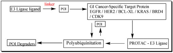
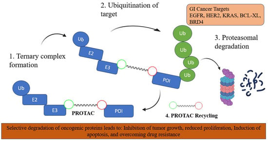
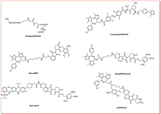
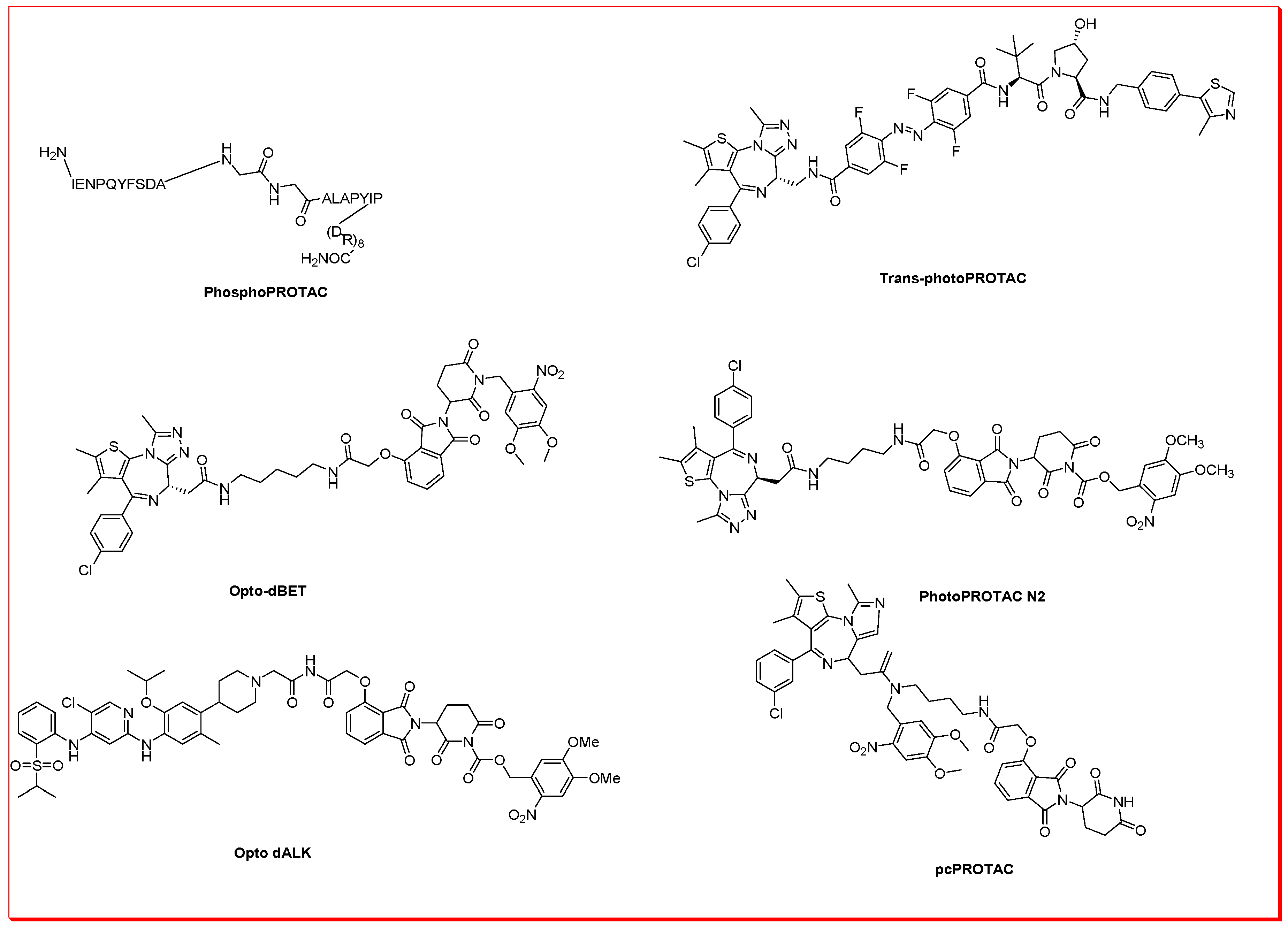
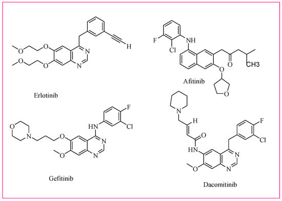
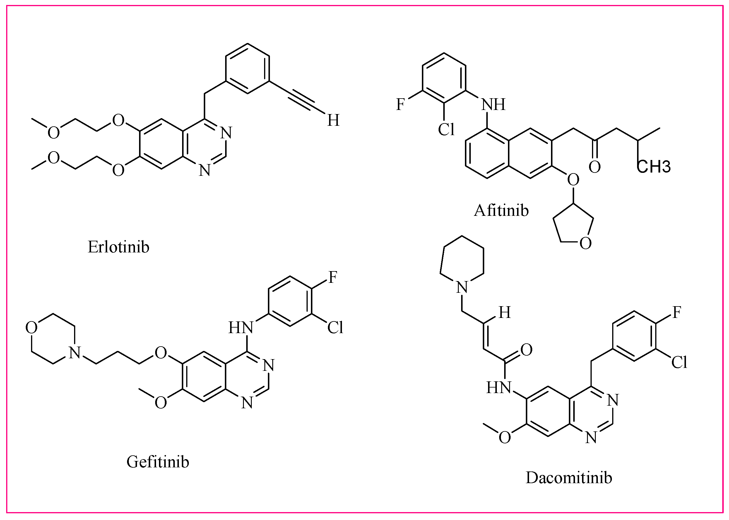
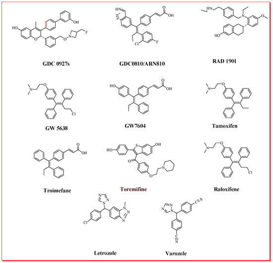
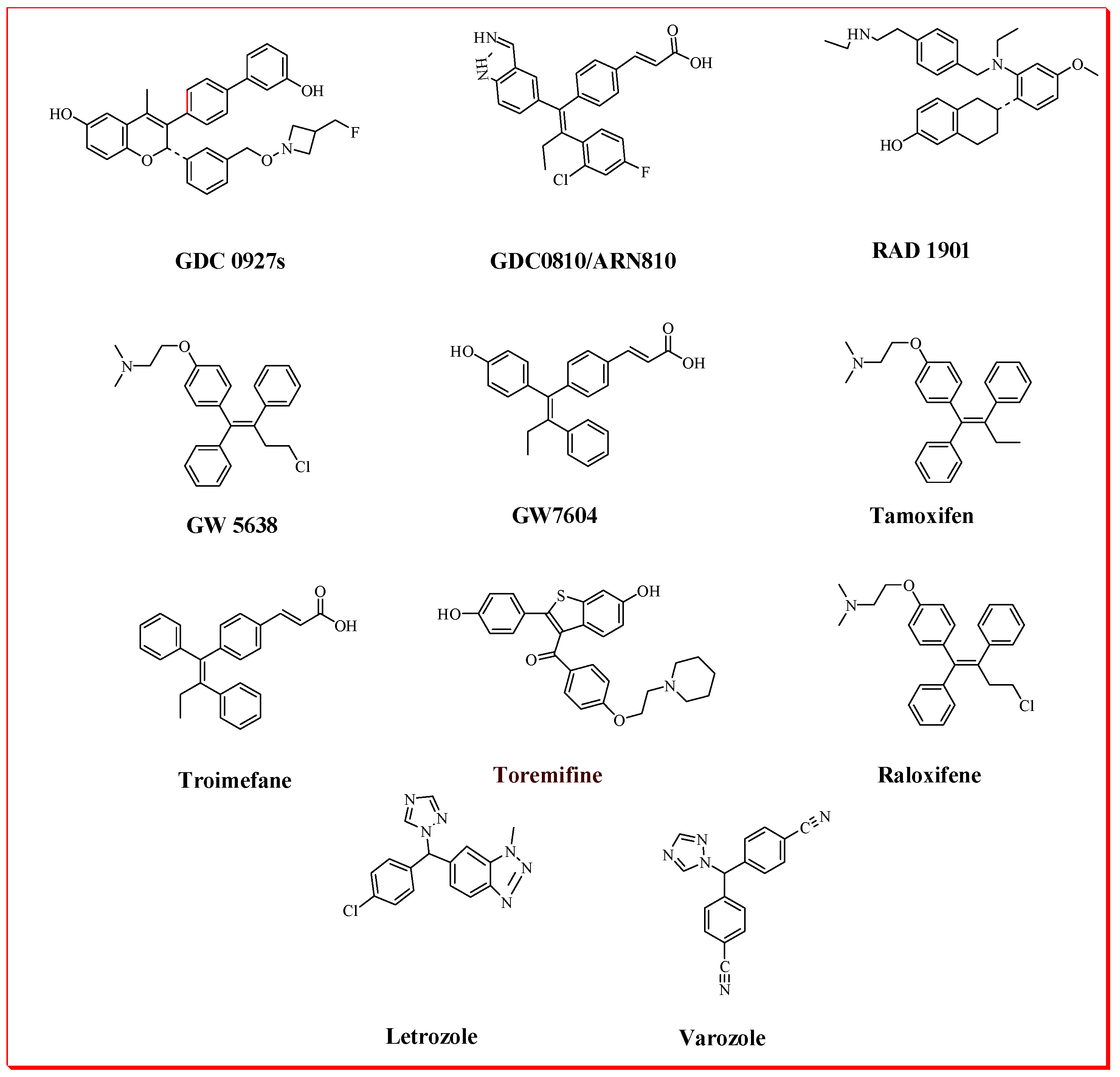
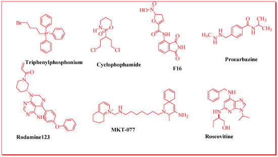
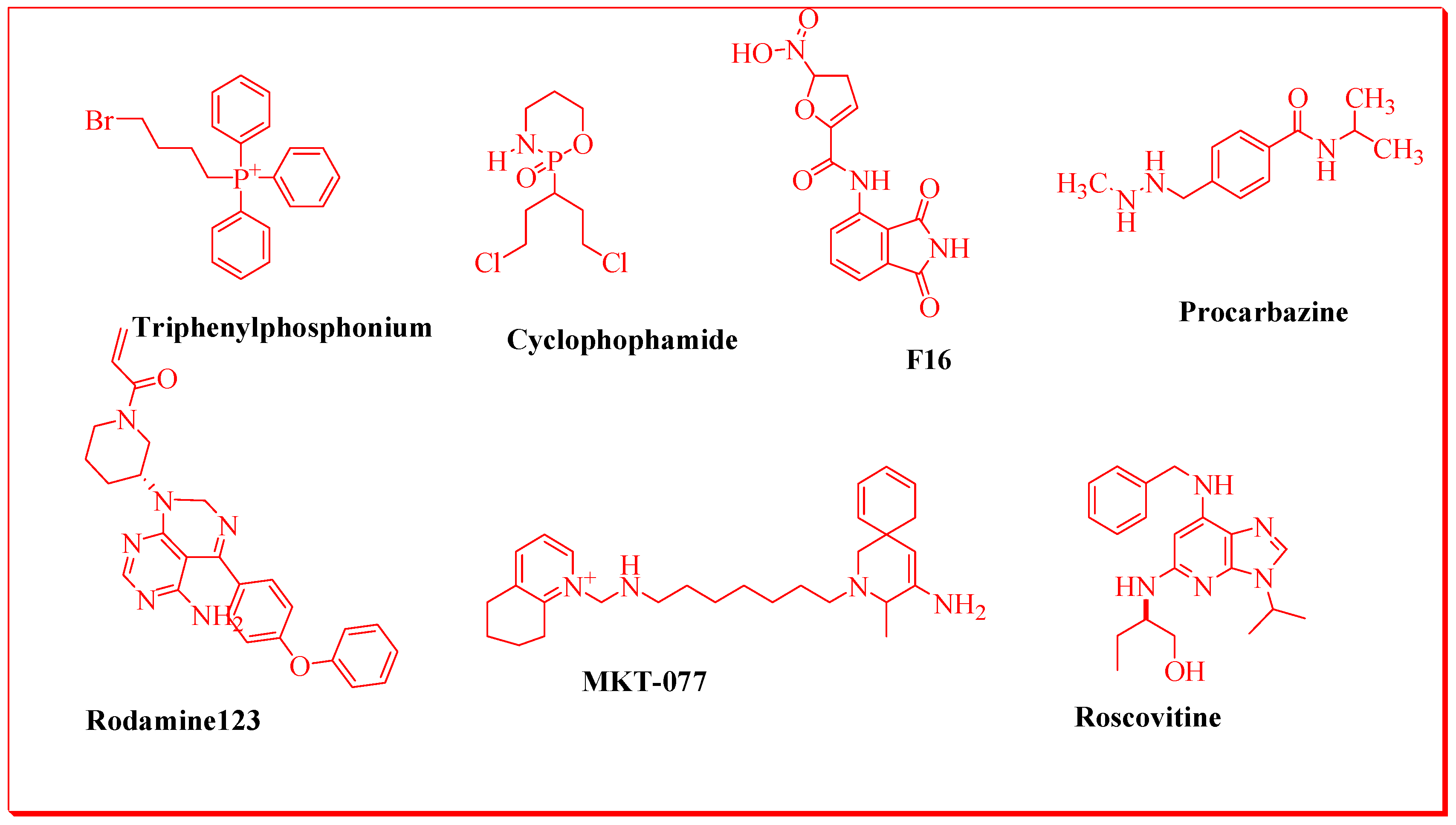
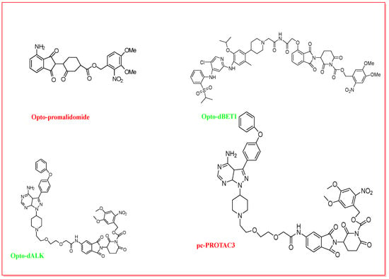
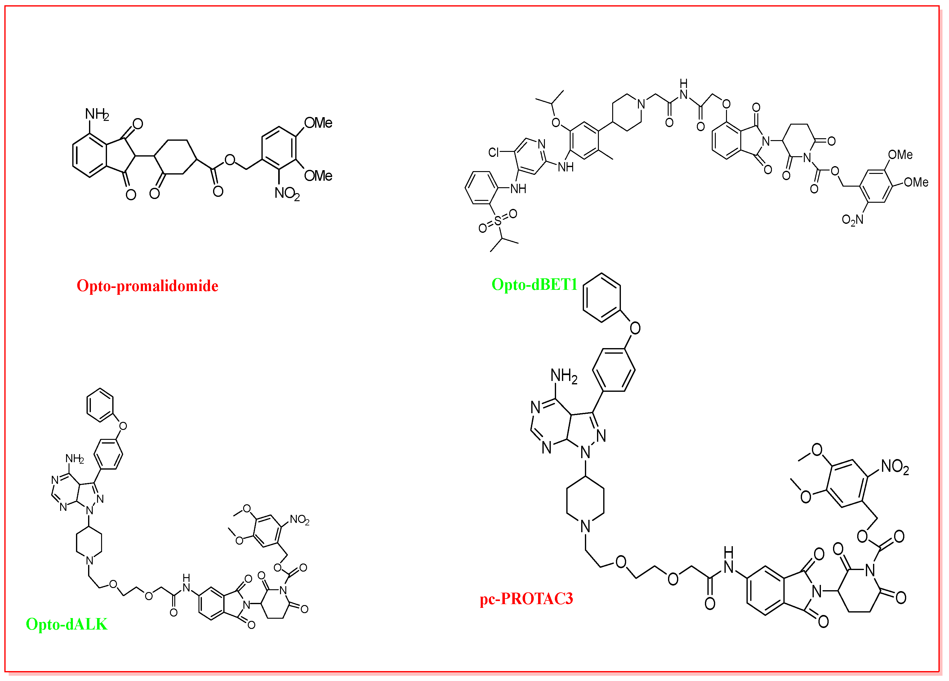
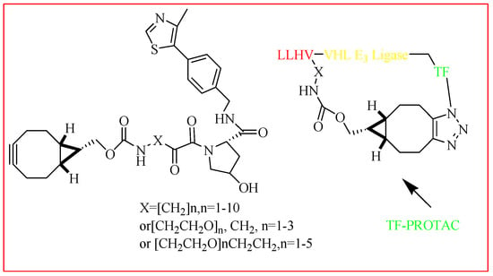
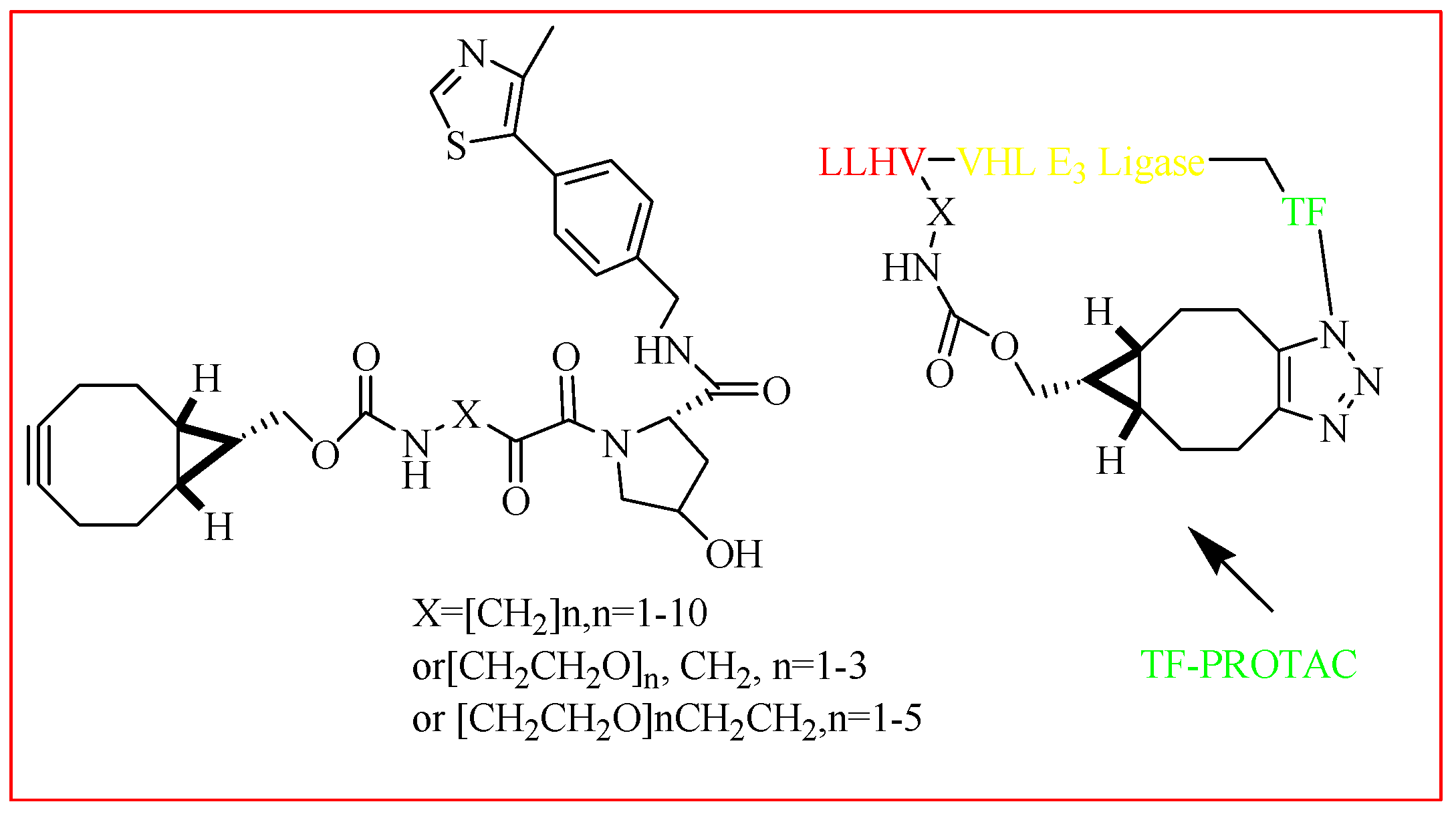
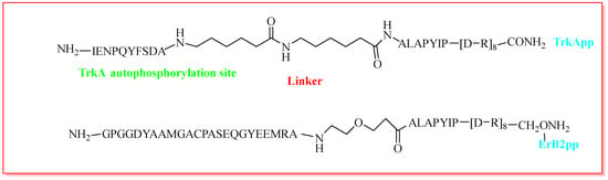
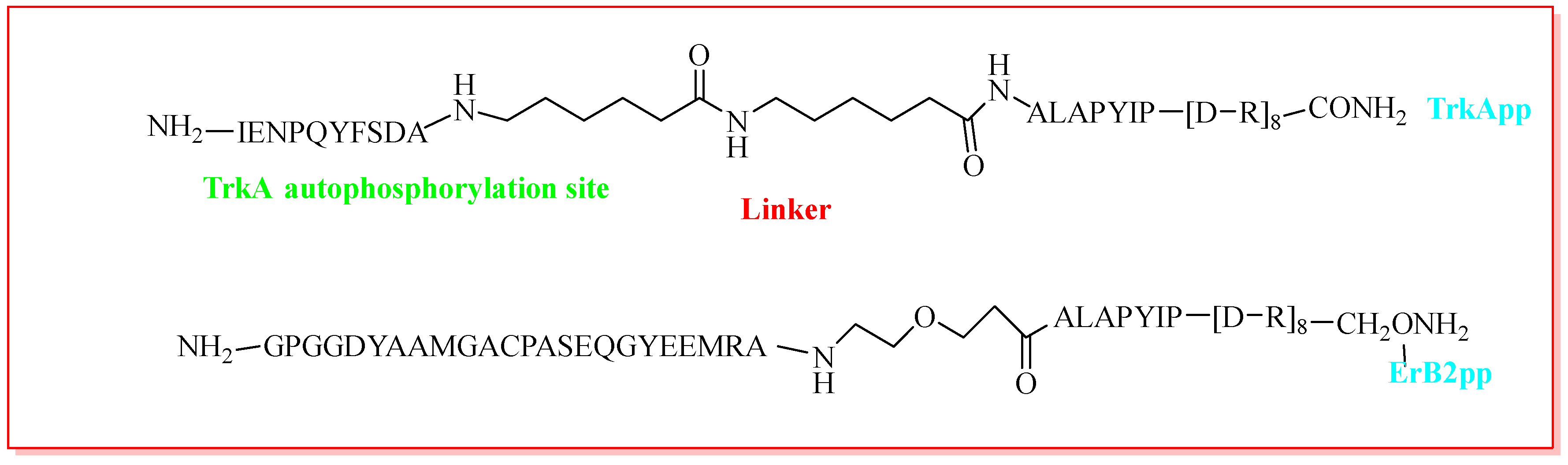
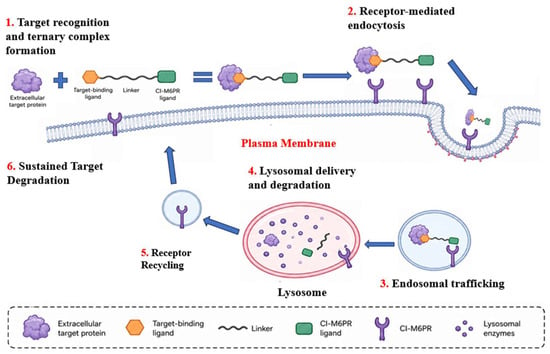
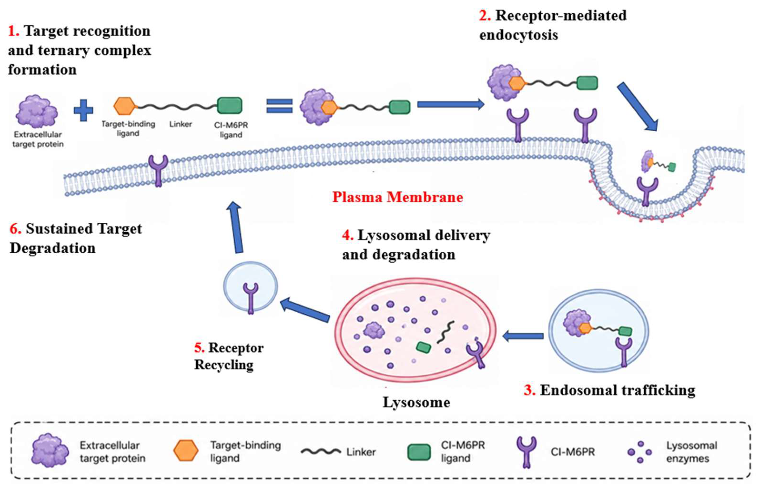
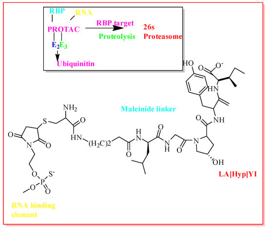
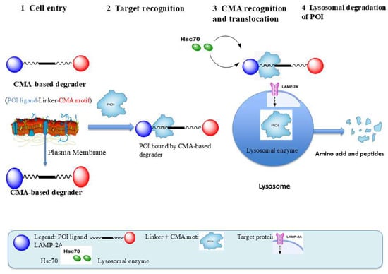
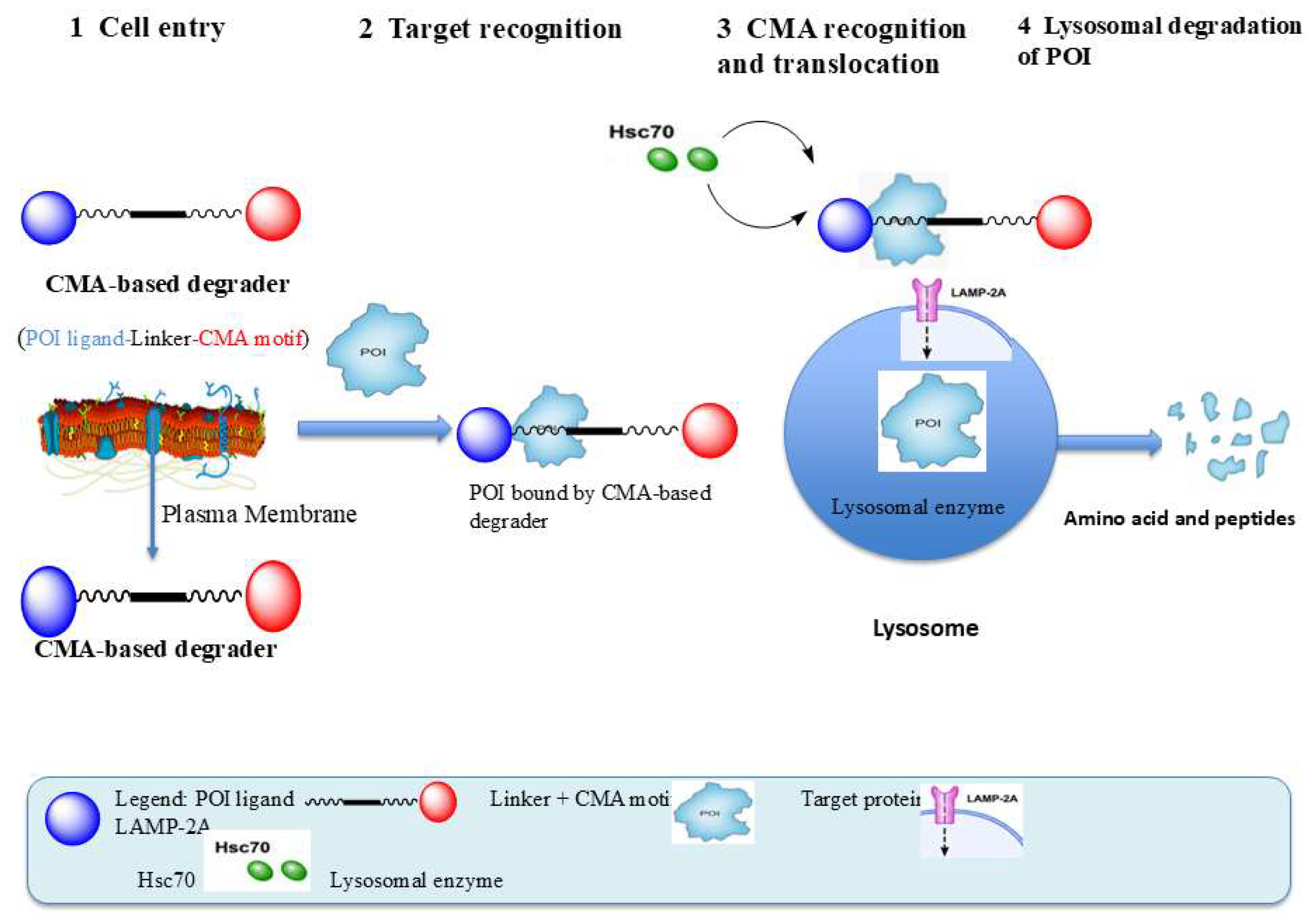
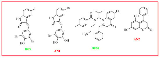
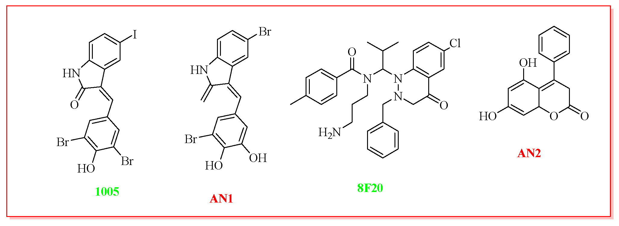
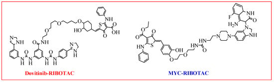
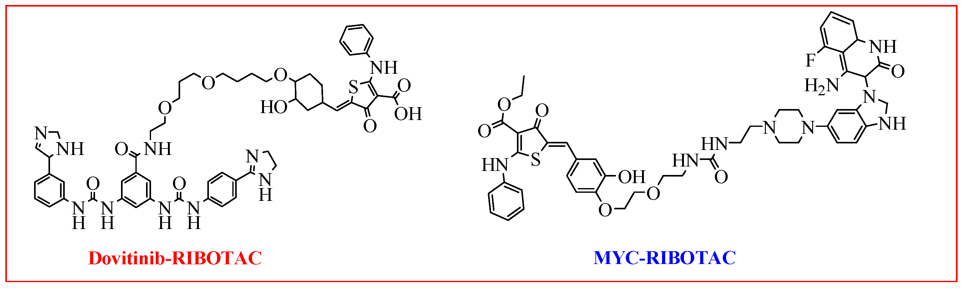
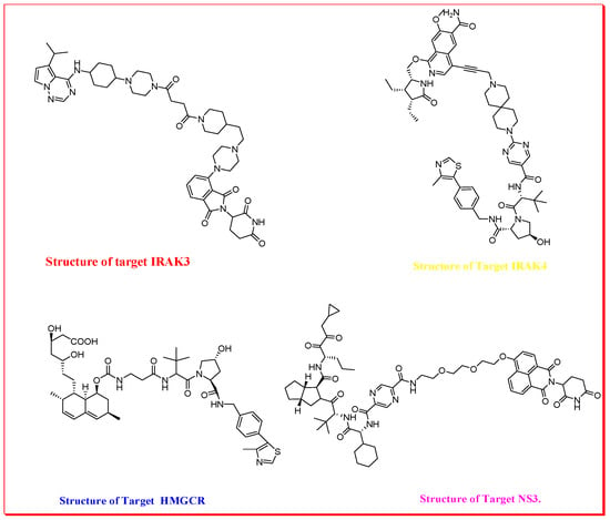
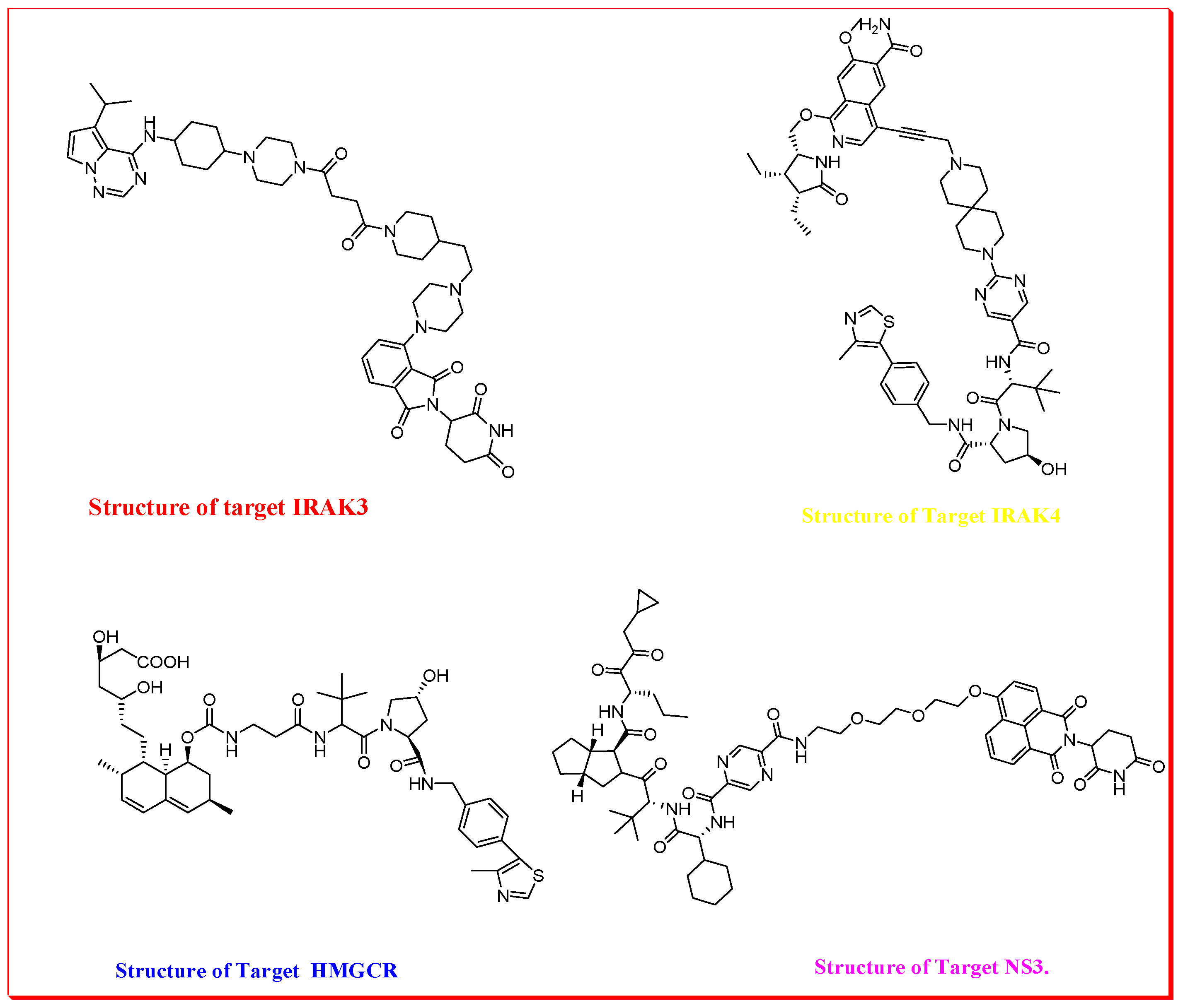

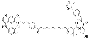
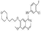
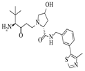
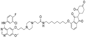
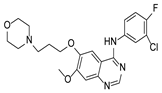
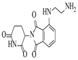
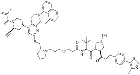
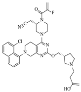
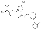
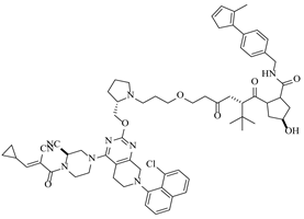
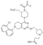
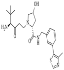
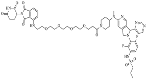
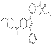
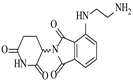
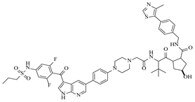
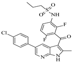
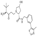
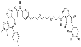
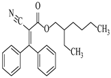
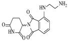
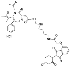
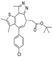
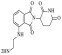
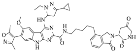
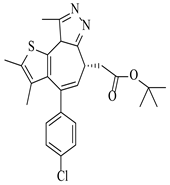
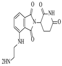
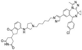
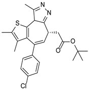
Disclaimer/Publisher’s Note: The statements, opinions and data contained in all publications are solely those of the individual author(s) and contributor(s) and not of MDPI and/or the editor(s). MDPI and/or the editor(s) disclaim responsibility for any injury to people or property resulting from any ideas, methods, instructions or products referred to in the content. |
© 2026 by the authors. Licensee MDPI, Basel, Switzerland. This article is an open access article distributed under the terms and conditions of the Creative Commons Attribution (CC BY) license.