


Abstract
Seminaphtofluorones (SNAFRs) are a family of benzannulated xanthene dyes that exhibit strong fluorescence in both neutral and anionic states and can reach emission wavelengths in the deep-red to near-infrared region. Their optical response is highly sensitive to regioisomerism and functionalization, making them attractive candidates for systematic structure–property investigations. Here, we computed the photophysical properties of six SNAFR regioisomers for both neutral and anionic species and correlate the calculated results with available experimental data. From the six dyes, we further chose two of them, SNAFR4 and SNAFR6, to further investigate how phenyl-ring functionalization modulates SNAFR properties by introducing methyl (–CH3) and carboxyl (–COOH) substituents at the ortho (o), meta (m), and para (p) positions. The calculations indicate that substitution induces measurable changes in geometries, as well as in excitation and emission energies, with particularly pronounced effects for the anionic derivatives. Overall, these results provide a computational framework for the rational tuning of SNAFRs’ optical properties and the design of derivatives with tailored optical characteristics for fluorescence imaging and applications in photodynamic therapy.
1. Introduction
Among the most important properties of fluorophores are the emission wavelength and the Stokes shift, which largely determine whether a dye can be used within a given spectral window and for fluorescence imaging [1].
Fluorophores with large Stokes shifts are particularly desirable in fluorescence imaging because they minimize self-absorption, thereby improving the signal-to-noise ratio, reducing interference from autofluorescence, and enabling deeper tissue imaging [2]. Moreover, long wavelength emitters are attractive in a wide range of applications in biological imaging, environmental sensing, and photodynamic therapy [3]. In particular, fluorescence in the near-infrared (NIR) region is highly advantageous for biological applications because radiation in this spectral region offers practical advantages for bioimaging due to improved tissue penetration and reduced background autofluorescence.
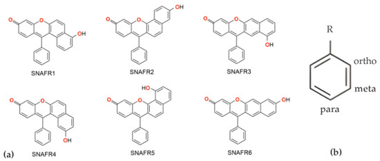
Seminaphtofluorones (SNAFRs) are benzannulated xanthene dyes developed as water-soluble, low-molecular-weight and low-cytotoxicity dyes [4]. Six SNAFR regioisomers have been reported, differing in the benzannulation pattern of the xanthene core (Scheme 1) [4,5]. Unlike most xanthenes, both the neutral and anionic forms of SNAFRs are fluorescent, and the anionic species can emit in the deep-red to near-infrared (NIR) region, making them highly attractive for biological imaging, environmental sensing, and photodynamic therapy [3]. Their photophysical properties, including absorption/emission wavelengths, extinction coefficients, Stokes shifts, lifetimes, and the degree of charge-transfer character, are strongly modulated by regioisomerism and substitution pattern [6,7,8]. Substituent effects such as electron donation or withdrawal can reshape the electronic structure, reorganization energy, and radiative lifetime [9].
Scheme 1.
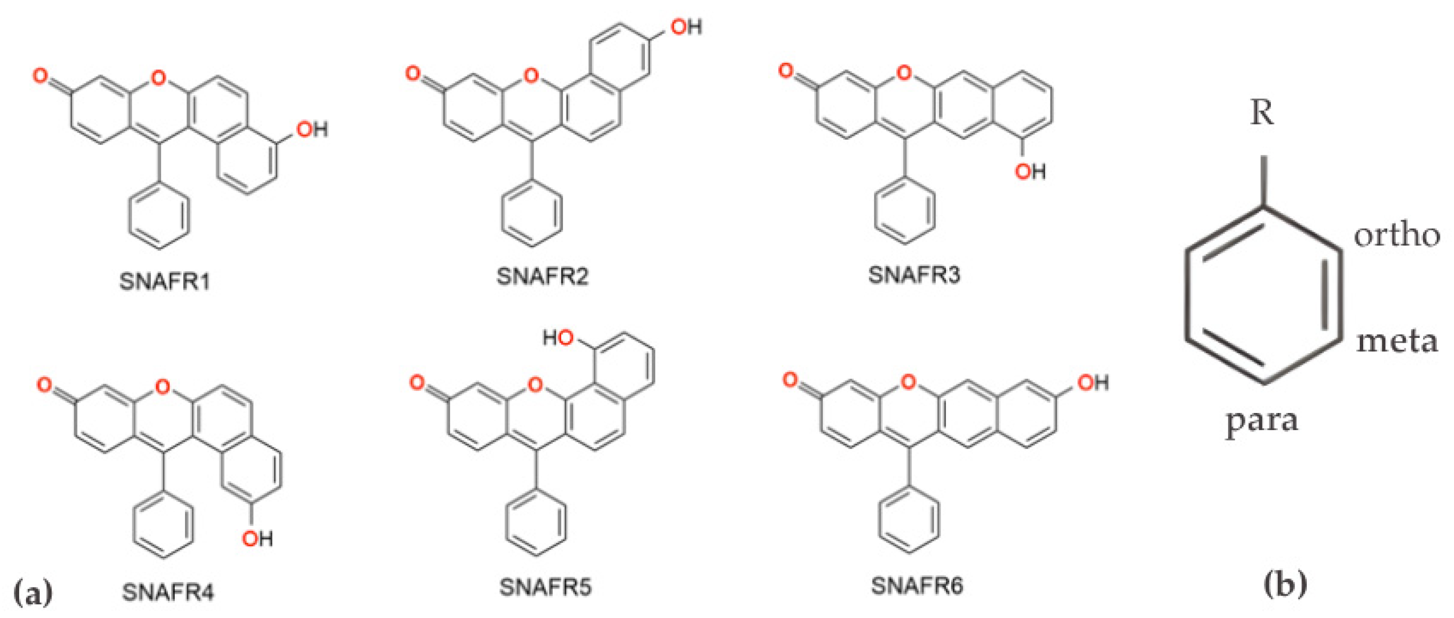
(a) Schematic illustration of the six investigated SNAFR regioisomers and (b) representation of the substituent placement.
In particular, the position of the hydroxyl group within the xanthene framework plays an important role in determining emission behavior [10].
The present study has two goals. First, we correlate the available experimental data with the calculated photophysical properties of six SNAFR regioisomers reported in ref. [4], considering both neutral and anionic species. For this purpose we employed time-dependent density functional (TD-DFT) methods and simulated vibronically resolved spectra using the Franck–Condon Herzberg–Teller (FCHT) approximation. Second, we investigate computationally how phenyl-ring functionalization affects the structural and photophysical properties of selected SNAFR molecules.
For this study we selected two representative substituents—an electron-withdrawing carboxyl group (COOH) and an electron-donating methyl group (CH3)—and examined computationally the effects of substitution at the ortho (o), para (p) and meta (m) positions of the phenyl ring. These substituents were chosen to modulate fluorophore solubility: COOH is expected to enhance water solubility and reduce solubility in non-polar solvents, whereas CH3 is expected to have the opposite effect.
Computational spectroscopy methods are highly valuable for such investigations due to their predictive power and because these approaches can simultaneously incorporate multiple factors in the modeling, such as solvent effects, pH effects (neutral, protonated, or deprotonated species), chemical functionalization, and fluorophore self-assembly, among others. In addition, they can predict structural parameters of electronically excited states, excitation-induced electron density redistribution, the vibronic structure of absorption and emission spectra, and radiative fluorescence lifetimes. These are the main reasons why we undertook the present computational study, aiming to validate computational protocols on the SNAFR systems, for which a substantial body of experimental photophysical data is available [4,5].
In this study we address two interconnected goals: first, benchmarking calculated photophysical properties of neutral and anionic SNAFRs against experiment, and second, using the validated protocol to investigate substituent effects via phenyl-ring functionalization. We therefore computed the photophysical properties of all six derivatives shown in Scheme 1, in both neutral (ne) and anionic (an) forms. To assess functionalization effects, we initially screened all derivatives; however, in this study we report functionalization results only for SNAFR4 and SNAFR6, as these unsubstituted systems showed the best agreement between the calculated and experimental data.
2. Materials and Methods
Electronic structure calculations were performed using Density Functional Theory (DFT) methods for ground-state (GS) optimization and Time-Dependent DFT (TD-DFT) for excited-state (ES) properties. The B3LYP hybrid functional [11,12,13] was used throughout, in combination with 6-311+G(d,p) basis set.
B3LYP hybrid functional was selected because it is widely used in DFT studies and has been shown to provide reliable performance for both DFT and TD-DFT calculations [14], including simulations of vibrationally resolved spectra, albeit with a modest tendency to red-shift peak positions in some cases [15,16,17,18]. The 6-311+G(d,p) basis set was chosen as a practical compromise between the size of the investigated molecules and the computational cost required to evaluate the targeted molecular properties [19].
All six molecules were investigated in both neutral and anionic forms in DMSO solution, considering the GS and the first ES, targeting properties such as: optimized geometries, vibrational frequencies, electronic spectra (absorption and emission).
Solvent effects were treated using the Polarizable Continuum Model (PCM). Tight and very tight criteria were used for the convergence of the molecular geometries and SCF calculations, respectively, and the ultrafine grid was employed for the numerical integration of the electronic density. For each optimized structure, the dihedral angle between the phenyl substituent and the xanthene core was extracted to quantify conformational changes upon excitation.
Vibrationally resolved one-photon absorption (OPA) and one-photon emission (OPE) spectra were computed following the procedure [20,21]. Briefly, GS and ES geometries were optimized and harmonic vibrational frequencies were computed for both states at the PCM(DMSO)-B3LYP/6-311+G(d,p) level of theory. The resulting normal modes and frequencies were then used to evaluate vibronic intensities within the Franck–Condon Herzberg–Teller (FCHT) approximation for the transition dipole moment. Unless stated otherwise, the Adiabatic Hessian (AH) model was used to describe the equilibrium geometries of each electronic state. In a few cases, the AH approach yielded weak vibronic progressions; therefore, the Vertical Hessian (VH) model was used for the SNAFR1 anion and the Vertical Gradient (VG) model for the SNAFR4 anion. All FCHT spectra have been calculated at 0 K.
To reproduce the experimental vibronic band shapes as closely as possible, Gaussian broadening was applied with half-width at half-maximum (HWHM) of 400 cm−1 for all neutral species, as well as for the SNAFR3 and SNAFR4 anions. For the remaining anions, broader line shapes were required: HWHM = 800 cm−1 for the SNAFR1, SNAFR2, and SNAFR6 anions, and HWHM = 600 cm−1 for the SNAFR5 anion.
The anionic species were generated by deprotonating the hydroxyl group.
Radiative lifetime has been calculated as:
where c is the speed of light, ΔE is the S0 ⟶ S1 transition energy and is the transition dipole moment strength [22].
Whenever available, computed properties were compared with the corresponding experimental values. All calculations were performed with Gaussian 16, Revision C.01 software package [23].
3. Results and Discussion
The experimental spectra of the six SNAFR derivatives in both the neutral and anionic forms were reported by Yang et al. [4]. A summary of these data, together with the calculated counterparts, is given in Table 1 and Table 2.
Table 1.
Calculated and experimental photophysical data for the six neutral SNAFR derivatives.
Table 2.
Calculated and experimental photophysical data for the six anionic SNAFR derivatives.
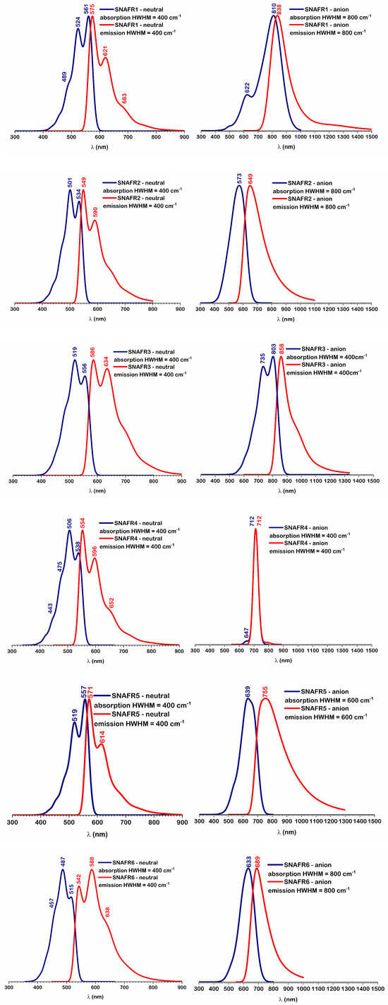
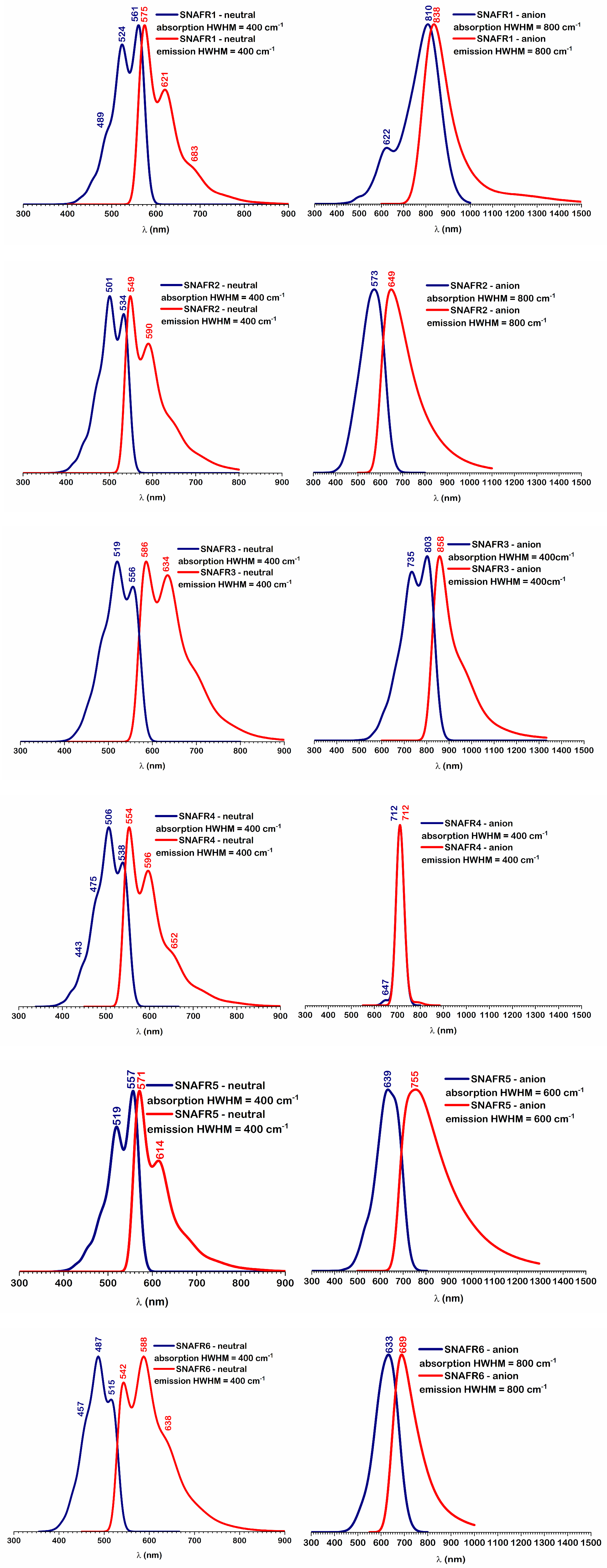
Vibrationally resolved one-photon absorption and one-photon emission spectra were computed for all derivatives in both neutral and anionic forms within the Franck–Condon Herzberg–Teller (FCHT) framework. The resulting spectra are shown in Figure 1. Gaussian broadening was applied using HWHM values selected to best reproduce the experimental line shapes (see Computational Details).
Figure 1.
Calculated absorption (blue curves) and emission (red curves) spectra (normalized to unity) of the neutral (left side) and anionic (right side) forms of the investigated unsubstituted molecules.
Comparing the TD-DFT vertical absorption wavelengths for the neutral species (first row in Table 1) with the middle experimental absorption peak (typically the most intense), we found that, excepting SNAFR6, the calculated wavelengths are blue-shifted. The deviations range from 34 nm for SNAFR4 to 12 nm for SNAFR 5.
Comparing the experimental spectrum with the spectrum computed using the FCHT approach shows not only a 5.2 nm reduction in the MAE (from 19.8 to 14.6 nm), but also perfect agreement in the ordering of the absorption wavelengths between experiment and theory.
A similar underestimation is observed for the TD-DFT vertical emission wavelengths, with deviations ranging from 41 nm for SNAFR5 to 18 nm for SNAFR6, yielding an MAE of 28.8 nm.
The experimental trend is largely reproduced, with a single inversion between SNAFR5 and SNAFR1:
If the calculated values are compared to the first peak of the experimental spectrum, the same ordering is preserved and the agreement improves substantially, with the MAE reduced to 12.8 nm.
When the TD-DFT vertical emission wavelengths are instead compared with the first experimental emission peak, which can be associated with the 0–0 transition, the agreement improves markedly: the deviations fall between −13 nm (SNAFR5) and +20 nm (SNAFR6), and the MAE decreases significantly to 12.8 nm.
Finally, if the middle peaks of the experimental spectrum and the calculated time-independent FCHT spectrum are compared, the MAE decreases to 8 nm.
A similar analysis of the anionic forms shows that, for all derivatives, the TD-DFT vertical absorption wavelengths are strongly blue-shifted relative to experiment. The deviations span from −89 nm for SNAFR5 to −39 nm for SNAfR1.
For the five anions with available experimental data, the resulting mean absolute error is 70.2 nm, indicating that the vertical TD-DFT description is substantially less accurate for the anionic species than for the neutral forms.
Despite this systematic underestimation, the relative ordering of the computed absorption wavelengths reproduces the experimental trend:
For vertical emission, the discrepancies are even larger: the calculated wavelengths differ from experiment by +178 nm for SNAFR1 and −68 nm for SNAFR5, with an overall MAE of 92.0 nm. This unacceptably large error is dominated by the SNAFR1 and SNAFR4 anions, indicating that the vertical TD-DFT description is not reliable for these species under the present computational setup.
In summary, the TD-DFT vertical absorption and emission data show acceptable agreement with experiment for the neutral derivatives, whereas for the anionic species the deviations are substantially larger, and the vertical TD-DFT approach alone does not provide a quantitatively adequate description.
However, when the calculated vibronic spectra are compared with the experimental spectra for these two anions, the discrepancies decrease markedly—to 70 nm for SNAFR1 and 18 nm for SNAFR4.
For the entire set, comparing the FCHT-calculated peak positions with the experimental spectra significantly improves the agreement, reducing the MAE to 35.4 nm for absorption and 36.4 nm for emission. Moreover, in contrast to vertical transitions, vibrationally resolved simulations can reproduce not only peak positions but also the overall band shapes.
It is well known that molecular electronic excitation energies are often underestimated by hybrid functionals, leading to calculated transition wavelengths that are red-shifted by a few tens of nanometers [24,25].
On the other hand, the range-separated density functionals provide significantly better values for some molecules [26,27], like those from the QuestDB benchmark set [28]. Among the hybrid GGA functionals tested by Liang et al. [29], from that set, the B3LYP functional gives the worst results, while ωB97X-D and cam-B3LYP are in first and third place, respectively. However, for the seminaphthofluorone series examined here, exemplified for SNAFR4 and SNAFR6 derivatives, the B3LYP functional provides the best excitation energies compared to ωB97XD, cam-B3LYP and LC-BLYP (see Table 3). This behavior underscores the system dependence of TD-DFT accuracy and the need to benchmark functionals against experimental data for the specific chromophore class under study.
Table 3.
Calculated excitation energies (in nm) to the first excited state for the neutral and anionic forms of SNAFR4 and SNAFR6 regioisomers, compared to experimental data.
The larger discrepancies obtained for the anionic species likely arise from a combination of structural, vibronic, and environmental effects:
- Enhanced conformational reorganization upon excitation (phenyl-ring torsion)
The phenyl–xanthene core dihedral angle (Φ) changes modestly between S0 and S1 for the neutral derivatives (6.6° for SNAFR1 to 11.6° for SNAFR2), whereas for the anions the corresponding changes are substantially larger (12.8° for SNAFR6 up to 29.1° for SNAFR4). The effect is particularly pronounced for SNAFR1 and SNAFR4 (≈25–29°), consistent with reduced steric constraints upon deprotonation. Such larger torsional reorganization increases sensitivity to the excited-state minimum and can strongly affect computed absorption/emission energies.
- Challenges in treating vibronic coupling for low-frequency, large-amplitude modes
The vibronic spectra of SNAFR1 and SNAFR4 anions were not reproduced satisfactorily using the Adiabatic Hessian (AH) model. A plausible contributor is the presence of very-low-frequency, large-amplitude phenyl-ring rocking modes (40.30 cm−1 for SNAFR1 and 13.38 cm−1 for SNAFR4). Such floppy modes are difficult to describe within harmonic-based vibronic formalisms and can make the predicted vibronic progression strongly model-dependent (AH vs. VH vs. VG), thereby increasing the uncertainty in peak positions and band shapes.
- Solvation model limitations for charged chromophores
Continuum solvation (PCM) captures bulk polarization but does not explicitly represent local solvent structuring around the deprotonated site. For anions, specific short-range interactions can differentially stabilize S0 and S1 states, shifting transition energies by tens of nanometers.
- Counterion and ion-pairing effects
In experimental conditions, anions are commonly associated with counterions and may form contact or solvent-separated ion pairs. Ion pairing can alter the electrostatic environment and stabilize one electronic state more than the other, leading to appreciable shifts in absorption and/or emission wavelengths.
- Hydrogen bonding to the anionic site
Deprotonated species can form strong hydrogen bonds that significantly perturb electronic structure and reorganization. Such interactions can stabilize either the ground or excited state and thus shift the spectra.
- Greater electronic reorganization and state relaxation after deprotonation
Deprotonation increases charge delocalization and can lead to larger excited-state structural relaxation compared with neutral derivatives. This amplifies sensitivity to excited-state optimization, to the chosen solvation treatment, and to the vibronic model used to relate vertical/adiabatic energies to the spectral maximum.
- Possible increase in charge-transfer (CT) character for anions
The anionic forms may have a larger intramolecular CT component, which is more challenging for the TD-DFT approach. However, in the present dataset of molecules, range-separated functionals do not systematically improve agreement (see Table 3), suggesting that CT-related functional error is unlikely to be the dominant factor for these anions under the current conditions.
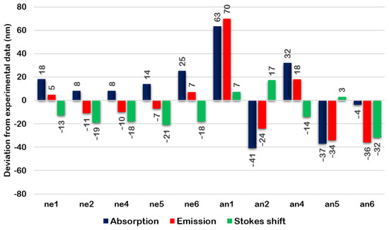
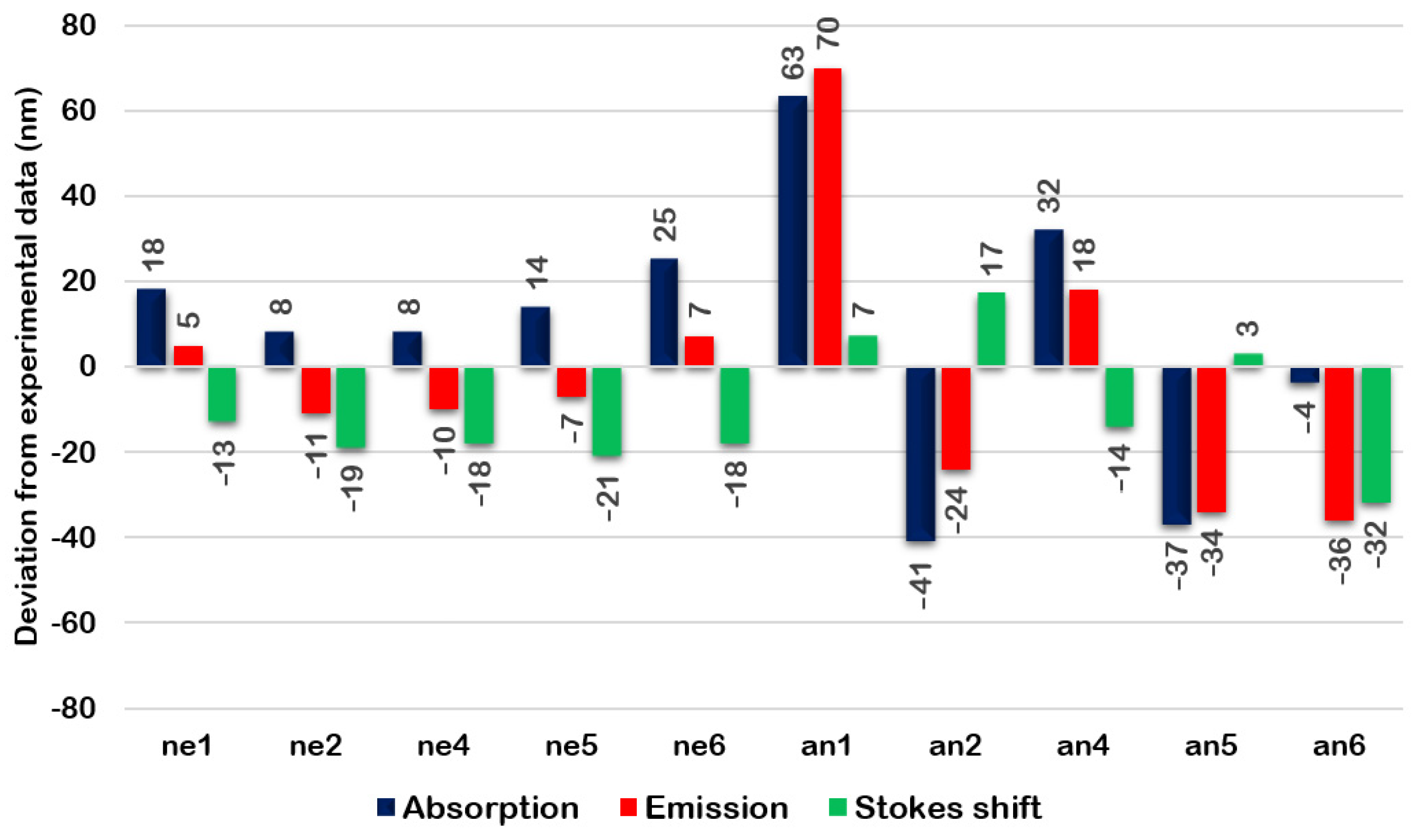
As summarized in Figure 2, both for the neutral and anionic species, the vibrationally resolved spectra yield substantially better agreement with experiment than the vertical TD-DFT values. Thus, for neutral species the mean absolute errors (MAEs) are 14.6 nm for absorption and 8.0 nm for emission wavelengths, while for anions the corresponding values are 35.4 and 36.4 nm.
Figure 2.
Difference (in nm) between the calculated vibronic and experimental data for the neutral (NE) and anionic (AN) forms of the investigated molecules. Data for SNAFR3 are missing because of the lack of experimental data for this derivative.
Experimentally, the largest Stokes shift (reported as Δ in cm−1) is observed for SNAFR6, and this finding is reproduced by the calculations. With the exception of SNAFR5, the calculated Stokes shift ordering follows the experimental trend:
Thus, all the neutral forms emit in the red spectral region and the largest Stokes shift corresponds to SNAFR6, followed by SNAFR 2 and SNAFR5, in line with the computational calculations.
It is worth mentioning that for the compounds studied here, an experimental quantum yield is available only for neutral SNAFR6 (Φ = 9%) which corresponds to an experimental fluorescence lifetime of 4.08 ns. The calculated radiative lifetime for this derivative is 48.14 ns; combined with the experimental quantum yield Φ, this implies a total fluorescence lifetime of 4.33 ns, in very good agreement with experiment. To our knowledge, no additional literature data on radiative or total fluorescence lifetimes are available for these SNAFR derivatives.
Larger discrepancies are observed for the anionic species, most notably for SNAFR1, for which the Vertical Hessian (VH) model was required to obtain a satisfactory vibronic progression. For the anions, the MAEs are 35.4 nm for absorption and 34.4 nm for emission. The Stokes shifts are reproduced with more reasonable accuracy, with an MAE of 14.6 nm.
It is important to note that, within the FCHT framework, we recover weak absorption features for the SNAFR1 and SNAFR4 anions, calculated at 622 nm and 647 nm, respectively, with experimental counterparts reported at 680 nm and 625 nm. Moreover, ref. [4] indicates that the SNAFR3 anion is expected to exhibit the longest wavelength absorption among the series; our calculations are consistent with this trend and predict a markedly red-shifted spectrum, including an emission wavelength of 858 nm.
Useful information for characterizing electronic excitation transitions is their nature (transition character). This information can be obtained either from an analysis of the frontier molecular orbitals (primarily HOMO and LUMO), or from difference density plots that compare the electron density in the excited and ground states. Both approaches were employed in the present work. All ground-to-excited-state transitions considered here are dominated by HOMO → LUMO excitations.
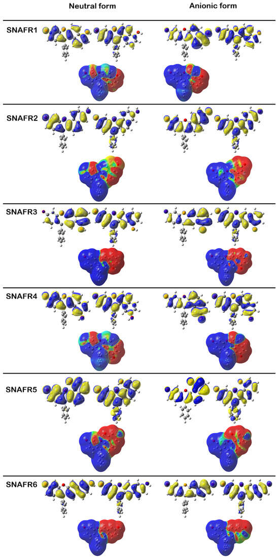
Since the lowest-energy electronic transition for all derivatives, in both neutral and anionic forms, corresponds to S0 → S1, the orbital-based analysis focuses on the HOMO and LUMO shown in Figure 3.
Figure 3.
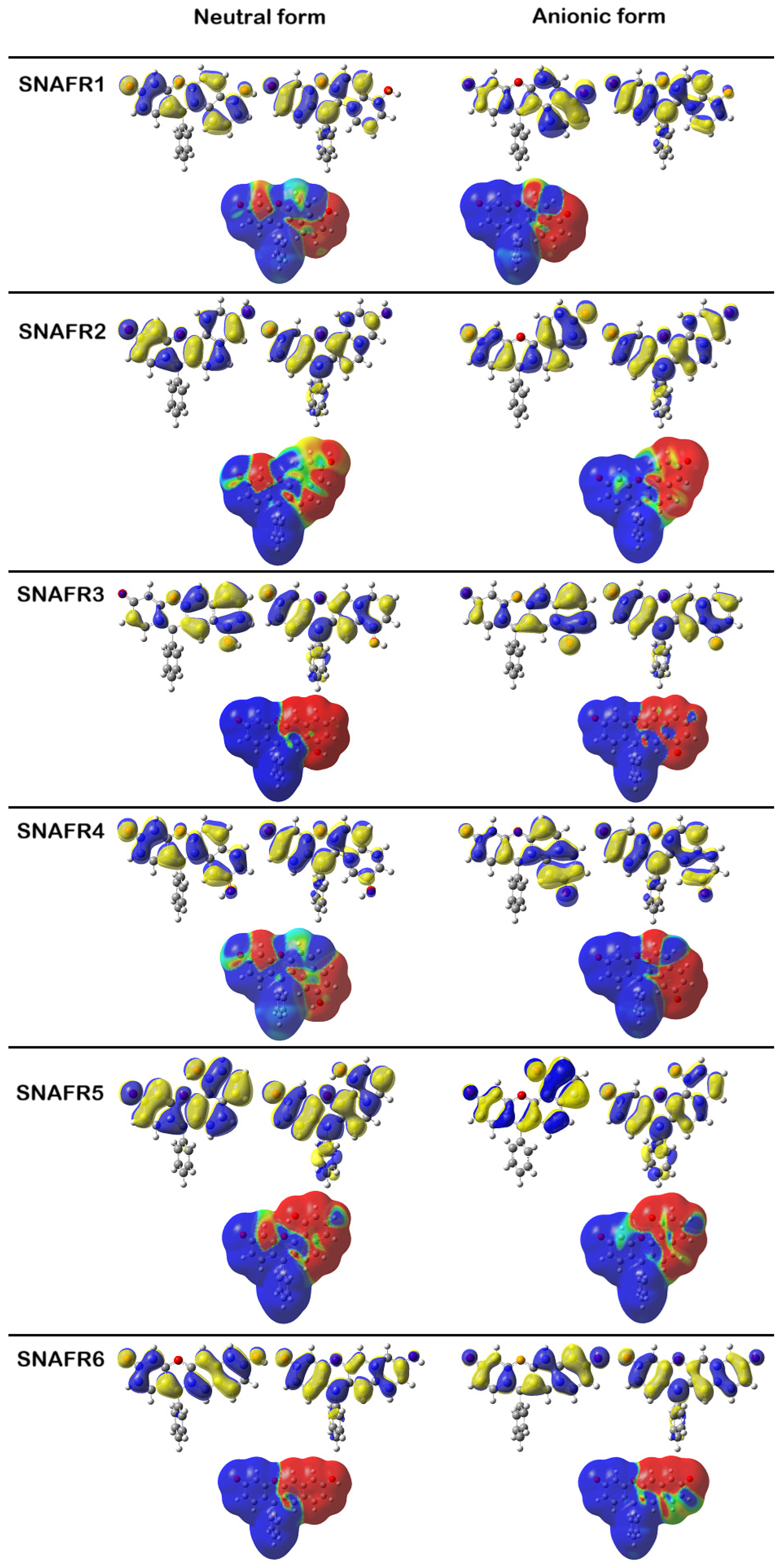
Frontier molecular orbitals (HOMO and LUMO) and excited–ground state electron density difference maps for the SNAFR derivatives in neutral and anionic forms, calculated at the PCM(DMSO)-B3LYP/6-311+G(d,p) level of theory. For each derivative, the HOMO (left) and LUMO (right) are shown in the upper panels, and the corresponding electron density difference map is shown in the lower panel. The blue (red) areas denote an increase (depletion) in electronic density in the excited state.
For both charge states, the HOMO has predominantly π character and is delocalized over the main chromophoric framework comprising the xanthene moiety and the additional fused ring. Particularly, the HOMO shows negligible contribution from atomic orbitals localized on the phenyl ring.
For the HOMO of neutral and anionic SNAFR3, a small contribution arises from atomic orbitals localized on the ring at the far left of the xanthene unit. In contrast, for the anionic HOMOs, the contribution from the oxygen atom in the central xanthene is nearly negligible.
With regard to the LUMO orbitals, an evident feature is the contribution from atomic orbitals centered on the phenyl group. As shown in Figure 3, the largest phenyl group contribution, regardless of charge state, is observed for SNAFR5. Along with the delocalization of the LUMO over the phenyl group, all derivatives (neutral and anionic) display a reduced LUMO contribution on the ring annulated to the xanthene framework, suggesting decreased excited-state electron density in that region.
This effect is also observed in the differences between the electron density corresponding to the excited state and that corresponding to the ground state. The total SCF electronic density isosurface (0.02 a.u.) is mapped with electronic density differences between the S1 singlet excited state and the ground state of the six SNAFR derivatives in the neutral and anionic forms. Blue regions indicate an increase in electron density upon excitation, whereas red regions denote electron density depletion in the excited state.
In the next section we address the phenyl group substitution of SNAFR4 and SNAFR6 derivatives to understand how such substitutions to the ortho (o), meta (m) and para (p) positions with an electron-donating methyl group (CH3) and an electron-withdrawing carboxyl group (COOH) influences their structural and photophysical behavior. We investigated all the derivatives but, in the following discussion, only selected SNAFR4 and SNAFR6 for further investigation because they display a consistently high Stokes shift, reaching the highest value among the neutral derivatives, while maintaining relatively large oscillator strengths in both neutral and anionic forms.
We consider both electronic states, ground state (GS) and excited state (ES), as well as neutral (ne) and anionic (an) species. An abbreviation example is: neutral form of the SNAFR6 derivative, functionalized with a methyl group in the ortho position = SNAFR6ne-CH3-o.
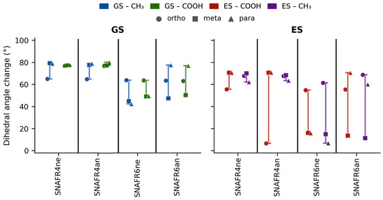
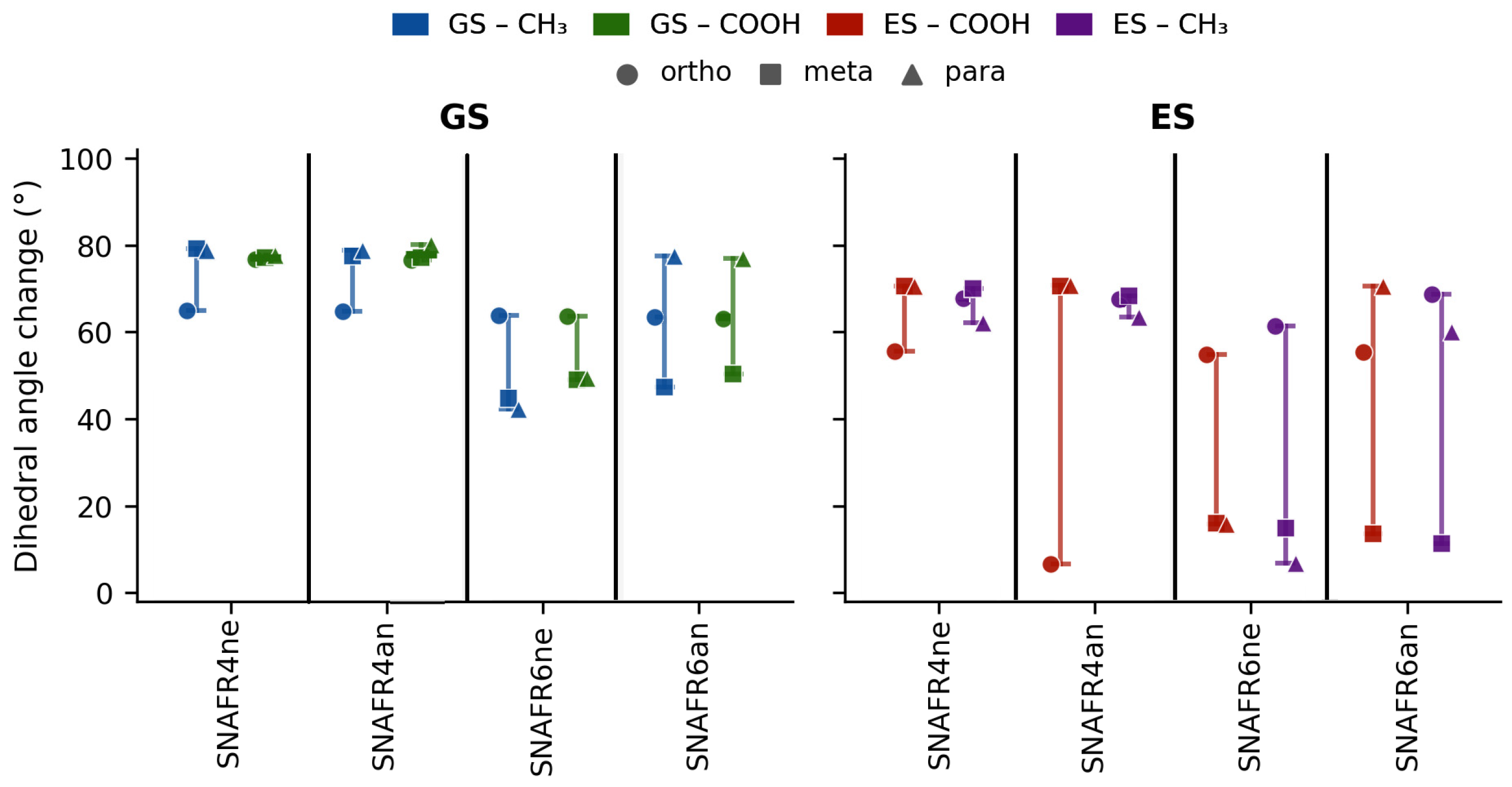
Functionalization of the SNAFR4 and SNAFR6 with CH3 or COOH induces substantial changes in the dihedral angle between the phenyl ring and the mean plane of the xanthene core (θ) relative to the unfunctionalized scaffold in both the neutral and anionic forms (Figure 4).
Figure 4.
Dihedral angle changes (°) between unfunctionalized and functionalized SNAFR derivatives in the ground state (left) and excited state (right). Each data point represents one substitution position: ortho (circle), meta (square), and para (triangle), for either a CH3 or COOH functional group. Vertical bars span the full range (minimum to maximum) of values for each functional group across the three substitution positions.
For SNAFR4ne, functionalization leads to a large Δθ and remains relatively uniform across all substituent types and positions in the GS, the difference compared to the unfunctionalized one ranging from 65° to 79°, while the Δθ for the ES is between 56° and 71°. Between the GS and ES there is minor variation; the average Δθ is below 2°.
SNAFR4an displays similarly large θ rotations where the smallest change is 63° and the largest is 78° compared to the unfunctionalized one in the GS. The angle change in the ES differs between the unfunctionalized and functionalized one from 16° to 49°. It is worth mentioning that the largest change here, in the case of SNAFR4an-CH3-o in the ES, is the only derivative that rotates in the opposite direction compared to the rest. The state change again shows low variation, as in the case of the neutral species; however, SNAFR4an–CH3–o represents the only inconsistency compared to the trends, the jump from GS to ES being 49°.
In contrast, SNAFR6 exhibits a more heterogeneous response overall, with a stronger dependence on substituent type, substitution position, and charge state. For SNAFR6ne in the GS, the Δθ is situated between 34° and 56°. In the ES, the pattern changes: the largest rotation compared to the unfunctionalized molecule is 52°, whereas the smallest is an almost negligible change of 2°. Both extremities can be found in the -COOH functionalized molecule. The average Δθ between the GS and ES is 17°. For SNAFR6an, the GS geometry displays the largest overall Δθ: 71°, while the smallest is 41°, both belonging to derivatives functionalized with -CH3. In the ES, the Δθ minimum response is 1.3°, while the largest rotation is 61°.
In both SNAFR4 and SNAFR6, a clear bathochromic shift is observed when going from the neutral to the anionic species, consistent with the extended conjugation and altered electron density distribution upon deprotonation. SNAFR4 exhibits larger variations in transition energies across the functionalized series, while SNAFR6 shows a more uniform behavior.
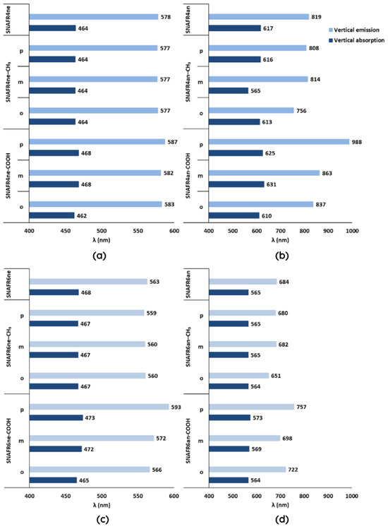
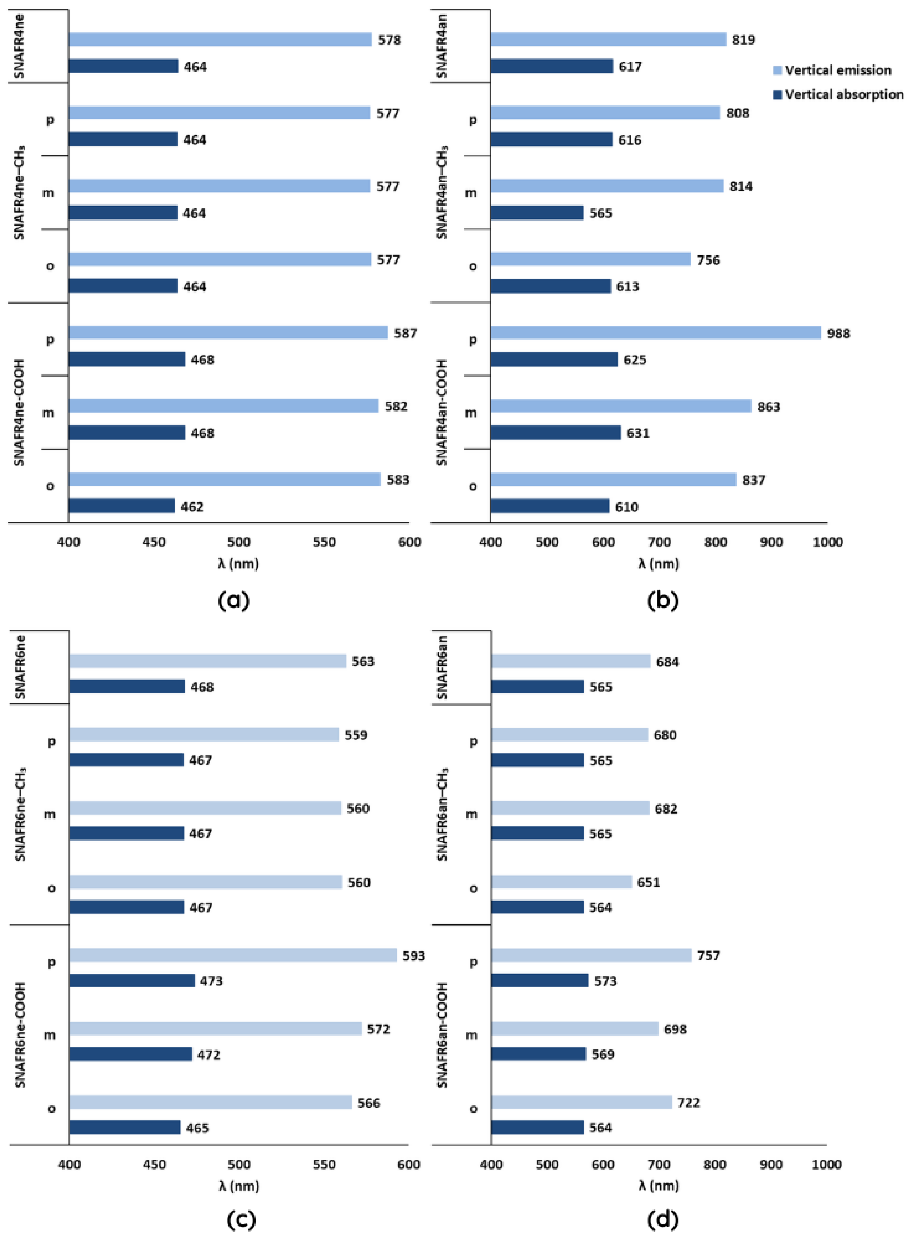
For the neutral derivatives, the transition energies are remarkably insensitive to substitution. The functionalized SNAFR4ne series shows average values of 465 nm (vertical absorption), deviating by less than 3 nm from the unfunctionalized reference (Figure 5a). SNAFR6ne behaves similarly, with the average value being 469 nm, compared to 468 nm for the parent compound (Figure 5c). In both neutral systems, absorption falls in the blue visible region and emission in the yellow/orange region, regardless of substituent identity or position.
Figure 5.
Relative changes in vertical absorption (dark blue) and emission (light blue) transition energies of functionalized SNAFR4 and SNAFR6 derivatives with respect to the corresponding unfunctionalized dye. Results are shown for neutral species (a,c) and anionic species (b,d), with substitution at the o, m, and p positions by CH3 and COOH groups.
The anionic derivatives show a different pattern. For SNAFR4an, functionalization produces a wider spread of transition energies compared to the neutral case, vertical absorption ranges from 565 to 631 nm, and vertical emission from 808 to 988 nm across the functionalized series, all shifted relative to the unfunctionalized anion (617 and 819 nm; Figure 5b). The emission range across the functionalized SNAFR4an series suggest NIR emission for these species with the most pronounced red shift observed for SNAFR4an–p–COOH (988 nm vertical emission). SNAFR6an displays a more restrained but still significant response, with average transition energies of 567 nm (vertical absorption), closely tracking the unfunctionalized reference (565, Figure 5d). Emission extends into the far-red and NIR region—680–757 nm—but the spread across substituents is narrower than for SNAFR4an. The relative insensitivity of SNAFR6an to substitution suggests that the electronic coupling between the phenyl substituent and the chromophore is more consistently attenuated in this regioisomer across all positions and charge states. Accordingly, SNAFR6an absorbs predominantly in the yellow/orange region, while its emission extends toward the far-red and into the NIR region (680–757 nm).
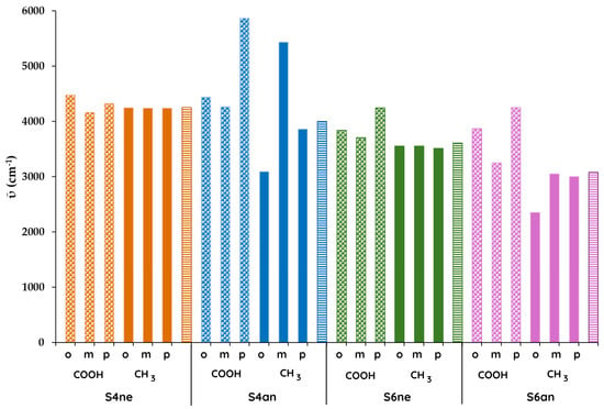
Based on Figure 6, the largest Stokes shift is exhibited by SNAFR4an, followed by SNAFR6an, SNAFR4ne, and finally SNAFR6ne.
Figure 6.
Stokes shifts in neutral and anionic SNAFR4 (orange and blue) and SNAFR6 (green and cyan) functionalized derivatives, compared to the unfunctionalized forms (horizontal fill). Bar patterns denote the substituent: COOH (checkered), CH3 (solid), and unsubstituted (horizontal stripes). For each substituent, the order is from left to right—ortho, meta, para.
For the neutral species, the unfunctionalized dyes do not show significant changes in Stokes shift relative to one another. The main exception is SNAFR6ne-p-COOH, which displays the most pronounced decrease in wavenumber, while the other positional COOH substitutions produce only slight decreases compared to the reference compound.
For the anionic species, all derivatives functionalized with COOH exhibit larger Stokes shifts than their corresponding unfunctionalized references. Notably, SNAFR4an-p-COOH shows the lowest wavenumber among all investigated species. In the case of CH3 functionalization in the anionic form, only SNAFR4an-m-CH3 demonstrates a significant improvement in Stokes shift, whereas for the sixth derivative the wavenumber increases relative to the reference compound.
The fluorescence radiative lifetimes exhibit notable differences across the various SNAFR molecular forms, with functionalization playing a significant role in modulating these values depending on both the scaffold and the position of the substituent.
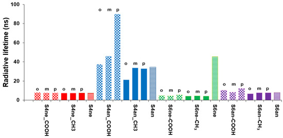
As shown in Figure 7, the SNAFR4 anion (SNAFR4an) exhibits radiative lifetimes that are substantially longer than those of the other molecular forms.
Figure 7.
Radiative lifetimes of neutral and anionic SNAFR4 (orange and blue) and SNAFR6 (green and indigo) functionalized derivatives, compared to the unfunctionalized forms (horizontal fill). Bar patterns denote the substituent: COOH (checkered), CH3 (solid), and unsubstituted (horizontal stripes). For each substituent, the order is from left to right—ortho, meta, para.
Functionalization with COOH results in a dramatic increase, particularly at the para position, while CH3 does not significantly change the lifetime, except in the ortho position, where it decreases. This strong positional dependence suggests that the electronic influence of the COOH group is highly sensitive to its placement on the SNAFR4an scaffold.
For SNAFR4ne, the radiative lifetimes remain largely unchanged upon functionalization, regardless of the substituent type or position. The unfunctionalized molecule shows a lifetime of 7.4 ns, and both COOH (7.8, 7.6, and 7.8 ns for ortho, meta, and para, respectively) and CH3 (7.5, 7.4, and 7.4 ns) functionalization produce values that are nearly identical to the baseline. This suggests that the neutral form of the SNAFR4 scaffold is relatively insensitive to the electronic perturbations introduced by these functional groups.
In contrast, SNAFR6ne displays a dramatic decrease in radiative lifetime upon functionalization. The unfunctionalized molecule has a remarkably long lifetime of 45 ns, which drops sharply to values in the range of 4–6 ns for both COOH and CH3 substituents across all positions.
Finally, SNAFR6an shows only a slight variation in radiative lifetime upon functionalization. The unfunctionalized molecule exhibits a lifetime of 8 ns, and this value remains in a comparable range for most substituted derivatives, with COOH yielding values of 10, 8, and 12 ns (ortho, meta, para) and CH3 giving 7, 8, and 8 ns. While the para-COOH substitution introduces a modest increase, the overall trend suggests limited sensitivity of this scaffold to functionalization.
4. Conclusions
Neutral SNAFRs are described reasonably well by vertical TD-DFT, especially for absorption, and the ordering of absorption wavelengths across neutral derivatives is reproduced correctly. Neutral vertical emission is less accurate unless compared to the 0–0 transition. Vertical emission shows larger blue shifts but agreement improves strongly when the calculated data are compared to the first experimental emission peak.
Anionic SNAFRs are not well described by TD-DFT calculations, the vertical absorption and emission wavelengths being strongly blue-shifted. However, the relative ordering of anionic absorption wavelengths is still consistent with experiment.
Vibronically resolved (FCHT) simulations drastically improve the agreement between experiment and theory and add band-shape information, both for neutral and anionic forms.
All neutral forms emit in the red region, and SNAFR6 remains the most favorable in terms of Stokes shift, followed by SNAFR2 and SNAFR5. For anions, the SNAFR3 is expected experimentally to have the most red-shifted absorption.
The lowest-energy transition is consistently S0 → S1 for all derivatives (neutral and anionic) and both charge states. The HOMO is π-delocalized over the xanthene core and fused ring, with a negligible phenyl-ring contribution.
Difference density plots are consistent with the HOMO → LUMO analysis, predicting that excitation shifts density toward the phenyl-associated regions and away from parts of the annulated framework.
Regarding the phenyl-ring functionalization, the calculations show that substituents introduced at the ortho, meta, and para positions lead to measurable changes in dihedral angles, transition energies, Stokes shifts, and radiative lifetimes. The neutral derivatives exhibit relatively small variations across all parameters, whereas the anionic species display more pronounced shifts, particularly in vertical emission and 0–0 energies, as well as in Stokes shifts and fluorescence radiative lifetimes. These results indicate that functionalization modulates the excited-state landscape more strongly in the anionic form, providing a practical route for tuning emission wavelengths and excited-state behavior.
Overall, the findings highlight the potential of substituent-based modulation for improving the optical performance of SNAFR4 and SNAFR6 derivatives, enabling the tuning of key photophysical parameters relevant for applications such as deep-tissue fluorescence imaging.
Author Contributions
Conceptualization, V.C. and S.-R.S.; methodology, V.C.; validation, V.C. and S.-R.S.; investigation, V.C. and S.-R.S.; data curation, S.-R.S.; writing—original draft preparation, S.-R.S.; writing—review and editing, V.C.; supervision, V.C. All authors have read and agreed to the published version of the manuscript.
Funding
This research received no external funding.
Institutional Review Board Statement
Not applicable.
Informed Consent Statement
Not applicable.
Data Availability Statement
The original contributions presented in this study are included in the article. Further inquiries can be directed to the corresponding author.
Conflicts of Interest
The authors declare no conflict of interest.
References
- Souza, R.C.; Duarte, J.C.; Goldschmidt, R.R.; Borges, I., Jr. Predicting Fluorescence Emission Wavelengths and Quantum Yields via Machine Learning. J. Chem. Inf. Model. 2025, 65, 3270–3281. [Google Scholar] [CrossRef] [Scilit]
- Sednev, M.V.; Belov, V.N.; Hell, S.W. Fluorescent dyes with large Stokes shifts for super-resolution optical microscopy of biological objects: A review. Methods Appl. Fluoresc. 2015, 3, 042004. [Google Scholar] [CrossRef] [Scilit]
- Fabian, J.; Nakazumi, H.; Matsuoka, M. Near-Infrared Absorbing Dyes. Chem. Rev. 1992, 92, 1197–1226. [Google Scholar] [CrossRef] [Scilit]
- Yang, Y.; Lowry, M.; Xu, X.; Escobedo, J.O.; Sibrian-Vazquez, M.; Wong, L.; Schowalter, C.M.; Jensen, T.J.; Fronczek, F.R.; Warner, I.M.; et al. Seminaphthofluorones Are a Family of Water-Soluble, Low Molecular Weight, NIR-Emitting Fluorophores. Proc. Natl. Acad. Sci. USA 2008, 105, 8829–8834. [Google Scholar] [CrossRef] [Scilit] [PubMed]
- Yang, Y.; Lowry, M.; Schowalter, C.M.; Fakayode, S.O.; Escobedo, J.O.; Xu, X.; Zhang, H.; Jensen, T.J.; Fronczek, F.R.; Warner, I.M.; et al. An Organic White Light-Emitting Fluorophore. J. Am. Chem. Soc. 2006, 128, 14081–14092. [Google Scholar] [CrossRef] [Scilit] [PubMed]
- Lee, L.G.; Berry, G.M.; Chen, C.-H. Vita Blue: A New 633-nm Excitable Fluorescent Dye for Cell Analysis. Cytometry 1989, 10, 151–164. [Google Scholar] [CrossRef] [Scilit]
- Whitaker, J.E.; Haugland, R.P.; Prendergast, F.G. Spectral and Photophysical Studies of Benzo[c]xanthene Dyes: Dual Emission pH Sensors. Anal. Biochem. 1991, 194, 330–344. [Google Scholar] [CrossRef] [Scilit]
- Yang, J.; Dass, A.; Rawashdeh, A.-M.M.; Sotiriou-Leventis, C.; Panzner, M.J.; Tyson, D.S.; Kinder, J.D.; Leventis, N. Arylethynyl-Substituted 9,10-Anthraquinones: Tunable Stokes Shifts by Substitution and Solvent Polarity. Chem. Mater. 2004, 16, 3457–3468. [Google Scholar] [CrossRef] [Scilit]
- Nijegorodov, N.; Vasilenko, V.; Monowe, P.; Masale, M. Systematic Investigation of the Influence of Methyl Groups upon Fluorescence Parameters and the Intersystem Crossing Rate Constant of Aromatic Molecules. Spectrochim. Acta A 2009, 74, 189–198. [Google Scholar] [CrossRef] [Scilit]
- Hammershøj, P.; Thyrhaug, E.; Harris, P.; Ek, P.K.; Andresen, T.L.; Clausen, M.H. Convenient one-step synthesis of 5-carboxy-seminaphthofluoresceins. Tetrahedron Lett. 2017, 58, 1611–1615. [Google Scholar] [CrossRef] [Scilit]
- Becke, A.D. Density-functional thermochemistry. III. The role of exact exchange. J. Chem. Phys. 1993, 98, 5648–5652. [Google Scholar] [CrossRef] [Scilit]
- Stephens, P.J.; Devlin, F.J.; Chabalowski, C.F.; Frisch, M.J. Ab Initio Calculation of Vibrational Absorption and Circular Dichroism Spectra Using Density Functional Force Fields. J. Phys. Chem. 1994, 98, 11623–11627. [Google Scholar] [CrossRef] [Scilit]
- Lee, C.; Yang, W.; Parr, R.G. Development of the Cole-Salvetti correlation-energy formula into a functional of the electron density. Phys. Rev. B 1988, 37, 785–789. [Google Scholar] [CrossRef] [Scilit] [PubMed]
- Narsaria, A.K.; Ruijter, J.D.; Hamlin, T.A.; Ehlers, A.W.; Fonseca Guerra, C.; Lammertsma, K.; Bickelhaupt, F.M. Performance of TDDFT Vertical Excitation Energies of Core-Substituted Naphthalene Diimides. J. Comput. Chem. 2020, 20, 1448–1455. [Google Scholar] [CrossRef] [Scilit]
- Ahn, D.-H.; Song, J.-W. Assessment of long-range corrected density functional theory on the absorption and vibrationally resolved fluorescence spectrum of carbon nanobelts. J. Comput. Chem. 2021, 42, 505–515. [Google Scholar] [CrossRef] [Scilit]
- Jacquemin, D.; Adamo, C. Computational Molecular Electronic Spectroscopy with TD-DFT. In Density-Functional Methods for Excited States; Topics in Current Chemistry; Springer: Cham, Switzerland, 2015; Volume 368, pp. 347–375. [Google Scholar] [CrossRef] [Scilit]
- Laurent, A.D.; Jacquemin, D. TD-DFT Benchmarks: A Review. Int. J. Quantum Chem. 2013, 113, 2019–2039. [Google Scholar] [CrossRef] [Scilit]
- Fortino, M.; Bloino, J.; Collini, E.; Bolzonello, L.; Trapani, M.; Faglioni, F.; Pedone, A. On the Simulation of Vibrationally Resolved Electronic Spectra of Medium-Size Molecules: The Case of Styryl-Substituted BODIPYs. Phys. Chem. Chem. Phys. 2019, 21, 3512–3526. [Google Scholar] [CrossRef] [Scilit]
- Raghavachari, K.; Binkley, J.S.; Seeger, R.; Pople, J.A. Self-Consistent Molecular Orbital Methods. 20. Basis set for correlated wave-functions. J. Chem. Phys. 1980, 72, 650–654. [Google Scholar] [CrossRef] [Scilit]
- Bloino, J. Development and Application of Time-Dependent and Time-Independent Models for the Study of Spectroscopic Properties in Compounds of Biological Interest. Ph.D. Thesis, Università degli Studi di Napoli Federico II, Naples, Italy, 2008. [Google Scholar]
- Bloino, J.; Biczysko, M.; Santoro, F.; Barone, V. General Approach to Compute Vibrationally Resolved One-Photon Electronic Spectra. J. Chem. Theory Comput. 2010, 6, 1256–1274. [Google Scholar] [CrossRef] [Scilit]
- Savarese, M.; Aliberti, A.; De Santo, I.; Battista, E.; Causa, F.; Netti, P.A.; Rega, N. Fluorescence Lifetimes and Quantum Yields of Rhodamine Derivatives: New Insights from Theory and Experiment. J. Phys. Chem. A 2012, 116, 7491–7497. [Google Scholar] [CrossRef] [Scilit]
- Frisch, M.J.; Trucks, G.W.; Schlegel, H.B.; Scuseria, G.E.; Robb, M.A.; Cheeseman, J.R.; Scalmani, G.; Barone, V.; Petersson, G.A.; Nakatsuji, H.; et al. Gaussian 16, Revision C.01; Gaussian, Inc.: Wallingford, CT, USA, 2016.
- Guido, C.A.; Knecht, S.; Kongsted, J.; Mennucci, B. Benchmarking Time-Dependent Density Functional Theory for Excited State Geometries of Organic Molecules in Gas-Phase and in Solution. J. Chem. Theory Comput. 2013, 9, 2209–2220. [Google Scholar] [CrossRef] [Scilit]
- Brémond, E.; Savarese, M.; Adamo, C.; Jacquemin, D. Accuracy of TD-DFT Geometries: A Fresh Look. J. Chem. Theory Comput. 2018, 14, 3715–3727. [Google Scholar] [CrossRef] [Scilit]
- Xu, L.; Kumar, A.; Wong, B.M. Linear polarizabilities and second hyperpolarizabilities of streptocyanines: Results from broken-Symmetry DFT and new CCSD(T) benchmarks. J. Comput. Chem. 2018, 39, 2350–2359. [Google Scholar] [CrossRef] [Scilit]
- Wong, B.M.; Hsieh, T.H. Optoelectronic and Excitonic Properties of Oligoacenes: Substantial Improvements from Range-Separated Time-Dependent Density Functional Theory. J. Chem. Theory Comput. 2010, 6, 3704–3712. [Google Scholar] [CrossRef] [Scilit]
- Véril, M.; Scemama, A.; Caffarel, M.; Lipparini, F.; Boggio-Pasqua, M.; Jacquemin, D.; Loos, P. QUESTDB: A database of highly accurate excitation energies for the electronic structure community. Wiley Interdiscip. Rev. Comput. Mol. Sci. 2021, 11, e1517. [Google Scholar] [CrossRef] [Scilit]
- Liang, J.; Feng, X.; Hait, D.; Head-Gordon, M. Revisiting the Performance of Time-Dependent Density Functional Theory for Electronic Excitations: Assessment of 43 Popular and Recently Developed Functionals from Rungs One to Four. J. Chem. Theory Comput. 2022, 18, 3460–3473. [Google Scholar] [CrossRef] [Scilit]

Disclaimer/Publisher’s Note: The statements, opinions and data contained in all publications are solely those of the individual author(s) and contributor(s) and not of MDPI and/or the editor(s). MDPI and/or the editor(s) disclaim responsibility for any injury to people or property resulting from any ideas, methods, instructions or products referred to in the content. |
© 2026 by the authors. Licensee MDPI, Basel, Switzerland. This article is an open access article distributed under the terms and conditions of the Creative Commons Attribution (CC BY) license.