


Abstract
Amygdalin is a cyanogenic diglycoside whose hydrolysis links plant defense with cyanide exposure in animals and humans. This review examines its activation, metabolism and associated stress responses. In intact plant tissues, cyanogenic glycosides are separated from hydrolytic enzymes; tissue disruption permits substrate-enzyme contact. Amygdalin hydrolysis produces prunasin and mandelonitrile, followed by benzaldehyde and hydrogen cyanide (HCN). After oral exposure, host and microbial β-glucosidases contribute to activation, while host and potentially microbial detoxification modify cyanide exposure. The biological response depends on dose, release rate, exposure route, detoxification capacity and mitochondrial susceptibility. Localized HCN production participates in plant stress signaling, whereas excessive exposure inhibits mitochondrial respiration and may lead to secondary oxidative and inflammatory injury. Mammalian cell studies have reported low-level regulatory effects, but these findings do not establish therapeutic benefit from oral amygdalin. Reports of antioxidant or anti-inflammatory activity require measurements of cyanogenic activation to distinguish responses to the parent glycoside from those associated with its degradation. Time-resolved measurements of amygdalin and its products of biotransformation should be paired with microbial, mitochondrial and redox endpoints and appropriate experimental controls.
1. Introduction
Amygdalin is a cyanogenic diglycoside found predominantly in seeds and kernels of Rosaceae species, including bitter almond, apricot, peach, plum and cherry [1,2]. Like other cyanogenic glycosides, it can release hydrogen cyanide (HCN) after enzymatic hydrolysis [1,3,4].
In plants, cyanogenic glycosides form part of a compartmentalized defense system. The glycoside and the enzymes capable of hydrolyzing it are normally separated within intact tissues. Tissue disruption caused by herbivory, infection or mechanical damage permits enzyme-substrate contact and initiates cyanogenesis [3,4,5]. Amygdalin is therefore best regarded as a stored precursor rather than a constitutively active toxicant. Its biological effect emerges when the tissue architecture that keeps the system inactive is lost.
The situation changes after ingestion. Once removed from the plant, amygdalin enters an enzymatic environment that is no longer controlled by plant compartmentation. Host intestinal enzymes and microbial β-glucosidases can hydrolyze the glycoside and increase the formation of cyanogenic intermediates [2,6,7]. The toxicological significance of oral exposure depends on both the amount of amygdalin ingested and the extent and rate of its conversion to HCN. Food matrix, tissue disruption, gastrointestinal transit, microbial metabolism and cyanide detoxification all influence this process.
Much of the existing literature has approached amygdalin from one of three directions. One body of work deals with plant cyanogenesis and the ecological role of cyanogenic glycosides [3,4,5,8]. A second focuses on toxicology, food safety and cyanide poisoning [1,2,9,10]. A third discusses pharmacological claims, including the historical use of amygdalin or laetrile in cancer treatment [6,11]. These areas overlap chemically, but they are rarely examined within the same biological model.
HCN provides the clearest mechanistic link between them. At sufficiently high concentrations, cyanide inhibits cytochrome c oxidase, disrupts oxidative phosphorylation and can cause rapid systemic toxicity [9,10,12]. In plants, however, low and spatially restricted HCN production has also been associated with redox regulation, stress responses, seed physiology and pathogen defense [9,13,14,15]. The same molecule can therefore participate in fundamentally different biological outcomes depending on concentration, compartment and exposure time.
Gut microbial β-glucosidases may increase cyanide formation after oral amygdalin exposure, whereas host and potentially microbial detoxification can reduce the resulting burden [6,16,17]. Mitochondrial inhibition may be accompanied by changes in reactive oxygen species (ROS), antioxidant responses and inflammatory signaling [18,19,20]. These effects are often reported separately in toxicological, pharmacological and microbiological studies, even though they represent connected stages of the same exposure pathway.
This review follows amygdalin from storage and activation in plant tissues to gastrointestinal biotransformation and host stress responses. It examines how cyanide release, microbial metabolism and detoxification influence mitochondrial function, redox balance and inflammatory signaling, and identifies experimental controls needed to distinguish effects of the intact glycoside from those of its degradation products.
Literature Identification and Selection
Literature searches were conducted in Scopus and Web of Science Core Collection using seven topic-specific queries, last updated on 9 October 2026. Search terms included amygdalin, prunasin, cyanogenic glycosides and hydrogen cyanide, combined with terms related to plant defense, hydrolysis, gut microbiota, mitochondrial function, oxidative and inflammatory responses, analytical determination and toxicity. The combined searches retrieved 36,845 records, reduced to 23,376 unique entries after automated deduplication. The broad search terms also returned numerous publications outside the scope of this review, particularly studies of general cyanide chemistry or plant physiology without direct relevance to amygdalin.
Several secondary publications discussed the same experimental findings and referred to a common set of original studies. Primary research was therefore prioritized, with emphasis on plant cyanogenesis, amygdalin hydrolysis, microbial metabolism, cyanide exposure, and mitochondrial, redox and inflammatory responses. Studies were considered according to their experimental models, exposure conditions, measured endpoints and mechanistic relevance. Review articles were used mainly for background information and identification of original sources. This was a targeted narrative review; no systematic screening of all retrieved records, formal risk-of-bias assessment or meta-analysis was undertaken. The seven Boolean search queries used to identify the literature were as follows:
Q1. Plant cyanogenesis and defense: (“cyanogenic glycosides” OR “cyanogenic glucosides” OR cyanogenesis OR amygdalin) AND (“plant defense” OR “plant defence” OR herbivory OR pathogen OR biosynthesis OR “Prunus”).
Q2. Amygdalin hydrolysis and activation: (“amygdalin” OR “prunasin”) AND (hydrolysis OR “beta-glucosidase” OR “amygdalin hydrolase” OR “prunasin hydrolase” OR mandelonitrile).
Q3. Gut microbiota and detoxification: (amygdalin OR prunasin) AND (microbiota OR microbiome OR intestinal OR “gut bacteria” OR “germ-free” OR detoxification).
Q4. Cyanide and mitochondrial responses: (cyanide OR “hydrogen cyanide”) AND (“cytochrome c oxidase” OR “complex IV” OR “S-cyanylation” OR gasotransmitter OR “endogenous cyanide”).
Q5. Oxidative and inflammatory responses: amygdalin AND (Nrf2 OR NLRP3 OR “NF-kappa B” OR inflammation OR “oxidative stress” OR “ischemia reperfusion” OR endothelial).
Q6. Analytical methods and food safety: (amygdalin OR prunasin OR “cyanogenic glycosides”) AND (quantification OR analytical OR “free cyanide” OR “cyanogenic potential” OR processing OR poisoning).
Q7. Toxicity and pharmacokinetics: (amygdalin OR laetrile) AND (“oral administration” OR pharmacokinetics OR “clinical trial” OR “apricot kernels” OR toxicity).
2. Cyanogenic Glycosides as Plant Stress-Defense Metabolites
Cyanogenic glycosides are widespread specialized metabolites found in numerous plant families, including Rosaceae, Poaceae, Fabaceae and Euphorbiaceae [3,4,21]. Their defining feature is the ability to release hydrogen cyanide after enzymatic hydrolysis. In intact tissues, however, this potential is usually kept under tight spatial control. The glycoside and the enzymes that hydrolyze it are separated until tissue integrity is disturbed [3,4,5,21].
This arrangement is well suited to defense against biotic stress. Herbivory, pathogen penetration or mechanical damage can collapse cellular compartmentation and bring cyanogenic substrates into contact with β-glucosidases and, depending on the system, hydroxynitrile lyases [3,4,5,21]. The resulting cyanohydrins are unstable and may release HCN rapidly. In ecological terms, this provides a preformed chemical defense that can be activated without the delay required for de novo synthesis. Cyanogenic glycosides are therefore often classified as phytoanticipins rather than inducible metabolites in the strict sense [5,22,23].
The defensive value of cyanogenesis is supported by both ecological observations and experimental manipulation. Cyanogenic tissues can reduce palatability, interfere with feeding and impair cellular respiration in herbivores or pathogens [21,23,24]. In Arabidopsis thaliana, introduction of the dhurrin biosynthetic pathway increased resistance to herbivory, providing direct evidence that cyanogenic capacity can alter plant–herbivore interactions [24]. Cyanogenic metabolites have also been implicated in inducible pathogen resistance in Arabidopsis, where a cyanogenic compound contributes to defense against Pseudomonas syringae [25].
A recent example of experimentally introduced cyanogenic defense was reported by Chen et al. in 2026 [26]. By reconstructing amygdalin biosynthesis in rice endosperm using enzymes from Prunus species, the authors obtained transgenic grains containing up to 14.54 μg g−1 amygdalin. These grains showed reduced attractiveness to the rice weevil (Sitophilus oryzae) and impaired insect development [26]. However, the engineered lines also exhibited changes in endogenous metabolic gene expression, and cyanide release was not established as the sole cause of resistance. The study therefore supports the defensive potential of engineered cyanogenic metabolism rather than demonstrating a naturally occurring amygdalin defense system in rice.
The system is not uniformly effective against all attackers. Specialist herbivores may avoid cyanide release, detoxify HCN, sequester cyanogenic glycosides or, in some cases, synthesize related compounds themselves [23,27]. The effectiveness of cyanogenic defense depends on the physiology and feeding behavior of the attacker.
Amygdalin distribution in Prunus varies across tissues and developmental stages. In almond, amygdalin and its monoglucoside precursor prunasin vary between tissues and developmental stages, while β-glucosidase localization also differs between sweet and bitter genotypes [28,29]. The cyanogenic phenotype therefore reflects more than the amount of stored glycoside. Enzyme localization, tissue organization and developmental timing all determine whether cyanide can be released efficiently.
Developmental variation in cyanogenic metabolism has recently been examined in Prunus mandshurica. In two clones studied across five stages of kernel development, Jiang et al. observed early accumulation of prunasin, whereas amygdalin reached its maximum at later stages, with different accumulation profiles in the two clones [30]. β-Glucosidase activity generally increased during later development, including in the clone with higher amygdalin content. Transcriptomic analyses identified candidate genes and transcription factors associated with these changes, although their regulatory functions remain to be established experimentally. In apricot, functional studies identified PabHLH2 as a regulator of CYP79D16/CYP71AN24 expression and prunasin accumulation [31].
In seeds and kernels, cyanogenic compounds may deter herbivores and seed predators and protect nutrient-rich reproductive tissues. In bitter almond, high cyanogenic potential is associated with bitterness [28,29].
Cyanogenic glycosides may also serve functions beyond immediate defense. Evidence from several plant systems suggests roles in nitrogen and carbon storage, endogenous turnover and metabolic remobilization [22,32]. These functions are not mutually exclusive with defense. The same metabolite pool may contribute to deterrence after tissue damage while also participating in developmental or metabolic processes under other conditions.
Amygdalin may also contribute to stress in the producing plant’s environment. Yang et al. observed its accumulation in the rhizosphere of repeatedly replanted peach orchards and demonstrated that exogenous amygdalin inhibited seedling growth, particularly in sterilized soil [33]. Selected rhizobacterial isolates degraded amygdalin and alleviated growth inhibition, accompanied by changes in jasmonate signaling and redox homeostasis. These findings extend the biological context of amygdalin beyond tissue-localized defense, although microbial degradation should not automatically be equated with complete removal of cyanogenic toxicity.
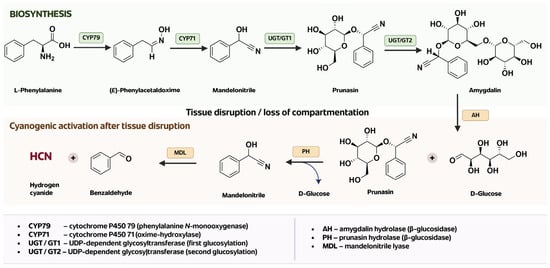
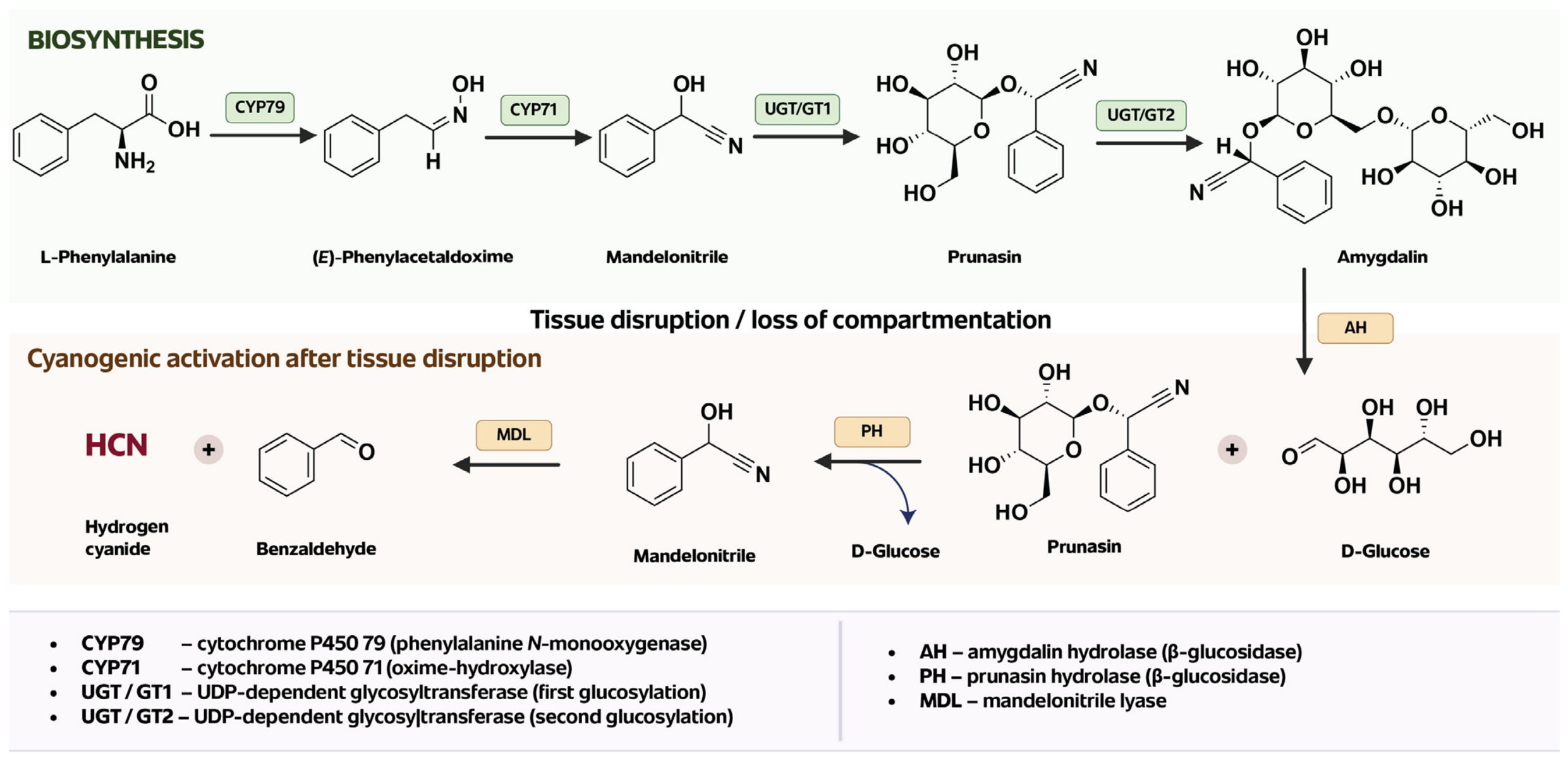
β-Glucosidases initiate cyanogenic glycoside hydrolysis [5,34]. Amygdalin biosynthesis and the enzymatic sequence leading to HCN release are summarized in Figure 1.
Figure 1.
Biosynthesis and cyanogenic activation of amygdalin in Prunus. The (upper) pathway shows the conversion of L-phenylalanine to prunasin and amygdalin, while the (lower) pathway illustrates sequential hydrolysis after tissue disruption, leading to mandelonitrile, benzaldehyde and HCN release. Schematic prepared by the authors based on published biochemical findings [26,28,29,35,36,37,38].
3. Amygdalin Activation: β-Glucosidases, Benzaldehyde and Hydrogen Cyanide
Amygdalin is a relatively stable cyanogenic precursor that requires hydrolysis before HCN can be released [1,5,28,29,35].
The degradation pathway is sequential. Amygdalin is first hydrolyzed to prunasin and glucose, after which prunasin is converted to mandelonitrile and a second glucose molecule. Mandelonitrile is unstable and decomposes, either spontaneously or enzymatically depending on local conditions, to benzaldehyde and HCN [35]. The different products should not be treated as interchangeable. Prunasin is an intermediate glycoside, mandelonitrile is the immediate cyanohydrin precursor, benzaldehyde is a coupled carbonyl product, and HCN accounts for the principal acute toxicological effect.
In plant tissues, this sequence is controlled by compartmentation. Cyanogenic glycosides and their hydrolytic enzymes are separated until tissue damage permits contact. Crushing, chewing, grinding, maceration, herbivory or infection can therefore accelerate cyanogenesis [5,28,29,35].
The enzymology of Prunus species has been studied in detail. Amygdalin hydrolase converts amygdalin to prunasin, whereas prunasin hydrolase catalyzes the next hydrolytic step toward mandelonitrile [29,36,37]. Mandelonitrile lyase then promotes cleavage of mandelonitrile to benzaldehyde and HCN [37,38]. In black cherry (Prunus serotina), these enzymes show distinct spatial and developmental patterns of expression [36,37,38].
The situation after ingestion is different. Amygdalin is transferred from a plant system in which activation is spatially regulated to the gastrointestinal tract, where host and microbial hydrolases provide a new activation environment [3,7,16,17]. Amygdalin- and prunasin-hydrolyzing glucosidase activity has been demonstrated in rat and human small intestinal mucosa, while comparable activity was not detected in the human tumor tissues examined in an early study [7].
Microbial β-glucosidases provide an additional source of cyanogenic activation [16,17]. Oral and parenteral administration differ in exposure to digestive and microbial enzymes, which affects their pharmacokinetic interpretation [2,3,39,40].
Mandelonitrile is a particularly important intermediate because it connects glycoside hydrolysis directly with HCN formation. Its stability is affected by pH, temperature and enzyme activity [35,38]. Hydroxynitrile lyases can accelerate its decomposition in plant tissues, whereas spontaneous cleavage may become more relevant under other physicochemical conditions. These differences matter when comparing intact kernels, processed food, purified amygdalin and gastrointestinal models.
Benzaldehyde is formed in parallel with HCN during mandelonitrile cleavage [1,28,35,38]. It contributes to the characteristic odor of damaged bitter almond and related cyanogenic tissues, and it can serve as evidence that the hydrolytic pathway has proceeded beyond the cyanohydrin stage. Its concentration, however, is influenced by volatility, oxidation and matrix effects. Benzaldehyde should therefore not be used as a direct quantitative proxy for cyanide formation.
Analytical factors can further complicate interpretation. Naturally occurring amygdalin is predominantly the R-epimer, while neoamygdalin may arise through epimerization under some conditions [1,41]. Recent rat pharmacokinetic data also demonstrate different oral exposure profiles for the two amygdalin epimers [42]. Analytical methods that do not distinguish these forms, or that quantify amygdalin without considering prunasin and free cyanide, can underestimate or misclassify cyanogenic potential [41,43]. Measuring the parent compound alone is therefore insufficient when the biological question concerns stress or toxicity.
Attribution of antioxidant, anti-inflammatory or cytotoxic responses to amygdalin requires characterization of β-glucosidase activity and cyanide formation [2,44,45,46]. The analytical controls needed for this distinction are discussed in Section 9.
4. Hydrogen Cyanide as a Dose-Dependent Stress Factor
Hydrogen cyanide is the main product that connects amygdalin hydrolysis with cellular stress. Its acute toxicity is well established and results primarily from inhibition of cytochrome c oxidase, the terminal complex of the mitochondrial respiratory chain [10,12]. The biological effects of HCN, however, depend strongly on concentration, site of production and duration of exposure. This is particularly evident in plants, where cyanide can participate in stress-related regulation at concentrations far below those associated with overt toxicity [9,13,14,15].
At toxic concentrations, cyanide binds to the heme a3-CuB center of cytochrome c oxidase and interferes with electron transfer to molecular oxygen [10,12]. Oxidative phosphorylation falls, ATP synthesis is impaired and cells lose the ability to use oxygen efficiently despite adequate oxygen availability. This histotoxic hypoxia particularly affects tissues with high energy demand, including the brain and cardiovascular system. Severe exposure can consequently produce lactic acidosis, neurological dysfunction, cardiovascular collapse and death [10,12].
The mitochondrial effects of cyanide extend beyond ATP depletion. Disruption of electron transport changes mitochondrial redox state, membrane potential and electron leakage, with consequences for reactive oxygen species formation [10,12]. Cyanide can also interact with other metalloproteins and influence nitric oxide- and thiol-dependent processes [10,12,47].
Plant HCN biology is more complex. Cyanide is generated not only through cyanogenic glycoside turnover but also as a by-product of other metabolic processes, including ethylene biosynthesis [9,14]. Plants possess dedicated detoxification mechanisms, particularly the β-cyanoalanine pathway, which limits intracellular accumulation. Within this controlled metabolic setting, low and transient HCN concentrations have been associated with seed germination, root development, pathogen responses and redox regulation [9,14,15].
Recent experiments in Arabidopsis thaliana have provided further evidence of interactions between endogenous HCN metabolism and abiotic stress responses. Zhang et al. linked H2S-dependent regulation of PAD3 to cyanide homeostasis, changes in cellular oxygen consumption and responses to salt and drought stress [48]. The study supports a role for regulated HCN production in plant stress physiology, but does not directly address cyanide formation through amygdalin hydrolysis.
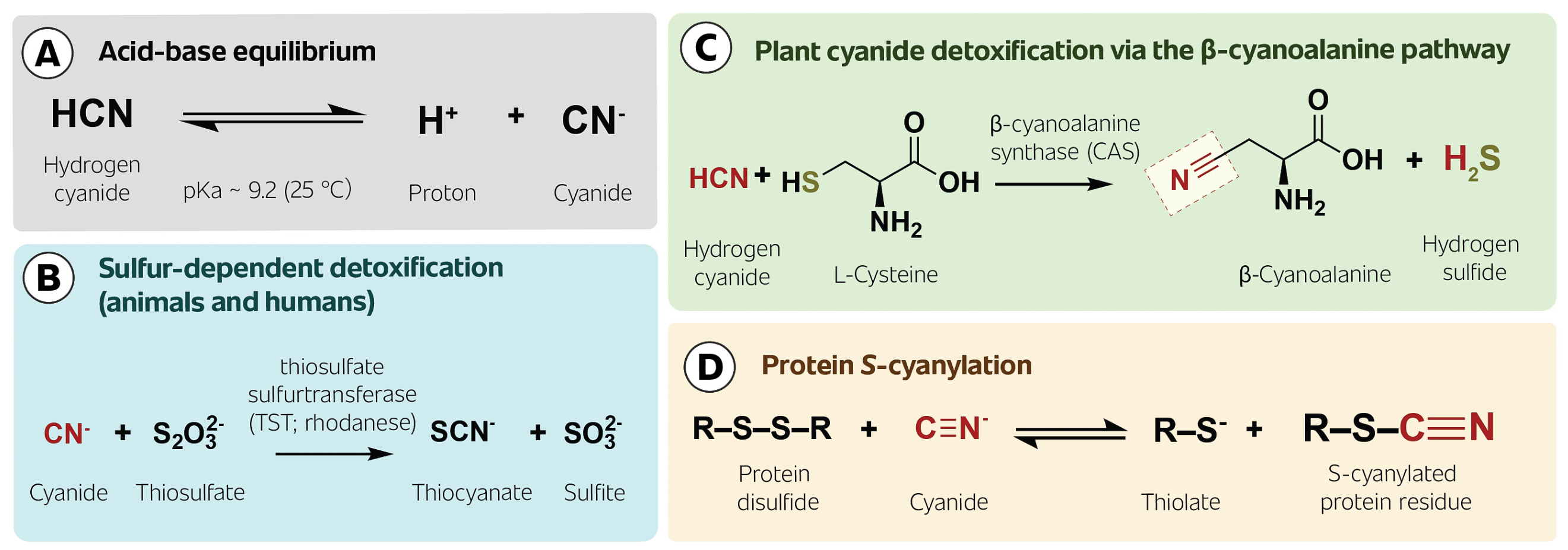
Protein S-cyanylation has also been implicated in cyanide-dependent regulation in plants. One characterized route of protein S-cyanylation involves nucleophilic attack by CN− on a disulfide bond, producing an S-cyanylated cysteine residue and a thiolate. Proteomic studies in Arabidopsis thaliana have identified proteins bearing this modification [13]. Such observations support a regulatory role for endogenous cyanide in plants that cannot be explained solely by respiratory toxicity. The effect is nevertheless highly dependent on local concentration and cellular context.
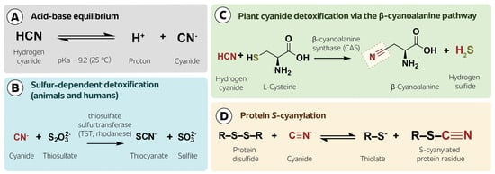
The major chemical routes linking cyanide speciation, detoxification and regulatory S-cyanylation are summarized in Figure 2.
Figure 2.
Selected chemical fates of hydrogen cyanide and cyanide relevant to stress biology. (A) HCN/CN− acid–base equilibrium; (B) sulfurtransferase-mediated thiocyanate formation in animals and humans; (C) β-cyanoalanine formation in plants; (D) protein S-cyanylation through reaction of cyanide with a disulfide bond. The dashed outline in panel (C) highlights the incorporated nitrile group. Schematic prepared by the authors based on [8,9,10,13,47,49].
Evidence for a regulatory role of low-level cyanide in mammals remains disputed, particularly with respect to mitochondrial Complex IV. Randi et al. [50] reported increased cytochrome c oxidase activity and oxygen consumption at nanomolar cyanide concentrations in cultured cells and purified-enzyme experiments. Giamogante et al. [51], using isolated mouse liver mitochondria, found no respiratory stimulation at 1 nM KCN and questioned whether the proposed deglutathionylation reaction could proceed sufficiently rapidly under those conditions. Neither study examined amygdalin hydrolysis.
Zuhra et al. [47,49] subsequently demonstrated glycine-dependent cyanide production in mammalian cells and tissues, with substantial involvement of lysosomal metabolism. They also identified endogenous protein S-cyanylation and measured increased blood cyanide after intraperitoneal amygdalin administration in mice. In cultured human endothelial cells, amygdalin or mandelonitrile (100 μM) increased cyanide release and was associated with enhanced proliferation, migration and tube formation [52]. The endothelial study did not directly measure mitochondrial respiration or Complex IV activity, and the pharmacological inhibitors used were not entirely specific. These findings provide evidence of cyanide-associated responses in defined experimental models, but do not establish a beneficial effect of oral amygdalin exposure in humans.
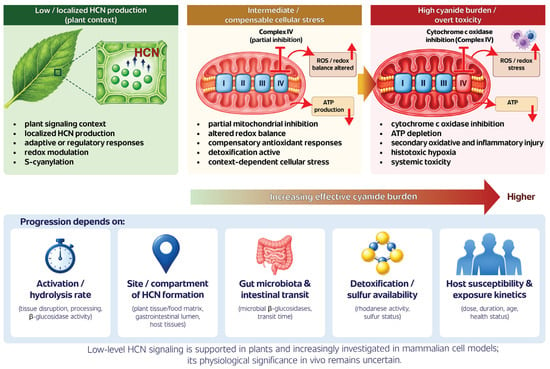
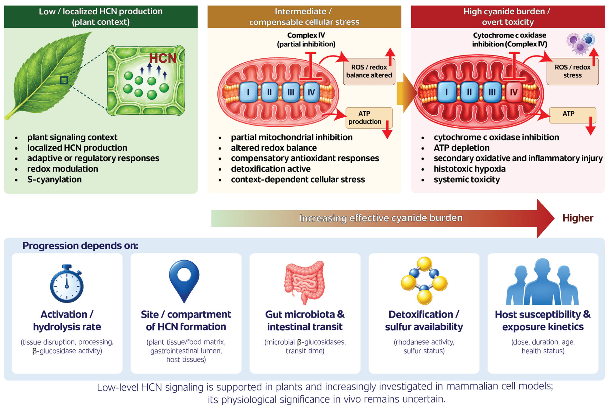
Localized HCN production has been implicated in plant stress responses [9,13,14,15]. Cyanide exposure in animal tissues presents a different experimental context: increased cyanide burden can impair mitochondrial respiration and, at sufficiently high exposure, cause systemic toxicity [10,12,53,54,55]. A compensated phase of cellular disturbance remains a plausible intermediate condition, but its limits have not been defined across biological systems. Figure 3 compares these contexts without assuming a common exposure scale or a single progression across species.
Figure 3.
Cyanide-related responses in distinct biological contexts. The (left) panel illustrates localized HCN production associated with plant stress regulation. The (middle,right) panels depict a proposed intermediate state of cellular stress and the established consequences of excessive cyanide exposure, respectively. These panels do not represent successive stages demonstrated in a single organism or a common quantitative exposure scale. Schematic prepared by the authors based on [9,10,12,13,14,15,17,50,51].
For amygdalin, the rate of cyanide release may be as important as the total amount produced. Rapid hydrolysis can create a high local cyanide burden before detoxification pathways compensate, whereas slower formation may permit more efficient conversion to thiocyanate [2,3,10,17]. Food matrix, β-glucosidase activity, intestinal microbiota and exposure route can therefore alter the toxicological significance of the same nominal amygdalin dose.
The term cyanogenic stress load is used here as a qualitative descriptor, not as a validated quantitative index. It refers to cyanide exposure within a biological compartment in relation to detoxification capacity and the preservation of mitochondrial respiration. Its individual components could be investigated through time-resolved cyanide measurements, thiocyanate formation, relevant enzyme activities and mitochondrial oxygen consumption. None of these measurements alone provides a numerical estimate of the proposed load [2,3,10,17].
5. Amygdalin as a Xenochemical Stressor in Animals and Humans
Once amygdalin is ingested, its biological role is fundamentally different from that in the plant. It no longer belongs to a compartmentalized defense system but becomes an exogenous cyanogenic precursor exposed to digestion, microbial metabolism, absorption and detoxification [2,3,7,10]. In animals and humans, its relevance is therefore best considered in terms of xenochemical stress: the response to a foreign compound whose toxicity depends on enzymatic activation and host handling.
Human exposure occurs mainly through cyanogenic plant material, particularly kernels and seeds of Prunus species such as apricot, bitter almond, peach and plum [1,53,56,57]. The amount of amygdalin can vary markedly between species and cultivars, and bitterness is often associated with higher cyanogenic potential [56,57]. Processing adds another source of variation. Chewing, grinding or crushing disrupts tissue organization and increases contact between cyanogenic glycosides and hydrolytic enzymes [1,5,53]. Thus, two products containing similar amounts of amygdalin may differ in the rate at which cyanide is generated.
Recent analytical data from Prunus mume syrups further demonstrate that cyanogenic precursor content and free cyanide must be considered separately. Lee et al. quantified amygdalin and prunasin in 670 homemade and commercial syrup samples, with mandelonitrile and free cyanide additionally determined in selected subsets [58]. Mean free cyanide concentrations were 2.65 mg/kg in the homemade subset and 0.11 mg/kg in commercial liquid tea-type products, whereas the corresponding calculated total cyanide potentials were 3.41 and 11.23 mg CN− equivalents/kg, respectively. Because the study was cross-sectional and the samples differed in processing and storage histories, these differences cannot be attributed to any single processing step.
Raw apricot kernels have received particular attention because enzymatic hydrolysis of their cyanogenic glycosides can release cyanide after mastication or grinding [53]. Clinical case reports describe severe cyanide poisoning following ingestion of apricot kernels or amygdalin-containing alternative preparations [54,55]. Neurological symptoms, lactic acidosis and cardiovascular deterioration reported in these cases are consistent with impaired mitochondrial oxygen utilization [10,12,54,55].
Route of administration has a major influence on the outcome. In an early human pharmacological study, intravenously administered amygdalin was largely recovered unchanged, whereas oral administration produced measurable blood cyanide [39]. Animal pharmacokinetic studies likewise showed limited systemic availability of unchanged amygdalin after oral dosing and evidence of metabolic conversion [40]. These findings indicate that gastrointestinal activation, rather than circulating parent compound, is a major determinant of cyanide exposure.
The balance between intestinal cyanide formation and detoxification contributes to variation in oral toxicity [3,7,11,16,17]. The microbial contribution is examined in Section 6.
Cyanogenic activation in cell cultures varies with enzyme activity and incubation conditions [7,52]. Such cultures do not necessarily reproduce the exposure to parent compound and metabolites that follows oral dosing.
The historical literature on laetrile creates an additional source of confusion. Amygdalin, laetrile and the term “vitamin B17” have often been used interchangeably, although they are not chemically or scientifically equivalent [2,11,59]. Amygdalin is a naturally occurring cyanogenic glycoside; laetrile has referred to semi-synthetic or preparation-dependent products, while “vitamin B17” has no accepted nutritional basis. Clinical studies have not established amygdalin or laetrile as effective anticancer treatments, whereas cyanide-related adverse effects remain well documented [11,59].
The biological meaning of amygdalin-associated cyanogenesis changes markedly between plant tissues, food matrices, the gastrointestinal tract and experimental systems, as summarized in Table 1.
Table 1.
Biological context determines the outcome of amygdalin-associated cyanogenesis.
6. Gut Microbiota and β-Glucosidases as Gatekeepers of Cyanogenic Stress
Gut microorganisms participate in xenobiotic metabolism through hydrolysis, reduction, deconjugation and other reactions [60,61,62,63]. In the case of amygdalin, microbial β-glucosidases contribute to the formation of cyanogenic intermediates [3,7,16,17].
Carter et al. [16] compared conventional and germ-free rats given a single oral dose of amygdalin (600 mg/kg). Conventional animals that developed toxicity had blood cyanide concentrations of 2.6–4.5 μg/mL, whereas germ-free rats remained clinically unaffected, with concentrations below 0.4 μg/mL. Intestinal contents from conventional animals also released benzaldehyde from amygdalin during incubation, unlike preparations obtained from germ-free rats. These experiments directly implicated the gastrointestinal microbiota in the formation of toxic amounts of cyanide in this animal model.
Wen et al. [17] subsequently compared conventional rats with antibiotic-treated pseudo-germ-free animals after oral administration of 440 mg/kg amygdalin. Cyanide concentrations rose more rapidly in conventional rats, which also developed acute poisoning symptoms. In separate incubation experiments, microbial enzyme preparations generated cyanide from amygdalin and thiocyanate from added cyanide in the presence of thiosulfate. Metagenomic analyses identified candidate sulfurtransferase genes, but their presence does not establish how much cyanide is detoxified by microorganisms in vivo. The authors could not quantify this contribution reliably because of acute toxicity in conventional animals. Moreover, antibiotic treatment altered intestinal β-glucosidase activity and may have affected other aspects of host and microbial physiology. The evidence for microbial activation of amygdalin is therefore substantially stronger than that for a protective microbial detoxification pathway.
This balance may vary considerably among individuals. Human gut microbiomes differ in species composition, gene content and enzyme activity [60,61,62]. These differences can influence the metabolism of orally administered xenobiotics, sometimes producing large interindividual variation in metabolite exposure [60,61]. Comparable variability is plausible for amygdalin, although direct human data remain limited. At present, most evidence linking microbiota composition to amygdalin-derived cyanide production comes from experimental models rather than controlled human studies.
Diet and exposure history may further modify this process. Dietary composition can alter microbial β-glucosidase activity and community structure, while antibiotic treatment can reduce bacterial populations capable of hydrolyzing glycosides [17,62,63]. Repeated exposure to plant glycosides could also select for microbial functions involved in their metabolism. Whether such adaptation increases cyanide production, enhances detoxification or produces both effects has not been established for amygdalin.
Microbial hydrolysis also depends on the accessibility of amygdalin in the ingested matrix, which differs between purified preparations, intact kernels and processed plant material [1,5,53].
These considerations also expose a limitation of many experimental systems. Standard cell cultures contain neither the microbial community nor the gastrointestinal chemistry present after oral exposure. Conversely, fecal incubations and gut fermentation models may reproduce microbial hydrolysis but lack normal absorption, hepatic detoxification and systemic distribution. Animal models capture more of the pathway, although differences in microbiota composition and cyanide metabolism complicate translation to humans.
Future studies should therefore move beyond taxonomic microbiome profiling. Knowing which bacterial groups are present is less informative than knowing what reactions they perform. Direct measurements of β-glucosidase activity, amygdalin disappearance, prunasin formation, cyanide release and thiocyanate production should be combined with metagenomic or metatranscriptomic data where possible [17,60,61,62,63]. Such designs could identify microbial functions that contribute most strongly to cyanogenic activation or detoxification.
7. Mitochondrial Stress, Oxidative Stress and Inflammatory Pathways
The main cellular consequence of amygdalin-derived cyanide is interference with mitochondrial respiration. HCN inhibits cytochrome c oxidase and restricts electron transfer through Complex IV, reducing oxidative phosphorylation and ATP production [10,12]. At high exposure, this mechanism accounts for the rapid toxicity of cyanide. At lower levels, partial respiratory inhibition may still alter membrane potential, electron flow, NADH/NAD+ balance and mitochondrial reactive oxygen species (ROS) production [10,12,64].
These mitochondrial changes provide a plausible link between cyanogenic exposure and redox disturbance. Mitochondrial ROS are normal participants in cellular signaling, but their effect depends on where and how rapidly they are produced [19,20]. A moderate increase may trigger adaptive responses, whereas sustained respiratory inhibition can shift the balance toward oxidative damage. Protein oxidation, lipid peroxidation, mitochondrial DNA injury and loss of membrane integrity may then reinforce mitochondrial dysfunction [18,19,64].
Changes in ROS or antioxidant enzyme activity do not, by themselves, demonstrate an intrinsic antioxidant effect of amygdalin. Nrf2 regulates glutathione metabolism and the expression of cytoprotective enzymes, including heme oxygenase-1 and NAD(P)H:quinone oxidoreductase 1 [65]. Increased Nrf2 activity may reflect protection in a particular injury model or a compensatory response to chemical stress. These alternatives require assessment of cyanogenic activation.
Experimental reports of anti-inflammatory activity differ substantially in exposure conditions and measured endpoints. Tang et al. [44] administered amygdalin intraperitoneally (2, 4 or 8 mg/kg, twice at a 12-h interval) before D-galactosamine/lipopolysaccharide-induced liver injury in mice. Treatment reduced serum ALT and AST, hepatic MDA and MPO, and inflammatory cytokines. Changes in NLRP3, NF-κB and Nrf2/NQO1-related proteins were also reported. In separate HepG2 experiments, amygdalin increased Nrf2 and NQO1 expression, an effect attenuated by brusatol. Cyanide production and mitochondrial respiration were not measured.
In DSS-induced colitis, Xu et al. [45] administered amygdalin orally (12.5, 25 or 50 mg/kg/day) and observed reduced intestinal injury, changes in TLR4/NF-κB/MAPK signaling, improved antioxidant indices and altered apoptosis- and ferroptosis-related markers. Fecal microbiota composition was assessed by 16S rRNA sequencing, but neither microbial β-glucosidase activity nor cyanide formation was determined. The microbial findings therefore cannot establish whether hydrolysis of amygdalin contributed to the observed effects.
Mohamed et al. [46] investigated amygdalin (5 mg/kg, intraperitoneally) alone and in combination with infliximab in a rat model of hepatic ischemia–reperfusion injury. Amygdalin monotherapy reduced TNF-α, phosphorylated MLKL and MDA while increasing SOD activity. In contrast, changes in Nrf2 and NF-κB were not statistically significant with amygdalin alone. Significant modulation of these markers was observed in the combination-treatment groups and should not be attributed to amygdalin monotherapy. The study did not measure cyanide formation or mitochondrial respiratory activity. Consequently, the reported protective responses cannot be assigned specifically to intact amygdalin or its cyanogenic metabolites.
Inflammation may also arise downstream of mitochondrial damage. Mitochondria influence innate immune signaling through ROS, cardiolipin, mitochondrial nucleic acids and other damage-associated signals [18]. Disturbance of mitochondrial homeostasis can therefore activate inflammatory pathways without requiring a direct interaction between amygdalin and an inflammatory receptor.
NF-κB and NLRP3 are relevant in this context because both respond to metabolic and redox disturbances. NF-κB controls the transcription of multiple inflammatory mediators, including TNF-α, IL-1β, IL-6, COX-2 and iNOS [44,65]. NLRP3 activation is sensitive to mitochondrial dysfunction, ROS, ionic changes and cellular damage [18,44]. Modulation of these pathways after amygdalin administration should therefore be interpreted in relation to mitochondrial status and cyanide formation rather than as isolated evidence of anti-inflammatory activity.
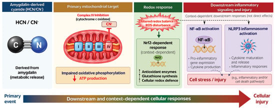
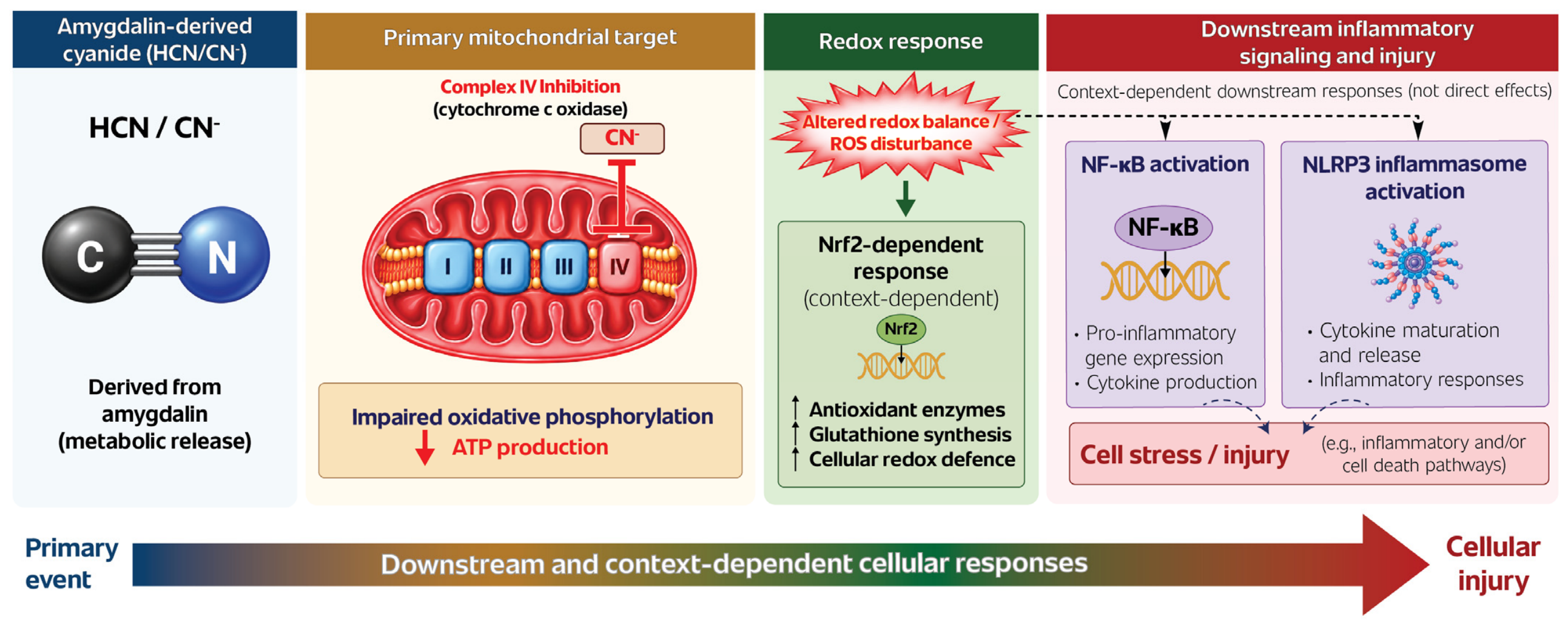
The proposed sequence linking amygdalin-derived cyanide with mitochondrial dysfunction, redox imbalance and secondary inflammatory signaling is summarized in Figure 4.
Figure 4.
Mitochondrial, redox and inflammatory responses potentially associated with amygdalin-derived cyanide. Inhibition of cytochrome c oxidase (Complex IV) is an established toxicological mechanism of cyanide [10,12]. Downstream redox alterations and responses involving Nrf2, NF-κB and NLRP3 are context-dependent; their causal link to cyanide formation was not established in the amygdalin intervention studies discussed here [44,45,46]. Dashed connections represent possible downstream associations rather than a single experimentally established pathway.
Cell death pathways provide an additional link. Mitochondrial depolarization and respiratory failure can promote apoptosis through cytochrome c release and caspase activation [19,20]. More severe metabolic injury may favor necrotic or regulated necrotic processes, while ferroptosis-related changes have been reported in intestinal inflammation models involving amygdalin [45]. Which pathway predominates will depend on exposure level, cell type, duration and the extent of cyanogenic activation. The experimental studies discussed above differ substantially in their models, exposure conditions and measured endpoints. Their main findings and mechanistic limitations are summarized in Table 2.
Table 2.
Experimental evidence for amygdalin-associated cyanogenic stress: study models, exposure conditions, measured endpoints and mechanistic limitations.
8. From Plant Defense to Host Stress: A Cross-System Model
Cross-system comparisons become unreliable when the site of cyanide formation is ignored. In damaged Prunus tissues, cellular disruption brings amygdalin into contact with plant hydrolases [4,5,28,29]. After ingestion, host and microbial enzymes act in the gastrointestinal tract, while cyanide removal depends partly on sulfur-dependent detoxification [7,10,16,17]. Equal amounts of the precursor can consequently produce different exposure profiles.
The cyanogenic stress load described in Section 4 is qualitative and does not predict mitochondrial impairment directly from the administered amygdalin dose.
Several predictions can be tested experimentally. Under otherwise comparable conditions, differences in microbial β-glucosidase activity should alter cyanide release after oral amygdalin exposure. Mechanical disruption of cyanogenic plant material should also affect activation kinetics. Measurements of cyanide, thiocyanate, oxygen consumption and cellular redox status may explain biological responses more reliably than amygdalin concentration alone. These comparisons require time-resolved chemical measurements alongside mitochondrial and redox endpoints.
The evidence supporting individual components of this model differs considerably. Cyanide-mediated inhibition of cytochrome c oxidase is well established [10,12], whereas microbiota-dependent detoxification and the physiological significance of low-level cyanide signaling in mammals remain less certain [17,47,49,50,51,52]. These differences in evidence strength are summarized in Table 3.
Table 3.
Current level of evidence for key components of amygdalin-associated cyanogenic stress.
9. Methodological Gaps and Priorities for Future Research
Nominal amygdalin dose is not a direct measure of cyanide exposure. Conversion through prunasin and mandelonitrile depends on the enzymatic environment, plant matrix and microbial metabolism [7,35]. Experiments addressing cyanogenic stress should, where analytically feasible, monitor amygdalin, prunasin, mandelonitrile, benzaldehyde, free cyanide/HCN and thiocyanate over time [7,17,35,43,58]. Mandelonitrile requires particular care because its stability depends on pH and sample handling; failure to detect it does not necessarily indicate that this intermediate was not formed [35,58].
The reliability of cyanide measurements also depends on sample handling. Free HCN is volatile, while residual enzymatic activity may continue to hydrolyze cyanogenic glycosides after sampling. Studies should therefore distinguish free cyanide from total cyanogenic potential and report the time between collection and stabilization, sample pH, storage conditions, analytical recovery, detection limits and relevant matrix effects. For example, Lee et al. applied matrix-matched calibration for mandelonitrile and free cyanide because both analytes showed substantial signal suppression in syrup matrices [58]. Appropriate controls should include systems with and without β-glucosidase activity, as well as comparisons with relevant degradation products or cyanide exposure. Cyanide scavengers may provide additional mechanistic information, although their effects should not be assumed to be entirely specific. These controls are particularly important when antioxidant or inflammatory responses are attributed to intact amygdalin rather than its cyanogenic metabolites [43,52].
Purified amygdalin, kernel powders, plant extracts, food products and laetrile-like preparations cannot be assumed to be equivalent [2,4,41]. Epimeric composition, prunasin content, endogenous enzyme activity, processing history and total cyanogenic potential may all influence the resulting exposure [41,43,56,57]. Precise characterization is particularly important for studies that compare biological effects across formulations.
Oral dosing exposes amygdalin to gastrointestinal enzymes and the gut microbiota before systemic absorption, whereas intravenous administration bypasses much of this activation environment [3,16,17,39,40]. In vitro studies may lack relevant β-glucosidase activity entirely. Conclusions should therefore be limited to the biochemical conditions actually present in the model rather than generalized to amygdalin as a compound.
Animal studies demonstrate that intestinal microorganisms can substantially alter cyanide formation and thiocyanate kinetics [16,17], yet most amygdalin studies do not measure microbial function. Taxonomic profiling alone is unlikely to resolve this problem. Enzyme activity, functional gene content and metabolite formation are more directly relevant to cyanogenic exposure [60,61,62,63]. Human data are particularly sparse, leaving the extent of interindividual microbiota-driven variability largely unresolved.
Cyanide toxicity depends not only on production but also on removal, particularly sulfur-dependent conversion to thiocyanate [10,17]. Sulfur availability, sulfurtransferase activity, nutritional state, liver and kidney function, and competing metabolic demands may therefore modify susceptibility. These variables are seldom incorporated into experimental designs, even though they may explain why similar cyanide burdens produce different biological outcomes.
Cytochrome c oxidase is the primary toxicological target of cyanide, yet many studies emphasize general oxidative or inflammatory markers without establishing whether mitochondrial respiration was impaired [10,12]. Oxygen consumption, Complex IV activity, ATP production, membrane potential and mitochondrial ROS should be prioritized when the aim is to characterize cyanogenic stress.
Redox and inflammatory endpoints are most informative when interpreted within that mitochondrial context. Changes in glutathione, antioxidant enzymes, lipid peroxidation, Nrf2, NF-κB, NLRP3 or cytokine concentrations can indicate several different processes: compensation, tissue protection, metabolic inhibition or injury [18,19,20,44,45,46,65]. None of these markers, considered alone, identifies the chemical origin of the response. Pairing them with cyanide kinetics and mitochondrial measurements would substantially improve mechanistic interpretation.
Amygdalin hydrolysis, cyanide production, thiocyanate formation, respiratory inhibition, ROS generation and inflammatory signaling do not occur simultaneously. Single time-point measurements can miss transient cyanide peaks or detect secondary inflammatory effects after the initiating chemical event has passed. Time-resolved designs are therefore needed to separate exposure, early mitochondrial response and later tissue-level consequences.
Acute cyanide poisoning is well described, whereas the effects of recurrent consumption of amygdalin-containing products are much less certain [10,53]. Questions remain about cumulative thiocyanate burden, mitochondrial adaptation, thyroid-related effects, persistent redox changes and microbiome remodeling. Existing evidence is insufficient to define an amygdalin-specific chronic stress phenotype.
Based on the limitations identified above, a minimum set of experimental and reporting parameters for mechanistic studies of amygdalin-associated cyanogenic stress is proposed in Table 4.
Table 4.
Core reporting information and mechanism-dependent measurements for studies of amygdalin-associated cyanogenic stress.
10. Conclusions
Amygdalin contributes to cyanogenic defense in plants, but its significance after ingestion depends on the amount and rate of cyanide released. Excessive HCN exposure inhibits cytochrome c oxidase and can produce severe mitochondrial and systemic toxicity [10,12,53,54,55]. In plants, localized HCN formation also participates in stress-related signaling [9,13,14,15]. The physiological relevance of comparable low-level effects in mammals remains uncertain [47,49,50,51,52].
For oral amygdalin, intestinal hydrolysis, microbial metabolism and sulfur-dependent detoxification influence the resulting cyanide exposure [16,17]. These factors complicate comparisons between purified compounds, processed plant materials, cell cultures and animal models. Reports of antioxidant or anti-inflammatory activity require particular caution when cyanogenic activation has not been measured. Future studies should combine time-resolved measurements of amygdalin and its products of biotransformation with appropriate controls and mitochondrial, redox and inflammatory endpoints to help distinguish responses to the parent glycoside from those associated with its degradation.
Author Contributions
Conceptualization, A.G.; methodology, A.G.; writing—original draft preparation, A.G.; writing—review and editing, A.G. and R.R.; visualization, A.G. All authors have read and agreed to the published version of the manuscript.
Funding
This research received no external funding.
Institutional Review Board Statement
Not applicable.
Informed Consent Statement
Not applicable.
Data Availability Statement
No new data were created or analyzed in this study. Data sharing is not applicable to this article.
Acknowledgments
The authors would like to thank Mariusz Malinowski, mariusz.malinowski@student.wsb.edu.pl, a student of English Philology at WSB University, for his assistance with English-language editing during the preparation of this manuscript.
Conflicts of Interest
The authors declare no conflicts of interest.
Abbreviations
The following abbreviations are used in this manuscript:
| AH | Amygdalin hydrolase |
| ATP | Adenosine triphosphate |
| CAS | β-Cyanoalanine synthase |
| CN− | Cyanide anion |
| COX-2 | Cyclooxygenase-2 |
| CYP71 | Cytochrome P450 family 71 |
| CYP79 | Cytochrome P450 family 79 |
| DSS | Dextran sulfate sodium |
| HCN | Hydrogen cyanide |
| IL-1β | Interleukin-1 beta |
| IL-6 | Interleukin-6 |
| iNOS | Inducible nitric oxide synthase |
| MDL | Mandelonitrile lyase |
| NADH/NAD+ | Reduced/oxidized nicotinamide adenine dinucleotide |
| NF-κB | Nuclear factor kappa B |
| NLRP3 | NLR family pyrin domain-containing protein 3 |
| NQO1 | NAD(P)H:quinone oxidoreductase 1 |
| Nrf2 | Nuclear factor erythroid 2-related factor 2 |
| PAD3 | Phytoalexin deficient 3 |
| PH | Prunasin hydrolase |
| ROS | Reactive oxygen species |
| SCN− | Thiocyanate anion |
| TNF-α | Tumor necrosis factor alpha |
| TST | Thiosulfate sulfurtransferase (rhodanese) |
| UDP | Uridine diphosphate |
| UGT | UDP-dependent glycosyltransferase |
| VEGF | Vascular endothelial growth factor |
References
- Jaszczak-Wilke, E.; Polkowska, Ż.; Koprowski, M.; Owsianik, K.; Mitchell, A.E.; Bałczewski, P. Amygdalin: Toxicity, Anticancer Activity and Analytical Procedures for Its Determination in Plant Seeds. Molecules 2021, 26, 2253. [Google Scholar] [CrossRef] [Scilit] [PubMed]
- He, X.-Y.; Wu, L.-J.; Wang, W.-X.; Xie, P.-J.; Chen, Y.-H.; Wang, F. Amygdalin-A Pharmacological and Toxicological Review. J. Ethnopharmacol. 2020, 254, 112717. [Google Scholar] [CrossRef] [Scilit] [PubMed]
- Zagrobelny, M.; Bak, S.; Møller, B.L. Cyanogenesis in Plants and Arthropods. Phytochemistry 2008, 69, 1457–1468. [Google Scholar] [CrossRef] [Scilit] [PubMed]
- Gleadow, R.M.; Møller, B.L. Cyanogenic Glycosides: Synthesis, Physiology, and Phenotypic Plasticity. Annu. Rev. Plant Biol. 2014, 65, 155–185. [Google Scholar] [CrossRef] [Scilit] [PubMed]
- Morant, A.V.; Jørgensen, K.; Jørgensen, C.; Paquette, S.M.; Sánchez-Pérez, R.; Møller, B.L.; Bak, S. β-Glucosidases as Detonators of Plant Chemical Defense. Phytochemistry 2008, 69, 1795–1813. [Google Scholar] [CrossRef] [Scilit] [PubMed]
- Jaswal, V.; Palanivelu, J.; Ramalingam, C. Effects of the Gut Microbiota on Amygdalin and Its Use as an Anti-Cancer Therapy: Substantial Review on the Key Components Involved in Altering Dose Efficacy and Toxicity. Biochem. Biophys. Rep. 2018, 14, 125–132. [Google Scholar] [CrossRef] [Scilit] [PubMed]
- Newmark, J.; Brady, R.O.; Grimley, P.M.; Gal, A.E.; Waller, S.G.; Thistlethwaite, J.R. Amygdalin (Laetrile) and Prunasin Beta-Glucosidases: Distribution in Germ-Free Rat and in Human Tumor Tissue. Proc. Natl. Acad. Sci. USA 1981, 78, 6513–6516. [Google Scholar] [CrossRef] [Scilit] [PubMed]
- Sánchez-Pérez, R.; Neilson, E.H. The Case for Sporadic Cyanogenic Glycoside Evolution in Plants. Curr. Opin. Plant Biol. 2024, 81, 102608. [Google Scholar] [CrossRef] [Scilit] [PubMed]
- Krasuska, U.; Ciacka, K.; Staszek, P.; Tyminski, M.; Wal, A.; Gniazdowska, A. Hormetic Action of Cyanide: Plant Gasotransmitter and Poison. Phytochem. Rev. 2024, 23, 705–719. [Google Scholar] [CrossRef] [Scilit]
- Lachowicz, J.I.; Alexander, J.; Aaseth, J.O. Cyanide and Cyanogenic Compounds—Toxicity, Molecular Targets, and Therapeutic Agents. Biomolecules 2024, 14, 1420. [Google Scholar] [CrossRef] [Scilit] [PubMed]
- Milazzo, S.; Horneber, M. Laetrile Treatment for Cancer. Cochrane Database Syst. Rev. 2015, 2015, CD005476. [Google Scholar] [CrossRef] [Scilit] [PubMed]
- Cooper, C.E.; Brown, G.C. The Inhibition of Mitochondrial Cytochrome Oxidase by the Gases Carbon Monoxide, Nitric Oxide, Hydrogen Cyanide and Hydrogen Sulfide: Chemical Mechanism and Physiological Significance. J. Bioenerg. Biomembr. 2008, 40, 533–539. [Google Scholar] [CrossRef] [Scilit] [PubMed]
- García, I.; Arenas-Alfonseca, L.; Moreno, I.; Gotor, C.; Romero, L.C. HCN Regulates Cellular Processes through Posttranslational Modification of Proteins by S-Cyanylation. Plant Physiol. 2019, 179, 107–123. [Google Scholar] [CrossRef] [Scilit] [PubMed]
- Díaz-Rueda, P.; Morales De Los Ríos, L.; Romero, L.C.; García, I. Old Poisons, New Signaling Molecules: The Case of Hydrogen Cyanide. J. Exp. Bot. 2023, 74, 6040–6051. [Google Scholar] [CrossRef] [Scilit] [PubMed]
- Piekarniak, M.; Weston, L.A.; Gniazdowska, A.; Krasuska, U. Hydrogen Cyanide Acts as a Regulator of Reactive Oxygen Species Metabolism. Planta 2025, 262, 134. [Google Scholar] [CrossRef] [Scilit] [PubMed]
- Carter, J.H.; McLafferty, M.A.; Goldman, P. Role of the Gastrointestinal Microflora in Amygdalin (Laetrile)-Induced Cyanide Toxicity. Biochem. Pharmacol. 1980, 29, 301–304. [Google Scholar] [CrossRef] [Scilit] [PubMed]
- Wen, Q.; Yu, S.; Wang, S.; Qin, Y.; Xia, Q.; Wang, S.; Chen, G.; Shen, C.; Song, S. Impact of Intestinal Microbiota on Metabolic Toxicity and Potential Detoxification of Amygdalin. Front. Microbiol. 2022, 13, 1030516. [Google Scholar] [CrossRef] [Scilit] [PubMed]
- Marchi, S.; Guilbaud, E.; Tait, S.W.G.; Yamazaki, T.; Galluzzi, L. Mitochondrial Control of Inflammation. Nat. Rev. Immunol. 2023, 23, 159–173. [Google Scholar] [CrossRef] [Scilit] [PubMed]
- Murphy, M.P. How Mitochondria Produce Reactive Oxygen Species. Biochem. J. 2009, 417, 1–13. [Google Scholar] [CrossRef] [Scilit] [PubMed]
- Sena, L.A.; Chandel, N.S. Physiological Roles of Mitochondrial Reactive Oxygen Species. Mol. Cell 2012, 48, 158–167. [Google Scholar] [CrossRef] [Scilit] [PubMed]
- Boter, M.; Diaz, I. Cyanogenesis, a Plant Defence Strategy against Herbivores. Int. J. Mol. Sci. 2023, 24, 6982. [Google Scholar] [CrossRef] [Scilit] [PubMed]
- Møller, B.L. Functional Diversifications of Cyanogenic Glucosides. Curr. Opin. Plant Biol. 2010, 13, 337–346. [Google Scholar] [CrossRef] [Scilit] [PubMed]
- Zagrobelny, M.; Bak, S.; Rasmussen, A.V.; Jørgensen, B.; Naumann, C.M.; Lindberg Møller, B. Cyanogenic Glucosides and Plant–Insect Interactions. Phytochemistry 2004, 65, 293–306. [Google Scholar] [CrossRef] [Scilit] [PubMed]
- Tattersall, D.B.; Bak, S.; Jones, P.R.; Olsen, C.E.; Nielsen, J.K.; Hansen, M.L.; Høj, P.B.; Møller, B.L. Resistance to an Herbivore Through Engineered Cyanogenic Glucoside Synthesis. Science 2001, 293, 1826–1828. [Google Scholar] [CrossRef] [Scilit] [PubMed]
- Rajniak, J.; Barco, B.; Clay, N.K.; Sattely, E.S. A New Cyanogenic Metabolite in Arabidopsis Required for Inducible Pathogen Defence. Nature 2015, 525, 376–379. [Google Scholar] [CrossRef] [Scilit] [PubMed]
- Chen, K.; Chai, N.; Chen, S.; Ye, C.; Sun, K.; Guo, J.; Zhou, X.; Chen, D.; Liu, J.; Hu, Y.; et al. Engineering of Amygdalin Biosynthesis in Rice Endosperm for Pharmaceutical Production and Sitophilus oryzae Resistance. Plant Biotechnol. J. 2026, 24, 4860–4862. [Google Scholar] [CrossRef] [Scilit] [PubMed]
- Martinez, M.; Diaz, I. Plant Cyanogenic-Derived Metabolites and Herbivore Counter-Defences. Plants 2024, 13, 1239. [Google Scholar] [CrossRef] [Scilit] [PubMed]
- Sánchez-Pérez, R.; Jørgensen, K.; Olsen, C.E.; Dicenta, F.; Møller, B.L. Bitterness in Almonds. Plant Physiol. 2008, 146, 1040–1052. [Google Scholar] [CrossRef] [Scilit] [PubMed]
- Sánchez-Pérez, R.; Belmonte, F.S.; Borch, J.; Dicenta, F.; Møller, B.L.; Jørgensen, K. Prunasin Hydrolases during Fruit Development in Sweet and Bitter Almonds. Plant Physiol. 2012, 158, 1916–1932. [Google Scholar] [CrossRef] [Scilit] [PubMed]
- Jiang, Y.; Chen, J.; Zhang, Y.; Wang, J.; Yang, H.; Sun, Y.; Dong, S. Transcriptional Regulation of Amygdalin Biosynthesis and Catabolism during Kernel Development in Prunus Mandshurica. Food Biosci. 2026, 78, 108494. [Google Scholar] [CrossRef] [Scilit]
- Zhang, M.; Jiang, F.; Yang, L.; Yu, W.; Ling, J.; Wang, Y.; Zhang, J.; Sun, H. Indel Mutation in Transcription Factor PabHLH2 Regulates Amygdalin Accumulation and Kernel Bitterness in Apricot. Plant J. 2025, 124, e70523. [Google Scholar] [CrossRef] [Scilit] [PubMed]
- Cuny, M.A.C.; La Forgia, D.; Desurmont, G.A.; Glauser, G.; Benrey, B. Role of Cyanogenic Glycosides in the Seeds of Wild Lima Bean, Phaseolus Lunatus: Defense, Plant Nutrition or Both? Planta 2019, 250, 1281–1292. [Google Scholar] [CrossRef] [Scilit] [PubMed]
- Yang, J.; Du, H.; Tao, F.; Ashraf, M.A.; Gao, X.; Huang, X.; Zhu, K.; Li, G.; Zheng, J.; Bonfante, P.; et al. Rhizosphere Microbial Shifts Drive Amygdalin Detoxification and Jasmonate-Mediated Alleviation of Peach Autotoxicity. ISME J. 2026, 20, wrag095. [Google Scholar] [CrossRef] [Scilit] [PubMed]
- Stathaki, A.; Pantidi, G.; Thomopoulou, M.; Koudounas, K. β-Glucosidases in Specialized Metabolism: Towards a New Understanding of the Gatekeepers of Plant Chemical Arsenal. Curr. Opin. Plant Biol. 2024, 82, 102638. [Google Scholar] [CrossRef] [Scilit] [PubMed]
- Haisman, D.; Knight, D. The Enzymic Hydrolysis of Amygdalin. Biochem. J. 1967, 103, 528–534. [Google Scholar] [CrossRef] [Scilit] [PubMed]
- Li, C.P.; Swain, E.; Poulton, J.E. Prunus serotina Amygdalin Hydrolase and Prunasin Hydrolase: Purification, N-Terminal Sequencing, and Antibody Production. Plant Physiol. 1992, 100, 282–290. [Google Scholar] [CrossRef] [Scilit] [PubMed]
- Zheng, L.; Poulton, J.E. Temporal and Spatial Expression of Amygdalin Hydrolase and (R)-(+)-Mandelonitrile Lyase in Black Cherry Seeds. Plant Physiol. 1995, 109, 31–39. [Google Scholar] [CrossRef] [Scilit] [PubMed]
- Wu, H.-C.; Poulton, J.E. Immunocytochemical Localization of Mandelonitrile Lyase in Mature Black Cherry (Prunus serotina Ehrh.) Seeds. Plant Physiol. 1991, 96, 1329–1337. [Google Scholar] [CrossRef] [Scilit] [PubMed]
- Moertel, C.G.; Ames, M.M.; Kovach, J.S.; Moyer, T.P.; Rubin, J.R.; Tinker, J.H. A Pharmacologic and Toxicological Study of Amygdalin. JAMA 1981, 245, 591–594. [Google Scholar] [CrossRef]
- Rauws, A.G.; Olling, M.; Timmerman, A. The Pharmacokinetics of Amygdalin. Arch. Toxicol. 1982, 49, 311–319. [Google Scholar] [CrossRef] [Scilit] [PubMed]
- Wahab, M.F.; Breitbach, Z.S.; Armstrong, D.W.; Strattan, R.; Berthod, A. Problems and Pitfalls in the Analysis of Amygdalin and Its Epimer. J. Agric. Food Chem. 2015, 63, 8966–8973. [Google Scholar] [CrossRef] [Scilit] [PubMed]
- Liu, W.; Hu, X.; Zhou, M.; Cai, F.; Zhang, S.; Lei, J.; Ma, W.; Qi, S.; Liu, W.; Wang, C. Stereoselective Pharmacokinetics and Anti-Inflammatory Activity of Amygdalin Epimers: Implications for Thermal and pH Stability in Amygdalin-Based Functional Foods. Food Funct. 2026, 17, 6849–6867. [Google Scholar] [CrossRef] [Scilit] [PubMed]
- Doria, E.; Lerno, L.; Chen, M.-A.; Lee, J.; Huang, G.; Mitchell, A.E. Novel UHPLC-(+ESI)MS/MS Method for Determining Amygdalin, Prunasin and Total Cyanide in Almond Kernels and Hulls (Prunus dulcis). J. Agric. Food Chem. 2025, 73, 5500–5510. [Google Scholar] [CrossRef] [Scilit] [PubMed]
- Tang, F.; Fan, K.; Wang, K.; Bian, C. Amygdalin Attenuates Acute Liver Injury Induced by D-Galactosamine and Lipopolysaccharide by Regulating the NLRP3, NF-κB and Nrf2/NQO1 Signalling Pathways. Biomed. Pharmacother. 2019, 111, 527–536. [Google Scholar] [CrossRef] [Scilit] [PubMed]
- Xu, D.; Xie, Y.; Cheng, J.; He, D.; Liu, J.; Fu, S.; Hu, G. Amygdalin Alleviates DSS-Induced Colitis by Restricting Cell Death and Inflammatory Response, Maintaining the Intestinal Barrier, and Modulating Intestinal Flora. Cells 2024, 13, 444. [Google Scholar] [CrossRef] [Scilit] [PubMed]
- Mohamed, R.A.; Ghoneim, M.E.-S.; Tawfiq, R.A.; El Sayed, N.F. Amygdalin Synergizes with the TNF-α Monoclonal Antibody Infliximab to Modulate HSP90 and Related Necro-Inflammatory/Oxidative Stress Pathways in a Rat Model of Hepatic I/R. Naunyn-Schmiedeberg’s Arch. Pharmacol. 2026, 399, 7643–7657. [Google Scholar] [CrossRef] [Scilit] [PubMed]
- Zuhra, K.; Szabo, C. The Two Faces of Cyanide: An Environmental Toxin and a Potential Novel Mammalian Gasotransmitter. FEBS J. 2022, 289, 2481–2515. [Google Scholar] [CrossRef] [Scilit] [PubMed]
- Zhang, W.; Zhang, J.; Bo, S.; Zhan, L.; Xue, Q.; Fan, L.; Pei, Y.; Jin, Z. H2S-Induced PAD3 Persulfidation Balances H2S-HCN Homoeostasis for Osmotic Stress Tolerance in Arabidopsis. Plant Cell Environ. 2026, 49, 5965–5978. [Google Scholar] [CrossRef] [Scilit] [PubMed]
- Zuhra, K.; Petrosino, M.; Janickova, L.; Petric, J.; Ascenção, K.; Vignane, T.; Khalaf, M.; Philipp, T.M.; Ravani, S.; Anand, A.; et al. Regulation of Mammalian Cellular Metabolism by Endogenous Cyanide Production. Nat. Metab. 2025, 7, 531–555. [Google Scholar] [CrossRef] [Scilit] [PubMed]
- Randi, E.B.; Zuhra, K.; Pecze, L.; Panagaki, T.; Szabo, C. Physiological Concentrations of Cyanide Stimulate Mitochondrial Complex IV and Enhance Cellular Bioenergetics. Proc. Natl. Acad. Sci. USA 2021, 118, e2026245118. [Google Scholar] [CrossRef] [Scilit] [PubMed]
- Giamogante, F.; Calì, T.; Malatesta, F. Physiological Cyanide Concentrations Do Not Stimulate Mitochondrial Cytochrome c Oxidase Activity. Proc. Natl. Acad. Sci. USA 2021, 118, e2112373118. [Google Scholar] [CrossRef] [Scilit] [PubMed]
- Kieronska-Rudek, A.; Petrosino, M.; Zuhra, K.; Szabo, C. Cyanide Is an Endogenous Stimulator of Endothelial Cell Proliferation, Migration and Differentiation. Exp. Biol. Med. 2026, 251, 10856. [Google Scholar] [CrossRef] [Scilit] [PubMed]
- EFSA Panel on Contaminants in the Food Chain (CONTAM). Acute Health Risks Related to the Presence of Cyanogenic Glycosides in Raw Apricot Kernels and Products Derived from Raw Apricot Kernels. EFSA J. 2016, 14, e04424. [Google Scholar] [CrossRef] [Scilit]
- Sauer, H.; Wollny, C.; Oster, I.; Tutdibi, E.; Gortner, L.; Gottschling, S.; Meyer, S. Severe Cyanide Poisoning from an Alternative Medicine Treatment with Amygdalin and Apricot Kernels in a 4-Year-Old Child. Wien. Med. Wochenschr. 2015, 165, 185–188. [Google Scholar] [CrossRef] [Scilit] [PubMed]
- Cigolini, D.; Ricci, G.; Zannoni, M.; Codogni, R.; De Luca, M.; Perfetti, P.; Rocca, G. Hydroxocobalamin Treatment of Acute Cyanide Poisoning from Apricot Kernels. BMJ Case Rep. 2011, 2011, bcr0320113932. [Google Scholar] [CrossRef] [Scilit] [PubMed]
- Bolarinwa, I.F.; Orfila, C.; Morgan, M.R.A. Amygdalin Content of Seeds, Kernels and Food Products Commercially-Available in the UK. Food Chem. 2014, 152, 133–139. [Google Scholar] [CrossRef] [Scilit] [PubMed]
- Lee, J.; Zhang, G.; Wood, E.; Rogel Castillo, C.; Mitchell, A.E. Quantification of Amygdalin in Nonbitter, Semibitter, and Bitter Almonds (Prunus dulcis) by UHPLC-(ESI)QqQ MS/MS. J. Agric. Food Chem. 2013, 61, 7754–7759. [Google Scholar] [CrossRef] [Scilit] [PubMed]
- Lee, H.; Cho, S.Y.; Kim, Y.-K.; Kim, H.-S.; Mitchell, A.; Chun, H.S.; Lee, K.-W.; Lee, J. Quantification of Amygdalin, Prunasin, Mandelonitrile, and Free Cyanide in Homemade and Commercial Prunus Mume Syrups Using UHPLC-(ESI)QqQ-MS/MS and Their Dietary Risk Assessment. Food Chem. 2026, 507, 148175. [Google Scholar] [CrossRef] [Scilit] [PubMed]
- Moertel, C.G.; Fleming, T.R.; Rubin, J.; Kvols, L.K.; Sarna, G.; Koch, R.; Currie, V.E.; Young, C.W.; Jones, S.E.; Davignon, J.P. A Clinical Trial of Amygdalin (Laetrile) in the Treatment of Human Cancer. N. Engl. J. Med. 1982, 306, 201–206. [Google Scholar] [CrossRef] [Scilit] [PubMed]
- Koppel, N.; Maini Rekdal, V.; Balskus, E.P. Chemical Transformation of Xenobiotics by the Human Gut Microbiota. Science 2017, 356, eaag2770. [Google Scholar] [CrossRef] [Scilit] [PubMed]
- Zimmermann, M.; Zimmermann-Kogadeeva, M.; Wegmann, R.; Goodman, A.L. Mapping Human Microbiome Drug Metabolism by Gut Bacteria and Their Genes. Nature 2019, 570, 462–467. [Google Scholar] [CrossRef] [Scilit] [PubMed]
- Spanogiannopoulos, P.; Bess, E.N.; Carmody, R.N.; Turnbaugh, P.J. The Microbial Pharmacists within Us: A Metagenomic View of Xenobiotic Metabolism. Nat. Rev. Microbiol. 2016, 14, 273–287. [Google Scholar] [CrossRef] [Scilit] [PubMed]
- He, J.; Liu, X.; Zhang, J.; Wang, R.; Cao, X.; Liu, G. Gut Microbiome-Derived Hydrolases—An Underrated Target of Natural Product Metabolism. Front. Cell. Infect. Microbiol. 2024, 14, 1392249. [Google Scholar] [CrossRef] [Scilit] [PubMed]
- Xu, X.; Pang, Y.; Fan, X. Mitochondria in Oxidative Stress, Inflammation and Aging: From Mechanisms to Therapeutic Advances. Sig. Transduct. Target. Ther. 2025, 10, 190. [Google Scholar] [CrossRef] [Scilit] [PubMed]
- Saha, S.; Buttari, B.; Panieri, E.; Profumo, E.; Saso, L. An Overview of Nrf2 Signaling Pathway and Its Role in Inflammation. Molecules 2020, 25, 5474. [Google Scholar] [CrossRef] [Scilit] [PubMed]

Disclaimer/Publisher’s Note: The statements, opinions and data contained in all publications are solely those of the individual author(s) and contributor(s) and not of MDPI and/or the editor(s). MDPI and/or the editor(s) disclaim responsibility for any injury to people or property resulting from any ideas, methods, instructions or products referred to in the content. |
© 2026 by the authors. Licensee MDPI, Basel, Switzerland. This article is an open access article distributed under the terms and conditions of the Creative Commons Attribution (CC BY) license.