


Abstract
Background/Objectives: This study evaluated whether walking speed and backward-directed resistive force interact to systematically alter knee joint kinematics and activity of knee flexor and extensor muscles in healthy adults. Methods: Fourteen active young adults (18–35 years) without lower-extremity injuries walked in a randomized order at 0.5 and 1.0 m/s while experiencing backward-directed resistive forces ranging from 0% to 25% of body weight. Knee joint kinematics, stride characteristics, and electromyographic (EMG) activity of the vastus lateralis, vastus medialis, and biceps femoris were collected and quantified. Results: No significant speed × force interactions were observed for step kinematic outcomes (p > 0.05). Walking speed significantly affected stride length and stride time (p < 0.05), whereas force did not (p > 0.05). For knee stance phase kinematics, force significantly affected outcomes (p < 0.05), whereas speed and the speed × force interaction did not (p > 0.05). For EMG activity, speed and the speed × force interaction was not significant for any muscle (p > 0.05). Resistive force significantly affected activation of all three muscles (p < 0.05). Conclusions: Backward-directed resistive forces alter knee kinematics and EMG activity, with effects becoming evident at approximately 15% of body weight regardless of walk speed. These findings suggest that posteriorly directed resistance to the center of mass at slower walk speeds may provide a novel approach for modifying knee joint kinematics and lower-extremity muscular activity while walking on a stable, level surface.
1. Introduction
Walking in the community requires individuals to modulate their speed while also adapting to environmental constraints such as hills, resulting in changes in joint kinematics and muscular activation. During level walking the knee joint primarily absorbs mechanical energy and provides body weight support, whereas increasing speed or treadmill incline requires greater generation of positive mechanical work at the knee [1,2]. This increased work is associated with greater knee kinematics (i.e., knee flexion) and subsequent muscular activation [3]. Although manipulation of treadmill speed and incline to generate greater mechanical work while walking is common, recent studies have also investigated the use of externally applied resistive forces. The application of backward-directed resistive forces to an individual’s center of mass (COM) requires greater propulsive forces compared to walking at the same speed without external forces [4]. However, it is unclear how altering these resistive forces interacts with speed to influence knee joint kinematics and muscular activation.
Previous research has shown that, acutely, a posterior-directed horizontal force applied to the COM during walking increases quadriceps muscle activation [5]. With respect to knee kinematics, knee flexion at heel strike and maximum knee extension during stance generally increase with walking speed [6,7], although this relationship may be nonlinear at slower walking speeds [6]. Additionally, peak knee flexion may be more sensitive to increases in treadmill incline than to increases in treadmill speed [3,8]. Although no study to date has directly compared the kinematic and kinetic outcomes of walking at increasing treadmill inclines with those of walking against equivalent increasing backward-directed resistive forces applied at the COM, our previous work has shown similar physiologic outcomes using backward forces matched to mechanical work of the same individual running uphill [9]. Thus, walking against backward resistive forces could provide a safe and controlled environment for challenging walking.
The purpose of this study was to evaluate the extent to which walking speed and backward-directed resistive force interact to create a systematic difference in stride kinematics, knee range of motion, and muscular activation of the knee in young, healthy individuals. Previous research suggests that walking slowly on an incline can result in a larger change in knee flexion than walking speed alone [3]. However, it remains unclear whether slower walking speed combined with increasing backward-directed resistive forces can similarly produce targeted changes in gait kinematics and muscular activation. We hypothesized that knee kinematics and EMG activity of muscles acting at the knee would increase to a greater degree with backward-directed resistive forces than increased walking speeds. Although the present study evaluates these responses in nonimpaired individuals, establishing these relationships may provide a foundation for developing targeted walking interventions for populations where increasing walking speed is difficult or unsafe.
2. Materials and Methods
We recruited 14 young, active individuals (10 female and 4 male, 1.71 ± 0.11 m, 77 ± 21.8 kg, 23.0 years old) between the ages of 18–35 without any previous or current musculoskeletal impairments such as anterior cruciate ligament (ACL) injury and who were moderately-to-vigorously active for a minimum of three days per week [10]. Definitions were provided to participants based on the ACSM guidelines. This sample reflects the most common population to sustain an ACL injury and undergo ACL reconstruction (ACLR) [11,12]. In the current investigation, limb dominance was not assessed. Informed consent was obtained from all subjects involved in the study. The study was conducted in accordance with the Declaration of Helsinki and approved by the Institutional Review Board of The University of Alabama at Birmingham IRB-300003740, October 2019.
2.1. Testing Protocol
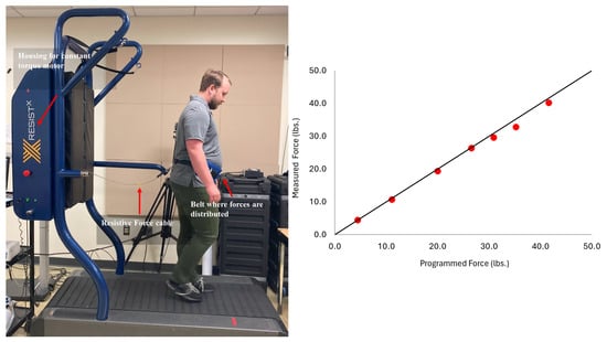
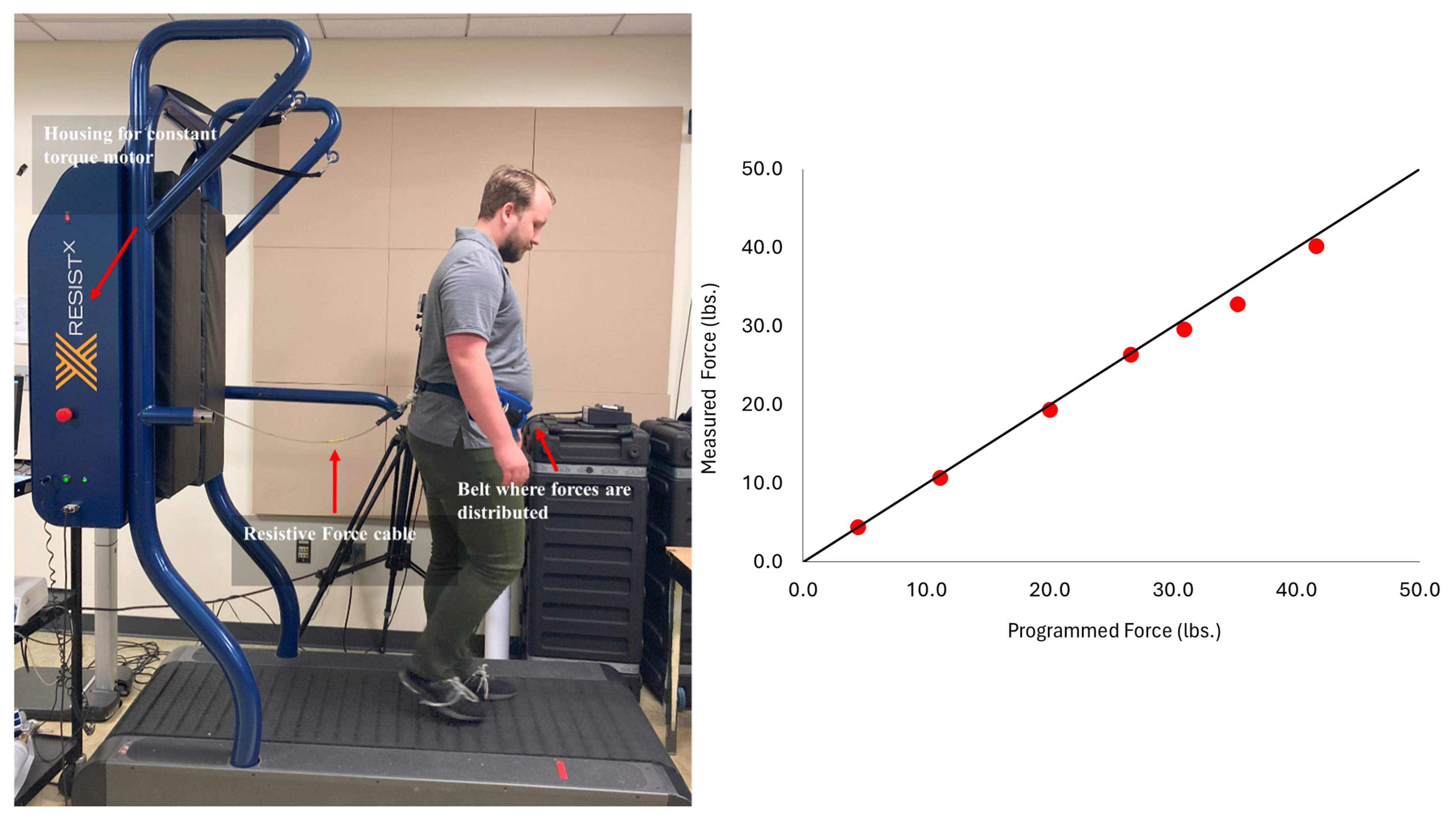
Participants walked at two speeds: 0.5 m/s and 1.0 m/s, which is slower than is typical for healthy young participants; however, these speeds are reasonable for patient populations in rehabilitation [13], and slower speeds can reduce joint contact forces [14] while walking against backward-directed resistive forces. The presentation of all walking trials was randomized to prevent an ordering effect. All walking trials were completed on a device (Figure 1) that integrates a custom resistive force mechanism with a commercially available treadmill [15] (Woodway, Waukesha, WI, USA). The application of force connected to the participant via a padded belt connected to braided cable with a worm drive height adjustment mechanism to ensure the forces were delivered to the height of the participant’s COM. Visual inspection on every trial ensured the cable remained level with the horizontal to ensure that primarily horizontal backward force was applied. We (Figure 1) and others have described the accuracy of the constant torque motor applying a near-constant backward force despite step-to-step fluctuations in body positioning on the treadmill as individuals walk on the device [15]. The device provides a safe walking environment and allows for independent control of: (1) resistive forces and (2) belt speed/gait speed [15].
Figure 1.
(Left) ResistX mounted on Woodway Treadmill (Woodway, Waukesha, WI, USA). Shown is the mechanism where the constant torque motor applies resistive force to the individual. Pictured is a co-author (CK). (Right) A scatterplot of validation results comparing the programmed and measured forces (lbs.) from the constant torque motor. The data are plotted (red circles) with the line of identity (black) showing close agreement.
Individuals walked against backward-directed forces that equated to 0–25% of their body weight in 5% increments. Individuals initially completed a 30 s walking trial with the harness on, while no pulling force was applied to acclimate to the experimental conditions. The experiment included two-minute walking trials. Transferring between trials took around 60 s, and individuals were asked if they needed a longer break between trials. Only two participants requested a total of 3 breaks between 22 and 36 s in addition to the transfer time. We collected gait kinematics using a 3-dimensional motion capture system operating at 100 Hz (Vicon, Oxford, UK). Marker position data were tracked and processed offline (Vicon Nexus) to construct a 7-segment unilateral rigid body biomechanical model for each participant (Visual 3D, Rockville, MD, USA), consisting of the right-side foot, lower leg, upper leg, pelvis, trunk, upper arm and lower arm segment. Knee joint angular data were processed and low-pass filtered at 6 Hz using a Butterworth filter. Stance phase knee joint kinematics were quantified as maximum and minimum knee joint flexion during stance as well as knee angle at heel strike. We also quantified max swing phase flexion angle to investigate if speed or resistive force affected knee joint kinematics during swing phase. Initial contact and foot off were calculated using kinematics of the heel and toe markers. We also quantified stride time and stride length to assess if stride kinematic changes with increasing resistive force. A wireless electromyography (EMG) data acquisition system with interelectrode distances of 10 mm spacing between parallel bar contacts was used to collect EMG data during each trial at 2000 Hz (Delysis, Natick, MA, USA). All data were collected and synced via the Vicon Nexus software. EMG data were collected from the vastus lateralis, vastus medialis, and biceps femoris, muscles responsible for knee flexion and extension. We prepped the EMG electrodes consistent with the Surface Electromyography for Non-Invasive Assessment of Muscle (SENIAM) guidelines [16]. The EMG data were zeroed and rectified, and a linear envelope was created using a low-pass Butterworth filter at 25 Hz and integrated from heel strike to toe off step-by-step to calculate integrated EMG (iEMG) during the stance phase. The cutoff frequency was chosen to create the linear envelope while maintaining variability between participants and is similar to previous work [17]. Average iEMG data over the stance phase was quantified.
2.2. Statistical Analyses
Statistical analyses tested the hypothesis that knee kinematics and EMG activity of muscles acting at the knee would increase to a greater degree with backward-directed resistive forces than increased walking speeds. Our statistical analyses consisted of two-factor repeated measures ANOVA for the kinematic and EMG data, respectively. The two factors were speed and resistive force. Mauchly’s Test of Sphericity was performed on all variables, and where the condition of sphericity was violated, the Greenhouse Geisser adjusted p-values are reported. Effect sizes are reported using partial ɳ2. For significant main effects, we performed post hoc comparisons with the Bonferroni correction that provided adjusted p-values for multiple comparisons within each dependent variable. For post hoc analysis, we were most interested in when the outcome variable was significantly different from the no force condition, i.e., 0% force condition. Significance level was set to p < 0.05.
3. Results
3.1. Kinematic Results
Kinematic data responded to increased speed and resistive forces differentially in our young participant group. For all variables across all participants, we collected between 85 and 105 strides. Stride kinematic showed a systematic effect of speed but not resistive force. Stride time showed a nonsignificant speed by force interaction (p = 0.649, partial ɳ2 = 0.10) and no effect of force (p = 0.335, partial ɳ2 = 0.198), but a significant effect of speed (p < 0.001, partial ɳ2 = 0.967). Stride time was less at 1.0 m/s (1.22 s ± 0.01 SEM) compared to 0.5 m/s (1.7 s ± 0.03 SEM). Stride length showed a nonsignificant speed by force interaction (p = 0.774, partial ɳ2 = 0.22) and no effect of force (p = 0.185, partial ɳ2 = 0.132), but a significant effect of speed (p < 0.01, partial ɳ2 = 0.646). Stride length was greater for 1.0 m/s (1.1 m ± 0.03 SEM) compared to 0.5 m/s (0.78 m ± 0.01 SEM).
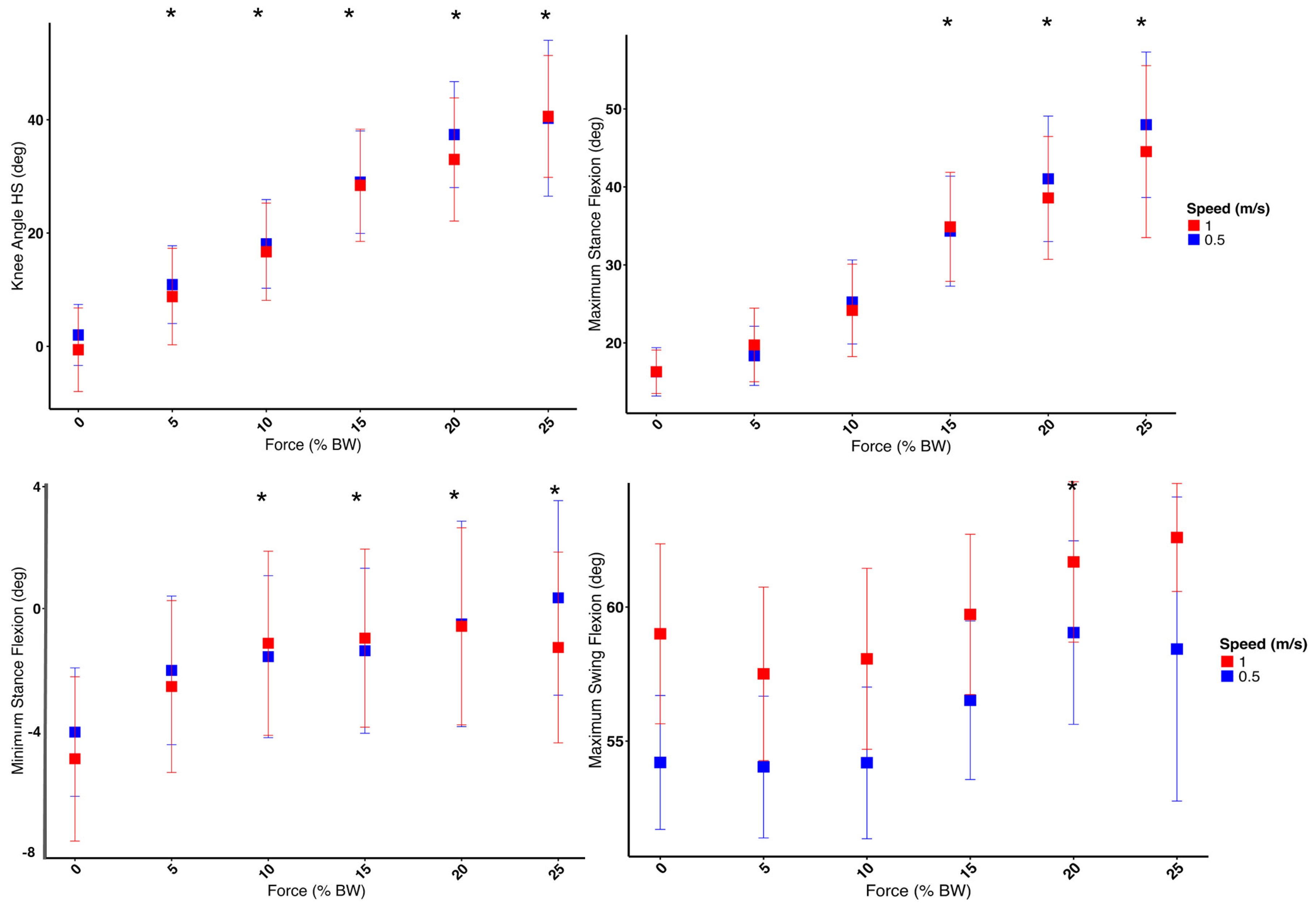
Stance phase knee joint kinematics were affected by force but not speed for all outcome variables (Figure 2). For minimum knee flexion angle during stance, there was no speed by force interaction (p = 0.342, partial ɳ2 = 0.073) or a main effect of speed (p = 0.382, partial ɳ2 = 0.059), but a significant main effect of force (p < 0.001, partial ɳ2 = 0.485). Our post hoc analysis revealed that 10% through the 25% force conditions resulted in a significant increase compared to the 0% force condition (p < 0.01 for all comparisons). For maximum knee flexion angle during stance, there was no speed by force interaction (p = 0.157, partial ɳ2 = 0.13) or a main effect of speed (p = 0.240, partial ɳ2 = 0.105), but a significant main effect of force (p < 0.001, partial ɳ2 = 0.784). Our post hoc analysis also revealed that the 15% through the 25% force conditions resulted in a significant increase compared to the 0% force condition (p < 0.01 for all comparisons). For knee flexion angle at heel strike, there was no speed by force interaction (p = 0.419, partial ɳ2 = 0.068) or a main effect of speed (p = 0.114, partial ɳ2 = 0.181), but a significant effect of force (p < 0.001, partial ɳ2 = 0.867). Our post hoc analysis revealed that the 5% through the 25% force conditions resulted in a significant increase compared to the 0% force condition (p < 0.01 for all comparisons). Finally, we assessed the effect on maximum knee flexion angle during swing phase. For maximum swing knee flexion, there was no speed by force interaction (p = 0.286, partial ɳ2 = 0.091); however we observed a significant main effect of speed (p = 0.007, partial ɳ2 = 0.437) and a significant main effect of force (p < 0.001, partial ɳ2 = 0.526). Only 20% was significantly different from the 0% condition but all other comparisons were not (p > 0.05). For 95% confidence intervals of post hoc differences, see Supplementary Table S1.
Figure 2.
Knee kinematic responses by speed and resistive forces for average knee angle at heel strike, minimum stance knee flexion and maximum stance flexion and maximum knee flexion during the swing phase. Post hoc analysis results showing significance from 0% trial denoted with *. Speed was not statistically significant across stance phase variables, only the maximum swing phase flexion where the faster speed resulted in larger swing phase flexion angles.
3.2. Muscle Activation During Stance
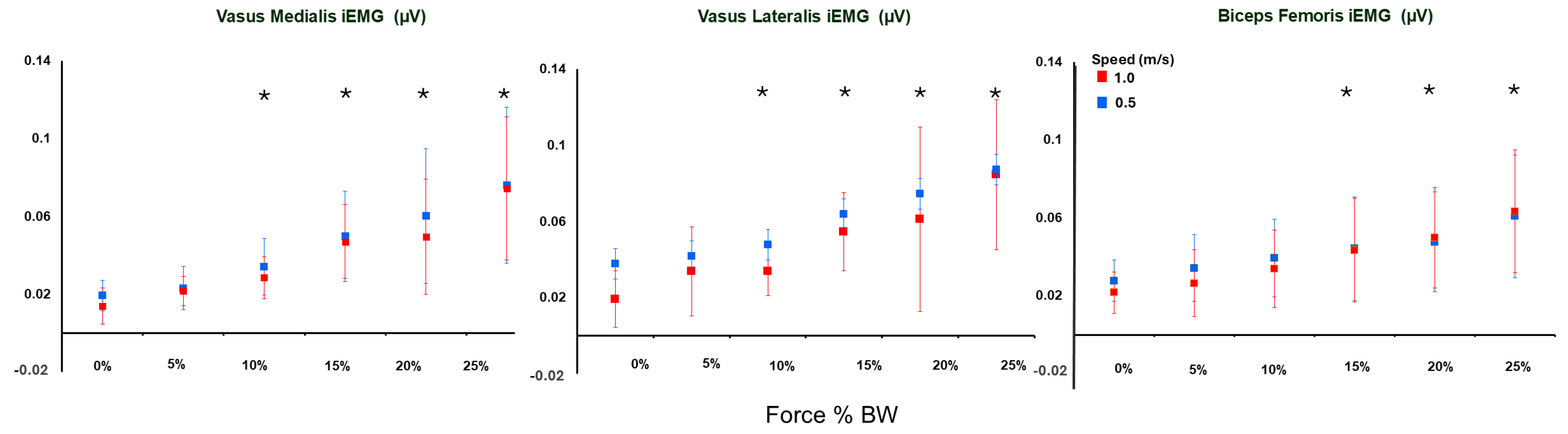
We observed a consistent effect of increasing muscular activation for the knee flexor and extensor muscles with increasing force, but with limited effect of increasing walk speed (Figure 3). For the vastus medialis, there was no interaction between walking speed and force trials (p = 0.287, partial ɳ2 = 0.099) or a significant main effect of walking speed (p = 0.07, partial ɳ2 = 0.254), but a significant main effect of force (p < 0.01, partial ɳ2 = 0.639). Our post hoc analysis revealed that the 5% through the 25% force condition resulted in a significant increase compared to 0% (p < 0.043 for all comparisons). For the vastus lateralis, there was no significant interaction between walking speed and force trials (p = 0.437, partial ɳ2 = 0.054) or significant main effect of speed (p = 0.71, partial ɳ2 = 0.012), but a significant main effect of force (p < 0.006, partial ɳ2 = 0.383). Our post hoc analysis revealed that the 15% through the 25% force trials were significantly increased compared to 0% force (p < 0.047 for all comparisons). Lastly, for the biceps femoris, the interaction between walking speed and force trials was close but failed to reach significance (p = 0.067, partial ɳ2 = 0.179) or a main effect of speed (p = 0.218, partial ɳ2 = 0.123), but a significant main effect of force (p < 0.001, partial ɳ2 = 0.583). Our post hoc analysis revealed that the 15% through the 25% force trials were all significantly increased compared to 0% force (p < 0.0016). For 95% confidence intervals for post hoc differences, see Supplementary Table S1.
Figure 3.
Integrated EMG (iEMG) during stance phase while walking against added backward-directed resistive force equal to 0–25% body weight (BW). We show a significant effect of added force but no difference with respect to treadmill walk speed. Post hoc (*) analysis shows significant differences for all muscles tested at 15–25% body weight (BW) compared to 0 and for the vastii muscles at 10% also.
4. Discussion
The purpose of this study was to evaluate the extent to which walking speed and backward-directed resistance force interacted to create a systematic difference in stride kinematics, knee range of motion, and muscular activation of the knee in young, healthy individuals. We hypothesized that knee kinematics and EMG activity of muscles acting at the knee would increase to a greater degree with backward-directed resistive forces than increased walking speeds. Our results supported this hypothesis. Knee kinematics and EMG variables increased with backward-directed resistive force, particularly at forces greater than 15% of an individual’s body weight for all dependent variables. In keeping with previous work, stride kinematics were impacted by speed and importantly not force. This suggests that changes observed with increasing external forces were not due to the participant taking longer or shorter strides but due to adjusting the knee biomechanics to greater resistive forces. It is unclear if the systematic changes we show at 15% body weight are mechanistic or would change with a different number of participants and require further study.
The change in knee kinematics with added external force is consistent with the literature. One study showed that increasing treadmill incline can significantly increase knee flexion to a greater degree than increasing walking speed [8]. Indeed, in the current study individuals walked against a large range of resistive forces (i.e., 5–25% body weight), and relatively large partial ɳ2 values show that these resistive forces significantly alter knee joint kinematics and muscular activation of walking. While much of the previous literature on added external forces while walking primarily uses treadmill incline [2,3,18], more recently, backward resistive forces applied to the COM are being used to increase external work of individuals while walking on the treadmill [4,8,19]. Both interventions apply greater external work to the individuals. Although we have demonstrated similar physiologic responses when external work rates of walking and running are matched [9], a detailed biomechanical comparison of these modalities has not been performed. Thus, direct comparisons of joint kinetics and kinematics between the two experimental modalities may not be straightforward and require future investigation.
The lack of speed effect on knee kinematics was somewhat surprising, although alterations in stride kinematics with speed are consistent with the literature [20]. Previous research demonstrates that knee kinematics generally increase with walking speed [6,7] and one study showed that increasing walking speed increases both knee range of motion and knee muscle EMG activity [21]. However, some evidence suggests that this relationship may be nonlinear at slower walking speeds [6]. Importantly it should be noted, however, that the reported effect sizes of nonsignificant speed effects range from 0.059 to 0.254, which represent a small to large effect size; thus, the lack of statistical significance may be a power issue related to sample size and affect generalizability of the current results.
The speeds used in the current study are relatively slow for a healthy young cohort [22] and may not have been sufficient to demonstrate the effect of speed on knee range of motion or muscle activation. However, these speeds are common for patient populations recovering from a musculoskeletal injury such as ACLR surgery [13]. The increases in kinematics and muscle activation with increasing resistive force we observed are in line with previous studies that used incline to increase the parallel forces that increased the external work on the individual while walking on a treadmill [3]. We have previously used both treadmill incline and backward-directed resistive force to challenge walking [2,19]. The collective results of investigations using incline walking and backward-directed resistive forces as an experimental manipulation create a hypothesis that increased external work under slower walk speeds could be a potential intervention to address well-described gait maladaptations such as decreased knee joint range of motion and alterations in knee joint moments after ACLR [23,24,25,26] using a flat stable walking surface. Since the current investigation only recruited healthy young individuals, much more work is required to know the extent that backward resistive forces can be an effective intervention for this patient population.
A key limitation of the current study is that we included only a small sample of healthy, active, mostly female individuals. Due to the small sample, we were unable to perform a sensitivity analysis to assess the extent to which sex may have played a role in the data. Thus, caution should be exercised when generalizing these results. However, this study was completed as a purposeful, proof-of-concept study for a future, larger study including clinical populations with knee injuries. Another limitation is related to the unilateral data collection without reference to limb dominance. Although healthy gait is not perfectly symmetrical, previous studies have demonstrated substantial bilateral similarity in lower-extremity gait biomechanics and have shown that the selection of the dominant, nondominant, or a randomly selected limb has minimal influence on biomechanical outcomes in healthy populations [27]. Further, the current study was interested in changes in lower limb behavior between multiple speeds and force levels, which we were able to assess. The current study does not include kinetic data, which would help to address the extent to which joint moments and power are modified by resistive force and treadmill speed. However, the treadmill apparatus used is not integrated with an instrumented treadmill. The ResistX device is not currently readily accessible for clinical practice. However, it is possible to generate similar backward-directed resistive forces in a controlled manner using different modalities [5,28,29]. The results of this study serve to further support the development of such clinical-applied tools, as they would provide a more practical means to use resistive treadmill walking as a clinical intervention.
In conclusion, the findings of this study suggest that slow treadmill walking against a moderate magnitude of force directed posteriorly at an individual’s center of mass can alter gait kinematics, resulting in increased knee flexion range of motion and increased muscle activation of knee flexors and extensors while walking.
Supplementary Materials
The following supporting information can be downloaded at: https://www.mdpi.com/article/10.3390/biomechanics6030084/s1. A supplementary table (Table S1) listing all adjusted p-values, effect sizes, and 95% confidence intervals of post hoc comparisons is included.
Author Contributions
Conceptualization, C.P.H. and K.S.; methodology, C.P.H., K.S., S.F. and C.K.; software, C.P.H. and H.B.S.; validation, C.P.H., K.S., S.F. and C.K.; formal analysis, C.P.H., K.S., S.F. and C.K.; investigation, C.P.H., K.S., S.F. and C.K.; resources, C.P.H.; data curation, C.P.H., K.S., S.F. and C.K.; writing—original draft preparation, C.P.H., K.S., S.F., C.K., C.S.B., M.P.I., L.V. and H.S.; writing—review and editing, C.P.H., K.S., S.F., C.K., C.S.B., M.P.I., L.V. and H.S.; visualization, C.P.H. and H.B.S.; supervision, C.P.H.; project administration, C.P.H. and K.S.; funding acquisition, N/A. All authors have read and agreed to the published version of the manuscript.
Funding
This research received no external funding.
Institutional Review Board Statement
The study was conducted in accordance with the Declaration of Helsinki and approved by the Institutional Review Board of The University of Alabama at Birmingham IRB-300003740, October 2019.
Informed Consent Statement
Informed consent was obtained from all subjects involved in the study.
Data Availability Statement
The data that support the findings of this study are available from the corresponding author, CPH, upon reasonable request.
Acknowledgments
We acknowledge the participants who gave their time to take part in this investigation.
Conflicts of Interest
The authors declare no conflicts of interest.
Abbreviations
The following abbreviations are used in this manuscript:
| COM | Center of Mass |
| ACLR | Anterior Cruciate Ligament Reconstruction |
| EMG | Electromyography |
| iEMG | Integrated Electromyography |
| SEM | Standard Error of Measurement |
References
- Farris, D.J.; Sawicki, G.S. The Mechanics and Energetics of Human Walking and Running: A Joint Level Perspective. J. R. Soc. Interface 2012, 9, 110–118. [Google Scholar] [CrossRef] [Scilit] [PubMed]
- Hurt, C.P.; Kuhman, D.J.; Reed, W.R.; Baumann, A.; Jiang, W.; Marsh, K. Asymmetric Walking on an Incline Affects Aspects of Positive Mechanical Work Asymmetrically. J. Biomech. 2022, 136, 111083. [Google Scholar] [CrossRef] [Scilit] [PubMed]
- Ehlen, K.A.; Reiser, R.F., 2nd; Browning, R.C. Energetics and Biomechanics of Inclined Treadmill Walking in Obese Adults. Med. Sci. Sports Exerc. 2011, 43, 1251–1259. [Google Scholar] [CrossRef] [Scilit] [PubMed]
- Lewek, M.D.; Raiti, C.; Doty, A. The Presence of a Paretic Propulsion Reserve during Gait in Individuals Following Stroke. Neurorehabil. Neural Repair 2018, 32, 1011–1019. [Google Scholar] [CrossRef] [Scilit] [PubMed]
- Ellis, R.G.; Sumner, B.J.; Kram, R. Muscle Contributions to Propulsion and Braking during Walking and Running: Insight from External Force Perturbations. Gait Posture 2014, 40, 594–599. [Google Scholar] [CrossRef] [Scilit] [PubMed]
- Kirtley, C.; Whittle, M.W.; Jefferson, R.J. Influence of Walking Speed on Gait Parameters. J. Biomed. Eng. 1985, 7, 282–288. [Google Scholar] [CrossRef] [Scilit] [PubMed]
- Mannering, N.; Young, T.; Spelman, T.; Choong, P.F. Three-Dimensional Knee Kinematic Analysis during Treadmill Gait: Slow Imposed Speed Versus Normal Self-Selected Speed. Bone Jt. Res. 2017, 6, 514–521. [Google Scholar] [CrossRef] [Scilit] [PubMed]
- Conway, K.A.; Franz, J.R. Increasing the Propulsive Demands of Walking to Their Maximum Elucidates Functionally Limiting Impairments in Older Adult Gait. J. Aging Phys. Act. 2020, 28, 1–8. [Google Scholar] [CrossRef] [Scilit] [PubMed]
- Hurt, C.P.; Bamman, M.M.; Naidu, A.; Brown, D.A. Comparison of Resistance-Based Walking Cardiorespiratory Test to the Bruce Protocol. J. Strength Cond. Res. 2020, 34, 3569–3576. [Google Scholar] [CrossRef] [Scilit] [PubMed]
- Liguori, G.; American College of Sports Medicine. ACSM’s Guidelines for Exercise Testing and Prescription; Lippincott Williams & Wilkins: Philadelphia, PA, USA, 2020. [Google Scholar]
- Kaeding, C.C.; Léger-St-Jean, B.; Magnussen, R.A. Epidemiology and Diagnosis of Anterior Cruciate Ligament Injuries. Clin. Sports Med. 2017, 36, 1–8. [Google Scholar] [CrossRef] [Scilit] [PubMed]
- Waldén, M.; Hägglund, M.; Werner, J.; Ekstrand, J. The Epidemiology of Anterior Cruciate Ligament Injury in Football (Soccer): A Review of the Literature from a Gender-Related Perspective. Knee Surg. Sports Traumatol. Arthrosc. 2011, 19, 3–10. [Google Scholar] [CrossRef] [Scilit] [PubMed]
- Almansouri, S.Y.; Sigward, S.M. Increased Gait Speed Changes Knee Mechanics Following Anterior Cruciate Ligament Reconstruction: Implications and Caution for Treadmill Training in Early Rehabilitation. Int. J. Sports Phys. Ther. 2025, 20, 354–363. [Google Scholar] [CrossRef] [Scilit] [PubMed]
- Wang, X.; Ma, Y.; Hou, B.Y.; Lam, W.K. Influence of Gait Speeds on Contact Forces of Lower Limbs. J. Healthc. Eng. 2017, 2017, 6375976. [Google Scholar] [CrossRef] [Scilit] [PubMed]
- Moradian, N.; Ko, M.; Hurt, C.P.; Brown, D.A. Effects of Backward-Directed Resistance on Propulsive Force Generation during Split-Belt Treadmill Walking in Non-Impaired Individuals. Front. Hum. Neurosci. 2023, 17, 1214967. [Google Scholar] [CrossRef] [Scilit] [PubMed]
- Stegeman, D.; Hermens, H. Standards for Suface Electromyography: The European Project Surface EMG for Non-Invasive Assessment of Muscles (SENIAM); Roessingh Research and Development: Enschede, The Netherlands, 2007; Volume 1. [Google Scholar]
- Shiavi, R.; Frigo, C.; Pedotti, A. Electromyographic Signals during Gait: Criteria for Envelope Filtering and Number of Strides. Med. Biol. Eng. Comput. 1998, 36, 171–178. [Google Scholar] [CrossRef] [Scilit] [PubMed]
- Franz, J.R.; Kram, R. The Effects of Grade and Speed on Leg Muscle Activations during Walking. Gait Posture 2012, 35, 143–147. [Google Scholar] [CrossRef] [Scilit] [PubMed]
- Hurt, C.P.; Wang, J.; Capo-Lugo, C.E.; Brown, D.A. Effect of Progressive Horizontal Resistive Force on the Comfortable Walking Speed of Individuals Post-Stroke. J. Neuroeng. Rehabil. 2015, 12, 12. [Google Scholar] [CrossRef] [Scilit] [PubMed]
- Ardestani, M.M.; Henderson, C.E.; Salehi, S.H.; Mahtani, G.B.; Schmit, B.D.; Hornby, T.G. Kinematic and Neuromuscular Adaptations in Incomplete Spinal Cord Injury after High- Versus Low-Intensity Locomotor Training. J. Neurotrauma 2019, 36, 2036–2044. [Google Scholar] [CrossRef] [Scilit] [PubMed]
- Holden, J.P.; Chou, G.; Stanhope, S.J. Changes in Knee Joint Function over a Wide Range of Walking Speeds. Clin. Biomech. 1997, 12, 375–382. [Google Scholar] [CrossRef] [Scilit] [PubMed]
- Brinkerhoff, S.A.; Murrah, W.M.; Hutchison, Z.; Miller, M.; Roper, J.A. Words Matter: Instructions Dictate “Self-Selected” Walking Speed in Young Adults. Gait Posture 2022, 95, 223–226. [Google Scholar] [CrossRef] [Scilit] [PubMed]
- Hart, H.; Culvenor, A.G.; Collins, N.J.; Ackland, D.C.; Machotka, Z.; Cowan, S.M.; Crossley, K.M. A Systematic Review and Meta-Analysis of Knee Kinematics and Moments after Anterior Cruciate Ligament Reconstruction: Implications for Post-Traumatic Knee Osteoarthritis. Osteoarthr. Cartil. 2015, 23, A93–A94. [Google Scholar] [CrossRef] [Scilit][Green Version]
- Kaur, M.; Ribeiro, D.C.; Theis, J.C.; Webster, K.E.; Sole, G. Movement Patterns of the Knee during Gait Following ACL Reconstruction: A Systematic Review and Meta-Analysis. Sports Med. 2016, 46, 1869–1895. [Google Scholar] [CrossRef] [Scilit] [PubMed]
- Slater, L.V.; Hart, J.M.; Kelly, A.R.; Kuenze, C.M. Progressive Changes in Walking Kinematics and Kinetics after Anterior Cruciate Ligament Injury and Reconstruction: A Review and Meta-Analysis. J. Athl. Train. 2017, 52, 847–860. [Google Scholar] [CrossRef] [Scilit] [PubMed]
- Shabani, B.; Bytyqi, D.; Lustig, S.; Cheze, L.; Bytyqi, C.; Neyret, P. Gait Knee Kinematics after ACL Reconstruction: 3D Assessment. Int. Orthop. 2015, 39, 1187–1193. [Google Scholar] [CrossRef] [Scilit] [PubMed]
- Kowalski, E.; Catelli, D.S.; Lamontagne, M. Side Does Not Matter in Healthy Young and Older Individuals—Examining the Importance of How We Match Limbs during Gait Studies. Gait Posture 2019, 67, 133–136. [Google Scholar] [CrossRef] [Scilit] [PubMed]
- Ogawa, T.; Obata, H.; Yokoyama, H.; Kawashima, N.; Nakazawa, K. Different Functional Networks Underlying Human Walking with Pulling Force Fields Acting in Forward or Backward Directions. Sci. Rep. 2023, 13, 1909. [Google Scholar] [CrossRef] [Scilit] [PubMed]
- Bin Shuwayyi, H.K.; Reed, W.R.; Lin, C.; Hill, J.D.; Varghese, A.M.; Liang, Y.Y.; Hurt, C.P. Intrinsic and Extrinsic Feedback Generate Similar Propulsion but Distinct Biomechanical Strategies during Split-Belt Walking. Front. Hum. Neurosci. 2025, 19, 1729051. [Google Scholar] [CrossRef] [Scilit] [PubMed]
Disclaimer/Publisher’s Note: The statements, opinions and data contained in all publications are solely those of the individual author(s) and contributor(s) and not of MDPI and/or the editor(s). MDPI and/or the editor(s) disclaim responsibility for any injury to people or property resulting from any ideas, methods, instructions or products referred to in the content. |
© 2026 by the authors. Licensee MDPI, Basel, Switzerland. This article is an open access article distributed under the terms and conditions of the Creative Commons Attribution (CC BY) license.