


Abstract
Medicinal plants are valuable sources of structurally diverse bioactive compounds with potential antiviral properties, although their chemical complexity complicates candidate prioritization. This study developed a hierarchical computational framework to prioritize metabolites from Uvaria chamae against Ebola virus glycoprotein GP1–GP2 (PDB ID: 5F1B) and SARS-CoV-2 papain-like protease PLpro (PDB ID: 7CMD). A library of 105 phytochemicals was evaluated using statistical analyses, molecular docking, Prime MM-GBSA, ADMET and medicinal chemistry assessments, induced-fit docking, 100 ns molecular dynamics simulations, and conceptual DFT. Although overall docking-score distributions did not differ significantly among plant organs, top-ranked candidates were predominantly associated with roots and stems, while flavonoids, acetogenins, and phenolics showed the most favorable profiles. Proanthocyanidin emerged as the leading Ebola GP candidate (Prime MM-GBSA ΔGbind = −45.66 kcal/mol), whereas uvangoletin showed the most balanced PLpro profile (−41.69 kcal/mol). MD simulations with toremifene and GRL0617 as references supported structural persistence of the prioritized complexes. DFT analysis yielded HOMO–LUMO gaps of 5.37 eV for proanthocyanidin and 4.81 eV for uvangoletin. This framework enables reproducible prioritization of plant organs, phytochemical classes, and metabolites before experimental validation.
1. Introduction
Ebola virus disease (EVD), caused by the Ebola virus (EBOV), and coronavirus disease 2019 (COVID-19), caused by severe acute respiratory syndrome coronavirus 2 (SARS-CoV-2), remain major global public health threats. The 2013–2016 West African EVD outbreak infected nearly 29,000 individuals and resulted in approximately 11,300 deaths, with subsequent outbreaks occurring in the Democratic Republic of Congo (2018–2020), Uganda (2021), and Guinea (2022) [1]. The SARS-CoV-2 pandemic has resulted in over 14 million estimated excess deaths worldwide as of late 2023, causing unprecedented disruption to healthcare systems globally [2]. Both viruses are enveloped pathogens with high transmissibility and limited therapeutic options. Although vaccines and monoclonal antibody-based therapeutics (Inmazeb, Ebanga) have been approved for EVD, and antiviral therapies (remdesivir, nirmatrelvir-ritonavir) are available for COVID-19, accessibility remains restricted in endemic regions, and the emergence of viral variants shows concerning patterns of resistance to existing therapeutics [1,2]. This pharmacological gap underscores the urgent need for novel, rationally designed antiviral agents with distinct mechanisms of action.
Natural products have historically contributed significantly to pharmaceutical innovation, with over 50% of all small-molecule drugs approved by the FDA between 1981 and 2019 originating from or being inspired by natural sources [3]. This continuing importance reflects the exceptional structural and chemical diversity inherent to plant secondary metabolites, which have been refined through evolutionary processes spanning millions of years. African traditional medicinal plants represent an underexploited reservoir of bioactive compounds with potential antiviral properties.
Uvaria chamae P. Beauv. (Annonaceae), commonly known as African pepper or cluster pepper, has been used in traditional African medicine for the empirical treatment of infectious diseases and inflammatory conditions. Systematic phytochemical investigations have yielded 106 structurally characterized metabolites from different plant organs (roots, stems, leaves, seeds), including diverse chemical classes: isoquinoline alkaloids, acetogenins, flavonoids, phenolic compounds, sterols, and essential oils [4,5]. Several metabolites identified in Uvaria chamae have demonstrated documented in vitro antimicrobial and anti-inflammatory activities [4,6,7]. Moreover, structurally related natural products belonging to similar phytochemical classes have exhibited antiviral properties against clinically relevant viruses. For instance, pinocembrin inhibited the post-entry replication of Zika virus in human placental (JEG-3) cells with an IC50 of 17.4 μM, whereas corydine inhibited HIV-1 reverse transcriptase with an IC50 of 356.8 μg/mL [4]. These findings suggest that natural products sharing related structural scaffolds may possess antiviral potential. Nevertheless, the antiviral activity of Uvaria chamae metabolites against major human pathogens, including Ebola virus and SARS-CoV-2, remains largely unexplored, highlighting an important knowledge gap that this study aims to address. Conventional phytochemical screening via high-throughput in vitro testing is resource-intensive, time-consuming, and economically prohibitive when screening large compound libraries. Structure-based computational chemistry offers a complementary, rapid, and cost-effective alternative for rational prioritization of natural products before experimental validation [8]. The integrated use of multiple computational techniques—including molecular docking, Prime MM-GBSA binding-energy refinement, predicted pharmacokinetic and toxicity profiling (ADMET), receptor-flexibility-informed docking, molecular dynamics (MD) simulations, and, finally, density functional theory (DFT) analysis—provides a multidimensional assessment of compound-binding potential and complex stability [9,10]. Molecular docking rapidly evaluates geometric complementarity and first-sphere ligand–receptor interactions across large chemical libraries; MM-GBSA rescoring refines affinity predictions by accounting for solvation contributions; induced-fit docking accommodates receptor flexibility; and 100-ns molecular dynamics simulations under pseudo-physiological conditions validate complex stability and elucidate conformational dynamics [11]. Such integrated workflows have proven effective in prioritizing lead compounds across diverse therapeutic areas and can be adapted specifically for antiviral natural product discovery.
To comprehensively investigate the antiviral potential of Uvaria chamae across distinct stages of viral infection, two functionally complementary and structurally characterized viral targets were selected. For Ebola virus, the GP1–GP2 glycoprotein complex represented by PDB ID 5F1B was chosen because this experimentally resolved structure captures the primed viral glycoprotein engaged with the endosomal receptor Niemann–Pick C1 (NPC1), a critical step preceding membrane fusion and viral genome release. This structure therefore provides a relevant model for investigating small-molecule interference with the GP–NPC1 entry machinery [1]. For SARS-CoV-2, the papain-like protease PLpro (PDB ID: 7CMD) was selected because of its dual role in viral polyprotein processing and suppression of host innate immunity through deubiquitinating and deISGylating activities. In addition, 7CMD contains the non-covalent inhibitor GRL0617 bound within the S3/S4 substrate-binding region, providing an experimentally characterized reference site for structure-based ligand prioritization [2,12]. Thus, the two targets were selected to interrogate complementary antiviral mechanisms: Ebola virus endosomal entry/fusion and SARS-CoV-2 proteolytic processing/immune evasion. Owing to its dual role in viral replication and immune modulation, PLpro has emerged as one of the most promising antiviral drug targets, with recent artificial intelligence-assisted discovery platforms identifying potent orally bioavailable inhibitors that exhibit encouraging preclinical efficacy [12]. By simultaneously targeting a viral entry protein and a viral replication enzyme from two major human pathogens, this study explores the antiviral potential of Uvaria chamae metabolites across complementary molecular mechanisms.
Despite the remarkable phytochemical diversity of Uvaria chamae, its antiviral potential remains largely unexplored, and no systematic strategy has been proposed to prioritize its metabolites for antiviral drug discovery. Furthermore, most virtual screening studies focus exclusively on ranking compounds according to docking scores, overlooking important factors such as phytochemical distribution among plant organs, chemical-class enrichment, pharmacokinetic properties, receptor flexibility, and dynamic binding stability. To overcome these limitations, we developed a hierarchical computational framework integrating complementary structure-based and cheminformatics approaches for progressive candidate prioritization.
Accordingly, this study aimed to investigate the influence of plant organs and phytochemical classes on the predicted antiviral potential of the 105 structurally characterized metabolites of Uvaria chamae; identify the most promising antiviral candidates against Ebola virus GP1 and SARS-CoV-2 PLpro through high-precision molecular docking, Prime MM-GBSA binding free-energy calculations, interaction profiling, ADMET prediction, physicochemical and medicinal chemistry evaluation, and induced-fit docking; validate the stability of the highest-priority protein–ligand complexes using 100 ns molecular dynamics simulations coupled with MM/PBSA binding free-energy analyses; perform DFT analysis; and establish a reproducible, evidence-based computational workflow capable of prioritizing not only individual metabolites but also the most promising phytochemical classes and plant organs for antiviral natural product discovery. Beyond the identification of lead compounds, this hierarchical strategy provides a rational framework for accelerating natural product-based antiviral drug discovery while reducing the time, cost, and experimental burden associated with conventional high-throughput screening approaches.
2. Materials and Methods
2.1. Computational Resources
All computational analyses were performed on a Dell Pro 16 Plus PB16250 laptop (Dell Technologies Inc., Round Rock, TX, USA) with an Intel® Core™ Ultra 7 255U processor (2.00 GHz), 16.0 GB of RAM, and the 64-bit Windows 11 Education operating system. Molecular modeling analyses were conducted using Maestro (v14.3; Schrödinger, LLC, New York, NY, USA). ADMET predictions were performed using the web-based ADMETlab 3.0 platform (https://admetlab3.scbdd.com/; accessed on 17 June 2026). Protein structures were retrieved from the RCSB Protein Data Bank (https://www.rcsb.org/; accessed on 7 October 2026), and phytochemical structures were obtained from PubChem (https://pubchem.ncbi.nlm.nih.gov/; accessed on 25 May 2026).
2.2. Ligand Selection and Preparation
The phytochemical dataset used in this study was adopted from our previously published comprehensive phytochemical dataset of Uvaria chamae P. Beauv. [5], which systematically compiled structurally characterized metabolites reported from different plant organs (roots, stems, leaves, and seeds). Briefly, the dataset was established through an extensive survey of the PubMed, Scopus, ScienceDirect, and Google Scholar databases using the keywords “Uvaria chamae”, “phytochemistry”, “phytochemical constituents”, “secondary metabolites”, and “chemical composition”. Studies reporting the isolation or structural characterization of metabolites from Uvaria chamae were retained, whereas publications lacking reliable structural identification, containing redundant information, or reporting compounds not specifically identified from Uvaria chamae were excluded.
The published dataset comprised 106 phytochemicals. However, one compound (joolanin) was excluded from the present study because its chemical structure could not be unambiguously assigned owing to inconsistent structural descriptions in the available literature. Consequently, 105 structurally characterized phytochemicals were retained for the computational analyses. The complete list of compounds is provided in Supplementary Table S1 [4,5,6,7,13,14,15,16,17,18,19,20,21,22,23,24].
Three-dimensional ligand structures were retrieved from the PubChem database whenever available and prepared using the LigPrep a module of the Schrödinger Suite (v2025-1; Schrödinger, LLC, New York, NY, USA). Ligand preparation included energy minimization, stereochemical verification, generation of ionization states at physiological pH (7.4 ± 2.0), and geometry optimization using the OPLS4 force field [5,25]. The co-crystallized ligand of 7CMD and the reference compound used for docking validation were prepared following the same protocol to ensure methodological consistency throughout the study.
2.3. Protein Selection and Preparation
Two experimentally resolved viral targets involved in distinct stages of the viral life cycle were selected to evaluate the antiviral potential of the identified phytochemicals: the Ebola virus glycoprotein in complex with its endosomal receptor Niemann–Pick C1 (PDB ID: 5F1B) [26] and the SARS-CoV-2 papain-like protease (PLpro) in complex with the non-covalent inhibitor GRL0617 (PDB ID: 7CMD) [27]. These proteins were selected because they represent validated therapeutic targets involved in viral entry and viral replication/immune evasion, respectively.
The three-dimensional crystal structures were retrieved from the Protein Data Bank (https://www.rcsb.org; accessed on 25 May 2026) and prepared using the Protein Preparation Wizard in the Schrödinger Suite (v2025-1; Schrödinger, LLC, New York, NY, USA) [28]. Protein preparation included bond-order assignment, addition of hydrogen atoms, optimization of hydrogen-bonding networks, assignment of protonation states at physiological pH (7.4 ± 2.0) using Epik, restrained energy minimization with the OPLS4 force field, and removal of crystallographic water molecules not directly involved in ligand recognition. Missing side chains and residues, when present, were automatically corrected during the preparation procedure to obtain energetically optimized receptor structures suitable for subsequent molecular docking studies.
2.4. Molecular Docking, Prime MM-GBSA and Induced-Fit Docking
To investigate the antiviral potential of the identified phytochemicals, molecular docking studies were performed against two experimentally resolved viral targets: the Ebola virus glycoprotein in complex with its endosomal receptor Niemann–Pick C1 (PDB ID: 5F1B) and the SARS-CoV-2 papain-like protease (PLpro) in complex with the non-covalent inhibitor GRL0617 (PDB ID: 7CMD). Protein structures were retrieved from the Protein Data Bank (PDB) and prepared using the Protein Preparation Wizard implemented in the Schrödinger Suite (version 2025-1). Protein preparation included bond-order assignment, addition of hydrogen atoms, optimization of hydrogen-bonding networks, assignment of protonation states at physiological pH (7.4 ± 2.0) using Epik, restrained energy minimization with the OPLS4 force field, and removal of crystallographic water molecules not directly involved in ligand recognition [29]. Because the 5F1B structure does not contain a suitable co-crystallized small-molecule ligand for docking validation, the binding pocket was identified using the SiteMap module(Figure 1B). The receptor grid was generated from the highest-ranked druggable pocket using the SiteMap centroid coordinates (X = 41.59, Y = −26.42, Z = −296.34). To enable direct comparison of ligand binding, the same receptor grid coordinates were applied to the complete GP–NPC1 complex as well as to the isolated chain A and chain B structures. For 7CMD, the receptor grid was centered on the co-crystallized inhibitor GRL0617 (X = −34.22, Y = −11.63, Z = −30.06). The docking protocol was validated by redocking the native ligand into its experimental binding site, yielding an RMSD of 0.24 Å between the crystallographic and predicted poses, thereby confirming the reliability of the docking procedure(Figure 1A).
Figure 1.
Docking protocol validation and binding-site identification. (A) Superposition of the co-crystallized ligand (blue) and its best Glide XP redocked pose (yellow) in SARS-CoV-2 PLpro (PDB ID: 7CMD). The RMSD of 0.24 Å supports the accuracy of the docking protocol. The inset shows a magnified view of the ligand overlap within the active site. (B) Highest-ranked binding pocket predicted by SiteMap on the Ebola virus glycoprotein–NPC1 complex (PDB ID: 5F1B), shown as a yellow surface. The inset displays the docking pose of toremifene within the selected pocket. The site exhibited a DScore of 1.092, a SiteScore of 1.027, and a volume of 338.198 Å3.
All phytochemicals were docked into the corresponding receptor grids using the Glide Extra Precision (XP) docking protocol and ranked according to their Glide XP docking scores. To determine whether docking performance was associated with the plant organ of origin or phytochemical class, XP docking scores were statistically analyzed (v8; GraphPad Software, San Diego, CA, USA). Data normality was assessed using the Shapiro–Wilk test. Since at least one group deviated from normality, differences among groups were evaluated using the Kruskal–Wallis test followed by Dunn’s multiple-comparison post hoc test, with statistical significance set at p < 0.05. To address the limitations of docking scores alone, all docked complexes were rescored using Prime MM-GBSA (Schrödinger Suite, v2025-1), with the VSGB 2.0 implicit solvation model and the OPLS4 force field. Protein–ligand complexes were then prioritized based on their XP docking scores, Prime MM-GBSA binding free energies, protein–ligand interaction profiles, and predicted ADMET properties. Selected complexes were further refined using the Induced Fit Docking (IFD) protocol (Schrödinger Suite, v2025-1) to account for local receptor flexibility. Finally, one representative protein–ligand complex per target, selected based on its overall computational performance, was retained for molecular dynamics simulations.
2.5. Drug-likeness Assessment and ADMET Property Prediction
To evaluate the drug-like potential and ADMET characteristics of the selected molecules, analyses were performed using the ADMETLab 3.0 web server (https://admetlab3.scbdd.com/; accessed on 17June 2026) [30,31]. This platform uses a deep multitasking neural network (DMPNN) model, trained on a large dataset of ADMET endpoints, to simultaneously predict pharmacokinetic properties (absorption, distribution, metabolism, and excretion), physicochemical parameters, and medicinal chemistry descriptors. For this purpose, SMILES representations of the studied ligands were imported into the software, which then generated predictions regarding drug-like potential, pharmacodynamic properties, and pharmacokinetic profiles [30,32].
2.6. Molecular Dynamics Simulations
To further assess the dynamic stability of the predicted protein–ligand complexes under explicit-solvent conditions, molecular dynamics (MD) simulations were performed on five solvated systems. For each viral target, the highest-ranked phytochemical was simulated together with a reference inhibitor bound to the identical receptor, thereby providing an internally controlled comparison: proanthocyanidin and the reference Ebola virus entry inhibitor toremifene bound to the Ebola virus glycoprotein GP1–GP2 heterodimer derived from PDB ID 5F1B (chains A and B) [26], and uvangoletin and the reference non-covalent inhibitor GRL0617 bound to the SARS-CoV-2 papain-like protease derived from PDB ID 7CMD (chain A) [27]. In 5F1B, chains A and B correspond to GP1 and GP2, respectively. A fifth system, consisting of proanthocyanidin bound to the isolated GP1 subunit, was used to assess the influence of the receptor construct and is reported in the Supplementary Material.
Candidate complexes were prioritized through the hierarchical computational workflow described above, integrating multiple selection criteria, including XP molecular docking scores, Prime MM-GBSA binding-energy estimates, induced-fit docking performance where applicable, predicted pharmacokinetic and toxicity properties (ADMET), and the consistency of protein–ligand interaction profiles. For each target, the highest-ranked complex resulting from the combined evaluation of these parameters was selected for MD simulation.
Protein–ligand MD simulations were performed using the ScientiFlow pipeline (https://scientiflow.com/; accessed on 26 June 2026) [33], with GROMACS 2025.4 [9] as the simulation engine. Protein topologies were generated using the AMBER99SB-ILDN force field, whereas ligand parameters were assigned using the General AMBER Force Field version 2 (GAFF2) through ACPYPE [34,35]. Atomic partial charges were derived using the GFN2-xTB semiempirical quantum-mechanical method [36]. Each protein–ligand complex was centered in a rhombic dodecahedral simulation box with a minimum distance of 1.0 nm between the solute and the box boundaries under periodic boundary conditions. The systems were solvated with the explicit TIP3P water model, and counter-ions were added as required to achieve overall charge neutrality.
Energy minimization was performed using the steepest-descent algorithm for a maximum of 50,000 steps or until the maximum force fell below 1000 kJ·mol−1·nm−1. The minimized systems were then equilibrated in two consecutive phases: a 100 ps NVT equilibration at 300 K using the V-rescale thermostat (τT = 0.1 ps), with the protein–ligand and water–ion groups coupled separately, followed by a 100 ps NPT equilibration at 300 K and 1 bar using the V-rescale thermostat and the Parrinello–Rahman barostat (τP = 2.0 ps; isothermal compressibility = 4.5 × 10⁻⁵ bar−1). All covalent bonds involving hydrogen atoms were constrained using the LINCS algorithm, allowing a 2 fs integration time step. Long-range electrostatic interactions were treated using the Particle Mesh Ewald (PME) method with fourth-order interpolation and a Fourier grid spacing of 0.16 nm. Coulombic and van der Waals interactions used a cutoff of 1.2 nm, with a force-switch applied to the van der Waals potential between 1.0 and 1.2 nm and no long-range dispersion correction. Neighbor searching was performed using the Verlet cutoff scheme under periodic boundary conditions.
Production simulations were subsequently performed for 100 ns using the leap-frog integrator. Trajectory coordinates were saved every 50 ps, yielding 2001 frames per trajectory. Binding-energy calculations were performed using gmx_MMPBSA v1.6.4 [37], based on MMPBSA.py v16.0 [38], using the MM-GBSA formalism. Calculations employed the OBC2 Generalized Born model (igb = 5) [39], mbondi2 intrinsic radii, an implicit salt concentration of 0.150 M, and a temperature of 298.15 K. The nonpolar solvation contribution (ESURF) was estimated from solvent-accessible surface areas using the LCPO method. MM-GBSA calculations were performed on 201 evenly spaced frames extracted from the final 20 ns of each production trajectory. Configurational entropy was not calculated; therefore, the reported ΔGbind values should be interpreted as effective MM-GBSA binding-energy estimates for relative comparison between ligands bound to the same receptor rather than as absolute experimental binding free energies.
Structural stability, conformational dynamics, and protein–ligand interactions were evaluated through root mean square deviation (RMSD), root mean square fluctuation (RMSF), radius of gyration (Rg), solvent-accessible surface area (SASA), and intermolecular hydrogen-bond analysis using a donor–acceptor distance cutoff of 0.35 nm. Additional analyses included geometric profiling of non-covalent protein–ligand interactions with PLIP [40], principal component analysis (PCA), free-energy landscape (FEL) analysis, secondary-structure analysis using DSSP, essential root mean square fluctuation (eRMSF) [41], and per-residue MM-GBSA energy decomposition. Residue-level dynamic cross-correlation matrices (DCCMs) were obtained from the Cartesian covariance matrix, with the corresponding 3 × 3 residue blocks contracted before normalization according to . Representative trajectory snapshots, two-dimensional protein–ligand interaction diagrams, and three-dimensional structural visualizations were generated using the ScientiFlow Data Visualizer and PyMOL (v3.1.0; Schrödinger, LLC, New York, NY, USA) [33,42,43].
2.7. Conceptual DFT Analysis of the Prioritized Ligands
The electronic structure of the prioritized phytochemicals and of the corresponding reference inhibitors was characterized by density functional theory. Starting geometries were extracted from the last frame of each molecular dynamics trajectory and optimized in implicit aqueous phase at the B3LYP-D3BJ/6-31G(d,p) level with the CPCM solvation model, using ORCA 6.1.1 [44,45]. The hybrid B3LYP functional [46,47] was corrected for dispersion with Grimme’s D3 scheme and Becke–Johnson damping [48,49]; the 6-31G(d,p) Pople basis set [50] and the CPCM conductor-like solvation model [51] were used for the optimization. Analytical harmonic frequencies were computed on every optimized geometry: the absence of imaginary frequencies confirms that each structure corresponds to a local minimum of the potential energy surface and provides the thermal corrections at 298.15 K. Because the 6-31G(d,p) basis set lacks diffuse functions and therefore describes the lowest unoccupied orbital poorly, a single-point calculation was performed on every optimized geometry at the B3LYP-D3BJ/6-311+G(d,p) level with the same CPCM solvation model [52,53], and the frontier orbital energies were taken from it. These energies were then used to derive the conceptual-DFT reactivity descriptors through Koopmans’ theorem [54]: ionization potential IP = −EHOMO, electron affinity EA = −ELUMO, electronegativity χ = (IP + EA)/2, chemical hardness η = (IP − EA)/2 [55], softness S = 1/(2η) and electrophilicity index ω = χ2/(2η) [56]. Consistent with the candidate-versus-reference logic applied throughout this study, descriptors are compared only between ligands bound to the same biological target. Toremifene was modelled in its amine-protonated form (net charge +1), consistent with the molecular dynamics setup, in which the simulation box was neutralized with chloride ions only; its dipole moment is therefore not reported. For a species of non-zero net charge Q the dipole transforms as μ′ = μ − Q·a under a translation of the coordinate origin by a vector a, so that only the lowest non-vanishing multipole moment is origin-independent and the dipole of an ion is not a well-defined molecular property [57,58]. Comparisons between toremifene and the neutral proanthocyanidin are accordingly restricted to the HOMO–LUMO gap and the chemical hardness, both being differences in orbital energies; the electrophilicity index is discussed qualitatively for that pair. This restriction does not apply to the SARS-CoV-2 PLpro pair, in which both uvangoletin and GRL0617 are neutral.
3. Results
3.1. Influence of Plant Organs and Phytochemical Classes on the Predicted Antiviral Potential of Uvaria chamae Metabolites
The XP docking scores of Uvaria chamae metabolites were analyzed according to their plant organ of origin and phytochemical class against the Ebola virus glycoprotein (5F1B) and SARS-CoV-2 papain-like protease (7CMD) (Table 1 and Table 2). Since the docking score distributions were not consistently normally distributed, non-parametric Kruskal–Wallis tests followed by Dunn’s multiple comparison test were performed.
Table 1.
Influence of plant organs on XP scores.
Table 2.
Influence of phytochemical classes on XP docking scores.
Regarding plant organs, no significant difference was observed for 5F1B (p > 0.05), suggesting that metabolites with favorable predicted affinity toward the Ebola virus target are broadly distributed among the investigated organs. In contrast, a significant overall difference was detected for 7CMD (p = 0.0316). However, none of the pairwise comparisons remained significant after Dunn’s multiple comparison correction, indicating that the influence of plant organ on docking performance was limited. Nevertheless, roots and stems consistently exhibited lower mean ranks than leaves and seeds, suggesting a greater enrichment of high-affinity metabolites in these organs. Conversely, phytochemical class exerted a pronounced effect on docking performance for both viral targets. Flavonoids consistently exhibited the lowest mean ranks (5F1B: 20.82; 7CMD: 13.09), corresponding to the most favorable XP docking scores. Post hoc analysis demonstrated that flavonoids performed significantly better than acetogenins for both proteins (5F1B: p = 0.0002; 7CMD: p = 0.0018) (Table 3). Likewise, flavonoids showed significantly better docking scores than the essential oils, sterols, and terpene group (5F1B: p = 0.0262; 7CMD: p < 0.0001). These findings are consistent with the structural characteristics of flavonoids, whose conjugated aromatic scaffolds and multiple hydroxyl groups facilitate hydrogen bonding, π-interactions, and hydrophobic contacts with amino acid residues lining the binding pockets.
Table 3.
Significant Multiple Comparisons (Dunn).
Overall, these results indicate that the predicted antiviral potential of Uvaria chamae metabolites is driven primarily by phytochemical class rather than plant organ. Nevertheless, the recurrent enrichment of highly ranked compounds in the roots and stems suggests that these organs constitute the most promising sources of antiviral phytochemicals. This statistical prioritization provided the first filtering step of the hierarchical computational workflow before structure-based virtual screening.
3.2. Structure-Based Prioritization of Antiviral Phytochemicals
Although the statistical comparison of XP docking scores according to plant organs did not reveal significant differences among roots, stems, leaves and seeds, the detailed docking and Prime MM-GBSA analyses showed that the highest-ranked hits were mainly associated with roots and stems. This suggests that, while the overall distribution of docking scores was not organ-dependent, the most promising antiviral candidates were preferentially found in these organs.
For 5F1B chain A, proanthocyanidin, a root-derived phenolic compound, showed the most favorable profile, with the best XP docking score and a favorable Prime MM-GBSA binding free energy. Its interaction pattern involved multiple hydrogen bonds and hydrophobic contacts with residues such as GLU103, GLU100, ARG64, PRO133, PHE132, TRP104, ALA101 and TYR99, supporting stable anchoring within the binding pocket (Figure 2A).
Figure 2.
(A) Interaction diagram of proanthocyanidin with Ebola virus GP1 (chain A) (PDB ID: 5F1B). (B) Interaction diagram of Uvangoletin with SARS-CoV-2 PLpro (PDB ID: 7CMD).
For 5F1B chain B, squamocin, an acetogenin identified from roots and seeds, displayed the most favorable MM-GBSA binding free energy, despite a moderate XP docking score. This suggests that acetogenins may form energetically stable complexes through extensive hydrophobic and hydrogen-bond interactions involving residues such as THR519, THR520, LEU585, PHE582, THR581, LEU579, TRP518, TYR543 and ILE584.
At the complete 5F1B complex level, neoannonin showed the most favorable Prime MM-GBSA energy, whereas chamanetin/chametin exhibited the strongest XP docking score. These compounds, mainly belonging to acetogenins and flavonoids and originating from roots and stems, interacted with residues located on both GP1 and GP2 chains, suggesting that they may stabilize the interfacial binding region of the Ebola virus glycoprotein complex (Table 4).
Table 4.
Prioritization of Uvaria chamae metabolites against Ebola virus GP (5F1B) and SARS-CoV-2 target 7CMD based on docking score, MM-GBSA binding energy and phytochemical classification.
For 7CMD, the redocked native ligand showed the most favorable reference profile, confirming the validity of the docking protocol. Among Uvaria chamae metabolites, uvangoletin, diuvaretin and uvaretin showed favorable docking and interaction profiles within the SARS-CoV-2 PLpro binding pocket. These flavonoids interacted with key residues such as LEU162, ASP164, ARG166, MET208, TYR264, TYR268, GLN269, TYR273, PRO247 and PRO248, indicating that the flavonoid scaffold is well accommodated within the PLpro active site (Table 4).
Overall, these results complement the statistical analyses by showing that, although plant organ origin was not significantly associated with global docking score distributions, the top-ranked candidates were mainly concentrated in roots and stems. In addition, the strongest molecular interaction profiles were predominantly observed for flavonoids, acetogenins and phenolic compounds. This supports the use of a hierarchical prioritization strategy combining statistical analysis, docking scores, Prime MM-GBSA energies and interaction profiling to identify not only individual lead candidates, but also the most promising plant organs and phytochemical classes for subsequent experimental antiviral validation.
3.3. In Silico Pharmacokinetic and Toxicity Profiling of Prioritized Antiviral Phytochemicals
To further refine the hierarchical selection strategy, the six highest-priority phytochemicals identified after molecular docking and Prime MM-GBSA analyses were subjected to in silico ADMET prediction (Table 5). Compound prioritization was not based solely on docking scores but on the integration of complementary criteria, including binding affinity, binding free energy, interaction profiles, chemical diversity and plant organ distribution. Consequently, three representative compounds were selected for each viral target to preserve the phytochemical diversity of Uvaria chamae while minimizing the risk of overlooking promising scaffolds.
Table 5.
Predicted absorption, distribution, metabolism, excretion, and toxicity (ADMET) profiles of the prioritized antiviral phytochemicals.
For the Ebola virus glycoprotein target (5F1B), Neoannonin, Chametin/Chamanetin and Proanthocyanidin were selected because they consistently represented the three most promising chemical scaffolds. Neoannonin exhibited the most favorable Prime MM-GBSA binding free energy, whereas Chametin/Chamanetin showed the strongest XP docking score against the complete GP1–GP2 complex. Proanthocyanidin was retained despite a slightly less favorable docking score because it displayed the best overall interaction profile for chain A, including four hydrogen bonds, extensive hydrophobic contacts and the highest structural stability during subsequent molecular dynamics simulations. Together, these three compounds represent the major phytochemical families identified in roots and stems, namely acetogenins, flavonoids and phenolic compounds.
For SARS-CoV-2 PLpro (7CMD), Uvangoletin, Diuvaretin and O,O-dimethylcoclaurine were prioritized based on their complementary binding characteristics. Uvangoletin showed the most favorable overall balance between docking score, interaction profile and binding stability, whereas Diuvaretin exhibited one of the strongest docking scores among the flavonoids. O,O-Dimethylcoclaurine was retained because, despite a more moderate docking score, it displayed the most favorable Prime MM-GBSA binding free energy and established key electrostatic, π–π and π-cation interactions with catalytic-site residues, suggesting a distinct binding mechanism compared with the flavonoid scaffold.
The predicted ADMET profiles revealed notable differences among the six prioritized compounds. Flavonoids generally exhibited comparable pharmacokinetic characteristics, including high plasma protein binding and limited blood–brain barrier penetration. Neoannonin showed the highest predicted volume of distribution, whereas Proanthocyanidin displayed the lowest membrane permeability, a finding consistent with its polyphenolic structure and high molecular weight. Several compounds were predicted to interact with cytochrome P450 isoforms, reflecting the metabolic complexity commonly associated with plant secondary metabolites. Importantly, none of the six candidates exhibited a prohibitive toxicity profile that would justify exclusion at this stage of the computational workflow. Overall, the ADMET analysis confirmed that the six prioritized phytochemicals combined favorable target affinity with acceptable predicted pharmacokinetic and safety characteristics.
3.4. Physicochemical and Medicinal Chemistry Profiling of Prioritized Antiviral Phytochemicals
To further evaluate the pharmaceutical potential of the six prioritized antiviral candidates, their physicochemical properties and medicinal chemistry descriptors were assessed (Table 6). These analyses complement the ADMET evaluation by estimating molecular complexity, drug-likeness and synthetic accessibility before structural refinement by induced-fit docking.
Table 6.
Physicochemical characteristics, drug-likeness and medicinal chemistry descriptors of the prioritized antiviral phytochemicals.
The selected compounds displayed marked structural diversity, reflecting the major phytochemical classes identified during the prioritization workflow. Flavonoids, represented by Uvangoletin, Diuvaretin and Chametin/Chamanetin, exhibited moderate molecular weights, balanced lipophilicity and topological polar surface areas compatible with oral drug development. In contrast, Neoannonin and Proanthocyanidin possessed substantially larger molecular frameworks and higher structural complexity, whereas O,O-dimethylcoclaurine represented the smallest and least polar scaffold among the six candidates. Drug-likeness analysis revealed favorable profiles for Uvangoletin, O,O-dimethylcoclaurine and Chametin/Chamanetin, which satisfied Lipinski’s rule of five, the Pfizer rule and the Golden Triangle criteria while showing high quantitative estimates of drug-likeness (QED). Diuvaretin also complied with Lipinski’s rule despite a lower QED value, whereas Neoannonin and Proanthocyanidin violated Lipinski’s rule, mainly because of their larger molecular size and increased structural complexity. Nevertheless, such deviations are frequently observed among bioactive natural products and do not necessarily preclude biological activity.
Interestingly, all six compounds exhibited favorable synthetic accessibility scores, suggesting that their chemical structures remain accessible for future optimization or semisynthetic modification. Moreover, none of the compounds triggered PAINS alerts, indicating a low probability of nonspecific assay interference and reinforcing the reliability of their predicted biological activities. Although several molecules generated ALARM NMR or BMS alerts, these structural alerts are commonly encountered among complex natural products and should therefore be interpreted cautiously pending experimental validation. From a medicinal chemistry perspective, Neoannonin and Proanthocyanidin exhibited the highest natural product scores (NPscore), reflecting their pronounced natural-product character and structural originality. Conversely, Uvangoletin and O,O-dimethylcoclaurine combined high drug-likeness with relatively simple molecular architectures, making them particularly attractive starting points for future lead optimization. Overall, the physicochemical and medicinal chemistry analyses further supported the hierarchical prioritization workflow. Rather than identifying a single optimal compound, these results highlight complementary pharmacological profiles among flavonoids, acetogenins, alkaloids and phenolic compounds, providing a rational basis for advancing structurally diverse candidates to induced-fit docking and subsequent molecular dynamics simulations.
3.5. Induced-Fit Docking Refinement of Prioritized Protein–Ligand Complexes
To account for local receptor flexibility and further refine the binding conformations obtained from rigid molecular docking, induced-fit docking (IFD) was performed on the prioritized protein–ligand complexes (Table 7). Unlike conventional docking, IFD simultaneously optimizes ligand geometry and neighboring protein side chains, providing a more realistic representation of molecular recognition.
Table 7.
Induced-fit docking refinement of the prioritized protein–ligand complexes showing docking scores, IFD binding energies and key hydrogen-bond interactions.
For the Ebola virus glycoprotein (5F1B), Neoannonin, Chametin/Chamanetin, and Proanthocyanidin were initially prioritized after XP docking and Prime MM-GBSA analyses of the complete GP1–GP2 complex. However, the IFD protocol could not be applied to the complete multichain structure. Because Neoannonin and Chametin/Chamanetin were primarily selected from the interfacial binding site involving both GP1 and GP2, transferring these ligands to an isolated-chain model would have modified the structural environment of their predicted binding modes. In contrast, Proanthocyanidin exhibited a consistent interaction profile across the complete GP1–GP2 complex as well as the isolated chain A and chain B analyses, making it the most robust candidate for receptor-flexibility refinement. Following IFD, its docking score improved from −6.17 to −7.43 kcal/mol, while preserving a favorable binding free energy (Prime MM-GBSA = −45.66 kcal/mol). The refined complex established eight hydrogen bonds involving GLU71, ASN69, GLU103, TRP104, ARG136 and GLU106, revealing a dense interaction network likely to enhance ligand stabilization within the binding pocket.
For SARS-CoV-2 PLpro (7CMD), the three compounds retained after the hierarchical prioritization workflow (Uvangoletin, Diuvaretin and O,O-dimethylcoclaurine) all showed improved binding conformations after receptor-flexibility refinement. Diuvaretin exhibited the most favorable IFD score (−11.57 kcal/mol) and formed five hydrogen bonds involving GLN269, TYR268 and GLY266. Uvangoletin showed an improved docking score (−9.43 kcal/mol) together with four hydrogen bonds involving LEU162, TYR264, GLY163 and GLN269, whereas O,O-dimethylcoclaurine reached an IFD score of −7.98 kcal/mol and established three hydrogen bonds with TYR268, ASP164 and ASP302. These results indicate that receptor flexibility strengthens ligand accommodation within the PLpro binding pocket while preserving the key interactions identified during the initial docking analyses.
Although Diuvaretin achieved the most favorable IFD score for SARS-CoV-2 PLpro, the final selection for molecular dynamics simulations was based on the overall consistency of the hierarchical prioritization workflow rather than on a single computational parameter. Uvangoletin was therefore retained because it combined a favorable XP docking score, a stable Prime MM-GBSA binding free energy, acceptable ADMET and medicinal chemistry profiles, and persistent interactions with key catalytic-site residues. Although Diuvaretin exhibited superior receptor-flexibility refinement, Uvangoletin presented the most balanced developability profile across all computational filters, making it the most suitable PLpro candidate for dynamic validation.
For the Ebola virus glycoprotein (5F1B), Proanthocyanidin was selected for molecular dynamics simulations because it exhibited a consistent binding profile throughout the entire prioritization workflow. Unlike Neoannonin and Chametin/Chamanetin, which were primarily identified from the complete GP1–GP2 complex, Proanthocyanidin ranked among the top candidates in both the complete complex and the individual GP1 (chain A) and GP2 (chain B) analyses. Furthermore, receptor-flexibility refinement confirmed the preservation and strengthening of its interaction network within the GP1 binding pocket, supporting its selection as the most structurally robust representative of the Ebola virus target.
Overall, the induced-fit docking analyses confirmed that the prioritized phytochemicals remained favorably accommodated within their respective binding pockets after local receptor relaxation. More importantly, these findings demonstrate that incorporating receptor flexibility strengthens the robustness of the hierarchical computational workflow by validating the structural plausibility of the selected protein–ligand complexes prior to molecular dynamics simulations.
3.6. Molecular Dynamics Simulations
To evaluate the dynamic stability of the prioritized protein–ligand complexes under physiological-like conditions, 100 ns molecular dynamics (MD) simulations were performed on five solvated systems. Because MM-GBSA binding free energies computed for different receptors are not transferable, the study was designed so that every quantitative comparison is made between a candidate and a reference inhibitor bound to an identical receptor: proanthocyanidin versus toremifene on the Ebola virus glycoprotein GP1–GP2 heterodimer, and uvangoletin versus GRL0617 on the SARS-CoV-2 papain-like protease. A fifth system, proanthocyanidin bound to the isolated GP1 subunit, was simulated as a control of the receptor construct and is reported as Supplementary Material. The trajectories were analysed using protein root mean square deviation (RMSD), root mean square fluctuation (RMSF), radius of gyration (Rg), ligand RMSD, hydrogen-bond analysis, solvent-accessible surface area (SASA), MM-GBSA binding free-energy calculations, secondary-structure evolution (DSSP), geometric interaction profiling with PLIP [40], dynamic cross-correlation matrices (DCCM), free-energy landscapes (FEL) and per-residue MM-GBSA energy decomposition. Together, these complementary analyses evaluate structural stability, conformational dynamics and protein–ligand interactions throughout the simulations.
The protein backbone RMSD profiles showed that all four complexes rapidly reached equilibrium and remained structurally stable throughout the 100 ns simulations (Figure 3A). On the SARS-CoV-2 PLpro, the reference inhibitor GRL0617 and the candidate uvangoletin produced almost superimposable profiles (0.19 ± 0.03 nm and 0.25 ± 0.04 nm, respectively), the lowest values of the study. On the Ebola virus GP1–GP2 heterodimer, proanthocyanidin (0.30 ± 0.04 nm) and toremifene (0.34 ± 0.04 nm) were likewise closely matched. In every system the plateau was reached within the first 10 ns, and neither progressive drift nor major conformational rearrangement was observed. The systematically higher values recorded for the glycoprotein are consistent with the intrinsically greater flexibility of viral glycoproteins compared with compact globular enzymes, and are observed to the same extent for the candidate and for the clinically characterized reference compound.
Figure 3.
Conformational stability of the four protein–ligand complexes during 100 ns MD simulations. Candidate compounds are shown as solid lines and reference inhibitors as dashed lines: uvangoletin versus GRL0617 for SARS-CoV-2 PLpro (left) and proanthocyanidin versus toremifene for Ebola virus GP1–GP2 (right). (A) Protein-backbone RMSD. (B) Residue-wise RMSF. (C) Radius of gyration. (D) Ligand RMSD after backbone alignment and correction of periodic-boundary imaging. Comparisons are made only within each receptor system.
Residue-level flexibility assessed by RMSF further supported the structural stability of all systems (Figure 3B). Mean fluctuations remained between 0.11 and 0.15 nm, and the residues forming the binding pockets consistently fluctuated less than the solvent-exposed surface loops, indicating that the protein–ligand interfaces were preserved throughout the trajectories. Within each target, the candidate and the reference inhibitor produced RMSF profiles of comparable amplitude and shape, showing that neither ligand rigidified nor destabilized the receptor relative to the other.
The radius of gyration remained essentially stable throughout the simulations for all complexes (Figure 3C), with mean values of 2.36 ± 0.02 nm and 2.39 ± 0.02 nm for the uvangoletin–PLpro and GRL0617–PLpro complexes, respectively, and 1.90 ± 0.02 nm and 1.87 ± 0.02 nm for the proanthocyanidin–GP1–GP2 and toremifene–GP1–GP2 complexes, respectively. These stable Rg profiles indicate that neither protein underwent major global expansion or collapse during the simulations. The difference in absolute Rg values between PLpro and GP1–GP2 mainly reflects their distinct overall dimensions and structural architectures.
Ligand stability within the binding pockets was evaluated using the ligand RMSD computed relative to the aligned protein backbone (Figure 3D). Because the automated analysis compares each frame with a reference structure without re-imaging the ligand, the quantity was recomputed for all four complexes directly from the trajectory snapshots: the protein chain and the ligand are first made whole again across the periodic boundaries, each frame is then superimposed on the first by a Kabsch fit on the backbone, and the heavy-atom RMSD of the ligand is measured against the reference pose. The recomputed ligand RMSD values closely reproduced the automated results for the three complexes in which the original analysis was unaffected by periodic-boundary artifacts: 0.175 ± 0.042 versus 0.18 ± 0.04 nm for proanthocyanidin, 0.467 ± 0.156 versus 0.47 ± 0.15 nm for toremifene, and 0.176 ± 0.066 versus 0.18 ± 0.06 nm for GRL0617. Protein backbone RMSD values, which were not affected by the periodic-image issue, were likewise reproduced for all systems within 0.01 nm. In contrast, the automated analysis of the uvangoletin complex yielded an anomalously high and nearly constant ligand RMSD from the first frame because the ligand had been imaged into a neighboring periodic box. After correcting the trajectory for periodic-boundary imaging and recomputing the analysis, the ligand RMSD was 0.278 ± 0.111 nm. Two main observations emerge from the ligand RMSD analysis. For the Ebola virus GP1–GP2 system, proanthocyanidin maintained a more persistent binding pose than the reference ligand, with a mean ligand RMSD of 0.175 nm, whereas toremifene displayed greater positional variability (0.467 nm), including transient excursions approaching 0.8 nm before adopting a more stable configuration after approximately 60 ns. For SARS-CoV-2 PLpro, both uvangoletin and GRL0617 remained associated with the binding pocket throughout the simulation, although uvangoletin was more mobile than the reference ligand, with mean RMSD values of 0.278 and 0.176 nm, respectively. Uvangoletin showed a transient increase in mobility between approximately 45 and 65 ns, during which its mean RMSD increased to 0.40 nm and reached a maximum of 0.58 nm at 64 ns, compared with approximately 0.25 nm over the remainder of the trajectory. This excursion was only weakly correlated with the first two backbone principal components (r = −0.31 for PC1 and r = +0.36 for PC2), suggesting that it was not strongly coupled to the dominant global protein motions and was more consistent with a local rearrangement of the ligand within the binding pocket. Importantly, this transient mobility did not indicate ligand dissociation: the Gln269 hydrogen bond was maintained in 92% of the analysed frames, and the trajectory analysis remained consistent with persistent occupation of the PLpro binding pocket. Hydrogen-bond analysis revealed persistent intermolecular interactions in all systems (Figure 4A). On the Ebola virus target, proanthocyanidin and toremifene maintained on average 1.4 and 1.3 hydrogen bonds, respectively, with transient increases of up to six for the polyphenol. On the PLpro, uvangoletin maintained more hydrogen bonds on average than the reference inhibitor (1.6 versus 1.1), consistent with its two phenolic hydroxyls and its conjugated ketone. Both ligands remained firmly anchored within their binding pockets, although they achieved stabilization through distinct interaction strategies.
Figure 4.
Intermolecular interactions and binding energetics during 100 ns MD simulations. (A) Ligand–protein hydrogen bonds (0.35 nm cut-off). (B) Complex SASA. (C) MM-GBSA energy components from 201 frames of the final 20 ns. Because entropy was not included, total values are interpreted as effective binding-energy estimates and compared only within the same receptor system.
The solvent-accessible surface area remained nearly constant throughout the simulations for all complexes (Figure 4B), indicating that ligand binding induced neither substantial exposure of buried residues nor major structural rearrangement, and confirming that the proteins preserved their overall three-dimensional organization in the aqueous environment.
Binding energies were estimated using the MM-GBSA approach on 201 evenly spaced frames extracted from the final 20 ns of each trajectory and were interpreted only within candidate–reference pairs sharing the same receptor (Figure 4C). For the Ebola virus GP1–GP2 system, toremifene showed a more favorable binding-energy estimate than proanthocyanidin (−49.7 ± 0.3 versus −37.2 ± 0.3 kcal·mol−1). Energy decomposition revealed distinct interaction patterns: toremifene exhibited a very large favorable electrostatic contribution (−142.9 kcal·mol−1) that was largely offset by an unfavorable polar desolvation term (+150.9 kcal·mol−1), whereas proanthocyanidin displayed a much smaller electrostatic contribution (−15.1 kcal·mol−1), a lower polar desolvation penalty (+31.1 kcal·mol−1), and the stronger van der Waals contribution of the pair (−46.6 kcal·mol−1). For SARS-CoV-2 PLpro, GRL0617 likewise showed a more favorable binding-energy estimate than uvangoletin (−33.7 ± 0.2 versus −26.8 ± 0.2 kcal·mol−1). This 6.9 kcal·mol−1 difference was mainly associated with the van der Waals term (−38.0 versus −30.7 kcal·mol−1), while the electrostatic contribution was slightly more favorable for uvangoletin (−20.6 versus −16.1 kcal·mol−1). Because configurational entropy was not included, these values should be interpreted as effective, enthalpy-dominated MM-GBSA binding-energy estimates and were used only for relative comparisons within each target system.
Secondary-structure analysis performed with DSSP demonstrated that α-helices and β-sheets remained globally conserved throughout the simulations (Figure 5). Only minor transitions were observed within a limited number of loop regions, without any significant loss of secondary structural elements, indicating that ligand binding did not perturb the overall protein architecture in any of the systems.
Figure 5.
Secondary-structure stability of candidate and reference complexes during 100 ns MD simulations. (A) DSSP profiles of the prioritized candidates. (B) DSSP profiles of the corresponding reference inhibitors. (C) Percentage of folded residues over time. Left: SARS-CoV-2 PLpro; right: Ebola virus GP1–GP2.
Geometric interaction profiling performed with PLIP on frames sampled every 4 ns provided the most informative result of the study (Figure 6A). On the SARS-CoV-2 PLpro, uvangoletin reproduced the binding mode of the reference inhibitor residue by residue. The candidate formed a nearly permanent hydrogen bond with Gln269 (92% of frames, against 100% for GRL0617); together with hydrophobic contacts with Tyr264 (76% versus 92%), Pro248 (52% versus 92%) and Tyr268 (44% versus 96%); and a hydrogen bond with Asp164 (44%). Tyr264, Tyr268, Gln269 and Pro247/248 delineate the aromatic sub-pocket of the BL2 loop, while Asp164, Leu162 and Gly163 form the opposite wall; this is precisely the residue set described in the crystal structure of the GRL0617–PLpro complex [59]. Gln269 and Tyr268 are the two residues that move towards the ligand when the BL2 loop closes [59], so their recruitment by uvangoletin indicates that the inhibition mechanism itself, and not only the binding location, is reproduced.
Figure 6.
Persistent protein–ligand interactions during 100 ns MD simulations. (A) Uvangoletin–PLpro. (B) GRL0617–PLpro. (C) Proanthocyanidin–GP1–GP2. (D) Toremifene–GP1–GP2. Interaction occupancies are shown for hydrogen bonds, hydrophobic contacts, and salt bridges.
On the Ebola virus GP1–GP2 heterodimer, the candidate and the reference inhibitor occupied the same interfacial cavity but engaged it through different chemistry (Figure 6B). Proanthocyanidin established a distributed, mainly apolar network spanning both subunits: hydrogen bonds with Gln551 of GP2 (88% of frames) and Asn40 of GP1 (76%), and hydrophobic contacts with Tyr517 (80%), Val66 (72%), Pro187 (68%), Ala101 (60%), Leu186 (56%), Leu515 (48%) and Ile38 (44%). Toremifene, by contrast, relied on a cationic anchor: PLIP detected a salt bridge with Glu523 of GP2 in 60% of the frames, formed by its protonated tertiary amine, together with hydrophobic contacts with Leu165 (84%), Phe582 (80%), Arg580 (76%) and Trp531 (44%). This directly explains the very large electrostatic and desolvation terms reported above for the reference compound.
Dynamic cross-correlation analysis was performed on residues, with the 3 × 3 blocks of the Cartesian covariance matrix being contracted before normalization as Cij/√(Cii·Cjj). All four complexes display predominantly correlated motions within contiguous structural elements, together with a limited number of localized anticorrelated regions (Figure 7, first two columns). Because each candidate and its reference inhibitor were simulated on an identical receptor, their difference map (third column) isolates the change in collective dynamics attributable to the ligand, and the two targets behave differently. On the SARS-CoV-2 PLpro the difference is small: the mean absolute deviation over off-diagonal residue pairs is 0.107 and only 2.4% of the pairs exceed 0.3, so uvangoletin essentially reproduces the collective dynamics imposed by GRL0617. On the Ebola virus GP1–GP2 heterodimer the difference is substantially larger—the mean absolute deviation is 0.178, with 18.7% of the pairs above 0.3 and 3.9% above 0.5. There, the changes are concentrated on the GP2 block and on the GP1–GP2 interface (mean absolute deviation 0.207 and 0.195, against 0.150 within GP1), with a small net strengthening of the interfacial coupling (mean signed deviation +0.041). Since the secondary structure is preserved in every system (Figure 5), these are redistributions of the coupling between segments rather than large-scale conformational transitions.
Figure 7.
Dynamic cross-correlation analysis of residue motions in candidate and reference complexes, with candidate–reference difference maps.
The free-energy landscapes reconstructed from the first two principal components show that every complex converges towards a dominant low-energy basin, but that the four landscapes are not equivalent (Figure 8). At the 2 kJ·mol−1 contour, each SARS-CoV-2 PLpro complex occupies a single basin, broader for GRL0617 (71% of the frames) than for uvangoletin (50%); the uvangoletin landscape shows a secondary minimum only 3.12 kJ·mol−1 above the global one, and the frames it contains all belong to the first 23 ns, so it is the starting state left behind during equilibration rather than a state revisited later. Both Ebola virus GP1–GP2 complexes, in contrast, resolve into two distinct basins at that same contour. For toremifene the secondary basin lies 1.35 kJ·mol−1 above the global minimum and is occupied between 16 and 42 ns, the trajectory settling into the main basin from 54 ns onwards. For proanthocyanidin the two basins are almost isoenergetic (0.76 kJ·mol−1 apart) and separate the first half of the trajectory from the second, the transition occurring near 51 ns. The fact that the backbone of the GP1–GP2 complex rearranges near mid-trajectory while the ligand RMSD of proanthocyanidin stays at 0.175 nm indicates that the receptor relaxes around a binding pose the ligand does not leave. No enthalpy decomposition of these landscapes is reported: the potential energy of the solvated box is uncorrelated with the two collective coordinates (R2 = 0.0004), so such a decomposition would describe solvent fluctuation rather than the conformational free energy.
Figure 8.
Free-energy landscapes (FELs) projected onto the first two backbone principal components for candidate and reference complexes. Free energies were calculated at 298.15 K from Gaussian kernel density estimates of the PC1–PC2 distributions using ΔG = −kBT ln(P/Pmax). Dark regions indicate the most favorable conformational basins. Candidate–reference comparisons are restricted to identical receptors. No enthalpy decomposition was performed because the solvated-system potential energy was essentially uncorrelated with PC1/PC2 (R2 = 0.0004).
Per-residue MM-GBSA free-energy decomposition identified the residues contributing most strongly to ligand binding (Figure 9A,B). On the PLpro, the five dominant contributors were identical and similarly ordered for both ligands: Tyr268 (−2.94 kcal·mol−1 for uvangoletin, −4.19 kcal·mol−1 for GRL0617), Tyr264 (−2.21 and −1.99), Gln269 (−1.72 and −2.50), Pro248 (−1.15 and −1.90) and Asp164 (−0.99 and −0.88). On the Ebola virus target, proanthocyanidin was anchored by Pro187 (−2.60), Met548 of GP2 (−2.30), Leu186 (−2.01), Ile38 (−1.56) and Tyr517 of GP2 (−1.41), whereas toremifene was dominated by a single ionic contact, Glu100 (−6.24 kcal·mol−1), followed by Asn98 (−2.69), Glu523 (−2.13), Arg580 (−2.02) and Phe582 (−1.84). The case of Glu100 is instructive: this contact was detected in only 16% of the frames yet dominates the energy decomposition, which shows that interaction occupancy and energetic contribution measure two different properties and should be reported together.
Figure 9.
Per-residue MM-GBSA decomposition of candidate and reference complexes. (A) SARS-CoV-2 PLpro: uvangoletin versus GRL0617. (B) Ebola virus GP1–GP2: proanthocyanidin versus toremifene.
Taken together, the molecular dynamics simulations support two distinct but biologically relevant structural interpretations. For SARS-CoV-2 PLpro, uvangoletin remained stably accommodated within the BL2 binding pocket occupied by the reference inhibitor GRL0617 and reproduced several of its key residue-level interaction and energetic hotspots. Although its MM-GBSA binding-energy estimate was 6.9 kcal·mol−1 less favorable than that of GRL0617, this difference was mainly associated with a weaker van der Waals contribution, suggesting that improved hydrophobic complementarity around ring A could represent a rational direction for structural optimization. Biologically, persistent occupation of the BL2 pocket is relevant because this region contributes to substrate recognition and is a validated site for non-covalent PLpro inhibition; stabilization of this pocket by uvangoletin could therefore interfere with PLpro-mediated viral polyprotein processing and with its deubiquitinating and deISGylating functions. For the Ebola virus GP1–GP2 system, proanthocyanidin showed a less favorable MM-GBSA binding-energy estimate than toremifene but maintained a markedly more persistent binding pose throughout the trajectory, as reflected by its lower ligand RMSD (0.18 versus 0.47 nm). Its interaction pattern was dominated by distributed hydrophobic contacts and shape complementarity across the GP1–GP2 interfacial region, whereas toremifene relied more strongly on a highly compensated electrostatic–desolvation contribution. From a biological perspective, occupation of the GP1–GP2 interfacial cavity is relevant because this region participates in the conformational rearrangements required for membrane fusion; persistent stabilization of this interface by proanthocyanidin could therefore hinder the structural transitions necessary for viral entry.
Overall, the two prioritized phytochemicals appear to engage mechanistically relevant regions of their respective targets through distinct interaction patterns. These findings provide biologically interpretable structural hypotheses, supported by direct comparison with reference ligands, while remaining computational predictions that require experimental validation.
3.7. Conceptual DFT Reactivity Profile of the Prioritized Ligands
The electronic structure of each prioritized phytochemical and of the reference inhibitor of its own target was characterized by density functional theory, starting from the final geometry of the corresponding molecular dynamics trajectory. Analytical harmonic frequencies confirmed that all four optimized structures are true minima on the potential energy surface, with none of them showing an imaginary frequency, so that the thermal corrections at 298.15 K are valid. Frontier orbital energies were taken from the 6-311+G(d,p) single-point calculation described in Section 2.7. The complete set of descriptors is reported in Table S5 and illustrated in Figure S7. In keeping with the candidate-versus-reference logic applied throughout this study, descriptors are compared only between ligands bound to the same biological target.
Augmenting the optimization basis set with diffuse functions had a systematic effect across all four ligands. The HOMO–LUMO gap proved robust, shifting by no more than 0.14 eV, whereas the electrophilicity index rose by 22 to 39% in every case (Figure S7E). The effect was most severe for proanthocyanidin, whose lowest unoccupied orbital lay at −0.02 eV in the 6-31G(d,p) basis—an essentially unbound orbital carrying no physical meaning—and moved to −0.55 eV once diffuse functions were included. Frontier-orbital descriptors computed without diffuse functions would therefore have underestimated the electrophilicity index by a quarter to two fifths, which is why the single-point step was introduced.
For the SARS-CoV-2 PLpro pair, both species being neutral, every descriptor is directly comparable. Uvangoletin and GRL0617 share essentially the same electrophilicity index (2.97 against 3.01 eV, a difference of 1.4%), indicating that neither ligand engages its target through electronic reactivity—as expected of two non-covalent inhibitors. Uvangoletin is the more electronically stable of the two, its HOMO–LUMO gap and chemical hardness exceeding those of the reference by 13% (4.81 against 4.26 eV, and 2.41 against 2.13 eV) and its ionization potential being higher by 0.47 eV, which suggests a lower propensity to frontier-orbital-controlled oxidation. It is also markedly more polar, with a dipole moment of 7.64 D against 5.65 D, consistent with the four hydroxyl groups of the dihydrochalcone and with the hydrogen-bond-dominated interaction profile observed along its trajectory, where GRL0617 relies more on hydrophobic stacking within the BL2 sub-pocket.
For the Ebola virus GP1–GP2 pair the comparison is deliberately restricted, toremifene having been modelled as its amine-protonated cation for consistency with the molecular dynamics setup (Section 2.7). On the two quantities that remain meaningful across unequal charges, proanthocyanidin is markedly more inert than its reference: its gap and its hardness exceed those of toremifene by 23.5% (5.37 against 4.35 eV, and 2.68 against 2.17 eV), twice the margin observed for the PLpro pair, and its gap of 5.37 eV is the widest of the four ligands examined. Qualitatively, proanthocyanidin is also the weakest electrophile of the set, with an electrophilicity index of 1.95 eV, which points in the same direction as the PLpro result: binding is not driven by electronic reactivity. Its computed dipole moment of 4.34 D is the lowest of the three neutral ligands despite its high hydroxyl content, most plausibly reflecting partial vectorial cancellation between intramolecularly hydrogen-bonded hydroxyls in the folded conformation sampled during the simulation; it should not be read as a measure of hydrophilicity.
These descriptors characterize the isolated ligand in solution and not the ligand–protein complex. They are therefore not a measure of binding affinity, and they do not contradict the MM-GBSA energies, which rank both reference inhibitors as the more strongly bound. What they support is narrower, and more useful within a prioritization framework: each candidate displays an electronic profile comparable to that of the validated inhibitor of the same target, with a somewhat greater intrinsic stability and no trace of the electrophilic reactivity that would flag a non-specific or covalent binder.
3.8. Workflow-Guided Prioritization of Antiviral Candidates from Uvaria chamae
Beyond the identification of stable protein–ligand complexes, the present computational strategy provides a rational framework for prioritizing Uvaria chamae phytochemicals and plant organs for subsequent biological investigation (Figure 10). Starting from the complete library of 105 previously reported phytochemicals, successive computational filters—including Glide XP docking, Prime MM-GBSA rescoring, ADMET and medicinal chemistry assessment, induced-fit docking, molecular dynamics simulations with reference-ligand benchmarking, trajectory-based MM-GBSA analysis, and conceptual DFT progressively narrowed the initial chemical space to a limited set of prioritized candidates.
Figure 10.
Hierarchical computational framework for antiviral phytochemical prioritization.
Although no statistically significant differences were observed in the overall docking-score distributions among plant organs, the highest-ranked compounds were predominantly associated with roots and stems, supporting their prioritization for subsequent phytochemical investigation. At the chemical-class level, flavonoids, acetogenins, and phenolic compounds showed the most favorable overall interaction profiles. Rather than identifying a single universal lead, the workflow enables evidence-based prioritization at three complementary levels plant organ, phytochemical class, and individual metabolite thereby providing a reproducible strategy for guiding compound isolation and subsequent experimental antiviral evaluation.
4. Discussion
The main contribution of this study lies not only in the identification of Uvangoletin and Proanthocyanidin, but also in the development of a hierarchical framework for progressively reducing a chemically heterogeneous library of 105 Uvaria chamae metabolites. This approach addresses a central challenge in computational pharmacognosy: natural products provide substantial structural diversity, yet their prioritization is complicated by their chemical complexity, uneven distribution across plant organs, and pharmacokinetic properties that often differ markedly from those of conventional drugs [3,60,61,62]. The statistically superior performance of flavonoids against both investigated proteins is consistent with their ability to combine aromatic interactions, hydrophobic contacts, and hydrogen-bond networks [60,61,62,63,64,65]. However, this finding does not demonstrate actual antiviral activity. Scoring functions may favor polyhydroxylated aromatic structures, while marked differences in sample size among chemical classes may influence rank-based comparisons. Interpretation at the plant-organ level requires even greater caution; none of the pairwise comparisons remained significant after correction, and because some metabolites occur in more than one organ, their scores may have been included in several groups, thereby limiting the assumption of statistical independence. Roots and stems should therefore be regarded as priority sources for further investigation rather than as organs that have demonstrated intrinsically superior antiviral activity. The selection of the two targets provides mechanistic complementarity, although its interpretation must remain precise. PLpro contributes to viral polyprotein processing and immune evasion through deubiquitination and deISGylation activities; its experimental inhibition reduces viral replication and may restore host antiviral responses [27,59,66,67,68,69]. The persistent interactions of uvangoletin with Asp164, Tyr264, Tyr268 and Gln269 involve residues implicated in the recognition of non-covalent inhibitors within the BL2 pocket. Crucially, the reference inhibitor GRL0617 was simulated on the same receptor under identical conditions in the present revision, and the candidate reproduces its interaction fingerprint and its per-residue energetic hot spots in the same rank order, with a binding free energy lower by 6.9 kcal·mol−1 that is carried entirely by the van der Waals term. This internal control is what makes the structural hypothesis testable, although it still does not demonstrate enzymatic inhibition. In the case of Ebola virus, the 5F1B structure represents a cleaved glycoprotein engaged with the endosomal receptor NPC1; the predictions therefore relate more directly to the GP–NPC1 endosomal engagement step than to the initial attachment of the virion to the cell surface. Nevertheless, the clinical validation of GP-directed antibodies confirms the therapeutic vulnerability of this glycoprotein [70,71,72,73,74]. The study also does not demonstrate compound-level polypharmacology, because different metabolites were prioritized against the two viral targets. Rather, it highlights the capacity of a single plant-derived chemical library to provide candidates against two distinct viral mechanisms.
The final selection illustrates the value of a multicriteria decision-making strategy. Uvangoletin was preferred over Diuvaretin despite its less favorable IFD score because its overall profile combined initial docking performance, energetic stability, physicochemical properties, and more balanced ADMET predictions. This decision is more defensible than a ranking based on a single energy value. Conversely, Proanthocyanidin showed greater structural consistency across the complete complex and isolated chains, but its high molecular weight, large polar surface area, numerous hydrogen-bond donors and acceptors, and low predicted permeability substantially limit its potential as a directly orally developable candidate. It would therefore be more appropriate to describe Proanthocyanidin as a bioactive scaffold, a mechanistic probe, or a candidate requiring pharmaceutical optimization. The docking–MM-GBSA–ADMET–IFD–MD workflow may reduce certain false-positive predictions, but its filters are not fully independent, as several rely on the same initial structures, protonation states, force fields, and energetic approximations. In addition, ADMET predictions depend on the applicability domain of the underlying models, which may be less reliable for chemically complex natural products [32,75,76,77,78].
Molecular dynamics simulations provide dynamic validation of the predicted binding poses rather than proof of antiviral activity. The convergence of protein RMSD, compactness, SASA, secondary-structure conservation, contact occupancies, MM/PBSA energies, and free-energy landscapes is more informative than any single descriptor. However, a single 100 ns trajectory per complex does not, on its own, allow inter-trajectory variability to be assessed or complete convergence to be established. Independent simulations and simulations of the unbound proteins could strengthen the analysis. For each target, the candidate compound was compared with an experimentally validated reference inhibitor, with both ligands simulated under identical conditions. This provides an internal benchmark for interpreting the energetic results. The initially high RMSD value for uvangoletin was also reassessed following recalculation.The ligand RMSD was recomputed for all four complexes from the trajectory snapshots after correcting the periodic images and re-fitting each frame on the protein backbone. The procedure reproduces the automated value in the three complexes where the latter is sound, and the backbone RMSD in all four, which validates it; for the uvangoletin complex it returns 0.278 ± 0.111 nm against 0.176 ± 0.066 nm for GRL0617 on the same receptor. The candidate therefore remains bound throughout, slightly more mobile than the reference inhibitor but without leaving the pocket. The results also depend on the selected force fields and water model [78,79,80]. For GP1, the absence of the complete trimer, the membrane, NPC1, and especially native glycosylation may alter binding-site accessibility and ligand dynamics [73,74,81]. Finally, MM/PBSA is an endpoint approximation whose performance is system dependent and that does not fully account for conformational entropy [75,76,77]. Accordingly, Uvangoletin and Proanthocyanidin should be described as prioritized computational candidates rather than confirmed antiviral agents. Experimental progression should include PLpro enzymatic inhibition assays, GP–NPC1 competition or pseudovirus-entry assays, cell-based antiviral testing, cytotoxicity assessment, solubility, permeability, microsomal stability, and metabolite profiling. Replicated simulations, ensemble receptor conformations, and a glycosylated GP model would further allow the robustness of the structural hypotheses to be tested. Within these limitations, the proposed framework retains substantial value: it transforms an extensive phytochemical inventory into experimentally testable hypotheses at three interconnected levels—plant organs, chemical classes, and individual metabolites—while avoiding confusion between in silico prioritization and pharmacological validation.
The conceptual-DFT analysis plays a confirmatory rather than a discriminating role in this framework. Because the descriptors describe the isolated ligand in solution, they cannot rank binding affinities, and they were not used to do so. Their value lies in what they exclude: both candidates are moderate electrophiles, comparable to or weaker than the reference inhibitor of their own target, which argues against non-specific or covalent reactivity as the origin of the binding observed in the simulations. The wider gaps and greater hardness of uvangoletin and proanthocyanidin relative to their references point to a modest advantage in intrinsic electronic stability, a property relevant to presumed metabolic robustness but one that remains to be confirmed experimentally.
5. Conclusions
This study presents a hierarchical computational framework for prioritizing phytochemicals from Uvaria chamae against Ebola virus glycoprotein GP1–GP2 and SARS-CoV-2 PLpro. By integrating statistical analysis, molecular docking, Prime MM-GBSA, ADMET and medicinal chemistry assessment, induced-fit docking, molecular dynamics simulations, MM-GBSA trajectory analysis, and conceptual DFT, the workflow progressively refined an initial library of 105 phytochemicals into a limited set of prioritized candidates.
Beyond individual compounds, the approach also enabled prioritization at the levels of phytochemical classes and plant organs. Although global docking-score distributions did not differ significantly among organs, the highest-ranked compounds were predominantly associated with roots and stems, while flavonoids, acetogenins, and phenolic compounds showed the most favorable overall interaction profiles.
Among the prioritized compounds, proanthocyanidin and uvangoletin showed distinct but biologically relevant binding behaviours when benchmarked against reference ligands on the same receptors. Uvangoletin remained within the validated BL2 pocket of PLpro and reproduced several key interactions and energetic hotspots observed for GRL0617, although its MM-GBSA binding-energy estimate was less favorable, mainly because of weaker van der Waals contributions. Proanthocyanidin displayed a less favorable MM-GBSA estimate than toremifene but maintained a more persistent binding pose within the GP1–GP2 interfacial region. Conceptual DFT further indicated comparatively large HOMO–LUMO gaps and chemical hardness values for both candidates, consistent with relatively low intrinsic frontier-orbital reactivity.
These findings support proanthocyanidin and uvangoletin as structurally and electronically plausible starting points for further investigation rather than as experimentally validated antivirals. More broadly, the workflow provides a reproducible strategy for prioritizing plant organs, chemical classes, and individual metabolites before experimental testing. Biochemical assays, cell-based antiviral studies, cytotoxicity evaluation, and bioassay-guided isolation will now be required to determine whether the predicted interactions translate into measurable antiviral activity and to clarify the underlying mechanisms of action.
Supplementary Materials
The following supporting information can be downloaded at https://www.mdpi.com/article/10.3390/compounds6040057/s1. Table S1: Uvaria chamae P. Beauv compounds selected for the in silico study; Table S2: systems simulated by molecular dynamics; Table S3: Summary of the molecular dynamics analyses; Table S4: Ligand RMSD of the four complexes, automated value and value recomputed with the periodic images corrected; Table S5: Conceptual-DFT reactivity descriptors of the four ligands at both basis-set levels; Figure S1: Essential-dynamics fluctuation (eRMSF) projected on the first principal component; Figure S2: Gibbs free energy landscapes of the four complexes over the first two principal components; Figure S3: Conformational sampling of the SARS-CoV-2 PLpro complexes; Figure S4: Conformational sampling of the Ebola virus GP1–GP2 complexes; Figure S5: Persistence of the binding-site contacts of the SARS-CoV-2 PLpro complexes; Figure S6: Persistence of the binding-site contacts of the Ebola virus GP1–GP2 complexes; Figure S7: Frontier molecular orbitals and conceptual-DFT reactivity descriptors, each candidate shown against the reference inhibitor of its own target.
Author Contributions
S.M.F. conceptualized the study and reviewed and edited the manuscript. T.S. conceptualized the study, developed the methodology, performed the computational analyses, analyzed and interpreted the data, prepared the figures, and wrote the original manuscript. M.B. contributed to the conceptualization and methodology of the study, participated in the interpretation of the results, and reviewed and edited the manuscript. M.S. contributed to data validation and reviewed and edited the manuscript. B.L. contributed to data validation and reviewed and edited the manuscript. K.E.H. contributed to the methodology, participated in data validation, and reviewed and edited the manuscript. J.-R.K. and V.D. supervised the study and reviewed and edited the manuscript. All authors have read and agreed to the published version of the manuscript.
Funding
The authors received no specific funding for this work.
Data Availability Statement
The original contributions presented in this study are included in the article/Supplementary Materials. Further inquiries can be directed to the corresponding author.
Acknowledgments
During the preparation of this manuscript, the authors used Claude (Anthropic, 2026) to design and generate the graphical abstract. The prompt used was as follows: “Create a graphical abstract summarizing our hierarchical computational framework for prioritizing antiviral metabolites from Uvaria chamae, showing the medicinal plant source and the library of 105 phytochemicals, the multi-step in silico workflow (statistical filtering of plant organs and chemical classes, Glide XP molecular docking, Prime MM-GBSA rescoring, ADMET and induced-fit docking, and 100 ns molecular dynamics simulations), and the two prioritized leads—proanthocyanidin against the Ebola virus GP1–GP2 glycoprotein (PDB 5F1B) and uvangoletin against the SARS-CoV-2 papain-like protease (PDB 7CMD).” ChatGPT (GPT-5.5 Instant; OpenAI) was used to assist with English-language editing and translation. The authors reviewed and edited the AI-generated outputs and take full responsibility for the content of this publication.
Conflicts of Interest
The authors declare no conflicts of interest.
References
- Durante, D.; Murugesh, V.; Kalanquin, T.; Gaisina, I.N.; Rong, L.; Moore, T.W. Small Molecule Drug Discovery for Ebola Virus Disease. RSC Med. Chem. 2025, 16, 4571–4598. [Google Scholar] [CrossRef] [Scilit] [PubMed]
- Garnsey, M.R.; Robinson, M.C.; Nguyen, L.T.; Cardin, R.; Tillotson, J.; Mashalidis, E.; Yu, A.; Aschenbrenner, L.; Balesano, A.; Behzadi, A.; et al. Discovery of SARS-CoV-2 Papain-like Protease (PLpro) Inhibitors with Efficacy in a Murine Infection Model. Sci. Adv. 2024, 10, eado4288. [Google Scholar] [CrossRef] [Scilit] [PubMed]
- Newman, D.J.; Cragg, G.M. Natural Products as Sources of New Drugs over the Nearly Four Decades from 01/1981 to 09/2019. J. Nat. Prod. 2020, 83, 770–803. [Google Scholar] [CrossRef] [Scilit] [PubMed]
- Agbebi, E.A.; Omotuyi, O.I.; Oyinloye, B.E.; Okeke, U.B.; Apanisile, I.; Okor, B.; Adefabijo, D. Ethnomedicine, Phytochemistry, and Pharmacological Activities of Uvaria chamae P. Beauv.: A Comprehensive Review. Naunyn-Schmiedeberg’s Arch. Pharmacol. 2024, 397, 5421–5436. [Google Scholar] [CrossRef] [Scilit] [PubMed]
- Sovegnon, T.; Fagla, S.M.; Legba, B.B.; Lorent, J.; Quetin-Leclercq, J.; Ganfon, H.; Klotoe, J.-R.; Gbaguidi, F.; Dougnon, V. Integrated In Silico Prioritization of Antidiabetic Phytochemicals from Uvaria chamae P. Beauv. Based on Docking, Induced-Fit Docking, QSAR, and ADMET Analyses. Molecules 2026, 31, 1879. [Google Scholar] [CrossRef] [Scilit] [PubMed]
- Abu, T.; Rex-Ogbuku, E.; Idibiye, K. A Review: Secondary Metabolites of Uvaria chamae p. Beauv. (Annonaceae) and Their Biological Activities. Int. J. Agric. Environ. Food Sci. 2018, 2, 177–185. [Google Scholar] [CrossRef] [Scilit]
- Koudokpon, H.; Armstrong, N.; Dougnon, T.V.; Fah, L.; Hounsa, E.; Bankolé, H.S.; Loko, F.; Chabrière, E.; Rolain, J.M. Antibacterial Activity of Chalcone and Dihydrochalcone Compounds from Uvaria chamae Roots against Multidrug-Resistant Bacteria. Biomed. Res. Int. 2018, 2018, 1453173. [Google Scholar] [CrossRef] [Scilit] [PubMed]
- Ferreira, L.G.; Dos Santos, R.N.; Oliva, G.; Andricopulo, A.D. Molecular Docking and Structure-Based Drug Design Strategies. Molecules 2015, 20, 13384–13421. [Google Scholar] [CrossRef] [Scilit] [PubMed]
- Abraham, M.J.; Murtola, T.; Schulz, R.; Páll, S.; Smith, J.C.; Hess, B.; Lindahl, E. GROMACS: High Performance Molecular Simulations through Multi-Level Parallelism from Laptops to Supercomputers. SoftwareX 2015, 1–2, 19–25. [Google Scholar] [CrossRef] [Scilit]
- Genheden, S.; Ryde, U. The MM/PBSA and MM/GBSA Methods to Estimate Ligand-Binding Affinities. Expert Opin. Drug Discov. 2015, 10, 449–461. [Google Scholar] [CrossRef] [Scilit] [PubMed]
- Valdés-Albuernes, J.L.; Díaz-Pico, E.; Alfaro, S.; Caballero, J. Modeling of Noncovalent Inhibitors of the Papain-like Protease (PLpro) from SARS-CoV-2 Considering the Protein Flexibility by Using Molecular Dynamics and Cross-Docking. Front. Mol. Biosci. 2024, 11, 1374364. [Google Scholar] [CrossRef] [Scilit] [PubMed]
- Velma, G.R.; Shen, Z.; Holberg, C.; Fu, J.; Soleymani, F.; Cooper, L.; Ramos, O.L.; Indukuri, D.; Musku, S.R.; Rychetsky, P.; et al. Non-Covalent Inhibitors of SARS-CoV-2 Papain-Like Protease (PLpro): In Vitro and In Vivo Antiviral Activity. J. Med. Chem. 2024, 67, 13681–13702. [Google Scholar] [CrossRef] [Scilit] [PubMed]
- Fall, D.; Gleye, C.; Franck, X.; Laurens, A.; Hocquemiller, R. Cis -Bullatencin, a Linear Acetogenin from Roots of Uvaria chamae. Nat. Prod. Lett. 2002, 16, 315–321. [Google Scholar] [CrossRef] [Scilit] [PubMed]
- Popoola, T.D.; Guetchueng, S.T.; Ritchie, K.J.; Awodele, O.; Dempster, N.M.; Akinloye, O.; Sarker, S.D.; Fatokun, A.A. Potent Nrf2-Inducing, Antioxidant, and Anti-Inflammatory Effects and Identification of Constituents Validate the Anti-Cancer Use of Uvaria chamae and Olax Subscorpioidea. BMC Complement Med. Ther. 2021, 21, 234. [Google Scholar] [CrossRef] [Scilit] [PubMed]
- Fall, D.; Pimentel, L.; Champy, P.; Gleye, C.; Laurens, A.; Hocquemiller, R. A New Adjacent Bis-Tetrahydrofuran Annonaceous Acetogenin from the Seeds of Uvaria chamae. Planta Med. 2006, 72, 938–940. [Google Scholar] [CrossRef] [Scilit] [PubMed][Green Version]
- Fall, D.; Duval, R.A.; Gleye, C.; Laurens, A.; Hocquemiller, R. Chamuvarinin, an Acetogenin Bearing a Tetrahydropyran Ring from the Roots of Uvaria chamae. J. Nat. Prod. 2004, 67, 1041–1043. [Google Scholar] [CrossRef] [Scilit] [PubMed]
- Philipov, S.; Ivanovska, N.; Istatkova, R.; Velikova, M.; Tuleva, P. Phytochemical Study and Cytotoxic Activity of Alkaloids from Uvaria chamae P. Beauv. Pharmazie 2000, 55, 688–689. [Google Scholar] [PubMed]
- Hufford, C.D.; Lasswell, W.L., Jr. Uvaretin and Isouvaretin. Two Novel Cytotoxic C-Benzylflavanones from Uvaria chamae L. J. Org. Chem. 1976, 41, 1297–1298. [Google Scholar] [CrossRef] [Scilit] [PubMed]
- Lasswell, W.L., Jr.; Hufford, C.D. Cytotoxic C-Benzylated Flavonoids from Uvaria chamae. J. Org. Chem. 1977, 42, 1295–1302. [Google Scholar] [CrossRef] [Scilit] [PubMed]
- Uwaifo, A.O.; Okorie, D.A.; Bababunmi, E.A. Mutagenicity of Chamuvaritin: A Benzyldihydrochalcone Isolated from a Medicinal Plant. Cancer Lett. 1979, 8, 87–92. [Google Scholar] [CrossRef] [Scilit] [PubMed]
- Okorie, D.A. New Benzyldihydrochalcones from Uvaria chamae. Phytochemistry 1977, 16, 1591–1594. [Google Scholar] [CrossRef] [Scilit]
- Hufford, C.D.; Lasswell, W.L., Jr.; Hirotsu, K.; Clardy, J. Uvarinol: A Novel Cytotoxic Tribenzylated Flavanone from Uvaria chamae. J. Org. Chem. 1979, 44, 4709–4710. [Google Scholar] [CrossRef] [Scilit]
- Madiba, M.; Oluremi, B.B.; Gulube, Z.; Oderinlo, O.O.; Marimani, M.; Osamudiamen, P.M.; Patel, M. Anti-Streptococcus Mutans, Anti-Adherence and Anti-Acidogenic Activity of Uvaria chamae P. Beauv. J. Ethnopharmacol. 2023, 300, 115673. [Google Scholar] [CrossRef] [Scilit] [PubMed]
- Akolade, O.R.; Chinwe, A.S.; Olalekan, B.T.; Halima, A.T.; Fatima, A.A.; Emuejevoke, T.T.; Herbert, C.A.B. Haematological and Genotoxicity Evaluations of Phytochemical Compounds from N-Hexane Extract of Uvaria chamae Stem on Selected Organs in Mice. AST 2018, 3, 28–34. [Google Scholar] [CrossRef] [Scilit]
- Yang, M.; Hu, H.; Gao, J.; Lai, Q.W.S.; Eshboev, F.; Leung, K.W.; Dong, T.T.; Xu, Q.; Tsim, K.W.K. The Identification of Gyrophoric Acid, a Phytochemical Derived from Lichen, as a Potent Inhibitor for Aggregation of Amyloid Beta Peptide: In Silico and Biochemical Evaluation. Int. J. Mol. Sci. 2025, 26, 8500. [Google Scholar] [CrossRef] [Scilit] [PubMed]
- Wang, H.; Shi, Y.; Song, J.; Qi, J.; Lu, G.; Yan, J.; Gao, G.F. Ebola Viral Glycoprotein Bound to Its Endosomal Receptor Niemann-Pick C1. Cell 2016, 164, 258–268. [Google Scholar] [CrossRef] [Scilit] [PubMed]
- Gao, X.; Qin, B.; Chen, P.; Zhu, K.; Hou, P.; Wojdyla, J.A.; Wang, M.; Cui, S. Crystal Structure of SARS-CoV-2 Papain-like Protease. Acta Pharm. Sin. B 2021, 11, 237–245. [Google Scholar] [CrossRef] [Scilit] [PubMed]
- Asfour, H.Z.; Mohamed, H.M.; Omar, A.M.; Alhakamy, N.A.; Mohamed, G.A.; El-Agamy, D.S.; Ibrahim, S.R.M. Molecular Docking and Dynamics Simulation Reveal Stable Binding of Tiliroside in the Colchicine Site of Tubulin. J. Pharm. Bioallied Sci. 2025, 17, 158. [Google Scholar] [CrossRef] [Scilit] [PubMed]
- Umar, H.I.; Alshehri, M.M.; Alaqarbeh, M.; Victor, O.; Hassan, T.A.; Bello, R.O.; Ahmad, A.-R.F.; Odetayo, I.K.; Alanazi, A.F.; Alqarni, Y.S.; et al. In Silico Exploration of Anti-Cancer Phytochemicals as Novel Inhibitors of LRRK2 via the WDR Domain. ChemistrySelect 2025, 10, e03726. [Google Scholar] [CrossRef] [Scilit]
- Muralidharan, V.; Sachdeo, R.A.; Singh, L.P.; Haque, M.A.; Panigrahy, U.P.; Mishra, A.K.; Begum, T.; Baig, M.S. Molecular Docking and Dynamic Simulation with ADMET Exploration of Natural Products Atlas Compound Library to Search for Potential Alpha Amylase Inhibitors for the T2DM Treatment. Chem. Methodol. 2025, 9, 81–102. [Google Scholar] [CrossRef]
- Yergaliyeva, E.; Khamit, A.; Zakiriyanova, G.; Kolbaeva, Z. Application of Admetlab-3.0 for Prediction of Pharmacokinetics and Toxicity of Chemical Compounds in the Educational Process. 3I Intellect Idea Innov.-Интеллект Идея Инновация 2025, 2, 84–94. [Google Scholar] [CrossRef] [Scilit]
- Fu, L.; Shi, S.; Yi, J.; Wang, N.; He, Y.; Wu, Z.; Peng, J.; Deng, Y.; Wang, W.; Wu, C.; et al. ADMETlab 3.0: An Updated Comprehensive Online ADMET Prediction Platform Enhanced with Broader Coverage, Improved Performance, API Functionality and Decision Support. Nucleic Acids Res. 2024, 52, W422–W431. [Google Scholar] [CrossRef] [Scilit] [PubMed]
- Scientiflow|Scientific Workflow Automation Tool. Available online: https://scientiflow.com/ (accessed on 4 July 2026).
- Kagami, L.; Wilter, A.; Diaz, A.; Vranken, W. The ACPYPE Web Server for Small-Molecule MD Topology Generation. Bioinformatics 2023, 39, btad350. [Google Scholar] [CrossRef] [Scilit] [PubMed]
- Lindorff-Larsen, K.; Piana, S.; Palmo, K.; Maragakis, P.; Klepeis, J.L.; Dror, R.O.; Shaw, D.E. Improved Side-Chain Torsion Potentials for the Amber ff99SB Protein Force Field. Proteins Struct. Funct. Bioinform. 2010, 78, 1950–1958. [Google Scholar] [CrossRef] [Scilit] [PubMed]
- Bannwarth, C.; Ehlert, S.; Grimme, S. GFN2-xTB—An Accurate and Broadly Parametrized Self-Consistent Tight-Binding Quantum Chemical Method with Multipole Electrostatics and Density-Dependent Dispersion Contributions. J. Chem. Theory Comput. 2019, 15, 1652–1671. [Google Scholar] [CrossRef] [Scilit] [PubMed]
- Valdés-Tresanco, M.S.; Valdés-Tresanco, M.E.; Valiente, P.A.; Moreno, E. gmx_MMPBSA: A New Tool to Perform End-State Free Energy Calculations with GROMACS. J. Chem. Theory Comput. 2021, 17, 6281–6291. [Google Scholar] [CrossRef] [Scilit] [PubMed]
- Miller, B.R., III; McGee, T.D., Jr.; Swails, J.M.; Homeyer, N.; Gohlke, H.; Roitberg, A.E. MMPBSA.py: An Efficient Program for End-State Free Energy Calculations. J. Chem. Theory Comput. 2012, 8, 3314–3321. [Google Scholar] [CrossRef] [Scilit] [PubMed]
- Onufriev, A.; Bashford, D.; Case, D.A. Exploring Protein Native States and Large-Scale Conformational Changes with a Modified Generalized Born Model. Proteins 2004, 55, 383–394. [Google Scholar] [CrossRef] [Scilit] [PubMed]
- Salentin, S.; Schreiber, S.; Haupt, V.J.; Adasme, M.F.; Schroeder, M. PLIP: Fully Automated Protein–Ligand Interaction Profiler. Nucleic Acids Res. 2015, 43, W443–W447. [Google Scholar] [CrossRef] [Scilit] [PubMed]
- Arantes, P.R.; Polêto, M.D.; Ligabue-Braun, R.; Palermo, G. eRMSF: A Python Package for Ensemble-Based RMSF Analysis of Biomolecular Systems. J. Chem. Inf. Model. 2025, 65, 12648. [Google Scholar] [CrossRef] [Scilit] [PubMed]
- Eisenhaber, F.; Lijnzaad, P.; Argos, P.; Sander, C.; Scharf, M. The double cubic lattice method: Efficient approaches to numerical integration of surface area and volume and to dot surface contouring of molecular assemblies. J. Comput. Chem. 1995, 16, 273–284. [Google Scholar] [CrossRef] [Scilit]
- Kabsch, W.; Sander, C. Dictionary of Protein Secondary Structure: Pattern Recognition of Hydrogen-Bonded and Geometrical Features. Biopolymers 1983, 22, 2577–2637. [Google Scholar] [CrossRef] [Scilit] [PubMed]
- Neese, F. Software Update: The ORCA Program System—Version 6.0. WIREs Comput. Mol. Sci. 2025, 15, e70019. [Google Scholar] [CrossRef] [Scilit]
- Neese, F. The ORCA Program System. WIREs Comput. Mol. Sci. 2012, 2, 73–78. [Google Scholar] [CrossRef] [Scilit]
- Becke, A.D. Density-Functional Thermochemistry. III. The Role of Exact Exchange. J. Chem. Phys. 1993, 98, 5648–5652. [Google Scholar] [CrossRef] [Scilit]
- Lee, C.; Yang, W.; Parr, R.G. Development of the Colle-Salvetti Correlation-Energy Formula into a Functional of the Electron Density. Phys. Rev. B 1988, 37, 785–789. [Google Scholar] [CrossRef] [Scilit] [PubMed]
- Grimme, S.; Antony, J.; Ehrlich, S.; Krieg, H. A Consistent and Accurate Ab Initio Parametrization of Density Functional Dispersion Correction (DFT-D) for the 94 Elements H-Pu. J. Chem. Phys. 2010, 132, 154104. [Google Scholar] [CrossRef] [Scilit] [PubMed]
- Grimme, S.; Ehrlich, S.; Goerigk, L. Effect of the Damping Function in Dispersion Corrected Density Functional Theory. J. Comput. Chem. 2011, 32, 1456–1465. [Google Scholar] [CrossRef] [Scilit] [PubMed]
- Hariharan, P.C.; Pople, J.A. The Influence of Polarization Functions on Molecular Orbital Hydrogenation Energies. Theor. Chim. Acta 1973, 28, 213–222. [Google Scholar] [CrossRef] [Scilit]
- Barone, V.; Cossi, M. Quantum Calculation of Molecular Energies and Energy Gradients in Solution by a Conductor Solvent Model. J. Phys. Chem. A 1998, 102, 1995–2001. [Google Scholar] [CrossRef] [Scilit]
- Krishnan, R.; Binkley, J.S.; Seeger, R.; Pople, J.A. Self-Consistent Molecular Orbital Methods. XX. A Basis Set for Correlated Wave Functions. J. Chem. Phys. 1980, 72, 650–654. [Google Scholar] [CrossRef] [Scilit]
- Clark, T.; Chandrasekhar, J.; Spitznagel, G.W.; Schleyer, P.v.R. Efficient Diffuse Function-Augmented Basis Sets for Anion Calculations. III. The 3-21+G Basis Set for First-Row Elements, Li–F. J. Comput. Chem. 1983, 4, 294–301. [Google Scholar] [CrossRef] [Scilit]
- Koopmans, T. Über die Zuordnung von Wellenfunktionen und Eigenwerten zu den einzelnen Elektronen eines Atoms. Physica 1934, 1, 104–113. [Google Scholar] [CrossRef] [Scilit]
- Parr, R.G.; Pearson, R.G. Absolute Hardness: Companion Parameter to Absolute Electronegativity. J. Am. Chem. Soc. 1983, 105, 7512–7516. [Google Scholar] [CrossRef] [Scilit]
- Parr, R.G.; von Szentpály, L.; Liu, S. Electrophilicity Index. J. Am. Chem. Soc. 1999, 121, 1922–1924. [Google Scholar] [CrossRef] [Scilit]
- Buckingham, A.D. Permanent and Induced Molecular Moments and Long-Range Intermolecular Forces. Adv. Chem. Phys. 1967, 12, 107–142. [Google Scholar] [CrossRef] [Scilit]
- Matar, I.K.; Matta, C.F. Origin-Dependence of Dipole Moments of Charged Proteins: Theoretical Foundations and Implications, Revisited. J. Comput. Chem. 2025, 46, e70207, Correction in J. Comput. Chem. 2026, 47, e70435. https://doi.org/10.1002/jcc.70435. [Google Scholar] [CrossRef] [Scilit] [PubMed]
- Fu, Z.; Huang, B.; Tang, J.; Liu, S.; Liu, M.; Ye, Y.; Liu, Z.; Xiong, Y.; Zhu, W.; Cao, D.; et al. The Complex Structure of GRL0617 and SARS-CoV-2 PLpro Reveals a Hot Spot for Antiviral Drug Discovery. Nat. Commun. 2021, 12, 488. [Google Scholar] [CrossRef] [Scilit] [PubMed]
- Ninfali, P.; Antonelli, A.; Magnani, M.; Scarpa, E.S. Antiviral Properties of Flavonoids and Delivery Strategies. Nutrients 2020, 12, 2534. [Google Scholar] [CrossRef] [Scilit] [PubMed]
- Badshah, S.L.; Faisal, S.; Muhammad, A.; Poulson, B.G.; Emwas, A.H.; Jaremko, M. Antiviral Activities of Flavonoids. Biomed. Pharmacother. 2021, 140, 111596. [Google Scholar] [CrossRef] [Scilit] [PubMed]
- Kaul, R.; Paul, P.; Kumar, S.; Büsselberg, D.; Dwivedi, V.D.; Chaari, A. Promising Antiviral Activities of Natural Flavonoids against SARS-CoV-2 Targets: Systematic Review. Int. J. Mol. Sci. 2021, 22, 11069. [Google Scholar] [CrossRef] [Scilit] [PubMed]
- Mouffouk, C.; Mouffouk, S.; Mouffouk, S.; Hambaba, L.; Haba, H. Flavonols as Potential Antiviral Drugs Targeting SARS-CoV-2 Proteases (3CLpro and PLpro), Spike Protein, RNA-Dependent RNA Polymerase (RdRp) and Angiotensin-Converting Enzyme II Receptor (ACE2). Eur. J. Pharmacol. 2021, 891, 173759. [Google Scholar] [CrossRef] [Scilit] [PubMed]
- Lin, L.; Chen, D.-Y.; Scartelli, C.; Xie, H.; Merrill-Skoloff, G.; Yang, M.; Sun, L.; Saeed, M.; Flaumenhaft, R. Plant Flavonoid Inhibition of SARS-CoV-2 Main Protease and Viral Replication. iScience 2023, 26, 107602. [Google Scholar] [CrossRef] [Scilit] [PubMed]
- Yang, J.-Y.; Ma, Y.-X.; Liu, Y.; Peng, X.-J.; Chen, X.-Z. A Comprehensive Review of Natural Flavonoids with Anti-SARS-CoV-2 Activity. Molecules 2023, 28, 2735. [Google Scholar] [CrossRef] [Scilit] [PubMed]
- Shin, D.; Mukherjee, R.; Grewe, D.; Bojkova, D.; Baek, K.; Bhattacharya, A.; Schulz, L.; Widera, M.; Mehdipour, A.R.; Tascher, G.; et al. Papain-like Protease Regulates SARS-CoV-2 Viral Spread and Innate Immunity. Nature 2020, 587, 657–662. [Google Scholar] [CrossRef] [Scilit] [PubMed]
- Klemm, T.; Ebert, G.; Calleja, D.J.; Allison, C.C.; Richardson, L.W.; Bernardini, J.P.; Lu, B.G.; Kuchel, N.W.; Grohmann, C.; Shibata, Y.; et al. Mechanism and Inhibition of the Papain-like Protease, PLpro, of SARS-CoV-2. EMBO J. 2020, 39, e106275. [Google Scholar] [CrossRef] [Scilit] [PubMed]
- Lu, Y.; Yang, Q.; Ran, T.; Zhang, G.; Li, W.; Zhou, P.; Tang, J.; Dai, M.; Zhong, J.; Chen, H.; et al. Discovery of Orally Bioavailable SARS-CoV-2 Papain-like Protease Inhibitor as a Potential Treatment for COVID-19. Nat. Commun. 2024, 15, 10169. [Google Scholar] [CrossRef] [Scilit] [PubMed]
- Ferreira, J.C.; Villanueva, A.J.; Al Adem, K.; Fadl, S.; Alzyoud, L.; Ghattas, M.A.; Rabeh, W.M. Identification of Novel Allosteric Sites of SARS-CoV-2 Papain-like Protease (PLpro) for the Development of COVID-19 Antivirals. J. Biol. Chem. 2024, 300, 107821. [Google Scholar] [CrossRef] [Scilit] [PubMed]
- Lee, H.-N.; McWilliams, I.L.; Lewkowicz, A.P.; Engel, K.; Ireland, D.D.C.; Kelley-Baker, L.; Thacker, S.; Piccardo, P.; Manangeeswaran, M.; Verthelyi, D. Characterization of the Therapeutic Effect of Antibodies Targeting the Ebola Glycoprotein Using a Novel BSL2-Compliant rVSVΔG-EBOV-GP Infection Model. Emerg. Microbes Infect. 2021, 10, 2076–2089. [Google Scholar] [CrossRef] [Scilit] [PubMed]
- Markham, A. REGN-EB3: First Approval. Drugs 2021, 81, 175–178. [Google Scholar] [CrossRef] [Scilit] [PubMed]
- Misasi, J.; Sullivan, N.J. Immunotherapeutic Strategies to Target Vulnerabilities in the Ebolavirus Glycoprotein. Immunity 2021, 54, 412–436. [Google Scholar] [CrossRef] [Scilit] [PubMed]
- Rayaprolu, V.; Fulton, B.O.; Rafique, A.; Arturo, E.; Williams, D.; Hariharan, C.; Callaway, H.; Parvate, A.; Schendel, S.L.; Parekh, D.; et al. Structure of the Inmazeb Cocktail and Resistance to Ebola Virus Escape. Cell Host Microbe 2023, 31, 260–272.e7. [Google Scholar] [CrossRef] [Scilit] [PubMed]
- Rijal, P.; Donnellan, F.R. A Review of Broadly Protective Monoclonal Antibodies to Treat Ebola Virus Disease. Curr. Opin. Virol. 2023, 61, 101339. [Google Scholar] [CrossRef] [Scilit] [PubMed]
- Sahakyan, H. Improving Virtual Screening Results with MM/GBSA and MM/PBSA Rescoring. J. Comput. Aided Mol. Des. 2021, 35, 731–736. [Google Scholar] [CrossRef] [Scilit] [PubMed]
- Poli, G.; Granchi, C.; Rizzolio, F.; Tuccinardi, T. Application of MM-PBSA Methods in Virtual Screening. Molecules 2020, 25, 1971. [Google Scholar] [CrossRef] [Scilit] [PubMed]
- Yau, M.Q.; Emtage, A.L.; Loo, J.S.E. Benchmarking the Performance of MM/PBSA in Virtual Screening Enrichment Using the GPCR-Bench Dataset. J. Comput. Aided Mol. Des. 2020, 34, 1133–1145. [Google Scholar] [CrossRef] [Scilit] [PubMed]
- Vidal-Limon, A.; Aguilar-Toalá, J.E.; Liceaga, A.M. Integration of Molecular Docking Analysis and Molecular Dynamics Simulations for Studying Food Proteins and Bioactive Peptides. J. Agric. Food Chem. 2022, 70, 934–943. [Google Scholar] [CrossRef] [Scilit] [PubMed]
- Brown, A.M.; Lemkul, J.A. Robustness in Biomolecular Simulations: Addressing Challenges in Data Generation, Analysis, and Curation. Cell Rep. Phys. Sci. 2025, 6, 102566. [Google Scholar] [CrossRef] [Scilit] [PubMed]
- Lohachova, K.O.; Kyrychenko, A.; Kalugin, O.N. Benchmarking Biomolecular Force Fields for Molecular Dynamics Simulations of Native Fold and Enzymatic Activity of SARS-CoV-2 Papain-like Protease. Heliyon 2025, 11, e43578. [Google Scholar] [CrossRef] [Scilit]
- Newby, M.L.; Allen, J.D.; Crispin, M. Influence of Glycosylation on the Immunogenicity and Antigenicity of Viral Immunogens. Biotechnol. Adv. 2024, 70, 108283. [Google Scholar] [CrossRef] [Scilit] [PubMed]
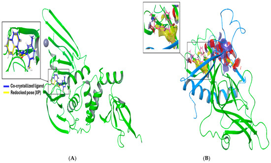
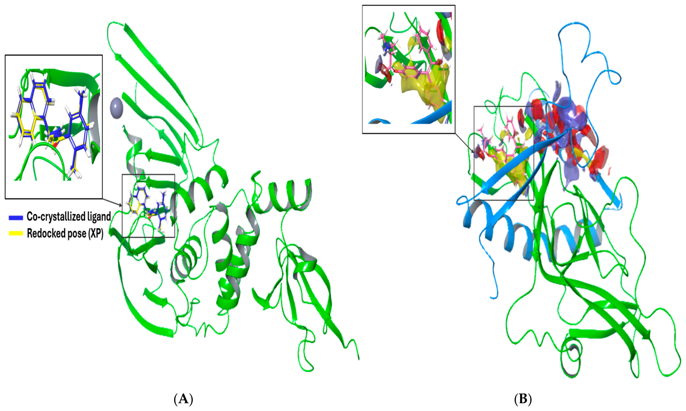
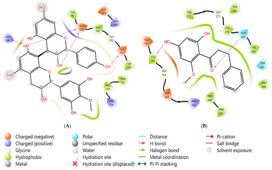
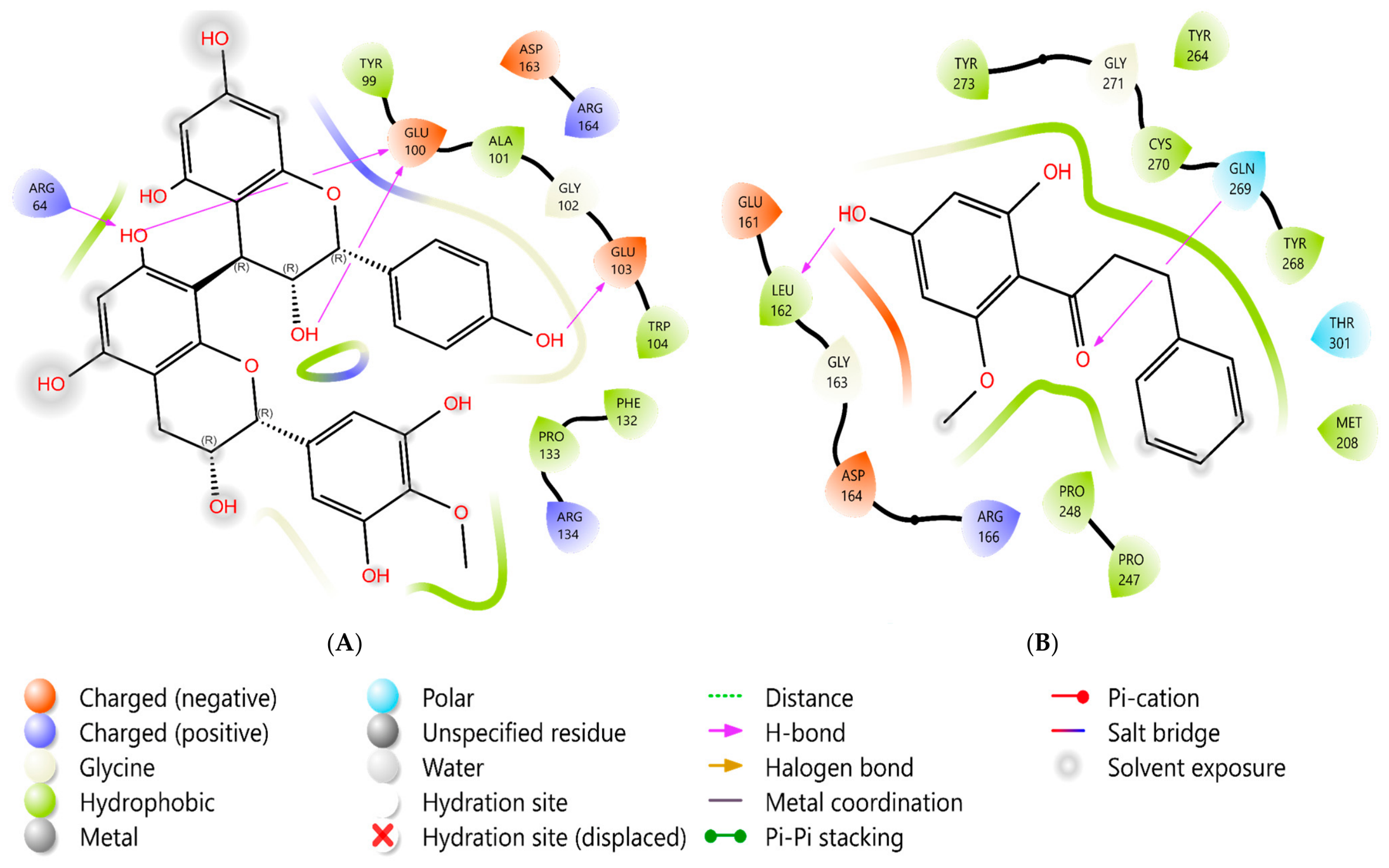
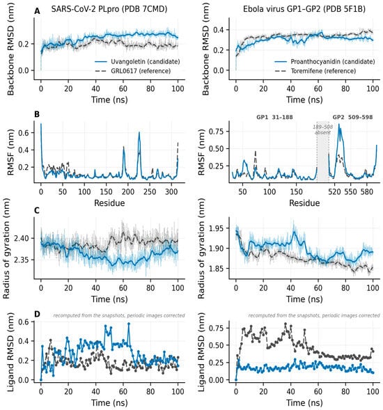
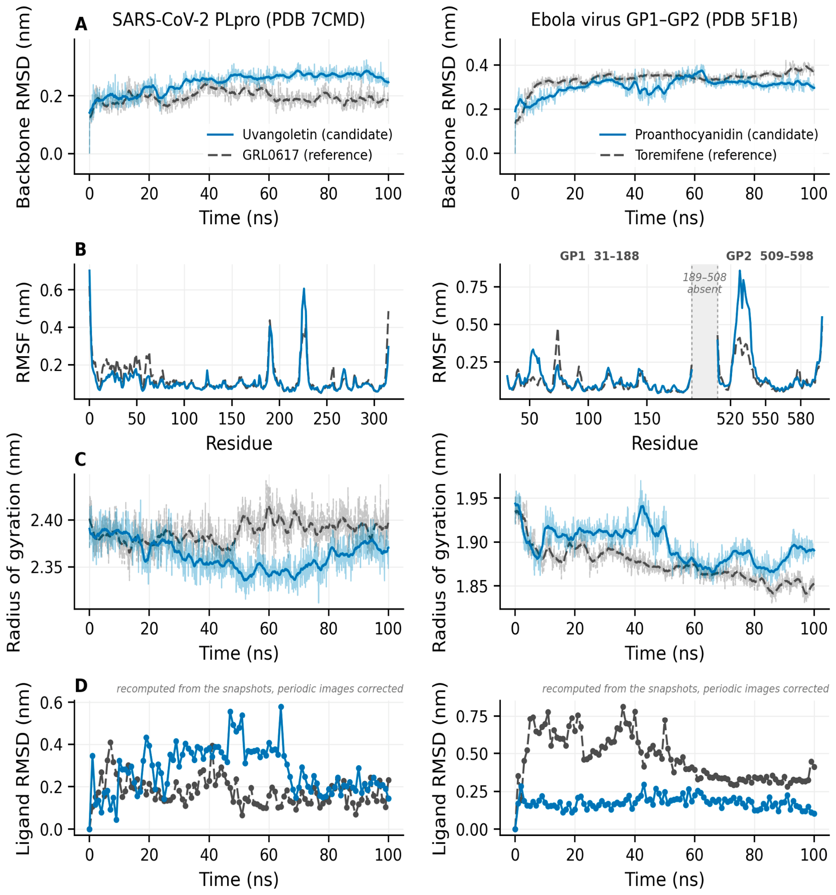
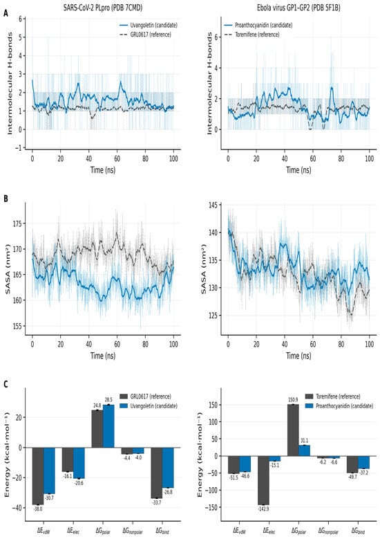
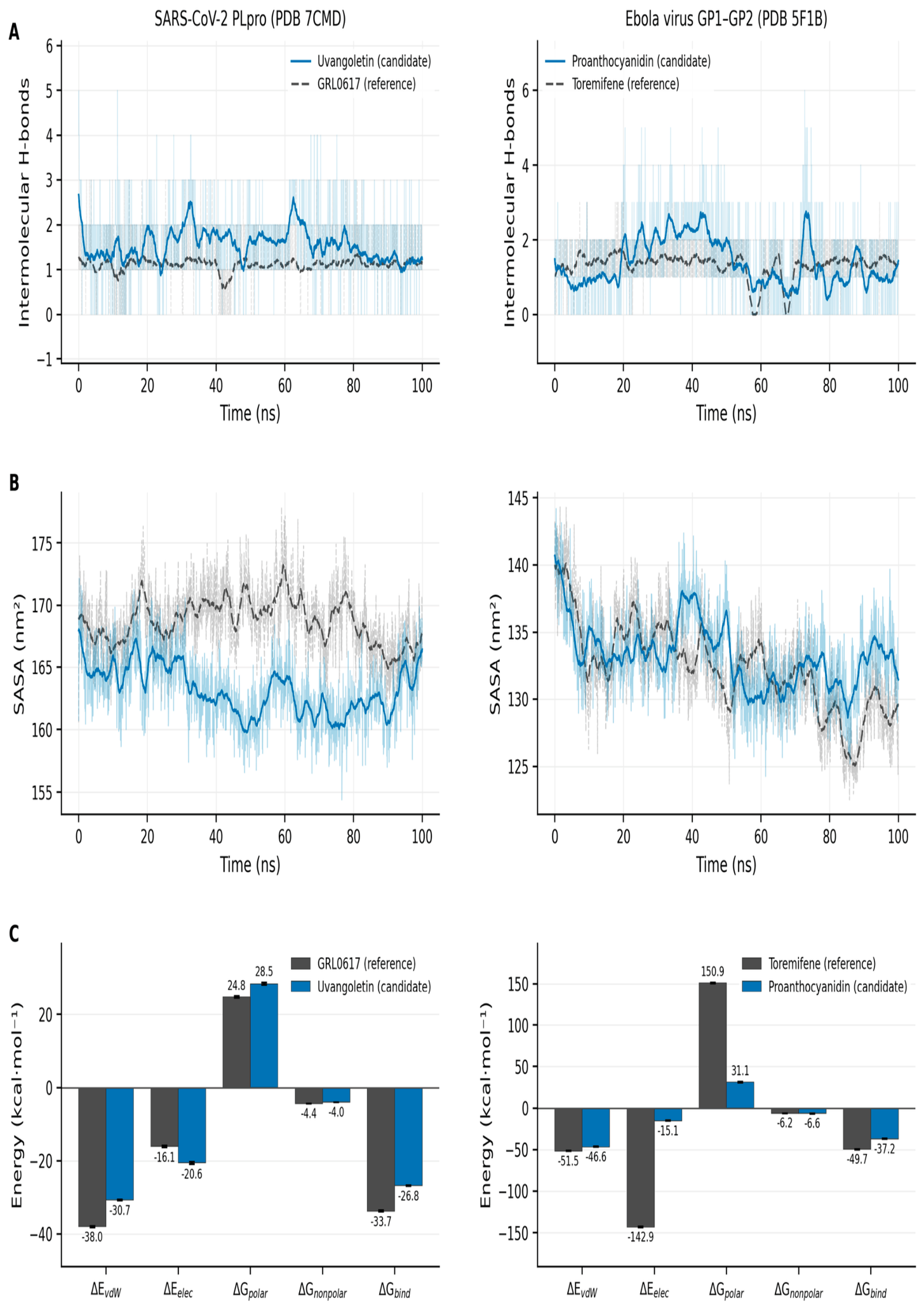
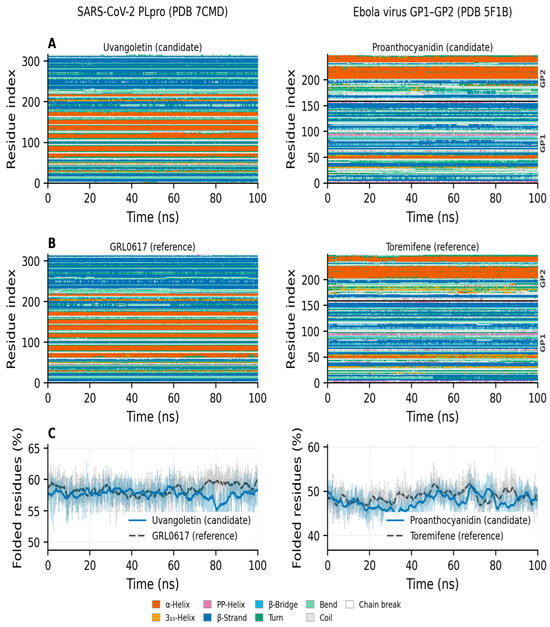
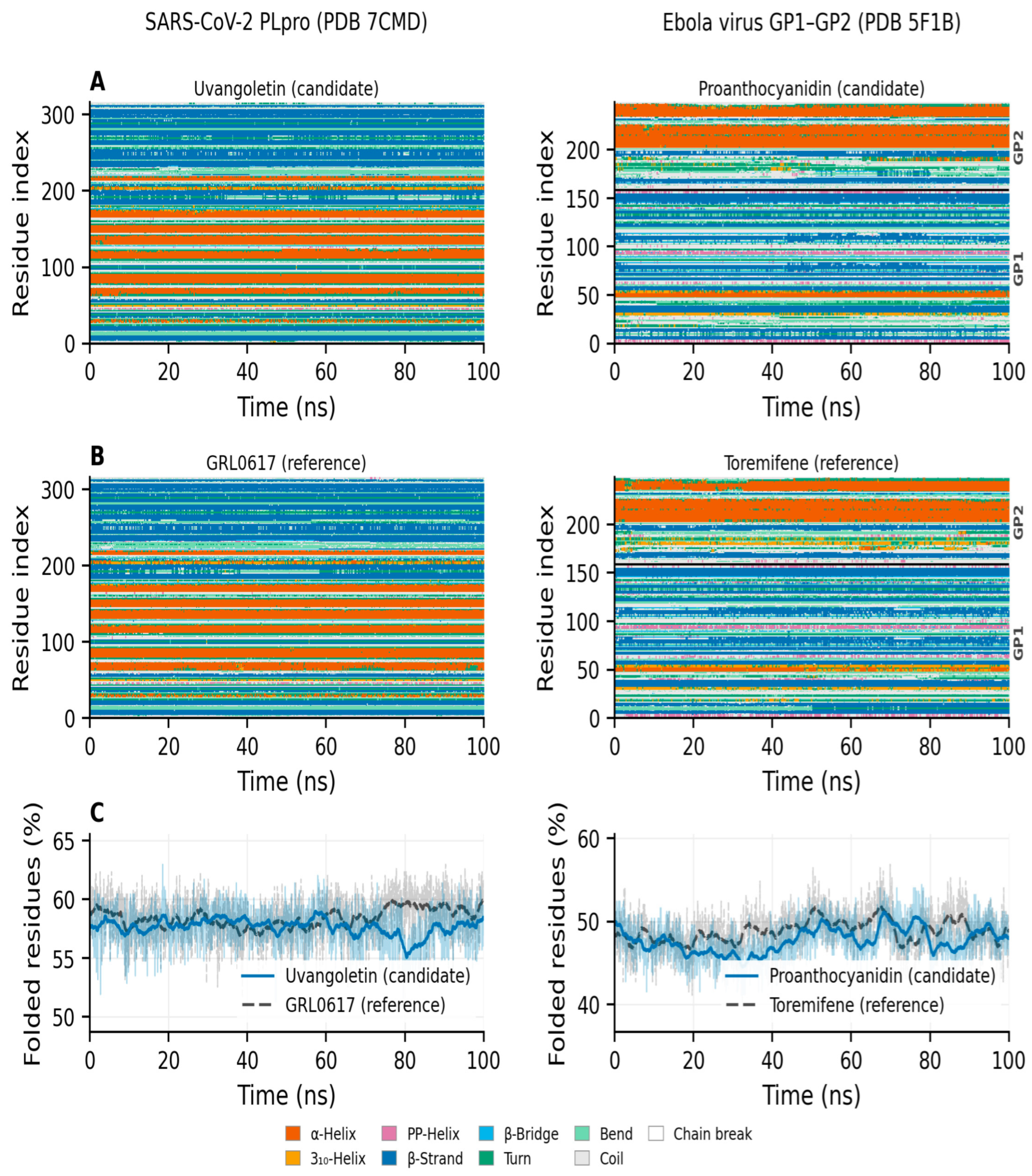
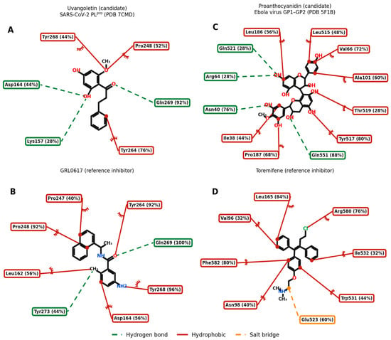
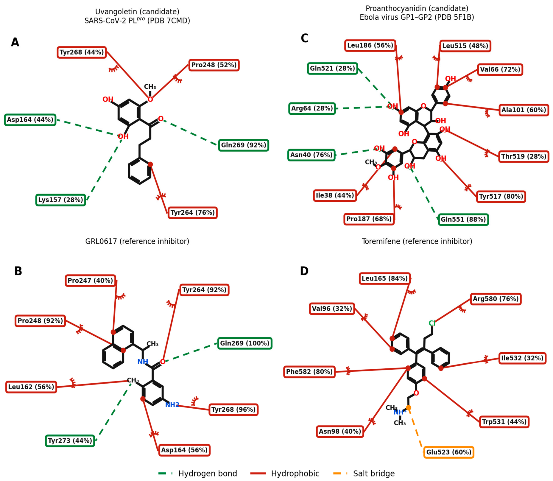
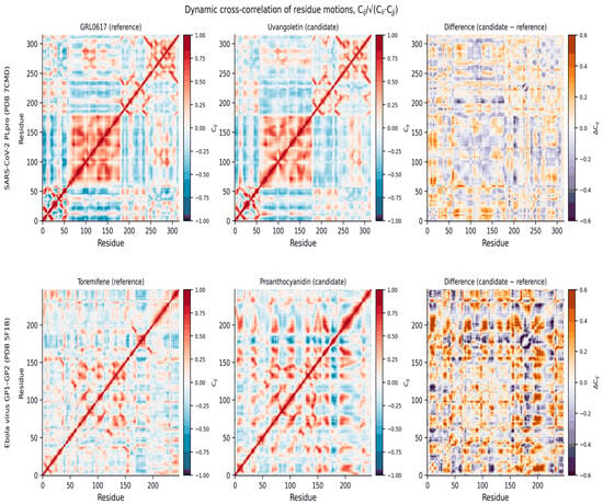
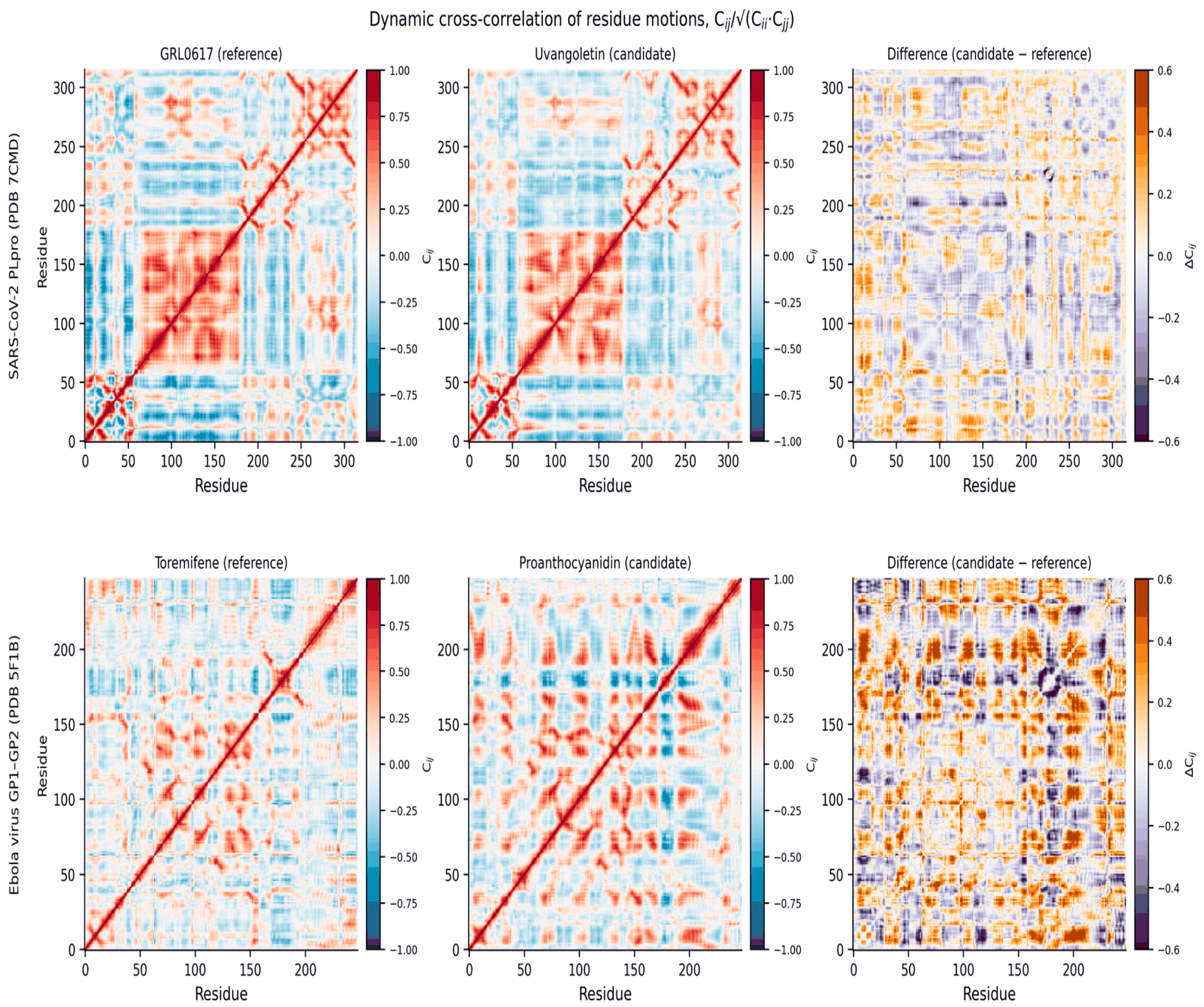
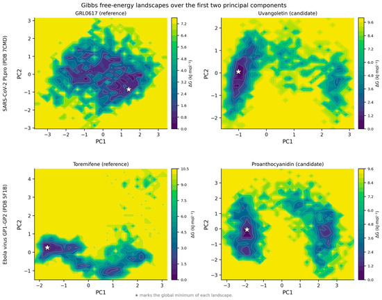
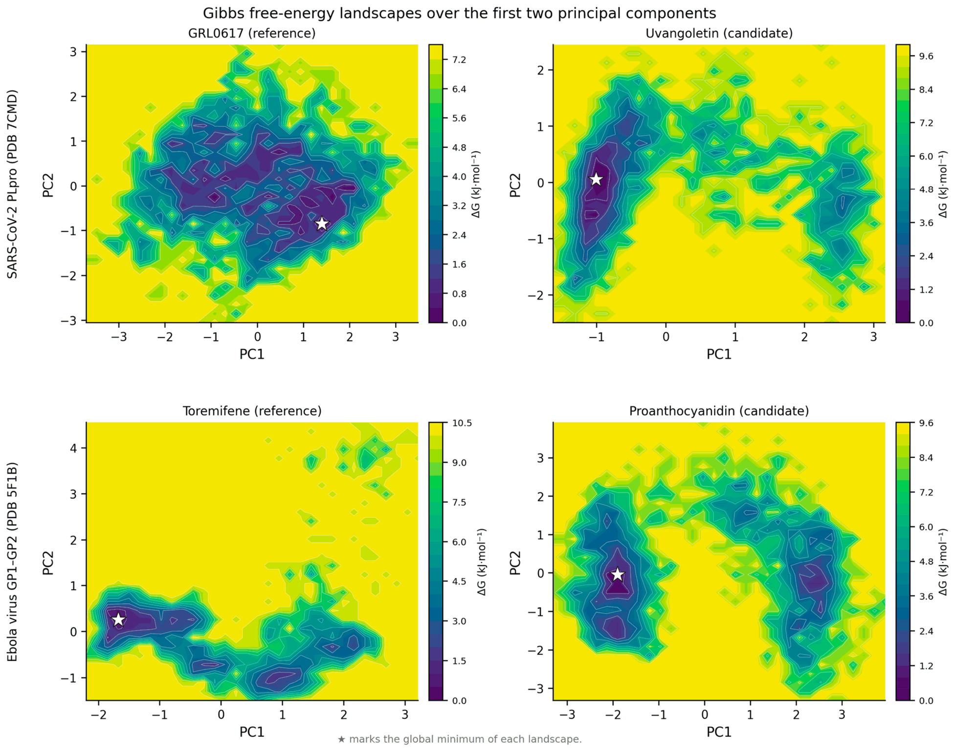
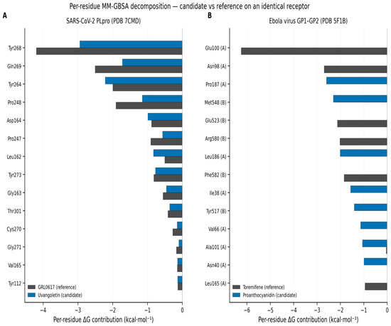
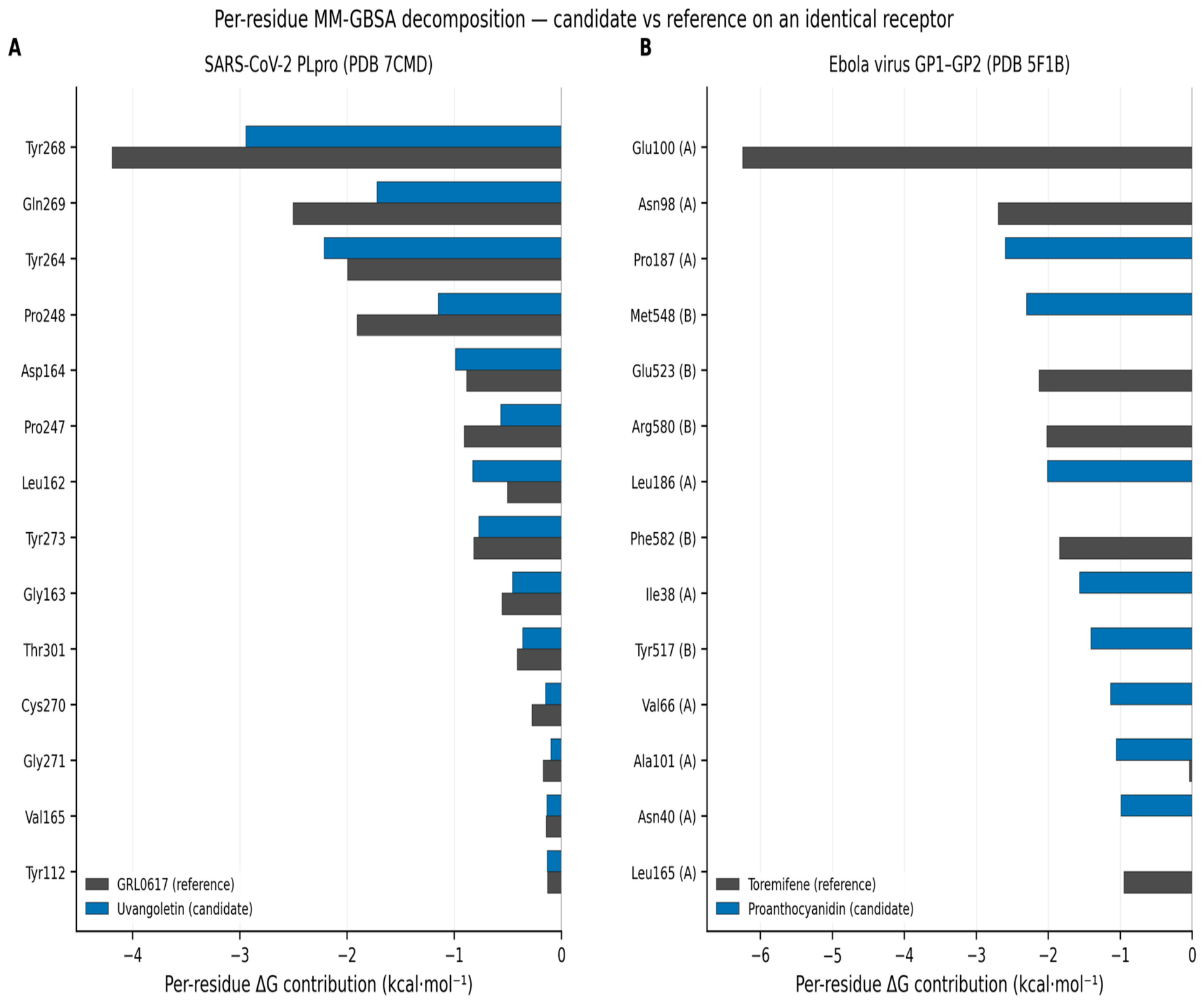
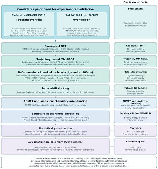
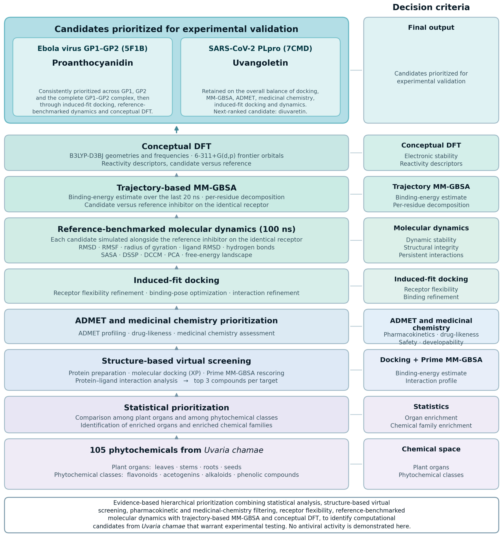

Disclaimer/Publisher’s Note: The statements, opinions and data contained in all publications are solely those of the individual author(s) and contributor(s) and not of MDPI and/or the editor(s). MDPI and/or the editor(s) disclaim responsibility for any injury to people or property resulting from any ideas, methods, instructions or products referred to in the content. |
© 2026 by the authors. Licensee MDPI, Basel, Switzerland. This article is an open access article distributed under the terms and conditions of the Creative Commons Attribution (CC BY) license.