


Abstract
Chamaecrista nigricans (Vahl) Greene leaves are investigated for their bioactive potential. Extracts were prepared using solvents of varying polarity to obtain a diverse range of compounds. The biological effectiveness of the extracts was analyzed against pathogens. Methanol extract showed antimicrobial activity. The methanol extract was subsequently partitioned with solvents of increasing polarity to obtain different fractions. Among these, the separated chloroform fraction was selected for further isolation. The separated chloroform fraction was subjected to column chromatography and visualized through thin-layer chromatography. Emodin was isolated and characterized by spectral analysis and compared with theoretical calculations using Gaussian software. Biological assessment was carried out using molecular docking. Among the cancer proteins, PDB ID: 7AH1 has a favourable binding energy (−3.42 kcal/mol) compared to other proteins. Emodin satisfies all criteria in ADMET profiling, supporting its potential as a promising candidate for further drug discovery.
1. Introduction
Nature serves as the primary source of pharmaceutical resources [1]. Most of the existing or newly discovered drugs are metabolites of plant origin [2,3,4]. Plants synthesize a wide range of secondary metabolites, such as alkaloids, flavonoids, terpenoids, saponins, and phenolics. These compounds defend the plant against pathogens and environmental stressors and exhibit therapeutic effects in humans [5]. Many plant-derived compounds have antimicrobial, anti-inflammatory, antioxidant, anticancer, and analgesic properties. Metabolomics stands out as a prominent and indispensable tool for the identification of primary and secondary metabolites [6,7,8]. Secondary metabolites play a crucial role as they are responsible for the clinical effects. Due to their diverse and specific biological activities, secondary metabolites are considered a valuable source of lead molecules for the development of new drugs. Therefore, they are continuously being studied and explored for their potential in drug development [9,10,11]. Some of these compounds can even act additively or synergistically. Through the implementation of advanced techniques, significant progress has been made in the high-throughput detection and characterization of these metabolites, thereby advancing the field of drug discovery [12,13,14]. PASS online prediction provides valuable insights into the diverse biological activities associated with these metabolites. Extraction is the first step of phytochemistry research, which is also the necessary work before the isolation of effective constituents [15,16,17].
A literature survey reveals that compounds extracted from the Chamaecrista species are widely used in traditional medicine. Chamaecrista nigricans (Vahl) Greene (Fabaceae/Caesalpiniaceae) was reported to be an annual, herbaceous, and leguminous plant. It is found in the wild and also grows along rural road sides [18]. C. nictitans (L.) Moench leaves extract was reported to contain proanthocyanidin oligomers with two structural features, namely, monohydroxy phenols and A-type linkages. These components are speculated to potentially underlie its observed antiviral properties. The efficacy of this extract is directed towards combating the Herpes Simplex Virus, showcasing promising potential in the realm of antiviral therapeutics [19,20]. The extract of C. nictitans (L.) Moench has a relevant therapeutic potential. Phenolic variation among C. nictitans (L.) Moench subspecies and varieties were revealed through UPLC-ESI (-)-MS/MS chemical Fingerprinting technique [21]. Tangavelou et al. reported three anthraquinones isolated from the leaves of C. nigricans (Vahl) Greene [22].
In this study, an active constituent from a plant extract is isolated and characterized through analytical and spectral techniques. The biological potency of the isolated compound was assessed using Gaussian 09w, Molecular docking, Swiss ADME [23], and ProTox II.
2. Materials and Methods
2.1. Collection of Plant Material
The plant samples were collected from natural habitats at Sivakasi, Virudhunagar District, Tamil Nadu, India. The plant specimen(air-dried) was identified and subsequently authenticated by Dr. M. U. Sharief (Scientist ‘F’ & Head of Office) at the Botanical Survey of India (BSI), Southern Regional Centre, Coimbatore, Tamilnadu, India (Certificate No. BSI/SRC/5/23/2024-25/Tech-319, dated 16 May 2024). A voucher specimen has been deposited and preserved in the herbarium of St. Xavier’s College, Pallayamkottai, Tirunelveli, Tamilnadu, India, for future reference.
2.2. Preparation of Plant Extracts
The collected plants were thoroughly washed under running water to clean up any dirt particles. Fresh leaves of Chamaecrista nigricans (Vahl) Greene were ground in a mortar and pestle. These ground leaves of C. nigricans (Vahl) Greene were successfully extracted with hexane, chloroform, and methanol. The extract was filtered to remove plant debris.
2.3. Antimicrobial Analysis
The three microbial strains, namely, E. coli (MTCC 739), Bacillus sp. (MTCC 441), and Staphylococcus sp. (MTCC 96) were procured from MTCC Chandigarh, India.
Antimicrobial activity was assessed using the Kirby-Bauer method. Different extracts of C. nigricans (Vahl) Greene leaves were evaluated for their antimicrobial activity against Gram-positive and Gram-negative bacterial strains. It shows a broad spectrum of antimicrobial activity. The extract was tested against three bacterial strains: E. coli, Bacillus species, and Staphylococcus species.
2.4. High-Resolution Liquid Chromatography Coupled to Electrospray Ionization Tandem Mass Spectrometry (HRLC-ESI-MS/MS)
The metabolite profile of the methanolic extract was obtained using HRLC-ESI-MS/MS (Santa Clara, CA, USA) at the Sophisticated Analytical Instrument Facility (SAIF), IIT, Mumbai.
2.5. Separation Protocol
The concentrated methanolic extract contains both polar and non-polar components. A separating funnel was used to separate the extract based on the polarity of phyto constituents such as hexane and chloroform. Polar phytochemicals are poorly soluble in hexane, limiting their extraction and isolation. Hexane fractions contain fewer bioactive compounds. Chloroform fractions are more effective for separating desired phytochemicals. The excess solvent was removed using a vacuum rotary evaporator.
2.6. Isolation of Phytoconstituent
Following tentative detection, the separated chloroform fraction was further subjected to chromatographic identification, including column and thin-layer chromatography. Thin-layer chromatography (TLC) is a basic chromatographic technique that is used to separate mainly non-volatile compounds. Fractions were collected separately and subjected to TLC (20 × 20 cm aluminium sheets coated with silica gel 60 F254) to detect phytocompounds. These techniques were utilized for the separation and purification of phytochemicals. A vertical (30 mm wide × 600 mm high) glass column was packed with silica gel (230–400 mesh). The introduction of hexane and ethyl acetate mixtures progressively enhanced the polarity of the eluting solvents.
2.7. Preliminary Phytochemical Screening
Phytochemical screening of the separated fraction was done by the standard procedure. The isolated pure compound was shaken vigorously with 10% ammonia solution. The red color indicates the presence of anthraquinones [24].
2.8. Computational Studies
Prediction of biological activity profiles has been performed by the computer program PASS 2022 (Prediction of Activity Spectra for Substances), which assesses the known biological activities with reasonable accuracy compared with other (Q)SAR (Quantitative Structure–Activity Relationship) methods. All calculations were performed in the gas phase using the Gaussian 09W software. Geometry optimizations were conducted using DFT at the B3LYP level with the 6-311+G(d,p) basis set. Vibrational frequency calculations were performed at the same level of theory to confirm that the optimized structures correspond to a true energy minimum via imaginary frequencies.
This specific functional and basis set combination was selected for its proven reliability and established accuracy in reproducing experimental spectroscopic data, including electronic, vibrational, and magnetic resonance, for comparable organic molecules. To establish a baseline electronic structure for the isolated compounds, all calculations were performed under default gas-phase conditions without solvent models.
Molecular docking was conducted using Autodock 4.2 with the Lamarckian Genetic algorithm to assess emodin against target proteins from the RCSB Protein Data Bank. Receptors and the DFT-optimized ligand were prepared in Autodock tools by assigning Kollman and Gasteiger charges. Grid maps for active-site pockets were created with a 0.375 Å spacing. Fifty independent runs were performed per receptor, with a maximum of 2,500,000 energy evaluations, and the best binding configurations were selected based on the lowest binding energy (kcal/mol). The ADMET (Absorption, Distribution, Metabolism, Excretion, and Toxicity) properties of a molecule are important factors in successful drug development. The physicochemical properties were compiled using the SwissADME web server and ProTox 3.0 [25,26,27,28,29].
3. Results and Discussion
3.1. Antimicrobial Activity
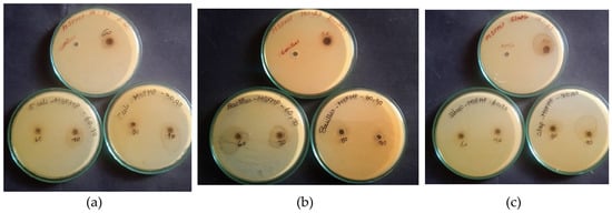
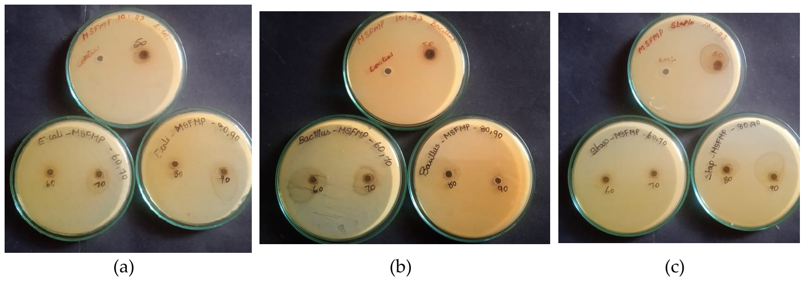
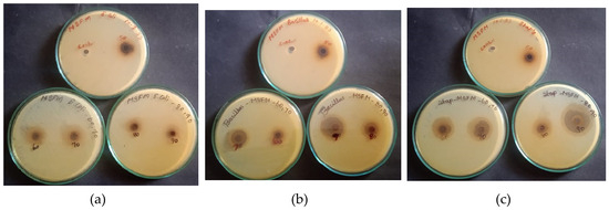
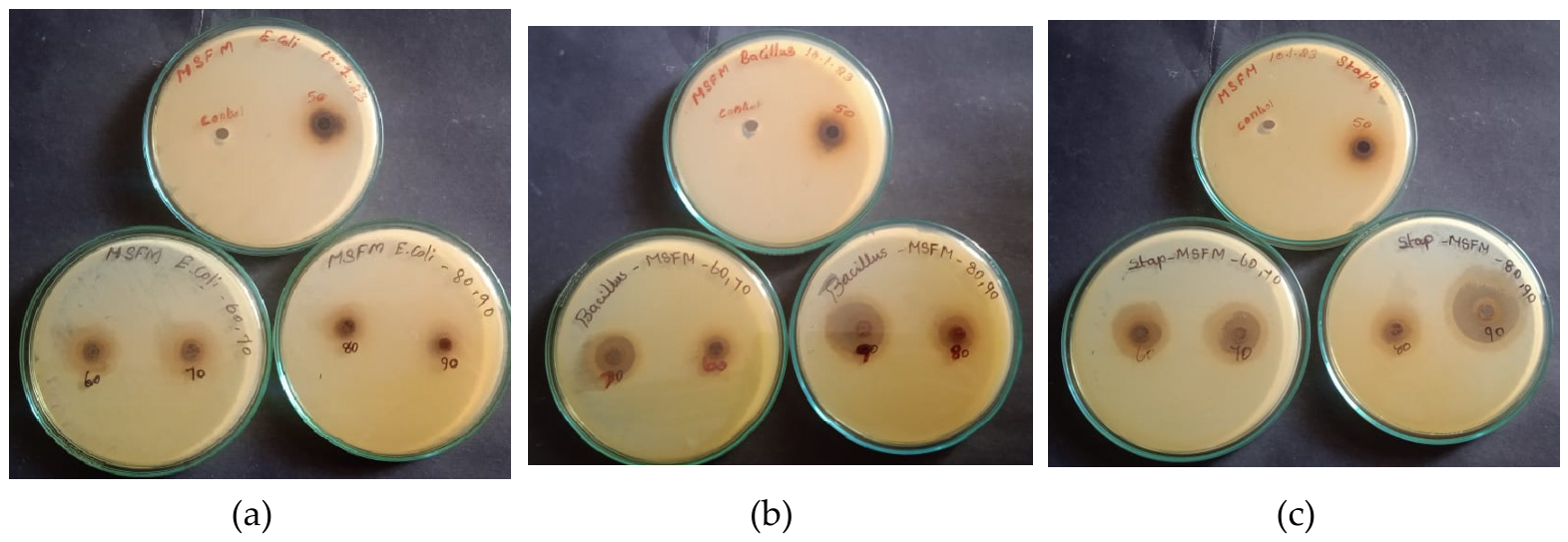
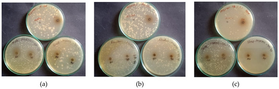
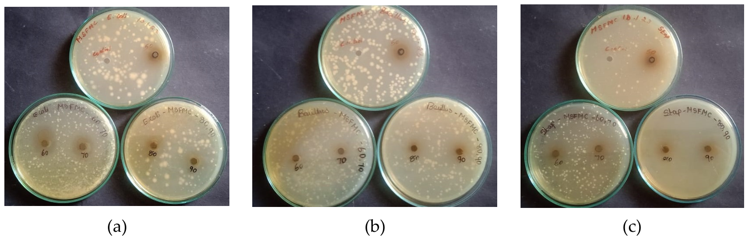
Hexane, Chloroform, and methanol extracts of C. nigricans (Vahl) Greene leaves were evaluated for their antimicrobial activity against certain Gram-positive and Gram-negative bacteria. The broad-spectrum action of these extracts suggests potential applications in antiseptic and disinfectant formulations, as well as chemotherapy. In this study, three pathogens showed more activity in the methanol extract of C. nigricans (Vahl) Greene leaves. The results are tabulated in Table 1. Figure 1, Figure 2 and Figure 3 show the zone of inhibition against pathogens. Compared with other extracts, the methanol extract of C. nigricans (Vahl) Greene leaves showed better activity against Staphylococcus sp.
Table 1.
Antimicrobial activity of plant extracts of C. nigricans (Vahl) Greene leaves.
Figure 1.
Antimicrobial activity of the Hexane extract of C. nigricans (Vahl) Greene leaves against (a) E. coli. (b) Bacillus sp. (c) Staphylococcus sp.
Figure 2.
Antimicrobial activity of CHCl3 extract of C. nigricans (Vahl) Greene leaves against (a) E. coli. (b) Bacillus sp. (c) Staphylococcus sp.
Figure 3.
Antimicrobial activity of CH3OH extract of C. nigricans (Vahl) Greene leaves against (a) E. coli. (b) Bacillus sp. (c) Staphylococcus sp.
In laboratory testing, this computational profile was confirmed by robust, concentration-dependent in vitro zones of inhibition against E.coli (21 mm), Bacillus sp. (22 mm), and Staphylococcus sp. (28 mm). To map the underlying mechanisms, molecular docking was utilized.
3.2. Metabolite Profiling
HRLC-ESI-MS/MS analysis of the methanolic extract revealed distinct major peaks in both positive and negative ESI modes, providing information on various phytochemical constituents. These metabolites were identified and characterized through the PASS prediction study. All identified metabolites exhibited multiple biological activities.
3.3. Preliminary Phytochemical Analysis
C. nigricans (Vahl) Greene leaf extracts were subjected to preliminary phytochemical screening (Table 2).
Table 2.
Phytochemical screening test for different extracts of C. nigricans (Vahl) Greene leaves.
The methanolic extract of C. nigricans (Vahl) Greene leaves contains the highest concentration of metabolites compared to hexane and chloroform extracts. Flavonoids and terpenoids were present in all three extracts. Alkaloids were present in the hexane extract. Phenol and phytosterol were present in chloroform and methanol extracts. Oils were present in hexane and methanol extracts. The methanolic extract of C. nigricans (Vahl) Greene leaves contains the highest concentration of metabolites compared to hexane and chloroform extracts.
3.4. Isolation of Emodin
The separated chloroform fraction was eluted with hexane and ethyl acetate. Waxy materials were removed by elution with hexane. The polarity of the solvent was slightly increased for effective separation. Emodin was effectively eluted in the ratio 4.5:0.5 (Hexane: Ethyl acetate).
3.5. Theoretical and Experimental Aspects of the Isolated Compound
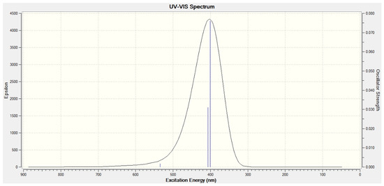
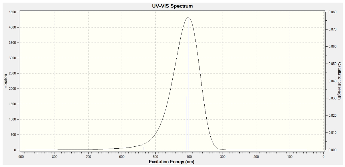
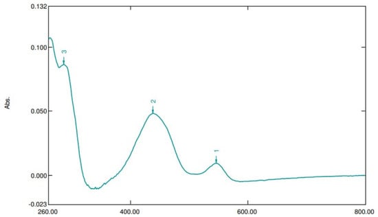
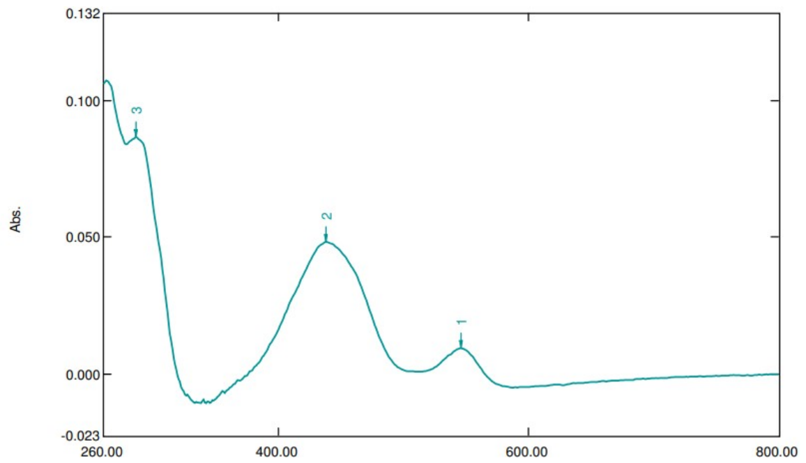
The electronic spectrum of isolated emodin was investigated using UV–visible spectroscopy (200–800 nm in methanol) and compared theoretically with Time-dependent Density Functional Theory (TD-DFT) calculations at the B3LYP/6-311+G(d,p) level (Figure 4 and Figure 5). The absorption maxima at 286 nm may be due to the aromatic π-electrons of the benzene ring. The peak at 438 nm can be attributed to the n → π* transition of the carbonyl groups, which was theoretically calculated at 410 nm. The peak at 546 nm corresponds to vibronic coupling of the C=O stretch, a characteristic of high-purity anthraquinones [30].
Figure 4.
Theoretical UV–Visible spectra of emodin.
Figure 5.
Experimental UV–Visible spectra of isolated emodin.
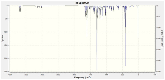
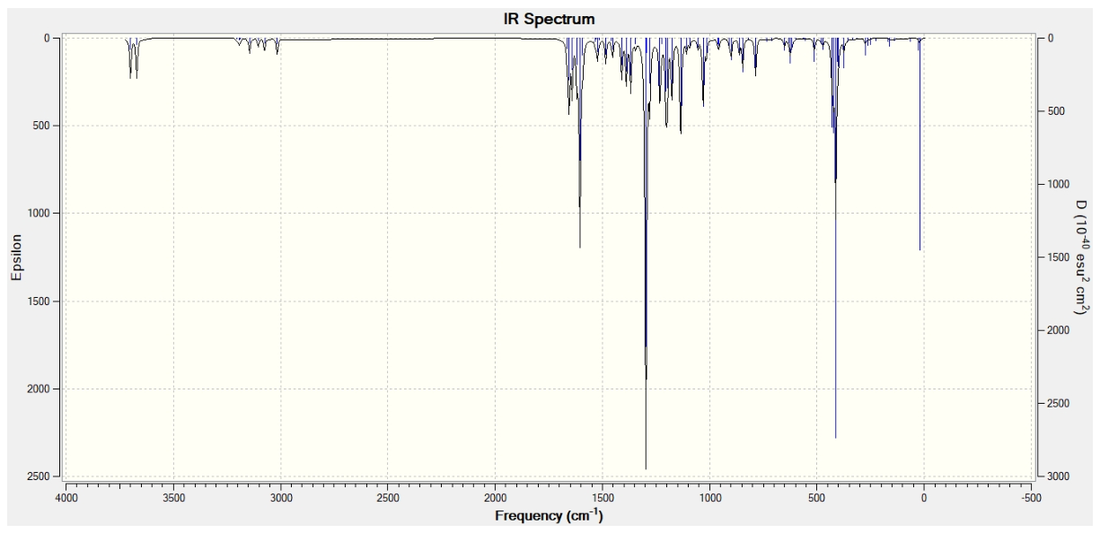
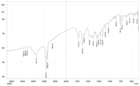
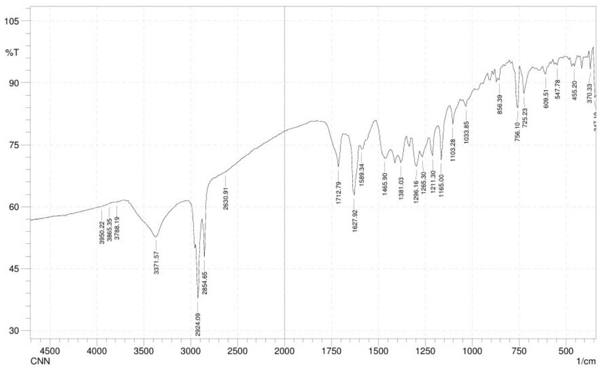
The FT-IR spectra of isolated emodin (Figure 6 and Figure 7) show a hydroxyl (O–H) stretching band at 3371 cm−1, with the free hydroxyl group appearing experimentally at 3788 cm−1 and theoretically at 3703 cm−1. A peak at 3073 cm−1 indicates a C–H stretch in the aromatic ring, while a characteristic peak at 1408 cm−1 results from C–OH in-plane bending and C–C stretching, confirming the phenolic fingerprint. CH3 deformations appear at 1465 cm−1 and 1381 cm−1, correlating well with DFT predictions of 1453 cm−1 and 1372 cm−1. Aromatic C=C stretching is observed at 1589 cm−1, and the CH3 symmetric stretch at 2854 cm−1 indicates aliphatic substitution. The peak at 1211 cm−1 corresponds to C–O phenolic stretching (theoretical: 1201 cm−1). The band at 1627 cm−1 represents C=O stretching conjugated quinones, which are theoretically predicted at 1606 cm−1 and 1642 cm−1 [31,32].
Figure 6.
Theoretical Infrared spectra of emodin.
Figure 7.
Experimental Infrared spectra of isolated emodin.
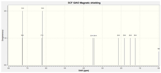
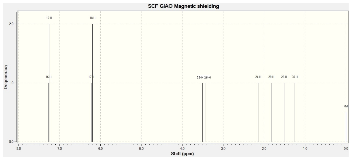
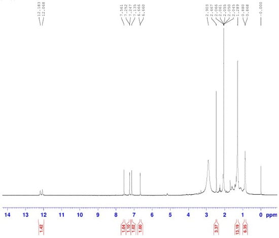
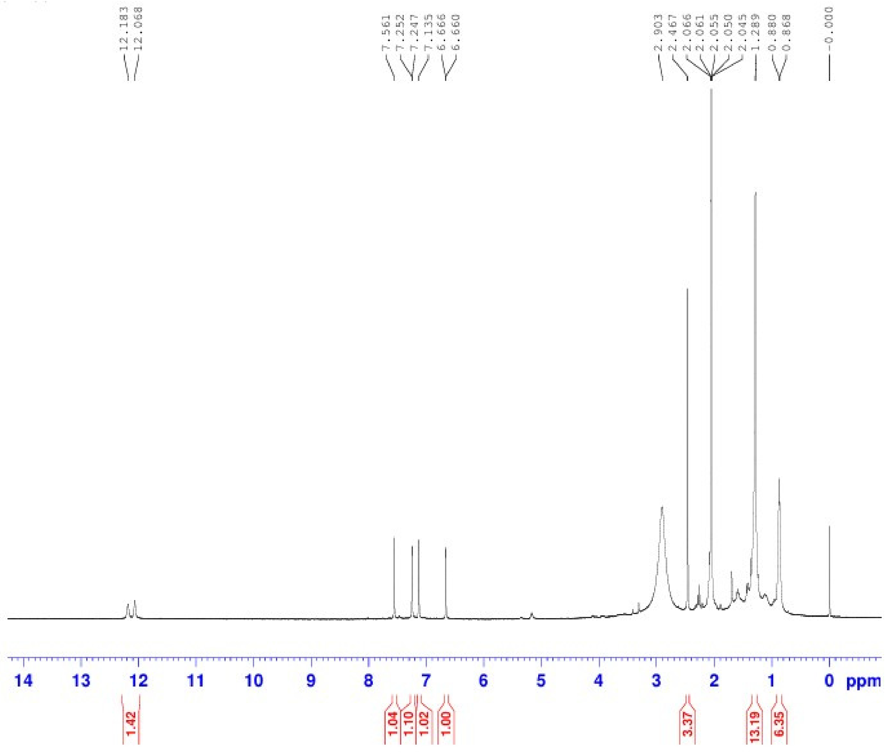
The 1H NMR spectra of the isolated emodin were predicted experimentally and theoretically in Figure 8 and Figure 9. The isotopic NMR chemical shifts obtained using the GIAO method for the isolated compound were compared with their experimental spectral data. The chemical shift value at δ 12–12.2 ppm for 1,8-Diol (intramolecular H-bonding to quinone carbonyls) and the methyl proton was observed as a broad singlet at δ 2.90 ppm, where the theoretical value is δ 2.142 ppm. Aromatic protons in the benzene moiety δ 6.5–7.8 ppm exhibited excellent GIAO-DFT correlation observed at δ 7–7.5 ppm.
Figure 8.
Theoretical 1H NMR spectra of emodin.
Figure 9.
Experimental 1H NMR spectra of isolated emodin.
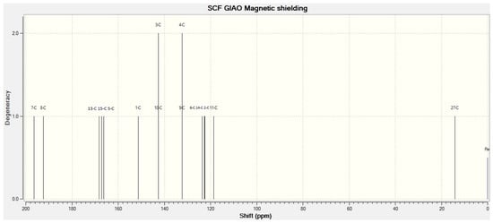
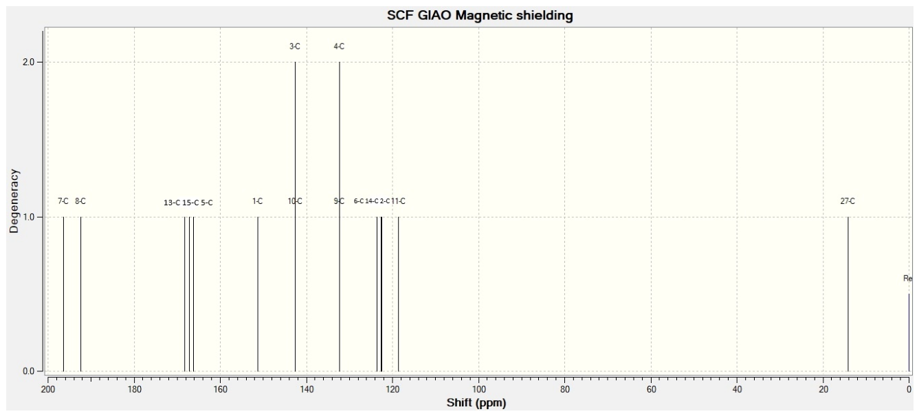
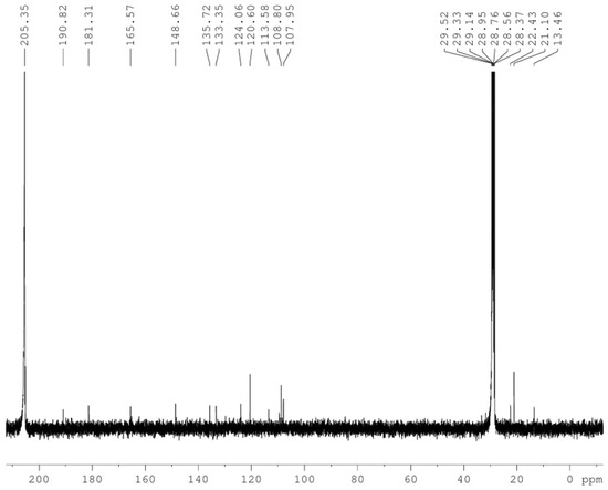
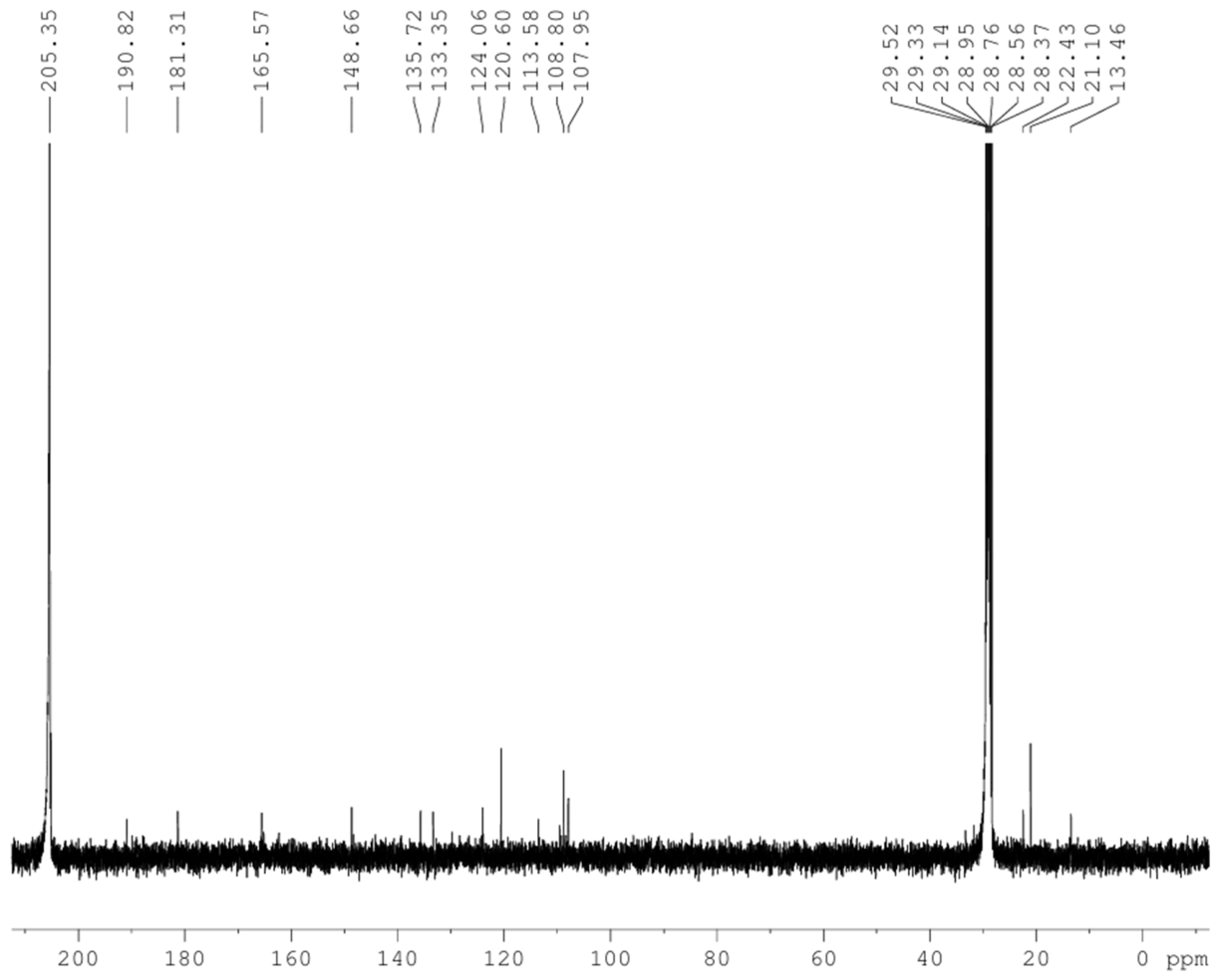
The 13C NMR spectra represented in Figure 10 and Figure 11 revealed 15 distinct carbon signals. The quinone C=O group signals are experimentally observed at δ 190.82 and 181.31 ppm, whereas they are theoretically observed at δ 192.41 and 196.39 ppm. The carbon attached to the hydroxyl group was observed at δ 165.57 ppm, while the theoretical values are 166.15, 167.17, and 168.31 ppm. The methyl proton signals are at δ 13.46, 21.10, 22.43 ppm experimentally, and at δ 14.129 ppm theoretically. Aromatic ring carbons appear at δ 107.95, 108.80, 113.58, 120.60, 124.06, 133.35, 135.72, 148.66, and 165.57 ppm experimentally and theoretically found at δ 118.61, 122.55, 122.68, 123.65, 132.30, 132.35, 142.49, 142.51, and 151.30 ppm [33].
Figure 10.
Theoretical 13C NMR spectra of emodin.
Figure 11.
Experimental 13C NMR spectra of isolated emodin.
GC-MS is a highly sensitive, robust, and reliable technique capable of detecting trace amounts of volatile and semi-volatile compounds. It provides accurate identification through the combination of retention time and mass spectral data, with the added advantage of wide applications and easy library-based spectral matching. A GC-MS study reveals that the major phytochemical is present in the chromatogram (Figure 12). GC-MS analysis confirmed emodin (C15H10O5) via molecular ion with characteristic anthraquinone fragmentation [M+] at 270, [M-OH]-253, [M-CO]-225, and [M-CO]-197, representing sequential quinone carbonyl and methyl group fragmentation [34].
Figure 12.
Experimental GC-MS spectra of isolated emodin.
3.6. Optimization of Emodin
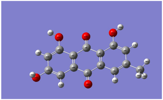
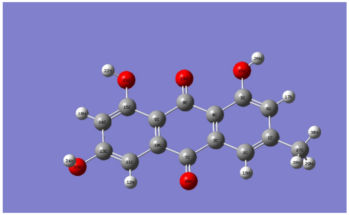
Full molecular geometry optimization was performed utilizing Time-Dependent Density Functional Theory at the B3LYP (Becke, 3-parameter, Lee-Yang-Parr) level and 6-311+G(d,p) basis set via the Gaussian09W software package. The optimized structure of emodin, as shown in Figure 13, facilitated the calculation of the molecular electrostatic potential, the highest occupied molecular orbital, and the lowest unoccupied molecular orbital energy using Gaussian09W. Ground-state geometry optimization of emodin (C15H10O5) yielded C1 point group symmetry with a total energy of −953.60764419 a.u. and dipole moment of 3.7248 Debye. B3LYP/6-311++G (d,p) optimization confirmed planar conjugated anthraquinone with 1,8-OH chelation to C-9 carbonyl.
Figure 13.
Optimized structure of emodin.
3.6.1. Bond Length and Bond Angle
Optimization of emodin in DFT provides information on bond lengths and bond angles, as indicated in Table 3.
Table 3.
Bond length and bond angle.
This geometric arrangement directly correlates with the molecule’s overall chemical reactivity profile. The spatial orientation defined by these bond angles minimizes total internal ring strain, rendering the ground-state structure thermodynamically favourable. Furthermore, the exposed polar bonds identified here may serve as highly flexible interaction sites, perfectly priming the compound to lock into target receptor pockets via directional hydrogen bonding networks during molecular docking and subsequent in vitro assays.
3.6.2. FMO (Frontier Molecular Orbital) Analysis
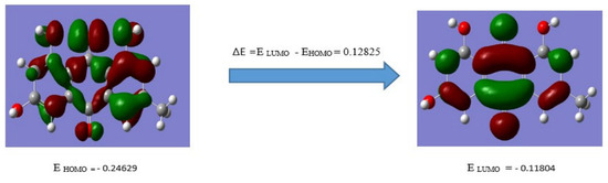
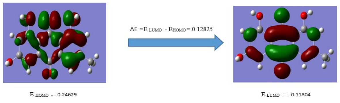
The HOMO–LUMO gap(∆E) (Figure 14) reflects molecular polarizability and chemical softness. However, absolute reactivity is determined by individual frontier orbital energies, which vary with the chemical process. The compound’s nucleophilic reactivity is governed by the absolute energy of HOMO (EHOMO), while its electrophilic reactivity is driven by the energy of the LUMO (ELUMO). The calculated HOMO–LUMO energy gap of the compound is 0.1285 a.u. This substantial energy gap indicates high kinetic stability, low ground-state polarizability, and an electronic excitation threshold characteristic of the ultraviolet region. Anthraquinones with ΔE < 0.15 eV exhibit enhanced bioactivity via facile electron donation to biological targets.
Figure 14.
FMO of Emodin.
3.6.3. MEP (Molecular Electrostatic Potential) Analysis
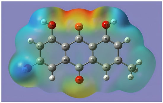
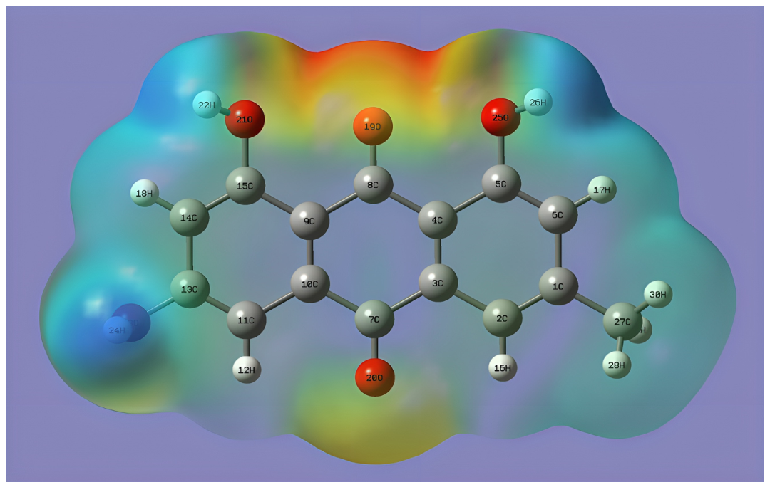
The electron density of emodin is uniform throughout the molecule, ranging from −8.429e−2 a.u. (represented in red color, indicating strongly nucleophilic regions localized around the carbonyl and phenolic oxygen atoms) to 8.429e−2 a.u. (represented in blue, indicating the electrophilic regions localized over the hydroxyl hydrogen atoms), as shown in Figure 15. Color variations on the molecular surface reflect the boundaries of the electronic potential energy surface, confirming that the oxygen centers act as primary electron-donating sites during molecular docking interactions.
Figure 15.
Total electron density mapped with the electrostatic potential of emodin.
3.7. Molinspiration
A Molinspiration study concludes that emodin exhibits drug-like properties, with promising enzyme inhibition and kinase activity. Emodin may serve as a natural lead for further drug development.
3.8. PASS Prediction
PASS prediction study reveals the structural features of emodin and predicted robust antibacterial (Pa = 0.887) and antineoplastic (Pa = 0.791) profiles. The probabilities (Pa) were well above the standard threshold of 0.7. This indicates a strong statistical likelihood of the core structure possessing the necessary chemical framework to disrupt both microbial and cancer systems.
The highly optimised HOMO–LUMO energy gap indicates that the molecule is chemically stable yet highly electronically polarisable, making it energetically ready to transfer charge or undergo chemical reactions with biological receptors. The molecular electrostatic potential proves that these exact groups are highly polarized and ready to engage in hydrogen bonding and electrostatic interactions.
3.9. Molecular Docking
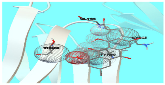
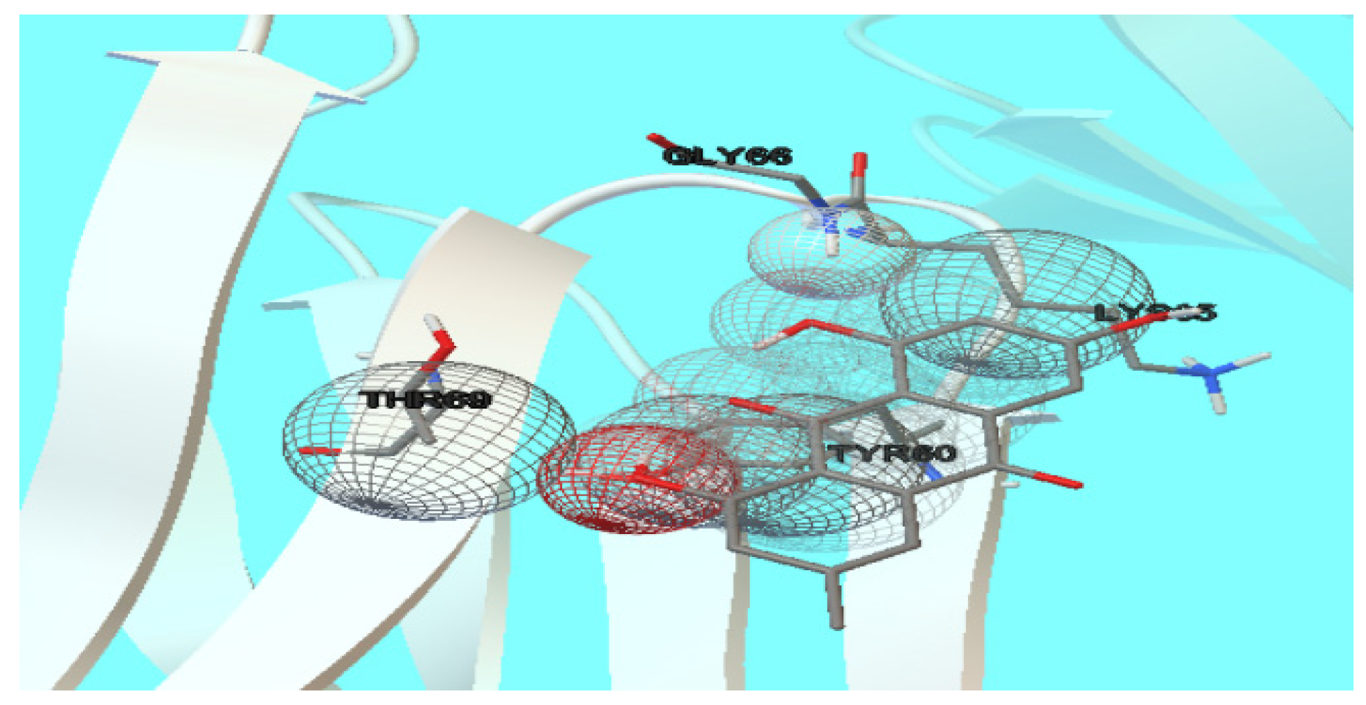
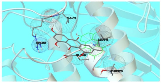
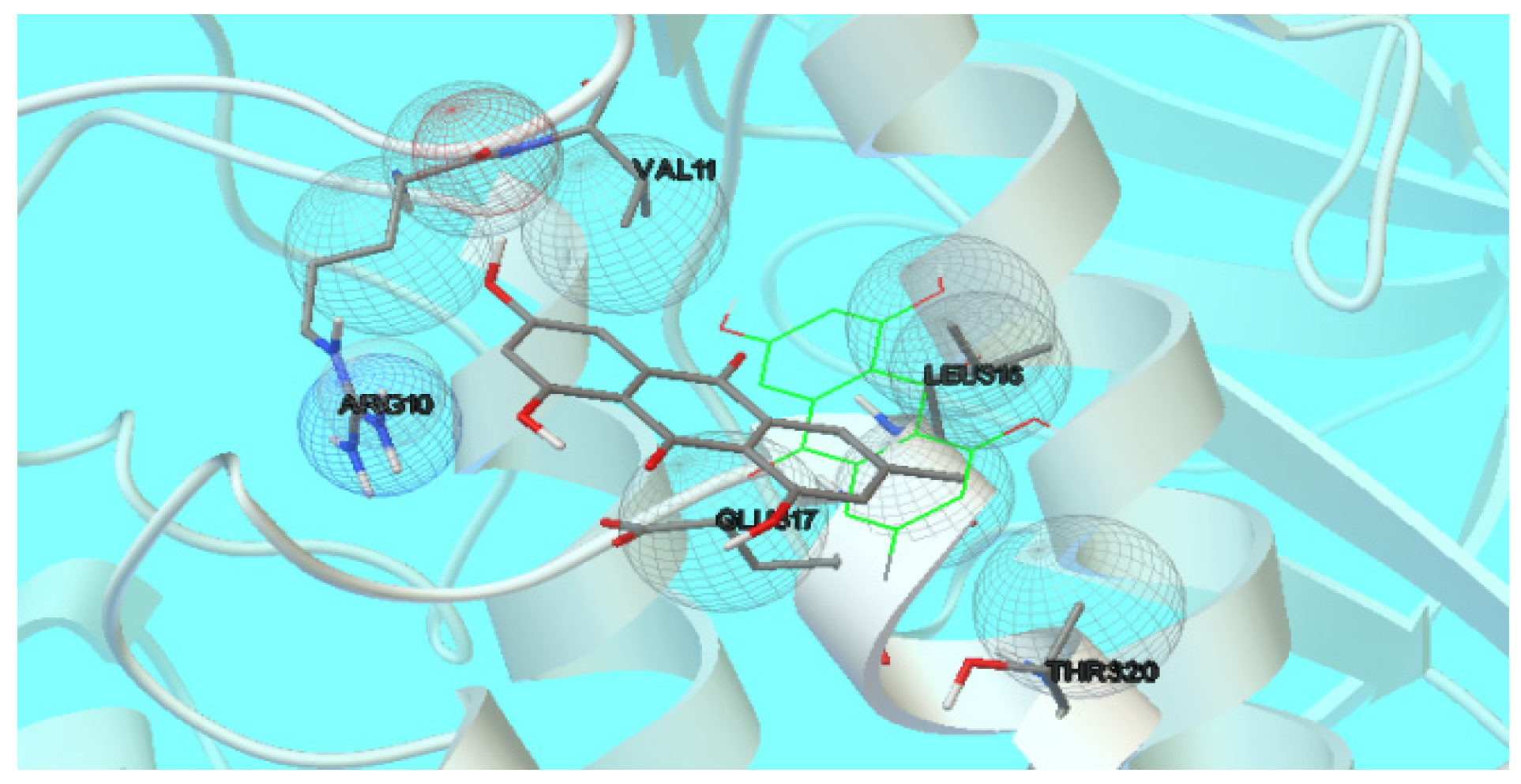
In this study, molecular docking of emodin with cancer drugs is performed using the cancer protein PDB IDs: 1M2P, 4R3E, 5WPU, and 7AH1. The glide score is shown in Table 4. The calculated binding energies of emodin with the target proteins 1M2P, 4R3E, 5WPU, and 7AH1 were −2.92, 0.86, 0.69, and −3.42 kcal/mol, respectively. The drug Doxorubicin was docked with these proteins. Emodin shows a better glide score than Doxorubicin. It revealed that the binding energy of the PDB ID: 7AH1 is higher than that of 1M2P. Based on the docking score, the new compound, emodin, selectively targets 7AH1 via various amino acid residues, including GLY66, THR69, LYS65, and TYR60 (Figure 16 and Figure 17), and this is further supported by Molinspiration studies [35].
Table 4.
The binding score of Isolated Emodin and the drug with proteins.
Figure 16.
Binding interaction between Emodin and PDB ID:1M2P.
Figure 17.
Binding interaction between Emodin and PDB ID:7AH1.
Pharmacokinetics
The ADMET properties of emodin are presented in Table 5. The two-dimensional chemical structure with canonical SMILES was shown in the first section. The drug likeness of isolated emodin was assessed by Lipinski’s rule of five. Permeability glycoprotein (P-gp) influences the ADMET properties of many drugs, and it is important to investigate the interaction between the transporter protein and the drug molecule. Cytochrome P450s (CYPs) represent a large class of heme-containing enzymes that are necessary for the detoxification of foreign chemicals and the metabolism of drugs. Cytochrome P450 enzymes can be inhibited or induced by drugs, resulting in clinically significant drug–drug interactions that can cause unanticipated adverse reactions or therapeutic failures. The target drug may need to be substituted or the dose adjusted to account for a potential decrease or increase in metabolism. Emodin meets the standard ADMET criteria. Emodin, adhering to Lipinski’s rule, was regarded as a potential drug candidate. Comprehensive correlations across spectroscopy, geometry, bioactivity, and ADMET profiling confirm preclinical treatment potential [36].
Table 5.
ADMET properties of Emodin.
4. Conclusions
This study concludes that the chloroform fraction of the methanolic extract from C. nigricans (Vahl) Greene leaves exhibits significant biological activities, including anti-microbial and anti-cancer activities. The secondary metabolite emodin was isolated from C. nigricans (Vahl) Greene leaves and identified through both theoretical and experimental studies. The electronic information from DFT supports macroscopic predictions from PASS, while molecular docking shows that PDB ID 7AH1 has a binding score of −3.42 Kcal/mol, indicating emodin’s competitive binding affinity to the targeted microbial receptors and favourable ADMET profiles. Emodin exhibits various pharmacological activities, including anti-cancer, anti-inflammatory, antioxidant, antimicrobial, and antiviral effects [37,38,39]. However, its clinical use is limited by dose-dependent toxicities (hepatotoxicity, nephrotoxicity, reproductive toxicity) and the absence of Western pharmaceutical approval, confining it to traditional Chinese medicine. Future research should focus on establishing safe therapeutic windows and positioning it as a complementary therapeutic agent [40,41,42,43].
Author Contributions
Writing—original draft, K.P.; Software, K.P.; Data curation, K.P.; Writing—original draft, S.S.S.; review, S.S.S. and M.K.M.; editing, S.S.S. and M.K.M.; Methodology, S.S.S.; Formal analysis, S.S.S. All authors have read and agreed to the published version of the manuscript.
Funding
This research received no external funding.
Data Availability Statement
The original contributions presented in this study are included in the article. Further inquiries can be directed to the corresponding author.
Acknowledgments
We gratefully acknowledge The Standard Fireworks Rajaratnam College for Women, Sivakasi for providing access to their Science Instrumentation Centre sponsored by DST-FIST which were essential for this study.
Conflicts of Interest
The authors declare no conflicts of interest.
References
- Usman, A.; Abubakar, S.; Alaku, C.; Nnadi, O. Plant: A Necessity of Life. Int. Lett. Nat. Sci. 2014, 20, 151–159. [Google Scholar] [CrossRef] [Scilit]
- Kumar, N.; Wani, Z.A.; Dhyani, S. Ethnobotanical study of the plants used by the local people of Gulmarg and its allied areas, Jammu & Kashmir, India. Int. J. Curr. Res. Biosci. Plant Biol. 2015, 2, 16–23. [Google Scholar]
- Bernhoft, A. A brief review of bioactive compounds in plants. In Proceedings of the Bioactive Compounds in Plants—Benefits and Risks for Man and Animals, Prague, Czech Republic, 8–10 September 2010; The Norwegian Academy of Science and Letters: Oslo, Norway, 2010; pp. 11–17. [Google Scholar]
- Chadwick, D.J.; Marsh, J. (Eds.) Ethnobotany and the Search for New Drugs. In Proceedings of the CIBA Foundation Symposium 185, London, UK, 19–21 July 1994; John Wiley and Sons (Hotel Praia Centro): Fortaleza, Brazil, 1994. [Google Scholar]
- Makunga, N.; Kai, G.; Sieniawska, E. Metabolomics as a tool in ethnobotany-driven drug discoveries. Front. Pharmacol. 2022, 13, 1066875. [Google Scholar] [CrossRef] [Scilit] [PubMed]
- Cock, I.E.; Matthews, B. Metabolomic profiling of antiviral Scaevola spinescens extracts by high resolution tandem mass spectrometry. In Proceedings of the 29th Int Horticultural Congress on Horticulture-Sustaining Lives, Livelihoods and Landscapes (IHC)/5th World Congress on Medicinal and Aromatic Plants (WOCMAP)/Int Symposium on Plants, as Factories of Nat Substances, Edible and Essential Oils (Brisbane, Australia), Brisbane, Australia, 17–22 August 2014. [Google Scholar] [CrossRef] [Scilit]
- Soodabeh, S.; Ahmad Reza, G. Application of spectroscopy in herbal metabolomics. DARU J. Pharm. Sci. 2012, 20, 91–92. [Google Scholar] [CrossRef] [Scilit] [PubMed]
- Rai, A.; Umashankar, S.; Swarup, S. Plant metabolomics: From experimental design to knowledge extraction, Legume Genomics. Methods Mol. Biol. 2013, 1069, 279–312. [Google Scholar] [CrossRef] [Scilit] [PubMed]
- Taamalli, A.; Arráez-Román, D.; Abaza, L.; Iswaldi, I.; Fernández-Gutiérrez, A.; Zarrouk, M.; Segura-Carretero, A. LC-MS-based metabolite profiling of methanolic extracts from the medicinal and aromatic species Mentha pulegium and Origanum majorana. Phytochem. Anal. 2015, 26, 320–330. [Google Scholar] [CrossRef] [Scilit] [PubMed]
- Susanti, I.; Pratiwi, R.; Rosandi, Y.; Hasanah, A.N. Separation methods of phenolic compounds from plant extract as antioxidant agents candidate. Plants 2024, 13, 965. [Google Scholar] [CrossRef] [Scilit] [PubMed]
- Zareena, B.; Khadim, A.; Jeelani, S.U.; Hussain, S.; Ali, A.; Musharraf, S.G. High-throughput detection of an alkaloidal plant metabolome in plant extracts using LC-ESI-QTOF-MS. J. Proteome Res. 2021, 20, 3826–3839. [Google Scholar] [CrossRef] [Scilit] [PubMed]
- Gajula, S.N.R.; Nanjappan, S. Metabolomics: A recent advanced omics technology in herbal medicine research. In Medicinal and Aromatic Plants, Expanding Their Horizons Through Omics, 1st ed.; Aftab, T., Hakeem, K.R., Eds.; Academic Press: London, UK, 2021; pp. 97–117. [Google Scholar] [CrossRef] [Scilit]
- Ali, K.; Ali, A.; Khan, M.N.; Rahman, S.; Faizi, S.; Ali, M.S.; Khalifa, S.A.; El-Seedi, H.R.; Musharraf, S.G. Rapid identification of common secondary metabolites of medicinal herbs using high-performance liquid chromatography with evaporative light scattering detector in extracts. Metabolites 2021, 11, 489. [Google Scholar] [CrossRef] [Scilit] [PubMed]
- Zhang, F.; La, M.; Gong, X.; Gao, S.; Wu, Z.; Sun, L.; Tao, X.; Chen, W. Metabolite identification and pharmacokinetic study of Lamiophlomis rotata in rats. RSC Adv. 2016, 6, 24331–24340. [Google Scholar] [CrossRef] [Scilit]
- Goel, R.K.; Singh, D.; Lagunin, A.; Poroikov, V. PASS-assisted exploration of new therapeutic potential of natural products. Med. Chem. Res. 2011, 20, 1509–1514. [Google Scholar] [CrossRef] [Scilit]
- Zhang, Q.W.; Lin, L.G.; Ye, W.C. Techniques for extraction and isolation of natural products: A comprehensive review. Chin. Med. 2018, 13, 20. [Google Scholar] [CrossRef] [Scilit] [PubMed]
- Filimonov, D.A.; Lagunin, A.A.; Gloriozova, T.A.; Rudik, A.V.; Druzhilovskii, D.S.; Pogodin, P.V.; Poroikov, V.V. Prediction of the biological activity spectra of organic compounds using the PASS online web resource. Chem. Heterocycl. Compd. 2014, 50, 444–457. [Google Scholar] [CrossRef] [Scilit]
- Available online: https://en.wikipedia.org/wiki/Chamaecrista_nictitans (accessed on 25 June 2022).
- Herrero Uribe, L.; Chaves Olarte, E.; Tamayo Castillo, G. In vitro antiviral activity of Chamaecrista nictitans (Fabaceae) against herpes simplex virus: Biological characterization of mechanisms of action. Rev. Biol. Trop. 2004, 52, 807–816. [Google Scholar] [PubMed]
- Mateos-Martín, M.L.; Fuguet, E.; Jiménez-Ardón, A.; Herrero-Uribe, L.; Tamayo-Castillo, G.; Torres, J.L. Identification of polyphenols from antiviral Chamaecrista nictitans extract using high-resolution LC–ESI–MS/MS. Anal. Bioanal. Chem. 2014, 406, 5501–5506. [Google Scholar] [CrossRef] [Scilit] [PubMed]
- Quirós-Guerrero, L.; Albertazzi, F.; Araya-Valverde, E.; Romero, R.M.; Villalobos, H.; Poveda, L.; Chavarría, M.; Tamayo-Castillo, G. Phenolic variation among Chamaecrista nictitans subspecies and varieties revealed through UPLC-ESI (-)-MS/MS chemical fingerprinting. Metabolomics 2019, 15, 14. [Google Scholar] [CrossRef] [Scilit] [PubMed]
- Tangavelou, A.C.; Viswanathan, M.B.; Balakrishna, K.; Patra, A. Phytochemical analysis in the leaves of Chamaecrista nigricans (Leguminosae). Pharm. Anal. Acta 2018, 3, 582–586. [Google Scholar] [CrossRef]
- Available online: https://www.swissadme.ch/ (accessed on 25 June 2022).
- Brinda, P.; Sasikala, P.; Purushothaman, K.K. Pharmacognostic studies on Merugan kizhangu. Bull. Med. Ethno-Bot. Res. 1981, 3, 84–96. [Google Scholar]
- Christopher Jeyaseelan, S.; Milton Franklin Benial, A. Spectroscopic characterization, DFT studies, molecular docking, and cytotoxic evaluation of 4-nitro-indole-3-carboxaldehyde: A potent lung cancer agent. J. Mol. Recognit. 2021, 34, e2872. [Google Scholar] [CrossRef] [Scilit] [PubMed]
- Bulbul, M.Z.H.; Hosen, M.A.; Ferdous, J.; Chowdhury, T.S.; Misbah, M.M.H.; Kawsar, S.M.A. DFT study, physicochemical, molecular docking, and ADMET predictions of some modified uridine derivatives. Int. J. New. Chem. 2021, 8, 88–110. [Google Scholar] [CrossRef] [Scilit]
- Adelusi, T.I.; Oyedele, A.Q.; Boyenle, I.D.; Ogunlana, A.T.; Adeyemi, R.O.; Ukachi, C.D.; Idris, M.O.; Olaoba, O.T.; Adedotun, I.O.; Kolawole, O.E.; et al. Molecular modeling in drug discovery. Inform. Med. Unlocked 2022, 29, 100880. [Google Scholar] [CrossRef] [Scilit]
- Mohammed, E.R.; El Fadili, M.; Mrabti, N.N.; Zarougui, S.; Elhallaoui, M. QSAR, molecular docking, ADMET properties in silico studies for a series of 7-propanamide benzoxaboroles as potent anti-cancer agents. Chin. J. Anal. Chem. 2022, 50, 100163. [Google Scholar] [CrossRef] [Scilit]
- Jamal, A.; Ferjani, H.; Faizi, M.S.; Alzahrani, A.Y. DFT calculation and molecular docking studies of designing quinoline-derived anti-Alzheimer agents with NLO response. J. Indian Chem. Soc. 2024, 101, 101181. [Google Scholar] [CrossRef] [Scilit]
- Gaikwad, S.; Asha, K.A.; Tushar, G.V.; Nirmala, D.R.; Jyoti, S.P. Standardization of Emodin-An Bioactive Molecule, Using Spectral Methods. Int. J. Drug Dev. Res. 2011, 3, 259–265. [Google Scholar]
- Cohen, P.A.; Towers, G.H.N. Anthraquinones and Phenanthroperylenequinones from Nephroma laevigatum. J. Nat. Prod. 1995, 58, 520–526. [Google Scholar] [CrossRef] [Scilit]
- Barik, B.K.; Mallya, H.M.; Sinha, R.K.; Chidangil, S. Molecular structure and spectra of danthron and emodin studied by FTIR, Raman spectroscopy and DFT techniques. Lith. J. Phys. 2021, 61, 1–26. [Google Scholar] [CrossRef] [Scilit]
- Mehta, J.P. Separation and characterization of anthraquinone derivatives from Cassia Fistula using chromatographic and spectral techniques. Int. J. Chem. Sci. 2012, 10, 306–316. [Google Scholar]
- Ayo, R.G.; Amupitan, J.O.; Yimin, Z. Cytotoxicity and antimicrobial studies of 1,6,8-trihydroxy-3-methyl-anthraquinone (emodin) isolated from the leaves of Cassia nigricans Vahl. Afr. J. Biotechnol. 2007, 6, 1276–1279. [Google Scholar]
- Suresh, R.; Sriram, S.; Garg, V.K.; Kamdar, Y.; Kuchibhotla, S.; Renganathan, G. In silico modeling of emodin’s interactions with serine/threonine kinases and chitosan derivatives. J. Emerg. Investig. 2022, 5, 1–8. [Google Scholar]
- Shadrack, D.; Ndesendo, V.M.K. Molecular Docking and ADMET Study of Emodin Derivatives as Anticancer Inhibitors of NAT2, COX2 and TOP1 Enzymes. Comput. Mol. Biosci. 2017, 7, 1–18. [Google Scholar] [CrossRef]
- Cui, Y.; Chen, L.-J.; Huang, T.; Ying, J.-Q.; Li, J. The pharmacology, toxicology and therapeutic potential of anthraquinone derivative emodin. Chin. J. Nat. Med. 2020, 18, 425–435. [Google Scholar] [CrossRef] [Scilit] [PubMed]
- Yu, L.; Zhao, Y.; Zhao, Y. Advances in the pharmacological effects and molecular mechanisms of emodin in the treatment of metabolic diseases. Front. Pharmacol. 2023, 14, 1240820. [Google Scholar] [CrossRef] [Scilit] [PubMed]
- Dong, X.; Fu, J.; Yin, X.; Cao, S.; Li, X.; Lin, L.; Huyiligeqi; Ni, J. Emodin: A review of its pharmacology, toxicity and pharmacokinetics. Phytother. Res. 2016, 30, 1207–1218. [Google Scholar] [CrossRef] [Scilit] [PubMed]
- Hsu, S.-C.; Chung, J.-G. Anticancer potential of emodin. Biomed. Pharmacother. 2012, 2, 108–116. [Google Scholar] [CrossRef] [Scilit] [PubMed]
- Zheng, Q.; Li, S.; Li, X.; Liu, R. Advances in the study of emodin: An update on pharmacological properties and mechanistic basis. Chin. Med. 2021, 16, 102. [Google Scholar] [CrossRef] [Scilit] [PubMed]
- Mihaylova, R.; Elincheva, V.; Simeonova, R.; Momekov, G. Emodin and the Anthraquinone Scaffold: Therapeutic Promise and Strategies to Overcome Translational Barriers. Molecules 2026, 31, 833. [Google Scholar] [CrossRef] [Scilit] [PubMed]
- Akkol, E.K.; Tatlı, I.I.; Karatoprak, G.Ş.; Ağar, O.T.; Yücel, Ç.; Sobarzo-Sánchez, E.; Capasso, R. Is emodin with anticancer effects completely innocent? Two sides of the coin. Cancers 2021, 13, 2733. [Google Scholar] [CrossRef] [Scilit] [PubMed]



Disclaimer/Publisher’s Note: The statements, opinions and data contained in all publications are solely those of the individual author(s) and contributor(s) and not of MDPI and/or the editor(s). MDPI and/or the editor(s) disclaim responsibility for any injury to people or property resulting from any ideas, methods, instructions or products referred to in the content. |
© 2026 by the authors. Licensee MDPI, Basel, Switzerland. This article is an open access article distributed under the terms and conditions of the Creative Commons Attribution (CC BY) license.