


Abstract
Allium sativum is widely consumed and studied plant for its potential health-promoting effects. Despite its widespread use, the impact of different extraction methods on the biological efficacy and specific phytochemical composition of garlic has not yet been fully elucidated. This study investigated the phytochemical profile, antibacterial, antioxidant, and anti-inflammatory properties of ethanolic and aqueous extracts of Moroccan-grown A. sativum using in vitro assays and in silico analyses. Total phenolic and flavonoid contents were determined by colorimetric methods, while phenolic aglycones were identified by HPLC. Antibacterial activity was evaluated by disc diffusion and determined the minimum inhibitory concentration (MIC) and minimum bactericidal concentration (MBC) values, antioxidant capacity by DPPH, TAC, and FRAP assays, and anti-inflammatory activity through protein denaturation inhibition. ADMET profiling was performed to predict pharmacokinetic and toxicological properties of the identified compounds. The ethanolic extract exhibited higher flavonoid and phenolic contents, reaching 13.27 ± 0.01 mg quercetin/gextract and 1.57 ± 0.02 mg GAE/gextract, respectively. HPLC analysis identified syringic, caffeic, ferulic, p-coumaric, and chlorogenic acids, as well as kaempferol and quercetin, whereas apigenin was detected only in the ethanolic extract under the present extraction and analytical conditions. Both extracts inhibited MRSA and E. coli but showed no activity against Pseudomonas aeruginosa. Docking analyses suggested favorable interactions between the identified compounds and bacterial target proteins. The ethanolic extract displayed stronger antioxidant activity, with DPPH IC50 and TAC EC50 values of 1.134 and 2.527 mg/mL, respectively. No ferric reducing activity was detected under the tested conditions. Protein denaturation inhibition ranged from 30.68% to 90.37%, with the aqueous extract showing significantly greater activity (p < 0.003). Overall, extraction-dependent differences in phenolic composition appear to influence the biological properties of A. sativum extracts, warranting further mechanistic and in vivo investigations.
1. Introduction
Bacterial infections remain a major public health concern, particularly in the context of increasing antimicrobial resistance [1]. In addition to their direct pathogenic effects, bacterial infections are often associated with oxidative stress and inflammatory responses, which may contribute to tissue damage when they become excessive or poorly controlled [2,3]. These interconnected mechanisms have encouraged the search for alternative or complementary bioactive agents able to modulate bacterial growth, oxidative imbalance, and inflammation [4].
Medicinal plants represent an important source of structurally diverse compounds with potential antibacterial, antioxidant, and anti-inflammatory properties [5,6,7,8,9]. Among them, Allium sativum L. (A. sativum) has attracted considerable attention because of its richness in bioactive metabolites, including organosulfur compounds and phenolic constituents [10]. However, the composition of garlic preparations is not fixed. It may vary according to cultivar, geographical origin, storage conditions, processing, temperature, and especially the extraction solvent used [11]. These factors can influence both the qualitative and quantitative recovery of bioactive compounds and, consequently, the biological activity of the resulting extracts.
In this regard, comparing aqueous and ethanolic extracts is particularly relevant because these solvents differ in their polarity and in their ability to extract distinct classes of bioactive compounds. Water, as a highly polar solvent, is more suitable for recovering water-soluble constituents, whereas ethanol can extract a broader range of polar and moderately polar compounds, including several phenolic compounds and flavonoids [12,13]. Previous studies have shown that the choice of solvent strongly influences the extraction yield, phenolic content, antioxidant potential, and biological properties of plant extracts, including garlic-derived preparations [14,15,16]. Therefore, a combined approach integrating phytochemical characterization and biological assays is needed to better understand how extraction conditions influence the activity of A. sativum extracts.
In addition to in vitro assays, molecular docking can provide complementary insight into the possible interactions between identified compounds and bacterial target proteins. Similarly, ADMET prediction may help estimate the pharmacokinetic and toxicological profile of these compounds at an early stage. Therefore, the present study aimed to compare the phytochemical composition of aqueous and ethanolic extracts of A. sativum from the Meknes region and to evaluate their antibacterial, antioxidant, and protein denaturation inhibitory activities. Molecular docking and ADMET analyses were also performed as predictive in silico approaches to support the possible interactions of the identified phenolic compounds with selected bacterial target proteins and to estimate their pharmacokinetic and toxicological profiles.
2. Materials and Methods
2.1. Plant Material
Fresh bulbs of A. sativum L. were harvested in May 2023 from agricultural fields located in Agourai (Meknes region, Morocco; 33.633333° N, 5.583333° W). The plant material belonged to a locally cultivated Moroccan red garlic accession. Taxonomic identification was carried out according to the procedure described by Wahnou et al. [17]. A reference specimen (voucher code: AS12052023) was archived in the herbarium of the Laboratory of Integrative Biology, Faculty of Sciences Aïn Chock, Hassan II University of Casablanca, Morocco. Following collection, the bulbs were washed, cut into thin slices, and dehydrated at 40 °C until a stable weight was obtained. The dried samples were ground into powder and preserved in sealed containers protected from moisture and light at −20 °C pending extraction.
2.2. Preparation of Extracts
Two extraction procedures were employed using 50 g of powdered garlic bulbs and 500 mL of solvent. For aqueous extraction, the plant powder was infused in distilled water maintained at 70 °C under continuous agitation (300 rpm) for 45 min. The suspension was subsequently cooled and filtered. For ethanolic extraction, the same quantity of plant material was immersed in absolute ethanol (99%) and left under constant stirring (300 rpm) for 72 h at ambient temperature in darkness.
After extraction, both preparations were centrifuged at 700× g for 10 min and filtered through Whatman No. 3 MM paper. Solvents were removed under reduced pressure using a BÜCHI Rotavapor R-300 (BÜCHI Labortechnik AG, Flawil, Switzerland) operated with a water bath between 50 and 60 °C. The extraction process yielded 8.59 g of aqueous extract and 5.75 g of ethanolic extract, corresponding to extraction efficiencies of 17.18% and 11.50%, respectively. Dry extracts were dissolved in sterile phosphate-buffered saline (PBS, pH 7.4) to obtain stock solutions of 1 g/mL and stored at −20 °C in the absence of light until further analyses.
2.3. Phytochemical Analysis of Secondary Metabolites
2.3.1. Total Phenolic and Flavonoids Compounds
The total phenolic content (TPC) of the extracts was assessed using the Folin–Ciocalteu colorimetric assay as previously described [18]. Absorbance values were recorded at 765 nm and phenolic concentrations were calculated from a gallic acid calibration curve. Results were expressed as milligrams of gallic acid equivalents per gram of extract (mg GAE/gextract) according to the equation:
Total flavonoid content (TFC) was quantified using the aluminum chloride method [19]. Measurements were performed at 420 nm and flavonoid concentrations were estimated using quercetin as the reference standard. Results were expressed as milligrams of quercetin equivalents per gram of extract (mg QE/gextract) using the following calibration curve:
2.3.2. HPLC Analysis
Preparation of Polyphenol Aglycones Hydrolysis
To obtain phenolic aglycones, extract samples were subjected to acid hydrolysis following a protocol adapted from previous reports [20]. Initially, the extracts were filtered and concentrated by evaporating the solvent at 60 °C under reduced pressure. The resulting residue was dissolved in 40 mL of 80% methanol. Subsequently, 0.5 mL of ascorbic acid solution (10 g/L) and 10 mL of hydrochloric acid (6 M) were added.
The reaction mixture was maintained at 90 °C for 2 h to cleave glycosidic bonds and release aglycone forms of phenolic constituents. Ascorbic acid was included to minimize oxidative degradation during hydrolysis. After cooling to room temperature, deionized water was added to reach a final volume of 100 mL. The solution was then passed through a syringe membrane filter before chromatographic analysis.
Analysis of Polyphenol Aglycones
Chromatographic analyses were conducted using an Agilent 1100 HPLC instrument (Agilent Technologies, Santa Clara, CA, USA) fitted with a Zorbax SB-C18 reversed-phase column (100 mm × 3.0 mm, 3.5 μm particle size; Agilent Technologies, Santa Clara, CA, USA). Column temperature was maintained at 48 °C throughout the analysis. Elution was performed under isocratic conditions using a solvent system composed of water, acetonitrile, and acetic acid (88:12:1, v/v/v). The mobile phase was delivered at a flow rate of 1 mL/min, and sample injections were performed with a volume of 5 μL. Detection was monitored at 330 nm.
Because all samples underwent complete acid hydrolysis prior to analysis, the detected compounds corresponded to aglycone derivatives liberated from conjugated phenolics rather than to the native phenolic profile of the crude extracts. Identification of individual constituents was achieved by comparing retention times with those of commercially available standards (Sigma-Aldrich, St. Louis, MO, USA). Quantification followed the AFNOR external-standard procedure [21] using caffeic acid calibration solutions (1–200 μg/mL; R2 ≥ 0.99), and concentrations were expressed as mg/100 gextract.
The analytical method had previously been validated, demonstrating satisfactory analytical performance, with correlation coefficients ranging from 0.9909 to 0.9997, LOD and LOQ values of 0.22–0.93 mg/L and 0.65–2.70 mg/L, respectively, recoveries exceeding 97.2%, and acceptable precision (retention-time RSD ≤ 2.57%; peak-area RSD ≤ 5.13%) [22]. Therefore, additional validation for each compound was not repeated in the present work.
2.4. Antibacterial Activity
2.4.1. Agar Well Diffusion Assay
The antibacterial potential of the extracts was evaluated against three reference strains: Escherichia coli (E. coli) ATCC 25922, Pseudomonas aeruginosa (P. aeruginosa) ATCC 27853, and methicillin-resistant Staphylococcus aureus (MRSA) ATCC 43300, all obtained from the American Type Culture Collection (ATCC, Manassas, VA, USA). Bacterial cultures were grown overnight in nutrient broth at 37 °C before use.
The agar well diffusion technique, adapted from Boughroud et al. [23]., was employed. Briefly, Mueller-Hinton (MH) agar plates were uniformly inoculated with bacterial suspensions adjusted to approximately 108 CFU/mL and allowed to dry for 5 min. Circular wells (5 mm diameter) were punched aseptically using sterile Pasteur pipettes. Each well received 15 μL of extract solution (1 g/mL). Following a diffusion period of 2 h at room temperature, plates were incubated at 37 °C for 24 h. Antibacterial activity was assessed by measuring the diameter of the growth inhibition zones (mm). The activity index (AI) was calculated according to:
where DIZs corresponds to the inhibition zone produced by the tested extract and DIZa represents the inhibition zone generated by the reference antibiotic.
2.4.2. Measurement of Minimal Inhibitory Concentration (MIC)
MIC values were established using a broth microdilution procedure performed in MH medium. Fifty microliters of culture medium were distributed into each well of a sterile 96-well microplate. Serial two-fold dilutions of the extracts were prepared by adding 50 μL of extract solution to the wells, followed by the addition of 50 μL of bacterial inoculum.
Control wells contained either inoculated medium without extract (growth control) or uninoculated medium (sterility control). After incubation at 37 °C for 24 h, bacterial viability was assessed using 30 μL of resazurin solution (0.01%, w/v). The microplates were further incubated for 3 h, and bacterial growth was evaluated visually according to the color transition of the indicator. The MIC was defined as the lowest extract concentration that prevented detectable bacterial growth.
2.4.3. Measurement of Minimum Bactericidal Concentration (MBC)
To establish the bactericidal concentration, aliquots (10 μL) were collected from wells showing no visible growth in the MIC assay and spread onto MH agar plates. Following incubation at 37 °C for 24 h, the lowest concentration yielding no colony formation was recorded as the MBC, corresponding to a bacterial killing rate of at least 99.9% of the initial inoculum.
2.5. Measurement of Antioxidant Activity
2.5.1. DPPH Radical Scavenging Assay
Free-radical scavenging activity was assessed using the DPPH method following Latif et al. [24] with minor modifications. Briefly, 50 μL of extract solutions at concentrations ranging from 0.5 to 10 mg/mL were mixed with 2 mL of ethanolic DPPH• solution (60 μM). The reaction mixtures were vortexed and maintained in darkness for 30 min at room temperature. Absorbance was subsequently measured at 517 nm. Ascorbic acid served as the positive antioxidant reference. Radical scavenging capacity was calculated using the following equation:
where A0 represents the absorbance of the control reaction and At corresponds to the absorbance measured in the presence of the extract.
Antioxidant activity was expressed as IC50 values (mg/mL).
2.5.2. Total Antioxidant Capacity
The phosphomolybdenum assay was employed to estimate the total antioxidant capacity of the extracts according to Elkoraichi et al. [25]. The reaction reagent consisted of ammonium molybdate (4 mM), sodium phosphate (28 mM), and sulfuric acid (0.6 M). For analysis, 100 μL of sample solution (0.5–10 mg/mL) were combined with 1 mL of reagent mixture and incubated at 95 °C for 90 min. After cooling to room temperature, absorbance was recorded at 765 nm. Ascorbic acid was used as the standard antioxidant compound. Results were expressed as EC50 values (mg/mL).
2.5.3. Ferric Reducing Antioxidant Power (FRAP)
The reducing capacity of the extracts was determined following the method described by Samiry et al. [26]. One milliliter of extract solution (0.5–10 mg/mL) was mixed with 2.5 mL of phosphate buffer (0.2 M, pH 6.6) and 2.5 mL of potassium ferricyanide solution (1%, w/v). After incubation at 50 °C for 20 min, 2.5 mL of trichloroacetic acid (10%) were added. The mixtures were centrifuged for 10 min, and 2.5 mL of the resulting supernatant were combined with an equal volume of distilled water and 0.5 mL of ferric chloride solution (0.1%). Absorbance was measured at 700 nm. Ascorbic acid was used as the reference antioxidant, and results were reported as EC50 values (mg/mL).
2.6. In Vitro Anti-Inflammatory Activity (BSA Denaturation Assay)
The ability of the extracts to inhibit protein denaturation was evaluated using bovine serum albumin (BSA) according to Williams et al. [27] with slight modifications. Reaction mixtures consisted of 1 mL of extract solution (50–500 μg/mL) combined with 0.2% BSA (Sigma, USA) prepared in Tris-HCl buffer (pH 6.6). Samples were first incubated at 37 °C for 15 min and subsequently heated at 72 °C for 5 min to induce protein denaturation. After cooling to room temperature, turbidity was determined spectrophotometrically at 660 nm. Indomethacin was included as a reference anti-inflammatory drug. The percentage inhibition of protein denaturation was calculated as follows:
where Ac denotes the absorbance of the control and At corresponds to the absorbance measured for the tested sample.
2.7. ADMET Prediction
The pharmacokinetic and toxicological properties of phenolic compounds experimentally identified by HPLC in the aqueous and ethanolic extracts of A. sativum were evaluated using SwissADME (Swiss Institute of Bioinformatics, Lausanne, Switzerland), ADMET-AI (Terray Therapeutics, Monrovia, CA, USA), and ProTox-II (University Medical Center Mainz, Mainz, Germany). No additional literature-derived compounds were included in the analysis [28]. Predictions related to intestinal absorption, oral bioavailability, blood–brain barrier permeability, plasma protein binding, metabolism, and elimination were obtained using ADMET-AI. The BOILED-Egg model generated by SwissADME was used to visualize the likelihood of gastrointestinal absorption and brain penetration based on lipophilicity and topological polar surface area. Toxicological parameters, including LD50 values and toxicity classes, were retrieved from ProTox-II. The qualitative descriptors “high”, “moderate”, and “low” correspond to the classifications provided by the respective software and should be interpreted as computational predictions rather than experimentally validated measurements.
2.8. Molecular Docking Analysis
Molecular docking simulations were performed using AutoDock Tools (ADT) version 1.5.7 (The Scripps Research Institute, La Jolla, CA, USA) [29] to evaluate the potential interactions between bacterial targets and the phenolic compounds experimentally identified by HPLC in the aqueous and ethanolic extracts of A. sativum. No compounds reported solely in the literature were included in the analysis. Chemical structures of the compounds were downloaded from the PubChem database in SDF format and converted into PDBQT files using Open Babel version 3.1.1. Two bacterial proteins involved in essential cellular functions were selected for docking studies: DNA gyrase B from E. coli (PDB ID: 1KZN) and transpeptidase/YcbB from E. coli (PDB ID: 6NTW). Three-dimensional protein structures were retrieved from the Protein Data Bank (RCSB-PDB).
Prior to docking, protein structures were processed by removing crystallographic water molecules and non-essential heteroatoms. Polar hydrogen atoms and Kollman charges were subsequently assigned. Ligands were prepared by adding Gasteiger charges and converting the structures into the PDBQT format. A cubic grid box (60 × 60 × 60 points, spacing 0.375 Å) was centered on the active site of each target protein using the coordinates reported in Table 1.
Table 1.
Molecular docking target of bacterial target proteins, PDB ID’s, and active site coordinates.
Docking calculations were performed with ADT version 1.5.7 using standard parameters. Binding affinity was evaluated through predicted free binding energies (ΔG, kcal/mol) and inhibition constants (Ki, μM). Protein-ligand interaction patterns were further examined using BIOVIA Discovery Studio Visualizer 2021.
3. Statistical Analysis
All measurements were carried out independently in triplicate, and results are presented as mean ± standard deviation (SD). Statistical comparisons were performed using one-way or two-way analysis of variance (ANOVA), depending on the experimental design, followed by Tukey’s post hoc multiple-comparison test. Differences were considered statistically significant when p < 0.05.
Concentration-response relationships were modelled by nonlinear regression analysis using GraphPad Prism software (version 8.0.2).
4. Results
4.1. Phytochemical Analysis of Secondary Metabolites
4.1.1. Determination of Total Phenolic and Flavonoid Contents
The quantification results for total phenolic and flavonoid content in A. sativum extracts are presented in Table 2.
Table 2.
Concentration of total phenolic and flavonoid compounds in ethanolic and aqueous extracts of Allium sativum L.
The results revealed that high levels of TPC and TFC were found in ethanolic extract compared to aqueous extract, with 1.57 ± 0.02 mg GAE/gextract and 13.27 ± 0.01 mg QE/gextract, and 0.32 ± 0.04 mg GAE/gextract and 8.14 ± 0.01 QE/gextract, respectively (p < 0.001).
4.1.2. Determination of Polyphenols Aglycones by HPLC
The identification of phenolic compounds was performed by comparing the peaks detected in the extracts with those of pure reference standards analyzed under the same chromatographic conditions. The retention times reported in Table 3 correspond to the reference standards used for compound identification.
Table 3.
Phenolic compounds screened and identified by HPLC in hydrolyzed aqueous and ethanolic extracts of Allium sativum L. expressed as mg/100 gextract.
The results showed considerable qualitative and quantitative differences between the aqueous and ethanolic extracts. In the ethanolic extract, eight compounds were identified, namely caffeic acid, syringic acid, ferulic acid, p-coumaric acid, chlorogenic acid, kaempferol, quercetin, and apigenin, representing a total concentration of 298.2 mg/100 g. In the aqueous extract, six compounds were detected, including gallic acid, caffeic acid, catechin, p-coumaric acid, sinapic acid, and quercetin, representing a total concentration of 106.8 mg/100 g. Apigenin was detected only in the ethanolic extract, suggesting that ethanol favored the recovery of this compound under the present extraction and hydrolysis conditions.
4.2. Antibacterial Activity
The antibacterial activities of the aqueous and ethanolic extracts of A. sativum were evaluated, and the results are presented in Table 4. Following a 24-h incubation period at a concentration of 1 g/mL, both aqueous and ethanolic extracts demonstrated effectiveness against MRSA, each exhibiting a DIZ of 14 ± 1 mm and an activity index of 73.68%. In the case of E. coli, only the aqueous extract showed efficacy, with a DIZ of 12 ± 1 mm and an activity index of 57.14%. No antibacterial effect was observed for P. aeruginosa with either extract. PBS and ethanol solvent controls showed no inhibition zones against MRSA, E. coli, or P. aeruginosa under the same experimental conditions.
Table 4.
Determination of diameter of inhibition zone (mm) and activity index of aqueous and ethanolic extracts of Allium sativum L.
The MIC and MBC values were determined against susceptible bacterial strains, and the results are summarized in Table 5. The aqueous extract exhibited MIC values of 167 mg/mL against MRSA and 83.3 mg/mL against E. coli, while the ethanolic extract showed a MIC value of 333 mg/mL against MRSA. The MIC of the ethanolic extract against E. coli was not determined because no inhibition was observed in the diffusion assay. Notably, the MBC values were identical to the MIC values, indicating that the aqueous extract exerted a bactericidal effect against both MRSA and E. coli, demonstrating superior efficacy compared to the ethanolic extract in bacterial eradication.
Table 5.
Determination of the MIC and MBC of aqueous and ethanolic extracts of Allium sativum L.
4.3. Measurement of Antioxidant Activity
The results of assessing antioxidant activity through DPPH•, TAC, and FRAP assays are detailed in Table 6.
Table 6.
Antioxidant activities of aqueous and ethanolic extracts of Allium sativum L.
The extracts showed measurable antioxidant responses in the DPPH• and TAC assays, whereas FRAP was weak under the tested conditions, with EC50 values higher than 10 mg/mL. Notably, the ethanolic extract demonstrated superior antioxidant activity compared to the aqueous extract. Specifically, for the DPPH assay, the IC50 values were 1.134 ± 0.019 mg/mL for the ethanolic extract and 2.103 ± 0.032 mg/mL for the aqueous extract (p < 0.001). Similarly, for the TAC assay, the EC50 values were 2.527 ± 0.099 mg/mL and 2.804 ± 0.510 mg/mL, respectively, with no significant difference between the extracts (p = 0.44). In contrast, the FRAP assay showed weak reducing activity, with EC50 values higher than 10 mg/mL for both extracts. These results indicate that the extracts displayed radical-scavenging and total antioxidant capacity responses, but only limited FRAP under the present experimental conditions.
4.4. In Vitro Anti-Inflammatory Activity
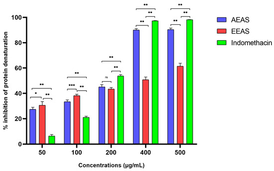
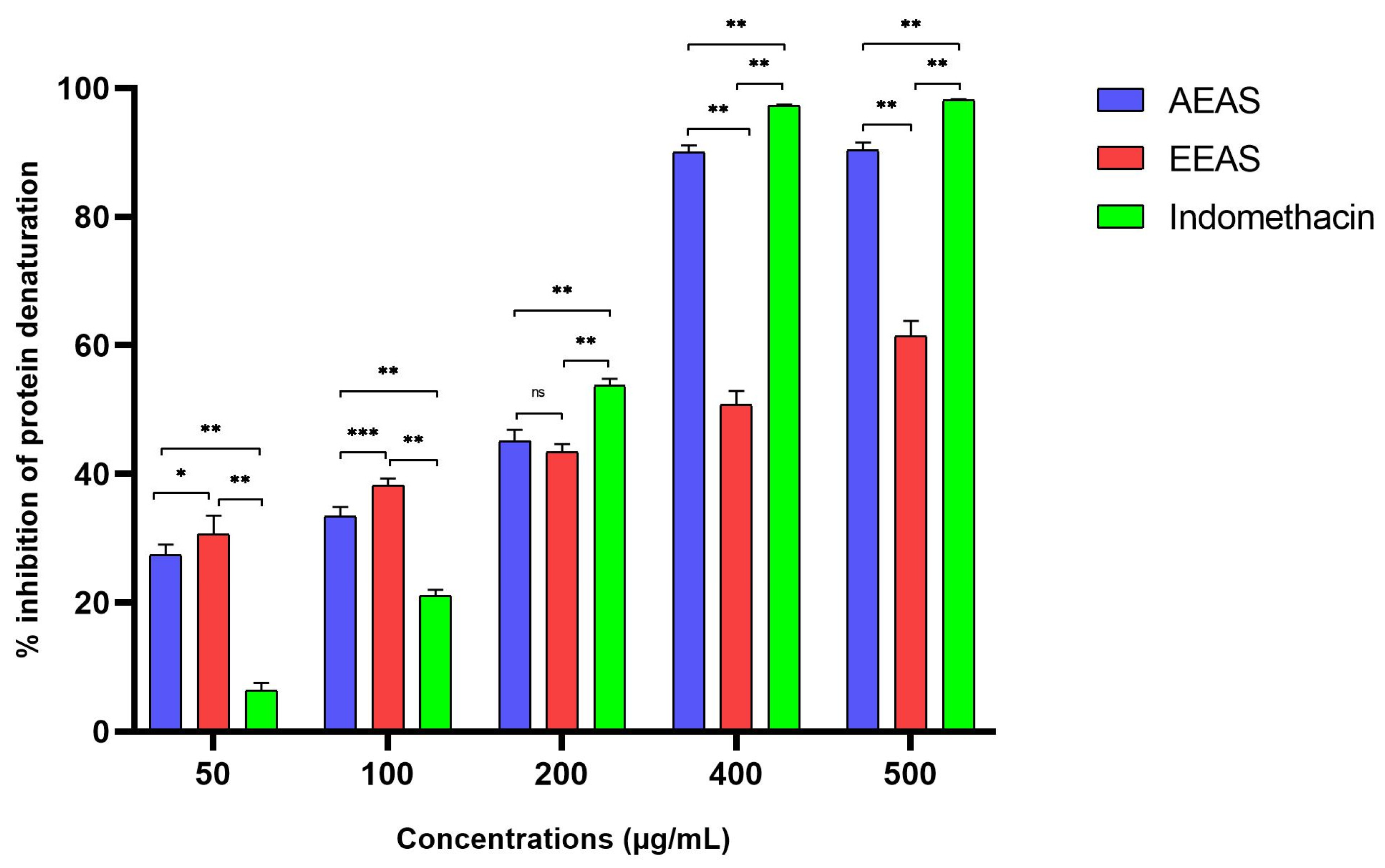
The anti-inflammatory effects of the extracts were evaluated using a protocol that inhibits thermally induced protein denaturation. In this study, varying concentrations (50–500 μg/mL) of both aqueous and ethanolic extracts from A. sativum were incubated with a BSA solution and subjected to heating, as the results are illustrated in Figure 1.
Figure 1.
Effect of aqueous (AEAS) and ethanolic (EEAS) extracts of Allium sativum on heat-induced protein denaturation. Protein denaturation inhibition (%) was measured at concentrations ranging from 50 to 500 μg/mL and compared with indomethacin as the reference anti-inflammatory drug. Data are expressed as mean ± SD of three independent experiments. Statistical significance was assessed by two-way ANOVA followed by simple effects analysis within each concentration. ns, not significant; * p < 0.05; ** p < 0.01; *** p < 0.0001.
Indomethacin, used as a positive control, showed marked inhibition of heat-induced BSA denaturation, confirming the responsiveness and validity of the assay. The maximum inhibition observed for the ethanolic and aqueous extracts was reached at 500 µg/mL, with inhibition percentages of 61.46 ± 2.35% and 90.37 ± 1.15%, respectively. In comparison, indomethacin showed maximum inhibition of 98.13 ± 0.18% at 500 µg/mL. Based on IC50 values, indomethacin showed the strongest inhibitory potency, with an IC50 of 122.96 ± 2.66 µg/mL, followed by the aqueous extract with an IC50 of 152.86 ± 2.44 µg/mL and the ethanolic extract with an IC50 of 283.30 ± 15.46 µg/mL (p < 0.003). These findings indicate that the aqueous extract had a stronger anti-denaturation effect than the ethanolic extract, while indomethacin remained the most potent treatment according to IC50 values.
4.5. ADMET Analysis
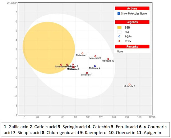
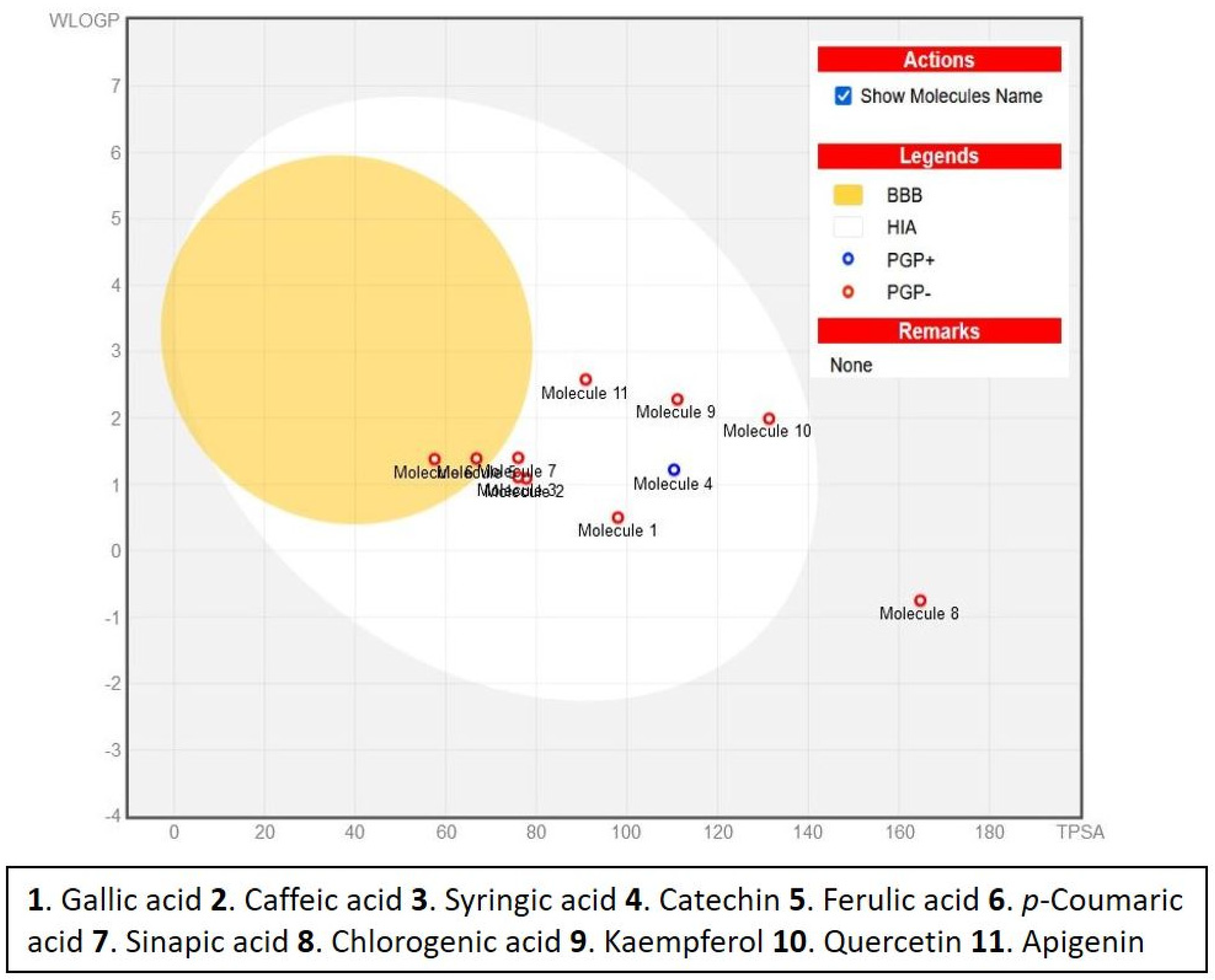
The absorption and distribution properties of compounds in the ethanolic and aqueous extracts of Allium sativum were evaluated using SwissADME and ADMET-AI tools (Table 7, Figure 2).
Table 7.
The percentages of intestinal absorption, oral bioavailability, blood–brain barrier permeability (BBB), and plasma protein binding (PBR) for the identified compounds in the aqueous and ethanolic extracts of Allium sativum L.
Figure 2.
Predictive results for the distribution of identified compounds in Allium sativum L. extracts, based on the BOILED-Egg model for intestinal and brain permeation. The eleven compounds are depicted as a small circle, where the yellow region indicates the space of compounds predicted to penetrate the brain. Abbreviations: tPSA (topological polar surface area) and WLogP (LogP) value calculated using the Wildman-Crippen method, reflecting the potential of a substance to be a substrate of P-glycoprotein, PGP+/PGP−).
All compounds exhibited notable intestinal absorption, with apigenin (67.63%) and kaempferol (53.63%) showing the highest rates, while chlorogenic acid had the lowest (24.93%). Sinapic acid (43.16%) and syringic acid (40.95%) displayed the highest oral bioavailability, whereas chlorogenic acid was the lowest (8.45%) (Table 7, Figure 3). According to the BOILED-Egg model, sinapic acid (50.52%) and ferulic acid (45.91%) showed significant BBB permeability, while quercetin (2.83%) and kaempferol (3.88%) had the lowest. Apigenin (76.31%), kaempferol (74.49%), and quercetin (70.07%) exhibited high PPBR, while chlorogenic acid (30.9%) and catechin (32.49%) were the lowest (Table 7, Figure 2).
Figure 3.
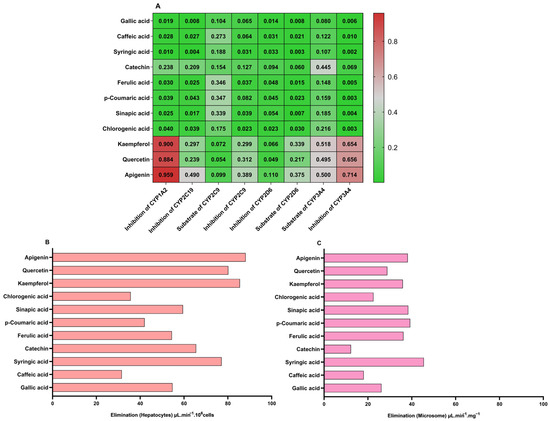
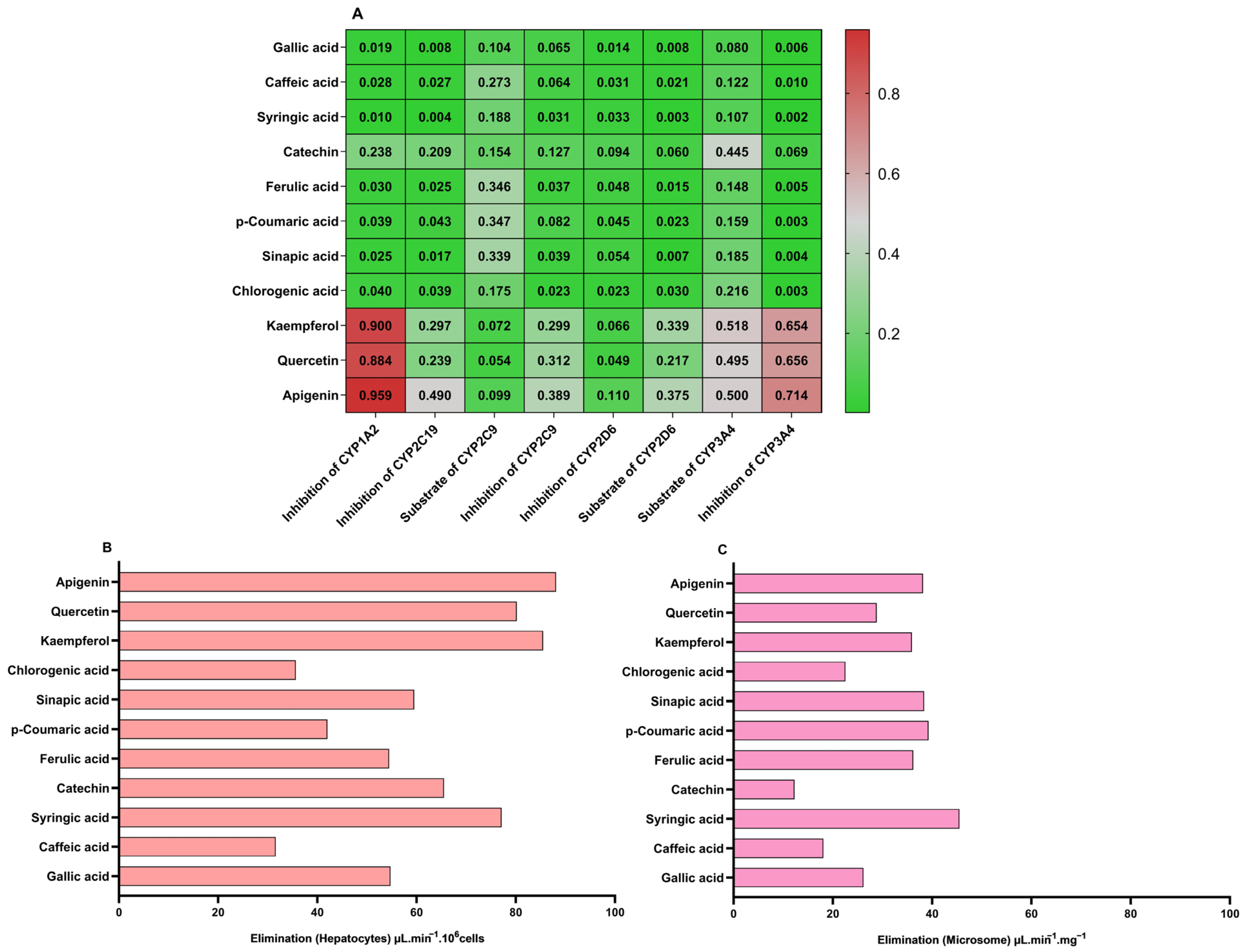
Predicted metabolism and excretion profiles of the identified compounds in Allium sativum L. extracts. (A) Heat map showing the likelihood of metabolism of the molecules by various enzymes and the predicted excretion of the selected metabolites. (B) Elimination of molecules by hepatocytes (μL·min−1 (106 cells). (C) Elimination of molecules by microsomes (µL·min−1·g−1).
Figure 3 presents the metabolism and excretion profiles. Apigenin, quercetin, and kaempferol strongly inhibited CYP1A2, while gallic and syringic acids showed minimal inhibition. Apigenin demonstrated moderate to high substrate potential for CYP2C9 and CYP3A4, while gallic acid and catechin had minimal activity. Hepatic elimination was highest for apigenin and quercetin, while gallic and syringic acids had slower rates, mirroring microsomal elimination trends.
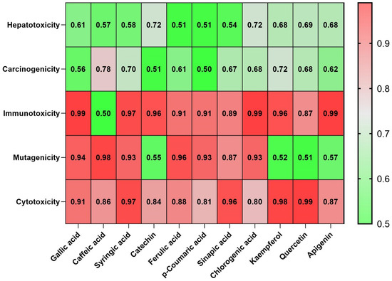
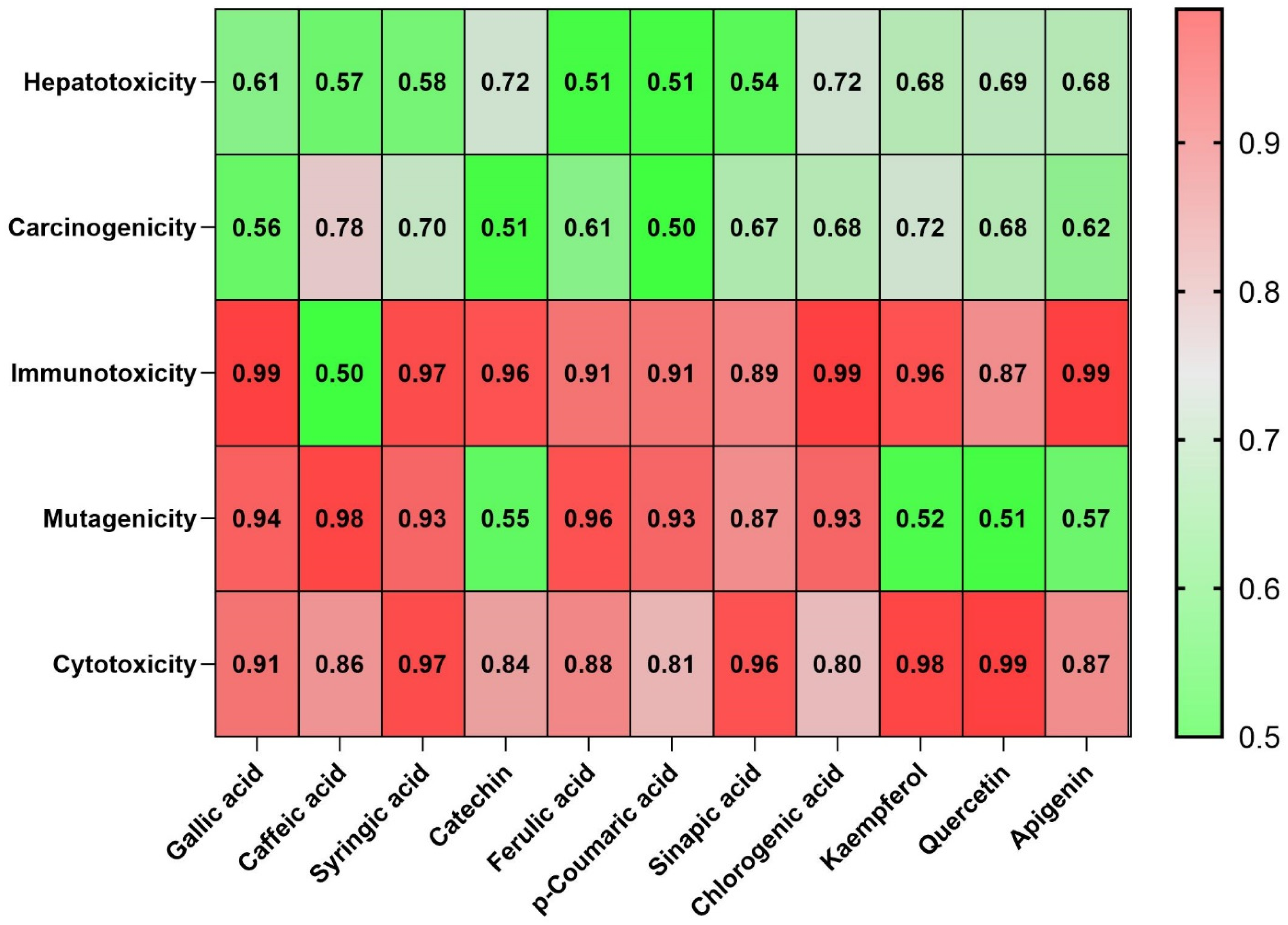
Toxicity predictions (Protox-II, Table 8, Figure 4) revealed low hepatotoxicity and carcinogenicity for most compounds. However, immunotoxicity, mutagenicity, and cytotoxicity were notable concerns. LD50 analysis classified catechin as low toxicity (Class 6), while caffeic acid, chlorogenic acid, and apigenin (Class 5) also showed favorable safety profiles. In contrast, quercetin (Class 3, LD50: 159 mg/kg) was moderately toxic, emphasizing the need for dosage considerations in therapeutic applications.
Table 8.
Toxicity predictions for the identified compounds in the aqueous and ethanolic extracts of Allium sativum L.
Figure 4.
Prediction of oral toxicity of identified compounds of Allium sativum L. extracts in rodents using PROTOX II: A heat map illustrates the probabilities of hepatotoxicity, carcinogenicity, mutagenicity, and cytotoxicity. Colors tending toward red indicate a higher predicted probability of toxicity, whereas colors tending toward green indicate a lower predicted toxicity risk.
4.6. Molecular Docking Analysis
Eleven bioactive compounds from the ethanolic and aqueous extracts of Allium sativum were docked with bacterial enzymes Gyrase B and Transpeptidase. The docking results (Table 9) include ΔGb and Ki values, which indicate binding affinity. Lower ΔGb values signify stronger interactions, with quercetin (−9.77 kcal/mol), chlorogenic acid (−9.5 kcal/mol), kaempferol (−9.34 kcal/mol), and catechin (−9.32 kcal/mol) exhibiting the strongest binding to Gyrase B. Chlorogenic acid showed the highest affinity for Transpeptidase (−11.79 kcal/mol), followed by quercetin (−8.88 kcal/mol), catechin (−8.88 kcal/mol), and kaempferol (−8.36 kcal/mol). These findings suggest that chlorogenic acid and quercetin showed favorable predicted binding affinities toward bacterial targets involved in DNA replication and cell wall synthesis.
Table 9.
Molecular docking analysis of the major compounds from the aqueous and ethanolic extracts of Allium sativum L. against bacterial enzymes.
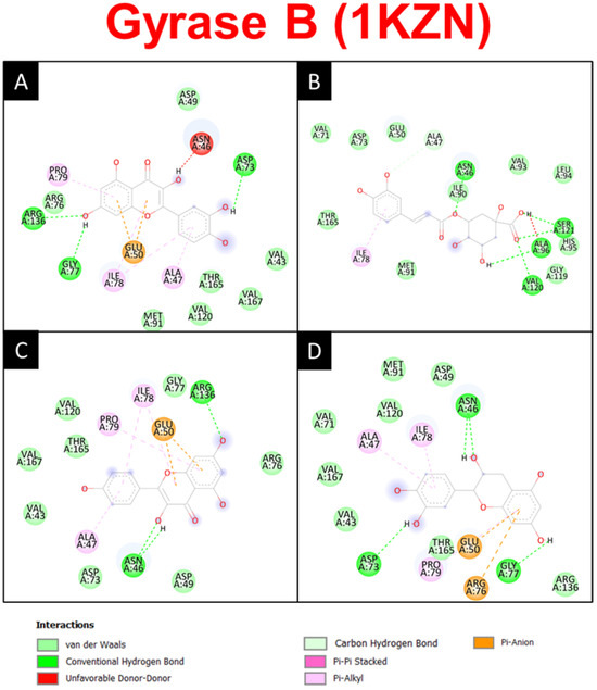
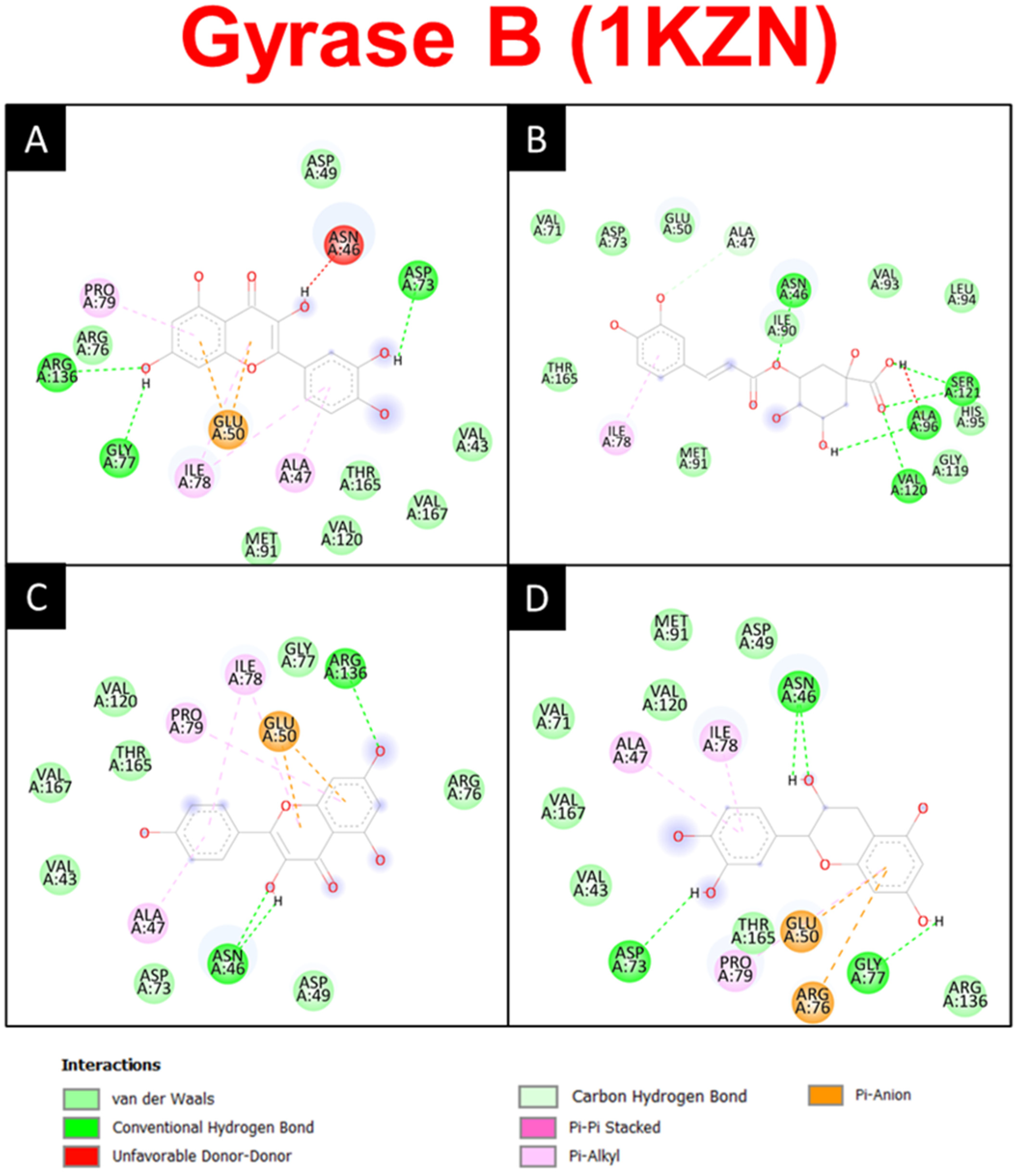
The calculated Ki values were consistent with the predicted binding energy results, with chlorogenic acid showing the most favorable predicted interaction with Transpeptidase and quercetin showing the most favorable predicted interaction with Gyrase B (Table 8). Hydrogen bond analysis (Table 9, Figure 5 and Figure 6) further supported these results. For Gyrase B, chlorogenic acid interacted with ASN A:46, ALA A:96, VAL A:120, and SER A:121, reinforcing its strong binding. Quercetin formed bonds with ASP A:73, GLY A:77, and ARG A:136, enhancing stability. Catechin and kaempferol also showed stable interactions with ASN A:46 and ARG A:136.
Figure 5.
Detailed 2D illustrations depicting the interaction modes between quercetin (A), chlorogenic acid (B), kaempferol (C), and catechin (D) with the crystal structure of Gyrase B.
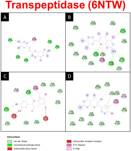
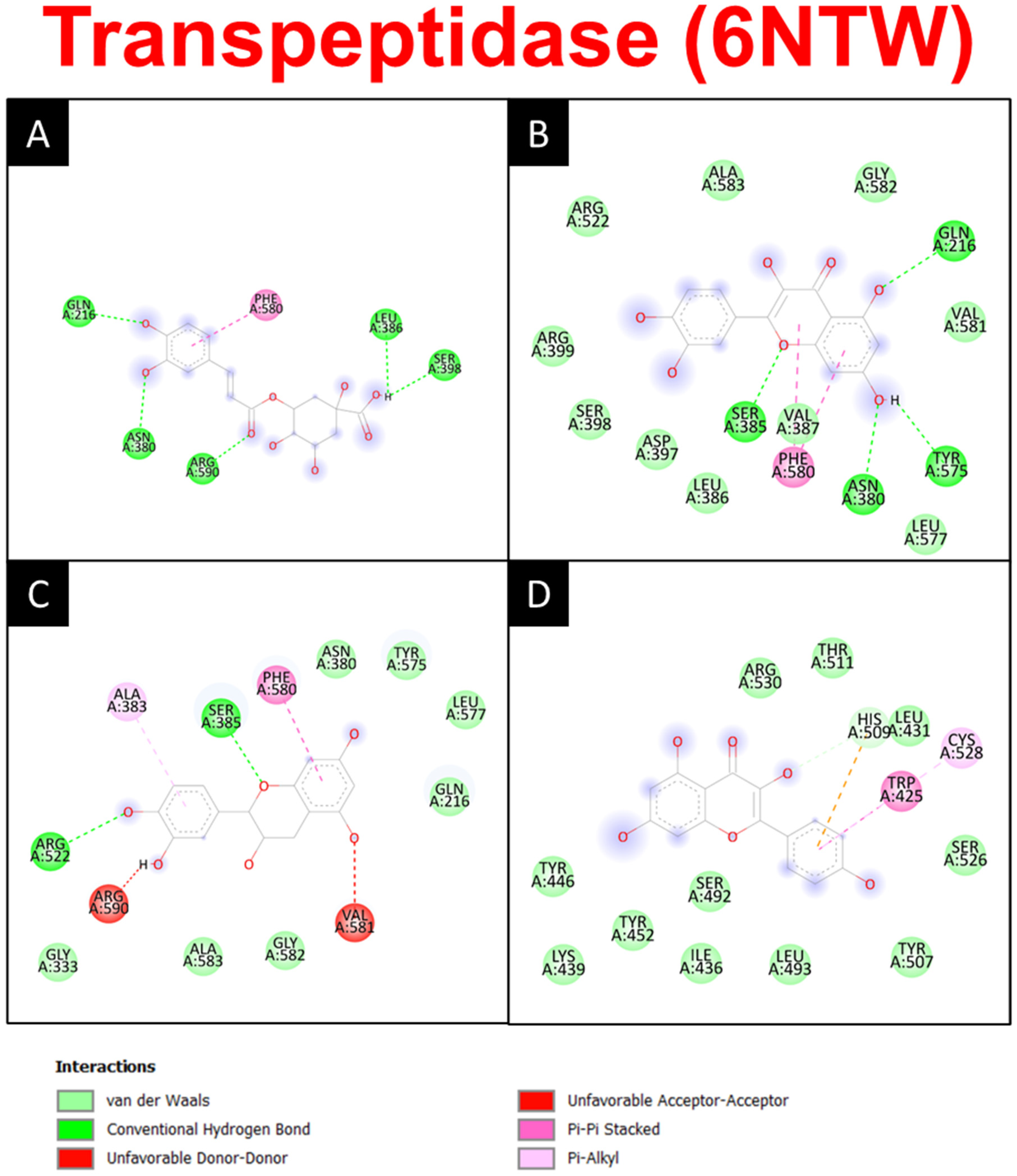
Figure 6.
Detailed 2D illustrations depicting the interaction modes between chlorogenic acid (A), quercetin (B), catechin (C), and kaempferol (D) with the crystal structure of Transpeptidase.
For Transpeptidase, chlorogenic acid displayed extensive bonding with GLN A:216, ASN A:380, LEU A:386, SER A:398, and ARG A:590, consistent with its lowest Ki. Quercetin bonded with GLN A:216, ASN A:380, SER A:385, and TYR A:575, while catechin and kaempferol interacted with SER A:385 and ARG A:522, supporting their inhibitory potential.
5. Discussion
The present study compared aqueous infusion and ethanolic maceration extracts of Moroccan-grown A. sativum using hydrolyzed phenolic aglycone profiling, antibacterial, antioxidant, and protein denaturation inhibition assays, supported by predictive docking and ADMET analyses.
Quantitative phytochemical analysis is an essential step in evaluating a plant’s composition. Our results revealed that the ethanolic extract of A. sativum contained significantly higher TPC and TFC than the aqueous extract (p < 0.001). HPLC analysis identified eight components in the ethanolic extract and six in the aqueous extract. Literature reports variation in TPC, TFC, and phenolic compounds in A. sativum. A study on Chinese garlic identified eight phenolic compounds, including ferulic acid (0.47–0.86 μg/g), p-coumaric acid (0.09–0.42 μg/g), and quercetin (0.13 μg/g) [30]. Beato et al. characterized Spanish A. sativum, reporting a TPC of 6.5 mg GAE/gextract, with caffeic acid (2.9 mg/kg) and ferulic acid (2.6 mg/kg) as major phenolics [31]. Another study from Finland found TPC levels ranging from 75 to 700 mg GAE/kg, with quercetin (82 mg/kg) and kaempferol (44 mg/kg) in garlic leaves [32].
Apigenin was detected only in the ethanolic extract of the Meknes A. sativum sample analyzed in the present study. However, previous studies have already identified this flavone in A. sativum from different geographical origins and cultivars [33,34,35,36]. Rather, our result highlights the influence of extraction solvent and experimental conditions on the phenolic profile of garlic extracts [14]. The absence of apigenin in the aqueous extract may be related to solvent selectivity, differences in compound solubility, matrix interactions, or concentration levels below the detection limit [16]. Therefore, the detection of apigenin in the ethanolic extract supports the broader observation that the phytochemical composition of garlic preparations depends strongly on origin, processing, extraction method, and analytical conditions [30].
In recent decades, the rise of bacterial antibiotic resistance has significantly impacted human and animal health, necessitating the search for new alternatives [37]. Consequently, there is an urgent need to discover new treatments for bacterial infections, underscoring the importance of identifying antibacterial compounds from natural sources [38]. The antibacterial activity observed in the present study was evaluated exclusively against bacterial strains, namely MRSA, E. coli, and P. aeruginosa. Both aqueous and ethanolic extracts inhibited MRSA, whereas only the aqueous extract showed activity against E. coli. No inhibitory effect was observed against P. aeruginosa.
These findings suggest that the antibacterial response of A. sativum extracts depends on both the bacterial strain tested and the solvent-dependent phytochemical composition of the extracts [39]. The MIC and MBC results further indicated a bactericidal effect against susceptible bacterial strains, as shown by MBC/MIC ratios equal to 1. Despite the detectable antibacterial activity observed against MRSA and E. coli, the relatively high MIC values obtained (83.3–333 mg/mL) indicate modest antibacterial potency when compared with conventional antimicrobial agents. Therefore, the observed effects should be interpreted as preliminary evidence of antibacterial potential rather than strong antimicrobial efficacy. This observation is not uncommon for crude plant extracts, which generally require substantially higher concentrations than purified antibiotics due to their complex chemical composition and the relatively low abundance of individual active constituents. The activity observed against MRSA with both extracts, and against E. coli only with the aqueous extract, may be related to differences in the solubility and distribution of antibacterial constituents according to the extraction solvent. In particular, the stronger activity of the aqueous extract against E. coli may reflect the presence of water-soluble compounds or synergistic interactions among constituents that are absent, less abundant, or less active in the ethanolic extract [40]. However, since crude extracts contain complex mixtures of major and minor phytochemicals, including phenolic compounds and flavonoids, the antibacterial effects cannot be attributed to a single compound without further fractionation and testing of isolated molecules. Therefore, these results should be interpreted as evidence of solvent- and strain-dependent antibacterial activity rather than as the effect of one specific bioactive constituent.
In our effort to explore the potential mechanisms underlying the antimicrobial effects of A. sativum extracts, an in silico analysis was carried out through molecular docking. The in silico study focused on bacterial targets such as gyrase B and transpeptidase, which were chosen for their prominence as common targets of antibiotic action or for their potential to help reduce microbial resistance to certain antimicrobial agents. Indeed, gyrase B is a component of DNA gyrase, which belongs to the type II topoisomerase family [41]. It plays a critical role in the energy generation process through hydrolysis, providing the energy necessary for the ligation of cleaved DNA strands by gyrase A subunits. This process is essential for the ATP-dependent introduction of negative supercoils into DNA, a key step in maintaining DNA structure and function [42]. Transpeptidase, an enzyme integral to the penicillin-binding proteins of classes A and B, facilitates the transpeptidation reaction. This process involves the linking of pentapeptide chains to adjacent peptide chains within the peptidoglycan layer of bacterial cell walls [43].
Among the 11 molecules identified in the ethanolic and aqueous extracts of A. sativum, chlorogenic acid, quercetin, kaempferol, and catechin exhibited the most favorable predicted binding affinities toward the selected bacterial targets, as indicated by their low Ki and favorable ΔGb values. These computational results suggest that these compounds may interact with regions involved in enzyme activity. However, molecular docking provides only a theoretical prediction of ligand-protein interactions and does not constitute direct evidence of enzyme inhibition or antibacterial activity.
The predicted binding poses observed for some compounds involve regions associated with the activity of DNA gyrase and may therefore be consistent with mechanisms targeted by fluoroquinolone antibiotics such as nadifloxacin, ciprofloxacin, and moxifloxacin. However, experimental studies would be required to confirm whether these compounds exert similar biological effects [43,44,45]. If these predicted interactions translate into effective enzyme inhibition, they could potentially interfere with essential bacterial processes such as DNA replication or cell wall biosynthesis. Nevertheless, this hypothesis remains to be experimentally validated.
Bacterial infections and oxidative stress are closely linked, with oxidative stress playing a crucial role in immune defense but requiring regulation to prevent tissue damage. Modulating oxidative stress through antioxidant compounds, such as those from medicinal plants, may help manage bacterial infection-related complications [2,46].
This study evaluated the antioxidant potential of ethanolic and aqueous extracts of A. sativum using three analytical methods. Both extracts exhibited notable antioxidant activity, with the ethanolic extract showing superior effects. This is can be attributed to its higher total polyphenol concentration (298.2 mg/100 g vs. 106.8 mg/100 g in the aqueous extract) and the presence of key phenolic compounds. Quercetin and kaempferol, found in high amounts in the ethanolic extract, contribute significantly to antiradical activity [47,48]. Kaempferol is also known for reducing chronic disease risks, including cancer [49]. Apigenin, detected only in the ethanolic extract, likely enhances antioxidant activity due to its strong antiradical, anti-inflammatory, and anticarcinogenic properties [50,51,52]. Additionally, bioactive compounds in the extract may act synergistically, enhancing antioxidant effects by regenerating depleted antioxidants, chelating metal ions involved in ROS formation, and providing broader oxidative defense [53].
The link between bacterial infection and inflammation plays a vital role in the body’s immune defense against invading pathogens [54]. If the bacterial infection is not adequately resolved, it may lead to persistent inflammation, which can cause tissue damage and contribute to the development of chronic diseases by leading to tissue destruction, protein denaturation and scar formation [55,56]. In the context of excessive inflammation, protein denaturation is a critical aspect of the biological impact of prolonged or uncontrolled inflammatory responses [57]. It is crucial to develop therapeutic strategies to mitigate the negative consequences of excessive inflammation. In fact, agents that can prevent protein denaturation, therefore, would be worthwhile for anti-inflammatory drug development [46].
In this context, the present work evaluated the anti-inflammatory effect of ethanolic and aqueous extracts of A. sativum on protein denaturation. Both extracts exerted an inhibitory effect on protein denaturation, and from the IC50 values it becomes evident that the aqueous extract was more protective than the ethanolic extract. The ability of the extracts to reduce BSA denaturation may be partly related to their phenolic composition and antioxidant properties [46]. However, the precise mechanism underlying this effect was not directly investigated in the present study. One possible explanation is that some extract constituents may contribute to protein stabilization through non-covalent interactions or by limiting oxidative changes that favor protein denaturation [58,59]. Nevertheless, this remains a hypothesis, and further studies using purified compounds, protein-binding assays, and structural approaches are required to confirm this mechanism.
The ADMET analysis of the aqueous and ethanolic extracts of A. sativum provided preliminary insights into the pharmacokinetic and toxicological profiles of the identified phenolic compounds. Apigenin and kaempferol exhibited higher predicted intestinal absorption and bioavailability than more polar compounds such as chlorogenic acid, likely due to their lower polarity and moderate molecular weight. Their greater lipophilicity was also associated with higher plasma protein binding, whereas compounds rich in hydroxyl groups, such as quercetin and kaempferol, showed limited predicted BBB permeability. Apigenin, quercetin, and kaempferol displayed stronger predicted interactions with CYP1A2, CYP2C9, and CYP3A4, suggesting extensive hepatic metabolism, while gallic and syringic acids exhibited slower predicted clearance. Toxicity predictions identified catechin as having a favorable safety profile, whereas quercetin showed a comparatively higher predicted toxicity. Overall, these results suggest that the physicochemical properties of the compounds may influence their predicted pharmacokinetic behavior and safety profiles. However, these findings were generated using computational models and should therefore be regarded as preliminary indicators rather than experimentally validated parameters, requiring confirmation through dedicated pharmacokinetic and toxicological studies.
6. Limitations and Future Perspectives
This study has several limitations that should be considered when interpreting the results. First, the antibacterial, antioxidant, and protein denaturation inhibition assays provide only preliminary in vitro evidence and do not fully reproduce the complexity of biological systems. Second, although phenolic compounds were identified using authentic reference standards analyzed under identical chromatographic conditions, identification was based primarily on retention-time matching and should therefore be considered tentative. Definitive structural confirmation would require complementary analytical techniques such as HPLC-DAD, LC-MS/MS, NMR, or other spectroscopic approaches. Third, because HPLC analysis was performed after acidic hydrolysis, the detected compounds represent hydrolyzed phenolic aglycones and do not fully reflect the native phenolic composition of the crude extracts. In addition, although the chromatographic procedure was based on a previously validated method, compound-specific validation parameters were not experimentally re-established for each analyte under the conditions of the present study. Finally, molecular docking and ADMET analyses are predictive computational approaches that provide theoretical estimates of molecular interactions and pharmacokinetic properties and therefore require experimental validation through enzymatic, cell-based, and in vivo studies. Future investigations should include advanced phytochemical characterization, testing of isolated compounds, broader biological evaluations, and in vivo models to further establish the biological relevance of the observed activities.
7. Conclusions
Overall, aqueous and ethanolic extracts of A. sativum showed antibacterial, antioxidant, and in vitro protein denaturation inhibitory activities under the present experimental conditions. These effects appear to be associated, at least in part, with solvent-dependent differences in phenolic composition. Molecular docking further suggested possible interactions between selected phenolic compounds and bacterial target proteins. However, further studies using additional controls, purified compounds, and in vivo models are required to confirm the pharmacological relevance of these findings.
Author Contributions
Conceptualization, O.E.F., A.E.F., I.E., Z.O., S.R., E.M.M.; data curation, O.E.F., A.E.A.; formal analysis, O.E.F., A.E.F., A.E.O.; software analysis, A.E.F.; methodology, O.E.F., A.E.F., I.E., A.E.A., A.E.O., H.B.; investigation, S.R., A.E.A.; writing—original draft preparation, O.E.F., A.E.F.; figures conception, O.E.F.; writing—review and editing, O.E.F., E.M.M. All authors have read and agreed to the published version of the manuscript.
Funding
This research received no external funding.
Data Availability Statement
The raw data supporting the conclusions of this article will be made available by the authors on request.
Conflicts of Interest
The authors declare no conflicts of interest.
Abbreviations
The following abbreviations are used in this manuscript:
| ΔGb | Binding energy |
| ADMET | Absorption, distribution, metabolism, excretion, and toxicity |
| ADT | AutoDock Tools |
| A. sativum | Allium sativum |
| BBB | Blood–brain barrier |
| BSA | Bovine Serum Albumin |
| DIZ | Diameter of the inhibition zone |
| DPPH• | 2,2-diphenyl-1-picrylhydrazyl |
| E. coli | Escherichia coli |
| EC50 | Effective concentration 50 |
| FRAP | Ferric reducing antioxidant power |
| HCl | Hydrochloric acid |
| HPLC | High-performance liquid chromatography |
| IC50 | Inhibitory concentration 50 |
| Ki | Inhibition constant |
| LD50 | Lethal dose 50 |
| LOD | Limits of detection |
| LOQ | Limits of quantification |
| MBC | Minimum bactericidal concentration |
| MIC | Minimum inhibitory concentration |
| MRSA | Methicillin-resistant Staphylococcus aureus |
| PPB | Plasma protein binding |
| P. aeruginosa | Pseudomonas aeruginosa |
| ROS | Reactive oxygen species |
| TAC | Total antioxidant capacity |
| TFC | Total flavonoids content |
| TPC | Total phenolic content |
References
- Fisher, R.A.; Gollan, B.; Helaine, S. Persistent Bacterial Infections and Persister Cells. Nat. Rev. Microbiol. 2017, 15, 453–464. [Google Scholar] [CrossRef] [Scilit] [PubMed]
- Ivanov, A.V.; Bartosch, B.; Isaguliants, M.G. Oxidative Stress in Infection and Consequent Disease. Oxidative Med. Cell. Longev. 2017, 2017, 3496043. [Google Scholar] [CrossRef] [Scilit] [PubMed]
- Grant, S.S.; Hung, D.T. Persistent Bacterial Infections, Antibiotic Tolerance, and the Oxidative Stress Response. Virulence 2013, 4, 273–283. [Google Scholar] [CrossRef] [Scilit] [PubMed]
- Pan, X.; Liu, W.; Du, Q.; Zhang, H.; Han, D. Recent Advances in Bacterial Persistence Mechanisms. Int. J. Mol. Sci. 2023, 24, 14311. [Google Scholar] [CrossRef] [Scilit] [PubMed]
- Gurib-Fakim, A. Medicinal Plants: Traditions of Yesterday and Drugs of Tomorrow. Mol. Asp. Med. 2006, 27, 1–93. [Google Scholar] [CrossRef] [Scilit] [PubMed]
- Ait Alla, K.; El Faqer, O.; Jahjah, S.; Labhar, A.; Alaoui Jamali, C.; Kasrati, A.; Souhail, B.; Legssyer, M.; Maouni, A.; Saidi, R. Volatile Compounds from Northern Moroccan Medicinal Plants: Phytochemical Analysis, Antioxidant and Antimicrobial Potential, and In Silico Investigations. Compounds 2025, 5, 49. [Google Scholar] [CrossRef] [Scilit]
- Alla, K.A.; El Faqer, O.; Jahjah, S.; Jaouani, M.; El Ouaddari, A.; Jamali, C.A.; Kasrati, A.; Souhail, B.; Maouni, A.; Saidi, R. Integrated In Vitro and In Silico Assessment of the Bioactivities of Essential Oils from Northern Moroccan Thymus Glandulosus Req. and Mentha Suaveolens Subsp. Eu-Rotundifolia: Promising Candidates against Healthcare-Associated Infections. J. Essent. Oil Bear. Plants 2025, 28, 1340–1356. [Google Scholar] [CrossRef] [Scilit]
- Bendiar, S.; El Faqer, O.; Benjelloun, N.; Hsseini, S.; Bellaoui, H.; Rais, S.; Zaid, Y.; Mtairag, E.M.; Oudghiri, M. Immunomosuppressive Effect of Ziziphus lotus L. (Desf.) Fruit’s Extract on Neutrophil Bactericidal Activity and on In Vivo Humoral Immune Response in Mice. Biomed. Pharmacol. J. 2022, 15, 2331–2341. [Google Scholar] [CrossRef] [Scilit]
- Salma, B.; Othman, E.F.; Sanaa, C.; Naima, B.; Mostafa, M.E.; Mounia, O. Phytochemical Screening and In Vivo Immunosuppressive, Antioxidant and Anti-Hemolytic Activities of Zea Mays Silk Aqueous Extract. Pharmacogn. J. 2020, 12, 1412–1420. [Google Scholar] [CrossRef] [Scilit]
- Savairam, V.D.; Patil, N.A.; Borate, S.R.; Ghaisas, M.M.; Shete, R.V. Allicin: A Review of Its Important Pharmacological Activities. Pharmacol. Res.-Mod. Chin. Med. 2023, 8, 100283. [Google Scholar] [CrossRef] [Scilit]
- Jang, H.-J.; Lee, H.-J.; Yoon, D.-K.; Ji, D.-S.; Kim, J.-H.; Lee, C.-H. Antioxidant and Antimicrobial Activities of Fresh Garlic and Aged Garlic By-Products Extracted with Different Solvents. Food Sci. Biotechnol. 2018, 27, 219–225. [Google Scholar] [CrossRef] [Scilit]
- Dai, J.; Mumper, R.J. Plant Phenolics: Extraction, Analysis and Their Antioxidant and Anticancer Properties. Molecules 2010, 15, 7313–7352. [Google Scholar] [CrossRef] [Scilit] [PubMed]
- Do, Q.D.; Angkawijaya, A.E.; Tran-Nguyen, P.L.; Huynh, L.H.; Soetaredjo, F.E.; Ismadji, S.; Ju, Y.-H. Effect of Extraction Solvent on Total Phenol Content, Total Flavonoid Content, and Antioxidant Activity of Limnophila Aromatica. J. Food Drug Anal. 2014, 22, 296–302. [Google Scholar] [CrossRef] [Scilit] [PubMed]
- Bar, M.; Binduga, U.E.; Szychowski, K.A. Methods of Isolation of Active Substances from Garlic (Allium sativum L.) and Its Impact on the Composition and Biological Properties of Garlic Extracts. Antioxidants 2022, 11, 1345. [Google Scholar] [CrossRef] [Scilit] [PubMed]
- Cavalcanti, V.P.; Aazza, S.; Bertolucci, S.K.V.; Rocha, J.P.M.; Coelho, A.D.; Oliveira, A.J.M.; Mendes, L.C.; Pereira, M.M.A.; Morais, L.C.; Forim, M.R.; et al. Solvent Mixture Optimization in the Extraction of Bioactive Compounds and Antioxidant Activities from Garlic (Allium sativum L.). Molecules 2021, 26, 6026. [Google Scholar] [CrossRef] [Scilit] [PubMed]
- Kallel, F.; Driss, D.; Chaari, F.; Belghith, L.; Bouaziz, F.; Ghorbel, R.; Chaabouni, S.E. Garlic (Allium sativum L.) Husk Waste as a Potential Source of Phenolic Compounds: Influence of Extracting Solvents on Its Antimicrobial and Antioxidant Properties. Ind. Crops Prod. 2014, 62, 34–41. [Google Scholar] [CrossRef] [Scilit]
- Wahnou, H.; Ndayambaje, M.; Ouadghiri, Z.; Benayad, S.; Elattar, H.; Chgari, O.; Naya, A.; Zaid, Y.; Oudghiri, M. Artemisia Herba-Alba: Antioxidant Capacity and Efficacy in Preventing Chronic Arthritis In Vivo. Inflammopharmacology 2024, 32, 1855–1870. [Google Scholar] [CrossRef] [Scilit] [PubMed]
- Ezaouine, A.; Salam, M.R.; Sbaoui, Y.; Nouadi, B.; Zouhir, S.; Elmessal, M.; Chegdani, F.; Bennis, F. Extraction, Chemical Composition and Antioxidant Activity of Phenolic Compounds from Moroccan Satureja nepeta L. Pharmacogn. J. 2022, 14, 182–192. [Google Scholar] [CrossRef] [Scilit]
- Latif, M.; Elkoraichi, I.; El Faqer, O.; Wahnou, H.; Mtairag, E.M.; Oudghiri, M.; Rais, S. Phytochemical Analysis and Immunomodulatory Activities In Vitro and In Vivo of Aframomum Melegueta K Schum Seed Extracts. Inflammopharmacology 2024, 32, 1621–1631. [Google Scholar] [CrossRef] [Scilit] [PubMed]
- Huang, Z.; Wang, B.; Eaves, D.H.; Shikany, J.M.; Pace, R.D. Phenolic Compound Profile of Selected Vegetables Frequently Consumed by African Americans in the Southeast United States. Food Chem. 2007, 103, 1395–1402. [Google Scholar] [CrossRef] [Scilit]
- Manoury, E.; Jourdon, K.; Boyaval, P.; Fourcassié, P. Quantitative Measurement of Vitamin K2 (Menaquinones) in Various Fermented Dairy Products Using a Reliable High-Performance Liquid Chromatography Method. J. Dairy Sci. 2013, 96, 1335–1346. [Google Scholar] [CrossRef] [Scilit] [PubMed]
- Kazusaki, M.; Ueda, S.; Takeuchi, N.; Ohgami, Y. Validation of Analytical Procedures by High−performance Liquid Chromatography for Pharmaceutical Analysis. Chromatography 2012, 33, 65–73. [Google Scholar] [CrossRef] [Scilit]
- Boughroud, H.; Chgari, O.; Wahnou, H.; El Amrani, A.; El Faqer, O.; Amarir, F.; Mtairag, E.M.; Rais, S.; Bourjilat, F. Assessing Syzygium Aromaticum: From Chemical Profiling to Antioxidant, Antistaphylococcal, and Biocompatibility Attributes. Chem. Biodivers. 2024, 22, e202402605. [Google Scholar] [CrossRef] [Scilit] [PubMed]
- Mounia, L.; Ismail, E.; Othman, E.F.; Hicham, W.; Rajaa, E.; El Mostafa, M.; Mounia, O.; Samira, R. Aframomum melegueta: Evaluation of Chronic Toxicity, HPLC Profiling, and In Vitro/In Vivo Antioxidant Assessment of Seeds Extracts. Chem. Biodivers. 2024, 22, e202400942. [Google Scholar] [CrossRef] [Scilit] [PubMed]
- Elkoraichi, I.; Latif, M.; El Faqer, O.; Elaje, R.; Thoume, A.; Mtairag, E.M.; Rais, S.; Daoudi, F. In Vivo and In Vitro Evaluation of Antioxidant, Antihemolytic, Acute Toxicity Effects and High-Performance Liquid Chromatography Analysis of an Aqueous Extract of Moroccan Punica granatum L. J. Herbmed Pharmacol. 2022, 12, 168–175. [Google Scholar] [CrossRef] [Scilit]
- Samiry, I.; El Faqer, O.; Rais, S.; Oudghiri, M.; Zaid, Y.; Mtairag, E.M. Biological Activities of Aqueous Extract of Caralluma Europaea. Int. J. Chem. Biomol. Sci. 2022, 21, 91–98. [Google Scholar]
- Ouadghiri, Z.; El Faqer, O.; Wahnou, H.; Soudassi, A.; Marnissi, F.; Rais, S.; Mtairag, E.M. In Vivo and In Vitro Assessment of Marrubium vulgare: Chemical, Antioxidant and Anti-Inflammatory Profiles. Arab. J. Sci. Eng. 2025, 51, 1227–1239. [Google Scholar] [CrossRef] [Scilit]
- El Faqer, O.; Rais, S.; Ouadghiri, Z.; El Faqer, A.; Benchama, Z.; El Ouaddari, A.; Dakir, M.; El Amrani, A.; Mtairag, E.M. Physicochemical Properties, GC–MS Profiling, and Antibacterial Potential of Allium Sativum Essential Oil: In Vitro and in Silico Approaches. Sci. Afr. 2024, 26, e02484. [Google Scholar] [CrossRef] [Scilit]
- El Faqer, O.; Elkoraichi, I.; Latif, M.; Debierre-Grockiego, F.; Ouadghiri, Z.; Rais, S.; Dimier-Poisson, I.; Mtairag, E.M. Pharmacological Insights into Laurus Nobilis: HPLC Profiling and Evaluation of Its Anti-Toxoplasma, Antioxidant, and Anti-Hemolytic Properties. Biochem. Syst. Ecol. 2024, 117, 104891. [Google Scholar] [CrossRef] [Scilit]
- Liu, J.; Ji, F.; Chen, F.; Guo, W.; Yang, M.; Huang, S.; Zhang, F.; Liu, Y. Determination of Garlic Phenolic Compounds Using Supercritical Fluid Extraction Coupled to Supercritical Fluid Chromatography/Tandem Mass Spectrometry. J. Pharm. Biomed. Anal. 2018, 159, 513–523. [Google Scholar] [CrossRef] [Scilit] [PubMed]
- Beato, V.M.; Orgaz, F.; Mansilla, F.; Montaño, A. Changes in Phenolic Compounds in Garlic (Allium sativum L.) Owing to the Cultivar and Location of Growth. Plant Foods Hum. Nutr. 2011, 66, 218–223. [Google Scholar] [CrossRef] [Scilit] [PubMed]
- Nuutila, A.M.; Puupponen-Pimiä, R.; Aarni, M.; Oksman-Caldentey, K.-M. Comparison of Antioxidant Activities of Onion and Garlic Extracts by Inhibition of Lipid Peroxidation and Radical Scavenging Activity. Food Chem. 2003, 81, 485–493. [Google Scholar] [CrossRef] [Scilit]
- Miean, K.H.; Mohamed, S. Flavonoid (Myricetin, Quercetin, Kaempferol, Luteolin, and Apigenin) Content of Edible Tropical Plants. J. Agric. Food Chem. 2001, 49, 3106–3112. [Google Scholar] [CrossRef] [Scilit] [PubMed]
- Fratianni, F.; Ombra, M.N.; Cozzolino, A.; Riccardi, R.; Spigno, P.; Tremonte, P.; Coppola, R.; Nazzaro, F. Phenolic Constituents, Antioxidant, Antimicrobial and Anti-Proliferative Activities of Different Endemic Italian Varieties of Garlic (Allium sativum L.). J. Funct. Foods 2016, 21, 240–248. [Google Scholar] [CrossRef] [Scilit]
- Park, M.-H.; Kim, J.-G. Low-Dose UV-C Irradiation Reduces the Microbial Population and Preserves Antioxidant Levels in Peeled Garlic (Allium sativum L.) during Storage. Postharvest Biol. Technol. 2015, 100, 109–112. [Google Scholar] [CrossRef] [Scilit]
- Azzini, E.; Durazzo, A.; Foddai, M.S.; Temperini, O.; Venneria, E.; Valentini, S.; Maiani, G. Phytochemicals Content in Italian Garlic Bulb (Allium sativum L.) Varieties. J. Food Res. 2014, 3, 26. [Google Scholar] [CrossRef] [Scilit]
- Stevanović, Z.; Bošnjak-Neumüller, J.; Pajić-Lijaković, I.; Raj, J.; Vasiljević, M. Essential Oils as Feed Additives—Future Perspectives. Molecules 2018, 23, 1717. [Google Scholar] [CrossRef] [Scilit] [PubMed]
- Manayi, A.; Kurepaz-mahmoodabadi, M.; Gohari, A.R.; Ajani, Y.; Saeidnia, S. Presence of Phthalate Derivatives in the Essential Oils of a Medicinal Plant Achillea Tenuifolia. DARU J. Pharm. Sci. 2014, 22, 78. [Google Scholar] [CrossRef] [Scilit] [PubMed]
- Bouarab-Chibane, L.; Forquet, V.; Lantéri, P.; Clément, Y.; Léonard-Akkari, L.; Oulahal, N.; Degraeve, P.; Bordes, C. Antibacterial Properties of Polyphenols: Characterization and QSAR (Quantitative Structure–Activity Relationship) Models. Front. Microbiol. 2019, 10, 829. [Google Scholar] [CrossRef] [Scilit] [PubMed]
- Ramata-Stunda, A.; Petriņa, Z.; Valkovska, V.; Borodušķis, M.; Gibnere, L.; Gurkovska, E.; Nikolajeva, V. Synergistic Effect of Polyphenol-Rich Complex of Plant and Green Propolis Extracts with Antibiotics against Respiratory Infections Causing Bacteria. Antibiotics 2022, 11, 160. [Google Scholar] [CrossRef] [Scilit] [PubMed]
- Skok, Ž.; Barančoková, M.; Benek, O.; Cruz, C.D.; Tammela, P.; Tomašič, T.; Zidar, N.; Mašič, L.P.; Zega, A.; Stevenson, C.E.M.; et al. Exploring the Chemical Space of Benzothiazole-Based DNA Gyrase B Inhibitors. ACS Med. Chem. Lett. 2020, 11, 2433–2440. [Google Scholar] [CrossRef] [Scilit] [PubMed]
- Tiz, D.B.; Skok, Ž.; Durcik, M.; Tomašič, T.; Mašič, L.P.; Ilaš, J.; Zega, A.; Draskovits, G.; Révész, T.; Nyerges, Á.; et al. An Optimised Series of Substituted N-Phenylpyrrolamides as DNA Gyrase B Inhibitors. Eur. J. Med. Chem. 2019, 167, 269–290. [Google Scholar] [CrossRef] [Scilit] [PubMed]
- Cochrane, S.A.; Lohans, C.T. Breaking down the Cell Wall: Strategies for Antibiotic Discovery Targeting Bacterial Transpeptidases. Eur. J. Med. Chem. 2020, 194, 112262. [Google Scholar] [CrossRef] [Scilit] [PubMed]
- Henderson, S.R.; Stevenson, C.E.M.; Malone, B.; Zholnerovych, Y.; Mitchenall, L.A.; Pichowicz, M.; McGarry, D.H.; Cooper, I.R.; Charrier, C.; Salisbury, A.-M.; et al. Structural and Mechanistic Analysis of ATPase Inhibitors Targeting Mycobacterial DNA Gyrase. J. Antimicrob. Chemother. 2020, 75, 2835–2842. [Google Scholar] [CrossRef] [Scilit] [PubMed]
- Lima, L.M.; Silva, B.N.M.D.; Barbosa, G.; Barreiro, E.J. β-Lactam Antibiotics: An Overview from a Medicinal Chemistry Perspective. Eur. J. Med. Chem. 2020, 208, 112829. [Google Scholar] [CrossRef] [Scilit] [PubMed]
- Hussain, T.; Tan, B.; Yin, Y.; Blachier, F.; Tossou, M.C.B.; Rahu, N. Oxidative Stress and Inflammation: What Polyphenols Can Do for Us? Oxidative Med. Cell. Longev. 2016, 2016, 7432797. [Google Scholar] [CrossRef] [Scilit] [PubMed]
- Formica, J.V.; Regelson, W. Review of the Biology of Quercetin and Related Bioflavonoids. Food Chem. Toxicol. 1995, 33, 1061–1080. [Google Scholar] [CrossRef] [Scilit] [PubMed]
- Dar, R.A.; Brahman, P.K.; Khurana, N.; Wagay, J.A.; Lone, Z.A.; Ganaie, M.A.; Pitre, K.S. Evaluation of Antioxidant Activity of Crocin, Podophyllotoxin and Kaempferol by Chemical, Biochemical and Electrochemical Assays. Arab. J. Chem. 2017, 10, S1119–S1128. [Google Scholar] [CrossRef] [Scilit]
- Chen, A.Y.; Chen, Y.C. A Review of the Dietary Flavonoid, Kaempferol on Human Health and Cancer Chemoprevention. Food Chem. 2013, 138, 2099–2107. [Google Scholar] [CrossRef] [Scilit] [PubMed]
- Eldahshan, O.A.; Azab, S. Anti-Inflammatory Effect of Apigenin-7-Neohesperidoside (Rhoifolin) in Carrageenin-Induced Rat Oedema Model. J. Appl. Pharm. Sci. 2012, 2, 74–79. [Google Scholar] [CrossRef] [Scilit]
- Rithidech, K.N.; Tungjai, M.; Reungpatthanaphong, P.; Honikel, L.; Simon, S.R. Attenuation of Oxidative Damage and Inflammatory Responses by Apigenin given to Mice after Irradiation. Mutat. Res. Toxicol. Environ. Mutagen. 2012, 749, 29–38. [Google Scholar] [CrossRef] [Scilit] [PubMed]
- Liu, R.; Zhang, T.; Yang, H.; Lan, X.; Ying, J.; Du, G. The Flavonoid Apigenin Protects Brain Neurovascular Coupling against Amyloid-Β25-35-Induced Toxicity in Mice. J. Alzheimer’s Dis. 2011, 24, 85–100. [Google Scholar] [CrossRef] [Scilit] [PubMed]
- Sonam, K.S.; Guleria, S. Synergistic Antioxidant Activity of Natural Products. Ann. Pharmacol. Pharm. 2017, 2, 1086. [Google Scholar]
- Serhan, C.N.; Ward, P.A.; Gilroy, D.W. (Eds.) Fundamentals of Inflammation; Cambridge University Press: Cambridge, UK; New York, NY, USA, 2010. [Google Scholar]
- Kouzu, K.; Tsujimoto, H.; Kishi, Y.; Ueno, H.; Shinomiya, N. Role of Microbial Infection-Induced Inflammation in the Development of Gastrointestinal Cancers. Medicines 2021, 8, 45. [Google Scholar] [CrossRef] [Scilit] [PubMed]
- Mizushima, Y. Screening Test for Anti-Rheumatic Drugs. Lancet 1966, 288, 443. [Google Scholar] [CrossRef] [Scilit]
- Juvekar, A.; Sakat, S.; Wankhede, S.; Juvekar, M.; Gambhire, M. Evaluation of Antioxidant and Anti-Inflammatory Activity of Methanol Extract of Oxalis Corniculata. Planta Med. 2009, 75, PJ178. [Google Scholar] [CrossRef] [Scilit]
- Rasouli, H.; Farzaei, M.H.; Khodarahmi, R. Polyphenols and Their Benefits: A Review. Int. J. Food Prop. 2017, 20, 1700–1741. [Google Scholar] [CrossRef] [Scilit]
- Nkeh-Chungag, B.N.; Oyedeji, O.O.; Oyedeji, A.O.; Ndebia, E.J. Anti-Inflammatory and Membrane-Stabilizing Properties of Two Semisynthetic Derivatives of Oleanolic Acid. Inflammation 2015, 38, 61–69. [Google Scholar] [CrossRef] [Scilit] [PubMed]

Disclaimer/Publisher’s Note: The statements, opinions and data contained in all publications are solely those of the individual author(s) and contributor(s) and not of MDPI and/or the editor(s). MDPI and/or the editor(s) disclaim responsibility for any injury to people or property resulting from any ideas, methods, instructions or products referred to in the content. |
© 2026 by the authors. Licensee MDPI, Basel, Switzerland. This article is an open access article distributed under the terms and conditions of the Creative Commons Attribution (CC BY) license.