


Abstract
Nematophagous fungi are important biological control agents that naturally reduce nematode populations through specialized predatory mechanisms. This study evaluated the viability and predatory capacity of the nematophagous fungus Duddingtonia flagrans (AC001) after fourteen years of refrigerated storage in sodium alginate pellets. Fungal viability was assessed by reactivating the pellets on Corn Meal Agar and Potato Dextrose Agar, with subsequent monitoring of mycelial growth and morphological confirmation by light microscopy. The predatory capacity was determined by means of in vitro assays using Panagrellus redivivus as a model organism. Plates previously colonized by the fungus received approximately 400 nematodes and were compared with fungus-free controls. Predatory activity was evaluated daily for five days by counting live nematodes in random microscopic fields. Final recovery of viable nematodes was performed using a modified Baermann technique. The data were analyzed using Student’s t-test (p < 0.05). Isolate AC001 exhibited successful reactivation, preserved morphological characteristics, and sustained high predatory activity, resulting in reductions in nematode populations ranging from 91% on the first day to 95% on subsequent days, with a final reduction of 67% in nematode recovery. These findings demonstrate that sodium alginate encapsulation combined with refrigeration enables the exceptionally long-term preservation of functional and predatory attributes of nematophagous fungi, supporting its applicability in biological control strategies.
1. Introduction
Nematophagous fungi have attracted increasing interest due to their potential as biological control agents of nematodes of agricultural and veterinary importance [1,2]. These organisms are capable of preying on plant-parasitic nematodes, such as Meloidogyne spp., Heterodera spp., and Pratylenchus spp., which are responsible for significant crop losses worldwide, as well as gastrointestinal nematodes of animals, including Haemonchus contortus, Trichostrongylus spp., and Ostertagia spp., which severely affect livestock production [3,4,5].
These fungi are prevalent in soil and rhizosphere and may occur as pathogens, saprophytes, or endophytes [6]. Their predatory activity is based on the formation of specialized trapping structures, such as three-dimensional adhesive networks and constricting rings, as well as the production of extracellular enzymes capable of degrading the nematode cuticle [3,7,8]. Due to their broad spectrum of action and environmental safety, nematophagous fungi have been increasingly studied as sustainable alternatives to chemical nematicides in both agriculture and animal production systems [1,2].
Among these microorganisms, Duddingtonia flagrans stands out for its ability to form specialized trapping structures, such as three-dimensional networks and constricting rings, which enable the capture and destruction of nematode larvae [7,8]. This characteristic has made the fungus a widely studied model in alternative strategies to the use of chemical nematicides [2].
In experimental contexts, the free-living nematode Panagrellus redivivus has been established as a valuable model organism for laboratory assays [9]. Due to its biological characteristics, including a short life cycle, high fecundity, ease of cultivation, and strong adaptability to laboratory conditions, this nematode has been widely employed in studies of functional biology, cell biology, toxicology, metabolism, pathogenicity, and biological control [9,10,11]. In addition, its ability to survive at relatively high temperatures, up to 37 °C, further expands its potential use in the screening of bioactive compounds and in the evaluation of nematicidal agents [12,13,14].
The relevance of P. redivivus also extends to studies involving nematophagous fungi, in which it acts as a natural inducer of the predatory process. Nematode movement and the release of excretory compounds stimulate the formation of fungal trapping structures, enabling the investigation of fungus–nematode interaction mechanisms and the real-time functional observation of the predatory cycle [7,8].
Despite advances in the understanding of nematophagous fungi and their application in biological control, the long-term preservation of filamentous fungi remains a significant challenge. Conservation methods must ensure not only fungi viability but also morphological, physiological, and functional stability over time [15,16]. In the case of D. flagrans, appropriate preservation is essential to maintain its predatory capacity, as storage-induced alterations may compromise its effectiveness against nematode larvae [8].
In this context, the use of sodium alginate pellets or microcapsules has emerged as an efficient strategy for the conservation of filamentous fungi [17]. This polymeric matrix acts as a protective system, promoting the maintenance of fungal viability during storage and protecting conidia during passage through the gastrointestinal tract in practical applications [18,19,20]. Alginate-based formulations have demonstrated stability under refrigeration (2–8 °C) for periods of up to 36 months, without significantly compromising the ability to capture and destroy larvae under experimental or field conditions [18].
However, information regarding the maintenance of viability and, particularly, the preservation of predatory functionality of D. flagrans after extremely prolonged storage periods remains scarce. In this context, the present study aimed to evaluate the viability and predatory capacity of D. flagrans (AC001) after fourteen years of refrigerated storage in sodium alginate pellets, using P. redivivus as a model organism.
2. Materials and Methods
2.1. Production of Sodium Alginate Pellets Containing Duddingtonia flagrans
The D. flagrans isolate AC001 was originally obtained from soil samples collected in the Viçosa region, Minas Gerais, Brazil, and is currently maintained in the collection of the Laboratório de Parasitologia Experimental e Controle Biológico, Universidade Vila Velha, Brazil. The initial fungal biomass was obtained from this isolate. Three culture discs (5 mm in diameter) were inoculated into 250 mL Erlenmeyer flasks containing 150 mL of Potato Dextrose liquid medium (PDB, Kasvi®, São José dos Pinhais, Brazil), adjusted to pH 6.5. The flasks were incubated on an orbital shaker at 120 rpm, in the dark, at 26 °C for ten days. For encapsulation, the grown mycelia were harvested and used for pellet preparation. The method was based on the techniques described by Walker and Connick [21] and Lackey et al. [22], with modifications. The formulation involved suspending the mycelium in a sodium alginate solution, which was then dropped into a 0.5 M calcium chloride solution to promote polymerization and the formation of pellets (or microspheres).
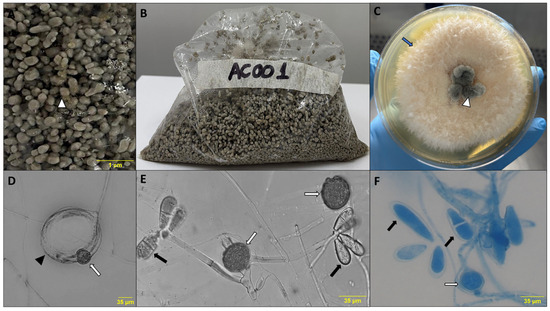
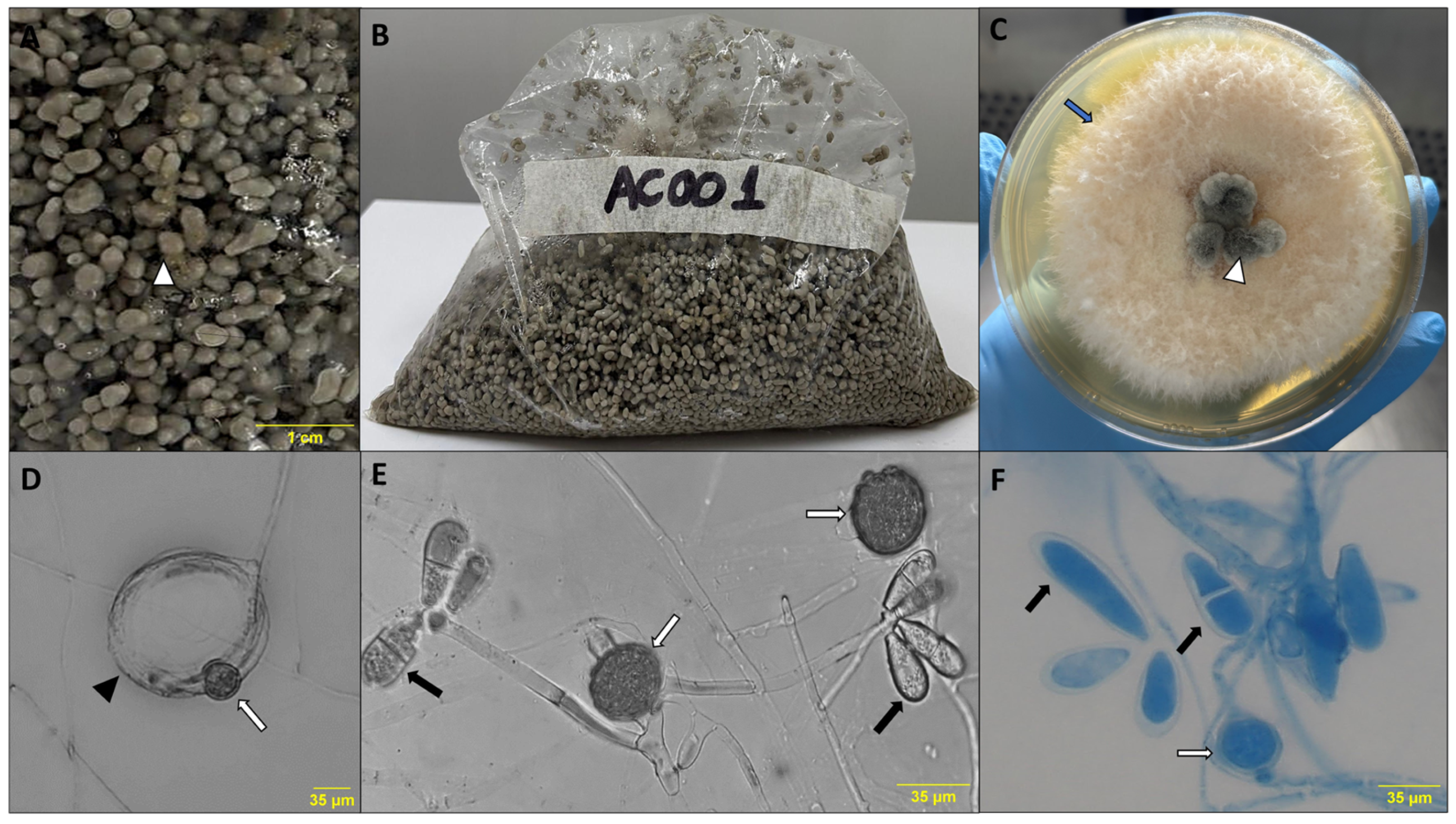
The formation of microspheres and encapsulation efficacy were confirmed according to Walker and Connick [21]. Pellet integrity was verified by their spherical shape upon contact with CaCl2 solution, indicating successful polymerization (Figure 1A,B). Additionally, fungal distribution within the microspheres was assessed by dissolving randomly selected pellets in 0.1 M sodium citrate, followed by observation under light microscopy, confirming the presence and uniform distribution of fungal structures (conidia and chlamydospores) within the matrix.
Figure 1.
(A,B) Formulation of the Duddingtonia flagrans isolate AC001 in sodium alginate pellets (white arrowhead). (C) Colony morphology of D. flagrans grown from pellets on Potato Dextrose Agar after 7 days of incubation (blue arrow). (D–F) Morphological structures of D. flagrans after reactivation from sodium alginate pellets stored under refrigeration: conidia (black arrow), chlamydospores (white arrow), and fungal trap (black arrowhead).
2.2. Storage of Pellets
After preparation, the sodium alginate pellets were placed in transparent bags (Figure 1A,B), previously labeled, and stored under constant refrigeration (4 °C ± 2 °C) for a period of fourteen years until experimental analysis at the Laboratório de Parasitologia Experimental e Controle Biológico, Universidade Vila Velha, Brazil.
2.3. Evaluation of Duddingtonia flagrans Viability in Pellets
The viability of the D. flagrans isolate was assessed by reactivation from alginate pellets. Sodium alginate pellets were inoculated onto Petri dishes containing Corn Meal Agar (CMA, Kasvi®, São José dos Pinhais, Brazil) and onto dishes containing Potato Dextrose Agar (PDA, Merck®, Darmstadt, Germany). Plates were incubated in a B.O.D. incubator at 27 °C, in the dark, for ten days. During incubation, fungal growth was monitored and recorded daily to confirm reactivation. At the end of the incubation period, to confirm fungal identity, microscope slides were prepared from the grown mycelium and stained with lactophenol cotton blue (Figure 1D–F). Morphological analysis of isolate AC001 was performed under light microscopy, and conidia and chlamydospores were identified using the 100× objective.
2.4. Maintenance of Nematodes and Preparation of the Suspension
Panagrellus redivivus was obtained from a commercial source and subsequently maintained under laboratory conditions at the Laboratório de Parasitologia Experimental e Controle Biológico, Universidade Vila Velha, Brazil. Nematodes were kept in continuous culture on Petri dishes containing commercial whole oat flakes (Quaker®, São Paulo, Brazil) as substrate. For the assays, nematodes were isolated by filtration using a commercially available plastic mesh sieve (Sanremo®, Esteio, Brazil) and distilled water to separate nematodes from the substrate. Nematode density in the filtered solution was determined by sampling: nematodes present in ten aliquots of 20 µL were counted and the mean value was calculated. Based on this count, the suspension was adjusted so that 200 µL contained approximately 400 nematodes, according to a modified technique described by Braga et al. [8].
2.5. Experimental Assay
The experiment consisted of two groups, each containing five Petri dishes:
Group I (Negative Control): Petri dishes containing only Corn Meal Agar (CMA), inoculated with 400 P. redivivus nematodes; Group II (Treatment): Petri dishes containing CMA previously colonized by D. flagrans, inoculated with 400 P. redivivus nematodes.
Although the exact number of spores was not quantified, plates were previously colonized under standardized conditions, ensuring similar fungal biomass across replicates.
Both groups were incubated in the dark at 28 °C for a period of five days.
2.6. Evaluation of Predatory Capacity and Nematode Recovery
Predatory efficacy was evaluated using two complementary approaches: direct microscopic monitoring and final recovery.
For direct microscopic monitoring, over the five-day period, evaluations were performed every 24 h. In each plate, ten random microscopic fields were examined using the 10× objective. The analysis consisted of quantifying nematodes not immobilized by fungal traps, i.e., live and motile nematodes. This approach allowed real-time monitoring of the interaction between D. flagrans and nematodes, including trap formation and immobilization dynamics.
For final recovery, at the end of the five-day incubation period, a modified Baermann technique was applied to quantify viable, non-predated nematodes remaining in the agar. This complementary method also enabled the recovery of nematodes that may have migrated to the agar–plate interface.
2.7. Statistical Analysis
The percentage reduction in nematode population was calculated by comparing the number of nematodes recovered in Group I (control) with the number recovered in Group II (treatment). Statistical analysis was performed using Student’s t-test (p < 0.05), using BioEstat software version 5.0 [23].
3. Results
The reactivation of isolate AC001 was successful in both culture media tested (PDA and CMA) after fourteen years of refrigerated storage in sodium alginate pellets. The growth of typical D. flagrans colonies was observed as early as the third day of incubation in both media. Microscopic analysis confirmed the presence of conidia, chlamydospores, and trapping structures (Figure 1D–F), indicating the preservation of the fungus’s morphological characteristics after the storage period.
The predatory activity of D. flagrans against P. redivivus was evaluated over five consecutive days, as well as through final nematode recovery using the modified Baermann technique (Table 1). On the first day of incubation (D1), a marked reduction in the number of free and motile nematodes was observed in the treated group compared with the control, with a reduction of 91%. On days D2 and D3, the percentage reduction increased to 95%, and on the fourth day (D4), nematode reduction remained high (93%), indicating sustained predatory activity throughout the experimental period. On the fifth day (D5), a less pronounced reduction was observed (55%).
Table 1.
Predatory activity of the Duddingtonia flagrans AC001 isolate against Panagrellus redivivus over five days of incubation and after final nematode recovery using the modified Baermann technique. Values represent the mean ± standard deviation of the number of free and motile nematodes observed per group. Different letters within the same row indicate a statistically significant difference between the control and treated groups (Student’s t-test, p ≤ 0.05).
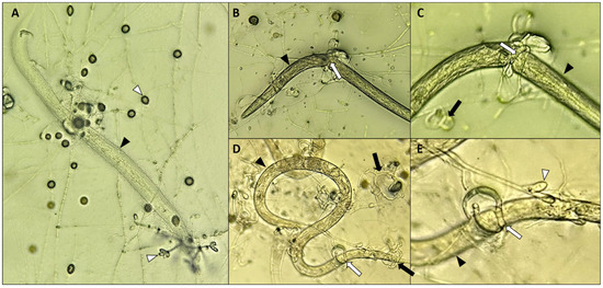
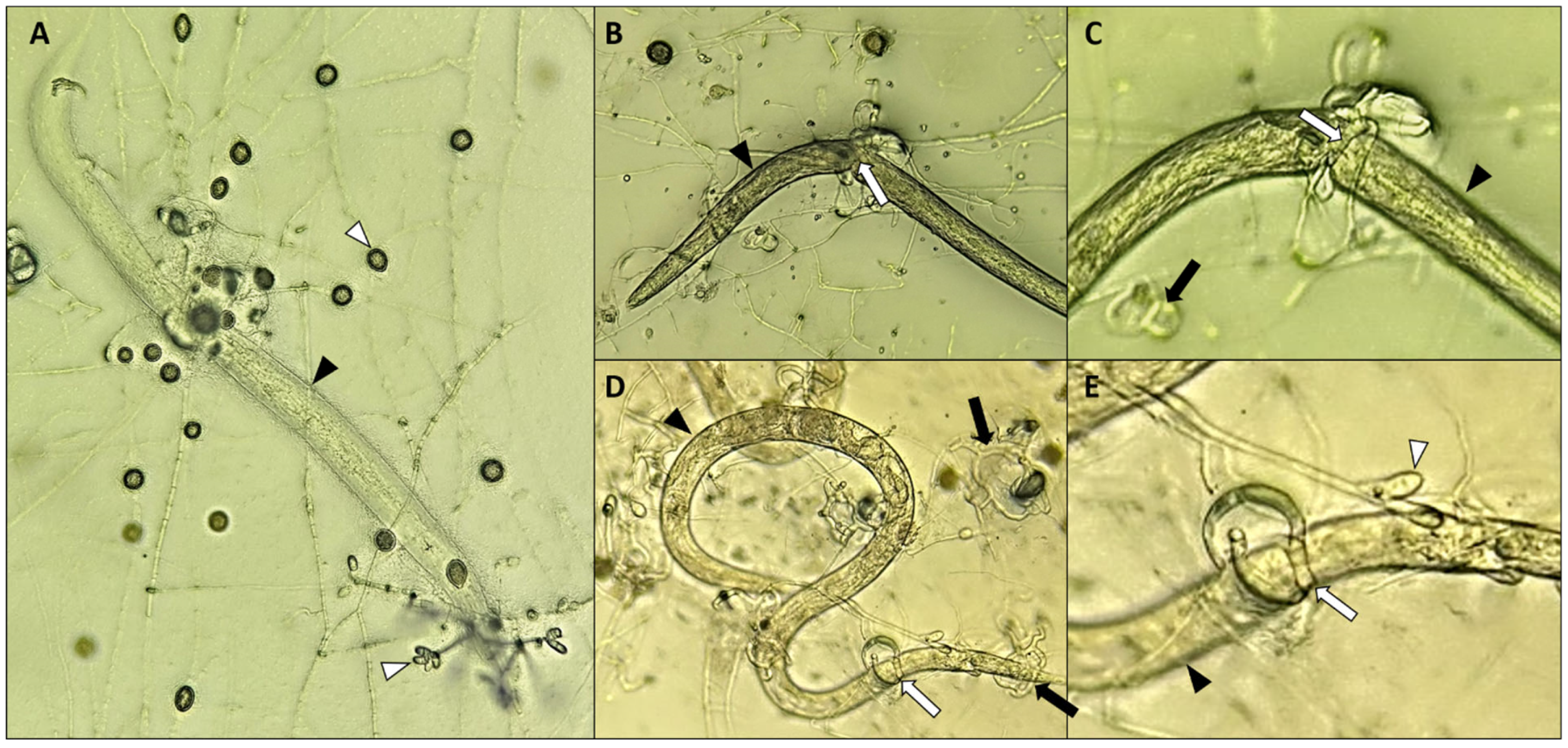
Final recovery using the Baermann technique revealed a 67% reduction in the number of viable nematodes in the treated group compared with the control. However, despite this noteworthy numerical reduction, the results did not differ significantly between groups for the Baermann assessment (p = 0.0890). Microscopic observations confirmed the formation of typical three-dimensional trapping structures around nematodes, followed by immobilization, predation, and death (Figure 2A–E).
Figure 2.
(A–E) Predatory capacity of Duddingtonia flagrans reactivated from sodium alginate pellets against Panagrellus redivivus (black arrowhead), showing the presence of conidia/chlamydospores (white arrowhead), adhesive hyphae modified into three-dimensional networks (black arrow), and hyphae modified into constricting rings (white arrow).
4. Discussion
The successful reactivation of AC001, together with the maintenance of conidia, chlamydospore production, and three-dimensional trapping structures, indicates that the combination of sodium alginate encapsulation and refrigeration provides favorable conditions for the stabilization of cellular and metabolic structures essential for the biocontrol activity of the fungus. The maintenance of viability and predatory activity of D. flagrans after prolonged storage may be associated with its high capacity for chlamydospore formation and the entry of the fungus into a reduced metabolic state, features that are well recognized as being related to tolerance to environmental stress and the survival of filamentous fungi under adverse preservation conditions [7,15]. In this context, the predatory efficacy observed, which reached up to 95% during the experimental period, reinforces the functional preservation of the isolate even after fourteen years of storage.
The high predatory efficacy (up to 95%) observed after fourteen years of storage is consistent with previous records for the AC001 strain. For instance, Braga et al. [24] evaluated this same isolate after 7 years of preservation in silica-gel, observing a reduction of 88.6% in cyathostomin L3 recovery. The fact that the isolate maintained a similarly high level of virulence (95%) even after doubling the storage period (14 years) confirms the exceptional stability of its predatory characteristics when preserved in an alginate matrix.
The temporal variation in predatory activity observed over the five-day assay reflects the natural dynamics of fungus–nematode interactions in in vitro systems (Table 1). The less pronounced reduction in predatory efficiency observed on day 5 (D5) may result from a combination of factors. These include decreased availability of motile nematodes throughout the assay and the progressive saturation of fungal trapping structures, as previously described in predatory-prey dynamics of nematophagous fungi [25,26]. Additionally, a gradual reduction in fungal growth, morphology, and predatory structures after reactivation supports the preservation of viability throughout the experiment.
It is important to consider that the direct microscopic observation method may not account for nematodes that died without being physically trapped by fungal structures, which could lead to a slight underestimation of total mortality. To address this, the modified Baermann technique was employed at the end of the experimental period as a complementary method, allowing confirmation of the reduction in viable, non-predated nematodes. This combined approach ensures both real-time monitoring of predatory interactions and accurate endpoint quantification.
Previous studies have demonstrated that different preservation strategies are capable of maintaining the predatory activity of nematophagous fungi. Braga et al. [24] showed that storage in silica gel was highly effective in preserving the predatory activity of D. flagrans and Arthrobotrys robusta isolates, highlighting the biological robustness of these fungi under conditions of dehydration and prolonged metabolic suspension. Similarly, Li et al. [27] demonstrated that nematophagous fungi encapsulated in alginate pellets retained germination capacity for more than two years when stored at 4 °C or −20 °C, with chlamydospore germination recovery exceeding initial values at certain storage periods. However, to date, no studies have evaluated the maintenance of D. flagrans over extremely long storage periods, making the present work the first report addressing this issue.
Moreover, Vilela et al. [18] evaluated the predatory viability of D. flagrans after 36 months of refrigerated storage in sodium alginate pellets and observed a reduction of up to 82% in the number of infective larvae in sheep when compared with the control group. Taken together, these findings support the ability of alginate to preserve functionally active fungal structures and corroborate the results obtained in the present study over a substantially longer temporal interval.
Additional evidence from studies involving entomopathogenic and antagonistic fungi further reinforces the applicability of sodium alginate as an encapsulation matrix. Alginate-encapsulated formulations of Metarhizium anisopliae maintained high counts of viable conidia for up to ten months of storage, while fungal structures of Trichoderma asperellum encapsulated using this model remained viable for at least 120 days under different temperature conditions [20,28]. Moreover, polymer compatibility studies have demonstrated that conidia of Beauveria bassiana and M. anisopliae encapsulated in alginate remained stable for up to twelve months under various environmental conditions [29].
In contrast to the storage periods reported in the literature for encapsulated fungi, which are generally limited to months or a few years, the present study demonstrated that D. flagrans preserved in sodium alginate pellets under refrigeration maintained predatory capacity with the production of trapping structures, conidia, and chlamydospores (Figure 2A–E) after fourteen years of storage. The durability observed in different encapsulated fungi has been attributed to general protective mechanisms conferred by the polymeric matrix, such as the formation of a physical barrier, isolation of reproductive structures, and reduction in environmental stress [30]. In this context, the functional stability observed in D. flagrans (AC001) suggests that the association between sodium alginate and refrigeration can substantially extend the period of functional preservation described to date for nematophagous fungi.
Over several decades, studies conducted in Brazil have demonstrated that alginate encapsulation allows conidia and chlamydospores of nematophagous fungi to maintain viability during passage through the gastrointestinal tract of domestic animals [18,31,32,33]. The consolidation of this experimental knowledge enabled the development of commercial formulations, such as Bioverm®, whose efficacy is directly related to the survival of encapsulated fungal structures and the maintenance of in vivo predatory activity [34,35].
Although the present study was conducted using a specific isolate, the physiological mechanisms associated with tolerance to prolonged storage, such as chlamydospore formation and metabolic downregulation, are widely described in nematophagous fungi, suggesting the potential applicability of this strategy to other isolates of the species. Furthermore, although molecular analyses were not performed, the maintenance of typical morphology and predatory activity indicates sufficient functional preservation for biocontrol applications [24].
The use of P. redivivus as a model organism provided standardized and reproducible conditions for evaluating long-term preservation of predatory activity. Nevertheless, we acknowledge that its rapid life cycle may introduce variability due to reproduction during the experimental period. Future studies should include nematode species of agricultural or livestock importance with longer life cycles to provide further validation of the functional preservation of D. flagrans.
5. Conclusions
The successful reactivation of the fungus, combined with the preservation of trapping structure formation and the high predatory efficacy observed in in vitro assays, confirms that sodium alginate encapsulation constitutes a robust strategy for the long-term functional conservation of nematophagous fungi. These findings expand current knowledge on preservation methods for microorganisms with applications in biological control and reinforce the potential of D. flagrans as a reliable agent for the control of gastro-intestinal nematodes, while also indicating that refrigerated storage in alginate represents a viable alternative for long-term experimental studies and future biocontrol programs.
Author Contributions
Conceptualization, F.R.B. and C.M.F.; methodology, V.B.S.S., C.M.F. and J.V.L.; formal analysis, F.E.d.F.S., J.V.d.A. and F.R.B.; writing—original draft preparation, V.B.S.S., J.A.G.F. and H.L.; writing—review and editing, F.E.d.F.S. and F.R.B.; funding acquisition, H.L. and F.R.B. All authors have read and agreed to the published version of the manuscript.
Funding
This research was funded by Fundação de Amparo à Pesquisa e Inovação do Espírito Santo (Edital FAPES n. 13/2025), under grant number 1484/2025—P: 2025-7ZFZV.
Institutional Review Board Statement
Not applicable.
Informed Consent Statement
Not applicable.
Data Availability Statement
The original contributions presented in this study are included in the article. Further inquiries can be directed to the corresponding author.
Acknowledgments
The authors would like to thank Conselho Nacional de Desenvolvimento Científico e Tecnológico (CNPq), Fundação de Amparo à Pesquisa e Inovação do Espírito Santo (FAPES), Fundação de Amparo à Pesquisa do Estado de Minas Gerais (FAPEMIG), and Fundação Coordenação de Aperfeiçoamento de Pessoal de Nível Superior (Capes) for their financial support.
Conflicts of Interest
The authors declare no conflicts of interest.
References
- Rahman, M.U.; Zhong, X.; Uzair, M.; Fan, B. Application of fungi as biological control strategies for nematode management in horticultural crops. Phytopathol. Res. 2024, 6, 38. [Google Scholar] [CrossRef] [Scilit]
- Faria, L.E.M.; Fonseca, J.D.S.; Araújo, J.V.; de Carvalho, L.M.; Albuquerque, G.R.; Perinotto, W.M.S. Nematophagous fungi to controlling gastrointestinal nematodes in small ruminants: A systematic review. Vet. Parasitol. 2025, 334, 110410. [Google Scholar] [CrossRef] [Scilit]
- Rahman, M.U.; Chen, P.; Zhang, X.; Fan, B. Predacious strategies of nematophagous fungi as bio-control agents. Agronomy 2023, 13, 2685. [Google Scholar] [CrossRef] [Scilit]
- Aguiar, A.A.R.M.; Lima, A.M.S.; Feitosa, T.F.; Ribeiro, W.L.C.; Soares, F.E.F.; Braga, F.R.; Vilela, V.L.R. In vitro efficacy of the monoterpene linalool isolated or combined with the nematophagous fungus Duddingtonia flagrans in the control of sheep gastrointestinal nematodes. Microbiol. Res. 2025, 16, 1. [Google Scholar] [CrossRef] [Scilit]
- Correia, G.S.; Ribeiro, G.B.; Sousa, G.C.E.; Moura, I.A.; Araújo, J.V.; Braga, F.R.; Carvalho, L.M.; Perinotto, W.M.S. Duddingtonia flagrans against anthelmintic-resistant gastrointestinal nematodes in goats. Small Rumin. Res. 2025, 252, 107599. [Google Scholar] [CrossRef] [Scilit]
- Soares, F.E.F.; Ferreira, J.M.; Genier, H.L.A.; Al-Ani, L.K.T.; Aguilar-Marcelino, L. Biological control 2.0: Use of nematophagous fungi enzymes for nematode control. J. Nat. Pestic. Res. 2023, 4, 100025. [Google Scholar] [CrossRef] [Scilit]
- Nordbring-Hertz, B.; Jansson, H.-B.; Tunlid, A. Nematophagous fungi. In Encyclopedia of Life Sciences (eLS); John Wiley & Sons: Hoboken, NJ, USA, 2011. [Google Scholar]
- Braga, F.R.; Araújo, J.V.; Soares, F.E.F.; Araujo, J.M.; Ferreira, S.R.; Queiroz, J.H. Use of statistical tools in the study of the conditions of predation of Duddingtonia flagrans versus Panagrellus sp. Biocontrol Sci. Technol. 2012, 22, 559–565. [Google Scholar] [CrossRef] [Scilit]
- Srinivasan, J.; Dillman, A.R.; Macchietto, M.G.; Heikkinen, L.; Lakso, M.; Fracchia, K.M.; Antoshechkin, I.; Mortazavi, A.; Wong, G.; Sternberg, P.W. The draft genome and transcriptome of Panagrellus redivivus are shaped by the harsh demands of a free-living lifestyle. Genetics 2013, 4, 1279–1295. [Google Scholar] [CrossRef] [Scilit]
- Masler, E.P. Digestion of invertebrate neuropeptides by preparations from the free-living nematode Panagrellus redivivus. J. Helminthol. 2008, 82, 279–285. [Google Scholar] [CrossRef] [Scilit]
- Masler, E.P. In vitro comparison of protease activities in preparations from free-living (Panagrellus redivivus) and plant-parasitic (Meloidogyne incognita) nematodes using FMRFa and FMRFa-like peptides as substrates. J. Helminthol. 2010, 4, 425–433. [Google Scholar] [CrossRef] [Scilit] [PubMed]
- Laws, T.R.; Smith, S.A.; Smith, M.P.; Harding, S.V.; Atkins, T.P.; Titball, R.W. The nematode Panagrellus redivivus is susceptible to killing by human pathogens at 37 degrees C. FEMS Microbiol. Lett. 2005, 1, 77–83. [Google Scholar] [CrossRef] [Scilit]
- Geniêr, H.L.A.; Soares, F.E.F.; Braga, F.R.; Queiroz, J.H. Activity of the fungus Pleurotus ostreatus and of its proteases on Panagrellus sp. larvae. Afr. J. Biotechnol. 2015, 14, 1496–1503. [Google Scholar] [CrossRef] [Scilit]
- Souza, D.C.; Moreira, T.F.; Braga, F.R.; Soares, F.E.F. In vitro evaluation of Bacillus licheniformis protease on the nematodes Panagrellus sp., Meloidogyne incognita, Haemonchus spp. and Trichostrongylus spp. Next Res. 2025, 2, 100308. [Google Scholar] [CrossRef] [Scilit]
- Borman, A.M.; Szekely, A.; Campbell, C.K.; Johnson, E.M. Evaluation of the viability of pathogenic filamentous fungi after prolonged storage in sterile water and review of recent published studies on storage methods. Mycopathologia 2006, 6, 361–368. [Google Scholar] [CrossRef] [Scilit] [PubMed]
- Oliveira, I.; Pereira, J.A.; Bento, A.; Baptista, P. Viability of Beauveria bassiana isolates after storage under several preservation methods. Ann. Microbiol. 2011, 61, 339–344. [Google Scholar] [CrossRef] [Scilit]
- Arias-Chavarría, L.D.; Batista-Menezes, D.; Orozco-Cayasso, S.; Vargas-Martínez, A.; Vega-Baudrit, J.R.; Montes de Oca-Vásquez, G. Evaluation of the viability of microencapsulated Trichoderma longibrachiatum conidia as a strategy to prolong the shelf life of the fungus as a biological control agent. Front. Chem. 2025, 12, 1473217. [Google Scholar] [CrossRef] [Scilit]
- Vilela, V.L.R.; Costa, P.W.L.; Silva, F.F.; Bezerra, H.M.F.F.; Oliveira, L.V.S.; Dantas, M.O.; Feitosa, T.F.; Araújo, J.V.; Braga, F.R. Predatory viability of the nematophagous fungus Duddingtonia flagrans on the infective larvae of gastrointestinal nematodes of sheep following storage and refrigeration. Braz. J. Vet. Res. Anim. Sci. 2019, 56, e154252. [Google Scholar] [CrossRef] [Scilit]
- Martínez-Cano, B.; Mendoza-Meneses, C.J.; García-Trejo, J.F.; Macías-Bobadilla, G.; Aguirre-Becerra, H.; Soto-Zarazúa, G.M.; Feregrino-Pérez, A.A. Review and Perspectives of the Use of Alginate as a Polymer Matrix for Microorganisms Applied in Agro-Industry. Molecules 2022, 13, 4248. [Google Scholar] [CrossRef] [Scilit]
- Sarma, B.D.; Puzari, K.C.; Dutta, P.; Pandey, A.K. An alginate-based encapsulation enhances shelf life and bioactivity of the entomopathogenic fungus, Metarhizium anisopliae. Egypt. J. Biol. Pest. Control 2023, 33, 69. [Google Scholar] [CrossRef] [Scilit]
- Walker, H.L.; Connick, W.J., Jr. Sodium alginate for production and formulation of mycoherbicides. Weed Sci. 1983, 3, 333–338. [Google Scholar] [CrossRef] [Scilit]
- Lackey, B.A.; Muldoon, A.E.; Jaffe, B.A. Alginate pellet formulation of Hirsutella rossiliensis for biological control of plant-parasitic nematodes. Biol. Control 1994, 3, 155–162. [Google Scholar] [CrossRef] [Scilit]
- Ayres, M.; Ayres, J.R.; Ayres, D.L.; Santos, A.S. BioEstat 3.0: Aplicações Estatísticas nas Áreas das Ciências Biológicas e Médicas; Sociedade Civil Mamirauá: Belém, Brazil; CNPq: Brasília, Brazil, 2003. [Google Scholar]
- Braga, F.R.; Araújo, J.V.; Araujo, J.M.; Tavela, A.O.; Ferreira, S.R.; Soares, F.E.F.; Benjamin, L.A.; Frassy, L.N. Influence of the preservation period in silica gel on the predatory activity of isolates of Duddingtonia flagrans on infective larvae of cyathostomins (Nematoda: Cyathostominae). Exp. Parasitol. 2011, 128, 460–463. [Google Scholar] [CrossRef] [Scilit]
- Jansson, H.B.; Nordbring-Hertz, B.; Tunlid, A. Polarized growth and trap formation in nematode-trapping fungi. FEMS Microbiol. Rev. 1997, 21, 1–29. [Google Scholar]
- Wang, B.B.; Li, Y.L.; Tian, S.Y.; Wang, H.Z.; Li, X.; Wang, F.H.; Cai, K.Z. Chlamydospore dormancy and predatory activity of nematophagous fungus Duddingtonia flagrans. J. Basic. Microbiol. 2024, 3, e2300365. [Google Scholar] [CrossRef] [Scilit]
- Li, Y.L.; Wang, B.B.; Wang, Y.F.; Chen, X.Y.; Wang, F.H.; Li, X.; Cai, K.Z.; Lv, S.Y. Storage impact on chlamydospore germination of Arthrobotrys flagrans in sodium alginate pellets. J. Basic. Microbiol. 2025, 7, e70043. [Google Scholar] [CrossRef] [Scilit] [PubMed]
- Lopes, A.R.O.; Locatelli, G.O.; Barbosa, R.M.; Junior, M.L.; Mascarin, G.M.; Finkler, C.L.L. Preparation, characterisation and cell viability of encapsulated Trichoderma asperellum in alginate beads. J. Microencapsul. 2020, 3, 270–282. [Google Scholar] [CrossRef] [Scilit] [PubMed]
- Rodrigues, I.M.W.; Filho, A.B.; Giordano, I.B.; Denadae, B.E.; Fernandes, J.B.; Forim, M.R. Compatibility of polymers to fungi Beauveria bassiana and Metarhizium anisopliae and their formulated products stability. Acta Sci. Agron. 2017, 4, 457–464. [Google Scholar] [CrossRef] [Scilit]
- Seabra, M.G.J.; Santos, T.S.; Varize, C.S.; Souto, E.B.; Severino, P.; Mendonça, M.C. New Beauveria bassiana aerial conidia-based bioinsecticide obtained by spray-dried microencapsulation of the entomopathogenic fungi in biopolymers for crop protection. Egypt. J. Biol. Pest. Control 2024, 34, 58. [Google Scholar] [CrossRef] [Scilit]
- Baiak, B.H.B.; Gasparina, J.M.; Ianke, L.; de Sousa, K.T.; Deniz, M.; Pereira, L.M.; Araújo, J.V.; da Rocha, R.A.; Dittrich, J.R. Predatory activity of nematophagous fungus Duddingtonia flagrans in infective larvae after gastrointestinal transit: Biological control in pasture areas and in vitro. J. Helminthol. 2021, 95, e31. [Google Scholar] [CrossRef] [Scilit]
- Assis, R.C.; Luns, F.D.; Araújo, J.V.; Braga, F.R.; Assis, R.L.; Marcelino, J.L.; Freitas, P.C.; Andrade, M.A. Comparison between the action of nematode predatory fungi Duddingtonia flagrans and Monacrosporium thaumasium in the biological control of bovine gastrointestinal nematodiasis in tropical southeastern Brazil. Vet. Parasitol. 2013, 193, 134–140. [Google Scholar] [CrossRef] [Scilit]
- Tavela, A.O.; de Araújo, J.V.; Braga, F.R.; da Silveira, W.F.; Silva, V.H.D.; Carretta Júnior, M.; Borges, L.A.; Araujo, J.M.; Benjamin Ldos, A.; Carvalho, G.R.; et al. Coadministration of sodium alginate pellets containing the fungi Duddingtonia flagrans and Monacrosporium thaumasium on cyathostomin infective larvae after passing through the gastrointestinal tract of horses. Res. Vet. Sci. 2013, 3, 568–572. [Google Scholar] [CrossRef] [Scilit]
- Braga, F.R.; Ferraz, C.M.; Silva, E.N.; Araújo, J.V. Efficiency of the Bioverm® (Duddingtonia flagrans) fungal formulation to control in vivo and in vitro Haemonchus contortus and Strongyloides papillosus in sheep. 3 Biotech. 2020, 10, 62. [Google Scholar] [CrossRef] [Scilit]
- Mendes, L.Q.; Ferraz, C.M.; Ribeiro, N.R.C.; Ulfeldt, K.B.; Ribeiro, J.C.C.; Merizio, M.F.; Rossi, G.A.M.; Aguiar, A.A.R.M.; Araújo, J.V.; Soares, F.E.F.; et al. Efficacy of Duddingtonia flagrans (Bioverm®) on the biological control of buffalo gastrointestinal nematodes. Exp. Parasitol. 2023, 253, 108592. [Google Scholar] [CrossRef] [Scilit]

Disclaimer/Publisher’s Note: The statements, opinions and data contained in all publications are solely those of the individual author(s) and contributor(s) and not of MDPI and/or the editor(s). MDPI and/or the editor(s) disclaim responsibility for any injury to people or property resulting from any ideas, methods, instructions or products referred to in the content. |
© 2026 by the authors. Licensee MDPI, Basel, Switzerland. This article is an open access article distributed under the terms and conditions of the Creative Commons Attribution (CC BY) license.