


Abstract
Cutaneous larva migrans (CLM) is a zoonotic parasitic infection traditionally associated with tropical and subtropical regions. In recent years, autochthonous cases have been reported in various European countries, suggesting a possible change in the epidemiological profile of the disease. We present a case of a patient with no history of international travel who developed a typical serpiginous, migrating skin lesion after local exposure. The diagnosis was made clinically, and treatment with albendazole resulted in rapid clinical improvement. To our knowledge, this is the first published probable autochthonous case of CLM in Bulgaria. The case highlights the role of climate change in expanding the geographical scope of the parasitosis and the need for increased clinical vigilance at the primary care level.
1. Introduction
Cutaneous larva migrans (CLM) is a zoonotic infection caused by larvae of canine and feline hookworms (Ancylostoma caninum, Ancylostoma braziliense, and Uncinaria stenocephala), which penetrate the skin of humans upon direct contact with infected soil or sand [1,2,3,4]. The disease is characterized by typical, intensely itchy, serpiginous skin lesions that progress by several centimeters daily.
Although traditionally endemic to tropical and subtropical regions like South America, Africa, and the South Pacific area [4,5,6], CLM is increasingly occurring in temperate climates, including southern and western Europe [3,7,8].
The increasing number of reports from Italy, France, Spain, Germany, Greece, and the United Kingdom shows a trend towards “Europeanization” of the infection. Climate change, the increase in average temperatures and humidity, and the extension of the warm seasons create favorable conditions for larval survival in temperate latitudes [9]. All reported cases demonstrate the importance of an interdisciplinary approach that could help specialists control the spread of the infection [4].
This report presents the first probable autochthonous case in Bulgaria. It emphasizes the need for better organization of the approach involving dermatologists, veterinarians, and occupational health specialists.
2. Clinical Case
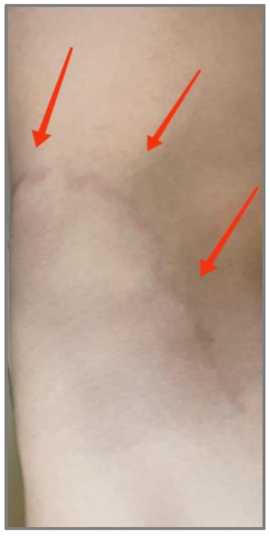
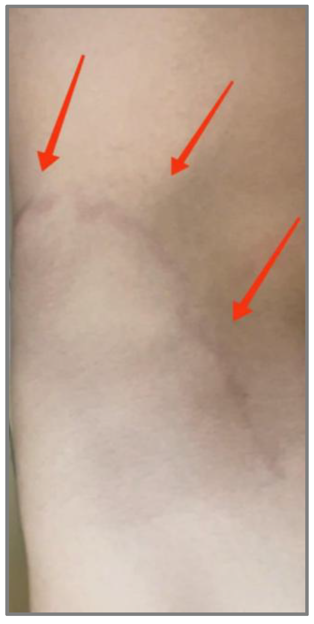
We report a case of a 23-year-old man who sought medical attention from his general practitioner regarding a severely itchy, serpiginous, cord-like rash on his back that had appeared a few days earlier (Figure 1). Symptomatic treatment with antihistamine was given without clinical improvement.
Figure 1.
Patient photography—serpiginous, cord-like rash on the lower left side of the back. The red arrows show the exact position of the rash.
Due to the persistence and gradual extension of the lesion, the patient referred themself to a dermatology clinic. Upon examination, a parasitic etiology was suspected due to the characteristic serpiginous course and the dynamic evolution of the rash. A visit to a specialized parasitology office was recommended. The patient reported that he had not left Bulgaria in the past year. A few days before the onset of symptoms, he had been lying directly on the sand on a beach on the Bulgarian Black Sea coast.
Examination revealed a lesion typical of CLM—an erythematous, slightly raised, serpiginous line with a width of about 2 mm and a length of 7–8 cm. Laboratory parameters (complete blood count and CRP levels) were within reference limits. The diagnosis was made clinically based on the characteristic morphology and dynamics of the lesion. Due to the typical clinical picture and rapid therapeutic response, no invasive diagnostic procedures were performed. Usually, the larva is located a few millimeters to centimeters in front of the visible trace, which makes skin biopsy often uninformative and rarely applicable in clinical practice for this disease.
Treatment with oral Albendazole was prescribed (400 mg once daily for 5 days), which led to a complete clinical recovery and resolution of pruritus within 48 h and gradual disappearance of the skin lesions over the following weeks.
The unique fact here is the lack of travel outside the country or any contact with potentially contaminated sand; therefore, our hypothesis is of a local (autochthonous) transmission. To date, no official cases of autochthonous CLM have been documented in Bulgaria.
3. Discussion
Cutaneous larva migrans (CLM) can be caused by various larvae of canine and feline hookworms. Normally, dogs and cats act as definitive hosts for these parasites, depositing eggs in nature through their feces. Humans are accidental hosts of the parasites, and in those cases, the life cycle is not completed. These parasites are able to penetrate the human epidermis but not the dermis itself. This leaves the parasite migrating within the epidermis due to its ability to produce enzymes like proteases and hyaluronidases that promote the invasion. The migration can last up to a few months. Such helminths can transfer to different organs and lead to symptoms that require systemic observation and treatment.
The presented case reflects the changing epidemiological profile of CLM in Europe. Data from the last decade show an increase in autochthonous cases in temperate latitudes [3,7,10,11]. They suggest a possible expansion of the ecological niche of the causative agent.
To our knowledge, this is the first published probable autochthonous case of CLM in Bulgaria. The lack of international travel and the presence of local exposure support the hypothesis of locally acquired infection. The occurrence of the case in Bulgaria suggests that the epidemiological conditions for the transmission of hookworm larvae are present along the Bulgarian Black Sea coast. Warm and humid sandy environments, typical of local beaches during the summer season, provide an ideal microclimate for the survival and maturation of Ancylostoma larvae. Furthermore, the increasing trend of milder winters in the Balkan region, attributed to climate change, may facilitate the overwintering of parasite eggs in the soil, potentially establishing new zoonotic outbreaks.
The increasing number of cases in Europe is likely the result of a complex interaction between environmental, climatic, and zoonotic factors. The increase in average temperatures, higher humidity, and the extended summer season may contribute to the longer survival of larvae in soil and sand, potentially increasing the possibility of local transmission [8,9].
Laboratory-confirmed autochthonous cases have been described in Italy, including in cases of occupational exposure and proven presence of the parasite in animal reservoirs [4,8]. Similar observations have been reported in Greece, Spain, and France [2,3,7,10,11], as well as in more northern regions such as Germany and the United Kingdom [12,13].
The diagnosis of CLM remains primarily clinical [9]. In our case, the clinical diagnosis was based on the pathognomonic serpiginous, erythematous, and pruritic track. However, several differential diagnoses were considered and excluded. Larva currens, caused by Strongyloides stercoralis, was ruled out due to the significantly slower migration rate of the lesion in our patient (1–2 cm per day compared to the rapid 5–10 cm per hour typical for larva currens). Phytophotodermatitis and allergic contact dermatitis were excluded as they lack the progressive migratory nature and the distinct cord-like morphology observed here. Additionally, migratory erythema (Lyme disease) was dismissed due to the absence of an expanding annular “bull’s-eye” pattern, a characteristic of the disease, and the patient’s clear history of skin contact with beach sand rather than a tick bite.
In isolated cases, eosinophilia or secondary bacterial infection may be observed, but laboratory tests are often nonspecific [14]. The therapeutic approach with albendazole or ivermectin is effective and leads to rapid clinical improvement, and both drugs are recommended in international guidelines and review publications [9].
The patient’s diagnostic pathway is particularly indicative. Initial ineffective treatment at the primary care level highlights the difficulties in recognizing parasitic infections that are not traditionally considered local. In the context of changing epidemiology, general practitioners should include CLM and other “imported” tropical parasites in the differential diagnosis of migratory skin lesions, even in the absence of a travel history.
This case illustrates the need for increased continuing medical education and adaptation of clinical algorithms to new climatic realities. Tracking such cases and implementing preventive measures are of key importance for public health since each day the world becomes more and more open, with the increasing levels of tourist trips and international trade markets operations. The majority of reported cases are from endemic regions, and pre-travel advice should reflect measures to prevent direct skin contact with sand on beaches and soil [14]. The governing organizations ought to promote strict campaigns for deworming animals and maintaining the cleanliness and safety of beaches. Preventive measures may include systematic veterinary deworming of domestic animals, control of stray animal populations, monitoring of public beach and park areas, and raising awareness among medical personnel and the population. A coordinated interdisciplinary approach, such as One health, could contribute to limiting future cases since it includes collaboration across sectors managing human health, animal health, and the environment [4,9].
4. Conclusions
The presented case illustrates a likely autochthonous infection with Cutaneous larva migrans (CLM) in Bulgaria, and fits into the growing number of reports of similar cases in various European countries. Observations over the last decade suggest a gradual expansion of the geographical distribution of the infection outside the traditionally endemic regions.
The case highlights the importance of increased clinical vigilance in patients with migrating skin lesions, even in the absence of a history of travel to tropical areas. Early recognition of the characteristic clinical picture allows for timely initiation of therapy and prevention of unnecessary prolonged symptomatic treatment.
In a broader epidemiological context, the present observation supports the need to adapt clinical algorithms to the changing environment and to systematically monitor potential autochthonous cases. The complex interaction between climatic factors, animal reservoirs, and human exposure highlights the importance of an integrated approach, including medical, veterinary, and environmental prevention strategies.
Author Contributions
Conceptualization: S.S. and B.P.-S.; methodology: S.S., B.P.-S., and N.K.; data collection and summarizing: S.S.; data analysis and interpretation: S.S., B.P.-S., N.K., and R.G.-G.; writing—preparation of the original project: S.S., B.P.-S., N.K., and R.G.-G.; writing—review and scientific editing: N.K. and B.P.-S.; project administration: N.K. All authors have read and agreed to the published version of the manuscript.
Funding
This research was funded by the Bulgarian Ministry of Education and Science (MES) in the frames of the Bulgarian National Recovery and Resilience Plan, Component “Innovative Bulgaria,” Project No. BGRRP-2.004-0006-C02, “Development of research and innovation at Trakia University in service of health and sustainable well-being”.
Institutional Review Board Statement
The study was conducted in accordance with the Declaration of Helsinki and was approved by the Research Ethics Committee of the Faculty of Medicine, Trakia University (Approval Code: No.47, Approval Date: 20 February 2026).
Informed Consent Statement
No personal data of the patient was presented in the report. Oral consent was provided, and the patient agreed to the use of the photographic image.
Data Availability Statement
The original contributions presented in this study are included in the article. Further inquiries can be directed to the corresponding author.
Conflicts of Interest
The authors declare no conflicts of interest related to this study.
Abbreviations
The following abbreviations are used in this manuscript:
| CLM | Cutaneous larva migrans |
References
- Traub, R.J.; Zendejas-Heredia, P.A.; Massetti, L.; Colella, V. Zoonotic hookworms of dogs and cats—Lessons from the past to inform current knowledge and future directions of research. Int. J. Parasitol. 2021, 51, 1233–1241. [Google Scholar] [CrossRef] [Scilit] [PubMed]
- Tamminga, N.; Bierman, W.F.W.; de Vries, P.J. Cutaneous larva migrans acquired in Brittany, France. Emerg. Infect. Dis. 2009, 15, 1856–1858. [Google Scholar] [CrossRef] [Scilit] [PubMed]
- Del Giudice, P.; Hakimi, S.; Vandenbos, F.; Magana, C.; Hubiche, T. Autochthonous cutaneous larva migrans in France and Europe. Acta Derm. Venereol. 2019, 99, 805–808. [Google Scholar] [CrossRef] [Scilit] [PubMed]
- Stufano, A.; Foti, C.; Lovreglio, P.; Romita, P.; De Marco, A.; Lia, R.P.; Otranto, D.; Iatta, R. Occupational risk of cutaneous larva migrans: A case report and systematic literature review. PLoS Neglected Trop. Dis. 2022, 16, e0010330. [Google Scholar] [CrossRef] [Scilit] [PubMed]
- Freedman, D.O.; Weld, L.H.; Kozarsky, P.E.; Fisk, T.; Robins, R.; von Sonnenburg, F.; Keystone, J.S.; Pandey, P.; Cetron, M.S. Spectrum of disease and relation to place of exposure among ill returned travelers. N. Engl. J. Med. 2006, 354, 119–130. [Google Scholar] [CrossRef] [Scilit] [PubMed]
- Maxfield, L.; Crane, J.S. Cutaneous Larva Migrans; StatPearls Publishing: Petersburg, FA, USA, 2023. Available online: https://www.ncbi.nlm.nih.gov/books/NBK507706/ (accessed on 2 April 2023).
- Blaizot, R.; Goiset, A.; Caumes, E.; Gabriel, F.; Milpied, B. Cutaneous larva migrans: A case in Bordeaux, France and a systematic review of locally acquired cases in Europe. Eur. J. Dermatol. 2017, 27, 426–429. [Google Scholar] [CrossRef] [Scilit] [PubMed]
- Veronese, F.; Graziola, F.; Farinelli, P.; Zavattaro, E.; Tarantino, V.; Esposto, E.; Savoia, P. North Italy: Welcome to the Tropics. Infect. Dis. Rep. 2021, 13, 215–218. [Google Scholar] [CrossRef] [Scilit] [PubMed]
- Heukelbach, J.; Feldmeier, H. Epidemiological and clinical characteristics of hookworm-related cutaneous larva migrans. Lancet Infect. Dis. 2008, 8, 302–309. [Google Scholar] [CrossRef] [Scilit] [PubMed]
- Panés-Rodríguez, A.; Piera-Tuneu, L.; López-Pestaña, A.; Ormaetxea-Pérez, N.; Gutiérrez-Támara, P.; Ibarbia-Oruezabala, S.; Tuneu-Valls, A. Autochthonous cutaneous larva migrans infection in Guipúzcoa. Actas Dermo-Sifiliogr. 2016, 107, 407–413. [Google Scholar] [CrossRef] [Scilit] [PubMed]
- Dimitriadis, I. Bilesional serpiginous cutaneous ectoparasitosis in a Greek grazier. IDCases 2019, 18, e00598. [Google Scholar] [CrossRef] [Scilit] [PubMed]
- Kienast, A.; Bialek, R.; Hoeger, P.H. Cutaneous larva migrans in northern Germany. Eur. J. Pediatr. 2007, 166, 1183–1185. [Google Scholar] [CrossRef] [Scilit] [PubMed]
- Baple, K.; Clayton, J. Hookworm-related cutaneous larva migrans acquired in the UK. BMJ Case Rep. 2015, 2015, bcr2015210165. [Google Scholar] [CrossRef] [Scilit] [PubMed]
- Sönmezer, M.Ç.; Erdinç, Ş.; Tülek, N.; Babür, C.; Büyükdemirci, A.; İlgar, T.; Ertem, G. Cutaneous larva migrans in Turkey: An imported case report. Mikrobiyoloji Bul. 2017, 51, 94–99. [Google Scholar] [CrossRef] [Scilit] [PubMed]
Disclaimer/Publisher’s Note: The statements, opinions and data contained in all publications are solely those of the individual author(s) and contributor(s) and not of MDPI and/or the editor(s). MDPI and/or the editor(s) disclaim responsibility for any injury to people or property resulting from any ideas, methods, instructions or products referred to in the content. |
© 2026 by the authors. Licensee MDPI, Basel, Switzerland. This article is an open access article distributed under the terms and conditions of the Creative Commons Attribution (CC BY) license.