


Abstract
Background/Objectives: The pituitary gland is highly susceptible to hypoxic–ischemic injury because of its distinctive vascular architecture, yet its forensic significance in non-traumatic deaths remains poorly defined. This study aimed to compare pituitary histopathology across deaths caused by different mechanisms of systemic hypoxia. Methods: This retrospective observational study investigated pituitary histopathology in 30 forensic autopsies performed between 2019 and 2025, including suicidal hanging (n = 10), fatal heroin intoxication with respiratory failure (n = 10), and sudden cardiac death controls (n = 10). Hematoxylin–eosin-stained sections were systematically evaluated for hemorrhagic, ischemic, and necrotic changes and their anatomical distribution. Results: Neurohypophyseal hemorrhage was detected in 6/10 hanging cases and 8/10 heroin-related deaths, but in no controls. Adenohypophyseal ischemic–necrotic lesions occurred in 2/10 hanging cases and 6/10 heroin cases and were absent in controls. Lesions were generally more extensive in heroin intoxication, where hemorrhage often coexisted with vascular congestion and coagulative necrosis. The preferential involvement of the posterior lobe by hemorrhage and the anterior lobe by ischemic–necrotic injury is consistent with their different vascular supplies. Conclusions: These exploratory findings require validation. Hypoxia duration and residual perfusion were not measured. Pituitary sampling may be a complementary tool worth investigating in selected forensic cases; its routine utility was not assessed. Because all sections were stained with hematoxylin–eosin only, these findings are preliminary and require confirmation.
1. Introduction
The pituitary gland is the central organ of the endocrine system and plays a fundamental role in maintaining homeostasis through the secretion of several hormones, including growth hormone (GH), thyroid-stimulating hormone (TSH), prolactin (PRL), adrenocorticotropic hormone (ACTH), follicle-stimulating hormone (FSH), luteinizing hormone (LH), antidiuretic hormone (ADH), and oxytocin. Deficiency of these hormones may result in severe endocrine disorders requiring long-term replacement therapy. Besides its endocrine function, the pituitary gland has distinctive anatomical and vascular features that make it particularly vulnerable to ischemic and hemorrhagic injury [1]. The anterior pituitary receives most of its blood supply through the hypothalamic-hypophyseal portal system, a low-pressure vascular network arising from the superior hypophyseal arteries, whereas the posterior lobe is supplied directly by the inferior hypophyseal arteries [2,3]. Because of this peculiar vascular organization, the gland is especially sensitive to alterations in systemic and cerebral perfusion [4,5]. Conditions associated with hypotension, hemorrhagic shock, cardiorespiratory failure, asphyxia, hypoxia, or sudden changes in intracranial pressure may impair pituitary blood flow, leading to vascular congestion, edema, ischemic infarction, necrosis, and intraparenchymal hemorrhage [4,6,7,8]. Likewise, traumatic brain injury may produce stretching or disruption of the pituitary stalk and its delicate vascular network, resulting in both ischemic and hemorrhagic damage [9,10,11,12]. Previous studies have shown that pituitary dysfunction develops in approximately 20–40% of patients following moderate or severe traumatic brain injury, leading to hormonal deficiencies such as growth hormone deficiency, hypogonadism, hypothyroidism, hypocortisolism, and central diabetes insipidus [13,14,15]. Different degrees of hypopituitarism have been described both during the acute phase and throughout long-term follow-up [16]. Nevertheless, these endocrine abnormalities often remain unrecognized in the acute setting because neurological assessment tools, including the Glasgow Coma Scale, provide no information on hypothalamic-pituitary function. Several pituitary disorders, including adenomas, ischemic necrosis, hypophysitis, abscesses, and pituitary apoplexy, have been reported as causes of sudden or unexpected death, with some cases being diagnosed only at post-mortem examination [17,18,19,20,21,22]. At the same time, injury to the hypothalamic-pituitary axis is increasingly recognized as a consequence of severe head trauma [23,24]. Histopathological changes involving the pituitary gland, however, are not limited to traumatic brain injury and may also occur in conditions characterized by systemic hypoxia or impaired tissue perfusion [5,6]. Kouroupi et al. [6] investigated hypoxia-inducible factor expression and angiogenesis-related changes in the pituitary gland in relation to different patterns of death, providing evidence that hypoxia-related molecular pathways may be activated in this tissue under conditions of systemic oxygen deprivation. However, their study did not specifically compare distinct non-traumatic hypoxic mechanisms or correlate molecular hypoxic responses with the detailed histomorphological distribution of hemorrhagic and ischemic–necrotic lesions within the adenohypophysis and neurohypophysis. More generally, systematic comparative data on pituitary histopathology across non-traumatic deaths characterized by different mechanisms and temporal patterns of hypoxia remain limited. It is therefore still unclear whether the anatomical distribution and severity of pituitary lesions may provide information on the mechanism and temporal evolution of the terminal hypoxic insult, rather than merely represent nonspecific agonal changes. In routine forensic practice, the pituitary gland is seldom sampled during autopsy. Consequently, findings such as hemorrhage, ischemia, necrosis, or vascular congestion are frequently overlooked or regarded as nonspecific agonal changes [20]. The present study was designed to investigate and compare the histopathological alterations of the pituitary gland observed at forensic autopsy in three groups of deaths: suicidal hanging, fatal heroin intoxication with respiratory failure, and sudden cardiac death controls. The primary objective was to compare the prevalence of neurohypophyseal hemorrhage and adenohypophyseal ischemic–necrotic lesions among these groups and to characterize their anatomical distribution and histopathological features. We hypothesized that these lesions would occur more frequently in deaths associated with asphyxia or severe systemic hypoperfusion than in sudden cardiac death controls. As an explanatory hypothesis, we further considered whether the observed histopathological patterns might be related to the duration and evolution of systemic hypoxia and residual tissue perfusion; however, these factors were not directly measured and therefore cannot be validated by the present study design.
2. Materials and Methods
A retrospective exploratory pilot study was performed on forensic autopsies carried out between April 2019 and December 2025 at a single university forensic institute. From the institutional autopsy archive, cases were screened by certified cause of death and assigned to three predefined categories: suicidal hanging, fatal acute heroin intoxication with respiratory failure, and sudden cardiac death (controls). Ten cases meeting the inclusion criteria were retained in each category (quota sampling within category, not an unselected consecutive series of all autopsies). The number of screened and excluded files was not retained in the study log; exclusion criteria applied at review were advanced decomposition or a postmortem interval exceeding 36 h. Whenever available, medical records and police reports were also reviewed. Individual-level data on ligature type, knot position, estimated suspension time, prior non-fatal hanging attempts, addiction history versus a single fatal heroin dose, antemortem treatment (cardiopulmonary resuscitation, ventilation, or naloxone), and survival after the incident were not uniformly available and were not analyzed. Manner of death as suicide in the hanging group followed the judicial certification, supported by scene investigation and vital reaction in the ligature mark; vitality of the mark does not by itself establish either suicidal manner or the duration of hypoxia. The three groups were selected because they represent different pathophysiological mechanisms leading to systemic hypoxia and impaired tissue perfusion. Hanging is characterized by acute mechanical asphyxia with rapid interruption of cerebral oxygen delivery, whereas heroin intoxication primarily causes progressive respiratory depression, resulting in severe hypoxemia and hypercapnia. In contrast, sudden cardiac death represents an abrupt circulatory failure in the absence of mechanical airway obstruction. This comparison was designed to evaluate whether different mechanisms of oxygen deprivation are associated with distinct histopathological changes within the pituitary gland. Cases with advanced decomposition or a postmortem interval exceeding 36 h were excluded to minimize autolytic changes that could interfere with histological interpretation. Comprehensive toxicological analyses were performed in all subjects. No drugs of abuse or other toxic substances capable of contributing to death were detected in either the hanging or the control groups. In all heroin-related deaths, the diagnosis was confirmed by toxicological analysis. Peripheral blood free morphine concentrations ranged from 111 to 690 ng/mL (median 286 ng/mL), values exceeding the laboratory reference threshold of 100 ng/mL, which is considered compatible with fatal opioid intoxication. Toxicological findings were interpreted together with the autopsy findings and the circumstantial evidence in establishing the cause of death. A complete forensic autopsy was carried out in every case. The pituitary gland was removed using a standardized protocol applied identically to all cases, irrespective of the cause of death. The sella turcica was opened with an oscillating bone saw by making two paramedian cuts extending from the anterior clinoid processes to the foramen magnum, followed by removal of the bony roof to expose the diaphragma sellae. The gland was then gently elevated from the sella floor using blunt dissection, taking care to avoid direct grasping of the parenchyma; traction was applied exclusively to the pituitary stalk or to the surrounding dural margins whenever feasible. The gland, together with the stalk and adjacent dural cuff, was fixed en bloc in 10% buffered formalin for at least 24 h before processing.
Histological evaluation specifically considered the possibility of mechanical artifacts related to sampling. Hemorrhagic foci located immediately adjacent to specimen margins, associated with tissue tears, or aligned along planes of traction were regarded as suspicious for iatrogenic origin and were carefully distinguished from intraparenchymal hemorrhages situated away from margins and not associated with tissue disruption. In all cases, the distribution of hemorrhagic and ischemic lesions was recorded in relation to the anatomical orientation of the gland and to the areas most susceptible to handling artifacts. No systematic differences in the relationship between lesion location and specimen margins were observed among the three study groups. Representative tissue samples were routinely collected from the brain, pituitary gland, heart, lungs, liver, and kidneys. In the hanging group, a full-thickness specimen of the ligature mark, including the underlying subcutaneous tissue and skeletal muscle, was routinely sampled for histopathological examination to assess tissue vitality and local morphological changes related to neck compression. Following fixation in 10% buffered formalin, all specimens were embedded in paraffin, sectioned at 8 μm, and stained with hematoxylin and eosin according to standard laboratory procedures. Histological examination was performed on hematoxylin–eosin–stained sections only, because the two target lesions—extravasation of erythrocytes and coagulative necrosis—are morphological diagnoses conventionally established on this stain. Ancillary stains were not applied; consequently, the study does not characterize individual tissue components (e.g., fibrin, collagen, basement membranes) or the pathogenesis of the lesions. Particular care was taken during removal of the pituitary gland to preserve both the adenohypophysis and neurohypophysis, together with the pituitary stalk whenever technically feasible. Despite these precautions, some degree of mechanical artifact cannot be entirely excluded, particularly in cases with advanced decomposition (which were excluded from the present series) or technically difficult extractions. Histological examination of the pituitary gland focused on identifying lesions potentially related to hypoxia or impaired vascular perfusion. The following features were systematically evaluated: vascular congestion, interstitial edema, intraparenchymal hemorrhage, ischemic changes, coagulative necrosis, cytoplasmic vacuolization, loss of staining affinity, membrane disruption, and nuclear pyknosis. Whenever present, the anatomical distribution of each lesion within the adenohypophysis, neurohypophysis, or pituitary stalk was recorded.
The prevalence of each histopathological finding was compared among the three groups using the two-sided Fisher–Freeman–Halton exact test for overall 3 × 2 comparisons in GraphPad Prism (version 10; GraphPad Software, Boston, MA, USA). Pairwise hanging-versus-heroin contrasts were not treated as confirmatory. Statistical significance was established at p < 0.05.
To better interpret the pathological findings, a review of the literature published over the last three decades was also performed. Relevant studies investigating pituitary histopathology in forensic practice were identified through searches of PubMed, Scopus, and Google Scholar. The observations from the present series were then compared with previously published data to evaluate the possible forensic significance of pituitary alterations in deaths characterized by severe hypoxia and systemic hypoperfusion, particularly those resulting from mechanical asphyxia and fatal heroin intoxication.
3. Results
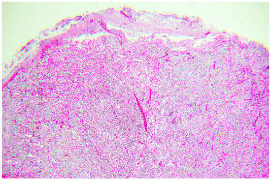
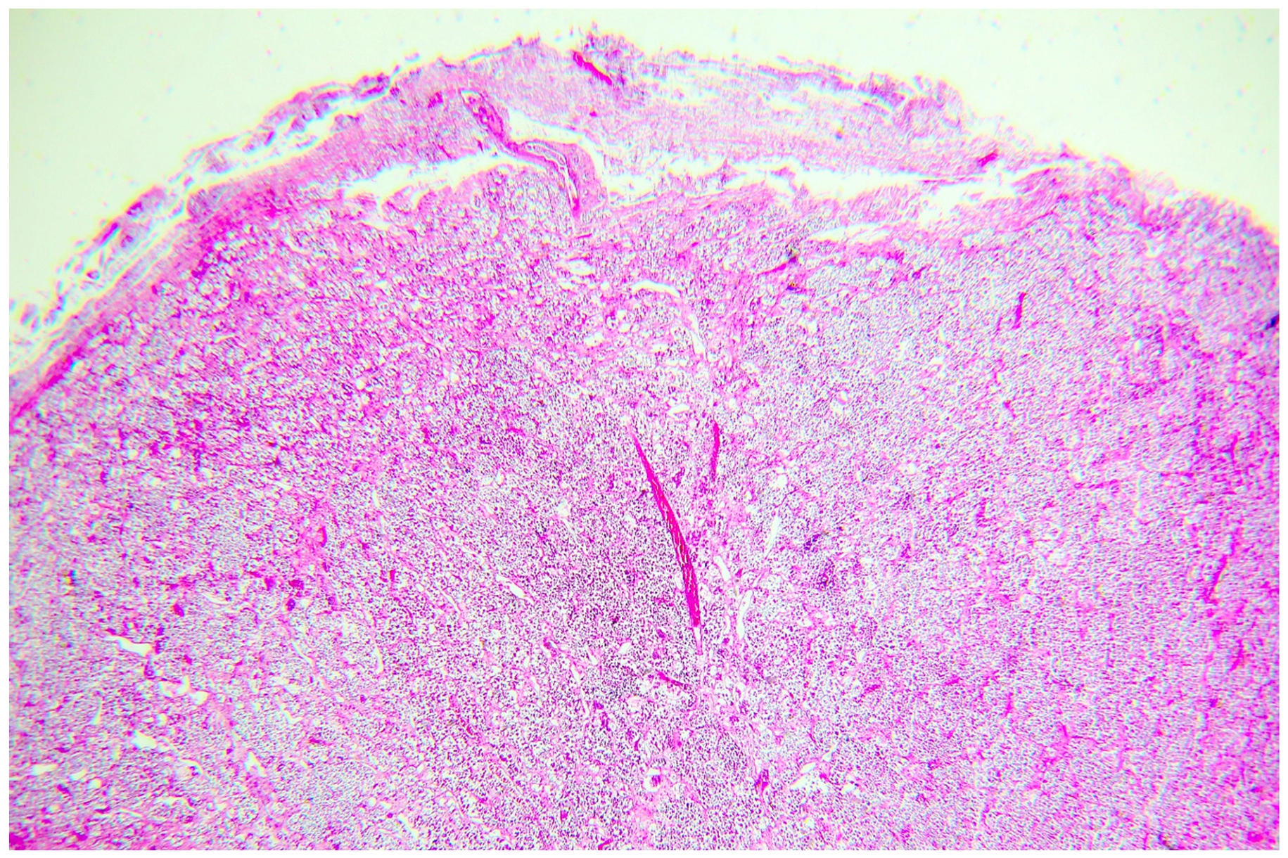
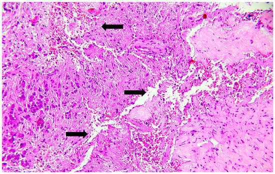
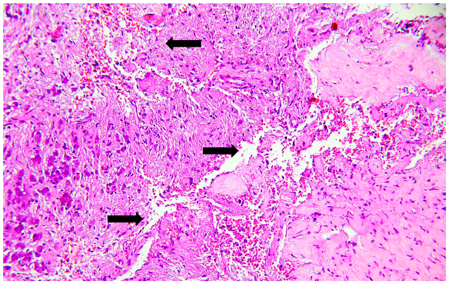
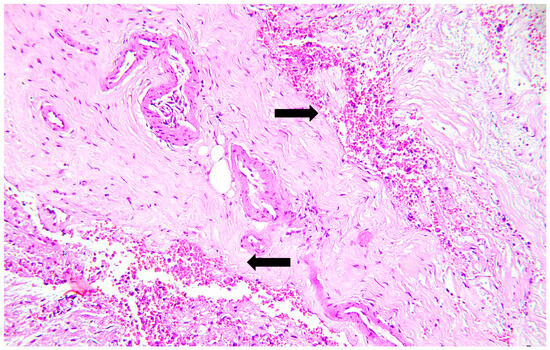
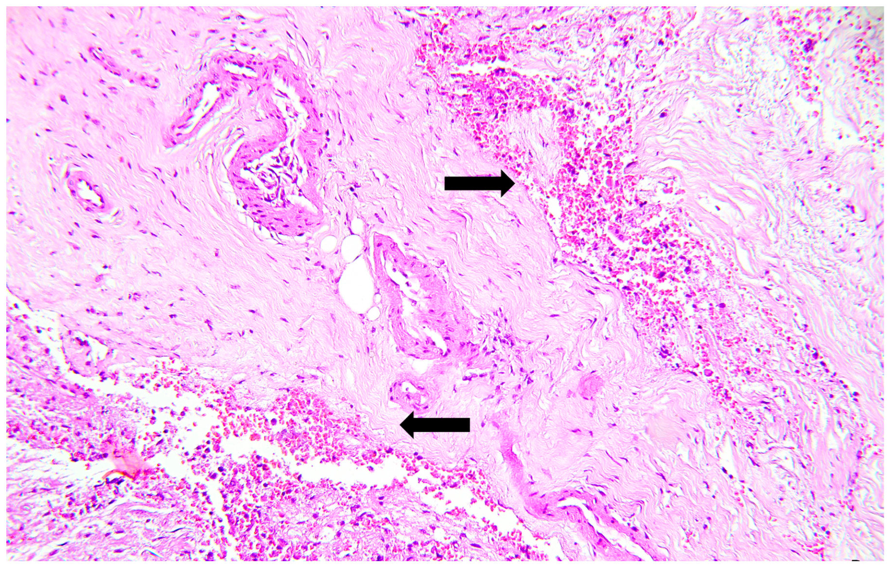
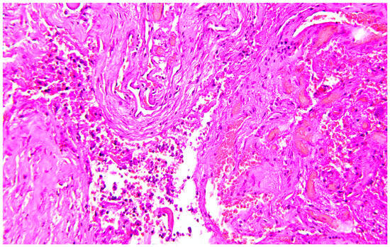
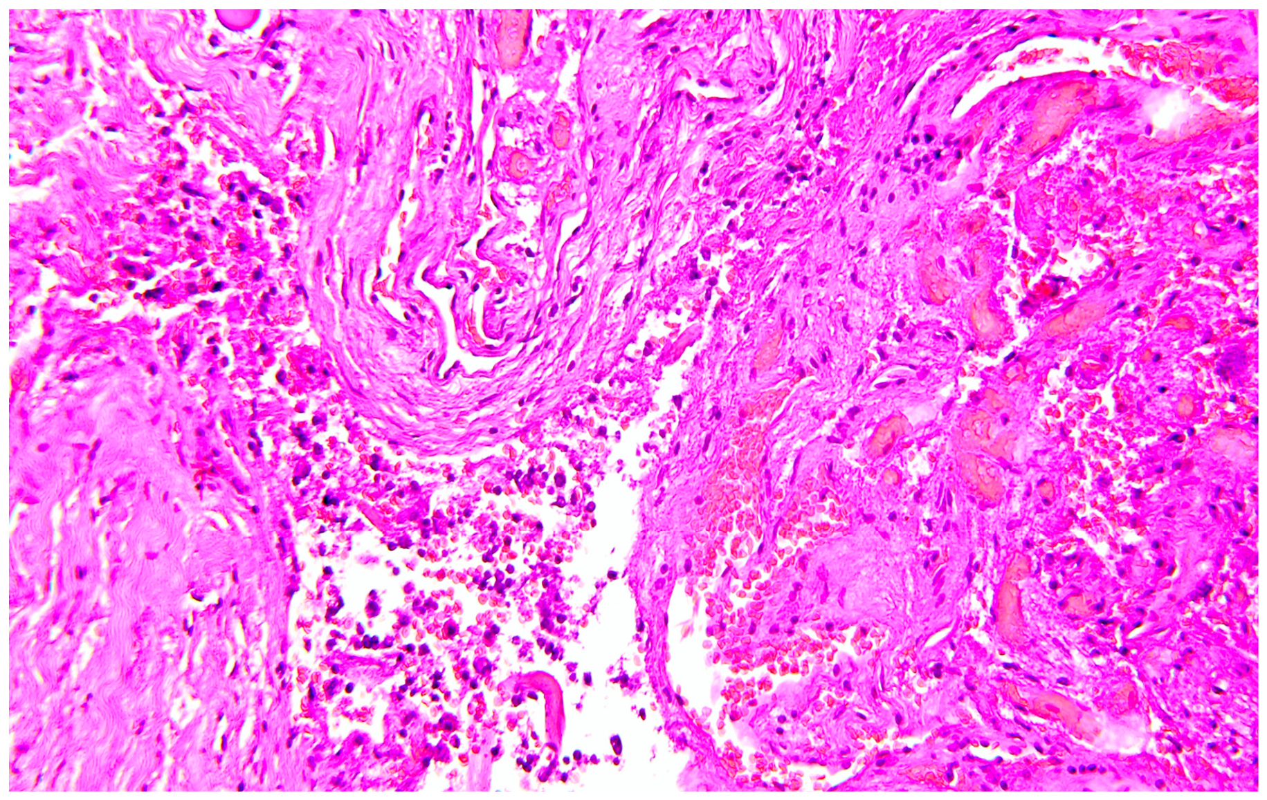
The study included 30 forensic autopsy cases, comprising 10 deaths due to suicidal hanging, 10 deaths attributed to acute heroin intoxication resulting in respiratory failure, and 10 sudden cardiac deaths, which served as the control group. The study population consisted of 17 females (56.7%) and 13 males (43.3%), ranging in age from 22 to 65 years (mean age: 40.0 ± SD 12.1 years; IQR 34–46). Information on medical history was obtained from police reports, available medical records, and autopsy documentation. Complete or partial clinical records were available in only a limited number of cases. In the remaining cases, information was limited to circumstantial data, autopsy findings, and toxicological results. No decedent had a documented diagnosis of pituitary disease or known endocrine disorder in the available records. Histological examination of the pituitary gland was specifically evaluated for features suggestive of pre-existing lesions. No case showed architectural distortion consistent with pituitary adenoma (e.g., monotonous cellular proliferation, loss of normal lobular architecture, compression of adjacent tissue), features of hypophysitis (dense lymphoplasmacytic infiltrate with parenchymal destruction), or findings typical of pituitary apoplexy (extensive hemorrhagic necrosis with disruption of glandular architecture beyond the focal changes described). Evidence of coagulopathy, shock, or treatments potentially relevant to hemorrhage was assessed through a combination of clinical documentation, autopsy findings, and histological examination of multiple organs. No case had a documented history of anticoagulant or antiplatelet therapy, and no autopsy or histological findings suggestive of a systemic coagulopathy (e.g., diffuse microthrombi, widespread hemorrhagic diathesis involving multiple organs) were identified. Signs of prolonged shock (e.g., acute tubular necrosis in the kidneys, centrilobular necrosis in the liver, ischemic changes in the gastrointestinal tract) were not observed in any case. Taken together, these findings support the interpretation that the pituitary lesions described are more likely related to the terminal hypoxic–ischemic insult than to pre-existing pituitary pathology, coagulopathy, or prolonged shock. Histological examination of the ligature mark in the hanging group confirmed evidence of vitality in all cases. Hemorrhagic infiltration of the cervical skeletal muscles was identified in 4 of the 10 cases (40%), while focal hemorrhagic infiltration involving the carotid adventitia was observed in 2 cases (20%), supporting antemortem neck compression. Cardiac examination in the control group demonstrated histopathological findings consistent with acute myocardial infarction, estimated to have occurred within the previous 24 h. None of these subjects underwent cardiopulmonary resuscitation before death, thereby excluding resuscitation-related artifacts as a potential source of pituitary or cerebral hemorrhage. In the heroin intoxication group, histopathological examination of the major organs showed changes typically associated with acute opioid toxicity. The lungs consistently demonstrated marked pulmonary edema associated with diffuse vascular congestion and focal intra-alveolar hemorrhagic areas, whereas no specific pathological alterations were identified in the remaining organs. Neuropathological examination of the control group did not reveal significant abnormalities involving the pituitary gland (Figure 1). Apart from occasional mild vascular congestion, no evidence of intraparenchymal hemorrhage, ischemic injury, or necrosis was observed in either the adenohypophysis or the neurohypophysis. In contrast, pituitary alterations were frequently identified in both groups characterized by severe hypoxia. Among the hanging cases, 6 of 10 subjects (60%) showed small hemorrhagic foci involving the neurohypophysis (Figure 2). These hemorrhages were generally limited in extent, occupying no more than two microscopic high-power fields, and were not associated with architectural disruption of the gland (Figure 3). In addition, 2 cases (20%) exhibited focal ischemic changes involving the adenohypophysis, characterized by increased eosinophilia of endocrine cells, nuclear pyknosis, and early loss of cellular architecture. More extensive pituitary lesions were observed in deaths due to heroin intoxication. Eight of the ten cases (80%) demonstrated hemorrhagic foci within the neurohypophysis, which were generally more extensive than those observed in the hanging group and frequently associated with marked vascular congestion (Figure 4). Furthermore, 6 cases (60%) showed ischemic injury of the adenohypophysis accompanied by focal coagulative necrosis, cytoplasmic hypereosinophilia, nuclear pyknosis, and disruption of the normal glandular architecture. In several cases, ischemic and hemorrhagic changes coexisted within the same specimen, suggesting a combined mechanism of prolonged hypoxemia and systemic hypoperfusion. Overall, pituitary lesions were numerically more frequent in the heroin intoxication group than in the hanging group (neurohypophyseal hemorrhage: 8/10 vs. 6/10; adenohypophyseal ischemic–necrotic injury: 6/10 vs. 2/10), whereas no comparable lesions were identified in the control group. In direct pairwise comparison between the heroin and hanging groups, these differences did not reach statistical significance (two-sided Fisher’s exact test: p = 0.63 for hemorrhage and p = 0.17 for ischemic–necrotic injury). These findings should be interpreted with caution given the small sample size; the lack of statistical significance does not imply equivalence between the two groups, but indicates that the present series is underpowered to detect moderate differences. Hemorrhagic lesions predominantly involved the posterior pituitary, while ischemic and necrotic changes were confined almost exclusively to the anterior pituitary, reflecting the different vascular supply of the two components of the gland. The frequency of each histopathological finding was compared among the three groups using the Fisher–Freeman–Halton exact test. Statistical analysis demonstrated a significantly higher prevalence of neurohypophyseal hemorrhage and adenohypophyseal ischemic-necrotic lesions in deaths due to hanging and heroin intoxication compared with sudden cardiac death (p < 0.05), supporting an association between severe hypoxic insults and pituitary injury.
Figure 1.
Pituitary tissue from a control case without the target lesions (hematoxylin–eosin).
Figure 2.
Focal neurohypophyseal hemorrhage in a hanging case, without architectural disruption (hematoxylin–eosin). Black arrows mark extravasated erythrocytes; adjacent clear spaces include congested vessels.
Figure 3.
Focal neurohypophyseal hemorrhage in a hanging case, occupying no more than two high-power fields and without architectural disruption (hematoxylin–eosin). Black arrows indicate foci of extravasated erythrocytes.
Figure 4.
Vascular congestion and neurohypophyseal hemorrhage in a heroin-intoxication case (hematoxylin–eosin).
The comparative distribution and morphology of pituitary lesions are summarized in Table 1.
Table 1.
Comparative pituitary histopathological findings across the three forensic autopsy groups.
4. Discussion
In this preliminary series, histopathological alterations of the pituitary gland were significantly more frequent in deaths characterized by severe systemic hypoxia than in sudden cardiac deaths. In particular, neurohypophyseal hemorrhage and adenohypophyseal ischemic-necrotic changes were identified in subjects who died from acute heroin intoxication and, to a lesser extent, in those who died by suicidal hanging, whereas comparable lesions were absent in the control group. This distribution points toward an interpretation in which pituitary injury is not merely a nonspecific agonal phenomenon but may reflect the pathophysiological mechanisms underlying prolonged oxygen deprivation while residual tissue perfusion is still maintained. This interpretation should, however, be contextualized within the limits of a small series assessed on hematoxylin–eosin-stained sections only. The vulnerability of the pituitary gland to ischemic injury is largely explained by its unique vascular organization. Unlike most endocrine organs, the anterior pituitary receives the majority of its blood supply through the hypothalamic-hypophyseal portal circulation, a low-pressure vascular system arising from the superior hypophyseal arteries [2,3]. Approximately 70–90% of the blood supply reaches the adenohypophysis through the long portal vessels, whereas only a smaller proportion derives from the short portal vessels originating from the inferior hypophyseal arteries [25,26]. Although anatomical anastomoses have been described, these do not appear sufficient to provide an effective collateral circulation during acute systemic hypoperfusion. Consequently, even relatively modest reductions in arterial oxygen content or perfusion pressure may substantially reduce oxygen delivery to the adenohypophysis, whose metabolic activity is particularly high because of its continuous endocrine secretory function. This concept is well established in endocrine pathology and explains why ischemic necrosis of the pituitary represents the hallmark of several clinical conditions associated with severe hemodynamic impairment, including Sheehan syndrome, pituitary apoplexy and prolonged hypotensive states [27,28,29]. The neurohypophysis differs substantially from the anterior lobe because it receives a direct arterial supply from the inferior hypophyseal arteries. Nevertheless, its rich capillary network may also become vulnerable during profound hypoxic states. Rather than developing extensive ischemic necrosis, the posterior lobe appears more prone to vascular congestion and focal hemorrhagic extravasation, probably reflecting endothelial dysfunction and microvascular injury occurring during the agonal phase. The different vascular architecture of the two lobes therefore provides a plausible explanation for the distinct histopathological pattern observed in the present series, with hemorrhagic lesions predominantly involving the neurohypophysis and ischemic-necrotic alterations almost exclusively affecting the adenohypophysis.
The absence of significant pituitary alterations in the control group represents one of the most relevant findings of the present study. Although all subjects ultimately died from circulatory arrest, no hemorrhagic or ischemic lesions were identified within the pituitary gland. This observation deserves particular attention because it argues against the hypothesis that the lesions observed in the hanging and heroin intoxication groups simply represent terminal or post-mortem artifacts. If pituitary hemorrhage and ischemia merely reflected nonspecific agonal events, similar findings would also be expected in sudden cardiac deaths. Their absence in this series is compatible with the hypothesis that the development of these lesions requires a specific pathophysiological setting in which oxygen delivery progressively declines while at least partial tissue perfusion is temporarily preserved [30]. This concept may also explain why the pituitary gland behaves differently from many other organs during systemic hypoxia. An abrupt interruption of circulation, such as that occurring in sudden cardiac death, immediately abolishes tissue perfusion, leaving insufficient time for the development of recognizable microscopic ischemic injury [31].
Conversely, conditions characterized by progressive respiratory failure or gradual impairment of cerebral perfusion expose the gland to a prolonged imbalance between oxygen demand and oxygen supply. Under these circumstances, endocrine cells remain metabolically active despite progressively decreasing oxygen availability, eventually leading to irreversible cellular damage [32].
Experimental and clinicopathological studies have consistently demonstrated that endocrine tissues supplied through portal vascular systems are particularly susceptible to this type of injury, emphasizing that both the duration and the mechanism of oxygen deprivation influence the final histological appearance [31]. From a forensic perspective, these observations suggest that pituitary histopathology may provide useful information regarding the dynamics of the terminal event rather than the cause of death itself. The lesions identified in the present study should not be regarded as disease-specific markers but rather as indicators of severe systemic hypoxia developing under conditions in which residual blood flow persists long enough to induce structural cellular injury. This distinction is particularly relevant because it shifts the interpretation of pituitary pathology from a purely descriptive finding toward a physiopathological marker that may contribute to reconstructing the mechanism of death when interpreted together with the complete autopsy findings, toxicological analyses and circumstantial evidence. The different distribution and severity of the lesions observed in the heroin intoxication and hanging groups can probably be explained by the distinct pathophysiological mechanisms leading to death. Fatal heroin intoxication is characterized by a progressive depression of the respiratory centres located within the medulla oblongata through activation of μ-opioid receptors [33,34,35]. As respiratory drive gradually decreases, ventilation becomes progressively inadequate, resulting in increasing hypercapnia, worsening arterial hypoxemia and, ultimately, respiratory arrest. Importantly, this process is usually not instantaneous. In many fatal overdoses, spontaneous circulation is maintained for a variable period despite progressively deteriorating gas exchange, allowing peripheral organs to remain perfused with increasingly hypoxemic blood [33,34,35]. Under these conditions, tissues continue to receive blood flow, but oxygen delivery becomes insufficient to meet their metabolic demands. The pituitary gland appears particularly vulnerable to this type of insult. Because the adenohypophysis depends predominantly on a portal vascular system operating under relatively low pressure, progressive reductions in arterial oxygen content may rapidly translate into tissue hypoxia despite the persistence of systemic circulation. Experimental studies have demonstrated that endocrine cells of the anterior pituitary are highly metabolically active and exhibit limited tolerance to prolonged oxygen deprivation [36]. Consequently, sustained respiratory hypoxia may initially produce reversible cellular injury, followed by irreversible ischemic damage characterized by cytoplasmic hypereosinophilia, nuclear pyknosis and coagulative necrosis, findings observed in 6 of the 10 heroin intoxication cases. The posterior pituitary appears to respond differently. Although its direct arterial blood supply theoretically offers greater protection against ischemia, severe systemic hypoxia is accompanied by profound endothelial dysfunction, alterations of capillary permeability and terminal hemodynamic instability. These events may facilitate vascular congestion and focal hemorrhagic extravasation, explaining the predominance of neurohypophyseal hemorrhages observed in our series. The coexistence of hemorrhagic and ischemic lesions in several glands therefore probably reflects different expressions of the same pathophysiological process rather than two independent mechanisms. Our findings are in agreement with the current understanding of fatal opioid intoxication. Neuropathological and experimental investigations have consistently demonstrated that respiratory depression represents the primary mechanism responsible for opioid-related deaths, whereas pulmonary edema and diffuse vascular congestion constitute the most frequent autopsy findings [37,38,39]. Although the brain has traditionally been considered the principal target organ of hypoxia, increasing evidence indicates that endocrine organs may be equally susceptible when oxygen deprivation develops progressively rather than abruptly. In this respect, the pituitary gland represents a unique model because its vascular anatomy amplifies the consequences of even moderate reductions in oxygen delivery. A similar interpretation may also explain the lesions observed in the hanging group, although both their frequency and severity were lower than those documented in heroin intoxication. The pathophysiology of hanging is considerably more complex because cerebral hypoxia results from the interaction of several mechanisms acting simultaneously [40]. Compression of the jugular veins causes rapid venous congestion and increased intracranial pressure, whereas compression of the carotid and vertebral arteries progressively reduces cerebral blood flow [41,42]. Airway obstruction contributes to arterial hypoxemia, while stimulation of the carotid sinus and adjacent autonomic structures may induce profound vagal reflexes with bradycardia, hypotension and, occasionally, sudden cardiac arrest. The relative contribution of each mechanism varies according to the position of the ligature, the degree of suspension and individual anatomical characteristics. Despite these differences, death by hanging is rarely instantaneous. Loss of consciousness generally occurs within seconds, but complete circulatory arrest usually develops only after a variable agonal period lasting several minutes. During this interval, residual cerebral and systemic perfusion may still be present despite progressively worsening hypoxia. This short survival period appears sufficient to induce early microscopic alterations within the pituitary gland, particularly in its most vulnerable vascular territories. However, compared with heroin intoxication, the duration of oxygen deprivation is generally shorter and circulatory impairment develops more abruptly. This may explain why hemorrhagic lesions remained limited in extent and ischemic-necrotic alterations involved only a minority of cases. The comparison between the two hypoxic groups therefore appears particularly informative. Both conditions share severe oxygen deprivation as the final common pathway, yet they differ substantially in the temporal evolution of the insult. In heroin intoxication, hypoxia develops progressively while circulation is initially preserved, exposing the pituitary to prolonged metabolic stress [37,38,39]. In hanging, hypoxia is accompanied by an early impairment of cerebral perfusion resulting from vascular compression, reducing the time available for irreversible ischemic injury to develop [40,43,44]. The higher prevalence of adenohypophyseal necrosis in heroin intoxication compared with hanging, although not statistically significant in the pairwise comparison, is biologically plausible and appears consistent with the different physiological mechanisms leading to death. Interestingly, the present observations also suggest that the severity of pituitary injury may depend not only on the intensity of hypoxia but also on its duration. This concept is supported by experimental and clinical studies demonstrating that irreversible endocrine cell damage requires a minimum period of persistent oxygen deprivation before becoming morphologically recognizable [4]. Accordingly, the absence of extensive necrosis in hanging should not be interpreted as evidence of a milder insult but rather as the consequence of a shorter interval between the onset of hypoxia and terminal circulatory arrest. Conversely, the coexistence of ischemic and hemorrhagic lesions observed in several heroin intoxication cases probably reflects a longer period during which residual perfusion persisted despite critically reduced oxygen delivery. Among the few studies investigating pituitary responses in relation to hypoxia and patterns of death, Kouroupi et al. [6] examined the expression of hypoxia-inducible factor (HIF) and angiogenesis-related markers in the pituitary gland across different modes of death. Their findings provided immunohistochemical evidence of activation of hypoxia-related and angiogenic pathways, supporting the concept that the pituitary gland actively responds to systemic oxygen deprivation at a molecular level. However, their study did not systematically compare distinct non-traumatic hypoxic mechanisms, such as progressive respiratory failure and mechanical asphyxia, nor did it correlate molecular findings with the detailed histomorphological distribution of hemorrhagic and ischemic–necrotic lesions within the adenohypophysis and neurohypophysis. The present study complements and extends these observations by providing a comparative histopathological analysis of fatal heroin intoxication, suicidal hanging, and sudden cardiac death, with specific attention to the frequency, severity, and lobar distribution of pituitary lesions. Thus, while Kouroupi et al. documented molecular evidence of hypoxic activation, our findings provide additional morphological information regarding the relationship between different mechanisms of systemic hypoxia and the pattern of pituitary injury.
Although pituitary lesions have been extensively investigated in traumatic brain injury, relatively few studies have explored their occurrence in non-traumatic causes of death characterized by systemic hypoxia. The work of Kibayashi and colleagues remains the principal forensic study addressing pituitary pathology after fatal head injury [45]. Examining 66 autopsy cases, the authors demonstrated that hemorrhage represents the earliest pituitary lesion following trauma, whereas ischemic infarction of the anterior lobe becomes increasingly evident with longer survival intervals. Their findings strongly support the concept that pituitary morphology is influenced not only by the nature of the insult but also by the duration of survival. In our opinion, this concept may be extended beyond traumatic brain injury. The present results are compatible with the possibility that the same temporal relationship may also characterize non-traumatic conditions in which oxygen delivery progressively declines while circulation is at least partially maintained. A similar concept has long been recognized in endocrine pathology. Sheehan syndrome represents the best-known clinical model of ischemic pituitary injury [28,29]. In this condition, severe postpartum hemorrhage produces systemic hypotension and reduced portal blood flow, leading to selective ischemic necrosis of the adenohypophysis. Likewise, pituitary apoplexy has been associated with acute vascular insufficiency and hemorrhagic infarction, particularly in patients with pre-existing adenomas. Although these disorders differ substantially from heroin intoxication or hanging, they share a common pathophysiological substrate: the inability of the pituitary vascular system to compensate adequately for sudden or sustained reductions in oxygen delivery. The characteristic vascular architecture of the gland therefore appears to represent the principal determinant of tissue vulnerability rather than the specific etiology of the systemic insult. The absence of significant pituitary alterations in the control group deserves consideration. All subjects included in this group died from acute myocardial infarction, yet none showed hemorrhagic or ischemic lesions involving either the anterior or posterior pituitary. This observation is particularly relevant because it argues against the possibility that the lesions identified in the heroin intoxication and hanging groups merely represent nonspecific terminal events or post-mortem artifacts. Cardiac arrest inevitably produces global tissue ischemia; however, when circulatory collapse occurs abruptly, there is little opportunity for morphologically recognizable pituitary injury to develop. Conversely, both heroin intoxication and hanging are characterized by a phase during which oxygen delivery progressively declines before complete circulatory arrest. Although the duration of this interval differs substantially between the two mechanisms of death, it appears sufficient to induce structural damage within one of the most metabolically active endocrine organs of the human body. The comparison among the three study groups therefore suggests that pituitary injury depends less on the absolute severity of hypoxia than on the relationship between oxygen deprivation and residual tissue perfusion. This concept may explain why the most extensive lesions were observed in heroin intoxication. Progressive respiratory depression allows circulation to persist while arterial oxygen content continuously decreases, exposing the gland to sustained hypoxic stress. In hanging, cerebral hypoxia develops more rapidly because respiratory impairment is accompanied by vascular compression and neurogenic mechanisms, thereby reducing the interval during which histologically evident ischemic injury can evolve. In contrast, sudden cardiac death abruptly interrupts perfusion and may not allow sufficient time for comparable microscopic alterations to become established. Taken together, these observations suggest that pituitary hemorrhage and ischemic–necrotic change may represent a candidate histological correlate of prolonged hypoxic–ischemic stress, rather than a marker of a specific cause of death. This interpretation requires validation against an independent standard for the duration of hypoxia; at present, these lesions cannot be used to indicate the probable duration of the agonal period in an individual case.
From a forensic perspective, these findings deserve careful interpretation. Neither pituitary hemorrhage nor ischemic necrosis should be regarded as pathognomonic of heroin intoxication, hanging or any other particular mechanism of death. Similar alterations may occur in traumatic brain injury, severe shock, disseminated intravascular coagulation, pituitary adenomas, pituitary apoplexy and other conditions associated with vascular instability. Consequently, histopathological examination of the pituitary cannot establish the cause of death in isolation. Nevertheless, when interpreted together with the autopsy findings, toxicological analyses, scene investigation and circumstantial evidence, pituitary lesions may provide useful additional information regarding the pathophysiological sequence leading to death. Their utility for reconstructing the circumstances of death should be tested through a separate protocol and cannot currently support inferences about the duration of the terminal hypoxic insult in an individual case.
Limitations
Several limitations of the present study should be acknowledged. The relatively small number of cases limits the statistical power of the analysis. The retrospective design precludes accurate reconstruction of the duration of the agonal period in individual subjects. Although police reports and, whenever available, medical records were reviewed, precise time points (e.g., last seen alive, estimated time of drug administration, discovery of the body) were often incomplete or unavailable, and no reliable individual estimates of the interval between onset of hypoxia and death could be established. Accordingly, the proposed differences in the duration and evolution of hypoxia between heroin intoxication and hanging should be interpreted as pathophysiological inferences based on the typical mechanisms of these modes of death, rather than as directly measured survival intervals. The observed histopathological patterns support an association between pituitary lesion type and category of death, but do not allow estimation of the duration of hypoxia from histology alone. Moreover, the diagnosis was based exclusively on routine hematoxylin-eosin staining. Hematoxylin–eosin allows the morphological recognition of hemorrhage and coagulative necrosis but does not permit a detailed assessment of the individual components of the examined tissue; in this respect, immunohistochemical markers of hypoxia, endothelial injury and apoptosis—including HIF-1α, VEGF, caspase-3 and CD34—could provide additional information regarding the timing and pathogenesis of these alterations. Future prospective studies should incorporate ancillary histochemical and immunohistochemical techniques to provide a more objective and mechanistic interpretation of the observed changes. In particular, Masson’s trichrome stain could be used to better delineate fibrosis, vascular walls, and the extent of hemorrhagic versus ischemic damage; the periodic acid–Schiff (PAS) reaction could highlight basement membranes, vascular structures, and potential glycoprotein-rich deposits; and phosphotungstic acid–hematoxylin (PTAH) or other fibrin stains could help characterize microthrombi and fibrin deposition in cases with suspected coagulopathy. In addition, immunohistochemical markers of hypoxia (e.g., HIF-1α), angiogenesis (e.g., VEGF, CD34), endothelial injury, and apoptosis (e.g., caspase-3) should be considered to provide more objective and quantifiable evidence of hypoxic stress and microvascular damage. The integration of these methods with conventional histology is likely to clarify the pathogenesis, timing, and forensic significance of pituitary alterations in different modes of death.
The relatively small number of cases (n = 30) limits the statistical power and generalizability of our findings. The three groups were selected to represent distinct pathophysiological mechanisms of systemic hypoxia rather than to provide an exhaustive survey of all possible causes of death. A further major limitation is that histological assessment was based exclusively on hematoxylin–eosin staining. Ancillary histochemical stains (e.g., Masson’s trichrome, PAS, PTAH) and immunohistochemical markers of hypoxia, angiogenesis, endothelial injury and apoptosis (e.g., HIF-1α, VEGF, CD34, caspase-3) were not applied in this retrospective series. These methods should be incorporated in future prospective studies to provide a more detailed and objective characterization of pituitary lesions. For findings intended to be novel, the distinctiveness of the histological patterns should first be confirmed with a range of complementary methods, if needed. The study should therefore be regarded as preliminary and hypothesis-generating, and its findings are not yet suitable for application to individual forensic cases. The sample is also limited in size and variability (a single centre, ten adult cases per category, and three categories of death), which further restricts generalizability. Age-related vascular and parenchymal changes in the pituitary gland cannot be completely excluded and may have contributed to the observed findings, although the complete absence of comparable lesions in the control group argues against age alone as a sufficient explanation. Similarly, while particular care was taken during gland removal, some degree of mechanical artifact cannot be entirely excluded. Finally, our study does not establish whether similar pituitary alterations may occur in other causes of death not associated with severe hypoxia or head trauma, such as shock, disseminated intravascular coagulation, pituitary adenomas, or pituitary apoplexy. Accordingly, the observed lesions should not be regarded as disease specific.
5. Conclusions
In this preliminary series, pituitary hemorrhagic and ischemic lesions were observed in selected hanging and heroin-related deaths but not in sudden cardiac death controls; the relative vulnerability of the gland is attributed in the literature to its distinctive vascular architecture. The different distribution of pituitary lesions observed among the three study groups is compatible with the hypothesis that the development of hemorrhagic and ischemic changes depends not only on the severity of oxygen deprivation but also on the duration of the hypoxic insult and the persistence of residual tissue perfusion. Fatal heroin intoxication, characterized by progressive respiratory depression with maintained circulation during the early stages of intoxication, was associated with the highest prevalence and severity of pituitary lesions. Suicidal hanging, although also producing profound hypoxia, resulted in less extensive alterations, probably reflecting the more rapid evolution of cerebral ischemia due to the combined effects of vascular compression, airway obstruction and neurogenic mechanisms. Conversely, the complete absence of comparable lesions in sudden cardiac deaths suggests that abrupt circulatory arrest alone may be insufficient to induce morphologically recognizable pituitary injury. In this context, the pituitary gland should not simply be regarded as a passive target of hypoxic injury; the observed lesions are better described as a candidate histological correlate of systemic oxygen deprivation that requires validation. Although these lesions cannot be considered disease-specific and should never be interpreted in isolation, their potential utility for reconstructing the circumstances of death should be tested through a separate protocol. At present, these lesions cannot be used to indicate the probable duration of the agonal period in an individual case. Further studies involving larger case series and the integration of conventional histology with immunohistochemical and molecular markers of hypoxia are warranted to clarify the diagnostic and forensic significance of these findings. Taken together, these findings support the hypothesis that pituitary histopathology may reflect the temporal evolution of systemic hypoxia rather than the specific cause of death. Nevertheless, the relatively small sample size, the exclusive use of H&E staining, and the retrospective design represent major limitations that preclude definitive etiological or diagnostic conclusions. Further studies involving larger and more diversified case series and the integration of conventional histology with histochemical and immunohistochemical markers of hypoxia are warranted to clarify the diagnostic and forensic significance of these findings. Until the distinctiveness of these patterns can be confirmed with complementary methods, they should be regarded as preliminary and should not be applied to individual forensic casework.
Author Contributions
Conceptualization, F.D.D. and A.M.; methodology, G.N., A.M. and F.D.D.; validation, A.D.M. and A.G.; formal analysis, G.N. and F.D.D.; investigation, G.N., A.D.M., A.G., M.R., B.T., G.D.A. and F.A.M.; resources, A.M. and P.F.; data curation, M.R., B.T. and G.D.A.; writing—original draft preparation, G.N. and F.D.D.; writing—review and editing, A.D.M., F.A.M., A.M., P.F. and F.D.D.; visualization, A.G. and F.A.M.; supervision, A.M., P.F. and F.D.D.; project administration, A.M. and F.D.D. All authors have read and agreed to the published version of the manuscript.
Funding
This research received no external funding.
Institutional Review Board Statement
Ethical review and approval were waived for this study. This research did not involve any live animal experiments or prospective human subject studies. The study solely retrospectively analyzed data from judicial autopsy cases without collecting new biological samples or conducting invasive research procedures on living individuals. All relevant investigative and autopsy procedures were legally mandated by local attorneys, and the research-related data analysis was performed purely for academic clarification of the cause and manner of death, complying with local and EU scientific research regulations.
Informed Consent Statement
Patient informed consent was not required for this study. The study did not involve identifiable personal data of individuals. All data utilized in this research conform to the provisions of the Italian Data Protection Authority regulations (Official Gazette no. 72 of 26 March 2012), EU GDPR 679/2016, 9/2016, and Italian Law Amendment No. 424/19 of July 2018 for scientific research exemptions. The retrospective analysis of judicial autopsy case data for academic research purposes qualifies for informed consent waiver under the above regulatory provisions.
Data Availability Statement
The data analyzed during the current study are available from the corresponding author upon reasonable request.
Conflicts of Interest
The authors declare no conflict of interest.
References
- Personnier, C.; Crosnier, H.; Meyer, P.; Chevignard, M.; Flechtner, I.; Boddaert, N.; Breton, S.; Mignot, C.; Dassa, Y.; Souberbielle, J.-C.; et al. Prevalence of Pituitary Dysfunction After Severe Traumatic Brain Injury in Children and Adolescents: A Large Prospective Study. J. Clin. Endocrinol. Metab. 2014, 99, 2052–2060. [Google Scholar] [CrossRef] [Scilit] [PubMed]
- Gorczyca, W.; Hardy, J. Arterial Supply of the Human Anterior Pituitary Gland. Neurosurgery 1987, 20, 369–378. [Google Scholar] [CrossRef] [Scilit] [PubMed]
- Leclercq, T.A.; Grisoli, F. Arterial Blood Supply of the Normal Human Pituitary Gland. An Anatomical Study. J. Neurosurg. 1983, 58, 678–681. [Google Scholar] [CrossRef] [Scilit] [PubMed]
- Chaturvedi, D.; Suri, A.; Kasliwal, M.K.; Mahapatra, A.K.; Mehta, V.S.; Shridhar, V.; Garg, A.; Sarkar, C.; Dogra, T.D.; Singh, R. Factors Affecting the Development of Hypothalamus and Pituitary Lesions in Fatal Closed Head Injury: A Prospective Study. J. Trauma 2010, 69, 290–293. [Google Scholar] [CrossRef] [Scilit] [PubMed]
- Milovanov, A.P.; Matsionis, A.E.; Makarenko, I.M. Pathomorphology of adenohypophysis in cases of massive obstetric hemorrhages (morphometric and immunohistochemical studies). Arkhiv Patol. 2005, 67, 14–17. [Google Scholar] [PubMed]
- Kouroupi, M.; Sivridis, E.; Papazoglou, D.; Koukourakis, M.I.; Giatromanolaki, A. Hypoxia Inducible Factor Expression and Angiogenesis—Analysis in the Pituitary Gland and Patterns of Death. In Vivo 2018, 32, 185–190. [Google Scholar] [CrossRef] [Scilit] [PubMed]
- Bevilacqua, G.; Fornaciari, G. Clinico-Pathological Correlations in a Case of Post Traumatic Pan-Hypopituitarism. Acta Neuropathol. 1975, 31, 171–177. [Google Scholar] [CrossRef] [Scilit] [PubMed]
- Dressler, J.; Mueller, E. High Thyroglobulin (Tg) Concentrations in Fatal Traumatic Brain Injuries. Am. J. Forensic Med. Pathol. 2006, 27, 280–282. [Google Scholar] [CrossRef] [Scilit] [PubMed]
- Vlad, R.M.; Albu, A.I.; Nicolaescu, I.D.; Dobritoiu, R.; Carsote, M.; Sandru, F.; Albu, D.; Păcurar, D. An Approach to Traumatic Brain Injury-Related Hypopituitarism: Overcoming the Pediatric Challenges. Diagnostics 2023, 13, 212. [Google Scholar] [CrossRef] [Scilit] [PubMed]
- Salehi, F.; Kovacs, K.; Scheithauer, B.W.; Pfeifer, E.A.; Cusimano, M. Histologic Study of the Human Pituitary Gland in Acute Traumatic Brain Injury. Brain Inj. 2007, 21, 651–656. [Google Scholar] [CrossRef] [Scilit] [PubMed]
- Ruszała, A.; Wójcik, M.; Krystynowicz, A.; Starzyk, J. Distinguishing between Post-Trauma Pituitary Stalk Disruption and Genetic Pituitary Stalk Interruption Syndrome—Case Presentation and Literature Overview. Pediatr. Endocrinol. Diabetes Metab. 2019, 25, 155–162. [Google Scholar] [CrossRef] [Scilit] [PubMed]
- Harper, C.G.; Doyle, D.; Adams, J.H.; Graham, D.I. Analysis of Abnormalities in Pituitary Gland in Non-Missile Head Injury: Study of 100 Consecutive Cases. J. Clin. Pathol. 1986, 39, 769–773. [Google Scholar] [CrossRef] [Scilit] [PubMed]
- Giordano, G.; Aimaretti, G.; Ghigo, E. Variations of Pituitary Function over Time after Brain Injuries: The Lesson from a Prospective Study. Pituitary 2005, 8, 227–231. [Google Scholar] [CrossRef] [Scilit] [PubMed]
- Agha, A.; Rogers, B.; Sherlock, M.; O’Kelly, P.; Tormey, W.; Phillips, J.; Thompson, C.J. Anterior Pituitary Dysfunction in Survivors of Traumatic Brain Injury. J. Clin. Endocrinol. Metab. 2004, 89, 4929–4936. [Google Scholar] [CrossRef] [Scilit] [PubMed]
- Schneider, H.J.; Schneider, M.; Saller, B.; Petersenn, S.; Uhr, M.; Husemann, B.; von Rosen, F.; Stalla, G.K. Prevalence of Anterior Pituitary Insufficiency 3 and 12 Months after Traumatic Brain Injury. Eur. J. Endocrinol. 2006, 154, 259–265. [Google Scholar] [CrossRef] [Scilit] [PubMed]
- Tanriverdi, F.; De Bellis, A.; Ulutabanca, H.; Bizzarro, A.; Sinisi, A.A.; Bellastella, G.; Amoresano Paglionico, V.; Dalla Mora, L.; Selcuklu, A.; Unluhizarci, K.; et al. A Five Year Prospective Investigation of Anterior Pituitary Function after Traumatic Brain Injury: Is Hypopituitarism Long-Term after Head Trauma Associated with Autoimmunity? J. Neurotrauma 2013, 30, 1426–1433. [Google Scholar] [CrossRef] [Scilit] [PubMed]
- Suzuki, H.; Hayashi, K.; Fukunaga, T. Two Forensic Autopsy Cases of Death from Unexpected Lesions of the Pituitary Gland. Leg. Med. 2014, 16, 36–39. [Google Scholar] [CrossRef] [Scilit] [PubMed]
- Blisard, K.S.; Pfalzgraf, R.R.; Balko, M.G. Sudden Death Due to Lymphoplasmacytic Hypophysitis. Am. J. Forensic Med. Pathol. 1992, 13, 207–210. [Google Scholar] [CrossRef] [Scilit] [PubMed]
- Hatake, K.; Kubota, A.; Taniguchi, T.; Negoro, M.; Ouchi, H.; Minami, T.; Hishida, S. A case of sudden death of a patient with hypopituitarism. Nihon Hoigaku Zasshi 1994, 48, 267–273. [Google Scholar] [PubMed]
- Leff, R.S.; Martino, R.L.; Pollock, W.J.; Knight, W.A. Pituitary Abscess after Autologous Bone Marrow Transplantation. Am. J. Hematol. 1989, 31, 62–64. [Google Scholar] [CrossRef] [Scilit] [PubMed]
- Wang, A.R.; Gill, J.R. The Pituitary Gland: An Infrequent but Multifaceted Contributor to Death. Acad. Forensic Pathol. 2016, 6, 206–216. [Google Scholar] [CrossRef] [Scilit] [PubMed]
- Shields, L.B.E.; Balko, M.G.; Hunsaker, J.C. Sudden and Unexpected Death from Pituitary Tumor Apoplexy. J. Forensic Sci. 2012, 57, 262–266. [Google Scholar] [CrossRef] [Scilit] [PubMed]
- Crompton, M.R. Hypothalamic Lesions Following Closed Head Injury. Brain 1971, 94, 165–172. [Google Scholar] [CrossRef] [Scilit] [PubMed]
- Maiya, B.; Newcombe, V.; Nortje, J.; Bradley, P.; Bernard, F.; Chatfield, D.; Outtrim, J.; Hutchinson, P.; Matta, B.; Antoun, N.; et al. Magnetic Resonance Imaging Changes in the Pituitary Gland Following Acute Traumatic Brain Injury. Intensive Care Med. 2008, 34, 468–475. [Google Scholar] [CrossRef] [Scilit] [PubMed]
- Biagetti, B.; Simò, R. Pituitary Apoplexy: Risk Factors and Underlying Molecular Mechanisms. Int. J. Mol. Sci. 2022, 23, 8721. [Google Scholar] [CrossRef] [Scilit] [PubMed]
- Ranabir, S.; Baruah, M.P. Pituitary Apoplexy. Indian J. Endocrinol. Metab. 2011, 15, S188–S196. [Google Scholar] [CrossRef] [Scilit] [PubMed]
- Tessnow, A.H.; Wilson, J.D. The Changing Face of Sheehan’s Syndrome. Am. J. Med. Sci. 2010, 340, 402–406. [Google Scholar] [CrossRef] [Scilit] [PubMed]
- Kilicli, F.; Dokmetas, H.S.; Acibucu, F. Sheehan’s Syndrome. Gynecol. Endocrinol. 2013, 29, 292–295. [Google Scholar] [CrossRef] [Scilit] [PubMed]
- Kovacs, K.; Yao, J. Pituitary Necrosis Following Major Heart Surgery. Z. Kardiol. 1975, 64, 52–57. [Google Scholar] [PubMed]
- Tani, N.; Ikeda, T.; Ishikawa, T. Effects of Prolactin on Brain Neurons under Hypoxia. Life 2024, 14, 152. [Google Scholar] [CrossRef] [Scilit] [PubMed]
- Todaro, D.R.; Volkow, N.D.; Langleben, D.D.; Shi, Z.; Wiers, C.E. Collateral Damage: Neurological Correlates of Non-Fatal Overdose in the Era of Fentanyl-Xylazine. Neurosci. Insights 2024, 19, 26331055241247156. [Google Scholar] [CrossRef] [Scilit] [PubMed]
- Kiyatkin, E.A. Hypoxic Effects of Heroin and Fentanyl and Their Basic Physiological Mechanisms. Am. J. Physiol. Lung Cell. Mol. Physiol. 2024, 327, L930–L948. [Google Scholar] [CrossRef] [Scilit] [PubMed]
- Lalley, P.M. Opioidergic and Dopaminergic Modulation of Respiration. Respir. Physiol. Neurobiol. 2008, 164, 160–167. [Google Scholar] [CrossRef] [Scilit] [PubMed]
- Dezfulian, C.; Orkin, A.M.; Maron, B.A.; Elmer, J.; Girotra, S.; Gladwin, M.T.; Merchant, R.M.; Panchal, A.R.; Perman, S.M.; Starks, M.A.; et al. Opioid-Associated Out-of-Hospital Cardiac Arrest: Distinctive Clinical Features and Implications for Health Care and Public Responses: A Scientific Statement From the American Heart Association. Circulation 2021, 143, e836–e870. [Google Scholar] [CrossRef] [Scilit] [PubMed]
- Pattinson, K.T.S.; Governo, R.J.; MacIntosh, B.J.; Russell, E.C.; Corfield, D.R.; Tracey, I.; Wise, R.G. Opioids Depress Cortical Centers Responsible for the Volitional Control of Respiration. J. Neurosci. 2009, 29, 8177–8186. [Google Scholar] [CrossRef] [Scilit] [PubMed]
- Stam, N.C.; Cogger, S.; Schumann, J.L.; Weeks, A.; Roxburgh, A.; Dietze, P.M.; Clark, N. The Onset and Severity of Acute Opioid Toxicity in Heroin Overdose Cases: A Retrospective Cohort Study at a Supervised Injecting Facility in Melbourne, Australia. Clin. Toxicol. 2022, 60, 1227–1234. [Google Scholar] [CrossRef] [Scilit] [PubMed]
- Stoermer, R.; Drewe, J.; Dursteler-Mac Farland, K.M.; Hock, C.; Mueller-Spahn, F.; Ladewig, D.; Stohler, R.; Mager, R. Safety of Injectable Opioid Maintenance Treatment for Heroin Dependence. Biol. Psychiatry 2003, 54, 854–861. [Google Scholar] [CrossRef] [Scilit] [PubMed]
- Bouillon, T.; Bruhn, J.; Roepcke, H.; Hoeft, A. Opioid-Induced Respiratory Depression Is Associated with Increased Tidal Volume Variability. Eur. J. Anaesthesiol. 2003, 20, 127–133. [Google Scholar] [CrossRef] [Scilit] [PubMed]
- Leino, K.; Mildh, L.; Lertola, K.; Seppälä, T.; Kirvelä, O. Time Course of Changes in Breathing Pattern in Morphine- and Oxycodone-Induced Respiratory Depression. Anaesthesia 1999, 54, 835–840. [Google Scholar] [CrossRef] [Scilit] [PubMed]
- Mitchell, J.R.; Roach, D.E.; Tyberg, J.V.; Belenkie, I.; Sheldon, R.S. Mechanism of Loss of Consciousness during Vascular Neck Restraint. J. Appl. Physiol. 2012, 112, 396–402. [Google Scholar] [CrossRef] [Scilit] [PubMed]
- Reay, D.T.; Holloway, G.A. Changes in Carotid Blood Flow Produced by Neck Compression. Am. J. Forensic Med. Pathol. 1982, 3, 199–202. [Google Scholar] [CrossRef] [Scilit] [PubMed]
- Maier, W.; Fradis, M.; Malatskey, S.; Krebs, A. Diagnostic and Therapeutic Management of Bilateral Carotid Artery Occlusion Caused by Near-Suicidal Hanging. Ann. Otol. Rhinol. Laryngol. 1999, 108, 189–192. [Google Scholar] [CrossRef] [Scilit] [PubMed]
- Sauvageau, A. Agonal Sequences in Four Filmed Hangings: Analysis of Respiratory and Movement Responses to Asphyxia by Hanging. J. Forensic Sci. 2009, 54, 192–194. [Google Scholar] [CrossRef] [Scilit] [PubMed]
- Sauvageau, A.; Laharpe, R.; King, D.; Dowling, G.; Andrews, S.; Kelly, S.; Ambrosi, C.; Guay, J.-P.; Geberth, V.J.; Working Group on Human Asphyxia. Agonal Sequences in 14 Filmed Hangings with Comments on the Role of the Type of Suspension, Ischemic Habituation, and Ethanol Intoxication on the Timing of Agonal Responses. Am. J. Forensic Med. Pathol. 2011, 32, 104–107. [Google Scholar] [CrossRef] [Scilit] [PubMed]
- Kibayashi, K.; Shimada, R.; Nakao, K.; Ro, A. Analysis of Pituitary Lesions in Fatal Closed Head Injury. Am. J. Forensic Med. Pathol. 2012, 33, 206–210. [Google Scholar] [CrossRef] [Scilit] [PubMed]

Disclaimer/Publisher’s Note: The statements, opinions and data contained in all publications are solely those of the individual author(s) and contributor(s) and not of MDPI and/or the editor(s). MDPI and/or the editor(s) disclaim responsibility for any injury to people or property resulting from any ideas, methods, instructions or products referred to in the content. |
© 2026 by the authors. Licensee MDPI, Basel, Switzerland. This article is an open access article distributed under the terms and conditions of the Creative Commons Attribution (CC BY) license.