


Abstract
Our exploration of Indian boletoid mushrooms (Boletaceae and related fleshy poroid fungi) led to the discovery of a species belonging to the genus Lanmaoa, marking the first record of this genus from the country. During macrofungal explorations in different parts of subtropical to temperate Uttarakhand (a Himalayan state in India), a new species of Lanmaoa was collected. After thorough studies of Indian collections this species is proposed herein as Lanmaoa atropurpurea sp. nov. and presented with conventional morpho-taxonomy and multigene molecular phylogeny. A provisional key to Asian species of Lanmaoa is also given.
1. Introduction
The genus Lanmaoa G. Wu & Zhu L. Yang was erected by Wu et al. (2016) [1], with Lanmaoa asiatica G. Wu & Zhu L. Yang from southwestern China designated as the type species. Species of Lanmaoa are characterized by stipitate-pileate basidiomata with a cream-yellow to lemon-yellow hymenophore that typically stains dull blue upon bruising. The tubes are concolorous with the hymenophoral surface or occasionally light red. The stipe is centrally positioned, yellow to lemon-yellow at the apex and gradually transitions to light or dark purplish red toward the base. Basidiospores are smooth, narrowly suboblong, subellipsoid to subfusiform, while the pileipellis is organized as an (ixo) trichoderm or (ixo) subcutis [1,2,3,4].
Lanmaoa is morphologically similar to another boletoid genus, Baorangia G. Wu & Zhu L. Yang; however, multilocus phylogenetic analyses have consistently resolved the two genera as distinct evolutionary lineages within the Boletaceae [5,6]. The circumscription of Lanmaoa remains a subject of taxonomic debate, with some authors proposing its inclusion within a third boletoid genus, Cyanoboletus Gelardi, Vizzini & Simonini based on their close phylogenetic relationship and partially overlapping morphological characteristics [1,4,6,7,8,9]. Cyanoboletus is generally distinguished by predominantly dull brown basidiomata, whereas species of Lanmaoa typically exhibit conspicuous yellow and red pigmentation [1,6,8,9,10]; however, this distinction is not always a reliable generic delimitation. Wu et al. [1,6] proposed the relatively thin hymenophore (approximately one-third to one-fifth the thickness of the pileus context) as a diagnostic character for Lanmaoa. However, this character has proven to be inconsistent, as it has also been observed in Cyanoboletus instabilis (W.F. Chiu) G. Wu & Zhu L. Yang and in some Baorangia species [e.g., B. emileorum (Barbier) Vizzini, Simonini & Gelardi] and varies within Lanmaoa, including in L. rubriceps N.K. Zeng & Hui Chai [8] or Lanmaoa macrocarpa N.K. Zeng, H. Chai & S. Jiang [9]. These observations indicate that no single morphological character can separate Lanmaoa from Cyanoboletus. Consequently, the delimitation of these genera should rely on an integrative approach combining morphology with multilocus phylogenetic evidence, as done by Wu et al. (2023) [11].
Currently, Lanmaoa comprises 12 accepted species (https://www.speciesfungorum.org/) (accessed on 30 August 2026). The genus exhibits a broad geographic distribution, with confirmed records from Asia, Europe, North Africa, North America, and Australia [1,3,12]. Species of Lanmaoa are ectomycorrhizal, forming symbiotic associations primarily with members of the Pinaceae and Fagaceae, indicating an important ecological role in temperate and subtropical forest ecosystems [1,3,4]. Despite its occurrence in China [1,8,9], no species of Lanmaoa has previously been confirmed from India, to the best of our knowledge.
Field surveys and integrative taxonomic approaches that combine morphological observations with molecular phylogenetic analyses are likely to uncover additional undescribed taxa from underexplored habitats. During a recent field survey in forested areas of Chamoli district, Uttarakhand (India), specimens of Lanmaoa were collected that closely resembled L. angustispora G. Wu & Zhu L. Yang; however, detailed morphological and molecular analyses confirm that they represent a new species forming a distinct lineage within the genus Lanmaoa.
2. Materials and Methods
2.1. Sampling and Morphological Observations
Fresh specimens ranging from young to mature were collected from mixed temperate forests of northwestern Himalaya. The macro-morphological features and substrate details were recorded both in the field and at the respective basecamps. Images of basidiomata were captured using a Nikon D3500 DSLR camera, (Nikon Corp., Tokyo, Japan). The terminology for macro-morphological characters was adopted from [13], while ref. [14] was primarily consulted for color codes and their respective terms. After collection and macro-morphological characterization, samples were dried in an electric field dryer at the temperature ranging from 40–50 °C, for approximately 10 h under continuous warm-air circulation, with regular monitoring to prevent overheating. Micro-morphological characters were examined with free-hand sections prepared from dried specimens. The sections were mounted in a mixture of 5% KOH and 1% ammoniacal Congo red and observed under an Olympus CX 41 compound microscope (Olympus, Tokyo, Japan), equipped with an Olympus DP-22 camera (Olympus, Tokyo, Japan). Illustrations of the micro-morphological features were observed under 1000× magnification and drawn with the help of a dedicated drawing tube at same magnification. Basidiospore measurements and length/width ratios (Q) are represented as minimum–mean–maximum. Basidia length measurements exclude the length of sterigmata. Herbarium acronym follows Thiers (continuously updated) [15].
2.2. DNA Extraction, Polymerase Chain Reaction (PCR) and Sequencing
Genomic DNA was isolated from approximately 100 mg of dried basidioma tissue using the HiPurA® Fungal DNA Purification Kit (HIMEDIA, Mumbai, India), following the manufacturer’s protocol. The amplification (PCR) of four nuclear loci, the internal transcribed spacer (ITS1-5.8S-ITS2 = ITS), partial 28S rDNA D1-D2 regions (28S), translation elongation factor 1-α (TEF1-α) and region between conserved domains 6 and 7 of second largest subunit of RNA polymerase II (RPB2), were done using the primer pairs ITS1 F and ITS4, LR0R and LR5, ef1-983F and ef1-1567R, brpb2-6F and frpb2-7cR [16,17,18,19] respectively in ProFlex PCR System (Applied Biosystems, Waltham, MA, USA). PCR reaction conditions were adapted from the protocols described by Zeng et al. (2013, 2015) [20,21] with certain modifications. The thermal cycling conditions included an initial denaturation at 95 °C for 3 min, followed by 35 cycles of denaturation at 94 °C for 30 s, annealing at 52–54 °C for 30 s, and extension at 72 °C for 1 min. A final extension step was performed at 72 °C for 10 min. The resulting PCR products were purified using the QIAquick PCR Purification Kit (QIAGEN, Hilden, Germany). Both strands of the amplicons were sequenced using the same primer set on an ABI 3500 automated DNA analyzer (Applied Biosystems, Waltham, MA, USA). Raw sequence data were assessed for quality using Sequence Scanner Software version 1 (Applied Biosystems). Sequence alignment assembly and editing were performed in Geneious version 5.1 [22]. All newly obtained sequences have been deposited in GenBank (https://www.ncbi.nlm.nih.gov/genbank/) (accessed on 26 September 2026). The GenBank accession numbers of all sequences used in the phylogenetic analysis are provided in corresponding Table 1 and Figure 1.
Table 1.
GenBank accession numbers correspond to the sequences used in the phylogenetic analyses. The newly generated sequences are indicated in bold and missing sequences are indicated by -.
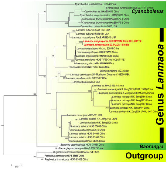
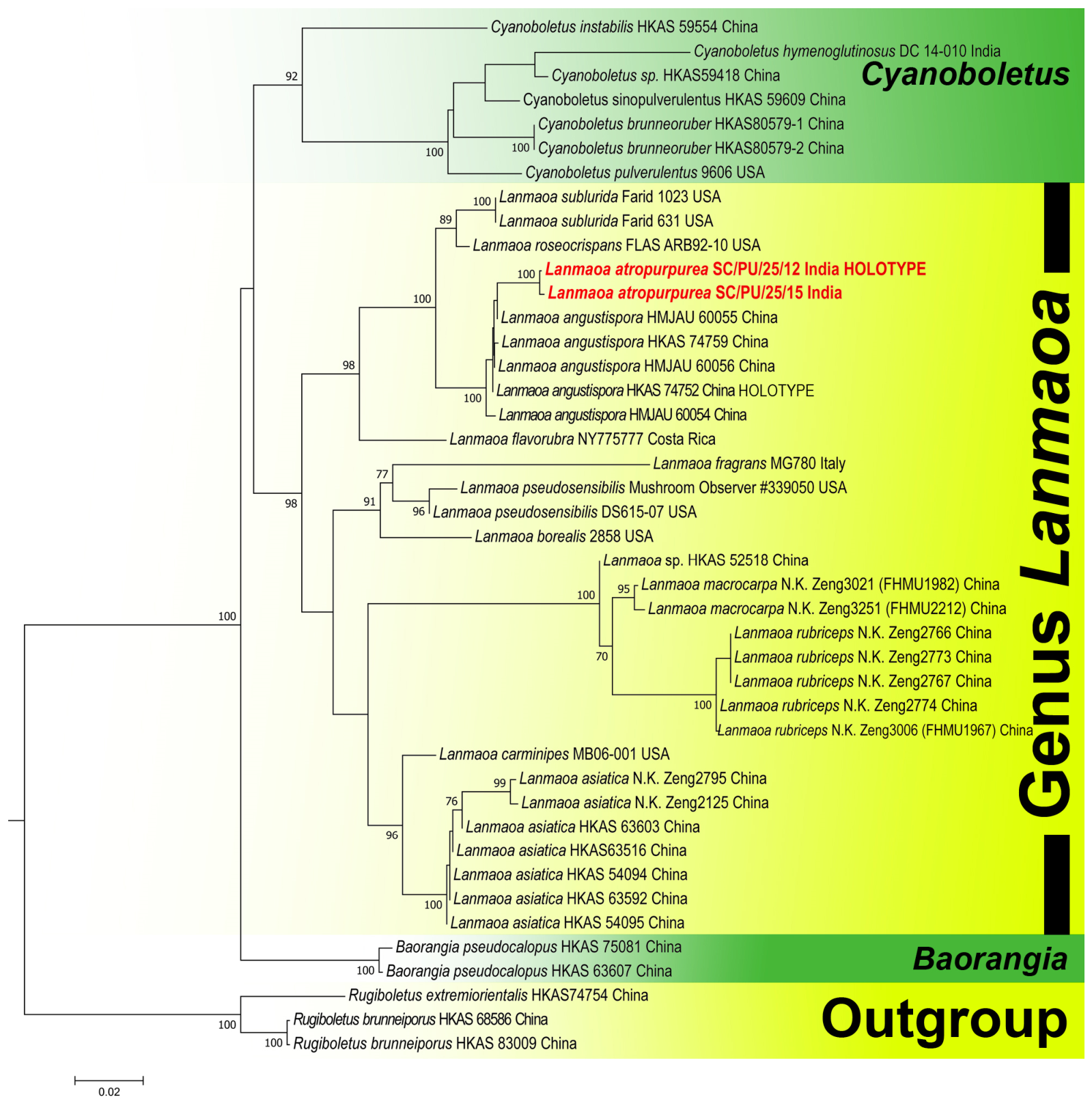
Figure 1.
Phylogram generated by maximum likelihood analysis based on 4-locus (ITS, 28S, TEF1-α and RPB2) sequence data for Lanmaoa atropurpurea and allied species. Maximum likelihood bootstrap support values (MLbs) ≥ 70% are shown above or below the branches at nodes. Lanmaoa atropurpurea is placed in bold red font to highlight its phylogenetic position in the tree.
2.3. Phylogenetic Analysis
Newly generated sequences of ITS, 28S, RPB2 and TEF1-α, and their close relatives, were retrieved from BLASTN search against GenBank (https://www.ncbi.nlm.nih.gov/genbank) (accessed on 30 August 2026) and from relevant published phylogenies [6,8,9]. Taxa representing the closest relatives outside the ingroup were chosen to ensure appropriate tree rooting. Four raw datasets (ITS, 28S, RPB2 and TEF1-α) were created separately. All the datasets were aligned separately using the online version of the multiple sequence alignment program MAFFT ver. 7 (https://mafft.cbrc.jp/alignment/software/) (accessed on 30 August 2026) with the L-INS-i [23] strategy, and normal alignment mode was used for alignment. The obtained alignment was checked and trimmed with the conserved motifs manually with MEGA ver. 7 [24]. The congruence of the datasets was tested using the incongruence length difference (ILD) test [25,26]. The analysis was performed in PAUP version 4.0b10 [27] using the partition-homogeneity test (PHT) option with 1000 heuristic search replicates. This test was used to assess the statistical congruence among datasets and to determine whether the individual data partitions could be combined for phylogenetic analysis. After testing congruence, the individual gene datasets were then concatenated into multi-locus dataset using BioEdit v. 7.0.9 [28] and processed for the phylogenetic analyses. Phylogenetic analysis was performed using the maximum likelihood (ML) method, the above-mentioned partitioned dataset with raxmlGUI 2.0 [29], and the GTRGAMMAI substitution model. This analysis was executed by applying the rapid bootstrap algorithm with 1000 replicates to obtain nodal support values. Maximum likelihood bootstrap (MLbs) values ≥ 70% were shown with the novel species being represented in the phylogenetic tree in bold red font (Figure 1). Phylogenetic trees for the individual markers ITS, 28S, RPB2, and TEF1-α are provided as Supplementary Figures S1–S4. Estimations of evolutionary divergence between sequences were conducted using the maximum composite likelihood model [30]. The analyses involved 15 nucleotide sequences of ITS, 39 nucleotide sequences of LSU (28S), 20 nucleotide sequences of RPB2 and 37 nucleotide sequences of TEF1-α. All positions containing gaps and missing data were eliminated. These evolutionary analyses were conducted in MEGA7 [24].
3. Results
3.1. Phylogenetic Inferences
A p-value ≥ 0.05 was considered to indicate no significant incongruence among the datasets. The ILD test resulted in a p-value of 0.539 indicating no significant incongruence among datasets. Therefore, the partitions were considered combinable for a combined phylogenetic analysis. The concatenated four-locus dataset (ITS + 28S + RPB2 + TEF1-α) of Lanmaoa comprised 43 voucher specimens and 3015 nucleotide positions (including gaps and missing data). The aligned sequence lengths were 773 bp for ITS, 928 bp for 28S, 687 bp for TEF1-α and 627 for RPB2. The combined dataset contained 991 distinct patterns, 557 parsimony-informative sites, 228 singleton sites and 2230 constant sites. Phylogenetic analysis (Figure 1) of Lanmaoa with Rugiboletus extremiorientalis (Lj.N. Vassiljeva) G. Wu & Zhu L. Yang and Rugiboletus brunneiporus G. Wu & Zhu L. Yang as the outgroup [8,9] strongly supports the clustering of the Indian specimens as placing them in the same lineage of the Chinese L. angustispora; however, the analysis does not consistently resolve the two species as separate clades exclusively with the help of four-locus phylogeny (ITS + 28S + RPB2 + TEF1-α). Even pairwise sequence-divergence analyses of the individual loci revealed varying levels of differentiation between the Indian species and L. angustispora. For ITS, L. atropurpurea (SC/PU/25/12 and SC/PU/25/15) were identical (d = 0.000) to L. angustispora HKAS 74752. The LSU (28S) locus also showed limited differentiation from L. angustispora HKAS 74759 and HKAS 74752 by only 0.002 substitutions per site. In contrast, RPB2 provided substantially greater differentiation, where the proposed new Indian species differed from L. angustispora HMJAU 60055 by 0.043 substitutions per site, from HKAS 74759 and HKAS 74752 by 0.054, and from HMJAU 60054 by 0.055, while, TEF1-α showed poor differentiation, with the proposed novel species distinguished from L. angustispora HMJAU 60055 by 0.012 substitutions per site, from HKAS 74759 and HKAS 74752 by 0.012, and from HMJAU 60054 and HMJAU 60056 by 0.000 and 0.012, respectively.
3.2. Taxonomy
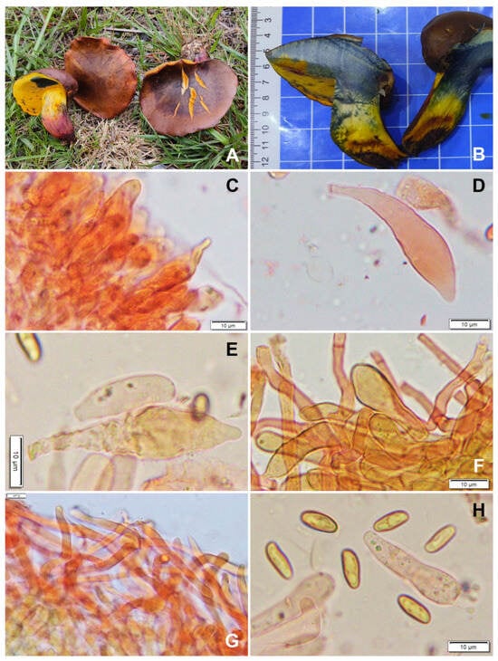
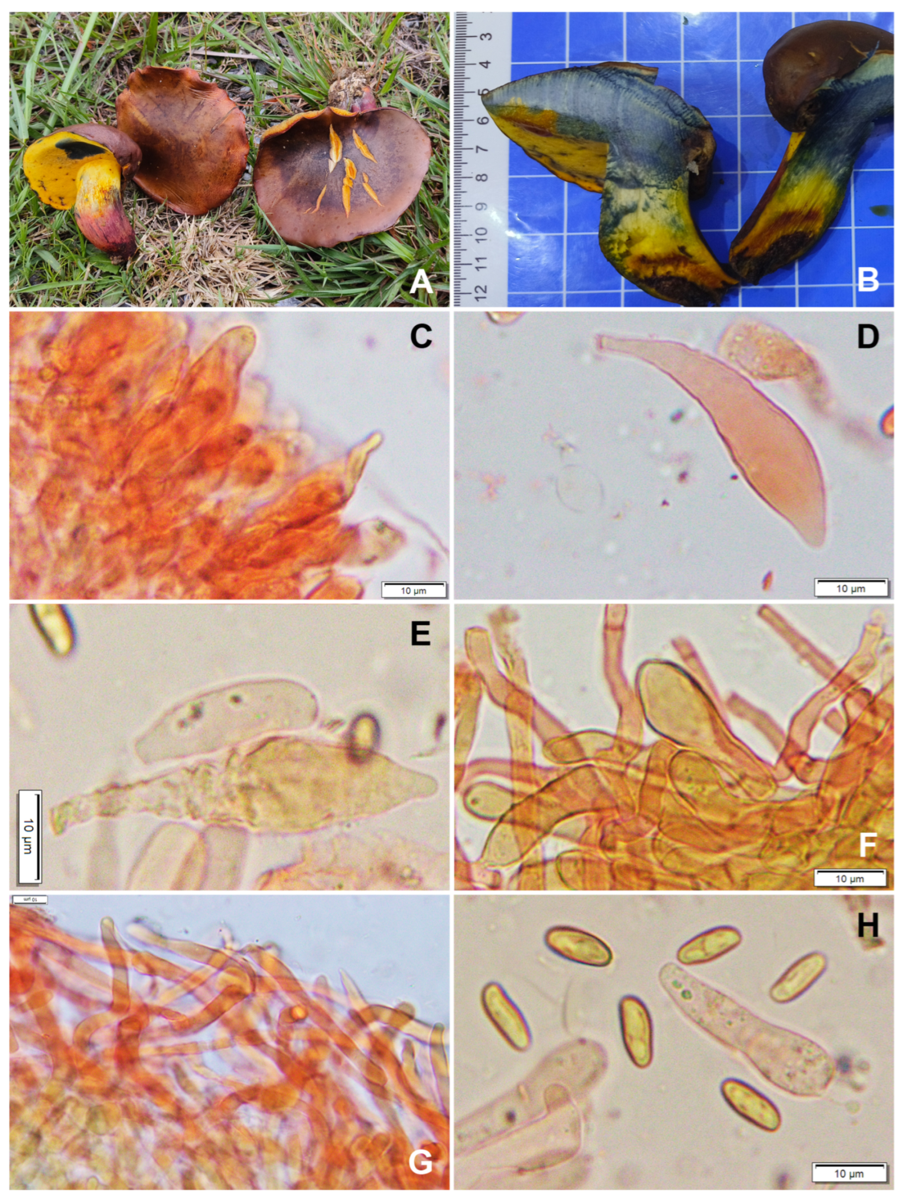
Figure 2.
Photoplate of Lanmaoa atropurpurea (CAL 2425, holotype). (A,B)—Fresh and dissected basidiomata in the field and basecamp; (C)—Cheilocystidia; (D,E)—Pleurocystidia; (F)—Clusters of caulocystidia; (G)—Transverse section through the pileipellis showing its hyphal elements; (H)—Basidiospores and basidia. Scale bars—(C–H) = 10 μm.
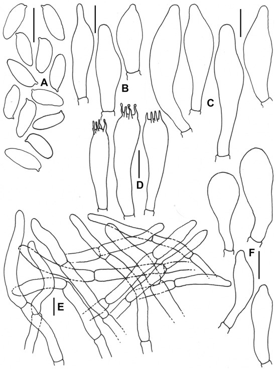
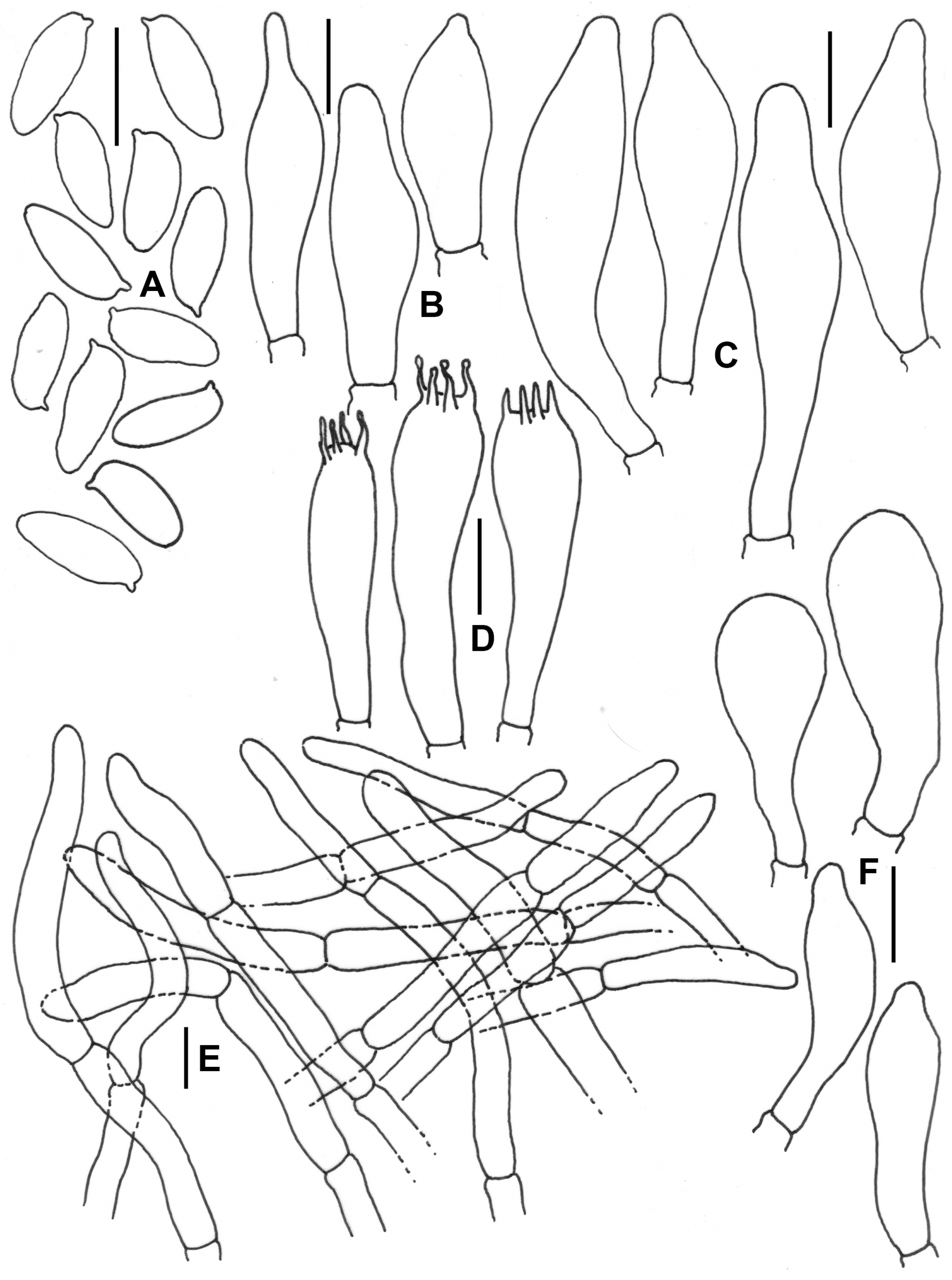
Figure 3.
Micro-morphological drawings of Lanmaoa atropurpurea (CAL 2425, holotype). (A)—Basidiospores; (B)—Cheilocystidia; (C)—Pleurocystidia; (D)—Basidia; (E)—Elements of the pileipellis; (F)—Caulocystidia. Scale bars—(A–F) = 10 μm.
MycoBank: MB 865299
GenBank: ITS: PZ747753 (holotype), PZ747754; 28S: PZ747759 (holotype), PZ747760; RPB2: PZ770682 (holotype), PZ770683; TEF1-α: QB001907 (holotype), QB001908
Type: INDIA, UTTARAKHAND Chamoli District, Devasthali, in mixed temperate forest, 2213 m, N 30°03′44.86′′ E 79°27′52.28′′, June 26, 2025, Shikha Choudhary and Priyanka Uniyal SC/PU/25/12 (holotype! CAL 2425).
Etymology: Refers to the dark purple pileus surface of mature basidiomata.
Diagnosis: Characterized by a light brown to greyish red or dark purple viscid and cracked pileus, a chalky to greyish white pileus context, an ixotrichoderm pileipellis, a reticulate stipe with reddish brown context at base. It differs from L. angustispora by a yellowish orange with a reddish tinged hymenophore, a finely reticulate stipe near apex, and occurrence in mixed forests of Quercus leucotrichophora A. Camus and Rhododendron arboreum Sm.
Description: Basidiomata medium to large-sized. Pileus 70–110 mm diam., convex when young, later becoming plano-convex with uplifted margins, light brown (7D5) to greyish red (8D4) or turning dark brown (7E5) to dark purple upon handling or with maturity; surface smooth, velvety, viscid when wet, cracked irregularly at maturity; margin entire, incurved to uplifted. Hymenophore adnate, pore surface melon yellow to apricot yellow (3A6–B6) when young, changing to yellowish orange (4B5) with reddish tinge at maturity, turning immediately to light blue (21D5) when bruised; pores angular to radially elongated, 1–2/mm; tubes up to 3–6 mm long, pale yellow (1A3), turning blue (21D4) when cut. Stipe 60–70 × 8–13 mm, subclavate to cylindrical, eccentric, concolorous to pore surface up to middle, gradually greyish red to pastel red (9B5–10B4), at base turning brown (7F5); surface striate to finely reticulate near apex, covered with irregularly scattered greyish ruby (11C6) dots, bruising light blue when handled. Context in pileus chalky to greyish white (1A2), 5–12 mm thick; context in stipe white to pale yellow (2A5), reddish brown (6E5) at base, bruising immediately bluish (21D4) on cut. Basal mycelium whitish to greyish white (1A1), unchanging when bruised. Taste not recorded, odor indistinctive. Spore print not obtained.
Basidiospores 9.0–11.00–13.3 × 3.2–4.63–5.3 μm, (Q = 1.90–2.40–3.17) elliptical to oblong, olive-greenish brown in 5% KOH, smooth. Basidia 28.6–35 × 6.4–10 μm, narrowly clavate, thin-walled, four-spored; sterigmata 2.7–4.3 μm long. Pleurocystidia (24.5) 29.2–48.8 × 8.5–11.7 μm, fusiform to subfusiform, ventricose to ventricose-rostrate, thin-walled, hyaline, sometimes yellowish pigmented in 5% KOH; emergent up to 22 μm. Tube edge fertile. Cheilocystidia 24.4–33.9 (34.7) × 6.9–9.5 (10.3) μm, ventricose to ventricose rostrate, subfusiform, thin-walled, hyaline, sometimes yellowish pigmented in 5% KOH, emergent up to 12.7 μm. Subhymenium 4–4.5 μm wide, composed of isodiametric cells. Hymenophoral trama divergent, composed of thin-walled, septate, gelatinous hyphae, 6–7.5 μm wide. Pileipellis an ixotrichoderm 240–255 μm thick, composed of erect to suberect, thin-walled, septate, rarely branched hyphae, compactly interwoven with a thin layer of gluten; terminal cells 23.9–51.4 × 4.2–8 μm, subcylindrical to subfusiform. Stipitipellis a trichoderm, up to 150 μm thick, composed of thin-walled, septate, branched, erect to suberect hyphae, brown pigmented in 5% KOH; with infrequent tufts of cystidia and basidia; caulocystidia 28–35.5 × 7.4–12.2 µm, clavate, pyriform, fusiform to subcylindric; caulobasidia 29–31 × 6.4–12 µm, narrowly clavate, 4-spored. Clamp connections absent in all tissues.
Habitat: Scattered on the ground in forests dominated by Quercus leucotrichophora and Rhododendron arboreum.
Additional materials examined: INDIA, UTTARAKHAND: Chamoli District, Devasthali, in mixed temperate forest, 2244 m, N 30°03′45.96′′ E 79°28′52.25′′, 30 June 2025, Shikha Choudhary and Priyanka Uniyal SC/PU/25/15 (CAL 2426).
4. Discussion
Lanmaoa angustispora G. Wu & Zhu L. Yang from China shows overall morphological resemblance and a close phylogenetic affinity to the proposed novel species L. atropurpurea. However, L. angustispora can be distinguished by its thicker (12–20 mm) pileus context (5–12 mm in L. atropurpurea), geranium red to greyish red hymenophore with maturity (yellowish orange with a reddish tinge in L. atropurpurea), stipe with purplish red dotted elements (finely reticulate near apex with striations in L. atropurpurea), and ecologically by its occurrence in mixed forests dominated by Castanea and Pinus [1]. Lanmaoa rubriceps N.K. Zeng & Hui Chai from China is readily distinguished by its red, crimson to orange-red pileus and a trichodermal pileipellis [8]. Similarly, L. macrocarpa N.K. Zeng, H. Chai & S. Jiang from Chian differs in possessing a dry, finely tomentose pileus, a yellow and thicker pileus context (up to 25 mm), longer tubes (up to 15 mm), a comparatively thinner pileipellis (120–160 μm thick) with a trichodermal structure, and a hymeniform stipitipellis [9]. Lanmaoa asiatica G. Wu & Zhu L. Yang from China can be distinguished by its dry, dull red to red or shell-pink pileus, thicker pileus context (10–30 mm), septate cheilocystidia, a trichodermal to subcutis pileipellis, and a hymeniform stipitipellis [1].
The combination of a light brown to greyish red or dark purple, viscid and cracked pileus, chalky white to greyish white pileus context, ixotrichodermal pileipellis, reddish-brown stipe context at the base, and a reticulate stipe, together with its distinct phylogenetic placement, supports recognition of the new taxon as a distinct species of Lanmaoa. The present study is based on two collections (CAL 2425 and CAL 2426) from the same locality, which may limit assessment of the full range of intraspecific variation. However, both collections comprised immature to mature basidiomata with consistent diagnostic macro- and micro-morphological characters, further supported by multilocus phylogenetic analysis. Additional later collections from the type locality and other localities will help characterize its morphological variability, ecological preferences, and geographical distribution.
It is worth noting that a four-locus molecular phylogeny alone does not clearly differentiate L. angustispora from the proposed new species, L. atropurpurea, as detailed in our ‘Results’ section (specifically under ‘Phylogenetic Inferences’). Instead, the robust delimitation of these species relies on combining morphological evidence with the concordance of multilocus data. This integration is crucial because pairwise sequence divergence is heavily influenced by sequence length. For instance, the ITS region included only one sequence for L. angustispora and exhibited greater variability compared to the 28S or RPB2 regions. Therefore, pairwise distances do not provide a consistent criterion for species recognition, particularly given the lack of an established species-level threshold for Lanmaoa.
The genus Lanmaoa is currently represented in Asia by five species, with four previously known only from China and the present species representing the first generic record from India. An identification key to the Asian species is provided to facilitate field identification and further species delimitation.
A provisional key to the species of Lanmaoa from Asia
- 1a
- Pileus dry, pileipellis as a trichoderm……………………………………………………………………………………………………………………………………………………..2
- 1b
- Pileus viscid when wet, pileipellis as an ixotrichoderm…………………………………………………………………………..…………………………………………………….4
- 2a
- Tubes long, up to 15 mm; pileus brownish red…………………………………………………………………………………………………………………………….L. macrocarpa
- 2b
- Tubes shorter, 3–7 mm; pileus crimson red or dull red to shell-pink, never brownish red………………………………………………………………………………………...3
- 3a
- Stipe comparatively smaller in length, 55–90 mm long, surface yellow, sometimes covered with brownish red minute squamules, reticulation absent; pore surface yellow, sometimes with reddish tinge at maturity, pores stuffed when young…………………………………………………………………………………………..L. rubriceps
- 3b
- Stipe comparatively longer, 80–110 mm long, yellow at the apex, greyish red to brownish red in the middle, greyish ruby towards the base, sometimes with reticulations at the upper part, squamules absent; pore surface yellow without red tinge, pores never stuffed………………………………………………………L. asiatica
- 4a
- Hymenophore surface geranium red to greyish red at maturity; stipe surface without any striation or reticulation, stipe context cream to pale yellow throughout the entire length…………………………………………………………………………………………………………………………………………………………………...L. angustispora
- 4b
- Hymenophore surface orangish yellow with reddish tinge at maturity; stipe surface with striation or faint reticulation, stipe context white to pale yellow, reddish brown at base…….…………………………………………………………………………………………………………………………………………………………...L. atropurpurea
Supplementary Materials
The following supporting information can be downloaded at: https://www.mdpi.com/article/10.3390/taxonomy6040064/s1, Figure S1: Maximum likelihood phylogram of the ITS sequence data of Lanmaoa atropurpurea and allied species; Figure S2: Maximum likelihood phylogram of the 28S sequence data of L. atropurpurea and allied species; Figure S3: Maximum likelihood phylogram of the RPB2 sequence data of L. atropurpurea and allied species; Figure S4: Maximum likelihood phylogram of the TEF1-α sequence data of L. atropurpurea and allied species.
Author Contributions
Conceptualization, P.U. and S.D.; methodology, S.C. and S.D.; software, S.D.; validation, K.D., S.D. and A.V.; formal analysis, S.D.; investigation, S.D. and K.D.; resources, P.U.; data curation, S.C. and S.D.; writing—original draft preparation, S.D.; writing—review and editing, K.D., P.U. and A.V.; visualization, S.C.; supervision, K.D.; project administration, K.D. and S.D.; funding acquisition, P.U. All authors have read and agreed to the published version of the manuscript.
Funding
This research was funded by DST-ANRF, India within grant number SPG/2021/003424.
Data Availability Statement
The original contribution presented in the study is included in the article, and further inquiries can be directed to the corresponding author.
Acknowledgments
PU is grateful to Principal, Govt. P.G. College, Gopeshwar for providing necessary facilities and DST-ANRF for funding the research. SC thanks the Head, Department of Botany, University of Jammu, for providing the necessary laboratory facilities.
Conflicts of Interest
The authors declare no conflicts of interest.
References
- Wu, G.; Zhao, K.; Li, Y.C.; Zeng, N.K.; Feng, B.; Halling, R.; Yang, Z.L. Four new genera of the fungal family Boletaceae. Fungal Divers. 2016, 81, 1–24. [Google Scholar] [CrossRef] [Scilit]
- Bessette, A.R. Lanmaoa roseocrispans Bessette, A.R. Bessette, Nuhn & Halling, sp. nov. Index Fungorum 2015, 259, 1. [Google Scholar]
- Phookamsak, R.; Hyde, K.D.; Jeewon, R.; Bhat, D.J.; Jones, E.B.G.; Maharachchikumbura, S.S.N.; Raspé, O.; Karunarathna, S.C.; Wanasinghe, D.N.; Hongsanan, S.; et al. Fungal diversity notes 929–1035: Taxonomic and phylogenetic contributions on genera and species of fungi. Fungal Divers. 2019, 95, 1–273. [Google Scholar] [CrossRef] [Scilit]
- Farid, A.; Bessette, A.E.; Bessette, A.R.; Bolin, J.A.; Kudzma, L.V.; Franck, A.R.; Garey, J.R. Investigations in the boletes (Boletaceae) of southeastern USA: Four novel species, and three novel combinations. Mycosphere 2021, 12, 1038–1076. [Google Scholar] [CrossRef] [Scilit]
- Nuhn, M.E.; Binder, M.; Taylor, A.F.; Halling, R.E.; Hibbett, D.S. Phylogenetic overview of the Boletineae. Fungal Biol. 2013, 117, 479–511. [Google Scholar] [CrossRef] [Scilit] [PubMed]
- Wu, G.; Feng, B.; Xu, J.; Zhu, X.T.; Li, Y.C.; Zeng, N.K.; Hosen, M.I.; Yang, Z.L. Molecular phylogenetic analyses redefine seven major clades and reveal 22 new generic clades in the fungal family Boletaceae. Fungal Divers. 2014, 69, 93–115. [Google Scholar] [CrossRef] [Scilit]
- Vizzini, A. Cyanoboletus Gelardi, Vizzini & Simonini, gen. nov. Index Fungorum 2014, 176, 1. [Google Scholar]
- Chai, H.; Liang, Z.Q.; Jiang, S.; Fu, X.L.; Zeng, N.K. Lanmaoa rubriceps, a new bolete from tropical China. Phytotaxa 2018, 347, 71–80. [Google Scholar] [CrossRef] [Scilit]
- Chai, H.; Liang, Z.-Q.; Xue, R.; Jiang, S.; Luo, S.-H.; Wang, Y.; Wu, L.-P.; Tang, L.-P.; Chen, Y.; Hong, D.; et al. New and noteworthy boletes from subtropical and tropical China. MycoKeys 2019, 46, 55–96. [Google Scholar] [CrossRef] [Scilit] [PubMed]
- Biketova, A.Y.; Garrido-Benavent, I.; Fachada, V.; Simonini, G.; Gelardi, M.; Assyov, B.; Polemis, E.; Conca, A.; Woods, R.; Zervakis, G.I.; et al. Revision of the Genus Cyanoboletus (Boletaceae) in the Mediterranean Basin with Notes on Arsenic Hyperaccumulation. J. Fungi 2026, 12, 315. [Google Scholar] [CrossRef] [Scilit] [PubMed]
- Wu, G.; Li, H.-J.; Horak, E.; Wu, K.; Li, G.-M.; Yang, Z.-L. New taxa of Boletaceae from China. Mycosphere 2023, 14, 745–776. [Google Scholar] [CrossRef] [Scilit]
- Crous, P.W.; Wingfield, M.J.; Lombard, L.; Roets, F.; Swart, W.J.; Alvarado, P.; Carnegie, A.J.; Moreno, G.; Luangsaard, J.; Thangavel, R.; et al. Fungal Planet description sheets: 951–1041. Pers. Mol. Phylogeny Evol. Fungi 2019, 43, 223–425. [Google Scholar] [CrossRef] [Scilit] [PubMed]
- Largent, D.L. How to Identify Mushrooms to Genus I-Macroscopic Features; Mad River Press: Eureka, CA, USA, 1986; 166p. [Google Scholar]
- Kornerup, A.; Wanscher, J.H. Methuen Handbook of Colour, 3rd ed.; Eyre Methuen: London, UK, 1978; 252p. [Google Scholar]
- Thiers, B. Index Herbariorum: A Global Directory of Public Herbaria and Associated Staff. New York Botanical Garden’s Virtual Herbarium. Available online: https://sweetgum.nybg.org/science/ih/ (accessed on 30 August 2026).
- White, T.J.; Bruns, T.; Lee, S.; Taylor, J.W. Amplification and direct sequencing of fungal ribosomal RNA genes for phylogenies. In PCR Protocols: A Guide to Methods and Applications; Innis, M.A., Gelfand, D.H., Sninsky, J.J., White, T.J., Eds.; Academic Press: New York, NY, USA, 1990; pp. 315–322. [Google Scholar] [CrossRef] [Scilit]
- Vilgalys, R.; Hester, M. Rapid genetic identification and mapping of enzymatically amplified ribosomal DNA from several Cryptococcus species. J. Bacteriol. 1990, 172, 4238–4246. [Google Scholar] [CrossRef] [Scilit] [PubMed]
- Matheny, P.B.; Wang, Z.; Binder, M.; Curtis, J.M.; Lim, Y.W.; Nilsson, R.H.; Hughes, K.W.; Hofstetter, V.; Ammirati, J.F.; Schoch, C.L.; et al. Contributions of rpb2 and tef1 to the phylogeny of mushrooms and allies (Basidiomycota, Fungi). Mol. Phylogenet. Evol. 2007, 43, 430–451. [Google Scholar] [CrossRef] [Scilit] [PubMed]
- Mikheyev, A.S.; Mueller, U.G.; Abbot, P. Cryptic sex and many-to-one coevolution in the fungus-growing ant symbiosis. Proc. Natl. Acad. Sci. USA 2006, 103, 10702–10706. [Google Scholar] [CrossRef] [Scilit] [PubMed]
- Zeng, N.K.; Tang, L.P.; Li, Y.C.; Tolgor, B.; Zhu, X.T.; Zhao, Q.; Yang, Z.L. The genus Phylloporus (Boletaceae, Boletales) from China: Morphological and multilocus DNA sequence analyses. Fungal Divers. 2013, 58, 73–101. [Google Scholar] [CrossRef] [Scilit]
- Zeng, N.K.; Su, M.S.; Liang, Z.Q.; Yang, Z.L. A geographical extension of the North American genus Bothia (Boletaceae, Boletales) to East Asia with a new species B. fujianensis from China. Mycol. Progr. 2015, 14, 1015. [Google Scholar] [CrossRef] [Scilit]
- Drummond, A.J.; Ashton, B.; Buxton, S.; Cheung, M.; Cooper, A.; Heled, J.; Kearse, M.; Moir, R.; Stones-Havas, S.; Sturrock, S.; et al. Geneious, Ver. 5.1; GraphPad Software, LLC: Boston, MA, USA, 2010. Available online: http://www.geneious.com (accessed on 30 August 2026).
- Katoh, K.; Rozewicki, J.; Yamada, K.D. MAFFT online service: Multiple sequence alignment, interactive sequence choice and visualization. Brief. Bioinform. 2019, 20, 1160–1166. [Google Scholar] [CrossRef] [Scilit] [PubMed]
- Kumar, S.; Stecher, G.; Tamura, K. MEGA7: Molecular evolutionary genetics analysis ver. 7.0 for bigger datasets. Mol. Biol. Evol. 2016, 33, 1870–1874. [Google Scholar] [CrossRef] [Scilit] [PubMed]
- Farris, J.S.; Källersjö, M.; Kluge, A.G.; Bult, C. Testing significance of incongruence. Cladistics 1995, 10, 315–319. [Google Scholar] [CrossRef]
- Farris, J.S.; Källersjö, M.; Kluge, A.G.; Bult, C. Constructing a significance test for incongruence. Syst. Biol. 1995, 44, 570–572. [Google Scholar] [CrossRef] [Scilit]
- Swofford, D.L. PAUP* Phylogenetic Analysis Using Parsimony (* and Other Methods), Ver. 4; Sinauer Associates: Sunderland, MA, USA, 2003. [Google Scholar]
- Hall, T.A. BioEdit: A user-friendly biological sequence alignment editor and analyses program for Windows 95/98/NT. In Nucleic Acids Symposium Series; Oxford University Press: Oxford, UK, 1999; Volume 41, pp. 95–98. [Google Scholar]
- Edler, D.; Klein, J.; Antonelli, A.; Silvestro, D. raxmlGUI 2.0: A graphical interface and toolkit for phylogenetic analyses using RAxML. Methods Ecol. Evol. 2021, 12, 373–377. [Google Scholar] [CrossRef] [Scilit]
- Tamura, K.; Nei, M.; Kumar, S. Prospects for inferring very large phylogenies by using the neighbor-joining method. Proc. Natl. Acad. Sci. USA 2004, 101, 11030–11035. [Google Scholar] [CrossRef] [Scilit] [PubMed]

Disclaimer/Publisher’s Note: The statements, opinions and data contained in all publications are solely those of the individual author(s) and contributor(s) and not of MDPI and/or the editor(s). MDPI and/or the editor(s) disclaim responsibility for any injury to people or property resulting from any ideas, methods, instructions or products referred to in the content. |
© 2026 by the authors. Licensee MDPI, Basel, Switzerland. This article is an open access article distributed under the terms and conditions of the Creative Commons Attribution (CC BY) license.