


Abstract
Our communication summarizes the recent changes in the paraphyletic species Orthoflavivirus encephalitidis. The paraphyly arises due to louping-ill virus (LIV), which is genetically closer to the European subtype of tick-borne encephalitis virus (TBEV). To maintain a concept of monophyly, we have split Orthoflavivirus encephalitidis into two monophyletic species: Orthoflavivirus zilberi (after Lev A. Zilber—the head of the expedition that identified and characterized TBEV in the Soviet Far East in 1937) and Orthoflavivirus neudoerflense, named after Neudörfl, a town in Austria where the reference strain Neudoerfl was first isolated. Also, to remove the resulting ambiguity in the virus name “tick-borne encephalitis virus”, we have proposed new names—Asian TBEV (TBEV-AS) for Orthoflavivirus zilberi, and European TBEV (TBEV-EU) for Orthoflavivirus neudoerflense.
1. Introduction
Tick-borne encephalitis virus (TBEV) (Flaviviridae: Orthoflavivirus) was discovered in 1937, making it a virus with a nearly 90-year research history [1]. The virus genome comprises a positive-sense single-strand RNA (ssRNA+) with a length of about 11,000 nucleotides. The genome contains one open reading frame (ORF) coding three structural and seven non-structural proteins which is flanked by untranslated (non-coding) regions (UTRs). The ORF is co- and post-translationally cleaved by viral and host proteases [2].
TBEV is one of the twelve tick-borne flaviviruses (TBFVs), and along with four closely related human pathogenic TBFVs, forms the so-called “Tick-borne encephalitis serocomplex” [3]. The virus circulates between its principal vectors, hard ticks (Ixodidae: mainly of the genus Ixodes), and small mammals (reservoir hosts) [4]. TBEV is a causative agent of tick-borne encephalitis with approximately 5000–10,000 human cases registered annually [5].
Intraspecific genetic diversity of TBEV is represented by five subtypes whose names reflect the areas where the viruses are found (listed chronologically by the date of the first complete genome sequence):
- European (TBEV-EU)—Mandl et al. (1989) [6];
- Far-Eastern (TBEV-FE)—Pletnev et al. (1990) [7];
- Siberian (TBEV-Sib)—Gritsun et al. (1997) [8];
- Baikalian (TBEV-Bkl)—Demina et al. (2010) [9];
- Himalayan (TBEV-Him)—Dai et al. (2018) [10].
Prior to the discovery of tick-borne encephalitis virus, louping-ill virus (LIV) was identified in 1931 [11]. LIV is the closest relative of TBEV, particularly of TBEV-EU (Figure 1), which has a similar genome composition in terms of length and the number of structural (3) and non-structural (7) proteins. Despite being RNA viruses, TBEV and LIV have relatively slow evolutionary rate with the same order of magnitude (10−5 nucleotide substitutions per site per year) [12,13]. The complete genome of LIV was sequenced and submitted to GenBank in 1997 [8]. Since then, phylogenies encompassing both TBEV and LIV clearly demonstrate their paraphyletic relationships (Figure 1) [13,14].
In April 2023, the taxonomic proposal to rename the genus Flavivirus to Orthoflavivirus was approved by the ICTV [15]. In addition, to adhere to a Latinized binomial format, the species names were also changed: Tick-borne encephalitis virus was renamed to Orthoflavivirus encephalitidis, and Louping-ill virus was modified to Orthoflavivirus loupingi. However, despite all changes, the phylogenetic relationships and, as a consequence, the paraphyly of TBEV and LIV, remained intact. Meanwhile, according to the ICTV Code, a species is “a monophyletic group of viruses whose properties can be distinguished from the properties of other species by multiple criteria” [16].
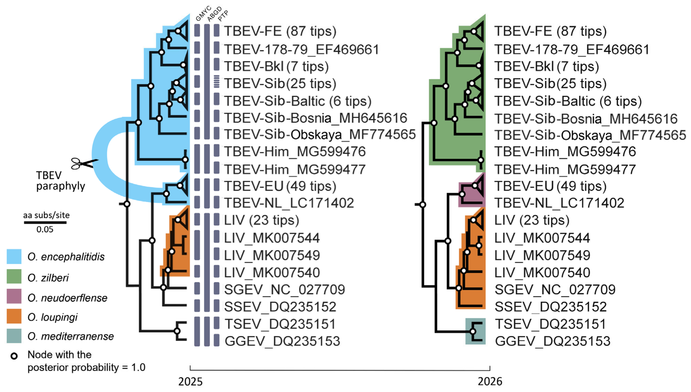
There were two main ways to resolve the paraphyly of TBEV and LIV and make them monophyletic: (1) to merge two viruses into a single species, or (2) to delimit the European subtype of TBEV as a separate species [17] (Figure 1).
Figure 1.
Schematic representation of tick-borne encephalitis virus (TBEV) taxonomy alteration that occurred in 2026. The phylogeny was reconstructed in BEAST v1.10.4 [18] using 211 amino acid (aa) sequences of a viral polyprotein with a length of 3414 aa. The nodes marked with white circles have a posterior probability of 1.0. The abbreviations used in tip names: TBEV-FE—Far-Eastern TBEV subtype; -178-79—178-79 lineage; -Bkl—Baikalian subtype; -Sib—Siberian subtype and its lineages: Baltic, Bosnia, Obskaya; -Him—Himalayan subtype; -EU—European subtype; -NL—the Netherlands strain; LIV—louping-ill virus; SGEV—Spanish goat encephalitis virus; SSEV—Spanish sheep encephalitis virus; TSEV—Turkish sheep encephalitis virus; GGEV—Greek goat encephalitis virus. The dark gray vertical bars in the left tree display delimited species entities according to delimitation analysis performed by three methods: Generalized Mixed Yule Coalescent (GMYC) method with 16 species, Automatic Barcode Gap Discovery (ABGD) with 5 species, and Poisson tree processes (PTP) model with 24 species.
The first approach implementing the merge of TBEV and LIV was proposed several times [19,20,21]. However, the supporting analyses either relied on a very limited number of sequences, or contradicted the species demarcation criteria for the genus Orthoflavivirus (https://ictv.global/report/chapter/flaviviridae/flaviviridae/orthoflavivirus (accessed on 15 August 2026)). The criteria include nucleotide and amino acid sequence data (i), antigenic characteristics (ii), geographic association (iii), vector association (iv), host association (v), disease association (vi) and ecological characteristics (vii).
In our taxonomic proposal, we followed the second approach of splitting TBEV-EU from the common TBEV clade and took into account all of the criteria listed above. In March 2026, the ICTV ratified our taxonomic proposal (https://ictv.global/ictv/proposals/2025.007S.Orthoflavivirus_2nsp_1spren.zip (accessed on 15 August 2026)) [22]. In the current communication, we aim to describe the reason for our proposal and outline its main taxonomic and virologic features.
2. Bioinformatic Analysis
In our species delimitation analysis, we scrutinized 211 complete amino acid sequences of a viral polyprotein to delineate species entities within the TBEV + LIV group. We applied three methods: Generalized Mixed Yule Coalescent (GMYC) method [23], Automatic Barcode Gap Discovery (ABGD) [24], and Poisson tree processes (PTP) model [25]. Briefly, GMYC detects the point on an ultrametric tree where branching shifts from a Yule (speciation) process to a coalescent (intraspecific) process. ABGD groups sequences by finding a significant gap between intra- and interspecific pairwise distances, without requiring a tree. PTP models speciation and coalescence as two Poisson processes on a tree and places species boundaries at the shift between them. Although the methods were not in concordance on the number of detected species (Figure 1), all of them rejected the null hypothesis that all TBEV subtypes and LIV form a single species. Furthermore, all three methods delineated TBEV-EU as a separate species entity (Figure 1).
The ABGD method was the most conservative and agreed almost completely with our taxonomic proposal for dividing the TBEV + LIV clade into four species (Figure 1). The only point of disagreement was that ABGD separated TBEV-Him as a distinct species. This may well be correct, but more data are needed to assess this against all the criteria established for the genus Orthoflavivirus.
3. Ecology, Antigenicity, and Disease Association
For genetically closely related species, the demarcation criteria of the genus Orthoflavivirus—such as host and vector associations, antigenic properties, and pathogenicity—can be particularly useful (Table 1).
Table 1.
Comparative characteristics of tick-borne encephalitis virus and louping-ill virus.
For example, LIV is primarily found in red grouse and sheep, where it induces encephalitis with a high mortality rate (78% in red grouse [26], 5–60% in sheep [27]), whereas TBEV mainly persists asymptomatically in small rodents, which act as its primary reservoir. Although rodents such as field voles (Microtus agrestis), bank voles (M. glareolus) and wood mice (Apodemus sylvaticus) raised an antibody response to LIV infection, they could not produce substantial viremia and did not support non-viraemic transmission between co-feeding ticks [28].
The existence of Eastern and Western geographic and antigenic variants of TBEV has been recognized for four decades [29]. Initial characterization of the virus’s antigenic properties identified two distinct subtypes—Eastern and Western [30]—which were also designated as ‘Persulcatus’ and ‘Ricinus’ variants based on their ecological characteristics [31].
Hubálek et al. (1995) [32] employed an indirect immunofluorescence assay (IIFT) with a panel of 17 monoclonal antibodies (MoAbs) to determine the reactivity patterns of flavivirus isolates. To compare these reactivity patterns, the authors used the unweighted pair group method with arithmetic mean (UPGMA), a hierarchical clustering method that groups objects by average linkage, which placed TBEV-EU, LIV, Turkish sheep encephalitis virus (TSEV), and Spanish sheep encephalitis virus (SSEV) in a single clade, with similarity values ranging from 84.0% (TBEV-EU vs. TSEV) to 96.6% (TBEV-EU vs. SSEV). In contrast, TBEV-FE (strain Sofjin) formed an outgroup with only 73% similarity, reflecting a distinct MoAbs reactivity profile. We reproduced their UPGMA dendrogram from the original data using R; the resulting tree (Figure S1), the data, and the R script is provided as the Supplementary Materials.
TBEV subtypes also have different clinical features. TBEV-EU has a clear biphasic course in about 74% of cases [33] (Gritsun et al. (2003) [34] reported a rate of 20–30%), whereas TBEV-FE and -Sib infections are mostly monophasic, with only a small fraction of cases demonstrating a biphasic pattern [35]. TBEV-FE’s fatality rate reaches 20–60% [36] and the disease in children is more severe than in adults. Conversely, TBEV-EU infection demonstrates a low case fatality rate of 1–2% and children tend to develop less severe disease than adults [34]. For LIV, the only one fatal case was documented [37].
Votiakov et al. (2002) [38] documented prominent clinical observations in laboratory-acquired TBE cases that occurred in Belarus (n = 9), Slovakia (n = 11), and the Russian Far East (n = 10). All five lethal cases were registered in the Far East, whereas in Belarus and Slovakia, the disease mostly manifested in a meningeal form. These outcomes, however, may reflect not only the virus subtype but also age, vaccination status, preexisting illnesses, infectious dose, route of exposure, and the timing and quality of medical care. Experiments on monkeys and sheep showed that TBEV-EU did not initially replicate in neuronal cells or damage them, even after intracerebral infection occurred [39]. Instead, the primary target of TBEV-EU was lymphoid tissue, and the virus later appeared in the brains of animals that developed encephalitis, predominantly in the cerebellum, 6–9 days after inoculation. In contrast, TBEV-FE directly infected and damaged brain neurons, causing severe encephalitis. These findings strongly indicate that TBEV-FE is more neurotropic than TBEV-EU (see also Gritsun, et al. (2003) [34]).
Votiakov, et al. (2002) [38] inoculated sheep subcutaneously or intracerebrally with TBEV-EU and TBEV-FE. Subcutaneous inoculation with TBEV-EU caused meningitis alone, while intracerebral TBEV-EU produced a biphasic illness with 12.5% mortality, lower CNS viral titres than in blood, and moderate glial pathology (glial nodules 14.6% FOV) with secondary neuronal damage. In contrast, intracerebral inoculation with TBEV-FE, by contrast, led to a monophasic, 100% fatal disease, with viral loads in the CNS exceeding those in blood and extensive primary degenerative changes (neuronophagia 37.5% FOV, glial nodules 56.1% FOV). These findings demonstrate that the two TBEV subtypes clearly differ in neurotropism, at least in the sheep model.
4. New Virus Names Proposed
The previous name “TBEV” is now paraphyletic as it refers to two distinct species. For now, this conflict is resolved by assigning temporary virus names: TBEV1 for Orthoflavivirus zilberi, and TBEV2 for Orthoflavivirus neudoerflense. In turn, we propose the following geographically derived names: Asian TBEV (TBEV-AS) for Orthoflavivirus zilberi and European TBEV (TBEV-EU) for Orthoflavivirus neudoerflense (Table 2). It should be noted that TBEV-EU is no longer considered a subtype but rather a distinct virus that represents the species Orthoflavivirus neudoerflense; the term now denotes this separate viral species, not a lineage.
Table 2.
Taxonomic reassignment of TBEV and LIV.
5. Conclusions
We have outlined the major points of our taxonomic proposal on splitting the paraphyletic species Orthoflavivirus encephalitidis into two monophyletic species. Further details are available in the proposal (2025.007S.Orthoflavivirus_2nsp_1spren) on the ICTV website (https://ictv.global/ictv/proposals/2025.007S.Orthoflavivirus_2nsp_1spren.zip (accessed on 15 August 2026)).
Supplementary Materials
The following supporting information can be downloaded at: https://www.mdpi.com/article/10.3390/taxonomy6030052/s1, Figure S1: UPGMA tree reconstructed using the similarity matrix (pairwise dice coefficients) derived from comparison of monoclonal antibody reactivity patterns of flaviviruses.
Author Contributions
Conceptualization, A.B. and Y.B.; writing—original draft preparation, A.B.; writing—review and editing, A.B. and Y.B.; supervision, Y.B. All authors have read and agreed to the published version of the manuscript.
Funding
This research received no external funding.
Data Availability Statement
R script for the UPGMA tree generating as well as a calibrated nexus tree are available from https://doi.org/10.6084/m9.figshare.33164036.
Acknowledgments
The authors sincerely thank the anonymous reviewers for their useful comments and suggestions. The authors would like to thank Andrey Lyamin for his invaluable assistance with the payment processing for this publication.
Conflicts of Interest
The authors declare no conflicts of interest.
References
- Zlobin, V.I.; Pogodina, V.V.; Kahl, O. A brief history of the discovery of tick-borne encephalitis virus in the late 1930s (based on reminiscences of members of the expeditions, their colleagues, and relatives). Ticks Tick-Borne Dis. 2017, 8, 813–820. [Google Scholar] [CrossRef] [Scilit] [PubMed]
- Růžek, D.; Yoshii, K.; Bloom, M.E.; Gould, E.A. Virology. In The TBE Book, 5th ed.; Dobler, G., Erber, W., Bröker, M., Chitimia-Dobler, L., Schmit, H.-J., Eds.; Global Health Press: Singapore, 2025. [Google Scholar]
- Shi, J.; Hu, Z.; Deng, F.; Shen, S. Tick-Borne Viruses. Virol. Sin. 2018, 33, 21–43. [Google Scholar] [CrossRef] [Scilit] [PubMed]
- Dobler, G.; Erber, W.; Bröker, M.; Schmitt, H.J. The TBE Book, 5th ed.; Global Health Press: Singapore, 2022; p. 388. [Google Scholar]
- Hills, S.L.; Gould, C.V. Travel-Related Infections and Diseases. In CDC Yellow Book 2026: Health Information for International Travel; Oxford University Press: Oxford, UK, 2026. [Google Scholar]
- Mandl, C.W.; Heinz, F.X.; Stockl, E.; Kunz, C. Genome sequence of tick-borne encephalitis virus (Western subtype) and comparative analysis of nonstructural proteins with other flaviviruses. Virology 1989, 173, 291–301. [Google Scholar] [CrossRef] [Scilit] [PubMed]
- Pletnev, A.G.; Yamshchikov, V.F.; Blinov, V.M. Nucleotide sequence of the genome and complete amino acid sequence of the polyprotein of tick-borne encephalitis virus. Virology 1990, 174, 250–263. [Google Scholar] [CrossRef] [Scilit] [PubMed]
- Gritsun, T.S.; Venugopal, K.; Zanotto, P.M.; Mikhailov, M.V.; Sall, A.A.; Holmes, E.C.; Polkinghorne, I.; Frolova, T.V.; Pogodina, V.V.; Lashkevich, V.A.; et al. Complete sequence of two tick-borne flaviviruses isolated from Siberia and the UK: Analysis and significance of the 5′ and 3′-UTRs. Virus Res. 1997, 49, 27–39. [Google Scholar] [CrossRef] [Scilit] [PubMed]
- Demina, T.V.; Dzhioev, Y.P.; Verkhozina, M.M.; Kozlova, I.V.; Tkachev, S.E.; Plyusnin, A.; Doroshchenko, E.K.; Lisak, O.V.; Zlobin, V.I. Genotyping and characterization of the geographical distribution of tick-borne encephalitis virus variants with a set of molecular probes. J. Med. Virol. 2010, 82, 965–976. [Google Scholar] [CrossRef] [Scilit] [PubMed]
- Dai, X.; Shang, G.; Lu, S.; Yang, J.; Xu, J. A new subtype of eastern tick-borne encephalitis virus discovered in Qinghai-Tibet Plateau, China. Emerg. Microbes Infect. 2018, 7, 74. [Google Scholar] [CrossRef] [Scilit] [PubMed]
- Greig, J.R.; Brownlee, A.; Wilson, D.R.; Gordon, W.S. The nature of louping ill. Vet. Rec. 1931, 11, 325–333. [Google Scholar]
- Clark, J.J.; Gilray, J.; Orton, R.J.; Baird, M.; Wilkie, G.; Filipe, A.D.S.; Johnson, N.; McInnes, C.J.; Kohl, A.; Biek, R. Population genomics of louping ill virus provide new insights into the evolution of tick-borne flaviviruses. PLoS Negl. Trop. Dis. 2020, 14, e0008133. [Google Scholar] [CrossRef] [Scilit] [PubMed]
- Bondaryuk, A.N.; Kulakova, N.V.; Belykh, O.I.; Bukin, Y.S. Dates and Rates of Tick-Borne Encephalitis Virus-The Slowest Changing Tick-Borne Flavivirus. Int. J. Mol. Sci. 2023, 24, 2921. [Google Scholar] [CrossRef] [Scilit] [PubMed]
- Uzcátegui, N.Y.; Sironen, T.; Golovljova, I.; Jääskeläinen, A.E.; Välimaa, H.; Lundkvist, Å.; Plyusnin, A.; Vaheri, A.; Vapalahti, O. Rate of evolution and molecular epidemiology of tick-borne encephalitis virus in Europe, including two isolations from the same focus 44 years apart. J. Gen. Virol. 2012, 93, 786–796. [Google Scholar] [CrossRef] [Scilit] [PubMed]
- Postler, T.S.; Beer, M.; Blitvich, B.J.; Bukh, J.; de Lamballerie, X.; Drexler, J.F.; Imrie, A.; Kapoor, A.; Karganova, G.G.; Lemey, P.; et al. Renaming of the genus Flavivirus to Orthoflavivirus and extension of binomial species names within the family Flaviviridae. Arch. Virol. 2023, 168, 224. [Google Scholar] [CrossRef] [Scilit] [PubMed]
- Walker, P.J.; Siddell, S.G.; Lefkowitz, E.J.; Mushegian, A.R.; Adriaenssens, E.M.; Alfenas-Zerbini, P.; Davison, A.J.; Dempsey, D.M.; Dutilh, B.E.; Garcia, M.L.; et al. Changes to virus taxonomy and to the International Code of Virus Classification and Nomenclature ratified by the International Committee on Taxonomy of Viruses (2021). Arch. Virol. 2021, 166, 2633–2648. [Google Scholar] [CrossRef] [Scilit] [PubMed]
- Bondaryuk, A.N.; Andaev, E.I.; Dzhioev, Y.P.; Zlobin, V.I.; Tkachev, S.E.; Kozlova, I.V.; Bukin, Y.S. Delimitation of the tick-borne flaviviruses. Resolving the tick-borne encephalitis virus and louping-ill virus paraphyletic taxa. Mol. Phylogenet. Evol. 2022, 169, 107411. [Google Scholar] [CrossRef] [Scilit] [PubMed]
- Suchard, M.A.; Lemey, P.; Baele, G.; Ayres, D.L.; Drummond, A.J.; Rambaut, A. Bayesian phylogenetic and phylodynamic data integration using BEAST 1.10. Virus Evol. 2018, 4, vey016. [Google Scholar] [CrossRef] [Scilit] [PubMed]
- Deviatkin, A.A.; Aleshina, Y.A.; Karganova, G.G.; Lukashev, A.N. Selection Pressure Profile Suggests Species Criteria among Tick-Borne Orthoflaviviruses. Viruses 2024, 16, 1554. [Google Scholar] [CrossRef] [Scilit] [PubMed]
- Grard, G.; Moureau, G.; Charrel, R.N.; Lemasson, J.J.; Gonzalez, J.P.; Gallian, P.; Gritsun, T.S.; Holmes, E.C.; Gould, E.A.; de Lamballerie, X. Genetic characterization of tick-borne flaviviruses: New insights into evolution, pathogenetic determinants and taxonomy. Virology 2007, 361, 80–92. [Google Scholar] [CrossRef] [Scilit] [PubMed]
- Charrel, R.N.; Zaki, A.M.; Attoui, H.; Fakeeh, M.; Billoir, F.; Yousef, A.I.; de Chesse, R.; De Micco, P.; Gould, E.A.; de Lamballerie, X. Complete coding sequence of the Alkhurma virus, a tick-borne flavivirus causing severe hemorrhagic fever in humans in Saudi Arabia. Biochem. Biophys. Res. Commun. 2001, 287, 455–461. [Google Scholar] [CrossRef] [Scilit] [PubMed]
- ICTV. Approved Proposals. Available online: https://ictv.global/files/proposals/approved (accessed on 10 April 2026).
- Fujisawa, T.; Barraclough, T.G. Delimiting species using single-locus data and the Generalized Mixed Yule Coalescent approach: A revised method and evaluation on simulated data sets. Syst. Biol. 2013, 62, 707–724. [Google Scholar] [CrossRef] [Scilit] [PubMed]
- Puillandre, N.; Lambert, A.; Brouillet, S.; Achaz, G. ABGD, Automatic Barcode Gap Discovery for primary species delimitation. Mol. Ecol. 2012, 21, 1864–1877. [Google Scholar] [CrossRef] [Scilit] [PubMed]
- Zhang, J.; Kapli, P.; Pavlidis, P.; Stamatakis, A. A general species delimitation method with applications to phylogenetic placements. Bioinformatics 2013, 29, 2869–2876. [Google Scholar] [CrossRef] [Scilit] [PubMed]
- Gilbert, L. Louping ill virus in the UK: A review of the hosts, transmission and ecological consequences of control. Exp. Appl. Acarol. 2016, 68, 363–374. [Google Scholar] [CrossRef] [Scilit] [PubMed]
- Jeffries, C.L.; Mansfield, K.L.; Phipps, L.P.; Wakeley, P.R.; Mearns, R.; Schock, A.; Bell, S.; Breed, A.C.; Fooks, A.R.; Johnson, N. Louping ill virus: An endemic tick-borne disease of Great Britain. J. Gen. Virol. 2014, 95, 1005–1014. [Google Scholar] [CrossRef] [Scilit] [PubMed]
- Gilbert, L.; Jones, L.D.; Hudson, P.J.; Gould, E.A.; Reid, H.W. Role of small mammals in the persistence of Louping-ill virus: Field survey and tick co-feeding studies. Med. Vet. Entomol. 2000, 14, 277–282. [Google Scholar] [CrossRef] [Scilit] [PubMed]
- Kahl, O.; Pogodina, V.V.; Poponnikova, T.; Süss, J.; Zlobin, V.I. A short history of TBE. In The TBE Book, 5th ed.; Dobler, G., Erber, W., Bröker, M., Schmitt, H.J., Eds.; Global Health Press: Singapore, 2022. [Google Scholar]
- Clarke, D.H. Further studies on antigenic relationships among the viruses of the Group B tick-borne complex. Bull. World Health Organ. 1964, 31, 45–56. [Google Scholar] [PubMed]
- Chumakov, M.P.; Rubin, S.G.; Linev, M.B. Three antigen types of tick-borne encephalitis virus, their dependence on arthropod vectors and geography. Probl. Med. Virol. 1975, 371–375. [Google Scholar]
- Hubálek, Z.; Pow, I.; Reid, H.W.; Hussain, M.H. Antigenic similarity of central European encephalitis and louping-ill viruses. Acta Virol. 1995, 39, 251–256. [Google Scholar] [PubMed]
- Kaiser, R. The clinical and epidemiological profile of tick-borne encephalitis in southern Germany 1994-98: A prospective study of 656 patients. Brain 1999, 122, 2067–2078. [Google Scholar] [CrossRef] [Scilit] [PubMed]
- Gritsun, T.S.; Lashkevich, V.A.; Gould, E.A. Tick-borne encephalitis. Antivir. Res. 2003, 57, 129–146. [Google Scholar] [CrossRef] [Scilit] [PubMed]
- Mansfield, K.L.; Johnson, N.; Phipps, L.P.; Stephenson, J.R.; Fooks, A.R.; Solomon, T. Tick-borne encephalitis virus—A review of an emerging zoonosis. J. Gen. Virol. 2009, 90, 1781–1794. [Google Scholar] [CrossRef] [Scilit] [PubMed]
- Pogodina, V.; Frolova, M.P.; Erman, B.A. Chronic Tick-Borne Encephalitis; Nauka: Novosibirsk, Russia, 1986. [Google Scholar]
- Williams, H.; Thorburn, H. Serum antibodies to louping-ill virus. Scott. Med. J. 1962, 7, 353–355. [Google Scholar] [CrossRef] [Scilit] [PubMed]
- Votiakov, V.I.; Zlobin, V.I.; Mishaeva, N.P. Tick-Borne Encephalitis of Eurasia (Ecology, Molecular Epidemiology, Nosology, Evolution); Nauka: Novosibirsk, Russia, 2002; p. 438. [Google Scholar]
- Votiakov, V.I.; Protas, I.I.; Zhdanov, V.M. Western Tick-Borne Encephalitis; Belarus: Minsk, Belarus, 1978; p. 256. [Google Scholar]

Disclaimer/Publisher’s Note: The statements, opinions and data contained in all publications are solely those of the individual author(s) and contributor(s) and not of MDPI and/or the editor(s). MDPI and/or the editor(s) disclaim responsibility for any injury to people or property resulting from any ideas, methods, instructions or products referred to in the content. |
© 2026 by the authors. Licensee MDPI, Basel, Switzerland. This article is an open access article distributed under the terms and conditions of the Creative Commons Attribution (CC BY) license.