


Abstract
Tin oxide thin films were deposited by the pulsed laser ablation of a metallic Sn target at different oxygen partial pressures, ranging from 10 to 40 mTorr. Langmuir plasma probe diagnostics were performed to evaluate the effect of pressure on mean kinetic energy and density of Sn ions. It was observed that the mean kinetic energy decreased from 34 to 11 eV while the ion density decreased from 10 to 1.5 × 1013 cm−3 with increasing pressure. The films exhibited enhanced optical transmittance, increasing from 10% for the sample grown at 10 mTorr to 70% for the film deposited at 40 mTorr. Furthermore, higher deposition pressures led to wider band gap values, increasing from 1.6 to 3.9 eV for direct transitions and from 2.2 to 3.2 eV for indirect transitions with increasing oxygen pressure. These trends are consistent with progressive oxidation and partial transparency characteristic of semiconducting tin oxides. Structural characterization, based on X-ray diffraction, revealed predominantly metallic Sn diffraction peaks across the entire oxygen pressure range. However, despite this structural signature, the films exhibited optical and electronic properties characteristic of tin oxides. This apparent discrepancy suggests the coexistence of metallic nanoparticles embedded within an amorphous or nanocrystalline SnO2/SnOx matrix. These findings provide insights into the non-equilibrium oxidation dynamics of tin and the formation of metastable SnOx phases during pulsed laser deposition.
1. Introduction
Tin oxides are multifunctional semiconductors extensively studied for their applications in gas sensing [1], transparent conductive electrodes [2], solar cells [3], and energy-efficient coatings [4]. Among the different oxidation states, SnO2 is the most stable and commonly reported phase, exhibiting n-type conductivity, a wide band gap (3.6–4.0 eV), and high optical transmittance [5,6]. Moreover, intermediate sub-oxides (SnO, Sn2O3, Sn3O4) are often observed under oxygen-deficient or metastable growth conditions and can strongly influence the optoelectronic response of the material. Furthermore, sub-stoichiometric oxides (SnOx) are characterized for having intermediate properties between oxides and metal, which make them ideal for defect-mediated applications [7].
Pulsed laser deposition (PLD) offers a versatile route to tune stoichiometry by adjusting the reactive gas pressure, enabling control over the oxidation degree of the ablated species. However, when ablating Sn under oxygen atmospheres, the balance between oxidation kinetics and plasma ions energy and density can produce heterogeneous structures where metallic tin coexists with amorphous or nanocrystalline stoichiometric and non-stoichiometric oxides. Understanding this competition is essential for tailoring SnOx films for optoelectronic or catalytic applications.
Oxidation may occur partially in flight, but mostly it occurs on the substrate surface, often freezing in metastable mixtures of metallic Sn0 and oxidized Sn2+/Sn4+ rather than a single equilibrium phase [8]. Mapping how oxygen partial pressure controls these mixed states is essential for applications that rely on simultaneous conductivity and transparency.
In this work, we present a study of the influence of the oxygen pressure on the deposition of thin films from the ablation of metallic tin. The main novelty relies on the plasma parameters determination as a function of pressure and their effect on the optical and structural properties of the deposited films, yielding a reliable method for controlling experimental parameters on the synthesis of metallic-oxide mixed phases.
2. Materials and Methods
Thin films were deposited by PLD from the ablation of a high-purity (99.99%) metallic Sn target. The vacuum chamber was evacuated to a base pressure of 2 × 10⁻⁶ Torr, and controlled Ar/O2 (80/20) was introduced to reach working pressures of 10, 20, 30, and 40 mTorr. A Q-switched Nd:YAG laser (Q-smart 850 from Quantel, Lannion, France) emitting at 1064 nm, with 6 ns pulse duration and 10 Hz repetition rate, was focused on the rotating target (15 rpm). The target–substrate distance was fixed at 4 cm. Depositions were carried out during 20 min on fused quartz substrates at room temperature. In all cases, the laser fluence was kept constant at 1.4 J/cm2.
2.1. Plasma Diagnostics
Plasma parameters were obtained in situ by a planar Langmuir probe mounted at the substrate position (the probe temporarily replaced the substrate). The probe, 6.7 mm in diameter, was biased at −45 V to collect the ion saturation current. The current was estimated from the voltage drop across a 22 Ω resistor recorded on a 500 MHz Tektronix digital oscilloscope (Beaverton, OR, USA), allowing time-of-flight (TOF) extraction for ion kinetic energy and plasma density calculations using the methodologies reported by Bulgakova [9] and Dogget [10], respectively.
2.2. Thin Films Characterization
Thin films thicknesses were determined by means of profilometry using a Veeco Dektak 150 system from Plainview (New York, NY, USA). UV-Vis measurements were used to estimate absorption, optical transparency, and band gap (Tauc method). The equipment was a Thermo Scientific Genesys 10 UV-Vis spectrophotometer (Madison, WI, USA). X-ray Photoelectron Spectroscopy (XPS) was performed on the Sn 3d and O 1s core levels to determine oxidation states and surface chemistry, and they were compared with reported binding energies for Sn0, Sn2+, and Sn4+ in tin oxides. The system was a SPECS spectrometer (Berlin, Germany) equipped with a PHOIBOS 150 analyzer and an Al-kα X-ray source (1486.7 eV). Finally, X-ray diffraction (XRD) measurements were performed with Cu Kα radiation from an empyrean panalytical system (Worcestershire, UK) to evaluate crystallinity and to identify metallic and oxide phases. Reference patterns for Sn metal and SnO2 were taken from standard databases.
3. Results and Discussion
3.1. Plasma Characterization
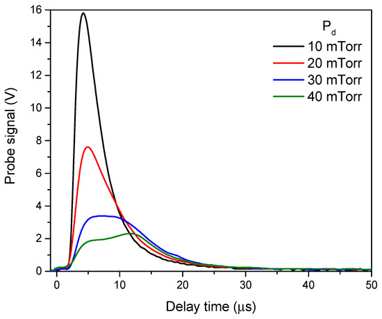
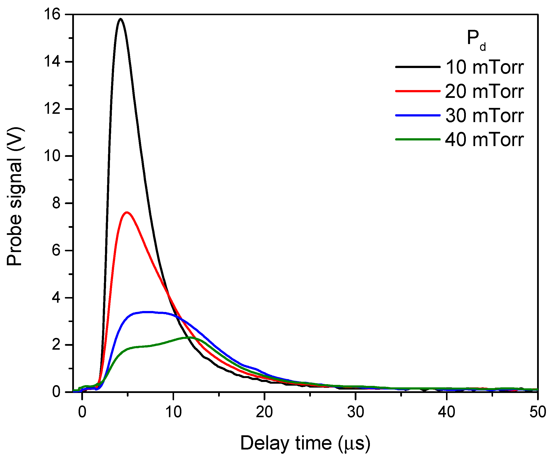
TOF curves obtained by Langmuir planar probe measurements of the plasmas produced by the ablation of Sn at different pressures are shown in Figure 1. The analysis of the curves revealed a strong dependence of plasma ion density and mean kinetic energy on the oxygen partial pressure. The probe signal decreased as oxygen pressure increased, indicating enhanced collisions occurrence and energy loss in the expanding plasma plume. Two main interesting effects can be observed from the TOF curves. First, we must recall that the maximum voltage is related to the ion density, through the ion saturation current; thus, a reduction in this maximum implies a decrease in ion density and consequently a reduction in the positive charged species arriving to the substrate. As a consequence, one would be tempted to think that under this reduced density of ions arriving to the substrate, oxidation may be limited; however, we need to consider that with increasing pressure there would be higher oxygen molecules available to neutrailze the ions and thus to form tion oxide more efficiently.
Figure 1.
TOF curves for plasmas produced at different pressures.
It has been widely reported [11] that neutral species play a key role on growth dynamics of pulsed laser deposited films; however, we have found in different studies, that positive ions can be effectively used to study and control physical properties of the growing films [12]. For example, ZnO crystallinity is highly influenced by the mean kinetic energy of the Zn ions in a reactive PLD process [13].
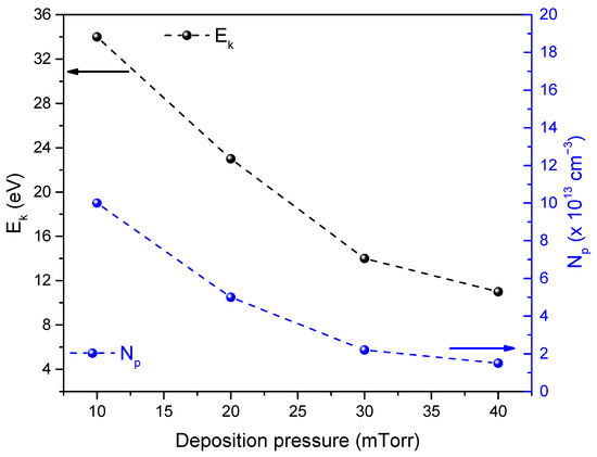
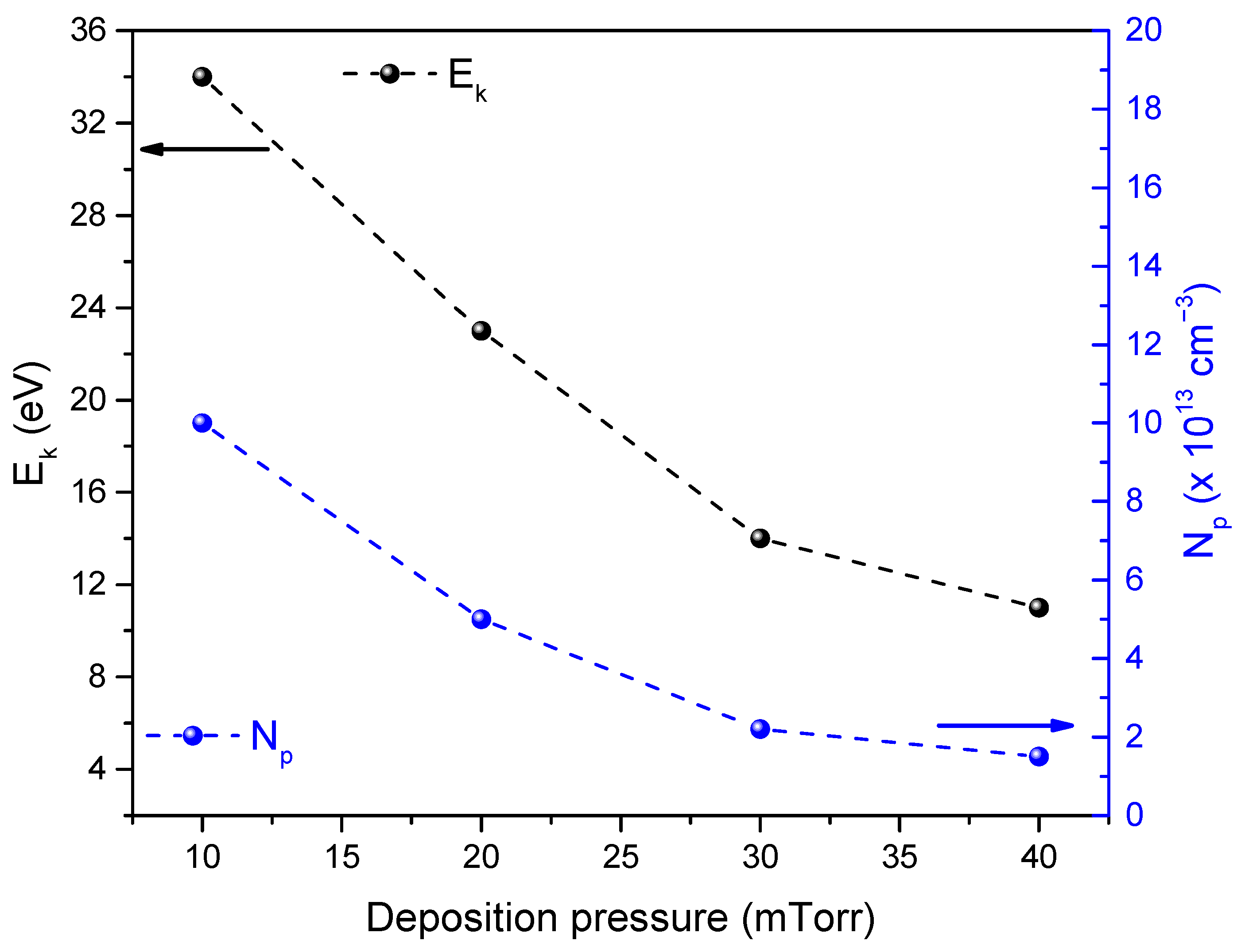
Following the methodologies described by Bulgakova and Dogget, the plasma parameters (ion mean kinetic energy and density) were estimated. Figure 2 shows the dependence of both mean kinetic energy and ion density on the deposition pressure. Notice that as pressure increases, both plasma parameters decrease, which indicates stronger collisional slowing and more efficient momentum transfer to the background gas at higher pressure. The reduction in ionic flux and kinetic energy with pressure suggests that part of the oxidation chemistry is already occurring in the plume, where Sn species interact with O2 before reaching the substrate. In other words, increasing O2 pressure not only changes the background during growth but actively modifies the energetic content of the arriving species.
Figure 2.
Plasma parameters as a function of the deposition pressure.
Notice that the mean kinetic energy of the tin ions is below 35 eV, which is considered low for PLD experiments, in which energies above 100 eV are commonly reported. The use of low kinetic energy has been studied for ZnO deposition from the ablation of Zn in similar atmpospheres and pressures leading to highly crystalline ZnO thin films [13]. This high degree of crystallization has been associated with the low surface mobility of arriving species that promotes the increase in low energy sites for crystallization [14].
The latter is important because both lower-energy arrival and partial oxidation in flight favor the formation of amorphous or nanocrystalline oxide networks rather than dense metallic tin.
Regarding thin films thickness, in general, for physical deposition methods, increasing the volume density of arriving species (ions, atoms, molecules, etc.) generated by the energy transfer from the evaporating and/or ionization source and the target would increase the thickness, as there would be more particles available for films growth. However, the effective adhesion of atoms, ions or molecules to the substrate surface would also be dependent on the surface energy. Thus, in the pulsed laser deposition method one would expect that higher ion densities yield thicker films, as more particles are reaching the surface. However, the kinetic energy of the species along with surrounding presssure will play a key role on the films thickness.
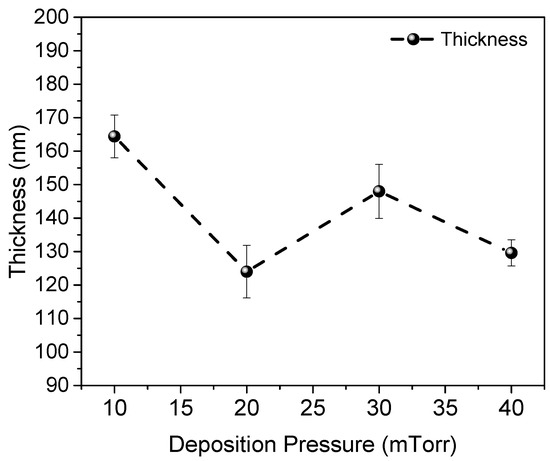
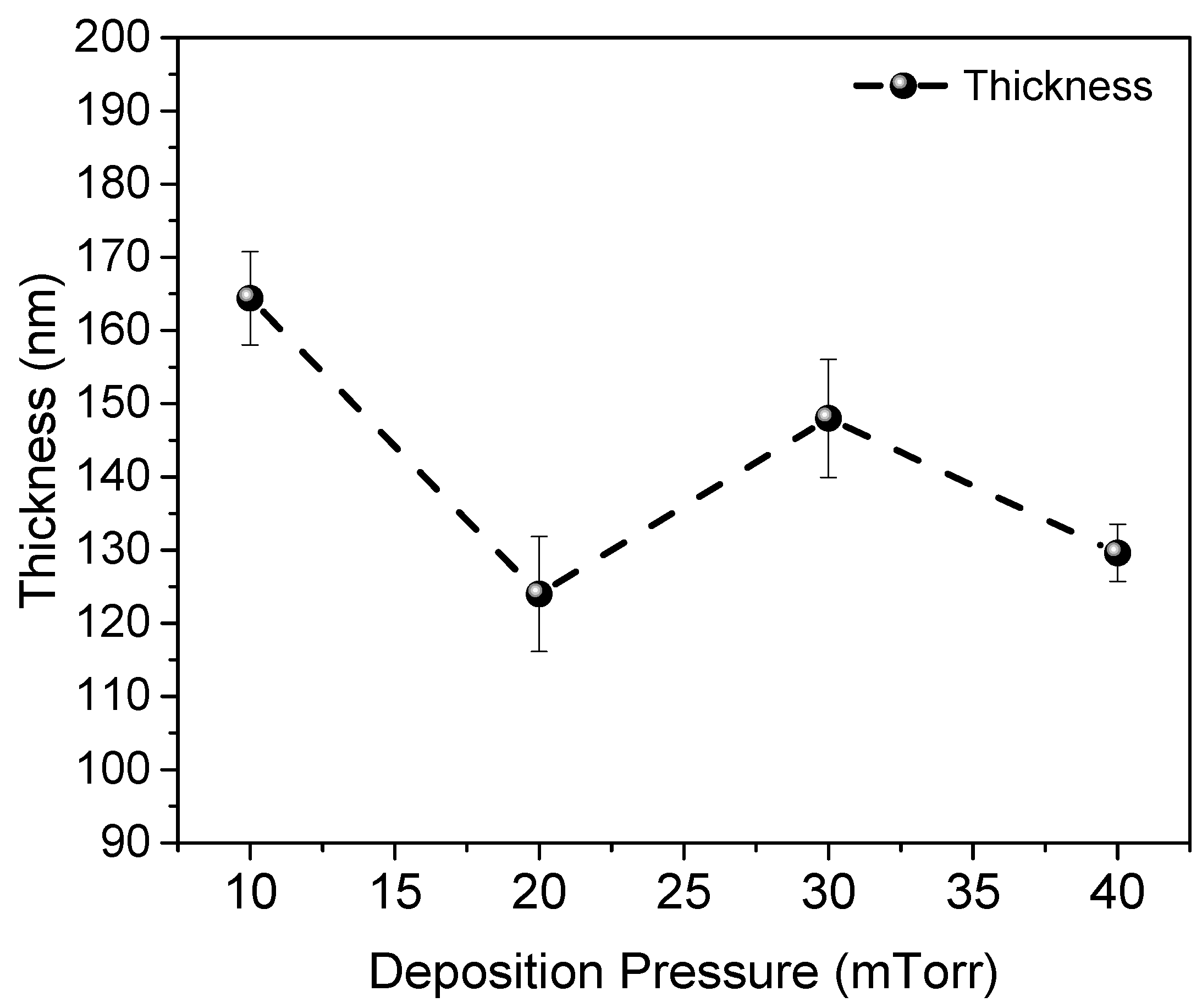
In general, for pulsed laser deposition, mean kinetic energies of the ions are considered high when compared to other plasma techniques, such as sputtering, allowing the appearance of resputering processes caused by the high energetic ions impinging the growing film. This phenomenon becomes important when using ion energies above 100 eV [15,16]. In the case of the present study, mean kinetic ion energies below 35 eV are being used, which allows for preventing resputtering processes and, thus, allows the ion density to be the main factor involved in the growth rate of the films. Figure 3 shows the variation in SnOx thin films’ thickness as a function of deposition pressure. It may be noticed that film grown at 10 mTorr has a thickness of 165 nm, while the thickness of the films grown at 20, 30, and 40 mTorr show an oscilatory trend around 140 nm. This initial reduction when pressure increases from 10 to 20 mTorr can be associated with the ion density decrease, which implies a lower number of species reaching the substrate. However, a further increase in pressure from 20 to 30 mTorr led to a thickness increase from 125 to 150 nm, followed by a decrease to 130 nm for 40 mTorr deposition pressure.
Figure 3.
SnOx films thicknesses a function of the deposition pressure.
To shed some light into the explanation of the thickness of films grown at 30 and 40 mTorr, note the shape of the TOF curves in Figure 2. Notice that for 30 and 40 mTorr there is a drastic lineshape change in the curves showing broad double-peaked signals. These lineshapes have been reported to be associated with the formaion of low velocity ion clusters [9], which are contributing to the deposition of the films generating this thickness increase. Nevertheless, a further increase in pressure to 40 mTorr reduces both the density of ions and ionic slow clusters and, thus, the film’s thickness.
3.2. Optical Characterization (UV-Vis)
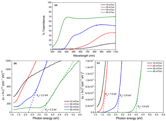
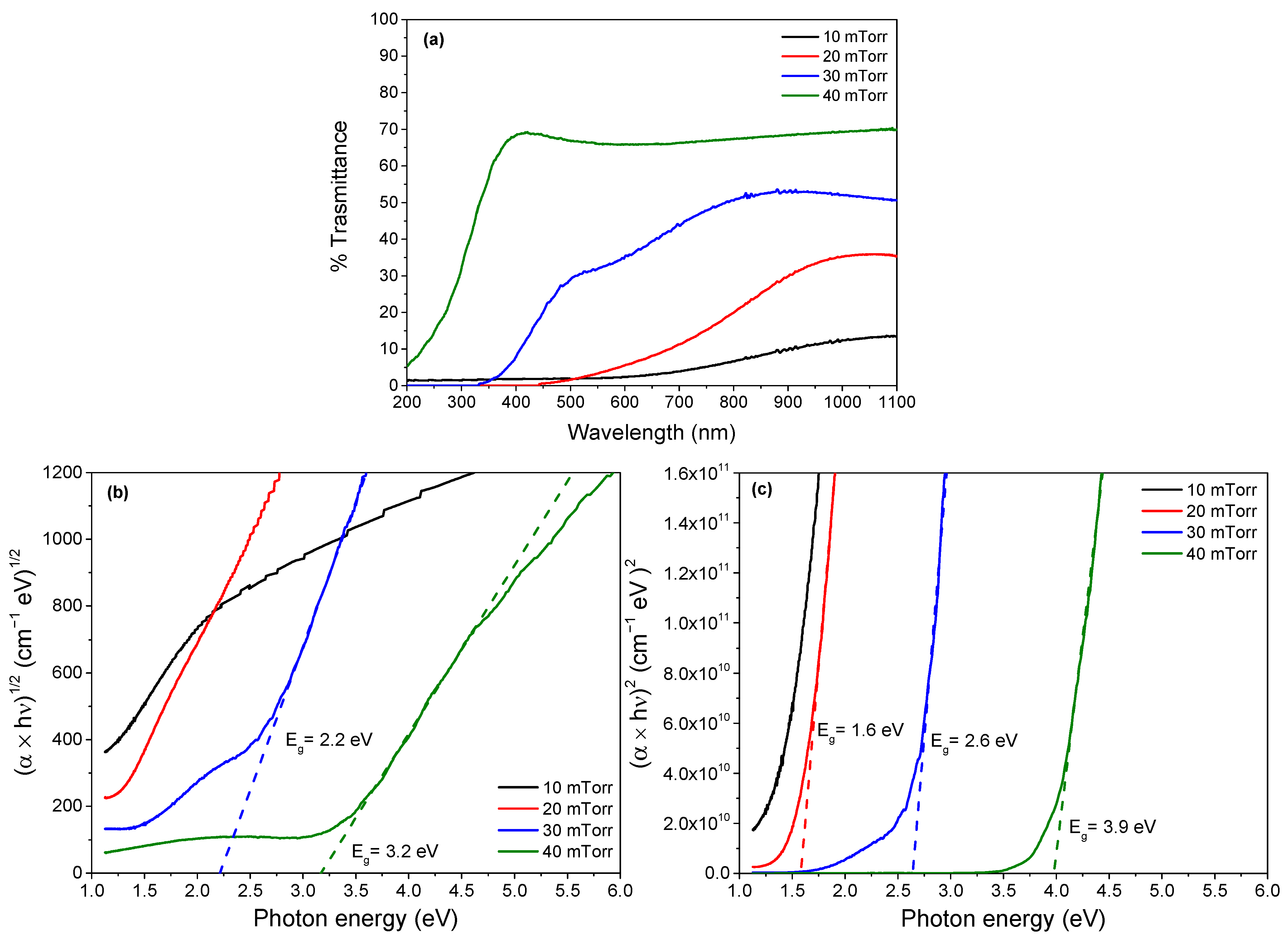
UV–Vis transmission spectra (Figure 4a) showed a gradual increase in transmittance for films deposited at higher oxygen pressures. Film deposited at 10 mTorr shows transmittance average values near to 10% in the 700–1100 nm range, while the transmittance in the same range for the film deposited at 20 mTorr increases to 30%. Notice that at these low-pressure values, the films have negligible transmittance in the visible range (400–700 nm) typical for highly defective or non-stoichiometric oxides [17]. On the other hand, for the samples grown at 30 and 40 mTorr the spectra change drastically, the transmittance increases considerably, and there is an important blue-shift in the absorption edge.
Figure 4.
(a) UV-Vis transmittance spectra and Tauc plots for (b) indirect and (c) direct transitions for SnOx films deposited at different pressure.
As it is noticeable from Figure 4a, the spectrum for the film deposited at 30 mTorr presents a band edge near 450 nm with average transmittance values of 40% in the 400–700 nm range, and 50% in the 700–1100 nm range. Meanwhile the film grown at 40 mTorr, has an almost constant transmittance of 70% from 400 to 1100 nm with its band edge located near to 350 nm. Visually, these samples are transparent, which is one of the main characteristics of tin oxide thin films.
Band gap values were estimated following the Tauc method. From the UV-Vis transmittance spectra, the absorption coefficient was calculated using the formula:
where T is the transmittance, R is the reflectance, and d is the film thickness. Then, using the relation: where for direct allowed transitions and for indirect allowed transitions, is the incident photon energy and is the band gap value. We used the fact that to express as a function of . The band gap values were then determined by the linear fit of the appropriate linear region of the Tauc plots, followed by extrapolation of the fitted line to the energy axis.
Figure 4b shows that the optical band gap, estimated from Tauc plots assuming indirect allowed transitions, increased from 2.2 eV (30 mTorr) to 3.2 eV (40 mTorr). This widening is consistent with the progressive formation of SnO2 and the reduced presence of metallic Sn clusters. We may notice that the bulk reported bandgap of SnO2 is indirect and has a value of 3.6 eV [2]; however, it has been observed that for the case of thin films, direct-like transitions may be observed at values above 3.4 eV [5], which is consistent with the value for sample grown at 40 mTorr (Figure 4c).
In general, an apparent discrepancy is observed between our data and the literature, as the indirect allowed transitions yield bandgap values lower than those reported for SnO2. However, this pronounced difference may be attributed to the combined effect of several factors, including partial oxidation, the presence of metallic tin, and the generation of a high density of structural and compositional defects, which introduce interband states and effectively reduce the optical bandgap.
The spectral transparency and tunable band gap confirm the semiconductor nature of the oxidized films combined with metallic Sn clusters embedded within the oxide matrix. This observation will be corroborated with the analysis of XRD results, which confirm the presence of metallic remnants.
UV-Vis measurements show that films deposited at higher oxygen pressures are more transparent in the visible range, and their apparent direct optical band gap (from Tauc analysis) shifts to higher energies, reaching values consistent with semiconducting tin oxides. At low oxygen pressure, the films are more absorbing, with a lower apparent gap. This optical evolution tracks perfectly with the XPS result: as O2 pressure increases, the surface and matrix behave optically like SnOx/SnO2; they tend to be transparent, wide-gap semiconductors even though metallic Sn crystallites are still present inside. Thus, optically, the films act as oxidized tin oxide layers, but, structurally, they still remember that they nucleated as metallic Sn.
3.3. Surface Chemical Analysis (XPS)
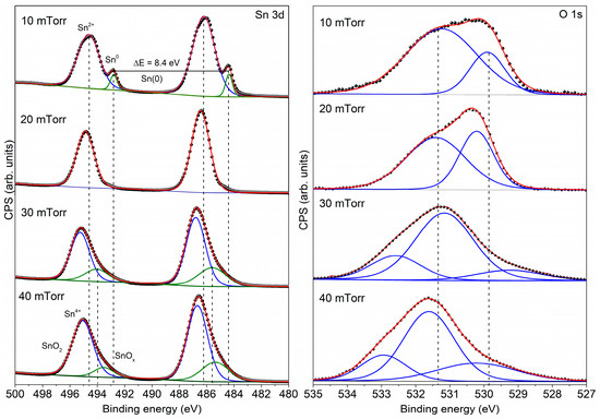
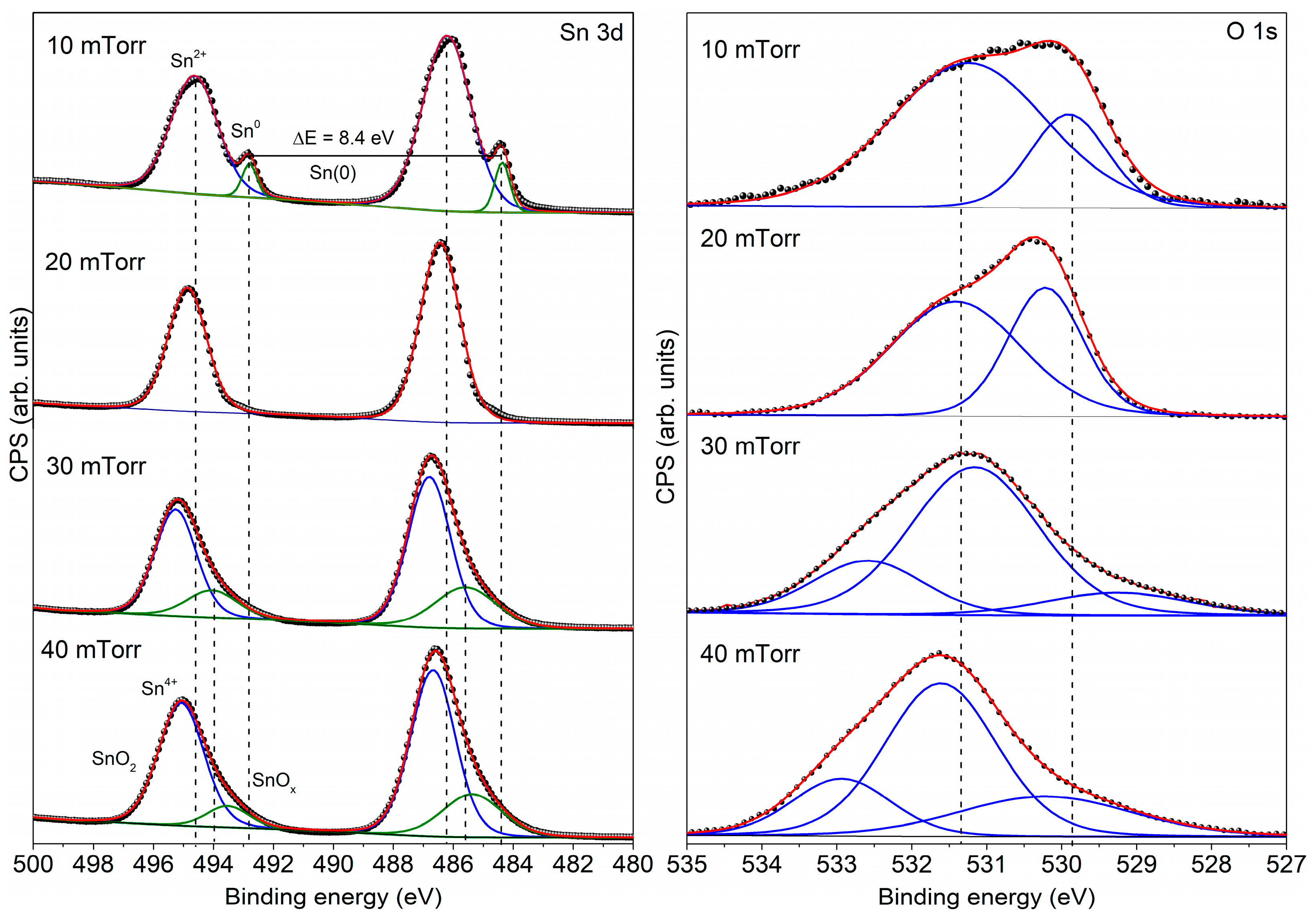
Figure 5 shows the XPS spectra of the Sn 3d and O 1s core levels in order to reveal the actual oxidation state at the films surface. The spectra revealed contributions consistent with oxidized tin species in addition to the metallic component. In particular: the spectrum of the sample grown at 10 mTorr, shows a lower-binding-energy doublet component consistent with Sn0 (metallic tin); the splitting energy is consistent with the reported value of 8.4 eV. Additionally, there is a higher binding energy component consistent with Sn–O bonding environments, assignable to Sn2+ state typically reported for oxidized SnO [18].
Figure 5.
High resolution XPS spectra for the Sn 3d and O 1s orbitals of the SnOx films grown at different pressures.
The coexistence of Sn2+/Sn4+ states components become evident as the oxygen pressure during deposition increases. For films grown at higher O2 pressure (≥ 20 mTorr), the oxidized contribution dominates the XPS envelope, indicating that the near-surface region is largely oxidized. At these pressures, the metallic Sn is no longer visible, which does not mean that the films have fully oxidized, but the surface oxide is thick enough to prevent metallic Sn signal to be revealed.
Moreover, the XPS signal of Sn2+/Sn4+ even at moderate pressures implies that oxidation proceeds efficiently at the surface once the energetic Sn species land, and/or that the plume already carries partially oxidized Sn to the substrate. This agrees with the plasma diagnostics: oxygen in the plume reduces ion energy and promotes in-flight reactions.
A quantitative deconvolution of the Sn 3d spectra into metallic and oxidized contributions was only feasible for the sample deposited at 10 mTorr, where a distinct Sn0 component could be clearly resolved. For films deposited at higher oxygen pressures, the Sn 3d spectra are dominated by oxidized Sn chemical states, and no unambiguous metallic Sn contribution can be reliably extracted within the experimental resolution and signal to noise ratio. Under these conditions, XPS provides reliable qualitative information on the presence and evolution of oxidized Sn species, while a quantitative determination of bulk Sn/SnOx ratios would require complementary depth-sensitive techniques.
On the other hand, from the O 1s spectra presented in Figure 5 we may notice two components centered near 530 and 531 eV associated with O-Sn2+ and O-Sn4+ bondings, confirming the observed coexistence of SnOx/SnO2-like oxides, while the signal above 532 eV corresponds to –OH groups [18].
Overall, the XPS results support a description in which metallic Sn cores or particles coexist with a SnOx/SnO2-like shell or surrounding matrix.
3.4. Structural Characterization (XRD)
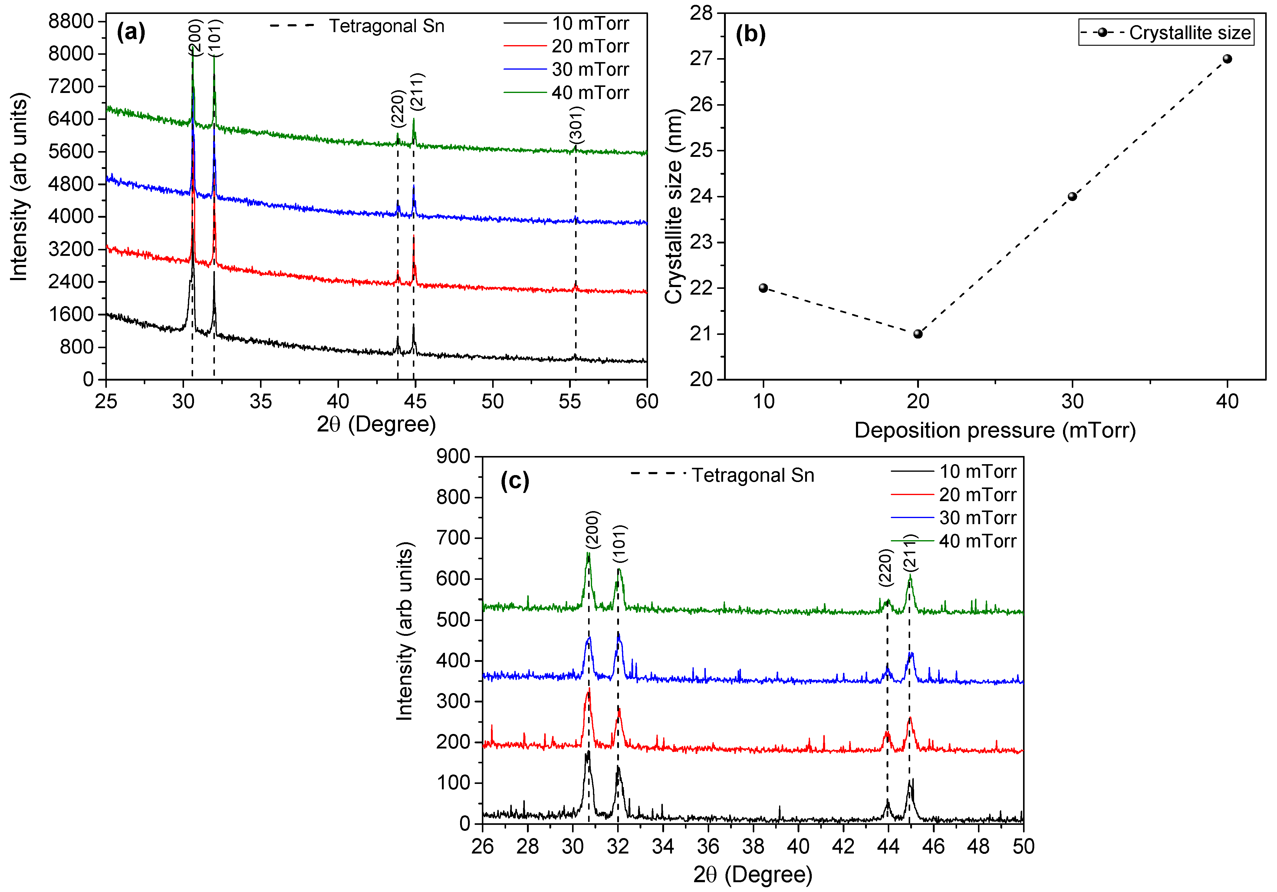
All the deposited films exhibited Bragg reflections that match metallic β-Sn, independent of the oxygen pressure [19]. Even at 40 mTorr, where oxidation is expected to dominate, clear metallic Sn peaks remained detectable. At the same time, no intense or sharp peaks unambiguously attributable to crystalline SnO2 were observed. However, UV-Vis spectroscopy reveals a progressive evolution of the optical response toward tin oxide-like behavior as the oxygen pressure increases. Consistently, XPS indicates a significant contribution from mixed tin oxides chemical states. The absence of well-defined SnO or SnO2 Bragg reflections in XRD therefore suggests that the oxide component is predominantly amorphous or poorly crystalline.
This constitutes a central result of the present study. From a structural standpoint, the long-range order of the films is dominated by metallic Sn crystallites, while a fully stoichiometric and long-range ordered SnO2 lattice does not appear to form under the present deposition conditions. XRD, as a bulk-sensitive technique probing depths on the order of hundreds of nanometers, and XPS, as a surface-sensitive probe with an information depth of only a few nanometers, provide complementary perspectives. Taken together, the simultaneous observation of metallic Sn Bragg reflections in XRD and SnO2-like chemical states in XPS indicates that metallic Sn persists as crystalline domains that are partially or fully covered by an oxidized shell. Accordingly, the deposited material should be described not as a uniform metallic film, but as a metal/oxide composite.
While electrical transport measurements (e.g., conductivity or Hall effect) would provide valuable complementary information on carrier type and mobility, such measurements were beyond the scope of the present study and are left for future work.
Overall, these results demonstrate that a description based solely on XRD would lead to an incomplete picture, as the optical and chemical characterizations reveal a substantial contribution from oxidized tin phases that strongly influence the observed bandgap evolution.
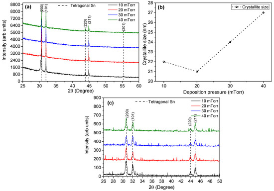
The crystallite size of the samples was estimated from the XRD patterns using the Debye–Scherrer formula, , where is the crystallite size, is the Scherrer constant, the X-ray wavelength (1.54 Å for the Cu Kα radiation used in the Empyrean system), and is the full width at half maximum (FWHM) [20]. Figure 6b shows the crystallite size as a function of deposition pressure. The crystallite size slightly decreases from 22 to 21 nm as the deposition pressure increases from 10 to 20 mTorr. This initial decrease can be attributed to a reduction in the mean kinetic energy of the plasma species, which lowers surface mobility and limits crystal coalescence [21,22].
Figure 6.
(a) XRD patterns for samples deposited at different pressures. (b) Crystallite size as function of deposition pressure. (c) Grazing angle XRD patterns of SnOx thin films.
At higher deposition pressures, the crystallite size increases to 24 nm at 30 mTorr and to 27 nm at 40 mTorr, despite the reduced kinetic energy. This behavior can be explained by the formation of ion clusters, as evidenced by the double-peaked signals observed in the TOF measurements at 30 and 40 mTorr. The presence of these clusters contributes to crystallite growth, overcoming the limitations imposed by reduced surface mobility.
4. Conclusions
Reactive PLD of metallic Sn in low-pressure oxygen does not immediately yield a single-phase crystalline SnO2 film. Instead, it yields a hierarchical composite with pressure tunable properties. Increasing oxygen partial pressure drives a transition from predominantly metallic behavior toward SnOx/SnO2-like behavior. XRD always detects metallic Sn, but XPS detects oxidized tin states (Sn2+/Sn4+), especially at pressures ≥ 20 mTorr. This implies metallic Sn nanocrystals embedded in or coated by an amorphous tin oxide matrix. On the other hand, films grown at higher O2 pressure become more transparent and exhibit a wider optical band gap, consistent with semiconducting SnO2-like domains, and therefore are promising for optoelectronic or sensing uses where visible transparency and conductivity are required.
Langmuir probe TOF shows that the plume loses kinetic energy as O2 pressure increases, meaning oxidation chemistry is already happening in flight. This supports a growth picture where energetic Sn species arrive partially oxidized and then rapidly passivate into a SnOx shell.
Author Contributions
Conceptualization, J.G.Q.-G.; Methodology, L.P.R. and C.D.R.-T.; Formal analysis, L.P.R.; Investigation, J.G.Q.-G.; Writing—original draft, L.P.R.; Writing—review & editing, L.P.R. and J.G.Q.-G. All authors have read and agreed to the published version of the manuscript.
Funding
This research was funded by University of Guadalajara, under the PRO-SNI program.
Data Availability Statement
The original contributions presented in this study are included in the article material. Further inquiries can be directed to the corresponding author(s).
Acknowledgments
The authors gratefully acknowledge the support from the program Apoyo al Fortalecimiento y Desarrollo de Infraestructura Científica y Tecnológica, project number 270662. We also thank the X-ray Photoelectron Spectroscopy (XPS) Laboratory at the Universidad de Guadalajara for providing access to their facilities and technical assistance, especially José Antonio Rivera Mayorga for his valuable support during the measurements. This work was carried out with the support of the Universidad de Guadalajara. Sergio Oliva and María Guadalupe Cárdenas are acknowledged for XRD measurements.
Conflicts of Interest
The authors declare no conflicts of interest.
References
- Song, Z.; Tian, Y.; Kang, Y.; Yan, J. Development of a Three-Dimensional Nanostructure SnO2-Based Gas Sensor for Room-Temperature Hydrogen Detection. Sensors 2025, 25, 4784. [Google Scholar] [CrossRef] [Scilit] [PubMed]
- Meddas, H.; Abdelghani, L.; Amel, D.; Saliha, B.; Randa, S.; Okba, A.M.S.B. Elaboration of spin coated deposition of tin oxide thin films: Effect of concentration. Results Opt. 2025, 19, 100811. [Google Scholar] [CrossRef] [Scilit]
- Rameshkumar, C.; Anderson, A.; Ananth, D.; Mohan, T.H.; Subalakshmi, R. An investigation of SnO2 nanofilm for solar cell application by spin coating technique. Mater. Today Proc. 2020, 45, 6042–6045. [Google Scholar] [CrossRef] [Scilit]
- Li, W.; Ji, S.; Qian, K.; Jin, P. Preparation and characterization of VO2(M)-SnO2 thermochromic films for application as energy-saving smart coatings. J. Colloid Interface Sci. 2015, 456, 166–173. [Google Scholar] [CrossRef] [Scilit]
- Kiruthiga, G.; Rajni, K.S.; Geethanjali, N.; Raguram, T.; Nandhakumar, E.; Senthilkumar, N. SnO2: Investigation of optical, structural, and electrical properties of transparent conductive oxide thin films prepared by nebulized spray pyrolysis for photovoltaic applications. Inorg. Chem. Commun. 2022, 145, 109968. [Google Scholar] [CrossRef] [Scilit]
- Turan, E.; Kul, M.; Akın, S. Structural and optical investigation of spray-deposited SnO2 thin films. J. Mater. Sci. Mater. Electron. 2022, 33, 15689–15703. [Google Scholar] [CrossRef] [Scilit]
- Medina, J.E.M.; Philippe, A.M.; Guillot, J.; Vergne, C.; Fleming, Y.; Barborini, E. Intermediate tin oxide in stable core-shell structures by room temperature oxidation of cluster-assembled nanostructured Sn films. Appl. Surf. Sci. 2024, 658, 159846. [Google Scholar] [CrossRef] [Scilit]
- Planche, M.P.; Liao, H.; Coddet, C. Oxidation control in atmospheric plasma spraying coating. Surf. Coat. Technol. 2007, 202, 69–76. [Google Scholar] [CrossRef] [Scilit]
- Bulgakova, N.M.; Bulgakov, A.V.; Bobrenok, O.F. Double layer effects in laser-ablation plasma plumes. Phys. Rev. E 2000, 62, 5624–5635. [Google Scholar] [CrossRef] [Scilit]
- Doggett, B.; Lunney, J.G. Langmuir probe characterization of laser ablation plasmas. J. Appl. Phys. 2009, 105, 033306. [Google Scholar] [CrossRef] [Scilit]
- Irimiciuc, S.A.; Chertopalov, S.; Lancok, J.; Craciun, V. Langmuir probe technique for plasma characterization during pulsed laser deposition process. Coatings 2021, 11, 762. [Google Scholar] [CrossRef] [Scilit]
- Quiñones-Galván, J.G.; Camps, E.; Muhl, S.; Flores, M.; Campos-González, E. Influence of plasma density on the chemical composition and structural properties of pulsed laser deposited TiAlN thin films. Phys. Plasmas 2014, 21, 053509. [Google Scholar] [CrossRef] [Scilit]
- de León, J.A.G.; Pérez-Centeno, A.; Gómez-Rosas, G.; Mariscal, A.; Serna, R.; Santana-Aranda, M.A.; Quiñones-Galván, J.G. Influence of the Zn plasma kinetics on the structural and optical properties of ZnO thin films grown by PLD. SN Appl. Sci. 2019, 1, 475. [Google Scholar] [CrossRef] [Scilit]
- de León, J.A.G.; Rivera, L.P.; Campos-González, E.; Camps, E.; Arias-Cerón, J.S.; Yee-Rendón, C.M.; López-Álvarez, M.Á.; Quiñones-Galván, J.G. Enhanced optical response of 1T/2H-MoS2 heterostructures thin films deposited by laser ablation. Mater. Sci. Eng. B 2026, 324, 118981. [Google Scholar] [CrossRef] [Scilit]
- Saenger, K.L. Angular Distribution of Ablated Material. In Pulsed Laser Deposition of Thin Films, 1st ed.; Chrisey, D.B., Hubler, G.K., Eds.; John Wiley and Sons: New York, NY, USA, 1994; pp. 199–227. [Google Scholar]
- Fähler, S.; Sturm, K.; Krebs, H.U. Resputtering during the growth of pulsed-laser-deposited metallic films in vacuum and in an ambient gas. Appl. Phys. Lett. 1999, 75, 3766–3768. [Google Scholar] [CrossRef] [Scilit]
- Quiñones-Galván, J.G.; Lozada-Morales, R.; Jiménez-Sandoval, S.; Camps, E.; Castrejón-Sánchez, V.H.; Campos-González, E.; Zapata-Torres, M.; Pérez-Centeno, A.; Santana-Aranda, M.A. Physical properties of a non-transparent cadmium oxide thick film deposited at low fluence by pulsed laser deposition. Mater. Res. Bull. 2016, 76, 376–383. [Google Scholar] [CrossRef] [Scilit]
- Lu, F.; Ji, X.; Yang, Y.; Deng, W.; Banks, C.E. Room temperature ionic liquid assisted well-dispersed core-shell tin nanoparticles through cathodic corrosion. RSC Adv. 2013, 3, 18791–18793. [Google Scholar] [CrossRef] [Scilit]
- Oehl, N.; Schmuelling, G.; Knipper, M.; Kloepsch, R.; Placke, T.; Kolny-Olesiak, J.; Plaggenborg, T.; Winter, M.; Parisi, J. In situ X-ray diffraction study on the formation of α-Sn in nanocrystalline Sn-based electrodes for lithium-ion batteries. CrystEngComm 2015, 17, 8500–8504. [Google Scholar] [CrossRef] [Scilit]
- Qu, S.; Zhang, J.; Gao, L.; Chen, H.; Ding, Y. Effect of Deposition Pressure and Temperature on Tungsten Thin-Film Heater for Phase-Change Switch Applications. Micromachines 2024, 15, 576. [Google Scholar] [CrossRef] [Scilit]
- Pawar, S.M.; Moholkar, A.V.; Kim, I.K.; Shin, S.W.; Moon, J.H.; Rhee, J.I.; Kim, J.H. Effect of laser incident energy on the structural, morphological and optical properties of Cu2ZnSnS4 (CZTS) thin films. Curr. Appl. Phys. 2010, 10, 565–569. [Google Scholar] [CrossRef] [Scilit]
- Rodríguez-Rosales, K.; Cruz-Gómez, J.; Serrano-Lázaro, A.; Santos-Cruz, J.; de Moure-Flores, F.; Villagrán-Muniz, M. Influence of laser fluence on plasma dynamics and the structural, optical, and electrical properties of CuI thin films deposited by pulsed laser ablation. Ceram. Int. 2025, 51, 54255–54266. [Google Scholar] [CrossRef] [Scilit]
Disclaimer/Publisher’s Note: The statements, opinions and data contained in all publications are solely those of the individual author(s) and contributor(s) and not of MDPI and/or the editor(s). MDPI and/or the editor(s) disclaim responsibility for any injury to people or property resulting from any ideas, methods, instructions or products referred to in the content. |
© 2026 by the authors. Licensee MDPI, Basel, Switzerland. This article is an open access article distributed under the terms and conditions of the Creative Commons Attribution (CC BY) license.