


Abstract
Background: Amelogenesis imperfecta (AI) has recently been realized as not only an inherited enamel anomaly, but also a genetically heterogeneous developmental disorder and a compelling model for the study of enamel biomineralization and craniofacial development. Methods: This review is a narrative synthesis of the state of the art of AI genetics, genotype–phenotype relationships, ameloblast biology, enamel biomineralization, molecular diagnosis and new approaches to regeneration by biomimetic nanomaterials, stem cell engineering, use of organoids, 3D bioprinting, and gene editing. Results: Pathogenic variants of enamel matrix proteins, proteolytic enzymes, ion transport, intracellular trafficking, and signaling between cells and the matrix provide insights into different molecular mechanisms underlying hypoplastic, hypomineralized (hypomaturation), and hypomineralized (hypocalcified) disease. The incorporation of new techniques, such as next-generation sequencing, functional genomics, multi-omics, and computational methods is changing the paradigm of AI from phenotype-based classification to one of molecularly stratified and personalized diagnosis. At the same time, emerging experimental approaches—including biomimetic mineralization, engineered ameloblast-like cells, organoid systems, and genome-editing technologies—provide promising platforms for investigating enamel regeneration, although their clinical applicability remains to be established. Conclusions: AI provides a valuable mechanistic framework for advancing research toward patient-specific, biologically informed approaches in developmental biology, precision dentistry, and regenerative craniofacial medicine; however, regenerative interventions remain investigational and require substantial preclinical validation before potential clinical translation.
1. Introduction
Amelogenesis Imperfecta (AI) is one of the most informative hereditary diseases of mineralized tissues in terms of understanding the cellular process of enamel biomineralization, the interaction between different types of tissues during development, and the molecular orchestra for the development of the craniofacial area [1]. Indeed, AI is now considered to be a developmental disease and not only a hereditary defect of enamel and therefore a reflection of the complex process of cell differentiation, extracellular matrix remodeling, mineral ion homeostasis and tissue-specific gene expression in odontogenesis [2]. Clinically, AI represents a spectrum of different hypoplastic, hypomaturation, hypocalcified enamel phenotypes, often with dentin hypersensitivity, rapid occlusal wear, masticatory dysfunction, esthetic disfigurement and significant psychosocial issues that affect the oral health-related quality of life. These phenotypes arise from disruptions along a coordinated developmental sequence of ameloblast differentiation, enamel matrix secretion, proteolytic processing, pH regulation, and hydroxyapatite crystal growth such that each clinical presentation reflects a distinct point of failure within this pathway [3,4]. Accordingly, the study of AI has moved beyond cataloging a rare, monogenic dental anomaly: mechanistic dissection of its genetic subtypes now offers a tractable entry point into the broader biology of mineralized tissue development and craniofacial morphogenesis, with direct relevance to precision approaches in oral medicine.
The genomic revolution has substantially reshaped the understanding of AI and shifted its entity from a morphology-based phenotype to a genetically defined mechanism/stratified disease. Due to the rapid expansion of next-generation sequencing, whole-exome or whole-genome sequencing and functional genomics analysis, an ever-growing list of pathogenic variants in genes involved in various stages of amelogenesis have been identified such as AMELX, ENAM, MMP20, KLK4, FAM83H, WDR72, SLC24A4, ITGB6 and LAMB3, as well as numerous newly identified loci. These genes all regulate the processes of enamel matrix formation, protein intracellular transport, processing for proteolysis, calcium and phosphate transport, acid–base regulation, ameloblast cytoskeletal organization and ameloblast survival [4]. Significantly, there is a growing body of evidence which shows that identical pathogenic changes can result in highly variable clinical manifestations, indicating the role of modifier genes, epigenetics, environment and developmental plasticity. This growing awareness has propelled AI into a paradigm for tackling complex genotype—phenotype associations and highlighted the need to shift the focus from descriptive, clinical diagnosis towards systems-level molecular disease modeling, which can help with predictive diagnostic, individual risk assessment and precision therapeutic decision making [5].
In addition to genetics, AI research is playing a huge part in the world of regenerative dentistry and bioengineered enamel replacement solutions. The main feature of conventional restorative approaches is that they mainly concentrate on the structural defects, but fail to correct the underlying developmental abnormalities, which cause enamel failures. The enormous opportunities for reconstructing enamel through biologically inspired regenerative processes have recently emerged thanks to the progress in developmental biology, stem cell engineering, biomimetic mineralization, organoid technology, gene editing and additive manufacturing [6]. All the four strategies—biomimetic peptide-guided mineralization systems, 3D bioprinted systems, amorphous artificial enamel organoids and ameloblast-like cells derived from induced pluripotent stem cells (iPSCs) and a hamster cell line together have expanded therapeutic strategies from conventional reparative approaches toward biologically informed regenerative interventions in dentistry [7]. However, there are still significant challenges in the field of translation that need to be addressed with regard to precise spatiotemporal regulation of ameloblast differentiation, durability of scaffold integration, controlled mineral deposition, long-term functional stability, immune compatibility of scaffolds, efficiency of scaffold delivery, regulatory approval, and the ethics of genome manipulation. To overcome these barriers will require an interdisciplinary approach to integrate molecular genetics, developmental biology, materials science, computational modeling, tissue engineering and clinical dentistry [8].
While existing reviews provide a thorough overview of clinical presentation or genetic categorisation of AI, few have attempted to combine recent advances in enamel developmental biology, the emerging mechanistic genotype–phenotype link with biomaterial innovation and regenerative approaches within the perspective of precision dentistry in general. This review synthesizes current knowledge spanning from molecular aspects of amelogenesis, to knowledge revolving around genes and pathways involved in various diseases, developmental signaling pathways, and the latest advances in amelogenesis-related diagnostics, biomaterials, and tissue regeneration. Moreover, it thoroughly reviews the current status of research, technology, and opportunities that can help to reassess the management of inherited enamel disorders. This review underscores the broader importance of AI in advancing the field of precision oral healthcare, serving as a developmental disease while also functioning as a model system to better understand precision medicine for mineralized dental tissues, pushing the boundaries of personalized diagnosis, biologically inspired regenerative therapeutics and towards next-generation approaches in mineralized tissue regeneration.
2. Methodology
The literature search included publications available through 2026. Original research articles, case reports, and prior review articles addressing the molecular, genetic, developmental, or translational aspects of AI were eligible for inclusion. Non-English publications, conference abstracts without accompanying full-text articles, and studies unrelated to enamel biomineralization or craniofacial development were excluded. Because this review followed a narrative rather than systematic methodology, no formal risk-of-bias assessment or quantitative synthesis was performed; study selection and thematic organization were investigator-directed. Retrieved literature was thematically grouped into three domains: (i) genetic heterogeneity and the mutational spectrum underlying AI, (ii) the molecular physiology of enamel formation, and (iii) the utility of AI-associated gene variants for probing craniofacial developmental biology. Representative pedigrees, clinical images, and mutational data from previously published, open-access sources were subsequently incorporated to illustrate genotype–phenotype relationships discussed in the text.
AI Disclosure
Artificial intelligence was used solely to assist in generating and revising selected graphical elements of Figure 1. OpenAI ChatGPT-6 was used for this purpose, and no AI-generated text, data, or scientific evidence was incorporated into the manuscript. The generated elements were critically reviewed, edited, and scientifically verified by the author, including the orientation of secretory-stage ameloblasts, Tomes’ process, and enamel matrix deposition. The author take full responsibility for the accuracy and final content of the figure.
Figure 1.
Conceptual overview of the molecular and translational dimensions of amelogenesis imperfecta, illustrating genetic determinants of enamel formation, phenotype–genotype relationships, precision diagnosis, and emerging regenerative strategies. (Disclaimer: The contents of this figure have partially been generated by OpenAI ChatGPT-6 and subsequently reviewed, edited, and scientifically verified by the author).
3. Molecular and Genetic Basis of AI
3.1. Genetic Heterogeneity and Key Mutations: Molecular Complexity Underlying
AI is one of the most genetically diverse inherited biomineralization disorders caused by the presence of pathogenic gene variants that affect genes involved in enamel matrix secretion, proteinase-mediated breakdown, protein transport and mineral ion homeostasis. In comparison to monogenic developmental disorders in which the phenotypic expression is largely uniform, the phenotypic expression of AI is quite variable, reflecting the complexity of the temporal and spatial co-ordination needed for amelogenesis. Enamel’s development is tightly regulated through reciprocal interactions among the various structure matrix proteins, proteases, signaling molecules, and ion transport systems acting together to regulate the nucleation, elongation, and maturation of hydroxyapatite crystals. Perturbation of these molecular networks compromises enamel ultrastructure and mechanical competence, giving rise to a clinically heterogeneous spectrum spanning hypoplastic, hypomaturation, and hypocalcified phenotypes. Consequently, elucidation of the specific molecular defects underlying AI subtypes has provided a valuable framework for dissecting the fundamental mechanisms governing tooth and mineralized tissue formation, and for understanding how their disruption manifests as disease.
Molecularly, mutations in canonical enamel-related genes AMELX, ENAM, MMP20, KLK4, FAM83H, WDR72, and SLC24A4 affect the various steps of amelogenesis in different pathogenic ways. Variants involved in the AMELX and ENAM genes mainly affect how extracellular matrix is assembled and scaffolded, which are likely to have a negative impact on crystal nucleation and enamel thickness. In contrast, mutations in MMP20 and KLK4 impair the ability to degrade matrix proteins leading to impaired matrix clearance and incomplete maturation of the mineral. Other recently identified genes including WDR72 and SLC24A4 underscore the essential role of zinc, vesicles, pH regulation, and calcium transport in regulating enamel mineral homeostasis. The importance of these mechanistic differences clearly highlights that enamel formation is not just a matter of protein synthesis and matrix formation but rather one of protein turnover and co-ordinated regulation of the protein transporting pathways and ionic equilibrium in the developmental process [9,10].
The clinicopathological consequences of the recurrent FAM83H c.973C>T (p.R325X) truncating mutation are shown in Figure 2 where the close association of molecular defects and hypocalcified AI phenotype is seen. The pedigree suggests that the condition is autosomal dominant, while the clinical photographs show generalized enamel hypocalcification with enamel breakdown easily detectable after eruption, significant erosion of enamel in occlusal areas, and poor esthetics. The decrease in relative enamel thickness and increase in relative dentinal thickness were accompanied by decreased enamel radiodensity, which is characteristic of defective mineral maturation and not impaired matrix secretion, suggesting a pathogenic difference between hypocalcified and hypoplastic AI. Mechanistically, the terminally located keratin cytoskeletal and casein kinase-1 (CK1)-mediated signaling events that are important for proper ameloblast activity are altered by truncating mutations in the terminal exon of FAM83H that shift protein localization from the cytoplasm to the nucleus. This abnormal cellular traffic eventually leads to disruption of mineralization and of the mechanical integrity of enamel. Such frequent recurrence of apparently identical variants in the p.R325X position in different populations also highlights the possible presence of mutational hotspots in FAM83H, which further highlights the need for genotype-driven diagnoses, molecular classification and treatment planning. Taken together these data highlight FAM83H as a key molecular player in enamel biomineralization and as a molecular target of regenerative and precision dentistry strategies in the future.
Figure 2.
Examination and mutational analysis of Family 4, showing (A) the pedigree with recruited members, (B) enamel defects characteristic of hypocalcified AI, (C,D) FAM83H exon 5 sequencing showing a c.973 C > T (p.R325X) mutation in the proband, and (E) the panoramic radiograph of the proband taken at 14 years. Adapted from [11].
A complex genotype–phenotype relationship is evident in hypomaturation AI where pathogenic variants in the gene coding for SLC24A4 lead to definite enamel defects yet enamel thickness is relatively preserved as shown in Figure 3. An autosomal recessive inheritance can be inferred from the pedigree and the clinical photos provide evidence of generalized brown discolored enamel, post-eruptive enamel loss, and weakened mechanical integrity typical of a defect in enamel maturation rather than enamel secretion. The reduction of radiographic contrast between enamel and dentin is indicative of poor acquisition of minerals during the maturing phase and of the importance of ion transport in hydroxyapatite crystal formation. At the functional level, SLC24A4 is the gene coding for the potassium-dependent sodium/calcium exchanger found in the maturation stage of the ameloblasts, which is necessary for proper calcium transport leading to progressive crystal enlargement and final enamel hardening. Loss-of-function mutations cause impaired calcium regulation and cause the enamel to become poorly mineralised, meaning the enamel is structurally weak and subject to rapid attrition after eruption. Importantly, this figure also illustrates that the same phenotypic clinical manifestations can be due to different molecular underlying defects, which demonstrates the need to integrate genomic sequencing with comprehensive phenotypic characterization for accurate diagnosis, prognostic stratification and precision therapeutic strategies [8,12,13]. These observations also corroborate the shift from a classification according to the resulting phenotype to a molecular classification according to mechanistically relevant genes, and give rise to future perspectives on biomimetic mineralization, gene-targeted interventions and personalized regenerative dentistry.
Figure 3.
(A) Pedigree of family 2 with the proband indicated by a black arrow and study participants labeled with plus signs; consanguinity is shown with double lines. (B–F) Clinical images of proband (IV:1) show generalized brown hypomatured enamel; (G) panoramic radiograph reveals hypomineralized enamel with reduced radiodensity and enamel breakdown in first molars. Adapted from [14].
Advances in next-generation and whole-exome sequencing, coupled with the expanding application of functional genomics, have substantially refined the resolution of genotype–phenotype relationships in AI, revealing a complexity that earlier phenotype-based classifications failed to capture. Notably, identical pathogenic variants can produce divergent clinical presentations, while phenotypically convergent cases frequently harbor distinct causative variants. This lack of strict genotype–phenotype concordance implicates modifier genes, epigenetic regulation, environmental exposures, and compensatory molecular pathways as critical determinants of disease severity and expressivity. This goes against the traditional approach of classifying AI by phenotype and suggests a shift in paradigm towards molecular classification of AI [15]. The application of genomic profiling data and clinical phenotyping, which is becoming more important is therefore now used as a method to improve the accuracy of diagnosis, aid disease prognosis and help personalize risk estimates in congenital enamel defects. Table 1 summarizes the biological function, enamel phenotype, stage of amelogenesis associated, and translational relevance of the principal AI-associated genes are summarized.
Table 1.
AI associated major genes and their roles in enamel biomineralization.
While significant progress has been made in identifying mutations associated with AI, the fields of associating genetic abnormalities with tissue level biomechanical abnormalities, and then with clinical outcome, remain still incomplete. To bridge this knowledge gap, multidimensional approaches that integrate genomics, transcriptomics, proteomics and computational modeling are needed to understand the regulatory networks associated with enamel biomineralisation network. Thus, AI as a bridge can be employed as a translational framework between developmental biology, molecular pathology and precision regenerative dentistry instead of an inherited dental anomaly. Elucidating these genotype–phenotype relationships may ultimately allow the development of mechanism-based treatments, through targeted molecular therapies or biomimetic regeneration as well as approaches involving the use of genome editing for physiologic enamel restoration [25].
3.2. Enamel Formation Pathophysiology
Molecular Mechanisms Underpinning Amelogenesis and Their Disruption in AI
In vertebrate development, one of the most tightly regulated mechanisms in biomineralization, and one that demands highly dynamic interactions amongst specific epithelial cells (ameloblasts), the extracellular matrix (ECM) proteins, ion transport mechanisms, and proteolytic remodeling pathways, is the formation of dental enamel (amelogenesis). In contrast to the other mineralized tissues, mature enamel is acellular, avascular and lacks intrinsic capacity for regeneration, which means that any interruptions of enamel development are not reversible and have clinical importance. Enamel formation is associated with well-regulated stages: secretory, transition and maturation stages, with specific cell phenotypes and activities. Enamel matrix proteins, such as enamelins (amelogenin and enamelin), and ameloblastin participate in the nucleation and hierarchical assembly of hydroxyapatite crystals in these stages, and proteases and ion channels mediate matrix degradation, mineral deposition, and pH homeostasis [26]. Inside these interconnected pathways lying at the border of enamel, perturbations lead to fundamental changes in enamel ultrastructure and impact hardness, resilience and long-term mechanical stability. Thus, knowledge of enamel pathophysiology will yield important insights into general biomineralization mechanisms and also developmental biology more generally, as well as hereditary enamel defects.
Amelogenesis is now recognized to be a highly dynamic molecular process, where various matrix proteins, cellular trafficking mechanisms, calcium signaling pathways, and extracellular ionic microenvironments are all involved in the regulation of the process in a reciprocal manner. The genetic variation is in structural proteins (AMELX and ENAM), in genes coding for proteolytic regulators (MMP20 and KLK4), and in genes coding for ion transport associated proteins (SLC24A4, WDR72 and GPR68), which affect specific stages of the developmental process, creating distinct pathological features that are seen in AI. Impairments that happen at the early secretory stages most often result in defects in scaffold formation and crystal orientation, which gives rise to hypoplastic enamel in which the thickness is diminished [6,27]. Disturbances in Proteolytic Remodeling or Mineral Ion transport, on the other hand, interfere with maturation processes, leading to hypomaturation or hypocalcified enamel, which has a low mineral density and is more prone to fracture. Current observations highlight that the AI phenomenon is not one disease, but a spectrum of diseases that are molecular disorders caused by stage-specific defects in amelogenesis.
Finally, recent work has shown that the maturation of enamel is also governed by highly regulated calcium flux, bicarbonate transport, vesicle trafficking and modulation of the extracellular pH which enables progressive growth of the hydroxyapatite crystals. Several groups of specialized cells called ameloblasts cycle through different morphologies, and at the same time regulate transcellular ion transport and clearance of enamel matrix proteins to achieve an enamel with the extraordinary mineral density and biomechanical properties of mature enamel. Defective crystal growth, persistence of organic matrix, and poor mineral homeostasis are the hallmarks of severe AI phenotypes, which are due to dysregulation of the above stated mechanisms. The results highlight the need to use a combination of molecular genetics and cell biology to provide insights as to how perturbation of intracellular signaling and/or ionic regulation induces clinically observable enamel abnormalities [28].
Conceptual understanding of AI has shifted markedly over the past decade: once classified purely on clinical phenotype, it is now recognized as a genetically and mechanistically heterogeneous group of developmental disorders. This reconceptualization has, in turn, positioned AI research as a productive platform for dissecting the molecular basis of enamel biomineralization more broadly—an approach that is beginning to inform precision diagnostic strategies and, more speculatively, early-stage regenerative therapeutic concepts. Understanding the molecular structure for normal and pathological amelogenesis will open avenues for biomimetic material development, modulation of mineralization processes at the molecular level, stem cell-based enamel regeneration and gene editing to restore enamel function. Understanding the molecular mechanisms of enamel formation is fundamental not only to elucidating the pathogenesis of enamel diseases, but also to establishing a conceptual framework for next-generation precision dentistry—one capable of shifting the management of hereditary enamel defects from restorative intervention toward biologically guided enamel regeneration [28].
4. AI as a Model for Developmental Tooth Disorders
4.1. Insights into Normal and Abnormal Tooth Development: Molecular Mechanisms, Pathophysiological Paradigms, and Broader Implications
AI has become an extremely informative biological model to study the molecular co-regulation of normal and pathological tooth formation processes. In addition to being a hereditary enamel defect, AI also offers important mechanistic insights into the regulatory processes at genetic, cellular and extracellular levels, that underpin odontogenesis and enamel biomineralisation process. All stages of tooth development are tightly regulated by epithelial–mesenchymal interactions, signaling cascades and temporally controlled expression of genes involved in the differentiation of the ameloblasts, type of extracellular matrix, transport of mineral ions and crystal maturation. Disruption of these very sophisticated developmental events from pathogenic variants leads to a profound change in enamel ultrastructure and biomechanics. Thus, AI is a bridge linking developmental biology and clinical dentistry, providing insight into how individual molecular anomalies in the gene networks responsible for enamel formation produce distinct pathological patterns (phenotypes) and more global abnormalities in the craniofacial field [13].
Gene-expression studies of these genes which are associated with AI, such as AMELX, ENAM, AMBN, MMP20, KLK4, WDR72 and SLC24A4, at the molecular level, have contributed significantly to the understanding of the assembly of enamel matrix, the nucleation of the enamel crystals, and mineral homeostasis. Structural enamel proteins, like amelogenin and enamelin, are not simply passive scaffolding materials but rather are highly dynamic proteins involved in regulating the orientation of hydroxyapatite crystals, the hierarchical prism structure of enamel, and the nanoscopic biomineralization in enamel. At the same time, extracellular remodeling through specific and well-regulated action of the Proteolytic enzymes such as MMP20 and KLK4 is also needed to attain progressive mineralization and enamel hardening [2]. Defective protease activity is associated with the retention of the protein which leads to a defect in crystal growth, providing a molecular explanation for hypomaturation phenotypes. Increasing evidence reveals that other cellular processes, such as intracellular vesicular trafficking, buffering of ions such as calcium and bicarbonate, and pH regulation, are also important in the maturation of enamel, underscoring the need for integration of extracellular matrix biology and ion transport during enamel development. These findings taken together shed new light on enamel, changing the paradigm of enamel as a nonliving mineralized structure to a highly regulated bioengineered tissue [13].
Remarkable phenotypic diversity of AI presented in Figure 4 shows how enamel defects that arise during different stages of amelogenesis result in clinically recognizable, but mechanistically distinct, enamel defects. In Hypoplastic AI the enamel thickness is reduced due to defects that arise during the secretory stage, while in Hypomaturation/Hypocalcified AI the enamel thickness is near normal, but the mineralization is severely affected, leading to extremely weak enamel with rapid post-eruptive fracture and wear. Mixed phenotypes also highlight the fact that it is not possible to classify AI into distinct clinical phenotypes as more than one molecular pathway often merge to give rise to an overlapping clinical manifestation. This clinical variability relates to the complex regulation of enamel matrix protein secretion, proteolytic processing, vesicular trafficking, ion transport, pH regulation and ameloblast maturation during enamel development. Therefore, mutations of different kind of proteins (such as ECM proteins, proteases, ion-transporters, and cell-adhesion molecules) can lead to similar clinical manifestations but with different pathogenic mechanisms.
Figure 4.
AI can vary in appearance: (A) Hypoplastic AI has flat teeth with little enamel; (B) Hypomaturation AI has normal enamel volume but is weak and prone to fast loss; (C) Hypomineralised AI is brown and loses enamel early; (D) Mixed AI has normal enamel volume but has pits, different opacities and no early fractures. Adapted from [1].
These observations support the increased understanding that AI should be regarded as a continuum or spectrum of molecularly heterogeneous developmental disorders, and not only as a purely phenomenologically-based disorder. Comprehensive clinical phenotyping, in combination with next-generation sequencing, functional genomics and experimental disease models will therefore be crucial for the accurate genotype–phenotype correlation, improvement of molecular diagnosis and development of mechanism-based precision therapy. Additionally, knowledge on the genetic defects that affect the enamel biomineralization process opens an interesting biological viewpoint to consider for biomimetic approaches to enamel mineralization, for engineering and regenerative enamel therapy, and for developing personalized strategies aimed at altering the developmental pathological process rather than only targeting the structural consequences of enamel failure.
Significantly, AI has offered an abundance of information on general rules of craniofacial morphogenesis and mineralised tissue biology. The molecular pathways affected in AI have common links with the pathways regulating stem cells, extracellular matrix, epithelial polarity and tissue mineralization in various craniofacial compartments. There is growing evidence that enamel anomalies may be a consequence of systemic abnormalities in ion transport, cytoskeletal organization and developmental signaling pathways. In addition, many AI-related phenotypes are also common in syndromic disorders, dentin defects, and abnormalities in the mineralization of all skeletons, lending further credence to the notion that enamel defects could represent tractable biomarkers of all other forms of developmental dysregulation. Finally, experimental models of AI, especially genetically modified murine models, have allowed ameloblast behavior, epithelial–mesenchymal communication and enamel organ dynamics to be studied in high resolution in sequential stages of tooth formation [21]. These investigations are still uncovering very important genotype–phenotype associations associated with both regional enamel abnormalities and large-scale craniofacial developmental abnormalities.
The implications of such findings in translation are huge and constantly undergoing transformation in current precision dentistry approaches. With the progress of both NGS and functional genomics, the assignment of each variant to a particular gene, as well as molecular phenotyping, makes individual risk stratification more and more possible with regard to hereditary enamel defects. In parallel, insight gained from AI in research is stimulating innovative biomimetic materials to restore enamel, remineralisation limited by nanotechnology, stem cell-based enamel regeneration and genome-modifying approaches for enamel regeneration. In particular, AI research is contributing to a paradigm shift from traditional symptom-centric restorative approaches toward mechanism-driven therapies [29]. Consequently, understanding the molecular pathophysiology of AI extends beyond characterizing enamel defects; it establishes a foundation for regenerative craniofacial medicine, precision oral care, and future biologically based approaches to treating developmental dental diseases.
4.2. Implications for Craniofacial Biology and Beyond
Amelogenesis imperfecta is increasingly recognized as more than a localized, hereditary enamel disorder; it functions as a tractable paradigm of developmental disease, offering insight into the molecular mechanisms governing craniofacial morphogenesis, epithelial–mesenchymal signaling, and tissue-specific biomineralization. The pronounced genetic heterogeneity of AI presents a distinctive opportunity to dissect how disruption of amelogenesis-specific pathways propagates into broader developmental programs that shape craniofacial growth and structural organization. Highly conserved mechanisms of molecular interactions are crucial for tooth formation, such as signaling involving BMP, Wnt/β-catenin, SHH, FGF, Notch and TGF-β, and the reciprocal interactions between oral epithelium and neural crest–derived mesenchymal cells. These coordinative systems regulate cell growth, cell commitment, ameloblast differentiation, extracellular matrix secretion and mineral deposition. Disruptions in genes involved in AI cause perturbations in these signaling cascades, which can not only disrupt enamel development, but also affect the dynamics of the stem cell niche, craniofacial tissue patterning and skeletal mineralization. Therefore, AI has become a biologically informative system to approach the study of developmental plasticity and pathological remodeling across various mineralized tissue types [21].
The molecular mechanisms of enamel formation are tightly coupled with epithelial–mesenchymal interactions, and signal gradients mediate progressive phases of odontogenesis and enamel maturation. Pathogenic mutations in AMELX, ENAM, FAM83H, ITGB6, MMP20 and KLK4 not only affect matrix assembly and proteolytic remodeling, but also might affect mechanotransduction pathways and cytoskeletal organization, which are important for tissue morphogenesis. In the example of mutations in FAM83H, the study revealed that mutations have been linked to abnormal keratin filament organization and disrupted subcellular localization suggesting that enamel defects could occur, in part, because of the epithelial polarity and cytoskeletal instability. Similarly, mutants of the ITGB6 gene that are associated with AI have pinpointed the importance of sensing extracellular matrix and activation of TGF-β by integrins in enamel and dentin development [29]. Abnormalities in the regulation of integrin signaling may result in abnormal control of many aspects of cellular adhesion, matrix turnover, and mineralization process, which may underlie the more extensive craniofacial abnormalities that have been reported such as changes in the architecture of the dentin enamel junction and taurodontism. The results highlight how AI-related mutations often have effects beyond enamel and on parts of the cytosolic communication systems that are essential to the organization of the craniofacial tissues.
Additionally, emerging evidence indicates AI’s potential as a valuable model for understanding the regulation of dental stem cell niches and their regenerational capacity in craniofacial tissues. The stem cell populations associated with the cervical loop region and within the enamel organ are tightly regulated by Wnt, FGF and Hedgehog pathways to retain its capacity to proliferate and to enable them to differentiate towards the ameloblast lineage. Disruptions in this genetic information can affect apposite signaling conditions and cause defective stem cell maintenance, abnormal lineage specification and incomplete enamels regeneration associated with AI. In addition, the defects in ion transport proteins (SLC24A4, WDR72) and in vesicular trafficking show new aspects of association of mineral homeostasis and programs of cellular differentiation [17,30,31]. As AI phenotypes often overlap with systemic diseases such as renal dysfunction, autism, and syndrome craniofacial disease, this observation suggests that enamel defects could be one of the many abnormalities resulting from broader disturbance in developmental pathways of signaling and systemic biomineralization. Therefore, enamel abnormalities can be used as easily accessible discrete markers of developmental dysregulation of multiple systems.
As the disease progresses (as seen in Figure 5) the craniofacial manifestations of AI are not limited to just enamel defects, but also reflect developmental craniofacial defects in tooth morphology, periodontal integrity, and craniofacial tissue organization. The extreme effects of impaired ameloblast function and perturbations in epithelial–mesenchymal interactions during odontogenesis are demonstrated by the clinical features of generalized yellow, roughened, and severely molted enamel, with enamel thinning, taurodontism, and alveolar bone loss. These abnormalities are especially pertinent in those patients with a pathogenic variant in genes involved in cell–matrix adhesion, TGF-β activation, and enamel biomineralization, linking extracellular matrix signaling with craniofacial morphogenesis, like ITGB6. The presence of taurodontism and periodontal changes also indicates that AI-related mutations transcend enamel development and contribute to the notion that enamel variants can function as convenient markers of general abnormalities in the process of mineralized tissue formation. Additionally, the lack of phenotypic expression in unaffected relatives also highlights the significance of molecular inheritance and detailed family history in making a correct diagnosis and genetic counseling. Overall, these datasets support the new paradigm that AI is not just an enamel disorder, but rather a multisystem developmental disorder. Thus, combining detailed clinical imaging, radiographic phenotyping, genomic sequencing, and functional molecular characterization will be important for early diagnosis, genotype/phenotype correlation, personalized risk assessment, and an escape from traditional restoration of enamel defect structures in order to consider more target-oriented strategies aimed at the underlying biological mechanisms. This multimodal precision medicine approach makes AI an incredibly useful model to further the field of regenerative craniofacial medicine and next-generation biomedically guided dental therapeutics.
Figure 5.
Clinical, radiographic, and familial manifestations of amelogenesis imperfecta. (A–F) Intraoral photographs showing generalized enamel defects, discoloration, tooth wear, and altered morphology; (G,H) panoramic radiographs at 8 and 15 years; (I,J) parental dentition; and (K) pedigree analysis demonstrating the familial inheritance pattern of AI. Adapted from [32].
These molecular findings carry more implications for the future of human dental applications than hereditary enamel defects and are also influencing the current paradigm for regenerative craniofacial medicine. The era of highly detailed characterization of the biology of ameloblasts and developmental paths on a tissue level is beginning, thanks to advances in genomic sequencing, single cell transcriptomics, spatial omics and organoid technologies. AI models provide mechanistic insights for future approaches using biomimetic scaffolds, stem cell engineering, gene correction technologies, and precision-guided regenerative therapies to regenerate enamel and other craniofacial tissues. More broadly, research into individual AI subtypes is illustrative of a wider shift away from phenotype-based diagnosis and toward systems-level, mechanism-informed medicine. Integrating genomic variant data with cellular signaling profiles and advances in biomaterials engineering may, in principle, permit intervention timed to the pre-symptomatic stage of amelogenesis—before enamel matrix defects become structurally irreversible—rather than relying on the current standard of restorative management after damage has occurred. Realizing this remains contingent on establishing robust genotype-to-mechanism pipelines for the many AI-associated variants that are not yet functionally characterized. Thus, AI cannot be considered just a disorder of enamel formation, but as a translational gateway for unraveling fundamental rules of craniofacial biology and pushing the envelope of the biologically enlightened therapeutic approaches in all branches of developmental dental medicine [33].
5. Translational Research Advances in AI
5.1. Genetic Diagnosis and Personalized Medicine
The fast pace of advancement in genomic technologies has revolutionized the diagnostic paradigm in AI from a paradigm of phenotype-deduced diagnosis to a molecularly based precision medicine approach to diagnosis. Traditionally, the diagnosis of amelogenesis imperfecta relied primarily on clinical examination and radiographic findings. However, substantial phenotypic overlap among hypoplastic, hypomaturation, and hypocalcified forms can make phenotype-based classification challenging. More recently, the advent of next-generation sequencing (NGS), whole-exome sequencing (WES), whole-genome sequencing (WGS), and targeted multigene panels has enabled more precise identification of pathogenic variants associated with enamel biomineralization disorders. Variants in genes such as AMELX, ENAM, MMP20, KLK4, FAM83H, WDR72, SLC24A4, and other genes involved in intracellular trafficking and ion homeostasis can thereby be identified and interpreted in the context of the corresponding clinical phenotype.
This molecular stratification has redefined AI, repositioning it from a single clinical phenotype to a spectrum of genetically distinct disorders that converge on a common enamel presentation. Importantly, the combination of genomics and clinical information has led to improved genotype–phenotype understanding which has enabled delivery of more precise diagnosis, earlier intervention and better prediction of disease progression [15]. Molecularly, pathogenic mutations involved in AI cause disruption of highly specialized biological processes such as secretion of the extracellular matrix, proteolytic remodeling, intracellular trafficking of vesicles, pH control and calcium trafficking which are crucial for the maturation of enamel crystals [21]. Thus, uncovering mechanisms related to mutations leads to more than a diagnostic resolution, and makes it possible to carry out functional characterization of pathways that cause the disease. For instance, mutations in AMELX mainly affect the self-assembly process of amelogenin, and the organization of hydroxyapatite crystals; on the other hand mutations in MMP20 or KLK4 cause insufficient degradation of the extracellular matrix which leads to incomplete clearance of proteins and to abnormal situations in the mineral maturation process.
Similarly, mutations in genes that regulate the transport of calcium and endocytic pathways that are critical to enamel mineral homeostasis, such as SLC24A4 and WDR72, cause defects. Mechanistic differences allow categorizing patients on the basis of affected molecular pathways, and not only based on their enamel phenotype. The eventual development of pathway-based stratification would help with personalized therapies, i.e., therapy in which treatments are tailored to fit the unique characteristics of the individual’s disease. Personalized medicine is another important application of genomic diagnostics, in which genomic and other multi-omics data, including transcriptomic, proteomic, epigenomic, and metabolomic profiles, are integrated to generate patient-specific molecular profiles. Evidence is growing that the same pathogenic variant(s) may result in different clinical manifestations due to modifier genes, epigenetic effects, environmental factors, and systemic factors that impact the biomineralization process. This observation indicates the shortcomings of the traditional monogenic models and the need for a systems-level approach to risk prediction and individualized treatment planning. Now, multidimensional biological data is starting to be integrated into AI-assisted analytics and machine learning algorithms for enhanced interpretation of mutations, prediction of severity of mutations and discovery of new therapeutic targets.
These computational methods could eventually be used to provide dynamic predictions of disease and allow treatment to be used to prevent irreversible enamel damage. In addition to diagnosis, molecular profiling is increasingly being utilized in next-generation therapeutic strategies which seek to address the mechanism of disease with the goal of correcting the disease, in addition to the mere structure of it. Causative variants identification can serve as the basis for the use of gene-editing technologies (CRISPR/Cas systems), RNA-based therapeutics, pharmacological chaperones, biomimetic regeneration platform, and stem cell—guided enamel restoration approaches [34]. However, significant hurdles are still left to surmount before such precision drugs can be clinically applicable, such as delivering them efficiently to ameloblast lineage cells, maintaining long-term genomic stability, off-target effects, considerations of ethics and their availability in the context of advanced molecular care in everyday clinical practice. Genomic diagnostics and regenerative medicine may together enable a shift in the management of hereditary enamel disorders from conventional restorative repair toward mechanism-based, precision therapeutic intervention. AI is in this context trending to become a model disease and a key force behind the future merger of genomics, bioengineering and personalized oral care [35].
5.2. Biomimetic and Restorative Strategies: Advanced Approaches to Functional and Esthetic Rehabilitation in AI
Conventional restorative management of AI has historically focused on replacing structurally compromised enamel using prosthetic or adhesive-based interventions, with limited capacity to address the underlying molecular abnormalities driving enamel dysfunction. Advances in understanding enamel biomineralization at the nanoscale and molecular level have driven a shift toward biologically informed, biomimetic restorative approaches designed to recapitulate the hierarchical architecture, anisotropic mechanical properties, and organic–mineral interfaces intrinsic to native enamel, rather than merely substituting for lost tissue. This mechanical resilience is not incidental: mature enamel’s exceptional fracture toughness and wear resistance arise from the highly ordered arrangement of hydroxyapatite nanocrystals into a prism/interprism architecture, a structure established through protein-guided mineralization during amelogenesis and not reproducible by conventional restorative materials. Replicating these multiscale structural properties remains a central challenge in restorative dentistry. Consequently, contemporary biomimetic strategies increasingly integrate nanotechnology, protein-inspired scaffolds, bioactive interfaces, and mineralization-guiding systems to emulate physiological enamel formation rather than merely replace lost tissue [36]. This transition reflects a broader movement from passive restorative materials toward dynamic regenerative platforms capable of interacting with biological microenvironments and promoting functional tissue repair.
At the molecular level, enamel formation depends on tightly regulated interactions among structural proteins (AMELX, ENAM, AMBN), proteases (MMP20, KLK4), and mineral ions governing hydroxyapatite crystal nucleation and elongation. Insights derived from these processes have inspired the development of protein-mimetic and peptide-mediated remineralization systems, designed to reproduce the regulatory functions of native enamel matrix proteins. Synthetic amelogenin analogs, self-assembling peptides, and phosphorylated biomolecules are increasingly investigated for their ability to direct oriented crystal growth, stabilize amorphous calcium phosphate precursors, and promote organized enamel-like mineral deposition. These biomolecules function by modulating nucleation kinetics, controlling ionic supersaturation, and guiding hydroxyapatite crystal alignment within demineralized enamel matrices. Unlike conventional remineralization agents that primarily increase mineral content, biomimetic peptide systems aim to restore hierarchical ultrastructure and biomechanical performance [2,27]. Such approaches suggest the possibility of recreating enamel-like architecture through controlled molecular self-assembly processes analogous to physiological amelogenesis.
Recent advances in nanotechnology have further expanded opportunities for targeted remineralization and precision restoration through the development of nanohydroxyapatite, amorphous calcium phosphate nanoparticles, bioactive glass systems, peptide-functionalized nanocarriers, and ion-releasing composites. Owing to their high surface area and tunable physicochemical properties, nanoparticles facilitate the controlled release of calcium, phosphate, and fluoride ions while enhancing penetration into enamel microporosities. Emerging multifunctional nanomaterials additionally incorporate antimicrobial, anti-inflammatory, and oxidative stress–modulating properties, potentially addressing secondary complications associated with AI, including hypersensitivity, bacterial colonization, and accelerated structural degradation. Moreover, nanoscale delivery systems capable of transporting biomineralization-promoting molecules or gene regulators may eventually enable localized modulation of enamel repair pathways [37]. Such developments increasingly position nanotherapeutics at the interface between restorative dentistry and regenerative medicine.
A major obstacle in AI rehabilitation remains the altered physicochemical nature of affected enamel substrates, characterized by abnormal prism organization, reduced mineral density, and impaired bonding properties. These abnormalities compromise adhesion strength and long-term restorative durability. Consequently, next-generation adhesive systems are being engineered with functional monomers, calcium-chelating molecules, bioactive primers, and matrix-interactive polymers designed to improve bonding performance under pathologic enamel conditions. Molecular interactions between adhesive monomers and residual hydroxyapatite facilitate formation of stable chemical complexes, enhancing interface integrity and reducing microleakage. Simultaneously, bioactive restorative materials capable of releasing remineralizing ions may contribute to continuous repair of surrounding tissues following restoration placement [38]. Such adaptive materials represent a substantial departure from inert restorative approaches, introducing the concept of therapeutically active restorations that participate in maintaining tissue homeostasis.
Emerging evidence further supports integration of stem cell biology, extracellular vesicles, organoid technology, and bioengineered scaffolds with biomimetic materials to promote biologically guided enamel regeneration. Enamel regeneration remains uniquely challenging because mature ameloblasts disappear following tooth eruption, eliminating endogenous regenerative capacity. Nevertheless, induced pluripotent stem cells (iPSCs), dental epithelial stem cells, and ameloblast-like organoids are increasingly explored as sources for reconstructing enamel-forming environments. Biomaterial scaffolds incorporating growth factors, signaling molecules, or extracellular matrix mimetics may provide spatiotemporal cues necessary for directing ameloblast differentiation and mineral deposition. Furthermore, advances in three-dimensional bioprinting and programmable biomaterials offer opportunities to recreate the spatial organization of enamel–dentin interfaces with unprecedented precision. Although translation into clinical practice remains constrained by challenges involving vascularization, cellular stability, immune compatibility, and long-term functionality, these approaches collectively signify a movement toward biological restoration rather than structural replacement [39].
Ultimately, the convergence of molecular biology, biomaterials engineering, nanotechnology, and regenerative medicine is redefining therapeutic strategies for hereditary enamel disorders. Future management of AI is likely to evolve beyond restorative rehabilitation toward mechanism-driven precision interventions integrating genomic profiling with individualized biomimetic therapies. In this context, biomimetic dentistry represents not simply an improvement in restorative materials but a conceptual transformation in oral healthcare—shifting treatment goals from replacing damaged enamel to recreating the biological processes responsible for its formation. Such advances position AI as a translational model for next-generation regenerative dentistry and precision craniofacial medicine.
5.3. Regenerative Medicine and Tissue Engineering: Pioneering Enamel and Tooth Tissue Restoration
One of the most important limitations in restorative dentistry and a challenge in the management of hereditary enamel diseases like AI is the absence of intrinsic regeneration of the mature enamel. In contrast to bone or dentin, enamel is formed by terminally differentiated cells in the epithelial compartment, called ameloblasts, which are lost after tooth eruption, thus losing all ability to sensitively regenerate. Thus, although the interventions of the last few decades minimize the defects in structure, it is very difficult to come close to the natural enamel formation. Recent progress in the fields of regenerative medicine, developmental biology and tissue engineering has started to question this dogma, with a goal of biomedical therapy now becoming to shift from mechanical towards biological reconstruction of enamel forming processes. The combination of stem cell biology, genome engineering, biomaterials science, three-dimensional (3D) bioprinting, and organoid systems encourages a new paradigm where enamel regeneration is a controlled phenomenon of mimicking the classical process of embryonic enamel formation and not replacing the defective enamel. This shift in the conceptual paradigm renders AI as a perfect fit for the challenges of designing next-generation regenerative therapies for restoration of structure and biological function [27].
Successful enamel regeneration necessitates the molecular level reconstruction of the highly coordinated networks that control ameloblast specification, secretion of enamel extracellular matrix, transport of enamel mineral ions and maturation of enamel hydroxyapatite crystals. The central processes of developmental pathways such as BMP, SHH, Wnt/β-catenin, FGF, TGF-β and Notch signaling are involved in controlling the proliferation of and lineage commitment to enamel-producing phenotypes from dental epithelial stem cells. Recently stem cell-based regeneration strategies have employed induced pluripotent stem cells (iPSCs), embryonic stem cells, dental epithelial stem cells and mesenchymal stem cells (MSCs) in order to produce the ameloblast-like populations that are responsible for the synthesis of enamel matrix proteins (EMPS). Yet, the differentiation of ameloblasts into fully functional ameloblasts is difficult, as the formation of enamel relies on the finely tuned temporal expression of genes, such as AMELX, ENAM, AMBN, MMP20 and KLK4, and controlled enamel calcium transport and matrix remodeling. Recent progress in organoid culture systems has paved the way to the in vitro recreation of the miniature tooth developmental environment, which has enabled research on the interaction between the enamel epithelium and the underlying mesenchyme and the study of the dynamics of enamel matrix with unprecedented level of detail [40]. These systems are increasingly being used as tools to model, screen therapeutic compounds, and optimize regenerative protocols in hereditary enamel disorders.
A second direction in which AI can signify a game changer is in genome engineering, aiming to inhibit disease mechanisms at a molecular level. The ability to edit genes using the CRISPR–Cas9 technique, base editing, prime editing, and also modulate genes with the help of RNA editing is an opportunity to fix pathogenic variants in a timely fashion before irreversible defects in amelogenesis take place. It is conceivably possible that mutations in genes like AMELX, ENAM, FAM83H and SLC24A4 could be corrected in the founder pool of progenitor or stem cells and pathological processes involved in enamel biomineralization could thus be restored to physiological levels. In addition to direct gene correction, epigenome editing strategies that can be able to change chromatin accessibility as well as the transcription of the gene may be developed for control of ameloblast differentiation and the expression of genes related to mineralization. However, there are significant challenges for its implementation in humans, such as delivery to the target populations of cells, lack of genomic stability, off-target effects, prolonged safety, immune responses, and ethical issues of the modification of the genes of all future generations [27]. Thus, although there is supporting proof-of-concept science, additional optimization would likely be needed prior to the clinical application of genomic interventions for enamel regeneration.
Importantly, the microenvironment that occurs during regeneration plays an integral part in successful tissue reconstruction. Engineering biomaterial scaffolds is becoming a popular method in making scaffolds with extracellular matrix structure of developing enamel, mimicking it, and supplying spatiotemporal cues for the regulation of cell behavior. Concurrently, a diverse array of biomaterial platforms including polymeric scaffolds, peptide-functionalized nanoshells, bioactive ceramics, extracellular-matrix-derived matrices, and hybrid polymer–ceramic composites has been engineered to recapitulate key aspects of the ameloblast microenvironment, supporting cell adhesion, proliferation, and lineage-specific differentiation while enabling spatially controlled mineral nucleation and growth. Significantly, scaffold properties such as stiffness, porosity, degradation kinetics, and ion release kinetics can have a biologic impact on stem cell fate via mechanotransduction and downstream signaling cascades. In addition, recent research has suggested that stem cell extracellular vesicles and exosomes could act as strong regulators of regeneration, carrying microRNAs, proteins and signaling molecules that play a part in the mineralization and tissue remodeling processes [41]. These acellular methods can overcome the problems of direct stem cell transplantation and conserve the regenerative ability.
Three-dimensional bioprinting has introduced more opportunities to re-create more complex tooth architectures by providing microscale precision of cell, growth factor and biomaterial deposition in 3D. 3D bioprinting offers an approach in contrast with traditional tissue engineering techniques which allows for the reconstruction of multilayered enamel–dentin interfaces and organized cellular microenvironments similar to the in vivo tooth. Combining ameloblast-like cells, odontoblast populations as well as vascular-supportive elements withiin printable bioinks holds potential for the generation of anatomically relevant constructs. However, it is still very difficult to ensure long term viability, vascularization and innervation and functional integration with surrounding tissues. Part of this difficulty is again in our inability to replicate the highly organized hierarchical enamel and incorporate the very high mineral density typical of full-grown enamel [42]. The limitations outlined indicate that to further advance the field, interdisciplinary approaches will be required, using developmental biology, computational modeling, biomaterials engineering and synthetic biology technologies.
In conclusion, regenerative approaches represent an emerging research direction for hereditary enamel disorders, with biomimetic mineralization, stem-cell engineering, organoid systems, gene-editing technologies, and biofabrication providing experimental platforms for investigating biologically informed enamel repair and regeneration. However, these strategies remain predominantly investigational and have not yet demonstrated the functional maturation, structural organization, durability, safety, delivery efficiency, and long-term stability required for clinical application. Future integration of genomic profiling with biomaterials, cell engineering, and developmental biology may enable more individualized regenerative strategies, but substantial preclinical and translational validation will be necessary before such approaches can be considered alternatives to established restorative management. Thus, AI currently serves primarily as a mechanistically informative model for developing and evaluating future precision and regenerative strategies rather than as an indication for an established regenerative treatment. The current evidence, translational potential, and major limitations of conventional restorative management and emerging precision and regenerative therapeutic strategies AI are summarized in Table 2.
Table 2.
New precision therapeutic approaches to AI.
6. Future Therapeutic Directions and Challenges
Therapeutic strategies for AI are increasingly conceptualized as shifting from purely restorative approaches toward mechanism-informed regenerative and precision strategies that aim to replace or biologically reconstitute defective enamel, rather than reverse the underlying genetic lesion itself, which persists in the patient’s genome regardless of treatment. Historically, management has focused on compensating for structural damage considered permanent once enamel has fully formed through prosthetic rehabilitation and biomimetic restoration. Yet, the recent progress in developmental biology, genomics, bioengineering and systems medicine is drawing new insights that indicate that hereditary enamel disorders could potentially shift to being a biologically modifiable instead of permanently managed disorder. AI is thus a useful model to study the possibility of targeting certain pathways known to influence extracellular matrix assembly, ion transport and intracellular trafficking and signaling alterations that affect ameloblast differentiation in therapy. The combination of multi-omics technologies, genome editing, stem cell engineering and programmable biomaterials introduces a new paradigm in the treatment of future diseases and enable the transition of dentistry to predictive, preventive, personalized and regenerative medicine. However, there are significant biological, technological and ethical hurdles to clinical use of these technologies, and these will all have to be met with interdisciplinary solutions [54].
One of the most promising of the new approaches is genome engineering, in which defective genes that can cause AI could be fixed before irreversible damage to enamel biomineralization occurs. Conventional CRISPR/Cas9 technologies have provided potential proof-of-concept for targeted modification of genes likely to play a role in enamel formation such as AMELX, ENAM, FAM83H, KLK4 and SLC24A4. The next generation of methods such as base editing, prime editing, epigenome editing and RNA-guided transcriptional modulation offer ways to achieve very specific correction and do not create double stranded breaks, which may minimize genomic instability and off-target mutagenesis [34]. In addition to direct mutations repair, chromatin accessibility manipulation and epigenetic regulators may allow re-expression of the transcriptional programs specific for ameloblast that are required for matrix secretion and mineral maturation. However, there are still some issues that need to be resolved such as efficient delivery of the genome edited product to enamel progenitor populations, long-term genomic safety, mosaicism, immunogenic response and ethical concerns surrounding hereditary editing. Therefore, well before clinical translation, a lot of work needs to be done to validate future strategies of gene correction in developmental models and develop frameworks for proper regulatory oversight.
Very encouraging also are the new precision technologies based on stem cells and developmental reprogramming. Reconstruction of enamel formation requires not only the generation of cells resembling ameloblasts but also recreation of the extremely complex signaling microenvironment involved in the physiological process of enamel formation. The stem cells from induced pluripotent stem cells (iPSCs), dental epithelial progenitors, and organoid systems are now gaining in popularity for their ability to execute developmental programs of cells depending on BMP, Wnt/β-catenin, SHH, FGF, and TGF-β signaling. However, synchronization of cell differentiation, secretion of matrix proteins, ion homeostasis and crystal nucleation is essential to successful regeneration. The increasing body of evidence suggests that extracellular vesicles and exosome-mediated signaling are able to influence tracks of mineralization and cellular plasticity without being delivered by cell transplantation techniques, which would provide acellular options to therapy. Combination of synthetic biology with stem cell engineering could potentially make it possible for programmable stem cell systems to respond dynamically to microenvironmental cues for further promoting in situ tissue regeneration. These developments suggest that future therapies could be based on the ability to control the reactivation of developmental programs instead of replacing damaged tissues [21].
The growing complexity of multi-omics technologies are transforming therapeutic prediction and stratification of disease. Combination of genomics, transcriptomics, proteomics, metabolomics, epigenomics and spatial transcriptomic profiling enables the generation of very fine-resolution molecular maps of enamel development and the pathological divergence. These methods have been uncovering hitherto unknown modifier genes, compensatory pathways, and regulatory networks in AI that contribute to phenotypic heterogeneity. More importantly, the same genetic change often manifests clinically in different ways, and explaining clinical variability by these monogenic changes fails to account for epigenetic and other environmental modifying factors. AI and ML platforms with the ability to combine multidimensional datasets could deliver a level of prediction as to the severity of disease, response to therapy, and long-term outcomes. In principle, such computational methods could eventually inform patient-specific disease-progression models—an aspirational analog to ‘digital twins’ in other fields of precision medicine—though no such model currently exists for AI, and substantial work in longitudinal phenotyping and mechanistic simulation would be required before this becomes feasible [15,55].
Yet, significant barriers remain in exploiting this progress to enable the use of these therapies as normal approaches to treating disease. Recreation of enamel remains carries particular challenges, as mature enamel has an extraordinary hierarchical organization and it is extremely mineralised with no inherent capacity for regeneration. Obstacles include integration issues with native tissues over the long-term, issues with the maintenance of functional biomechanics, immune compatibility issues, vascularisation issues and reproducibility in patient populations. Furthermore, for the future precision dental medicine, issues of equitable access to advanced molecular therapies, ethical governance of genetic technologies and management of genomic privacy are important considerations. Together, molecular biologists, bioengineers, computational scientists, ethicists and clinicians will have to work together to develop robust translational frameworks in order to overcome these challenges [56,57,58,59].
Finally, the future management of AI will probably go beyond traditional restorative approaches and evolve towards prediction of molecular diagnostics, biologically tailored regenerative treatments, and biologically directed correction of developmental abnormalities. AI should therefore be considered not only as a hereditary enamel disorder but also as a valuable model for investigating tissue engineering, craniofacial regeneration, and precision medicine. Hereditary enamel defects are life-long structural disorders that may be transformed into biologically repairable disorders using the integration of genome editing, stem cell technologies, computational biology and biomimetic engineering. Such a transition may redefine the future of dentistry itself, from damage restoration, to controlled regeneration of function.
Knowledge Gaps and Future Research Priorities
Despite marked progress in the genetic and molecular characterization of AI, several gaps still constrain its translation into precision and regenerative dentistry. Genotype–phenotype correlations remain unresolved: identical or closely related pathogenic variants can produce widely divergent clinical outcomes, while phenotypically indistinguishable enamel defects can arise from distinct molecular lesions. This discordance points to a largely uncharacterized contribution from modifier genes, epigenetic regulation, and environmental factors. Next-generation sequencing has improved variant detection, but the functional consequences of most identified variants remain unvalidated, underscoring the need for longitudinal phenotyping integrated with transcriptomic, proteomic, and epigenomic data, alongside functional validation in cellular, organoid, and animal models.
Conventional restorative care remains the clinical standard, and mechanism-based or regenerative approaches to AI should be regarded as active research directions rather than imminent alternatives. Each faces unresolved challenges in ameloblast maturation, spatiotemporal patterning, hierarchical crystal assembly, mineral density, mechanical durability, tissue integration, biosafety, delivery, and scalability, none of which has yet been demonstrated at a level approaching clinical readiness. Closing these gaps will require standardized outcome measures, multicenter longitudinal cohorts, reproducible human-relevant disease models, systematic functional annotation of pathogenic variants, and rigorously designed translational studies. Until this evidence base is established, conventional restorative care remains the clinical standard, and mechanism-based or regenerative approaches to AI should be regarded as an active research direction rather than an imminent alternative.
7. Conclusions
Amelogenesis imperfecta (AI) provides a valuable mechanistic framework for understanding enamel biomineralization, ameloblast differentiation, and the molecular basis of developmental enamel defects. Advances in next-generation sequencing, functional genomics, and multi-omic approaches have progressively clarified the genetic and molecular heterogeneity underlying AI and strengthened genotype–phenotype interpretation. In parallel, biomimetic mineralization, bioactive nanomaterials, stem-cell-derived ameloblast-like cells, organoid systems, gene-editing technologies, and 3D bioprinting are generating promising experimental platforms for investigating biologically informed enamel repair and regeneration. These developments may ultimately support more mechanism-driven and individualized approaches to hereditary enamel disorders.
Nevertheless, these regenerative and molecular strategies remain investigational and predominantly preclinical, rather than established clinical treatments. Important challenges involving ameloblast maturation, hierarchical enamel organization, mineral density, mechanical durability, tissue integration, genomic stability, biosafety, delivery, scalability, and regulatory requirements must be resolved before clinical translation can be considered. Accordingly, conventional restorative management remains the established clinical approach, while regenerative and mechanism-based interventions should currently be regarded as research strategies with potential future applications. Continued integration of molecular genetics, developmental biology, biomaterials science, tissue engineering, computational approaches, and clinical research will be essential to determine whether these emerging technologies can ultimately achieve safe, reproducible, and clinically effective translation in AI and regenerative craniofacial medicine.
Funding
The author conforms that there was no financial support for this submitted work.
Institutional Review Board Statement
Not applicable. This manuscript is a review article and does not involve human participants, human tissue, animal subjects, or the collection of identifiable human data.
Informed Consent Statement
Not applicable. This manuscript is a review article and does not involve human participants or the collection of identifiable personal data.
Data Availability Statement
No data was used for the research described in the article. Not applicable.
Acknowledgments
Figure 1 includes selected graphical elements generated with the assistance of OpenAI ChatGPT; these elements were reviewed, edited, integrated, and scientifically verified by the author.
Conflicts of Interest
The author declares no conflicts of interest.
Abbreviations
AI—Amelogenesis Imperfecta; AMELX—Amelogenin, X-Linked; ENAM—Enamelin; MMP20—Matrix Metalloproteinase 20; KLK4—Kallikrein-Related Peptidase 4; SLC24A4—Solute Carrier Family 24 Member 4; WDR72—WD Repeat Domain 72; FAM83H—Family With Sequence Similarity 83 Member H; NGS—Next-Generation Sequencing; WES—Whole-Exome Sequencing; WGS—Whole-Genome Sequencing; CRISPR/Cas9—Clustered Regularly Interspaced Short Palindromic Repeats/CRISPR-Associated Protein 9; iPSCs—Induced Pluripotent Stem Cells; 3D—Three-Dimensional; ACP—Amorphous Calcium Phosphate; PCL—Polycaprolactone; “omics”—Genomics, Transcriptomics, Proteomics, and Metabolomics.
References
- Smith, C.E.; Poulter, J.A.; Antanaviciute, A.; Kirkham, J.; Brookes, S.J.; Inglehearn, C.F.; Mighell, A.J. Amelogenesis imperfecta; genes, proteins, and pathways. Front. Physiol. 2017, 8, 435. [Google Scholar] [CrossRef] [Scilit] [PubMed]
- Unterbrink, P.; Ganss, B.; Limeback, H.; Hollmann, B.; Fandrich, P.; Winschel, I.; Schulze zur Wiesche, E.; Amaechi, B.T.; Pawinska, M.; Paszynska, E. Human Enamel Formation: A Scoping Review for Oral Health Professionals. Dent. J. 2026, 14, 421. [Google Scholar] [CrossRef] [Scilit] [PubMed]
- Cao, X.; Gao, Y.; Liu, W.; Sun, X. Metabolic programming in tooth development: A regulatory network from energy substrates to signaling instructions. Front. Cell Dev. Biol. 2026, 14, 1779410. [Google Scholar] [CrossRef] [Scilit] [PubMed]
- Leban, T.; Fidler, A.; Trebušak Podkrajšek, K.; Pavlič, A.; Tesovnik, T.; Jenko Bizjan, B.; Vrhovšek, B.; Šket, R.; Kovač, J. Integrating Imaging and Genomics in Amelogenesis Imperfecta: A Novel Diagnostic Approach. Genes 2025, 16, 822. [Google Scholar] [CrossRef] [Scilit] [PubMed]
- Yin, H.; Jang, S.; Kim, H.; Simmer, J.P.; Hu, J.C.-C.; Kim, J.-W. Novel ITGB6 Mutations Causing Amelogenesis Imperfecta. Genes 2026, 17, 431. [Google Scholar] [CrossRef] [Scilit] [PubMed]
- Alsuraifi, A.; Mouzan, M.M.; Ali, A.A.A.; Algzaare, A.; Aqeel, Z.; Ezzat, D.; Ayad, A. Revolutionizing tooth regeneration: Innovations from stem cells to tissue engineering. Regen. Eng. Transl. Med. 2025, 11, 625–650. [Google Scholar] [CrossRef] [Scilit]
- Hermans, F.; Hasevoets, S.; Vankelecom, H.; Bronckaers, A.; Lambrichts, I. From pluripotent stem cells to organoids and bioprinting: Recent advances in dental epithelium and ameloblast models to study tooth biology and regeneration. Stem Cell Rev. Rep. 2024, 20, 1184–1199. [Google Scholar] [CrossRef] [Scilit] [PubMed]
- Kourtis, S. Full Mouth Rehabilitation with All-Ceramic Restorations in a Patient with Amelogenesis Imperfecta: A Case Report with 10-Year Follow-Up. Dent. J. 2025, 13, 546. [Google Scholar] [CrossRef] [Scilit] [PubMed]
- Wang, S.-K.; Zhang, H.; Lin, H.-C.; Wang, Y.-L.; Lin, S.-C.; Seymen, F.; Koruyucu, M.; Simmer, J.P.; Hu, J.C.-C. AMELX mutations and genotype–phenotype correlation in X-linked amelogenesis imperfecta. Int. J. Mol. Sci. 2024, 25, 6132. [Google Scholar] [CrossRef] [Scilit] [PubMed]
- Santos, M.C.L.G.d.; Line, S.R.P. The genetics of amelogenesis imperfecta: A review of the literature. J. Appl. Oral Sci. 2005, 13, 212–217. [Google Scholar] [CrossRef] [Scilit] [PubMed]
- Xin, W.; Wenjun, W.; Man, Q.; Yuming, Z. Novel FAM83H mutations in patients with amelogenesis imperfecta. Sci. Rep. 2017, 7, 6075. [Google Scholar] [CrossRef] [Scilit] [PubMed]
- Lin, K.; Ngu, J.; Le, S.U.; Zhang, Y. Impact of Amelogenesis Imperfecta on Junctional Epithelium Structure and Function. Biology 2025, 14, 853. [Google Scholar] [CrossRef] [Scilit] [PubMed]
- Wright, J.T. Enamel phenotypes: Genetic and environmental determinants. Genes 2023, 14, 545. [Google Scholar] [CrossRef] [Scilit] [PubMed]
- Seymen, F.; Zhang, H.; Kasimoglu, Y.; Koruyucu, M.; Simmer, J.P.; Hu, J.C.-C.; Kim, J.-W. Novel mutations in GPR68 and SLC24A4 cause hypomaturation amelogenesis imperfecta. J. Pers. Med. 2021, 12, 13. [Google Scholar] [CrossRef] [Scilit] [PubMed]
- Bloch-Zupan, A.; Rey, T.; Jimenez-Armijo, A.; Kawczynski, M.; Kharouf, N.; Consortium, O.-R.; Dure-Molla, M.d.L.; Noirrit, E.; Hernandez, M.; Joseph-Beaudin, C. Amelogenesis imperfecta: Next-generation sequencing sheds light on Witkop’s classification. Front. Physiol. 2023, 14, 1130175. [Google Scholar] [CrossRef] [Scilit] [PubMed]
- Hart, P.; Aldred, M.; Crawford, P.; Wright, N.; Hart, T.; Wright, J. Amelogenesis imperfecta phenotype–genotype correlations with two amelogenin gene mutations. Arch. Oral Biol. 2002, 47, 261–265. [Google Scholar] [CrossRef] [Scilit] [PubMed]
- Yu, S.; Zhang, C.; Zhu, C.; Quan, J.; Liu, D.; Wang, X.; Zheng, S. A novel ENAM mutation causes hypoplastic amelogenesis imperfecta. Oral Dis. 2022, 28, 1610–1619. [Google Scholar] [CrossRef] [Scilit] [PubMed]
- Kegulian, N.C.; Visakan, G.; Bapat, R.A.; Moradian-Oldak, J. Ameloblastin and its multifunctionality in amelogenesis: A review. Matrix Biol. 2024, 131, 62–76. [Google Scholar] [CrossRef] [Scilit] [PubMed]
- Wang, S.-K.; Zhang, H.; Lin, H.-C.; Wang, Y.-L.; Wright, J.T.; Bartlett, J.D.; Simmer, J.P.; Hu, J.C.-C. Novel MMP20 (matrix metalloproteinase 20) mutations causing hypoplastic-hypomaturation amelogenesis imperfecta. J. Dent. Sci. 2025, 21, 305–314. [Google Scholar] [CrossRef] [Scilit] [PubMed]
- Perez, V.A.; Mangum, J.E.; Hubbard, M.J. Direct evidence that KLK4 is a hydroxyapatite-binding protein. Biochem. Biophys. Res. Commun. 2018, 495, 1896–1900. [Google Scholar] [CrossRef] [Scilit] [PubMed]
- Leban, T.; Kunej, T. Multiomics Data Synthesis of FAM83H in Amelogenesis Imperfecta. Int. Dent. J. 2025, 76, 109293. [Google Scholar] [CrossRef] [Scilit] [PubMed]
- Katsura, K.; Nakano, Y.; Zhang, Y.; Shemirani, R.; Li, W.; Den Besten, P. WDR72 regulates vesicle trafficking in ameloblasts. Sci. Rep. 2022, 12, 2820. [Google Scholar] [CrossRef] [Scilit] [PubMed]
- Zarinfar, M.; Aghazadeh, M.; Bapat, R.A.; Ji, Y.; Paine, M.L. Enamel maturation as a systems physiology: Ion transport and Pi flux. Cells 2025, 14, 1821. [Google Scholar] [CrossRef] [Scilit] [PubMed]
- Wang, S.-K.; Choi, M.; Richardson, A.S.; Reid, B.M.; Lin, B.P.; Wang, S.J.; Kim, J.-W.; Simmer, J.P.; Hu, J.C.-C. ITGB6 loss-of-function mutations cause autosomal recessive amelogenesis imperfecta. Hum. Mol. Genet. 2014, 23, 2157–2163. [Google Scholar] [CrossRef] [Scilit] [PubMed]
- Wright, J.T.; Torain, M.; Long, K.; Seow, K.; Crawford, P.; Aldred, M.J.; Hart, P.S.; Hart, T.C. Amelogenesis imperfecta: Genotype-phenotype studies in 71 families. Cells Tissues Organs 2011, 194, 279–283. [Google Scholar] [CrossRef] [Scilit] [PubMed]
- Alghadeer, A.; Hanson-Drury, S.; Patni, A.P.; Ehnes, D.D.; Zhao, Y.T.; Li, Z.; Phal, A.; Vincent, T.; Lim, Y.C.; O’Day, D. Single-cell census of human tooth development enables generation of human enamel. Dev. Cell 2023, 58, 2163–2180. e9. [Google Scholar] [CrossRef] [Scilit] [PubMed]
- Mohabatpour, F.; Chen, X.; Papagerakis, S.; Papagerakis, P. Novel trends, challenges and new perspectives for enamel repair and regeneration to treat dental defects. Biomater. Sci. 2022, 10, 3062–3087. [Google Scholar] [CrossRef] [Scilit] [PubMed]
- Yong, R.; Ranjitkar, S.; Townsend, G.C.; Smith, R.N.; Evans, A.R.; Hughes, T.E.; Lekkas, D.; Brook, A.H. Dental phenomics: Advancing genotype to phenotype correlations in craniofacial research. Aust. Dent. J. 2014, 59, 34–47. [Google Scholar] [CrossRef] [Scilit] [PubMed]
- Lacruz, R.S.; Habelitz, S.; Wright, J.T.; Paine, M.L. Dental enamel formation and implications for oral health and disease. Physiol. Rev. 2017, 97, 939–993. [Google Scholar] [CrossRef] [Scilit] [PubMed]
- Collins, M.A.; Mauriello, S.M.; Tyndall, D.A.; Wright, J.T. Dental anomalies associated with amelogenesis imperfecta: A radiographic assessment. Oral Surg. Oral Med. Oral Pathol. Oral Radiol. Endodontology 1999, 88, 358–364. [Google Scholar] [CrossRef] [Scilit] [PubMed]
- Tan, L.; Guo, Y.; Zhong, M.-M.; Zhao, Y.-Q.; Zhao, J.; Aimee, D.M.; Feng, Y.; Ye, Q.; Hu, J.; Ou-Yang, Z.-Y. Tooth ultrastructure changes induced by a nonsense mutation in the FAM83H gene: Insights into the diversity of amelogenesis imperfecta. Clin. Oral Investig. 2023, 27, 6111–6123. [Google Scholar] [CrossRef] [Scilit] [PubMed]
- Sriwattanapong, K.; Theerapanon, T.; Boonprakong, L.; Srijunbarl, A.; Porntaveetus, T.; Shotelersuk, V. Novel ITGB6 variants cause hypoplastic-hypomineralized amelogenesis imperfecta and taurodontism: Characterization of tooth phenotype and review of literature. BDJ Open 2023, 9, 15. [Google Scholar] [CrossRef] [Scilit] [PubMed]
- Quandalle, C.; Boillot, A.; Fournier, B.; Garrec, P.; De La Dure-Molla, M.; Kerner, S. Gingival inflammation, enamel defects, and tooth sensitivity in children with amelogenesis imperfecta: A case-control study. J. Appl. Oral Sci. 2020, 28, e20200170. [Google Scholar] [CrossRef] [Scilit] [PubMed]
- Lundgren, G.P.; Dahllöf, G. Advances in clinical diagnosis and management of amelogenesis imperfecta in children and adolescents. J. Dent. 2024, 147, 105149. [Google Scholar] [CrossRef] [Scilit] [PubMed]
- Joy-Thomas, A.; Lalwani, Z.; Guajardo, L.; Valenza, J.; Fakhouri, W.D. The Role of Genetics in Human Oral Health: A Systematic–Narrative Review. Dent. J. 2025, 13, 133. [Google Scholar] [CrossRef] [Scilit] [PubMed]
- Collignon, A.-M.; Wulfman, C.; Boris, J.; Derbanne, M.; Bruno, Z.; Lebon, N.; Aurélie, D.; Tapie, L.; Attal, J.-P. The CAD/CAM unit in Paris Descartes University: For and by Students. In Proceedings of the ConsEuro Conference 2015, London, UK, 14–16 May 2015; pp. 1701–1754. [Google Scholar]
- Helal, M.B.; Alsherif, A.A. Advanced biomimetic mineralization approaches for enamel and dentin regeneration: A review. Odontology 2026. [Google Scholar] [CrossRef] [Scilit] [PubMed]
- Gerdolle, D.; Mortier, E.; Richard, A.; Vailati, F. Full-mouth adhesive rehabilitation in a case of amelogenesis imperfecta: A 5-year follow-up case report. Int. J. Esthet. Dent. 2015, 10, 12–31. [Google Scholar] [PubMed]
- Tekçe, N.; Demirci, M.; Tuncer, S.; Güder, G.; Sancak, E.I. Clinical performance of direct composite restorations in patients with amelogenesis imperfecta–anterior restorations. J. Adhes. Dent. 2022, 24, b2838105. [Google Scholar] [CrossRef] [Scilit] [PubMed]
- Singer, L.; Fouda, A.; Bourauel, C. Biomimetic approaches and materials in restorative and regenerative dentistry. BMC Oral Health 2023, 23, 105. [Google Scholar] [CrossRef] [Scilit] [PubMed]
- Angelopoulos, I. Bioengineered teeth and regenerative strategies: Advances in tissue engineering and stem cell applications. Ann. Biomed. Eng. 2026, 54, 338–352. [Google Scholar] [CrossRef] [Scilit] [PubMed]
- Jamal, H.; Elhussein, M. Integration of regenerative dentistry into the dental undergraduate curriculum. Front. Dent. Med. 2020, 1, 596189. [Google Scholar] [CrossRef] [Scilit]
- Londono, J.; Rattansi, S.; Patel, P.; Fortson, W.J.; Torosian, A.; Silas, J. Transformative comprehensive functional and esthetic rehabilitation of amelogenesis imperfecta: An interdisciplinary approach. J. Prosthet. Dent. 2026, 136, 637–641. [Google Scholar] [CrossRef] [Scilit] [PubMed]
- Woolfolk, S.K.; Cloyd, A.K.; Ye, Q.; Boone, K.; Spencer, P.; Snead, M.L.; Tamerler, C. Peptide-enabled nanocomposites offer biomimetic reconstruction of silver diamine fluoride-treated dental tissues. Polymers 2022, 14, 1368. [Google Scholar] [CrossRef] [Scilit] [PubMed]
- Inchingolo, A.M.; Inchingolo, A.D.; Viapiano, F.; Ciocia, A.M.; Ferrara, I.; Netti, A.; Dipalma, G.; Palermo, A.; Inchingolo, F. Treatment approaches to molar incisor hypomineralization: A systematic review. J. Clin. Med. 2023, 12, 7194. [Google Scholar] [CrossRef] [Scilit] [PubMed]
- Toupenay, S.; Fournier, B.P.; Manière, M.-C.; Ifi-Naulin, C.; Berdal, A.; de La Dure–Molla, M. Amelogenesis imperfecta: Therapeutic strategy from primary to permanent dentition across case reports. BMC Oral Health 2018, 18, 108. [Google Scholar] [CrossRef] [Scilit] [PubMed]
- Wu, Y.; Li, X.; Liu, H.; Yang, X.; Li, R.; Zhao, H.; Shang, Z. Organoids in the oral and maxillofacial region: Present and future. Int. J. Oral Sci. 2024, 16, 61. [Google Scholar] [CrossRef] [Scilit] [PubMed]
- Wu, S.-S.; Li, Q.-C.; Yin, C.-Q.; Xue, W.; Song, C.-Q. Advances in CRISPR/Cas-based gene therapy in human genetic diseases. Theranostics 2020, 10, 4374. [Google Scholar] [CrossRef] [Scilit] [PubMed]
- Gadhia, K.; McDonald, S.; Arkutu, N.; Malik, K. Amelogenesis imperfecta: An introduction. Br. Dent. J. 2012, 212, 377–379. [Google Scholar] [CrossRef] [Scilit] [PubMed]
- Agarwal, S.; Mistry, L.N.; Kamath, S.; Thorat, R.; Gupta, B.; Kondkari, S. Pioneering the future of oral healthcare: Bioprinting and its transformative clinical potential in dentistry. Cureus 2025, 17, e79030. [Google Scholar] [CrossRef] [Scilit] [PubMed]
- Hany, U.; Watson, C.; Liu, L.; Nikolopoulos, G.; Smith, C.; Poulter, J.; Brown, C.; Patel, A.; Rodd, H.; Balmer, R. Novel ameloblastin variants, contrasting amelogenesis imperfecta phenotypes. J. Dent. Res. 2024, 103, 22–30. [Google Scholar] [CrossRef] [Scilit] [PubMed]
- Klein, P.; Hoppe, J.S.; Bramswig, N.C.; Gierthmuehlen, P.C. Amelogenesis imperfecta: Analysis of the genetic basis and treatment with a digital workflow: A clinical report. J. Prosthet. Dent. 2025, 133, 1387–1396. [Google Scholar] [CrossRef] [Scilit] [PubMed]
- Das, N. Advancing precision dentistry: The integration of multi-omics and cutting-edge imaging technologies—A systematic review. Front. Dent. Med. 2025, 6, 1581738. [Google Scholar] [CrossRef] [Scilit] [PubMed]
- Morr, T. Amelogenesis imperfecta: More than just an enamel problem. J. Esthet. Restor. Dent. 2023, 35, 745–757. [Google Scholar] [CrossRef] [Scilit] [PubMed]
- Rhaiem, M.; Chalbi, M.; Bousaid, S.; Zouaoui, W.; Chemli, M.A. Dental treatment approaches of amelogenesis imperfecta in children and young adults: A systematic review of the literature. J. Esthet. Restor. Dent. 2024, 36, 881–891. [Google Scholar] [CrossRef] [Scilit] [PubMed]
- Lafferty, F.; Albadri, S.; Parekh, S.; Soldani, F. Specialist and transitional care provision for amelogenesis imperfecta: A UK-wide survey: Specialist and transitional care provision for amelogenesis imperfecta: A UK-wide survey. Br. Dent. J. 2022, 1–5. [Google Scholar] [CrossRef] [Scilit] [PubMed]
- Malaiappan, S. Biocompatibility and periodontal regenerative potential of hydroxyapatite nanoparticles from Portunus Sanguinolentus Shells: A crystallographic, morphological, and molecular gene expression analysis. J. Dent. 2025, 157, 105762. [Google Scholar] [CrossRef] [Scilit] [PubMed]
- Kaur, K.; Kaura, S.; Saini, R.S.; Manjunath, M.; Bavabeedu, S.S.; Alarcón-Sánchez, M.A.; Flores-Fraile, J.; Heboyan, A. Advanced nanoparticle-based antibacterial delivery for endodontic disinfection: A systematic review and meta-analysis. J. Dent. 2026, 166, 106347. [Google Scholar] [CrossRef] [Scilit] [PubMed]
- Mosaddad, S.A.; Khorasani, E.; Schimmel, M.; Santos, A.T.; Pieralli, S.; Çakmak, G.; Molinero-Mourelle, P. Do resin-modified 3D-printed complete dentures show improved mechanical, physical, surface, and biological properties? A systematic review and meta-analysis. J. Dent. 2025, 161, 105983. [Google Scholar] [CrossRef] [Scilit] [PubMed]
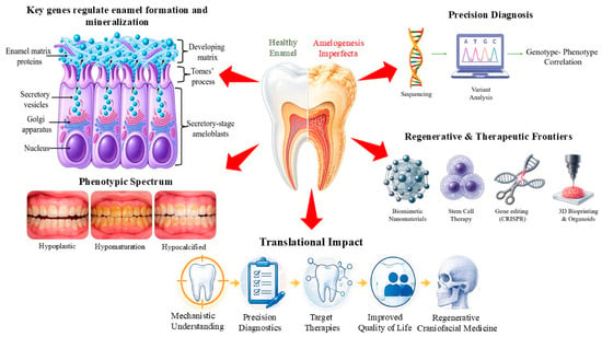
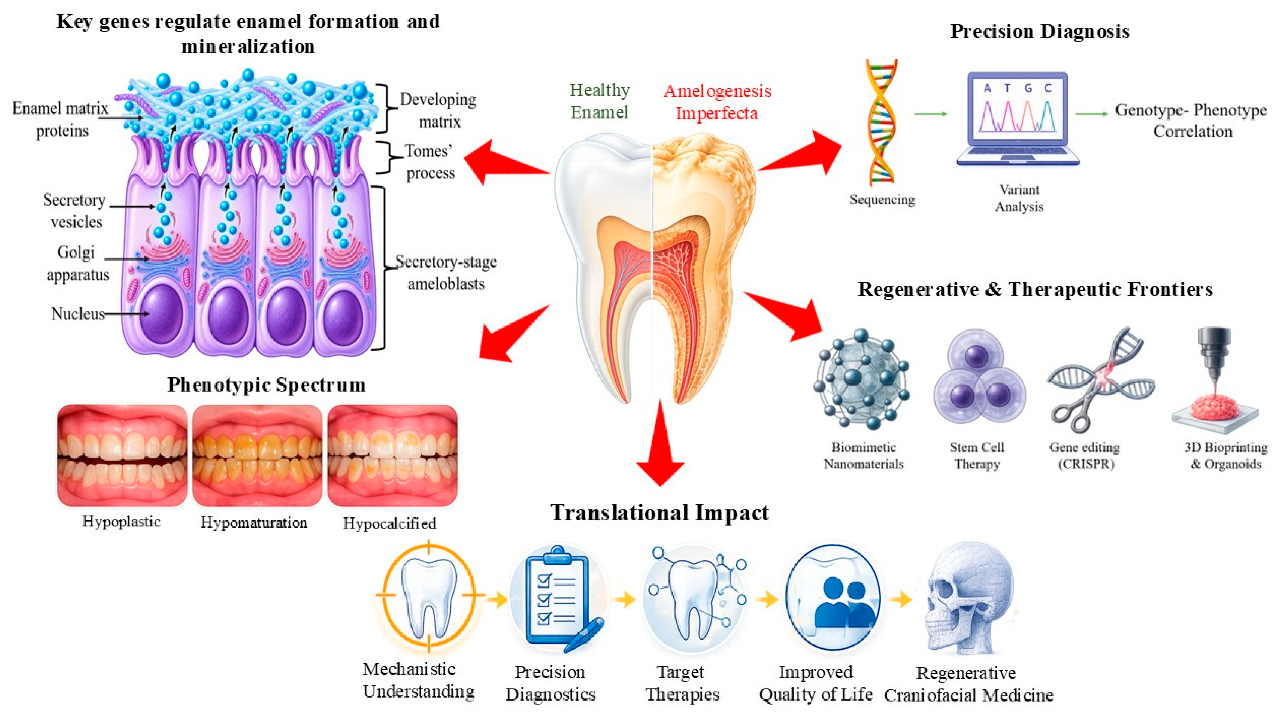
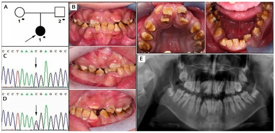
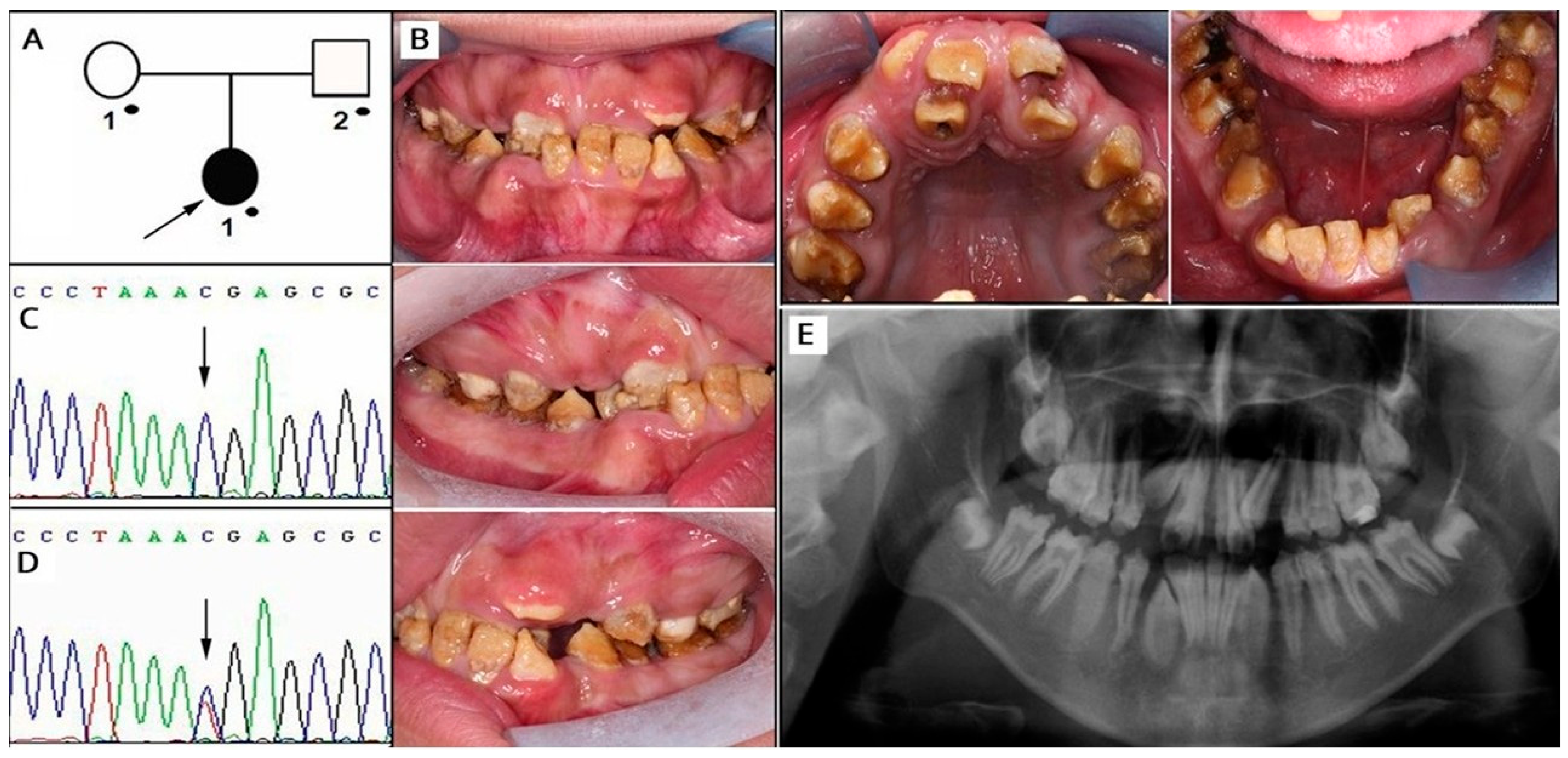
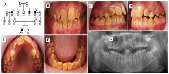
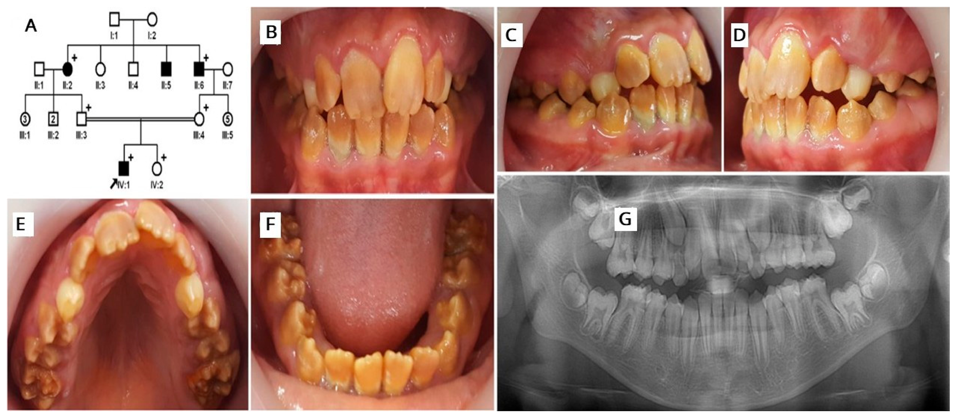
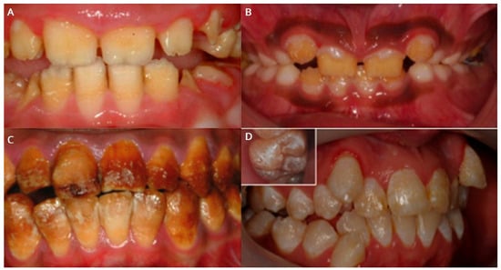
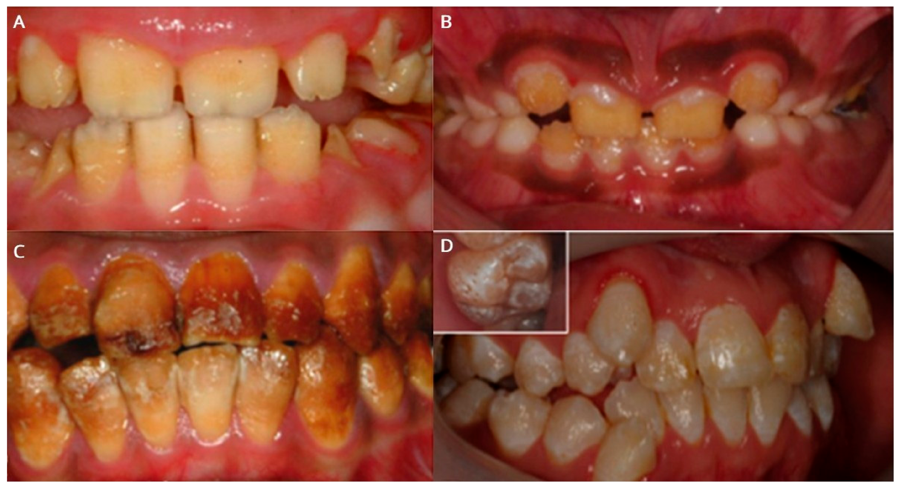
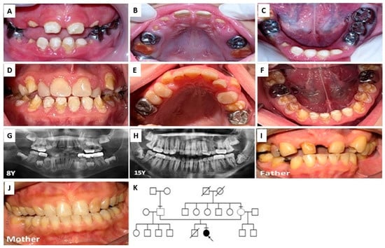
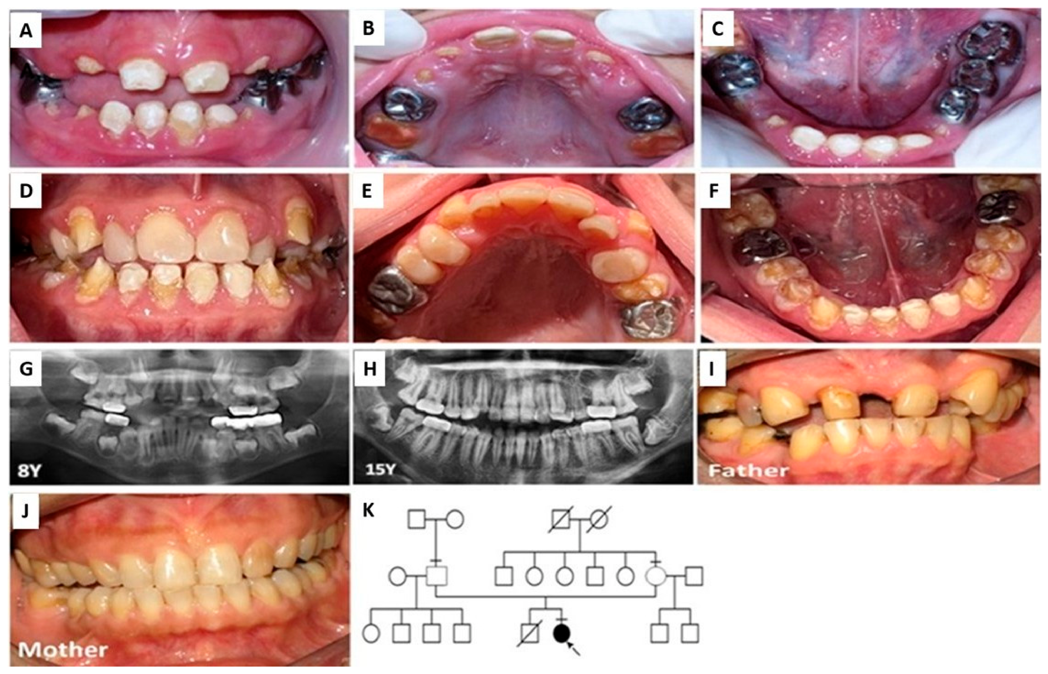

Disclaimer/Publisher’s Note: The statements, opinions and data contained in all publications are solely those of the individual author(s) and contributor(s) and not of MDPI and/or the editor(s). MDPI and/or the editor(s) disclaim responsibility for any injury to people or property resulting from any ideas, methods, instructions or products referred to in the content. |
© 2026 by the author. Licensee MDPI, Basel, Switzerland. This article is an open access article distributed under the terms and conditions of the Creative Commons Attribution (CC BY) license.