


Abstract
Background/Objectives: This study compared the capabilities of four periodontal assessment instruments (CP11 probe, WHO probe, explorer, and #11/12 explorer) to detect subgingival calculus and assess calculus morphology when used by dental hygienists, with a focus on their clinical and educational relevance. Methods: Typodont models with artificially placed subgingival calculus of various shapes were used by 27 dental hygienists to assess the presence and morphology of calculus using each instrument, and a questionnaire on the usability of the #11/12 explorer was completed. Results: Subgingival calculus detection rates did not significantly differ between instruments in either entire mandibular jaw model or individual anterior and posterior regions. Calculus morphology detection did not differ for the entire model or anterior regions but did significantly differ in posterior regions, with the #11/12 explorer showing the highest detection accuracy. Morphology-dependent differences were observed between CP-11 and WHO probes. Qualitative analysis of free-text responses regarding #11/12 explorer use with KH Coder identified clusters related to tactile perception, operability, and experiential/educational aspects. Conclusions: Although the overall calculus detection performance did not significantly differ among the instruments, the #11/12 explorer demonstrated higher detection accuracy in the posterior regions and was favorably rated in terms of tactile feedback and operability. These findings suggest that the #11/12 explorer may be a useful instrument for subgingival calculus assessment and may have educational value for developing tactile assessment skills.
1. Introduction
Periodontitis is a multifactorial, biofilm-induced inflammatory disease characterized by chronic destruction of the tooth-supporting tissues, including the periodontal ligament and alveolar bone [1,2]. Dental calculus acts as a plaque-retentive factor that facilitates biofilm accumulation. Although calculus itself is not considered a direct cause of periodontitis, it is frequently associated with periodontal disease sites because it provides a favorable surface for biofilm retention [3]. Therefore, accurate detection and removal of calculus are essential for the prevention and management of periodontitis. Calculus is categorized as supragingival or subgingival according to its location. While supragingival calculus can be easily identified on visual examination, subgingival calculus requires identification with periodontal assessment instruments. Accurate detection and removal of subgingival calculus are essential in periodontal treatment [4,5]. In clinical practice, detection of subgingival calculus largely depends on tactile feedback from periodontal instruments to evaluate root surface characteristics. Although technologies have been developed to detect subgingival calculus, such as autofluorescence and laser devices [6,7], hand-held instruments remain indispensable because of their cost-effectiveness and practical applicability.
A previous study reported that the #11/12 explorer had a significantly higher capability to detect subgingival calculus compared with that of the WHO probe [8], and its use is mandated in some Western licensure examinations [9]. Thus, the #11/12 explorer is widely regarded as a gold standard for detecting subgingival calculus [8]. However, evidence comparing its performance with other commonly used periodontal assessment instruments remains limited.
Traditionally, in dental hygienist education in Japan, subgingival calculus detection has been performed using probes [10]. However, recent revisions of educational textbooks now describe using both probes and explorers (including the #11/12 explorer) for plaque and calculus detection and present periodontal assessment instruments as being selected according to the site and purpose of use [11,12]. This change reflects a shift toward a more comprehensive approach to periodontal assessment using a variety of hand-held instruments. Nevertheless, studies comparing the detection performance of commonly used periodontal assessment instruments, including the CP-11 probe and explorers other than the WHO probe, remain limited. Therefore, further investigation is needed to clarify the relative effectiveness of these instruments in subgingival calculus detection.
According to the Survey of Dental Diseases in Japan [13], although the 8020 campaign, which encourages people to retain at least 20 of their own teeth by the age of 80, has contributed to an increase in the number of remaining teeth in older adults, the prevalence of periodontal pockets measuring ≥4 mm has also increased. Therefore, the prevention of periodontitis has become a more urgent issue as older people are maintaining their teeth for longer. Thus, this study aimed to compare the calculus detection abilities of various periodontal assessment instruments used by dental hygienists and identify the most useful tools for subgingival calculus detection. By clarifying the effectiveness of these instruments, this study aimed to improve clinical assessment skills and enhance periodontal treatment and preventive care in dental hygiene practices.
2. Materials and Methods
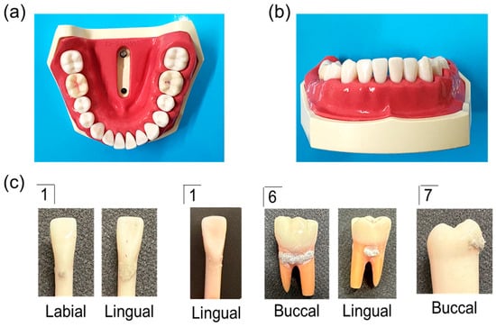
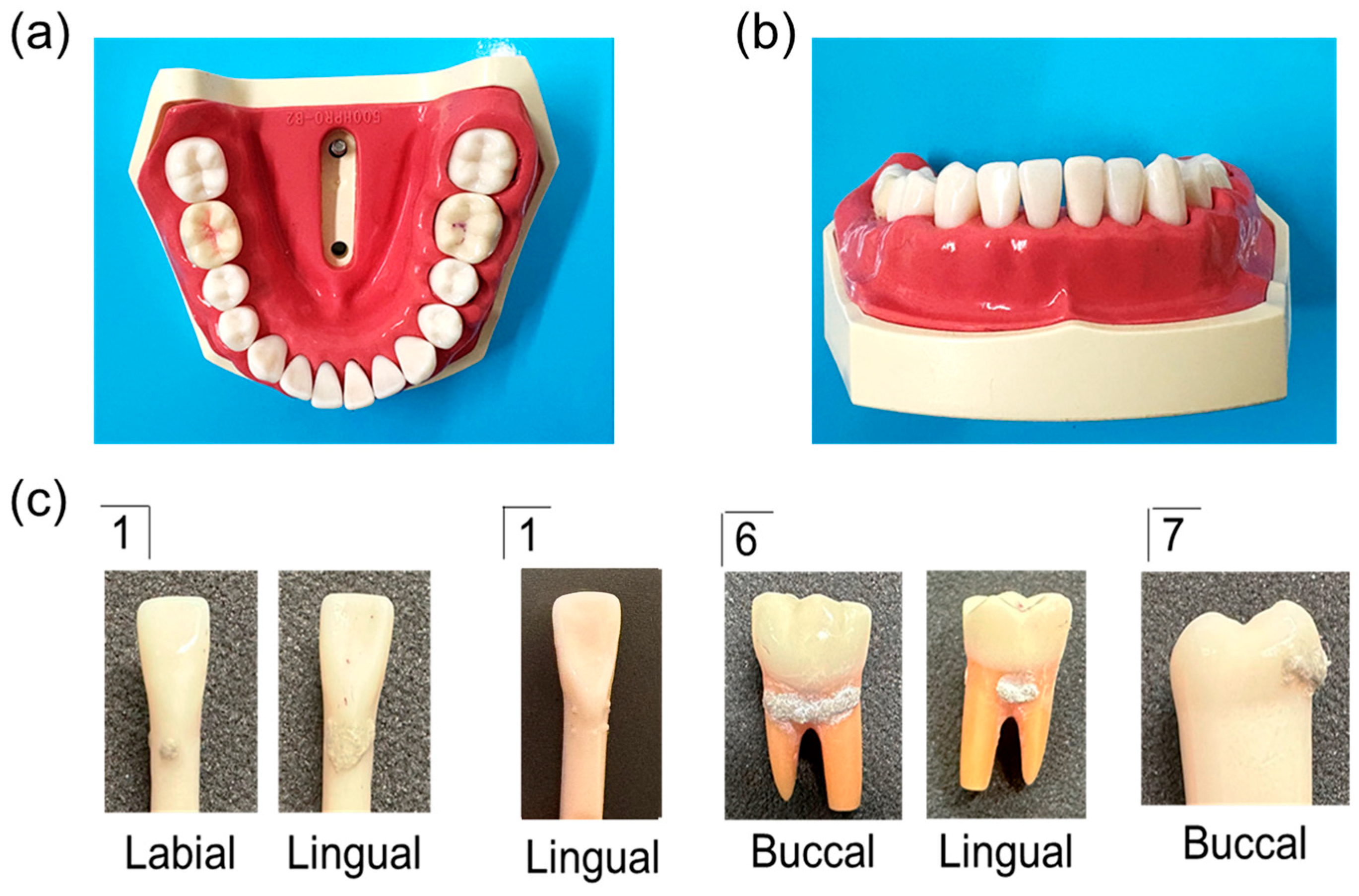
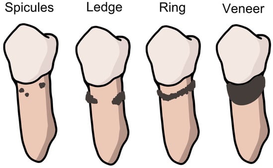
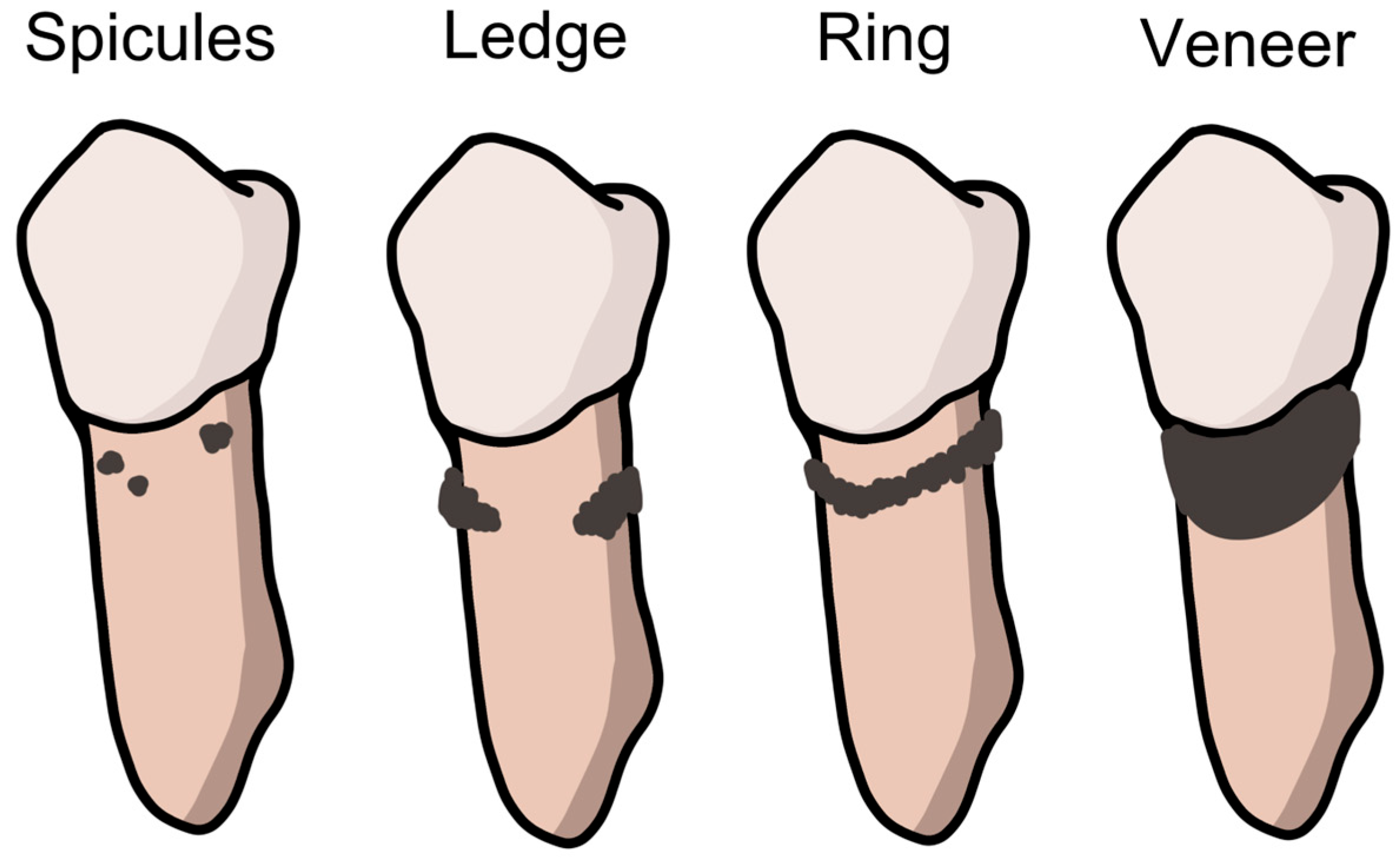
Typodont models with artificial subgingival calculus were prepared using a mandibular jaw model (Nissin, Kyoto, Japan). Artificial calculus was created using a Dental Calculus Set (Nissin) and attached to four teeth: the right and left mandibular central incisors and the left mandibular first and second molars (Figure 1). For the left mandibular central incisor, a typodont tooth with pre-attached artificial calculus was used. Artificial calculus was fabricated in four common morphological forms: spicules, ledges, rings, and veneers (Figure 2) [14].
Figure 1.
Typodont model simulating human dentition with artificially placed dental calculus on selected root surfaces. (a), (b) Mandibular jaw model; (c) Adhesion of artificial dental calculus. The numbers (1, 6, and 7) indicate the tooth types according to the Palmer (Zsigmondy) notation.
Figure 2.
Four types of subgingival calculus shapes. Spicule: small, discrete deposit of calculus typically observed beneath contact points along line angles or near the midline of the tooth surface. Ledge: linear accumulation of calculus forming a ridge parallel to the gingival margin, commonly found on various tooth surfaces. Ring: circumferential ridge of calculus that surrounds the tooth and runs parallel to the gingival margin. Veneer: thin, smooth layer of calculus covering part of the root surface, often presenting a shield-like appearance. Illustration created by the authors based on a periodontal instrumentation textbook [12].
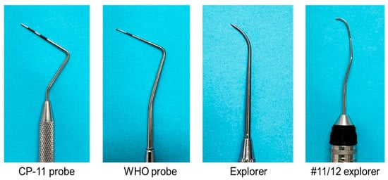
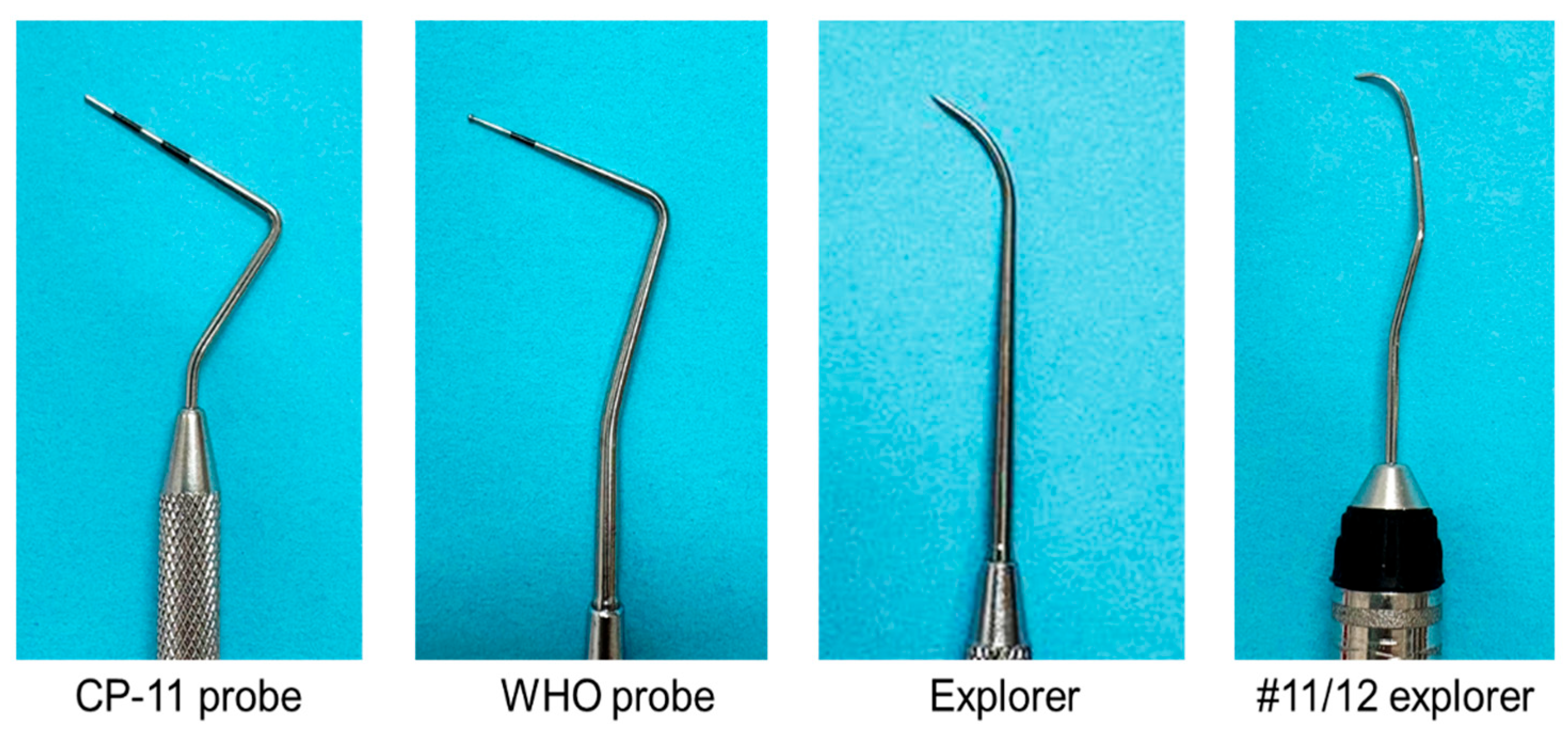
Four periodontal assessment instruments were used in this study: CP11 probe (Hu-Friedy Manufacturing Company, Chicago, IL, USA), WHO probe (YDM, Tokyo, Japan), explorer (YDM), and #11/12 explorer (Hu-Friedy Manufacturing Company) (Figure 3). This study included 27 dental hygienists practicing clinical dental hygiene at a dental hospital in Kumamoto Prefecture, Japan. Participants were recruited using convenience sampling from this institution. The hospital provides a wide range of dental services, including oral surgery, periodontics, pediatric dentistry, orthodontics, general dentistry, and dental anesthesia. Therefore, the participants represented a variety of clinical experiences and professional backgrounds. Each participant was assigned a unique identification number at random. All participants were adults, and the study was explained to them orally and using a document. Written informed consent was obtained from all participants prior to participation. For each instrument, participants were given 5 min to detect the presence and morphology of subgingival calculus. Different jaw models were provided for each instrument; however, participants were not informed that the location and morphology of the artificial calculus were identical across the models. Separate models were used to minimize potential learning and carry-over effects that could occur if participants repeatedly examined the same calculus deposits using multiple instruments. The typodont models were not mounted in a phantom head and were evaluated as standalone jaw models. Because the assessments were conducted at the participants’ clinical facilities, the study was performed under practical space and equipment constraints. Participants were instructed to hold and position the jaw models freely to simulate routine clinical conditions, including adjustments corresponding to maxillary angulation and head rotation typically used during patient care. No auxiliary instruments, such as mouth mirrors or air-water syringes, were used during the assessments. To minimize potential order effects, the sequence of instrument use was counterbalanced across participants. Because four instruments were evaluated, 24 unique instrument orders were possible. All 24 orders were generated, and participants were assigned to these orders as evenly as possible across the study population. The participants were asked to detect the subgingival areas using each instrument and answer questions about the presence and shape of dental calculus. Prior to the experiment, standardized instructions were provided regarding the correct use of the #11/12 explorer. Because most participants had limited experience using this instrument in routine clinical practice, they were also allowed to handle and practice with a typodont model without artificial calculus before the assessment. This familiarization procedure was conducted to standardize instrument use and reduce variability associated with differences in prior experience.
Figure 3.
Periodontal assessment instruments. Examples of CP-11 probe, WHO probe, explorer, and #11/12 explorer used to evaluate the presence and morphology of subgingival calculus.
The participants were also asked to complete a questionnaire regarding their basic attributes (age, sex, educational background, years of clinical experience, and certifications) and their impressions of using the #11/12 explorer (free response).
This study was approved by the Ethics Committee of the Kyushu University of Nursing and Social Welfare (Approval No. 05-020). Data analysis was performed using IBM SPSS Statistics version 25 (IBM Corp., Armonk, NY, USA). Normality of the data was assessed prior to statistical analysis. For overall detection accuracy data that met the assumption of normality, repeated-measures analysis of variance (ANOVA) was used to compare the four periodontal instruments. For subgroup analyses in which the assumption of normality was not satisfied, including comparisons by tooth region and calculus morphology, the Friedman test was applied. When significant differences were identified, post-hoc pairwise comparisons were conducted using the Wilcoxon signed-rank test with Bonferroni correction. A post hoc power analysis for the primary outcome (overall calculus detection accuracy) was conducted using G*Power version 3.1.9.7 (Heinrich Heine University Düsseldorf, Düsseldorf, Germany). Based on the observed effect size (partial η2 = 0.063), the achieved statistical power was 0.891 (α = 0.05, n = 27). p-values < 0.05 were considered statistically significant.
Free-text responses were analyzed using the text-mining software KH Coder version 3. Beta. 08e (Koichi Higuchi, Tokyo, Japan; http://khcoder.net/en, accessed on 1 July 2026) [15]. This software allows quantitative analysis of textual data, including identification of frequently occurring terms and the visualization of relationships among words.
3. Results
3.1. Participant Characteristics
Table 1 summarizes participants’ background characteristics and instrument preferences. All 27 participants were female dental hygienists, with a mean age of 36.2 ± 10.5 years and a mean clinical experience of 13.8 ± 10.1 years. In terms of educational background, 44.4% (n = 12) had completed a 2-year vocational program and 33.3% (n = 9) had graduated from a 4-year university degree. Approximately half of the participants (48.1%, n = 13) had previous training experience with the #11/12 explorer, whereas the remaining participants (51.9%, n = 14) did not. Regarding routinely used periodontal assessment instruments, the CP-11 probe was the most frequently reported instrument (92.6%, n = 25), whereas only 14.8% (n = 4) of participants reported routine use of the #11/12 explorer (multiple responses allowed). After the experiment, the majority of the dental hygienists (74.1%, n = 20) rated the #11/12 explorer as the most user-friendly instrument, whereas the remaining 25.9% (n = 7) selected the CP-11 probe.
Table 1.
Participants’ backgrounds and characteristics.
3.2. Detection of the Presence and Morphology of Subgingival Calculus
Table 2 presents the correct detection rates for the presence and morphology of dental calculus across the four periodontal assessment instruments. No significant differences among the instruments were observed in dental calculus detection in the overall mandibular jaw model (p = 0.166), anterior teeth (p = 0.088), or posterior teeth (p = 0.792). Similarly, for the detection of calculus morphology, there were no significant differences among the instruments in the overall mandibular jaw model (p = 0.694) or anterior teeth (p = 0.384). However, for the posterior teeth, the #11/12 explorer demonstrated a significantly higher correct detection rate for calculus morphology (32.1%) than the CP-11 probe (18.5%), WHO probe (19.8%), and explorer (17.3%) (p = 0.038).
Table 2.
Proportion (%) of correctly detected calculus (presence and morphology) for each periodontal instrument by 27 dental hygienists.
3.3. Influence of Calculus Morphology on Detection Performance
Table 3 shows the detection performance of each periodontal instrument according to calculus shape (spicules, ledge, ring, and veneer). For the CP-11 probe, detection rates varied significantly according to calculus morphology (p = 0.018), with lower detection observed for ring-shaped calculus (39.5%) compared with other forms. Similarly, the WHO probe showed significant variability across calculus shapes (p = 0.002), with the lowest detection rate also noted for ring-shaped calculus (38.3%). In contrast, no significant differences in detection performance across calculus shapes were observed for the explorer (p = 0.201) or the #11/12 explorer (p = 0.671).
Table 3.
Detection of each calculus morphology by each periodontal assessment instrument.
3.4. Qualitative Analysis of Impressions of the #11/12 Explorer
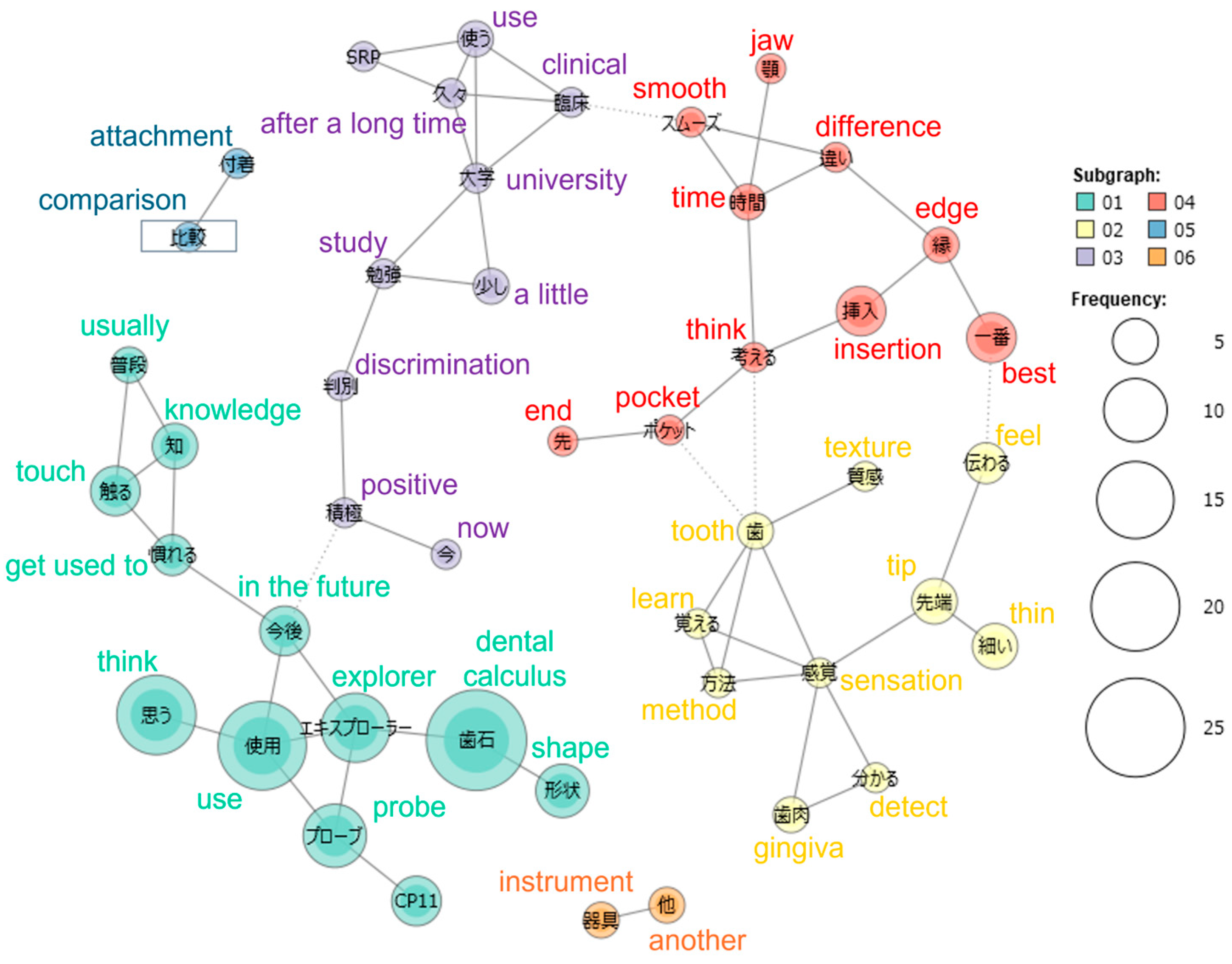
Open-ended questionnaire responses regarding the usability of the #11/12 explorer were analyzed using KH Coder. Figure 4 shows the co-occurrence network of frequently occurring terms. In this network, node size represents the frequency of each term, and node colors indicate clusters automatically identified by the KH Coder algorithm based on co-occurrence relationships among terms. Several clusters of co-occurring terms were identified in the network. One cluster contained words related to tactile perception, such as “feel,” “sensation,” and “thin.” Another cluster included terms associated with operability and handling, including “insertion,” “smooth,” and “best.” In addition, a cluster related to learning and experience was observed, with words such as “university,” “clinical,” “study,” “after a long time,” and “positive” appearing within this cluster.
Figure 4.
Co-occurrence network of terms from open-ended responses regarding the usability of the #11/12 explorer.
4. Discussion
In this study, we compared the capability of four periodontal assessment instruments (CP-11 probe, WHO probe, explorer, and #11/12 explorer) when used by dental hygienists to detect subgingival calculus and assess calculus morphology in typodont models with standardized artificial calculus formations. Although no significant differences were observed among the four instruments in detection of subgingival calculus, a significant difference was found in the assessment of calculus morphology in the posterior regions. This finding suggests that, while simple detection of calculus may be achieved regardless of the instrument used, accurate assessment of calculus morphology requires greater tactile sensitivity and instrument adaptability. While detection of calculus primarily depends on identifying distinct surface irregularities, evaluation of calculus morphology demands finer tactile discrimination of surface characteristics and, thus, is more susceptible to variations in instrument design and tactile feedback. The #11/12 explorer is characterized by a fine tip, a long and complex shank, and a working end bent at approximately 90° to the terminal shank [14]. These structural features may contribute to its improved ability to distinguish a calculus’s subtle surface characteristics.
Anatomical factors likely contributed to the observed results. Anterior teeth generally offer favorable visibility and accessibility, allowing the instruments to perform comparably. In contrast, the posterior regions have more complex anatomical structures, limited visibility, and restricted access, which increase the difficulty of precise assessment. The detection and removal of subgingival calculus is highly dependent on the clinician’s skill and is influenced by anatomical and clinical conditions, resulting in variable accuracy [16,17]. Under such conditions, the structural characteristics of the #11/12 explorer may provide advantages in accessing complex areas and detecting subtle variations in surface morphology.
The influence of calculus morphology further influences the differences between the instruments. Both the CP-11 and WHO probes showed significant variability in detection performance depending on the calculus shape, with lower detection rates observed for ring-shaped calculus. These findings may indicate that periodontal probes are more sensitive to certain morphologies but less effective for detecting circumferential deposits. In contrast, the detection performance of the explorer and #11/12 explorer was not significantly affected by calculus morphology, indicating that explorer-type instruments may provide more consistent tactile discrimination across a range of deposit forms. Such consistency may be particularly beneficial in clinical situations where calculus morphology exhibits substantial variability.
Qualitative findings from the open-ended questionnaire responses provided additional insights into why the #11/12 explorer was frequently selected as the easiest instrument to use. Co-occurrence network analysis identified clusters of terms related to tactile perception, operability, and prior learning experiences. Frequently occurring terms included those associated with tactile sensation (“feel,” “sensation”), instrument handling (“insertion,” “smooth”), and familiarity through education and clinical experience (“university,” “clinical,” “study”). These findings suggest that participants valued the #11/12 explorer primarily because of its perceived tactile feedback and ease of handling. Although a direct relationship between perceived usability and objective detection accuracy was not evaluated in the present study, the qualitative findings provide complementary information regarding participants’ experiences with the instrument. Therefore, the qualitative analysis should be interpreted as an exploration of user perceptions rather than evidence of superior detection performance. Nevertheless, these findings may help explain why the #11/12 explorer was frequently selected by participants as the easiest instrument to use and provide additional context for interpreting the quantitative findings. Future studies should investigate whether individual perceptions of usability are associated with objective detection performance.
At the same time, no single periodontal assessment instrument appeared to be universally superior under all conditions. Rather, appropriate instrument selection according to the anatomical site, calculus morphology, and clinical purpose may be important for accurate subgingival calculus assessment. Therefore, dental hygiene education and clinical training should emphasize not only proficiency with individual instruments but also the ability to appropriately select and use instruments according to specific clinical situations.
This study has some limitations that should be considered. The study was conducted using typodont models, which cannot fully replicate clinical conditions, such as gingival tissue resistance, saliva, or patient-related factors. The tactile feedback obtained from typodont models may differ from that in clinical settings. In addition, the overall detection accuracy observed in this study was relatively low, which may reflect the inherent limitations of the model conditions. Participants were required not only to detect the presence of calculus but also to distinguish among four different calculus morphologies, which may have increased the difficulty of the assessment task. Furthermore, the tactile characteristics of the simulated gingival tissues may have differed from those encountered in clinical practice, potentially affecting the perception of subtle surface irregularities. These factors may partly explain the relatively low detection accuracy observed despite the participants’ substantial clinical experience. Previous research has suggested that subgingival calculus detection is influenced not only by the instrument itself but also by the clinician’s tactile perception and clinical context [16,17,18]. Furthermore, the use of magnification devices significantly improves both calculus detection and removal performance [19], indicating that visual support is likely to enhance detection accuracy. These findings suggest that future studies should incorporate clinical conditions and adjunctive tools, such as magnification, to better evaluate detection performance.
Although participants were blinded to the calculus location and morphology, prior experience with certain instruments may have influenced their detection performance. Previous studies have demonstrated variability in calculus detection ability among examiners [18], suggesting that individual skills and experience influence detection performance. Therefore, structured educational interventions aimed at improving tactile detection skills when using these tools could improve dental hygiene training outcomes.
Finally, the sample size of the study was small. This made it challenging to show strong correlations and may have introduced biases, as the sample consisted only of female dental hygienists. The small sample size and sampling biases mean the results may have limited generalizability. In addition, as all participants were trained within a specific educational and clinical context in Japan, the findings may not be fully generalizable to other regions or settings where training systems and clinical practices differ. This study could be considered a pilot study to investigate the relationship between tools and clinical outcomes in subgingival calculus detection. Future studies should include more diverse and larger samples, as well as participants from different educational and clinical backgrounds, and should be conducted in clinical settings to further validate and expand upon these findings.
Overall, the findings of this study emphasize the importance of selecting periodontal assessment instruments that provide reliable tactile discrimination, particularly in the posterior regions where anatomical complexity may hinder calculus detection. The findings suggest that the #11/12 explorer may be a useful instrument for subgingival calculus assessment and may have educational value for developing tactile assessment skills. However, appropriate instrument selection according to the clinical situation remains important, as no single instrument demonstrated clear superiority under all conditions.
5. Conclusions
Although the overall detection of calculus presence did not differ significantly among the instruments, the #11/12 explorer showed higher accuracy for calculus morphology evaluation in the posterior regions and consistent performance across calculus shapes. In addition, many participants perceived the #11/12 explorer as easy to use, suggesting potential educational value for developing tactile assessment skills. However, no single instrument demonstrated clear superiority under all conditions, highlighting the importance of selecting periodontal assessment instruments according to the clinical situation and assessment purpose. Enhancing the ability and accuracy of subgingival calculus detection in both educational and clinical settings is increasingly important in the context of older individuals retaining their natural teeth for longer periods, with potential implications for long-term oral health outcomes.
Author Contributions
Conceptualization, M.N. (Mako Naniwa) and T.Y.; methodology, M.N. (Mako Naniwa) and T.Y.; software, M.N. (Mako Naniwa); validation, M.N. (Mako Naniwa) and T.Y.; formal analysis, M.N. (Mako Naniwa); investigation, M.N. (Mako Naniwa), M.N. (Masayo Nakamura) and T.Y.; data curation, M.N. (Mako Naniwa); writing—original draft preparation, M.N. (Mako Naniwa); writing—review and editing, M.N. (Masayo Nakamura) and T.Y.; visualization, M.N. (Mako Naniwa); project administration, M.N. (Mako Naniwa). All authors have read and agreed to the published version of the manuscript.
Funding
This research received no external funding.
Institutional Review Board Statement
The study was conducted in accordance with the Declaration of Helsinki, and approved by the Ethics Committee of the Kyushu University of Nursing and Social Welfare (Approval No. 05-020, 5 November 2023).
Informed Consent Statement
Informed consent was obtained from all subjects involved in the study.
Data Availability Statement
The data presented in this study are available on request from the corresponding author due to ethical reasons.
Acknowledgments
The authors would like to thank the staff of the Itoh Dento-Maxillofacial Hospital for their support and cooperation.
Conflicts of Interest
The authors declare no conflicts of interest.
References
- Tonetti, M.S.; Greenwell, H.; Kornman, K.S. Staging and grading of periodontitis: Framework and proposal of a new classification and case definition. J. Periodontol. 2018, 89, S159–S172. [Google Scholar] [CrossRef] [Scilit] [PubMed]
- Könönen, E.; Gursoy, M.; Gursoy, U.K. Periodontitis: A multifaceted disease of tooth-supporting tissues. J. Clin. Med. 2019, 8, 1135. [Google Scholar] [CrossRef] [Scilit] [PubMed]
- Wilson, T.G.; Harrel, S.K.; Nunn, M.E.; Francis, B.; Webb, K. The relationship between the presence of tooth-borne subgingival deposits and inflammation found with a dental endoscope. J. Periodontol. 2008, 79, 2029–2035. [Google Scholar] [CrossRef] [Scilit] [PubMed]
- Cobb, C.M. Non-surgical pocket therapy: Mechanical. Ann. Periodontol. 1996, 1, 443–490. [Google Scholar] [CrossRef] [Scilit] [PubMed]
- Van der Weijden, G.A.; Timmerman, M.F. A systematic review on the clinical efficacy of subgingival debridement in the treatment of chronic periodontitis. J. Clin. Periodontol. 2002, 29, 55–71; discussion 90. [Google Scholar] [CrossRef] [Scilit] [PubMed]
- Chang, J.J.; Chen, C.; Chang, J.; Koka, S.; Jokerst, J.V. A narrative review of imaging tools for imaging subgingival calculus. Front. Oral Maxillofac. Med. 2023, 5, 4. [Google Scholar] [CrossRef] [Scilit] [PubMed]
- Kamath, D.G.; Umesh Nayak, S. Detection, removal and prevention of calculus: Literature Review. Saudi Dent. J. 2014, 26, 7–13. [Google Scholar] [CrossRef] [Scilit] [PubMed]
- Rams, T.E.; Manos, M.P. Comparative in vitro evaluation of WHO periodontal probe and #11/12 dental explorer for subgingival calculus detection. J. Contemp. Dent. Pract. 2021, 22, 13–17. [Google Scholar] [CrossRef] [Scilit]
- American Board of Dental Examiners. ADEX Dental Hygiene Candidate Manual: Clinical Examination. 2026. Available online: https://adextesting.org/wp-content/uploads/2026/01/2026-HYG-CAN-Manual-Clinical_20251024-1.pdf (accessed on 10 May 2026).
- Sato, S.; Kosaka, T.; Inukai, J. (Eds.) Dental Hygiene Series: Periodontology; Ishiyaku Publications Inc.: Tokyo, Japan, 2023. (In Japanese) [Google Scholar]
- Sato, S.; Kosaka, T.; Inukai, J. (Eds.) Dental Hygiene Series: Periodontology, 2nd ed.; Ishiyaku Publications Inc.: Tokyo, Japan, 2026. (In Japanese) [Google Scholar]
- Kosaka, T.; Aiba, C.; Ishikawa, Y.; Endo, K.; Shiratori, T.; Hatanaka, Y.; Mizukami, M.; Yamada, S.; Yoshida, N. (Eds.) Dental Hygiene Series: Preventive Dental Treatment and Oral Health Instruction, 2nd ed.; Ishiyaku Publications Inc.: Tokyo, Japan, 2026. (In Japanese) [Google Scholar]
- Ministry of Health, Labour and Welfare. Report on the Survey of Dental Diseases. 2024. Available online: https://www.mhlw.go.jp/toukei/list/62-28.html (accessed on 10 May 2026).
- Gehrig, J.S.; Sroda, R.; Saccuzzo, D. (Eds.) Fundamentals of Periodontal Instrumentation and Advanced Root Instrumentation, 8th ed.; Wolters Kluwer: Philadelphia, PA, USA, 2017. [Google Scholar]
- Higuchi, K. New quantitative text analytical method and KH Coder software. Jpn. Sociol. Rev. 2017, 68, 334–350, (In Japanese with English Abstract). [Google Scholar] [CrossRef] [Scilit]
- Kato, T.; Sena, K.; Ishiko, R.; Tanda, N.; Yoda, N.; Hihara, H.; Koseki, T. Development of subgingival calculus detector utilizing optical fiber: Verification of its potential for clinical application. PLoS ONE 2024, 19, e0314563. [Google Scholar] [CrossRef] [Scilit] [PubMed]
- Gagnot, G.; Mora, F.; Poblete, M.G.; Vachey, E.; Michel, J.F.; Cathelineau, G. Comparative study of manual and ultrasonic instrumentation of cementum surfaces: Influence of lateral pressure. Int. J. Periodontics Restor. Dent. 2004, 24, 137–145. [Google Scholar]
- Garland, K.V.; Newell, K.J. Dental hygiene faculty calibration in the evaluation of calculus detection. J. Dent. Educ. 2009, 73, 383–389. [Google Scholar] [CrossRef] [Scilit]
- Nakaue, K.; Ikeda, M.; Ichimiya, R.; Kita, K.; Shirayama, Y. Practical effects of using a dental magnifier in a dental hygienist Training School. J. Oral Health Biosci. 2019, 31, 87–91, (In Japanese with English Abstract). [Google Scholar]

Disclaimer/Publisher’s Note: The statements, opinions and data contained in all publications are solely those of the individual author(s) and contributor(s) and not of MDPI and/or the editor(s). MDPI and/or the editor(s) disclaim responsibility for any injury to people or property resulting from any ideas, methods, instructions or products referred to in the content. |
© 2026 by the authors. Licensee MDPI, Basel, Switzerland. This article is an open access article distributed under the terms and conditions of the Creative Commons Attribution (CC BY) license.