


Highlights
What are the main findings?
- The experimental propolis/chitosan varnish did not prevent erosive mineral loss in primary teeth enamel.
- Fluoride varnish (Duraphat®) also failed to protect primary enamel against repeated acidic challenges.
What are the implications of the main findings?
- Conventional and natural-based varnishes may be insufficient to control dental erosion in deciduous teeth under severe acidic conditions.
- Preventive strategies for erosion in primary teeth require alternative or combined approaches beyond topical varnish application.
Abstract
Objective: To evaluate the ability of an experimental propolis/chitosan varnish to inhibit the erosive process in primary tooth enamel. Materials and Methods: Forty-two primary incisors were selected and divided into three groups (n = 14): control/no treatment, Duraphat® varnish, and propolis/chitosan varnish. The varnishes were applied to the experimental area and left in place for 4 h. After varnish removal, the specimens were subjected to erosive challenges with Coca-Cola®. The quantitative outcome variables were mineral loss assessed by longitudinal microhardness, surface roughness, and wear profile. The data were analyzed using ANOVA and Tukey’s test (p < 0.05). Results: Wear profile and surface roughness analyses showed no significant differences among the groups (p > 0.05). In the microhardness analysis, the control and experimental groups showed significant differences between the control and demineralized areas and between the control and experimental areas (p < 0.05). In the Duraphat group, a significant difference was observed between the control and experimental areas (p < 0.05). Conclusions: The experimental propolis/chitosan varnish was unable to prevent the progression of mineral loss in the enamel of primary teeth subjected to erosive challenge. Duraphat® fluoride varnish was also unable to protect the enamel. Clinical relevance: Several studies have investigated the use of propolis and chitosan in dentistry, and promising results have been reported when these agents are used individually; however, the propolis/chitosan combination remains poorly explored. To the best of our knowledge, this is the first study to investigate this combination for dental erosion in primary teeth.
1. Introduction
The modern lifestyle has resulted in a significant change in eating habits, with an increase in the intake of acidic beverages and foods [1,2,3,4]. In recent years, the consumption of soft drinks and energy drinks has increased substantially, especially among children and adolescents [5]. These changes have directly affected oral health by increasing the risk of dental erosion, which results from repeated contact between acids and the tooth surface [2,6,7].
Early diagnosis of dental erosion is difficult, and at the macroscopic level, the enamel becomes smooth, silky, and sometimes opaque [2,6,7]. As the process progresses, rounding of cusps and restorations may occur, along with dentin hypersensitivity due to exposure of the dentinal tubules, esthetic changes, and even loss of vertical dimension [1,3,4,6,7].
To prevent and control the erosive process, it is necessary to investigate new materials, and interest in natural substances and fluoride alternatives has increased in the search for products with lower toxicity, greater therapeutic potential, and lower cost [8]. One substance that has been studied is propolis, a natural resin produced by bees that has several beneficial properties [8,9,10]. It has been used in several areas, including the treatment of dentin hypersensitivity and aphthous ulcers, because of its antimicrobial and anti-inflammatory properties [8,9,10]. Propolis extract has also been incorporated with dentifrices, varnishes, and mouth rinses as an alternative for reducing the Streptococcus mutans counts and preventing caries [10,11].
Chitosan is another biomaterial that has been widely studied because of its biodegradability, biocompatibility, antimicrobial, antioxidant, and anti-inflammatory properties, among others [12,13,14,15]. With regard to tooth erosion, chitosan has been shown to form an acid-resistant layer on the tooth surface, thereby protecting it from contact with erosive acids [15,16,17,18]. Chitosan shows affinity for enamel because of its strong positive zeta potential, which allows it to adsorb through electrostatic interactions and to bind to proteins present in the salivary pellicle, especially mucin. Furthermore, some studies have shown that its association with fluoride dentifrices and solutions reduces erosive wear [15,16,17,18]. Given the complementary properties of propolis and chitosan, combining these agents into a single varnish may theoretically enhance enamel protection against acidic challenges.
Therefore, this study aimed to evaluate the ability of an experimental propolis/chitosan varnish to inhibit enamel mineral loss in primary teeth subjected to repeated erosive challenges, using microhardness, surface roughness, and wear profile analyses.
2. Materials and Methods
This study was approved by the Research Ethics Committee of the Ribeirão Preto School of Dentistry, University of São Paulo (USP)—CAAE: 38187420.6.0000.5419. The study was approved at the 241st Ordinary Meeting of the CEP/FORP on 11 November 2020.
This in vitro study had a factorial design, with the treatment factor evaluated at three levels: no treatment (control), Duraphat® fluoride varnish (5% sodium fluoride, Colgate-Palmolive, Hamburg, Germany), and experimental propolis/chitosan varnish. The experimental units were 42 human deciduous incisors, randomly divided into three groups (n = 14). The quantitative outcome variables were mineral loss assessed by changes in longitudinal microhardness, surface roughness, and wear profile measured by confocal microscopy. The enamel surface images were analyzed morphologically and qualitatively to identify possible changes in this substrate after the proposed treatments.
Forty-two human primary incisors were selected from the tooth bank of the Ribeirão Preto School of Dentistry, University of São Paulo. The criteria for tooth selection were the absence of caries lesions, cracks, or enamel development defects. The teeth were initially cleaned with pumice stone and water, using brushes. The teeth were disinfected by immersion in 0.1% thymol solution for one week. Afterward, the teeth were thoroughly rinsed and stored in deionized water, which was changed daily during specimen preparation.
When present, the roots were sectioned 1 mm below the cementoenamel junction using a diamond disk (Buehler®, Lake Bluff, IL, USA) under water cooling in a cutting machine (Isomet 1000, Buehler, Lake Bluff, IL, USA). Subsequently, the teeth were fixed onto acrylic resin blocks with the aid of a parallelometer so that the enamel surface remained parallel to the block. Then, half of the buccal surface was covered with composite resin (Filtek™ Z350, Solventum, Maplewood, MN, USA), hereafter referred to as the control area (CA).
The specimens were subjected to an initial erosive cycle, in which each tooth was immersed, under agitation, in 5 mL of Coca-Cola® (Coca-Cola®, São Paulo, SP, Brazil) (4 °C) for 20 s on a shaking table (Orbital CT-155, Cientec Equipamentos, São Paulo, Brazil) [19]. Six erosive challenges were performed with an interval of one hour between each challenge, during this interval, the specimens were kept in artificial saliva (methyl P-hydroxybenzoate, 2.00; sodium carboxymethyl cellulose, 10.0; KCl, 0.625; MgCl2-6H2O, 0.059; CaCl2-2H2O, 0.166; K2HPO4, 0.804; KH2PO4, 0.326) in an incubator (Odontobrás, Ribeirão Preto, SP, Brazil) at 37 °C [20].
After the initial erosive challenge, half of the remaining exposed area was covered with composite resin (Filtek™ Z350, Solventum, Maplewood, MN, USA), which was considered the demineralized untreated area (DA), that is, the area that was subjected to an erosive challenge. The specimens were randomly divided into three groups (n = 14): G1—Control/no treatment; G2—Duraphat® 5% fluoride varnish; G3—Experimental varnish.
Duraphat® varnish and the experimental varnish were actively applied to the enamel surface in the exposed area (experimental area) with the aid of a microbrush for 15 s and left in artificial saliva at 37 °C for 4 h. Subsequently, the varnish was removed from the surface. At the end of the treatment, the specimens were stored in artificial saliva at 37 °C for 24 h before starting the erosive challenges. Each specimen was immersed under agitation in 5 mL of Coca-Cola® (4 °C) for the 20 s on a shaking table. Six erosive challenges were performed per day for 5 days, with a 1 h interval between challenges. During this interval, the specimens were kept in artificial saliva at 37 °C. At the end of each cycle, the saliva was replaced, and the specimens were kept at 37 °C until the next cycle on the following day. In summary, all erosive challenges were performed under standardized temperature conditions. Coca-Cola® was used at 4 °C, and the specimens were maintained in artificial saliva at 37 °C between erosive cycles and overnight. Artificial saliva was renewed daily. In addition, varnish application was standardized by using the same amount of material (20 mg), active application for 15 s, and careful removal of excess material. However, the dynamic pH variation in the artificial saliva and the thickness/uniformity of the varnish film were not instrumentally monitored and should be considered limitations of the present study.
After the challenges were completed, the teeth were immersed in artificial saliva at 37 °C for 24 h. Then, the composite resin was gently removed, exposing the control and demineralized areas, and the specimens were cleaned in an ultrasonic cleaner (Bio Wash, Bio-art, São Carlos, SP, Brazil) and stored in deionized water for analysis.
The wear profile and enamel surface roughness were evaluated using a laser confocal microscope (LEXT OLS4100, Olympus, Tokyo, Japan) connected to a computer with specific software (OLS4100®, Olympus, Tokyo, Japan). The specimens were positioned parallel to the objective lens with the aid of a parallelometer and the images were captured with a 10× objective (216× magnification). To analyze surface roughness (Sa) in µm, images of the control, demineralized, and experimental areas were taken.
To evaluate the wear profile (Rv), images were obtained at the interfaces: control-demineralized area and control-experimental area, with the control area used as reference. Six measurements of each region were performed in µm and the mean value was obtained, and then the values of these interfaces were added for data analysis. Morphological evaluation of the enamel surface was performed by obtaining images of the control, demineralized, and experimental areas using a 100× objective with a final magnification of 2131×.
After microscope analysis, the specimens were embedded in acrylic resin and subsequently sectioned with a diamond disk (Buehler®, Lake Bluff, IL, USA) in a cutting machine (Isomet 1000, Buehler, Lake Bluff, IL, USA) obtaining a fragment containing the control, demineralized and experimental areas. The lateral surface of the fragment was flattened and polished in an Arotec APL-4 polisher (Arotec S/A Ind. e Comércio, São Paulo, SP, Brazil) with #1200 grain aluminum oxide sandpaper and felt disk and 0.3 and 0.05 μm alumina suspension (Arotec SA Ind. e Comércio, São Paulo, SP, Brazil). After polishing, the specimens were ultrasonically cleaned (Bio Wash, Bio-Art, São Carlos, SP, Brazil) for five minutes in deionized water to wash and remove residues.
A Shimadzu Micro Hardness Tester HMV-2000 (Shimadzu Corporation, Kyoto, Japan) with a Knoop pyramidal diamond penetrator was used to analyze the longitudinal microhardness in depth using a load of 25 gf, applied for 10 s. Indentations were made in the subsurface region (30 µm below the enamel surface and the following markings at 60 µm and 100 µm from the surface) in the 3 areas. Between each of these areas, 1000 μm was maintained to avoid any interference between the areas. For each depth and area, three indentations were performed, with 200 μm between them, totaling nine indentations in each area. Finally, the mean value for each area was calculated (control, demineralized and experimental) and these values were used for data analysis.
The data were analyzed for normality and homogeneity and were found to be normally distributed and homogeneous; therefore, one-way analysis of variance (ANOVA) was performed for the analyses of wear profile, surface roughness, and longitudinal microhardness. For comparison of means, Tukey’s test was used at a 5% significance level. The images obtained by laser confocal microscopy were analyzed qualitatively.
3. Results
In the analysis of the wear profile, it was observed that there was no statistically significant difference (p > 0.05) among the control, Duraphat®, and experimental groups. All groups showed similar superficial enamel mineral loss (Table 1).
Table 1.
Mean and standard deviation, in µm, of the wear profile of the analyzed groups.
When superficial roughness was analyzed separately for each area, no statistically significant difference was found among the groups (p > 0.05). In the intragroup roughness analysis, in all of them, the roughness in the control area was statistically different from the demineralized and experimental areas (p < 0.05). However, between the demineralized and experimental areas, there was no statistically significant difference (p > 0.05) (Table 2).
Table 2.
Mean and standard deviation, in µm, of the enamel surface roughness of the analyzed areas.
Thus, at baseline, the enamel surface was rougher, and after the first erosive challenge, there was a decrease in surface roughness that was maintained until the end of the study.
Microhardness was analyzed separately for each area, and it was observed that for the control, demineralized, and experimental areas, there was no statistically significant difference (p > 0.05) (Table 3). When analyzing the groups internally, it was observed that in the control and experimental groups, there was a statistically significant difference between the control/demineralized and control/experimental areas (p < 0.05). A decrease in microhardness values was observed between the demineralized and experimental areas; however, this difference was not statistically significant (p > 0.05). In the Duraphat group, there was a statistically significant difference only between the control/experimental areas (p < 0.05).
Table 3.
Mean and standard deviation, in KHN, of the longitudinal microhardness of the analyzed areas.
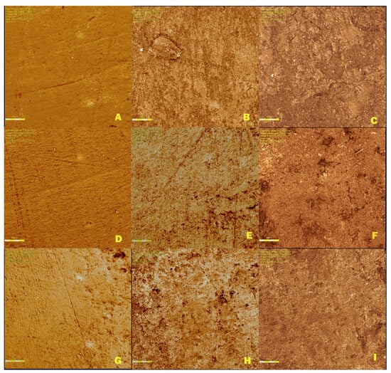
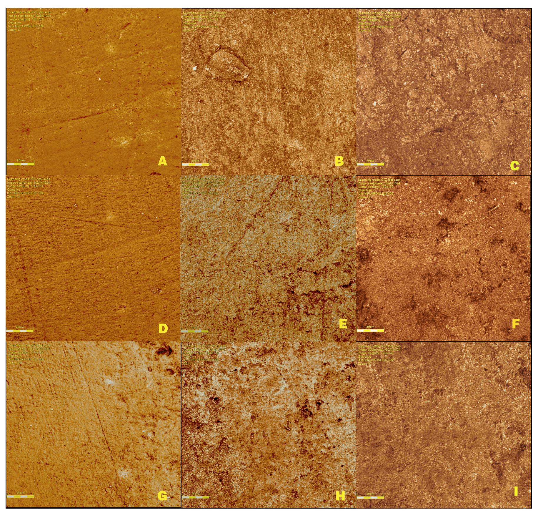
In the morphological analysis, it was observed that all analyzed areas in all groups showed alterations in the enamel surface. Figure 1 illustrates the progression of the erosive process throughout the study. In the control area, the enamel presented a more uniform aspect, without porosity or demineralized areas. In the demineralized and experimental areas, the enamel had a rough appearance, with some porosities and extensive areas of demineralization, and in some images, it was possible to observe small depressions on the surface, making mineral loss evident. The images were similar; therefore, morphologically, there were no morphological differences among the control, Duraphat®, and experimental groups.
Figure 1.
Enamel surfaces of the groups at different time points (baseline, after demineralization, and after treatment/erosive challenge). Control group: (A), control area; (B), demineralized area; (C), experimental area. Duraphat® group: (D), control area; (E), demineralized area; (F), experimental area. Experimental group: (G), control area; (H), demineralized area; (I), experimental area.
4. Discussion
In the present study, it was observed that all analyzed groups allowed the progression of enamel surface demineralization, and this mineral loss was similar among them. In the control group, as no treatment was performed, continued mineral loss was expected. In the Duraphat and experimental groups, it was possible to observe that the varnishes used were not efficient in protecting the enamel and preventing the progression of mineral loss caused by the erosive process, which was induced by successive immersion in Coca-Cola.
Importantly, the lack of a protective effect in the positive control group should not be interpreted as evidence of an invalid experimental model. The erosive protocol produced measurable substrate changes, as confirmed by the significant differences observed between the control and eroded areas in the intragroup analyses. Thus, the findings suggest that the present protocol represented a severe erosive condition capable of overwhelming the protective effect of the tested varnishes, particularly considering the greater susceptibility of primary enamel.
In this study, primary teeth were used because they have specific structural characteristics and differ from permanent teeth. In addition to being smaller and having a thinner enamel layer [5,21], primary teeth enamel has a larger amount of carbonate incorporated in its crystals, and these CO32− ions can replace an OH- group and form type A hydroxyapatite or replace a PO43− group and form type B hydroxyapatite. The primary teeth enamel is composed of type A hydroxyapatite that is significantly more soluble [5,21]. These hydroxyapatite crystals are arranged in bundles forming the enamel prisms, which also differ in deciduous teeth as they are smaller, less organized, and more spread out, having a greater interprismatic distance [5,21,22]. The deciduous enamel is also less mineralized, while the permanent tooth has about 97% of its volume mineral content, the deciduous tooth has about 81–94% [2]. These differences make primary teeth more vulnerable to demineralization; therefore, the behavior of the varnishes used may differ from that observed in permanent teeth.
For the erosive challenges, Coca-Cola® was used because it is a widely consumed beverage that has a low pH (2.5) and high titratable acidity, important characteristics to induce enamel demineralization [23]. The aim was to simulate the daily life of a child with a high consumption of soft drinks [24,25]. Several studies have also used Coca-Cola to induce demineralization, but there was a variation in the way the erosive cycles were performed [24,26,27]. The methodology employed in this study has already been used by the research group in a previous study [19], and the proposal of performing six erosive cycles per day for five consecutive days was to simulate a condition of intense erosive wear.
Duraphat® fluoride varnish was used because it has been extensively studied for the prevention of dental erosion [5,22] and served as a commercial control; moreover, it is a product commonly used in clinical practice. In this study, Duraphat® varnish was not effective in preventing the progression of enamel erosive wear. Similar to the study by Murakami et al. (2009) [22] in which fluoride varnish failed to protect the enamel surface of deciduous teeth subjected to erosive challenges with Coca-Cola®, there was also a reduction in Knoop hardness throughout the experiment, which corroborates the results of this study. Another factor that must be taken into account is that the varnish contained higher concentrations of fluoride. The literature has shown that the low concentration suppressed demineralization in the erosion process as a function of the application rate [28].
Another important factor to consider is that the mechanism of sodium fluoride (NaF) in helping to prevent the erosive process involves the formation of a calcium fluoride (CaF2) layer on the enamel. This layer acts as a physical barrier against acids. For a continuous CaF2 coating to form, the fluoride agent must be concentrated, such as Duraphat, have a low pH, and be applied for a sufficiently long time. However, the CaF2 layer is quickly dissolved under acidic conditions, which limits the effectiveness of conventional fluorides. To be effective, the layer must be dense enough to form a stable barrier against acid attacks. Additionally, the time required for this precipitation to form on the enamel surface remains unknown [5].
Considering that the action of Duraphat is time-dependent and that the reactivity of the entire varnish with enamel may only stabilize after 24 h [29], this could affect the formation of the CaF2 layer. It is possible that the exposure time used in the present study was insufficient to allow the formation of a CaF2 layer with adequate thickness and surface homogeneity to protect the enamel.
Propolis has been used to modify the salivary pellicle due to the polyphenols that are part of its chemical composition, thereby increasing its protective effect, although the results have not been satisfactory [30,31]. And to remineralize dental enamel after the erosive process, which was effective when propolis oil was used, and there was an increase in Vickers microhardness [32]. These studies were in vitro and performed on permanent teeth. In the present study, the extract was used in artificial saliva, which does not contain proteins capable of interacting with propolis; this may explain the results obtained.
On the other hand, Chitosan is a promising material in studies on dental erosion. Its use on both enamel and dentin suggests that this biopolymer can adhere to dental substrates and thus protect the surface, reducing mineral loss [33,34,35,36]. In this context, a previous study conducted by the research group obtained interesting results with the use of a chitosan solution like the one used in the present study [33]. The solution used was able to control the erosive process, reducing the loss of dentin structure. However, bovine teeth were used, presenting similar characteristics to permanent human teeth [37]. The association of chitosan, fluoride, and metallic ions (tin/titanium tetrafluoride) has also been widely investigated and has shown encouraging results [16,17,18,36]. However, the literature on chitosan solutions applied to primary teeth is still scarce, and as these differ from permanent teeth, their study is necessary. In the present study, it was observed that the chitosan-based solution was not effective in halting the erosive process; this difference with the literature is probably due to the particularities of the structure analyzed.
Few studies have investigated the association between propolis and chitosan, particularly with regard to their efficacy against Enterococcus faecalis biofilms. As both have antimicrobial properties, this synergism was able to inhibit bacterial growth and biofilm formation, including in deciduous teeth, in which the propolis/chitosan solution proved to be effective as an intracanal medication [38,39]. To our knowledge, there are no studies on propolis/chitosan varnish in the context of erosive tooth wear that could serve as a basis for comparison.
When surface roughness (Sa) values were evaluated, a decrease was observed throughout the study, i.e., in the beginning, the enamel was rougher. This can be explained by the demineralizing power of Coca-Cola, which when in contact with the enamel surface causes mineral loss and consequently provides a certain homogeneity of this surface. Thus, both the erosive effect of Coca-Cola and the inefficiency of the varnishes used did not prevent these effects on the tooth surface. However, when analyzing in detail the morphology of the enamel surface at the beginning of the study the enamel showed a less irregular topography and at the end, the irregular appearance increased. This difference probably occurred because the morphological analysis was performed at a much higher magnification than the roughness analysis, and thus the details of a very specific area of the surface were observed.
Continuous mineral loss due to the erosive challenges was observed in all groups, as demonstrated by the decrease in microhardness values throughout the study. The same was observed in the wear profile, and there was also similar mineral loss in the three groups, in the areas submitted to the erosive challenges, demineralized and experimental areas. This shows that no treatment was able to stop or reduce structural loss.
Although the results show that there was no efficacy of the experimental varnish, it is important to emphasize the innovative proposal of the study by using a varnish of natural origin and two biomaterials with pharmacological properties well reported in the literature [8,9,13,14,40]. Moreover, the search for new materials to prevent or minimize the damage caused by dental erosion is warranted, especially in the deciduous dentition, since this condition has become more and more prevalent and increasingly affects the young population [24,41]. Carbonated soft drinks are widely consumed by children, and because of their high demineralizing potential, dentists should advise parents/guardians regarding their consumption, to prevent tooth erosion and avoid further damage to the deciduous teeth, given the importance of maintaining healthy deciduous teeth until their exfoliation. Thus, further studies aimed at the treatment of dental erosion in deciduous teeth are necessary [42,43].
This study has limitations that should be acknowledged. The dynamic pH variation in artificial saliva was not monitored throughout the experimental period, and the thickness/uniformity of the varnish layer was not instrumentally measured, although the application protocol was standardized. In addition, no isolated propolis or chitosan groups were included, which prevents any conclusion regarding a possible synergistic interaction between these components. The physicochemical stability of the experimental varnish under acidic conditions and its cytotoxicity/biocompatibility were also not evaluated. Furthermore, the control, demineralized, and experimental areas were assessed within the same tooth. Although this design reduces inter-specimen variability and allows internal comparison under standardized substrate conditions, these measurements may not be completely independent from a statistical perspective. For these reasons, the present findings should be interpreted as exploratory in vitro data and not as evidence of clinical applicability.
5. Conclusions
Within the limitations of this in vitro study, the experimental propolis/chitosan varnish was not effective in preventing enamel mineral loss in primary teeth subjected to erosive challenge. Similarly, Duraphat® fluoride varnish did not show protective effects under the tested conditions. The continuous mineral loss observed across all groups highlights the high erosive potential of acidic beverages such as Coca-Cola®, especially for the more susceptible enamel of primary teeth. Despite the lack of efficacy, the innovative use of natural biomaterials such as propolis and chitosan remains a promising biocompatible approach that warrants further investigation. Future studies should explore alternative formulations, longer application times, the incorporation of salivary proteins, and in situ or in vivo models to better simulate clinical conditions and potentially improve the protective performance of these natural agents.
Author Contributions
B.C.d.F.R.; Conceptualization, Data curation, Formal analysis, Investigation, Methodology, Project administration, Resources, Validation, Visualization, Writing—original draft, Writing—review and editing. J.J.F.; Data curation, Formal analysis, Methodology, Project administration, Writing—original draft, Writing—review and editing. A.C.d.A.; Data curation, Formal analysis, Investigation, Methodology, Writing—original draft, Writing—review and editing. R.S.T.F.; Data curation, Formal analysis, Investigation, Methodology, Writing—original draft, Writing—review and editing. E.R.d.S.; Data curation, Formal analysis, Investigation, Methodology, Writing—original draft, Writing—review and editing. R.G.P.-D.; Conceptualization, Data curation, Formal analysis, Funding acquisition, Investigation, Methodology, Project administration, Resources, Software, Supervision, Validation, Visualization, Writing—original draft, Writing—review and editing. All authors have read and agreed to the published version of the manuscript.
Funding
This work was supported by the Coordenação de Aperfeiçoamento de Pessoal de Nível Superior—Brasil (CAPES)—Finance Code 001.
Institutional Review Board Statement
The study was conducted in accordance with the Declaration of Helsinki, and the protocol was approved by the Ethics Committee of the Ribeirão Preto School of Dentistry, University of São Paulo (USP) (CAAE: 38187420.6.0000.5419) on 11 November 2020.
Informed Consent Statement
The CT data used for model construction were fully anonymized, and no identifiable personal information was included. Therefore, the requirement for informed consent was waived by the Institutional Ethics Committee of the Ribeirão Preto School of Dentistry, University of São Paulo (USP).
Data Availability Statement
All data supporting the findings of this study are available within the paper.
Conflicts of Interest
The authors declare no conflicts of interest. All authors declare that they have no affiliations with or involvement in any organization or entity with any financial interest or non-financial interest in the subject matter or materials discussed in this manuscript.
References
- Assunção, C.M.; Schlueter, N.; Rodrigues, J.A.; Carvalho, T.S.; Lussi, A. Do fluoride toothpastes have similar preventive effect in permanent and primary teeth against erosive tooth wear? Int. J. Paediatr. Dent. 2018, 29, 228–236. [Google Scholar] [CrossRef] [Scilit]
- Rajeev, G.; Lewis, A.J.; N, S. A time based objective evaluation of the erosive effects of various beverages on enamel and cementum of deciduous and permanent teeth. J. Clin. Exp. Dent. 2020, 12, e1–e8. [Google Scholar] [CrossRef] [Scilit]
- Chan, A.S.; Tran, T.T.K.; Hsu, Y.H.; Liu, S.Y.S.; Kroon, J. A systematic review of dietary acids and habits on dental erosion in adolescents. Int. J. Paediatr. Dent. 2020, 30, 713–733. [Google Scholar] [CrossRef] [Scilit]
- Donovan, T.; Nguyen-Ngoc, C.; Abd Alraheam, I.; Irusa, K. Contemporary diagnosis and management of dental erosion. J. Esthet. Restor. Dent. 2021, 33, 78–87. [Google Scholar] [CrossRef] [Scilit] [PubMed]
- Lussi, A.; Buzalaf, M.A.R.; Duangthip, D.; Anttonen, V.; Ganss, C.; João-Souza, S.H.; Baumann, T.; Carvalho, T.S. The use of fluoride for the prevention of dental erosion and erosive tooth wear in children and adolescents. Eur. Arch. Paediatr. Dent. 2019, 20, 517–527. [Google Scholar] [CrossRef] [Scilit]
- Spinola, S.G.; Sobral, A.P.T.; Mandetta, A.R.H.; Gimenez, T.; Gonçalves, M.L.L.; Soares, P.V.; Santos, E.M.; Imparato, J.C.P.; Horliana, A.C.R.T.; Mesquita-Ferrari, R.A.; et al. Childhood early oral ageing syndrome: Prevalence and association with possible aetiological factors and consequences for the vertical dimension of occlusion: Protocol for a cross-sectional study. BMJ Open 2024, 14, e090085. [Google Scholar] [CrossRef] [Scilit] [PubMed] [PubMed Central]
- Yip, K.; Lam, P.P.Y.; Yiu, C.K.Y. Prevalence and Associated Factors of Erosive Tooth Wear among Preschool Children-A Systematic Review and Meta-Analysis. Healthcare 2022, 10, 491. [Google Scholar] [CrossRef] [Scilit]
- da Silva Barboza, A.; Aitken-Saavedra, J.P.; Ferreira, M.L.; Fábio Aranha, A.M.; Lund, R.G. Are propolis extracts potential pharmacological agents in human oral health?—A scoping review and technology prospecting. J. Ethnopharmacol. 2021, 271, 113846. [Google Scholar] [CrossRef] [Scilit]
- Zulhendri, F.; Felitti, R.; Fearnley, J.; Ravalia, M. The use of propolis in dentistry, oral health, and medicine: A review. J. Oral Biosci. 2021, 63, 23–34. [Google Scholar] [CrossRef] [Scilit] [PubMed]
- Saeed, M.A.; Khabeer, A.; Faridi, M.A.; Makhdoom, G. Effectiveness of propolis in maintaining oral health: A scoping review. Can. J. Dent. Hyg. 2021, 55, 167–176. [Google Scholar] [PubMed]
- Kodgi, V.; Shetty, P.; Thimmaiah, C.; Thomas, N.A.; Vergis, B.A.; Shetty, K. Comparative Assessment of Antimicrobial Activity of Propolis and Chlorhexidine on Salivary Isolates of Candida albicans and Streptococcus mutans in Children with Severe Early Childhood Caries: An In Vitro Study. Int. J. Clin. Pediatr. Dent. 2024, 17, 591–595. [Google Scholar] [CrossRef] [Scilit] [PubMed] [PubMed Central]
- Fakhri, E.; Eslami, H.; Maroufi, P.; Pakdel, F.; Taghizadeh, S.; Ganbarov, K.; Yousefi, M.; Tanomand, A.; Yousefi, B.; Mahmoudi, S.; et al. Chitosan biomaterials application in dentistry. Int. J. Biol. Macromol. 2020, 162, 956–974. [Google Scholar] [CrossRef] [Scilit]
- Zhang, C.; Hui, D.; Du, C.; Sun, H.; Peng, W.; Pu, X.; Li, Z.; Sun, J.; Zhou, C. Preparation and application of chitosan biomaterials in dentistry. Int. J. Biol. Macromol. 2021, 167, 1198–1210. [Google Scholar] [CrossRef] [Scilit]
- Sharifianjazi, F.; Khaksar, S.; Esmaeilkhanian, A.; Bazli, L.; Eskandarinezhad, S.; Salahshour, P.; Sadeghi, F.; Rostamnia, S.; Vahdat, S.M. Advancements in Fabrication and Application of Chitosan Composites in Implants and Dentistry: A Review. Biomolecules 2022, 12, 155. [Google Scholar] [CrossRef] [Scilit] [PubMed]
- França, R.C.S.; Dias, R.T.A.; Reis, R.M.; Sousa, F.B.; Carlo, H.L.; Santos, R.L.D.; Carvalho, F.G. Chitosan nanoparticles suspension can minimize enamel loss after in vitro erosive challenge. J. Appl. Oral Sci. 2025, 33, e20240445. [Google Scholar] [CrossRef] [Scilit] [PubMed] [PubMed Central]
- Carvalho, T.S.; Lussi, A. Combined effect of a fluoride-, stannous- and chitosan-containing toothpaste and stannous-containing rinse on the prevention of initial enamel erosion-abrasion. J. Dent. 2014, 42, 450–459. [Google Scholar] [CrossRef] [Scilit]
- Schlueter, N.; Klimek, J.; Ganss, C. Effect of a chitosan additive to a Sn2+-containing toothpaste on its anti-erosive/anti-abrasive efficacy—A controlled randomised in situ trial. Clin. Oral Investig. 2014, 18, 107–115. [Google Scholar] [CrossRef] [Scilit] [PubMed]
- de Souza, B.M.; Santi, L.R.P.; João-Souza, S.H.; Carvalho, T.S.; Magalhães, A.C. Effect of titanium tetrafluoride/sodium fluoride solutions containing chitosan at different viscosities on the protection of enamel erosion in vitro. Arch. Oral Biol. 2020, 120, 104921. [Google Scholar] [CrossRef] [Scilit]
- dos Reis Derceli, J.; Faraoni-Romano, J.J.; Azevedo, D.T.; Wang, L.; Bataglion, C.; Palma-Dibb, R.G. Effect of pretreatment with an Er:YAG laser and fluoride on the prevention of dental enamel erosion. Lasers Med. Sci. 2015, 30, 857–862. [Google Scholar] [CrossRef] [Scilit]
- Faraoni, J.J.; Toro, C.V.T.; de Matos, L.L.M.; Palma-Dibb, R.G. Efficacy of different strategies to treat root dentin eroded by liquid or gaseous hydrochloric acid associated with brushing abrasion. Arch. Oral Biol. 2018, 89, 65–69. [Google Scholar] [CrossRef] [Scilit]
- Carvalho, T.S.; Lussi, A.; Schlueter, N.; Baumann, T. Differences in susceptibility of deciduous and permanent teeth to erosion exist, albeit depending on protocol design and method of assessment. Sci. Rep. 2022, 12, 4153. [Google Scholar] [CrossRef] [Scilit]
- Murakami, C.; Bönecker, M.; Corrêa, M.S.; Mendes, F.M.; Rodrigues, C.R. Effect of fluoride varnish and gel on dental erosion in primary and permanent teeth. Arch. Oral Biol. 2009, 54, 997–1001. [Google Scholar] [CrossRef] [Scilit]
- Salas, M.M.; Nascimento, G.G.; Vargas-Ferreira, F.; Tarquinio, S.B.; Huysmans, M.C.; Demarco, F.F. Diet influenced tooth erosion prevalence in children and adolescents: Results of a meta-analysis and meta-regression. J. Dent. 2015, 43, 865–875. [Google Scholar] [CrossRef] [Scilit]
- Poggio, C.; Gulino, C.; Mirando, M.; Colombo, M.; Pietrocola, G. Preventive effects of different protective agents on dentin erosion: An in vitro investigation. J. Clin. Exp. Dent. 2017, 9, e7–e12. [Google Scholar] [CrossRef] [Scilit]
- Jiménez-Núñez, J.C.; González-Aragón Pineda, Á.E.; Villanueva-Gutiérrez, T.; Longinos-Huerta, R.L.; Cruz-Hervert, L.P.; Lussi, A.; Borges-Yáñez, S.A. Erosive Tooth Wear and Associated Risk Indicators in Schoolchildren from Rural and Urban Areas of the State of Mexico: A Cross-Sectional Study. Children 2024, 11, 1090. [Google Scholar] [CrossRef] [Scilit] [PubMed] [PubMed Central]
- Haghgou, H.R.; Haghgoo, R.; Asdollah, F.M. Comparison of the microhardness of primary and permanent teeth after immersion in two types of carbonated beverages. J. Int. Soc. Prev. Community Dent. 2016, 6, 344–348. [Google Scholar] [CrossRef] [Scilit]
- Farias-Neto, A.M.; Landmayer, K.; Liberatti, G.A.; Shimokawa, C.A.K.; Wang, L.; Honório, H.M.; Matos, A.B.; Francisconi-Dos-Rios, L.F. Bond strength to eroded dentin as per chlorhexidine use for controlling erosive wear or interface aging: An 18-month assay. Braz. Oral Res. 2025, 39, e003. [Google Scholar] [CrossRef] [Scilit] [PubMed] [PubMed Central]
- Yilmaz, N.; Baltaci, E.; Baygin, O.; Tüzüner, T.; Ozkaya, S.; Canakci, A. Effect of the usage of Er,Cr:YSGG laser with and without different remineralization agents on the enamel erosion of primary teeth. Lasers Med. Sci. 2020, 35, 1607–1620. [Google Scholar] [CrossRef] [Scilit] [PubMed]
- Fernández, C.E.; Tenuta, L.M.; Zárate, P.; Cury, J.A. Insoluble NaF in Duraphat® may prolong fluoride reactivity of varnish retained on dental surfaces. Braz. Dent. J. 2014, 25, 160–164. [Google Scholar] [CrossRef] [Scilit] [PubMed]
- Habluetzel, A.; Schmid, C.; Carvalho, T.S.; Lussi, A.; Eick, S. Impact of honey on dental erosion and adhesion of early bacterial colonizers. Sci. Rep. 2018, 8, 10936. [Google Scholar] [CrossRef] [Scilit]
- Niemeyer, S.H.; Baumann, T.; Lussi, A.; Meyer-Lueckel, H.; Scaramucci, T.; Carvalho, T.S. Salivary pellicle modification with polyphenol-rich teas and natural extracts to improve protection against dental erosion. J. Dent. 2021, 105, 103567. [Google Scholar] [CrossRef] [Scilit]
- Ali, S.; Farooq, I.; Bugshan, A.; Siddiqui, I.A.; Al-Khalifa, K.S.; Al-Hariri, M. Efficacy of propolis in remineralising artificially induced demineralisation of human enamel—An in-vitro study. J. Taibah Univ. Med. Sci. 2020, 16, 283–287. [Google Scholar] [CrossRef] [Scilit] [PubMed]
- Yamakami, S.A. Effect of Different Treatments on the Dental Erosion Interception. Ph.D Thesis, University of São Paulo, School of Dentistry, Ribeirão Preto, Brazil, 2019. [Google Scholar]
- Souza, B.M.; Machado, P.F.; Vecchia, L.R.; Magalhães, A.C. Effect of chitosan solutions with or without fluoride on the protection against dentin erosion in vitro. Eur. J. Oral Sci. 2020, 128, 495–500. [Google Scholar] [CrossRef] [Scilit] [PubMed]
- Arora, S.; Das, G.; Alqarni, M.; Grover, V.; Manzoor Baba, S.; Saluja, P.; Hassan, S.A.B.; Abdulla, A.M.; Bavabeedu, S.S.; Abullais, S.S.; et al. Role of Chitosan Hydrogels in Clinical Dentistry. Gels 2023, 9, 698. [Google Scholar] [CrossRef] [Scilit] [PubMed] [PubMed Central]
- Francese, M.M.; Urasaki, B.A.N.; de Barros, M.C.; Ferrari, C.R.; Grizzo, L.T.; Magalhães, A.C. Toothpaste containing TiF4 and chitosan against erosive tooth wear in situ. J. Dent. 2024, 145, 104977. [Google Scholar] [CrossRef] [Scilit] [PubMed]
- Wiegand, A.; Attin, T. Design of erosion/abrasion studies–insights and rational concepts. Caries Res. 2011, 45, 53–59. [Google Scholar] [CrossRef] [Scilit] [PubMed]
- Ong, T.H.; Chitra, E.; Ramamurthy, S.; Siddalingam, R.P.; Yuen, K.H.; Ambu, S.P.; Davamani, F. Chitosan-propolis nanoparticle formulation demonstrates anti-bacterial activity against Enterococcus faecalis biofilms. PLoS ONE 2017, 12, e0174888. [Google Scholar]
- Shamma, B.; Arrag, E.; Rajab, A.; Kurdi, S. Anti-bacterial activity of applying chitosan and propolis dressing against Enterococcus faecalis in primary teeth: In vitro study. J. Stomatol. 2022, 75, 36–43. [Google Scholar] [CrossRef] [Scilit]
- Jahanimoghadam, F.; Gisour, E.F.; Ranjbar, M.; Amdjadi, P.; Dehesh, T.; Tabatabaei Rad, M. A comparison of conventional sodium fluoride varnish and nano-sodium fluoride varnish regarding enamel microhardness of deciduous teeth: An in-vitro study. Eur. Arch. Paediatr. Dent. 2024, 25, 837–844. [Google Scholar] [CrossRef] [Scilit] [PubMed]
- Honório Mandetta, A.R.; Gimenez, T.; Sobral, A.P.T.; Spinola, S.G.; Gonçalves, M.L.L.; Soares, P.V.; Santos, E.M.; Imparato, J.C.P.; Horliana, A.C.R.T.; Mesquita-Ferrari, R.A.; et al. Development and content validation of the Childhood Early Oral Aging Syndrome (CEOAS) index for the deciduous dentition: Research protocol. PLoS ONE 2024, 19, e0310543. [Google Scholar] [CrossRef] [Scilit] [PubMed] [PubMed Central]
- Figueiredo, R.S.T.; Paranhos, L.R.; Palazzo, G.M.T.; Mendonça, G.H.F.; Menegazzo, E.B.; Soares, P.V.; Bulgareli, J.V. Associations Between Lifestyle, Habits, Quality of Life and Non-Carious Diseases: A Scoping Review. Appl. Sci. 2025, 15, 13265. [Google Scholar] [CrossRef] [Scilit]
- Soares, M.P.C.M.; Figueiredo, R.S.T.; Andrade, M.D.; Soares, P.V. Aesthetic smile modification associating Periodontology and Operative Dentistry in patient with non-carious lesion: Case report with 12-month follow-up. J. Clin. Dent. Res. 2023, 20, 75–91. [Google Scholar] [CrossRef] [Scilit]

Disclaimer/Publisher’s Note: The statements, opinions and data contained in all publications are solely those of the individual author(s) and contributor(s) and not of MDPI and/or the editor(s). MDPI and/or the editor(s) disclaim responsibility for any injury to people or property resulting from any ideas, methods, instructions or products referred to in the content. |
© 2026 by the authors. Licensee MDPI, Basel, Switzerland. This article is an open access article distributed under the terms and conditions of the Creative Commons Attribution (CC BY) license.