Systematic Review

Vascular Occlusion Following Dermal Filler Injections: A Systematic Review of Clinical Evidence and Emergency Management


by
Luigi Sardellitti
1,2,*,
Alessio Pirino
3,
Armando Di Palma
4,
Enrica Filigheddu
2 and
Egle Patrizia Milia
1,2
1
Department of Medicine, Surgery and Pharmacy, University of Sassari, 07100 Sassari, Italy
2
Dental Unit, Head and Neck Department, Azienda Ospedaliero Universitaria, 07100 Sassari, Italy
3
Department of Biomedical Sciences, University of Sassari, 07100 Sassari, Italy
4
Department of Emergency, Admission, Anesthesia and Critical Care, Azienda Ospedaliero-Universitaria Policlinico Umberto I, 00161 Rome, Italy
*
Author to whom correspondence should be addressed.
Submission received: 25 February 2026 / Revised: 10 April 2026 / Accepted: 29 April 2026 / Published: 7 May 2026

Abstract

Background/Objectives: Vascular complications associated with dermal filler injections are rare but potentially severe, particularly when ocular or neurological structures are involved. Current evidence on pathophysiology, clinical management, and prevention remains heterogeneous. This systematic review aimed to synthesize available clinical evidence on vascular occlusion related to dermal filler injections, focusing on clinical presentation, management strategies, and preventive approaches. Methods: This systematic review was conducted and reported according to PRISMA 2020 guidelines and prospectively registered in PROSPERO (ID: CRD420261323578). PubMed/MEDLINE, Scopus, and the Cochrane Library were searched for studies published between January 2015 and February 2025. Clinical trials, observational studies, case series, and case reports describing vascular complications following facial dermal filler injections were included. Study selection and data extraction were performed independently by two reviewers. Risk of bias was assessed using RoB 2.0 for randomized trials, the Newcastle–Ottawa Scale for observational studies, and Joanna Briggs Institute checklists for case reports and case series. Due to methodological heterogeneity, results were synthesized qualitatively. Results: A total of 1922 records were identified, and 91 studies met the inclusion criteria. Vascular complications were primarily related to intravascular embolization or external vascular compression. Hyaluronic acid fillers accounted for most reported events and were associated with more favorable outcomes owing to enzymatic reversibility with hyaluronidase. Cutaneous ischemia represented the most frequent complication and generally resolved with early high-dose hyaluronidase administration, whereas ocular and neurological events were less common but frequently resulted in permanent deficits. Imaging-guided approaches, particularly ultrasound-assisted techniques, emerged as promising tools for both prevention and targeted management. Conclusions: Vascular occlusion following dermal filler injections represents a time-critical clinical emergency requiring prompt recognition and intervention. The available evidence supports a time-dependent therapeutic model in which early treatment appears to improve cutaneous outcomes, whereas ocular complications remain associated with poor prognosis despite aggressive rescue strategies. Complications related to non-hyaluronic or permanent fillers and autologous fat appear to be associated with more limited therapeutic options and a higher risk of irreversible damage. The certainty of evidence is limited by heterogeneity of study designs and the predominance of descriptive and observational reports. Registration: PROSPERO (ID: CRD420261323578).

1. Introduction

1.1. Diffusion of Dermal Fillers in Aesthetic Medicine

Over the past two decades, dermal fillers have become a cornerstone of minimally invasive facial aesthetic procedures, being widely used for volume restoration, contour enhancement, and correction of static wrinkles. Among the available materials, hyaluronic acid (HA) is currently the most widely used because of its biocompatibility, hydrophilic properties, and reversibility, whereas other fillers, including calcium hydroxyapatite (CaHA), poly-L-lactic acid (PLLA), and polymethylmethacrylate (PMMA), are used in selected indications [1].
Current injection frameworks emphasize careful facial assessment, detailed knowledge of vascular anatomy, and accurate plane selection to optimize outcomes and reduce complications [2]. Although dermal fillers are generally considered safe and are associated with high patient satisfaction, adverse events may occur, ranging from transient local reactions, such as erythema, edema, ecchymosis, pain, and infection, to delayed complications including nodules, granulomas, malar edema, migration, and delayed hypersensitivity [3,4,5,6].
Among these adverse events, vascular complications are the most feared because they may result in cutaneous ischemia, necrosis, visual loss, or neurological injury [4,7,8,9,10,11]. These events are rare but potentially irreversible and occur most frequently in high-risk anatomical regions such as the glabella, nose, periorbital area, and lips [4,9,10,11]. Prevention therefore remains essential and relies on slow, low-volume injections, retrograde micro-bolus techniques, cautious use of blunt cannulas or fine needles, aspiration with awareness of its limitations, and a thorough understanding of injection planes [2,6,12].
Overall, dermal fillers show a favourable safety profile, with most adverse events consisting of transient local reactions and severe complications remaining uncommon [13,14,15]. Nevertheless, the most serious complications tend to involve high-risk areas such as the nose, glabella, and periocular region, highlighting the importance of risk awareness, appropriate patient counselling, and prompt recognition of warning signs [16,17].

1.2. Vascular Occlusion: Definition, Clinical Relevance, and Impact

Vascular occlusion (VO) is defined as the partial or complete interruption of arterial or venous blood flow caused either by intravascular filler injection or by extrinsic vascular compression. Clinically, it may present with acute pain, pallor, livedo reticularis, and, in severe cases, tissue necrosis or visual loss [18].
The underlying mechanism primarily involves retrograde flow of injected material through anastomotic networks between the external and internal carotid systems, most notably from the angular or dorsal nasal arteries toward the supratrochlear and ophthalmic arteries, leading to retinal ischemia and, in extensive cases, cerebral infarction [19,20,21]. The extent of injury may also be influenced by the microvascular architecture, including true and choke anastomoses and the angiosome concept [22]. Ocular prognosis is generally poor, with permanent visual deficits reported in a substantial proportion of cases and stroke occurring in a minority of severe presentations; outcomes appear particularly unfavorable after autologous fat embolization compared with HA fillers [23,24,25].
The nose and glabella are considered the highest-risk injection sites, followed by the forehead and nasolabial fold, and the therapeutic window for preventing irreversible retinal damage is extremely narrow, estimated at approximately 12–15 min [18,26,27,28]. Salvage strategies have shown variable effectiveness, and retrobulbar hyaluronidase administration remains controversial because of limited evidence and relevant procedural risks [27,29,30,31,32]. In addition to HA, CaHA, PLLA, poly-D, L-lactic acid (PDLLA), and platelet-rich plasma (PRP) have also been implicated in vascular complications [33]. A global analysis of 511 cases reported permanent blindness in 68% of affected patients [34].
To improve the characterization of ischemic patterns, the Facial Occlusion Emergency Model (FOEM) classification has been proposed as a structured clinical framework [35]. Overall, VO represents a rare but devastating emergency in which prevention, safe injection techniques, prompt recognition, and immediate activation of emergency management pathways are critical [2,6,36,37].

1.3. State of the Art: Existing Protocols and Guidelines

Current management strategies for vascular occlusion largely derive from expert consensus, case reports, and case series, which explains the marked heterogeneity across published recommendations and routine clinical practice. For HA-related occlusions, hyaluronidase remains the cornerstone of treatment, with prompt administration—ideally within the first hours—through distributed micro-injections at doses ranging approximately from 150 to 1500 U [11,38]. DeLorenzi further systematized this approach by introducing the High-Dose Pulsed Hyaluronidase (HDPH) protocol, based on repeated treatment cycles until reperfusion is achieved [39].
Adjunctive measures described in the literature include topical nitroglycerin, local warming, acetylsalicylic acid, and corticosteroids, as incorporated in the algorithm proposed by Bailey, Fagien, and Rohrich [11]. More recently, high-frequency ultrasound (HFUS) with Doppler imaging has emerged as a promising tool for both diagnosis and treatment, allowing more accurate localization of filler material and more targeted delivery of hyaluronidase [40,41]. A recent meta-analysis reported complete resolution in 94.6% of cases managed with ultrasound-guided hyaluronidase using lower doses, suggesting a potential role for imaging-guided strategies in optimizing outcomes [42].
Nevertheless, substantial gaps in standardization persist, particularly regarding management in high-risk anatomical areas such as the nose and periocular region, where injection planes, cannula use, conservative volumes, and caution in post-surgical anatomy remain crucial [43,44,45,46,47]. In ophthalmologic settings, salvage measures continue to show inconsistent efficacy, and retrobulbar approaches remain controversial because of limited success rates and significant procedural risks [26,27,29,31,48,49,50]. Thus, despite increasing clinical attention to the problem, an internationally standardized evidence-based management protocol is still lacking.

1.4. Rationale and Objectives of the Systematic Review

Despite the growing body of literature, evidence on dermal filler–related vascular occlusion remains fragmented and heterogeneous. Many studies focus exclusively on ocular complications or specific anatomical regions, without integrating pathophysiological mechanisms, therapeutic protocols, and preventive strategies into a comprehensive framework. Important discrepancies also persist regarding hyaluronidase timing and dosage, the role of adjunctive therapies such as vasodilators, corticosteroids, anticoagulants, and hyperbaric oxygen, and the integration of ultrasound-guided techniques into routine emergency management.
Considering these limitations, an updated and clinically oriented synthesis of the available evidence is needed. Therefore, the present systematic review was designed to comprehensively collect and comparatively analyze the recent clinical literature on vascular occlusion related to dermal fillers, including different filler materials, anatomical sites, and management strategies published between 2015 and 2025.
The primary objective of this review was to provide an updated and clinically relevant synthesis of the available evidence to support the development of more uniform and evidence-based preventive and therapeutic strategies. The specific objectives were: (1) to analyze the principal pathophysiological mechanisms and anatomical patterns underlying filler-related vascular occlusion; (2) to critically evaluate reported therapeutic protocols and preventive strategies, with particular emphasis on hyaluronidase and emerging ultrasound-guided techniques; and (3) to identify key knowledge gaps and propose directions for more standardized clinical management.

2. Materials and Methods

This systematic review was conducted in accordance with the PRISMA 2020 (Preferred Reporting Items for Systematic Reviews and Meta-Analyses) guidelines [51]. The aim was to provide an integrated synthesis of the incidence, risk factors, clinical manifestations, management protocols, and outcomes of vascular occlusion associated with dermal filler injections for aesthetic purposes.
A systematic review with qualitative synthesis was performed, focusing on clinical evidence published between January 2015 and February 2025. The research question was formulated according to the PICOS framework (Population, Intervention, Comparison, Outcomes, Study design). The population included patients undergoing facial dermal filler injections. Interventions comprised hyaluronic acid (HA), calcium hydroxyapatite (CaHA), poly-L-lactic acid (PLLA), poly-D, L-lactic acid (PDLLA), and polymethyl-methacrylate (PMMA) fillers. No mandatory comparator was required; when available, comparisons between injection techniques (needle vs. cannula), management protocols, or filler materials were considered.
Primary outcomes included cutaneous ischemia, tissue necrosis, visual impairment or blindness, and neurological complications associated with vascular occlusion following dermal filler injections. Secondary outcomes included anatomical site, filler type, injection technique, diagnostic approaches, therapeutic interventions (e.g., hyaluronidase, vasodilators, corticosteroids, hyperbaric oxygen therapy, thrombolysis), use of imaging guidance, time to treatment when available, and overall clinical outcomes. Eligible study designs included clinical trials, prospective or retrospective observational studies, case series, and case reports.
Given the rarity and clinical severity of vascular occlusion events, case reports and case series were included to capture early warning signals, management strategies, and rare complications. However, to avoid over-representation of low-level evidence, studies were stratified according to study design and methodological quality, and their relative weight in the synthesis was considered during interpretation.
The protocol was registered in the PROSPERO International Prospective Register of Systematic Reviews (ID: CRD420261323578). The review was conducted from 1 November 2025 to 23 February 2026. The record was first submitted to PROSPERO on 22 February 2026 and formally registered on 28 February 2026.

2.1. Search Strategy

A comprehensive electronic search was conducted in PubMed/MEDLINE, Scopus, and the Cochrane Library from database inception to February 2025. The search strategy combined controlled vocabulary (MeSH terms, when applicable) and free-text terms related to dermal fillers and vascular occlusion. The full database-specific search strategies were as follows:
PubMed/MEDLINE: (“dermal filler” [Title/Abstract] OR “hyaluronic acid filler” [Title/Abstract] OR “soft tissue filler” [Title/Abstract]) AND (“vascular occlusion” [Title/Abstract] OR “embolization” [Title/Abstract] OR “necrosis” [Title/Abstract] OR “blindness” [Title/Abstract] OR “vision loss” [Title/Abstract]); filters: Humans, English.
Scopus: TITLE-ABS-KEY (“dermal filler” OR “hyaluronic acid filler” OR “soft tissue filler”) AND TITLE-ABS-KEY (“vascular occlusion” OR embolization OR necrosis OR blindness OR “vision loss”); Language: English.
Cochrane Library: (“dermal filler” OR “hyaluronic acid”) AND (“vascular occlusion” OR embolization OR necrosis OR blindness).
Filters were applied to include studies involving humans and articles published in English.

2.2. Eligibility Criteria

Studies conducted in humans reporting vascular complications associated with facial dermal filler injections and providing data on diagnosis, management, or outcomes were included. Given the rarity and potential severity of filler-related vascular occlusion, case reports and case series were also included to capture uncommon but clinically significant complications, early warning signals, and detailed descriptions of emergency management strategies. Although these designs provide lower levels of evidence, they contribute valuable information in the context of rare adverse events that are unlikely to be investigated through randomized controlled trials.
Exclusion criteria included studies reporting no vascular complications related to dermal fillers; non-clinical studies (cadaveric, anatomical, or in vitro investigations); review articles, editorials, guidelines, expert opinions, and letters without original clinical data; conference abstracts without full data; studies with insufficient clinical outcome data or unclear complication reporting; procedures not involving facial dermal filler injections; duplicate publications or studies with overlapping patient populations; non-English publications; and studies focusing exclusively on non-vascular complications.

2.3. Study Selection and Quality Assessment

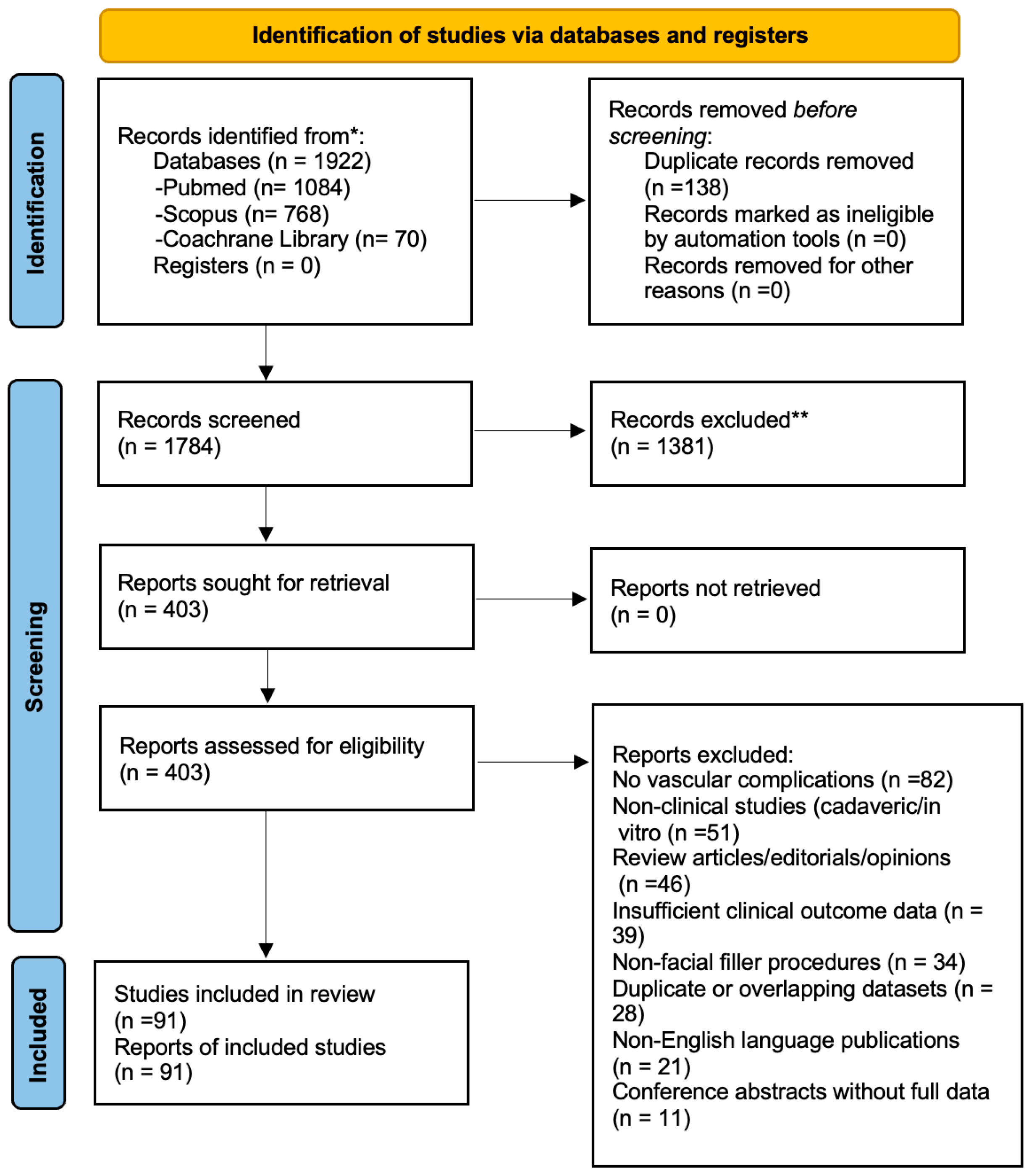
Study selection was independently performed by two reviewers using the Rayyan QCRI platform. After duplicate removal, titles and abstracts were screened, followed by full-text assessment of potentially eligible studies. Any discrepancies between reviewers were resolved by consensus through discussion and, when required, adjudication by a third reviewer. Inter-reviewer agreement for title/abstract screening, assessed using Cohen’s kappa coefficient, was 0.79, indicating substantial agreement. A total of 1922 records were identified. After removal of 138 duplicates, 1784 records were screened and 403 full-text articles were assessed for eligibility. Of these, 91 studies met the inclusion criteria and were included in the final qualitative synthesis. The selection process is summarized in the PRISMA flow diagram (Figure 1).
Risk-of-bias assessment was performed independently by two reviewers. Methodological quality was assessed using the Risk of Bias 2.0 tool for randomized trials, the Newcastle–Ottawa Scale for observational studies, and the Joanna Briggs Institute critical appraisal checklists for case reports and case series. Due to the predominance of descriptive studies and heterogeneity of designs, no numerical scoring system was applied; studies were qualitatively categorized as high, moderate, or low methodological quality.
In addition, studies were stratified according to their level of evidence using an Oxford-style hierarchy to facilitate interpretation of heterogeneous designs. This classification was used for descriptive purposes only and not as a quantitative measure of methodological quality. A summary of methodological quality and evidence level across included studies is presented in Table 1. A summary of methodological quality and evidence level across included studies is presented in Table 1.

2.4. Data Extraction and Synthesis

Data extraction and risk-of-bias assessment were also performed independently by two reviewers. For each included study, data relevant to the predefined primary and secondary outcomes were extracted, including author and year, country, study design, number of patients, anatomical site, filler type, injection technique, type of complication, diagnostic approach, treatment protocol, use of imaging guidance when reported, time to treatment when available, and clinical outcomes. Any discrepancies were resolved by consensus through discussion and, when required, adjudication by a third reviewer.
Given the marked heterogeneity in study design, outcome definitions, anatomical sites, filler materials, treatment protocols, and reporting methods, a quantitative meta-analysis was not considered appropriate. Pooling of data would have generated clinically misleading summary estimates because of the predominance of descriptive studies and the variable reporting of both exposures and outcomes.
A structured qualitative synthesis was therefore performed using a thematic approach. Studies were grouped into five predefined domains: (1) cutaneous complications, (2) ocular and neurological complications, (3) complications associated with non-hyaluronic and permanent fillers, (4) clinical management and emergency therapies, and (5) preventive strategies and ultrasound-guided approaches. Within each domain, findings were synthesized by considering study design, methodological quality, anatomical site, filler type, type of complication, therapeutic approach, and consistency of results across studies. Greater interpretative weight was attributed to higher-level evidence, including randomized trials, prospective studies, and multicenter datasets, while case reports and case series were primarily used to identify rare complications, early warning signals, and recurrent clinical patterns.
Due to the descriptive nature of the included literature and the predominance of case-based evidence, formal statistical assessment of reporting bias was not feasible. Potential reporting bias was therefore considered qualitatively by evaluating consistency of findings across studies, publication patterns, and the likelihood of selective reporting of severe complications. Certainty of evidence was interpreted using a GRADE-informed approach, considering study design, methodological quality, consistency of findings, and directness of evidence; however, no quantitative certainty rating was performed.

3. Results

3.1. Vascular Complications and Clinical Manifestations

Of the 91 included studies, most were descriptive in nature (40 case reports and 16 case series), whereas 28 were observational studies, 4 were registry/interventional datasets, and 3 were randomized controlled trials.
Overall, vascular complications following dermal filler injections were attributable to two principal and often overlapping mechanisms: intravascular embolization and extrinsic vascular compression. Intravascular embolization appeared to underlie the most severe complications, particularly when retrograde migration of filler material involved terminal arterial branches of the ophthalmic circulation, whereas extrinsic vascular compression was more often associated with localized ischemia and progressive tissue compromise in anatomically confined regions [52,53,54,55,56,57].
Cutaneous ischemia and skin necrosis represented the most frequently reported manifestations across the included studies. These events were mainly associated with hyaluronic acid injections and most involved the nose, glabella, nasolabial folds, lips, and adjacent perioral tissues [54,55,56,57,58,59,60,61,62,63,64,65,66,67,68,69,70,71,72,73,74,75,76,77,78,79,80,81,82,83,84,85,86,87].
Venous occlusion was less frequently reported than arterial compromise but was histopathologically documented in association with frontal skin necrosis, supporting the possibility that impaired superficial venous drainage may contribute to livedo reticularis and more localized cutaneous ischemia in selected cases [54]. In contrast to arterial embolization, venous compromise appeared to be associated predominantly with regional skin changes rather than ocular or neurological sequelae.
In addition, previous surgery or scarring, particularly in rhinoplasty patients, appeared to increase local ischemic susceptibility by altering vascular anatomy and perfusion patterns [56,58].
Most cutaneous complications were managed with early hyaluronidase administration, often combined with adjunctive measures such as acetylsalicylic acid, vasodilators, corticosteroids, antibiotics, hyperbaric oxygen therapy, nitroglycerin, platelet-rich plasma, or local wound care [55,56,57,61,62,65,66,67,68,69,70,71,72,74,76,77,78,79,80,84,85,86,87,88]. Across the included reports, outcomes were generally more favorable in cutaneous vascular compromise than in ocular events, especially when treatment was initiated within the first 48–72 h [55,57,62,67,69,71,78,87]. Delayed recognition or extensive ischemia was more frequently associated with scarring, incomplete recovery, or tissue loss [60,68,73,85].
Although hyaluronic acid accounted for most reported cutaneous events, similar complications were occasionally reported with other filler materials, confirming that cutaneous vascular injury is not exclusive to HA-based products [60,63,88].
Less frequent but clinically relevant presentations included mucosal necrosis, lingual ischemia, ischemic alopecia, and auricular necrosis with facial palsy, further highlighting the broad clinical spectrum of filler-related vascular injury [76,79,83,84]. Overall, the available evidence supports a marked time-dependence of cutaneous outcomes and emphasizes the importance of early recognition and immediate treatment. Detailed study-level data are reported in Supplementary Table S1.

3.2. Ocular and Neurological Complications

Ocular complications represented the most severe clinical manifestations across the included studies and were consistently associated with poor visual prognosis. These events predominantly followed injections in the mid and upper facial thirds, particularly the glabella, nasal dorsum and radix, forehead, and periorbital region, where extensive anastomoses facilitate retrograde migration of filler material into the ophthalmic circulation [40,52,53,83,89,90,91,92,93,94,95,96,97,98,99,100,101,102,103,104,105,106,107,108,109,110,111,112,113,114,115,116,117,118,119,120,121,122,123,124,125,126,127].
Reported complications included ophthalmic artery occlusion, central or branch retinal artery occlusion, posterior ciliary artery occlusion, choroidal ischemia, ischemic optic neuropathy, orbital infarction syndrome, anterior segment ischemia, ischemic keratitis, ophthalmoplegia, cranial nerve palsy, cerebral ischemia, cerebral infarction, and, in severe cases, blindness associated with diffuse facial ischemia [52,53,89,90,91,92,93,94,95,96,97,98,99,100,101,102,103,104,105,106,107,108,109,110,111,112,113,114,115,116,117,118,119,120,121,122,123,124,125,126,127].
Across studies, complete visual recovery was uncommon despite aggressive rescue strategies. Partial improvement was reported in selected cases treated with intra-arterial recanalization protocols, vasodilator-based approaches, or early hyaluronidase administration, whereas many reports described persistent blindness despite multimodal management including retrobulbar hyaluronidase, thrombolysis, hyperbaric oxygen therapy, corticosteroids, anterior chamber paracentesis, and ocular massage [89,90,91,92,93,94,100,103,104,105,106,107,108,109,110,111,112,113,115,120,123,124,125,126,127]. A few isolated cases reported substantial or complete recovery, but these remained exceptional rather than representative of the overall pattern [103,105,108,120].
In contrast, cases presenting with isolated ophthalmoplegia or cranial nerve palsy without established retinal ischemia showed more favorable functional outcomes, with partial or complete motor recovery after early corticosteroid and/or hyaluronidase treatment [95,96,114,118,119]. This distinction suggests that the prognosis is substantially worse once retinal or ophthalmic arterial occlusion is established.
Neurological complications were less frequently reported but included cortical ischemia, multifocal stroke, cerebral infarction, and peripheral nerve palsy [97,98,100,116,121,122,127]. Severe ocular and neurological events were also documented after non-hyaluronic fillers or mixed injectables, generally with poorer outcomes and limited reversibility [98,100,101,102,116]. Recent reports also identified the auricular region as a potential high-risk site because of vascular anatomical variability, with both auricular necrosis and irreversible visual loss described after postauricular injections [83,99]. Overall, ocular and neurological vascular events, although less common than cutaneous ischemia, carried a substantially worse prognosis and remained only partially responsive to currently available rescue strategies. Detailed study-level data are reported in Supplementary Table S2.

3.3. Complications Associated with Non-Hyaluronic and Permanent Fillers

Although most vascular complications reported across the included studies involved hyaluronic acid fillers, severe events were also described following injections of non-hyaluronic and permanent materials. These complications appeared to be associated with distinct clinical profiles and generally poorer outcomes, mainly because of the absence of a specific enzymatic reversal agent.
Autologous fat grafting represented one of the most severe causes of embolic vascular occlusion. Irregular lipid particles may induce extensive retrograde embolization, leading to retinal or cerebral ischemia. Kim et al. [100] reported multiple cases of permanent blindness following facial fat injection, with no visual recovery despite immediate intervention.
Among synthetic fillers, poly-DL-lactic acid (PDLLA) was associated with severe vascular complications, including stroke and bilateral blindness [98]. Calcium hydroxyapatite embolization has also been documented, resulting in ischemic optic neuropathy and cranial nerve involvement [101]. In addition, severe ocular complications, including irreversible blindness, have also been reported after non-hyaluronic injectables and related procedures [102].
Overall, the available evidence suggests that HA-related vascular events are more often associated with potentially reversible cutaneous outcomes when recognized early, whereas non-hyaluronic or permanent fillers and autologous fat appear to be linked to more limited therapeutic options and a higher likelihood of irreversible ocular or neurological damage. However, these observations should be interpreted cautiously, as the included studies do not allow formal estimation of comparative risk across filler materials.

3.4. Clinical Management and Therapeutic Protocols

Management of filler-related vascular complications requires immediate and multidisciplinary intervention aimed at restoring tissue perfusion and limiting irreversible damage. However, the available literature suggests that arterial and venous vascular compromise have different clinical implications and should be interpreted differently.
Arterial occlusion represents the most time-critical presentation and is typically associated with acute pain, blanching, retinal ischemia, or neurological sequelae. In these cases, management is primarily directed toward rapid reperfusion. For hyaluronic acid–related arterial occlusions, hyaluronidase remains the primary treatment, promoting enzymatic degradation of the filler and facilitating microvascular reperfusion. Therapeutic efficacy appeared strongly time-dependent, with better outcomes reported when treatment was initiated within the first hours after symptom onset [55,57]. Reported protocols described hyaluronidase doses ranging from 150 to 1500 IU administered through multiple superficial and deep injections according to high-dose pulsed regimens [39]. In cases of extensive or ocular involvement, peribulbar or retrobulbar administration of higher doses has been reported, although supporting evidence remains limited and procedural risks persist [106,128].
By contrast, venous occlusion has been reported less frequently and is supported by a much more limited evidence base. Available reports suggest that venous compromise is more often associated with localized livedo, congestion, and progressive cutaneous ischemia rather than immediate ocular or neurological injury. In such cases, management has generally overlapped with that of cutaneous vascular compromise, including hyaluronidase when filler-related obstruction or compression was suspected, together with supportive measures such as warm compresses, vasodilators, antiplatelet agents, hyperbaric oxygen therapy, and close clinical monitoring [54,55,57]. However, no clearly standardized venous-specific treatment pathway could be derived from the available literature.
Adjunctive treatments described in the literature included systemic corticosteroids, vasodilators, antiplatelet or anticoagulant agents, topical nitroglycerin, and hyperbaric oxygen therapy [57,110]. Endovascular approaches have been reported in selected severe cases of arterial embolization, with partial or complete reperfusion described after superselective intra-arterial recanalization with hyaluronidase, papaverine, or thrombolytic agents, particularly when performed within 24–48 h of symptom onset [81,89]. A multicenter survey conducted by the American Academy of Ophthalmology highlighted the lack of standardized emergency protocols across centers and the limited use of retrobulbar hyaluronidase [129].
Overall, the available evidence supports three main take-home messages: first, arterial occlusion should be treated as a true reperfusion emergency; second, early hyaluronidase-based treatment is most consistently associated with better cutaneous outcomes in HA-related events; and third, ocular rescue strategies remain heterogeneous and are associated with limited and inconsistent visual recovery.

3.5. Preventive Strategies and Ultrasound-Guided Approaches

Prevention emerged as a central theme across the included studies. The available evidence consistently identified high-risk anatomical regions, particularly the glabella, nasal dorsum and tip, infraorbital region, forehead, nasolabial folds, lips, and tear trough, as areas requiring especially cautious injection techniques [59,130,131,132,133,134,135,136,137,138,139,140,141]. Across preventive studies, safer approaches were generally characterized by low-volume micro-bolus delivery, reduced injection pressure, careful selection of deep or supraperiosteal planes, preference for midline trajectories in selected nasal procedures, aspiration with awareness of its limitations, and use of blunt cannulas in high-risk regions when appropriate [59,130,133,136,138,139,140,141].
Several preventive series reported no vascular occlusion or no major ischemic complications when standardized injection protocols were applied in rhinofiller, nasolabial fold correction, tear trough treatment, lip procedures, oral commissure enhancement, and combined aesthetic protocols [59,130,133,134,135,136,137,138,139,140,141,142,143]. These findings support the concept that risk reduction depends on procedural standardization and strict respect for anatomical planes rather than on any single preventive manoeuvre.
Ultrasound-guided approaches have increasingly been proposed both for prevention and for early management of suspected vascular compromise. High-frequency Doppler ultrasound enabled pre-procedural vascular mapping, real-time guidance during filler placement, and post-procedural assessment of filler position and tissue perfusion, thereby improving anatomical precision and facilitating safer injection planning [131,132,134,140]. Ultrasound-based assessment also improved diagnostic accuracy in identifying retained filler material, while targeted ultrasound-guided hyaluronidase delivery was reported as a useful adjunct in suspected vascular adverse events [40,82,88,132].
Emerging imaging modalities, including Laser Speckle Contrast Imaging, further support the concept of real-time perfusion monitoring and may improve the early recognition of ischemic change and the guidance of rescue interventions [80]. Overall, the preventive literature suggests that some strategies are supported more consistently than others. The most consistently supported measures across studies were detailed anatomical planning, cautious injection in appropriate planes, low-volume and low-pressure delivery, and procedural standardization in high-risk regions [59,130,133,136,138,139,140,141]. By contrast, strategies such as aspiration, cannula use, and imaging-guided approaches appeared promising but were supported by more heterogeneous and predominantly non-comparative evidence [130,131,132,134,140]. Therefore, preventive risk reduction should be viewed as a hierarchical multimodal strategy, in which core anatomical and technical principles are supported more consistently than adjunctive or technology-assisted measures. Detailed study-level information on preventive and safety studies is provided in Supplementary Table S3.

4. Discussion

Vascular complications associated with dermal filler injections are rare but potentially devastating adverse events. Large prospective series report an overall incidence below 1%, with early vascular compromise occurring in approximately 0.07% of procedures and complete resolution rates exceeding 90% when promptly recognized and treated [87]. Despite this low incidence, complications occurring in high-risk regions—such as the glabella, nasal dorsum, periorbital area, and nasolabial folds—may lead to severe functional and aesthetic sequelae, explaining the growing emphasis on vascular safety in aesthetic practice.
The predominance of hyaluronic acid (HA) fillers among reported complications largely reflects their widespread clinical use rather than a higher intrinsic risk. Importantly, HA-related vascular events generally show a more favorable prognosis due to enzymatic reversibility. In contrast, non-resorbable fillers and autologous fat are associated with more severe and often irreversible outcomes because they lack specific antidotes and are difficult to remove once embolization occurs [60,98,101]. These findings support cautious use of non-HA fillers in anatomically high-risk areas, particularly in less experienced settings.
The included literature also suggests that vascular complications may differ according to filler material. Hyaluronic acid accounted for most reported events, likely reflecting its wider clinical use, but HA-related complications were more frequently associated with potentially salvageable cutaneous outcomes because of enzymatic reversibility with hyaluronidase. In contrast, non-hyaluronic fillers and autologous fat were less frequently reported but appeared more often associated with severe ocular or neurological injury and poorer reversibility. Nevertheless, because the included studies were heterogeneous and largely descriptive, the present review cannot determine the true comparative incidence or relative risk of vascular complications across filler materials.
Two principal pathogenic mechanisms emerged: intravascular embolization and extrinsic vascular compression. Intravascular embolization underlies the most severe ocular and neurological complications and may occur even with minimal injected volumes (<0.1 mL), given the small calibre of terminal vessels and the presence of retrograde arterial flow through anastomotic networks [52,57]. This challenges the assumption that small volumes are inherently safe and highlights the importance of injection pressure, speed, and real-time awareness of tissue resistance. Extrinsic compression, in contrast, results from excessive or superficial filler placement and leads to progressive ischemia due to sustained capillary compromise, particularly in nasal and perinasal regions where vascular density and limited tissue compliance increase vulnerability [55,57].
Ocular complications remain the most feared outcomes, with visual prognosis generally poor. Occlusion of retinal or ophthalmic vessels frequently results in permanent visual loss. Although partial recovery has been reported following intra-arterial hyaluronidase and vasodilator therapy in selected cases [89,90], complete visual restoration remains uncommon. Conversely, isolated ophthalmoplegia without retinal involvement appears to have a more favorable prognosis when treated promptly with corticosteroids and hyaluronidase [95,96]. Reports of silent cortical infarctions and multifocal strokes further underscore the systemic relevance of filler-related embolization and support neuroimaging evaluation in selected cases [97,98].
Cutaneous ischemia and necrosis represent the most common vascular complications and generally show better outcomes when treated early. Several studies indicate that prompt, high-dose, multi-site hyaluronidase administration improves clinical recovery, particularly when combined with vasodilators, anti-inflammatory therapy, and hyperbaric oxygen [55,57,67,69,71]. Timing remains critical, as early intervention is associated with higher rates of complete healing, whereas delayed treatment increases the likelihood of tissue loss and scarring.
Management strategies increasingly rely on combined, protocol-driven approaches integrating enzymatic, pharmacological, and physical interventions. High-Dose Pulsed Hyaluronidase protocols have gained acceptance in HA-related ischemia [39,120]. Adjunctive treatments and, in selected cases, endovascular techniques such as intra-arterial hyaluronidase or thrombolysis have shown variable but occasionally promising results in specialized centers [81,83,89,90]. These approaches highlight the importance of multidisciplinary collaboration and rapid referral pathways.
Prevention remains the most effective strategy. Safe injection techniques, low-pressure delivery, and appropriate anatomical plane selection significantly reduce embolic risk [59,133]. Increasing use of high-frequency Doppler ultrasound allows vascular mapping, real-time injection guidance, and targeted treatment in suspected occlusion [131,132]. Emerging perfusion-monitoring technologies may further improve early detection of ischemia and support a shift toward proactive vascular safety [80].
Interpretation of the available evidence is also influenced by several confounding variables that were inconsistently reported across studies. These include practitioner experience, injection technique, plane of injection, bolus volume, injection pressure, use of needle versus cannula, previous surgery or scarring, local anatomical variability, and individual patient-related factors. Although these variables are likely to play an important role in both the occurrence and severity of vascular complications, the included literature did not allow a formal comparative analysis of their independent effects. Their influence should therefore be regarded as clinically plausible but not quantifiable on the basis of the current evidence.
Our findings are broadly consistent with previous review-based literature on dermal filler complications. Recent systematic reviews and review articles have shown that severe vascular adverse events are uncommon overall but tend to cluster in anatomically high-risk regions such as the nose, glabella, and periocular area [34,43,144]. In line with these data, our synthesis found that cutaneous ischemia was the most frequently reported vascular presentation, whereas ocular complications remained associated with a poor visual prognosis despite aggressive rescue strategies. Kroumpouzos and Treacy (2024) and Xiao et al. (2024) [145,146] support the central role of hyaluronidase in the management of hyaluronic acid–related vascular events, while emphasizing the persistent uncertainty regarding retrobulbar administration in ophthalmic vascular occlusion. Likewise, Fakih-Gomez et al. (2025) [147] supports our observation that imaging-assisted and time-sensitive approaches may improve diagnostic precision and facilitate more targeted treatment, although standardization and high-level comparative evidence remain limited.

4.1. Clinical Algorithm and Practical Implications

Based on the synthesis of the available evidence, a structured clinical approach to suspected vascular occlusion is essential to optimize outcomes. Early recognition remains the most important determinant of prognosis. Immediate warning signs include sudden blanching, severe disproportionate pain, livedo reticularis, visual disturbances, or neurological symptoms occurring during or shortly after injection.
In practical terms, suspected arterial occlusion should be considered a true reperfusion emergency, whereas venous compromise, although clinically relevant, more often presents as a progressive cutaneous event with less clearly standardized management pathways. In cases of suspected cutaneous vascular compromise, prompt administration of high-dose hyaluronidase should be considered the first-line intervention, ideally within the first 24–48 h, using multiple injection points across the affected vascular territory. Adjunctive measures such as warm compresses, gentle massage, vasodilators, antiplatelet agents, and hyperbaric oxygen therapy may be considered according to clinical severity and institutional availability.
In the presence of ocular symptoms, immediate emergency referral is mandatory. Retrobulbar or intra-arterial hyaluronidase has been attempted in selected cases; however, current evidence indicates limited and inconsistent visual recovery. Therefore, prevention and rapid multidisciplinary coordination remain fundamental.
Overall, although high-level comparative evidence is lacking, the consistent pattern across studies supports a time-dependent management model in which early intervention improves cutaneous outcomes, while ocular complications continue to show a poor prognosis despite aggressive rescue strategies.

4.2. Strength of Evidence

The overall certainty of evidence remains limited due to the predominance of case reports and small case series. However, this reflects the ethical and practical challenges of studying acute vascular complications following dermal filler injections. Randomized controlled trials are rare in this field, and most available data derive from observational and descriptive studies. In this review, higher-level evidence was prioritized when available, while case-based reports were used primarily to identify recurring clinical patterns, complications, and management strategies. A similar limitation applies to preventive strategies, for which the available evidence supports core anatomical and technical precautions more consistently than adjunctive measures such as aspiration, cannula use, or routine imaging guidance, although the latter appear promising in selected settings. Therefore, conclusions regarding treatment effectiveness and preventive strategies should be interpreted cautiously and in the context of heterogeneous study designs.

4.3. Limitations and Future Directions

This review has several limitations. The available evidence is largely based on case reports and case series, with substantial heterogeneity in study design, outcome definitions, and management protocols. Variability in hyaluronidase dosing, timing, and routes of administration, as well as inconsistent reporting of visual and cutaneous outcomes, limits direct comparison across studies. In addition, potentially relevant confounding factors—such as practitioner experience, injection technique, anatomical variability, prior surgery or scarring, and patient-related factors—were inconsistently reported across studies, precluding formal subgroup or comparative analyses.
Future research should prioritize multicenter prospective registries with standardized reporting of anatomical site, filler type, injection technique, time-to-treatment, cumulative hyaluronidase dose, imaging findings, and functional outcomes. Comparative studies evaluating ultrasound-guided approaches and the role of adjunctive therapies, including hyperbaric oxygen therapy, are warranted. Closer integration with interventional neuroradiology and the development of shared emergency pathways may further improve outcomes.
From a clinical perspective, preventive strategies remain essential. Detailed knowledge of facial vascular anatomy, cautious injection techniques, low-pressure delivery, and appropriate injection planes are key to minimizing risk. The integration of high-frequency Doppler ultrasound and emerging perfusion-monitoring technologies may further enhance procedural safety. Management of vascular complications should be multidisciplinary and supported by standardized emergency protocols, immediate availability of hyaluronidase, and structured referral pathways. Further progress in vascular safety will likely depend on prospective data collection, protocol standardization, and continued technological innovation.

5. Conclusions

Vascular complications associated with dermal filler injections, although rare, represent potentially severe medical emergencies with important functional and aesthetic consequences. The available evidence suggests that cutaneous ischemia has a more favorable prognosis when promptly recognized and treated, whereas ocular and neurological complications remain associated with poorer outcomes.
The findings of this systematic review support a time-dependent therapeutic model in which early intervention appears to improve cutaneous outcomes, particularly in hyaluronic acid–related events. In contrast, complications related to non-hyaluronic or permanent fillers and autologous fat appear to be associated with more limited therapeutic options and a higher risk of irreversible damage.
These conclusions should be interpreted with caution because the current evidence base is limited by substantial heterogeneity and by the predominance of descriptive and observational studies.

Supplementary Materials

The following supporting information can be downloaded at: https://www.mdpi.com/article/10.3390/oral6030051/s1, Table S1: Cutaneous and Mixed Vascular Complications Associated with Dermal Filler Injections; Table S2: Ocular and Neurological Complications Associated with Dermal Filler Injections; Table S3: Prevention and Safety Studies in Dermal Filler Procedures.

Author Contributions

Conceptualization, L.S. and E.P.M.; methodology, L.S. and A.D.P.; validation, L.S., A.D.P., A.P. and E.P.M.; formal analysis, L.S.; investigation, L.S.; data curation, L.S. and E.F.; writing—original draft preparation, L.S. and A.P.; writing—review and editing, A.D.P., E.F., A.P. and E.P.M.; visualization, L.S.; supervision, E.P.M.; project administration, L.S. All authors have read and agreed to the published version of the manuscript.

Funding

This research received no external funding.

Institutional Review Board Statement

Not applicable.

Informed Consent Statement

Not applicable.

Data Availability Statement

Extracted data, study selection records, and quality assessment materials supporting the findings of this review are available from the corresponding author upon reasonable request.

Conflicts of Interest

The authors declare no conflicts of interest.

Abbreviations

The following abbreviations are used in this manuscript:
ASAacetylsalicylic acid
bFGFbasic fibroblast growth factor
BoNTAbotulinum toxin type A
CaHAcalcium hydroxyapatite
CNcranial nerve
CRAOcentral retinal artery occlusion
HAhyaluronic acid
HAsehyaluronidase
HBOThyperbaric oxygen therapy
IAintra-arterial
IONischemic optic neuropathy
IUinternational units
LDIlaser Doppler imaging
LSCIlaser speckle contrast imaging
Nd:YAGneodymium-doped yttrium aluminum garnet
NLFnasolabial fold
OAOophthalmic artery occlusion
PCAposterior cerebral artery
PCAOposterior ciliary artery occlusion
PGE1prostaglandin E1
PLLApoly-L-lactic acid
PMMApolymethylmethacrylate
PRPplatelet-rich plasma
RAOretinal artery occlusion
RCTrandomized controlled trial
SVFstromal vascular fraction
USultrasound
VOvascular occlusion

References

  1. Wollina, U.; Goldman, A. Facial vascular danger zones for filler injections. Dermatol. Ther. 2020, 33, e14285. [Google Scholar] [CrossRef]
  2. De Maio, M.; DeBoulle, K.; Braz, A.; Rohrich, R.J. Facial assessment and injection guide for botulinum toxin and injectable hyaluronic acid fillers: Focus on the midface. Plast. Reconstr. Surg. 2017, 140, 540e–550e. [Google Scholar] [CrossRef]
  3. Bailey, S.H.; Cohen, J.L.; Kenkel, J.M. Etiology, prevention, and treatment of dermal filler complications. Aesthetic Surg. J. 2011, 31, 110–121. [Google Scholar] [CrossRef]
  4. Fitzgerald, R.; Bertucci, V.; Sykes, J.; Duplechain, J. Adverse reactions to injectable fillers. Facial Plast. Surg. 2016, 32, 532–555. [Google Scholar] [CrossRef]
  5. Chiang, Y.Z.; Pierone, G.; Al-Niaimi, F. Dermal fillers: Pathophysiology, prevention and treatment of complications. J. Eur. Acad. Dermatol. Venereol. 2017, 31, 405–413. [Google Scholar] [CrossRef]
  6. Galadari, H.; Krompouzos, G.; Kassir, M.; Gupta, M.; Wollina, U.; Katsambas, A. Complication of soft tissue fillers: Prevention and management review. J. Drugs Dermatol. 2020, 19, 829–832. [Google Scholar] [CrossRef]
  7. Yamauchi, P. Emerging permanent filler technologies: Focus on Aquamid. Clin. Cosmet. Investig. Dermatol. 2014, 7, 261–266. [Google Scholar] [CrossRef]
  8. Ginat, D.T.; Schatz, C.J. Imaging features of midface injectable fillers and associated complications. AJNR Am. J. Neuroradiol. 2013, 34, 1488–1495. [Google Scholar] [CrossRef]
  9. Juhász, M.L.W.; Marmur, E.S. Temporal fossa defects: Techniques for injecting hyaluronic acid filler and complications after hyaluronic acid filler injection. J. Cosmet. Dermatol. 2015, 14, 254–259. [Google Scholar] [CrossRef]
  10. Ferneini, E.M.; Hapelas, S.; Watras, J.; Ferneini, A.M.; Weyman, D.; Fewins, J. Surgeon’s guide to facial soft tissue filler injections: Relevant anatomy and safety considerations. J. Oral Maxillofac. Surg. 2017, 75, 2667.e1–2667.e5. [Google Scholar] [CrossRef]
  11. Bailey, S.H.; Fagien, S.; Rohrich, R.J. Changing role of hyaluronidase in plastic surgery. Plast. Reconstr. Surg. 2014, 133, 127e–132e. [Google Scholar] [CrossRef] [PubMed]
  12. Albornoz, C.A.; Jhawar, N.; Durso, T.A.; Hazan, E.; Wang, J.V.; Saedi, N. Preinjection aspiration for injectable fillers in aesthetic dermatology: Trust or bust? J. Cosmet. Dermatol. 2020, 19, 1063–1064. [Google Scholar] [CrossRef] [PubMed]
  13. Rivkin, A. Nonsurgical rhinoplasty using injectable fillers: A safety review of 2488 procedures. Facial Plast. Surg. Aesthetic Med. 2021, 23, 6–11. [Google Scholar] [CrossRef]
  14. Song, D.; Wang, X.; Yu, Z. Nonsurgical rhinoplasty: An updated systematic review of technique, outcomes, complications, and its treatments. Aesthetic Plast. Surg. 2024, 48, 4902–4915. [Google Scholar] [CrossRef]
  15. Janovskiene, A.; Chomicius, D.; Afanasjevas, D.; Petronis, Z.; Razukevicius, D.; Jagelaviciene, E. Safety and potential complications of facial wrinkle correction with dermal fillers: A systematic literature review. Medicina 2024, 61, 25. [Google Scholar] [CrossRef] [PubMed]
  16. Lohman, M.E.; Ghobadi, C.W.; Xu, S. Device safety implications of the clinical data leading to US Food and Drug Administration approval of soft-tissue fillers: A systematic review. JAMA Facial Plast. Surg. 2017, 19, 421–429. [Google Scholar] [CrossRef]
  17. Arlette, J.P.; Froese, A.L.; Singh, J.K. Soft tissue filler therapy and informed consent: A Canadian review. J. Cutan. Med. Surg. 2022, 26, 50–56. [Google Scholar] [CrossRef] [PubMed]
  18. Beleznay, K.; Carruthers, J.D.A.; Humphrey, S.; Jones, D. Avoiding and treating blindness from fillers: A review of the world literature. Dermatol. Surg. 2015, 41, 1097–1117. [Google Scholar]
  19. Li, X.; Du, L.; Lu, J. A novel hypothesis of visual loss secondary to cosmetic facial filler injection. Ann. Plast. Surg. 2015, 75, 258–260. [Google Scholar] [CrossRef]
  20. Ricci, L.H.; Navajas, S.V.; Carneiro, P.R.; Söderberg, S.A.; Ferraz, C.A. Ocular adverse effects after facial cosmetic procedures: A review of case reports. J. Cosmet. Dermatol. 2015, 14, 145–151. [Google Scholar] [CrossRef]
  21. Rauso, R.; Sesenna, E.; Fragola, R.; Zerbinati, N.; Nicoletti, G.F.; Tartaro, G. Skin necrosis and vision loss or impairment after facial filler injection. J. Craniofac. Surg. 2020, 31, 2289–2293. [Google Scholar] [PubMed]
  22. Ashton, M.W.; Taylor, G.I.; Corlett, R.J. The role of anastomotic vessels in controlling tissue viability and defining tissue necrosis with special reference to complications following injection of hyaluronic acid fillers. Plast. Reconstr. Surg. 2018, 141, 818e–830e. [Google Scholar] [CrossRef] [PubMed]
  23. Lee, J.S.; Kim, J.Y.; Jung, C.; Woo, S.J. Iatrogenic ophthalmic artery occlusion and retinal artery occlusion. Prog. Retin. Eye Res. 2020, 78, 100848. [Google Scholar] [CrossRef]
  24. Kato, J.M.; Matayoshi, S. Visual loss after aesthetic facial filler injection: A literature review on an ophthalmologic issue. Arq. Bras. Oftalmol. 2022, 85, e20220048. [Google Scholar] [CrossRef]
  25. Lazzeri, D.; Agostini, T.; Figus, M.; Nardi, M.; Pantaloni, M.; Lazzeri, S. Blindness following cosmetic injections of the face. Plast. Reconstr. Surg. 2012, 129, 995–1012. [Google Scholar] [CrossRef]
  26. Beleznay, K.; Carruthers, J.D.A.; Humphrey, S.; Carruthers, A.; Jones, D. Update on avoiding and treating blindness from fillers: A recent review of the world literature. Aesthetic Surg. J. 2019, 39, 662–674. [Google Scholar] [CrossRef]
  27. Hwang, C.J.; Chon, B.H.; Perry, J.D. Blindness after filler injection. Facial Plast. Surg. Clin. N. Am. 2021, 29, 359–367. [Google Scholar] [CrossRef]
  28. Mortada, H.; Seraj, H.; Barasain, O.; Bamakhrama, B.; Alhindi, N.I.; Arab, K. Ocular complications post-cosmetic periocular hyaluronic acid injections: A systematic review. Aesthetic Plast. Surg. 2022, 46, 760–773. [Google Scholar] [CrossRef] [PubMed]
  29. Carruthers, J.D.A.; Fagien, S.; Rohrich, R.J.; Weinkle, S.; Carruthers, A. Blindness caused by cosmetic filler injection: A review of cause and therapy. Plast. Reconstr. Surg. 2014, 134, 1197–1201. [Google Scholar]
  30. Murthy, R.; Roos, J.C.P.; Goldberg, R.A. Periocular hyaluronic acid fillers: Applications, implications, complications. Curr. Opin. Ophthalmol. 2019, 30, 395–400. [Google Scholar] [CrossRef]
  31. Paap, M.K.; Milman, T.; Ugradar, S.; Goldberg, R.; Silkiss, R.Z. Examining the role of retrobulbar hyaluronidase in reversing filler-induced blindness: A systematic review. Ophthalmic Plast. Reconstr. Surg. 2020, 36, 231–238. [Google Scholar] [CrossRef]
  32. Tran, A.Q.; Staropoli, P.; Rong, A.J.; Lee, W.W. Filler-associated vision loss. Facial Plast. Surg. Clin. N. Am. 2019, 27, 557–564. [Google Scholar] [CrossRef] [PubMed]
  33. Lee, K.W.A.; Chan, L.K.W.; Lee, A.W.K.; Lee, C.H.; Wan, J.; Yi, K.H. Biodegradable polymers and platelet-rich plasma causing visual impairment: A literature review. J. Dermatol. Treat. 2024, 35, 2402909. [Google Scholar] [CrossRef] [PubMed]
  34. Doyon, V.C.; Liu, C.; Fitzgerald, R.; Humphrey, S.; Jones, D.; Carruthers, J.D.A. Update on blindness from filler: Review of prognostic factors, management approaches, and a century of published cases. Aesthetic Surg. J. 2024, 44, 1091–1104. [Google Scholar] [CrossRef] [PubMed]
  35. Soares, D.J.; Bowhay, A.; Blevins, L.W.; Patel, S.M.; Zuliani, G.F. Patterns of filler-induced facial skin ischemia: A systematic review of 243 cases and introduction of the FOEM scoring system and grading scale. Plast. Reconstr. Surg. 2023, 151, 592e–608e. [Google Scholar] [CrossRef]
  36. Moon, H.J. Use of fillers in rhinoplasty. Clin. Plast. Surg. 2016, 43, 307–317. [Google Scholar] [CrossRef] [PubMed]
  37. Arlette, J.P.; Ashenhurst, M.; Hill, V.; Jiang, K. Prevention and management of filler-induced iatrogenic stroke of the eye. J. Cutan. Med. Surg. 2021, 25, 543–552. [Google Scholar] [CrossRef]
  38. Cavallini, M.; Gazzola, R.; Metalla, M.; Vaienti, L. The role of hyaluronidase in the treatment of complications from hyaluronic acid dermal fillers. Aesthetic Surg. J. 2013, 33, 1167–1174. [Google Scholar] [CrossRef] [PubMed]
  39. DeLorenzi, C. New high-dose pulsed hyaluronidase protocol for hyaluronic acid filler vascular adverse events. Aesthetic Surg. J. 2017, 37, 814–825. [Google Scholar]
  40. Schelke, L.W.; Velthuis, P.; Kadouch, J.; Swift, A. Early ultrasound for diagnosis and treatment of vascular adverse events with hyaluronic acid fillers. J. Am. Acad. Dermatol. 2023, 88, 79–85. [Google Scholar] [CrossRef]
  41. Kroumpouzos, G.; Harris, S.; Bhargava, S.; Wortsman, X. Complications of fillers in the lips and perioral area: Prevention, assessment, and management focusing on ultrasound guidance. J. Plast. Reconstr. Aesthetic Surg. 2023, 84, 656–669. [Google Scholar]
  42. Boey, J.J.E.; Boey, J.J.J.; Chen, Z.; Cao, T.; Tan, A.S.M.; Ng, Z.Y. Superior outcomes with ultrasound-guided hyaluronidase for impending filler-induced facial skin necrosis: A systematic review and pilot meta-analysis. Aesthetic Plast. Surg. 2025, 49, 3519–3525. [Google Scholar] [PubMed]
  43. DeVictor, S.; Ong, A.A.; Sherris, D.A. Complications secondary to nonsurgical rhinoplasty: A systematic review and meta-analysis. Otolaryngol. Head Neck Surg. 2021, 165, 611–616. [Google Scholar] [CrossRef]
  44. Agamawi, Y.M.; Ziai, K.; Lighthall, J.G.; Walen, S.G. Noninvasive rhinoplasty. Adv. Cosmet. Surg. 2021, 4, 71–81. [Google Scholar] [CrossRef]
  45. Moustafa, F.; Ibrahim, O.; Dover, J.S. Perioral rejuvenation. Adv. Cosmet. Surg. 2020, 3, 89–98. [Google Scholar] [CrossRef]
  46. Bouaoud, J.; Belloc, J.B. Use of injectables in rhinoplasty retouching: Towards an evolution of surgical strategy? Literature review. J. Stomatol. Oral Maxillofac. Surg. 2020, 121, 550–555. [Google Scholar]
  47. Hwang, K.; Han, S.H. Digital compression of the origin of the nasal branch of the ophthalmic artery during filler augmentation: A review of the anatomical literature and an analysis of plastinated specimens. J. Craniofac. Surg. 2023, 34, e692–e694. [Google Scholar] [CrossRef]
  48. Pavicic, T.; Funt, D. Dermal fillers in aesthetics: An overview of adverse events and treatment approaches. Clin. Cosmet. Investig. Dermatol. 2013, 6, 295–316. [Google Scholar] [CrossRef] [PubMed]
  49. Gonzalez-Hermosillo, L.M.; Ramos-Pacheco, V.H.; Gonzalez-Hermosillo, D.C.; Cervantes-Sanchez, A.M.; Vega-Gutierrez, A.E.; Ternovoy, S.K. MRI visualization and distribution patterns of foreign modeling agents: A brief pictorial review for clinicians. Biomed. Res. Int. 2021, 2021, 2838246. [Google Scholar] [CrossRef]
  50. Hong, G.W.; Hu, H.; Chang, K.; Park, Y.; Lee, K.W.A.; Chan, L.K.W. Review of the adverse effects associated with dermal filler treatments: Part I nodules, granuloma, and migration. Diagnostics 2024, 14, 1640. [Google Scholar] [CrossRef] [PubMed]
  51. Page, M.J.; McKenzie, J.E.; Bossuyt, P.M.; Boutron, I.; Hoffmann, T.C.; Mulrow, C.D.; Shamseer, L.; Tetzlaff, J.M.; Akl, E.A.; Brennan, S.E.; et al. The PRISMA 2020 statement: An updated guideline for reporting systematic reviews. BMJ 2021, 372, n71. [Google Scholar] [CrossRef]
  52. Hu, X.Z.; Hu, J.Y.; Wu, P.S.; Yu, S.B.; Kikkawa, D.O.; Lu, W. Posterior ciliary artery occlusion caused by hyaluronic acid injections into the forehead: A case report. Medicine 2016, 95, e3124. [Google Scholar] [CrossRef]
  53. Thanasarnaksorn, W.; Cotofana, S.; Rudolph, C.; Kraisak, P.; Chanasumon, N.; Suwanchinda, A. Severe vision loss caused by cosmetic filler augmentation: Case series with review of cause and therapy. J. Cosmet. Dermatol. 2018, 17, 712–718. [Google Scholar] [CrossRef]
  54. Maruyama, S. A histopathologic diagnosis of vascular occlusion after injection of hyaluronic acid filler: Findings of intravascular foreign body and skin necrosis. Aesthetic Surg. J. 2017, 37, NP102–NP108. [Google Scholar] [CrossRef] [PubMed]
  55. Sun, Z.S.; Zhu, G.Z.; Wang, H.B.; Xu, X.; Cai, B.; Zeng, L. Clinical outcomes of impending nasal skin necrosis related to nose and nasolabial fold augmentation with hyaluronic acid fillers. Plast. Reconstr. Surg. 2015, 136, 434e–441e. [Google Scholar] [CrossRef]
  56. Kim, J.L.; Shin, J.Y.; Roh, S.G.; Lee, N.H. Demarcative necrosis along previous laceration line after filler injection. J. Craniofac. Surg. 2017, 28, e481–e482. [Google Scholar] [CrossRef]
  57. Ouyang, H.; Li, G.; Zhu, Y.; Lei, Y.; Gold, M.H.; Tan, J. Treatment of skin soft tissue embolism after hyaluronic acid injection for injection rhinoplasty in Asian patients. J. Cosmet. Dermatol. 2019, 18, 747–754. [Google Scholar] [CrossRef] [PubMed]
  58. Robati, R.M.; Moeineddin, F.; Almasi-Nasrabadi, M. The risk of skin necrosis following hyaluronic acid filler injection in patients with a history of cosmetic rhinoplasty. Aesthetic Surg. J. 2018, 38, 883–888. [Google Scholar] [CrossRef]
  59. Chen, B.; Ma, L.; Ji, K.; Lu, H.; Hu, X.; Qi, Y. Rhinoplasty with hyaluronic acid: A standard 5-step injection procedure using sharp needle. Ann. Plast. Surg. 2020, 85, 595–600. [Google Scholar] [CrossRef] [PubMed]
  60. Kim, S.K.; Kim, J.H.; Hwang, K. Skin necrosis of the nose after injection of ribose cross-linked porcine atelocollagen. J. Craniofac. Surg. 2015, 26, 2211–2212. [Google Scholar] [CrossRef] [PubMed]
  61. Kang, B.K.; Kang, I.J.; Jeong, K.H.; Shin, M.K. Treatment of glabella skin necrosis following injection of hyaluronic acid filler using platelet-rich plasma. J. Cosmet. Laser Ther. 2016, 18, 111–112. [Google Scholar] [CrossRef] [PubMed]
  62. Hong, J.Y.; Seok, J.; Ahn, G.R.; Jang, Y.J.; Li, K.; Kim, B.J. Impending skin necrosis after dermal filler injection: A “golden time” for first-aid intervention. Dermatol. Ther. 2017, 30, e12440. [Google Scholar] [CrossRef] [PubMed]
  63. Goldman, A.; Wollina, U. Polymethylmethacrylate-induced nodules of the lips: Clinical presentation and management by intralesional neodymium: YAG laser therapy. Dermatol. Ther. 2019, 32, e12755. [Google Scholar] [CrossRef]
  64. Han, J.; He, Y.; Liu, K.; Yang, Q. Necrosis of the glabella after injection with hyaluronic acid into the forehead. J. Craniofac. Surg. 2018, 29, e726–e727. [Google Scholar] [CrossRef]
  65. Zambacos, G.J.; Hapsas, D.A.; Mandrekas, A.D. Glabella impending skin necrosis: A case report. Eur. J. Plast. Surg. 2019, 42, 193–196. [Google Scholar] [CrossRef]
  66. Ciancio, F.; Tarico, M.S.; Giudice, G.; Perrotta, R.E. Early hyaluronidase use in preventing skin necrosis after treatment with dermal fillers: Report of two cases. F1000Research 2019, 7, 1388. [Google Scholar] [CrossRef]
  67. Fahavani, F.F.; Tabrizi, R.; Bahmanzadeh, M.; Nematollahi, A.; Farhadi, M.; Sadri, S. Management of vascular complications following hyaluronic acid filler injection: A case series. J. Cosmet. Dermatol. 2019, 18, 1025–1031. [Google Scholar]
  68. Chauhan, A.; Singh, S. Management of delayed skin necrosis following hyaluronic acid filler injection using pulsed hyaluronidase. J. Cutan. Aesthetic Surg. 2019, 12, 183–186. [Google Scholar] [CrossRef] [PubMed]
  69. Kruize, R.G.F.; Teguh, D.N.; Van Hulst, R.A. Hyperbaric oxygen therapy in hyaluronic acid filler–Induced dermal ischemia. Dermatol. Surg. 2020, 46, 1755–1757. [Google Scholar] [CrossRef]
  70. Zeltzer, A.; Geeroms, M.; Antoniazzi, E.; Giunta, G.; De Baerdemaeker, R.; Hendrickx, B. The “ART” of facial filler injections: Avoid, recognize, and treat hyaluronic acid-induced complications. J. Cosmet. Dermatol. 2020, 19, 2229–2236. [Google Scholar] [CrossRef]
  71. Ors, S. The effect of hyaluronidase on depth of necrosis in hyaluronic acid filling-related skin complications. Aesthetic Plast. Surg. 2020, 44, 1778–1785. [Google Scholar] [CrossRef] [PubMed]
  72. Halepas, S.; Peters, S.M.; Goldsmith, J.L.; Ferneini, E.M. Vascular compromise after soft tissue facial fillers: Case report and review of current treatment protocols. J. Oral Maxillofac. Surg. 2020, 78, 440–445. [Google Scholar] [CrossRef]
  73. Hirsch, P.; Infanger, M.; Kraus, A. A case of upper lip necrosis after cosmetic injection of hyaluronic acid soft-tissue filler—Does capillary infarction play a role in the development of vascular compromise, and what are the implications? J. Cosmet. Dermatol. 2020, 19, 1316–1320. [Google Scholar] [CrossRef] [PubMed]
  74. Lee, A.L.; Chen, Y.F.; Yao, W.T.; Liu, Y.C.; Yu, C.M.; Yu, C.M. Laser Doppler imaging for treating vascular complications from procedures involving dermal fillers: Case series and literature review. Diagnostics 2021, 11, 1640. [Google Scholar] [CrossRef]
  75. Li, D.; Gao, Z.; Sun, J.; Li, Q.; Jiang, P.; Zhang, L. Safe and effective lip enhancement with VYC-15L in Chinese adults. Clin. Cosmet. Investig. Dermatol. 2022, 15, 2427–2436. [Google Scholar] [CrossRef]
  76. Jalilian, F.; Hetz, S.P.; Bostwick, J.; Boet, S. Hyperbaric oxygen therapy for treatment of a late presenting ischaemic complication from hyaluronic acid cosmetic filler injection. BMJ Case Rep. 2022, 15, e249190. [Google Scholar] [CrossRef]
  77. Rahimi, M.K.; Shariati, N.; Aboofazeli, A.; Ghasemi, Z. Nasal necrosis following injection of hyaluronic acid filler into the nasal tip. Oral. Maxillofac. Surg. Cases 2022, 8, 100265. [Google Scholar] [CrossRef]
  78. Choi, S.Y.; Shin, S.H.; Seok, J.; Yoo, K.H.; Kim, B.J. Management strategies for vascular complications in hyaluronic acid filler injections: A case series analysis. J. Cosmet. Dermatol. 2023, 22, 3261–3267. [Google Scholar] [CrossRef]
  79. Thanasarnaksorn, W.; Thanyavuthi, A.; Prasertvit, P.; Rattanakuntee, S.; Jitaree, B.; Suwanchinda, A. Case series of tongue necrosis from vascular complications after chin augmentation with hyaluronic acid: Potential pathophysiology and management. J. Cosmet. Dermatol. 2023, 22, 784–791. [Google Scholar] [CrossRef]
  80. Tsai, M.F.; Yu, C.M.; Chen, Y.F.; Chung, T.Y.; Lin, G.H.; Lee, A.L. Laser speckle contrast imaging guides needling treatment of vascular complications from dermal fillers. Aesthetic Plast. Surg. 2024, 48, 1067–1075. [Google Scholar] [CrossRef] [PubMed]
  81. Fu, H.; Fu, Q.; Yu, Y.; Yang, Y.; Zheng, C.; Xu, X. Efficacy of superselective intra-arterial recanalization of embolized arteries resulting from facial hyaluronic acid injection. Aesthetic Plast. Surg. 2024, 48, 3561–3567. [Google Scholar] [CrossRef] [PubMed]
  82. Mojallal, A.; Schelke, L.; Velthuis, P.J.; Boucher, F.; Henry, G. Ultrasound-assisted hyaluronic acid vascular adverse event management based on the vascular anatomy. Ann. Chir. Plast. Esthet. 2024, 69, 674–680. [Google Scholar] [CrossRef]
  83. Zheng, C.; Pei, H.N.; Zhou, G.W.; Xu, X.; Wu, Q.; Tang, S. Super-selective intra-arterial thrombolytic therapy for peripheral facial paralysis after postauricular hyaluronic acid injection for the esthetic correction of lying ears. Aesthetic Plast. Surg. 2025, 49, 13–19. [Google Scholar] [CrossRef]
  84. Albargawi, S.; Nagshabandi, K.N.; Shadid, A. Dermal filler-induced alopecia: A case report and literature review. J. Cosmet. Dermatol. 2025, 24, e16684. [Google Scholar] [CrossRef]
  85. Nomoto, S.; Ogawa, R. Management of secondary embolization that arose after intraarterial thrombolytic treatment of cosmetic facial filler-induced arterial occlusion. JPRAS Open 2021, 28, 25–28. [Google Scholar] [CrossRef]
  86. Oley, M.H.; Oley, M.C.; Mawu, F.O.; Aling, D.M.R.; Faruk, M. Hyperbaric oxygen therapy in managing minimally invasive aesthetic procedure complications: A report of three cases. Clin. Cosmet. Investig. Dermatol. 2022, 15, 63–68. [Google Scholar] [CrossRef] [PubMed]
  87. Nishikawa, A.; Aikawa, Y.; Kono, T. Current status of early complications caused by hyaluronic acid fillers: Insights from a descriptive, observational study of 41,775 cases. Aesthetic Surg. J. 2023, 43, 893–904. [Google Scholar] [CrossRef]
  88. Kadouch, J.; Schelke, L.; Groh, O.; Sokol, V.; Velthuis, P. Intralesional hyaluronidase injection to relieve non-hyaluronic acid filler-induced vascular adverse events. Int. J. Dermatol. 2024, 63, 1252–1255. [Google Scholar] [CrossRef] [PubMed]
  89. Wang, J.; Shen, H.; Liu, T.; Li, Q.; Lyu, Z.; Yu, Y. An efficacy and safety study of intra-arterial recanalization of occluded ophthalmic arteries in patients with monocular blindness caused by injection of hyaluronic acid in facial tissues. Aesthetic Plast. Surg. 2021, 45, 1573–1578. [Google Scholar] [CrossRef]
  90. Xu, X.; Zhou, G.; Fu, Q.; Zhang, L.; Yu, Y.; Dong, Y. Efficacy of intra-arterial thrombolytic therapy for vision loss resulting from hyaluronic acid filler embolization. J. Cosmet. Dermatol. 2021, 20, 3205–3212. [Google Scholar] [CrossRef]
  91. Chen, Y.C.; Wu, H.M.; Chen, S.J.; Lee, H.J.; Lirng, J.F.; Lin, C.J.; Luo, C.-B.; Guo, W.-Y.; Chang, F.-C. Intra-arterial thrombolytic therapy is not a therapeutic option for filler-related central retinal artery occlusion. Facial Plast. Surg. 2018, 34, 325–329. [Google Scholar] [CrossRef]
  92. Zhang, L.; Pan, L.; Xu, H.; Yan, S.; Sun, Y.; Wu, W.T.L.; Wu, S. Clinical observations and the anatomical basis of blindness after facial hyaluronic acid injection. Aesthetic Plast. Surg. 2019, 43, 1054–1060. [Google Scholar] [CrossRef]
  93. Jolly, R.; Bhalla, M.; Zakir, R.; Joshi, N. Visual loss from dermal fillers. Eur. J. Ophthalmol. 2021, 31, NP102–NP105. [Google Scholar]
  94. Gracia Rovira, E.; Natividad Andreu, S.; Montalt Barrachina, N.; Navarro Hernández, E.; Agustí Martínez, J.; Lanuza García, A. Ocular ischemia syndrome after aesthetic treatment with hyaluronic acid. Arch. Soc. Esp. Oftalmol. 2024, 99, 504–507. [Google Scholar] [PubMed]
  95. Downie, E.M.; Chen, Y.; Lucarelli, M.J.; Burkat, C.N. Isolated ophthalmoplegia following filler injections to the upper face. Ophthalmic Plast. Reconstr. Surg. 2020, 36, e152–e154. [Google Scholar] [CrossRef] [PubMed]
  96. Tabatabaei, F.S.; Azimi, A.; Tabatabaei, S.S.; Pakdaman, H. Recovery of acute ophthalmoplegia after hyaluronic acid filler injections to the temples: A case report and review of the literature. Arch. Plast. Surg. 2023, 50, 148–152. [Google Scholar] [CrossRef]
  97. Moore, R.M.; Mueller, M.A.; Hu, A.C.; Evans, G.R.D. Asymptomatic stroke after hyaluronic acid filler injection: Case report and literature review. Aesthetic Surg. J. 2021, 41, NP602–NP608. [Google Scholar]
  98. Tan, Y.J.; Sugianto, N.; Li, Y. Multifocal strokes and vision loss from PDLLA filler injections. J. Stroke Cerebrovasc. Dis. 2024, 33, 107556. [Google Scholar] [CrossRef]
  99. Huang, L.; Luo, S.T.; Xie, G.H.; Fu, X.; Ye, Z.W.; He, Q.Y.; Tan, B.-W.; Lu, J.-Q.; Li, S.-H.; Liao, X.; et al. Permanent vision loss due to vascular variation after ear hyaluronic acid injection: Two case reports and literature review. Aesthetic Plast. Surg. 2025, 49, 3968–3975. [Google Scholar] [CrossRef] [PubMed]
  100. Kim, Y.K.; Jung, C.; Woo, S.J.; Park, K.H. Cerebral angiographic findings of cosmetic facial filler-related ophthalmic and retinal artery occlusion. J. Korean Med. Sci. 2015, 30, 1847–1852. [Google Scholar] [CrossRef] [PubMed]
  101. Chou, C.C.; Chen, H.H.; Tsai, Y.Y.; Li, Y.L.; Lin, H.J. Choroid vascular occlusion and ischemic optic neuropathy after facial calcium hydroxyapatite injection: A case report. BMC Surg. 2015, 15, 21. [Google Scholar] [CrossRef]
  102. Bhalla, M.; El-Housseini, Z.; Asaria, R. Blindness associated with platelet-rich plasma temporomandibular joint injections. Br. J. Oral Maxillofac. Surg. 2020, 58, 1197–1199. [Google Scholar] [CrossRef]
  103. Chen, W.; Wu, L.; Jian, X.L.; Zhang, B.; Li, J.Y.; Qin, X.L.; Yu, B. Retinal branch artery embolization following hyaluronic acid injection: A case report. Aesthetic Surg. J. 2016, 36, NP219–NP224. [Google Scholar] [CrossRef]
  104. Zhu, G.Z.; Sun, Z.S.; Liao, W.X.; Cai, B.; Chen, C.L.; Zheng, H.H.; Zeng, L.; Luo, S.-K. Efficacy of retrobulbar hyaluronidase injection for vision loss resulting from hyaluronic acid filler embolization. Aesthetic Surg. J. 2018, 38, 12–22. [Google Scholar] [CrossRef]
  105. Chesnut, C. Restoration of visual loss with retrobulbar hyaluronidase injection after hyaluronic acid filler. Dermatol. Surg. 2018, 44, 435–437. [Google Scholar] [CrossRef]
  106. Ramesh, S.; Fiaschetti, D.; Goldberg, R.A. Orbital and ocular ischemic syndrome with blindness after facial filler injection. Ophthalmic Plast. Reconstr. Surg. 2018, 34, e108–e110. [Google Scholar] [CrossRef]
  107. Yang, H.K.; Lee, Y.; Woo, S.J.; Park, K.H.; Kim, J.S.; Hwang, J.M. Natural course of ophthalmoplegia after iatrogenic ophthalmic artery occlusion caused by cosmetic filler injections. Plast. Reconstr. Surg. 2019, 144, 28e–34e. [Google Scholar] [CrossRef] [PubMed]
  108. Worley, N.; Lupo, M.; Holcomb, K.; Kullman, G.; Elahi, E.; Elison, J. Hyperbaric oxygen treatment of keratitis following facial hyaluronic acid injection. Ther. Adv. Ophthalmol. 2020, 12, 193–196. [Google Scholar] [CrossRef]
  109. Eldweik, L. Orbital infarction syndrome following hyaluronic acid filler rhinoplasty. Am. J. Ophthalmol. Case Rep. 2021, 22, 101063. [Google Scholar] [CrossRef] [PubMed]
  110. Cecchini, M.P.; Marchese, A.; Pichi, F.; Tiosano, L.; Freund, K.B.; Gal-Or, O. A case of retinal and choroidal ischemia following cosmetic facial filler injection. Surv. Ophthalmol. 2021, 66, 915–922. [Google Scholar]
  111. Davidova, P.; Müller, M.; Wenner, Y.; König, C.; Kenikstul, N.; Kohnen, T. Ophthalmic artery occlusion after glabellar hyaluronic acid filler injection. Am. J. Ophthalmol. Case Rep. 2022, 26, 101407. [Google Scholar] [CrossRef]
  112. Pee, X.K.; Low, A.; Ab Kahar, M.E.P.I.; Mohamed, S.O.; Chong, Y.J. Purtscher-like retinopathy and paracentral acute middle maculopathy following breast filler injection. BMC Ophthalmol. 2023, 23, 444. [Google Scholar] [CrossRef]
  113. Si, S.; Su, W.; Wang, L.; Ji, Y.; Chen, A.; Hu, Y. A novel predictive model for phthisis bulbi following facial hyaluronic acid cosmetic injection. BMC Ophthalmol. 2023, 23, 244. [Google Scholar] [CrossRef]
  114. Chung, C.W.; Shiuey, E.J.; Briceño, C.A.; Lee, V. Acute diplopia after glabellar hyaluronic acid filler injection. Am. J. Ophthalmol. Case Rep. 2023, 31, 101860. [Google Scholar] [CrossRef]
  115. Zhang, L.; Zhou, Q.; Xu, H.; Gu, Q.; Shi, H.; Pan, L.; Sun, Y.; Wu, S. Long-term prognosis of vision loss caused by facial hyaluronic acid injections and the potential approaches to address this catastrophic event. Aesthetic Surg. J. 2023, 43, 484–493. [Google Scholar] [CrossRef]
  116. Prado, G.; Rodríguez-Feliz, J. Ocular pain and impending blindness during facial cosmetic injections: Is your office prepared? Aesthetic Plast. Surg. 2017, 41, 199–203. [Google Scholar] [CrossRef] [PubMed]
  117. Salval, A.; Ciancio, F.; Margara, A.; Bonomi, S. Impending facial skin necrosis and ocular involvement after dermal filler injection: A case report. Aesthetic Plast. Surg. 2017, 41, 1198–1201. [Google Scholar] [CrossRef] [PubMed]
  118. Lee, J.I.; Kang, S.J.; Sun, H. Skin necrosis with oculomotor nerve palsy due to a hyaluronic acid filler injection. Arch. Plast. Surg. 2017, 44, 340–343. [Google Scholar] [CrossRef] [PubMed]
  119. Bae, I.H.; Kim, M.S.; Choi, H.; Na, C.H.; Shin, B.S. Ischemic oculomotor nerve palsy due to hyaluronic acid filler injection. J. Cosmet. Dermatol. 2018, 17, 1016–1018. [Google Scholar] [CrossRef] [PubMed]
  120. Wibowo, A.; Kapoor, K.M.; Philipp-Dormston, W.G. Reversal of post-filler vision loss and skin ischaemia with high-dose pulsed hyaluronidase injections. Aesthetic Plast. Surg. 2019, 43, 1337–1344. [Google Scholar] [CrossRef]
  121. Harb, A.; Brewster, C.T. The nonsurgical rhinoplasty: A retrospective review of 5000 treatments. Plast. Reconstr. Surg. 2020, 145, 661–667. [Google Scholar] [CrossRef] [PubMed]
  122. Yang, Q.; Lu, B.; Guo, N.; Li, L.; Wang, Y.; Ma, X.; Su, Y. Fatal cerebral infarction and ophthalmic artery occlusion after nasal augmentation with hyaluronic acid—A case report and review of literature. Aesthetic Plast. Surg. 2020, 44, 543–548. [Google Scholar] [CrossRef]
  123. Si, S.; Lai, L.; Ding, X.; Su, W.; Zhou, G.; Wang, Q.; Wang, L.; Ji, Y.; Chen, A.; Yu, Y.; et al. A new injury severity scale for ocular complications following cosmetic filler injection. Front. Biosci. (Landmark Ed.) 2022, 27, 59. [Google Scholar] [CrossRef]
  124. Lucaciu, A.; Samp, P.F.; Hattingen, E.; Kestner, R.I.; Davidova, P.; Kohnen, T.; Rudolph, J.; Dietz, A.; Steinmetz, H.; Strzelczyk, A. Sudden vision loss and neurological deficits after facial hyaluronic acid filler injection. Neurol. Res. Pract. 2022, 4, 40. [Google Scholar] [CrossRef]
  125. Asensio-Sánchez, V.M. Central retinal artery occlusion following facial injection of hyaluronic acid. Arch. Soc. Esp. Oftalmol. 2023, 98, 410–412. [Google Scholar] [CrossRef]
  126. Wang, R.; Li, Y.; Li, Z.; Yao, H.; Zhai, Z. Hyaluronic acid filler-induced vascular occlusion—Three case reports and overview of prevention and treatment. J. Cosmet. Dermatol. 2024, 23, 1217–1223. [Google Scholar] [CrossRef] [PubMed]
  127. Zhao, F.; Chen, Y.; He, D.; You, X.; Xu, Y. Disastrous cerebral and ocular vascular complications after cosmetic facial filler injections: A retrospective case series study. Sci. Rep. 2024, 14, 3495. [Google Scholar] [CrossRef]
  128. Carruthers, J.; Fagien, S.; Dolman, P. Retro- or peribulbar injection techniques to reverse visual loss after filler injections. Dermatol. Surg. 2015, 41, S354–S357. [Google Scholar] [CrossRef]
  129. Foster, J.; Aakalu, V.K.; Freitag, S.K.; McCulley, T.J.; Tao, J.P.; Vagefi, M.R.; Yen, M.T.; Kim, S.J.; Wladis, E.J. Vision-threatening complications of soft tissue fillers. Ophthalmology 2025, 132, 935–944. [Google Scholar] [CrossRef] [PubMed]
  130. Han, X.; Hu, J.; Cheng, L.; Li, F. Multiplane hyaluronic acid (EME) in female Chinese rhinoplasty using blunt and sharp needle technique. J. Plast. Reconstr. Aesthetic Surg. 2015, 68, 1504–1509. [Google Scholar] [CrossRef]
  131. Rocha, P.S.; Guerra, T.A.; Teixeira, D.A. Description of a safe Doppler ultrasound-guided technique for hyaluronic acid filler in the face—A method to avoid adverse vascular events. J. Cosmet. Dermatol. 2022, 21, 2783–2787. [Google Scholar] [CrossRef]
  132. Jiang, L.; Yuan, L.; Li, Z.; Su, X.; Hu, J.; Chai, H. High-frequency ultrasound of facial filler materials in the nasolabial groove. Aesthetic Plast. Surg. 2022, 46, 2972–2978. [Google Scholar] [CrossRef]
  133. Rho, N.K.; Park, J.Y.; Youn, C.S.; Lee, S.K.; Kim, H.S. Early changes in facial profile following structured filler rhinoplasty: An anthropometric analysis using a 3-dimensional imaging system. Dermatol. Surg. 2017, 43, 255–263. [Google Scholar] [CrossRef] [PubMed]
  134. Lee, W.; Kim, J.-S.; Moon, H.-J.; Yang, E.-J. A safe Doppler ultrasound-guided method for nasolabial fold correction with hyaluronic acid filler. Aesthetic Plast. Surg. 2020, 40, NP569–NP581. [Google Scholar] [CrossRef]
  135. Jeong, T.K. Mouth corner lift with botulinum toxin type A and hyaluronic acid filler. Plast. Reconstr. Surg. 2020, 145, 538e–541e. [Google Scholar] [CrossRef]
  136. Kassir, R.; Venkataram, A.; Malek, A.; Rao, D. Non-surgical rhinoplasty: The ascending technique and a 14-year retrospective study of 2130 cases. Aesthetic Plast. Surg. 2021, 45, 1154–1168. [Google Scholar] [CrossRef]
  137. Braccini, F.; Fanian, F.; Garcia, P.; Delmar, H.; Loreto, F.; Benadiba, L.; Nadra, K.; Kestemont, P. Comparative clinical study for the efficacy and safety of two different hyaluronic acid-based fillers with TRI-HYAL versus Vycross technology: A long-term prospective randomized clinical trial. J. Cosmet. Dermatol. 2023, 22, 473–485. [Google Scholar] [CrossRef]
  138. Jalali, A. Nonsurgical rhinoplasty using the hyaluronic acid filler VYC-25L: Safety and patient satisfaction in a retrospective analysis of 492 patients. J. Cosmet. Dermatol. 2024, 23, 426–433. [Google Scholar] [CrossRef] [PubMed]
  139. Zhou, X.; Yi, Y.; Liu, Z.; Zhang, Q.; Wu, Y.; Luo, X. Clinical efficacy and safety of composite non-cross-linked hyaluronic acid for treating tear trough–lower eyelid bag deformity. J. Cosmet. Dermatol. 2024, 23, 2556–2562. [Google Scholar] [CrossRef] [PubMed]
  140. Bravo, B.S.F.; Carvalho, R.D.M.; Elias, M.C.; Nobre, C.S.; Alves, G.L.T.; Bravo, L.G.; de Almeida, T.S.C. Nasal filling guided by high-frequency ultrasound: Reducing risks. J. Cosmet. Dermatol. 2024, 23, 3256–3262. [Google Scholar] [CrossRef]
  141. Menezes, M.V.D.A.; Dezanetti, C.B.G.; Penna, F.W.P.; Cury, R.S.L.; De Aquino, S.B.G. Aesthetic treatment of the lips with hyaluronic acid filler: The multivector lip technique. J. Cosmet. Dermatol. 2025, 24, e70445. [Google Scholar]
  142. Gentile, P.; Sterodimas, A.; Calabrese, C.; De Angelis, B.; Trivisonno, A.; Pizzicannella, J.; Dionisi, L.; De Fazio, D.; Garcovich, S. Regenerative application of stromal vascular fraction cells enhanced fat graft maintenance: Clinical assessment in face rejuvenation. Expert Opin. Biol. Ther. 2020, 20, 1503–1513. [Google Scholar] [CrossRef]
  143. Wang, J.V.; Akintilo, L.; Geronemus, R.G. Combining cosmetic injectables with low-energy, low-density 1927 nm fractional thulium fiber laser: A 2.5-year examination on the safety of same-day treatments. J. Cosmet. Dermatol. 2021, 20, 3289–3290. [Google Scholar]
  144. Kyriazidis, I.; Spyropoulou, G.; Zambacos, G.; Tagka, A.; Rakhorst, H.A.; Gasteratos, K.; Berner, J.E.; Mandrekas, A. Adverse Events Associated with Hyaluronic Acid Filler Injection for Non-surgical Facial Aesthetics: A Systematic Review of High Level of Evidence Studies. Aesthetic Plast. Surg. 2024, 48, 719–741. [Google Scholar] [CrossRef] [PubMed]
  145. Kroumpouzos, G.; Treacy, P. Hyaluronidase for Dermal Filler Complications: Review of Applications and Dosage Recommendations. JMIR Dermatol. 2024, 7, e50403. [Google Scholar] [CrossRef]
  146. Xiao, H.; Kou, W.; Yang, Y.; Dai, E.; Zhang, X.; Wen, Y.; Peng, J.; Fei, P.; Zhao, P. Administration Method and Potential Efficacy of Hyaluronidase for Hyaluronic Acid Filler-Related Vision Loss: A Systematic Review. Aesthetic Plast. Surg. 2024, 48, 709–718. [Google Scholar] [CrossRef] [PubMed]
  147. Fakih-Gomez, N.; Muñoz-Gonzalez, C.; Porcar Plana, C.A.; Puzo Bayod, M.; Madero, J. Retrobulbar Hyaluronidase in Hyaluronic Acid-Induced Ocular Vascular Occlusion: Efficacy, Challenges, and Implications for Clinical Practice. Aesthetic Plast. Surg. 2025, 49, 1458–1468. [Google Scholar] [PubMed]
Figure 1. PRISMA 2020 flow diagram illustrating the study selection process, including identification, screening, eligibility assessment, and inclusion of studies in the qualitative synthesis.
Oral 06 00051 g001
Table 1. Methodological Quality and Level of Evidence of Included Studies.
Study DesignNumber of
Studies (n)
Evidence Level *Risk of BiasContribution to Overall SynthesisMain Limitations
Randomized controlled trials3Level IModerateHighSmall sample sizes, safety-focused outcomes
Prospective studies/series14Level IIModerateModerate–HighLack of control groups, heterogeneous protocols
Large registry/multicenter datasets2Level II–IIIModerateHigh (epidemiological relevance)Descriptive design, limited outcome detail
Retrospective observational studies14Level IIIModerate–HighModerateSelection bias, retrospective design
Interventional series2Level IIIModerateModerateSmall sample size, non-comparative design
Case series16Level IVHighLow–ModerateSmall cohorts, heterogeneity
Case reports40Level VHighLow (descriptive only)Anecdotal evidence, publication bias
Total91
* Based on an Oxford Centre for Evidence-Based Medicine–style hierarchy.
Disclaimer/Publisher’s Note: The statements, opinions and data contained in all publications are solely those of the individual author(s) and contributor(s) and not of MDPI and/or the editor(s). MDPI and/or the editor(s) disclaim responsibility for any injury to people or property resulting from any ideas, methods, instructions or products referred to in the content.