


Abstract
Acute lymphoblastic leukemia (ALL) is a genetically and epigenetically heterogeneous malignant disease characterized by different subtypes with varying sensitivities to conventional chemotherapy. Despite significant improvements in survival rates, relapse remains the primary cause of treatment failure, often associated with intrinsic or acquired drug resistance. First-line therapy at diagnosis represents a major determinant of relapse in ALL. In this study, we performed a transcriptome and drug response profiling analysis to identify subtype-specific cell surface proteins that are overexpressed in patients with poor response to induction therapy. We summarize the current state of knowledge regarding chemotherapy responses, resistance mechanisms to standard cytostatic drugs and the increasing importance of cell biomarkers as predictors of an adverse disease course and potential therapeutic targets. We discuss the results of clinical and molecular studies linking specific genomic alterations—such as KMT2A-rearrangements, Ph-like, DUX4-rearrangements and T-ALL—to drug resistance and highlight surface antigens like CSPG4, HER2, MCAM and ROR1 that define high-risk leukemia phenotypes. The integration of transcriptomic, immunophenotypic and drug response data could enable a new generation of risk-adapted, surface-directed strategies for relapse treatment in ALL. Our analysis therefore provides subtype-specific predictive therapeutic targets for relapse treatment in ALL.
1. Introduction
Despite significant advances in intensive multidrug chemotherapy, acute lymphoblastic leukemia (ALL) remains one of the most common causes of cancer-related deaths in children and young adults. Leukemia arises from the clonal expansion of immature lymphatic precursor cells whose differentiation and proliferation are impaired by genetic and epigenetic alterations. Modern treatment protocols lead to remission in most cases. However, depending on age and risk group, relapse occurs in approximately 15–20% of pediatric and over 30% of adult ALL patients [1,2,3,4]. Relapse is associated with multidrug resistance and the prognosis remains poor after a relapse [5,6].
ALL represents a prototypical example of a genetically diverse malignancy with defined subtypes, where response to therapy is determined not only by cytogenetics but also by transcriptional and epigenetic programs [7,8]. Molecular classification has divided ALL into several subtypes—such as ETV6-RUNX1 (ETS variant transcription factor 6/Runt-related transcription factor 1), Ph-positive (Philadelphia chromosome-positive), Ph-like (Philadelphia chromosome-like), KMT2A-rearranged (histone lysine N-methyltransferase 2A) and DUX4-rearranged (double homeobox 4)—each characterized by unique signaling dependencies, relapse risks and prognoses. For example, ETV6-RUNX1 is highly chemosensitive, while KMT2A-rearranged, Ph-like and T-ALL (T-cell ALL) exhibit intrinsic resistance to glucocorticoids, anthracyclines and antimetabolites [9,10,11,12]. Recent multi-omics studies in ALL suggest that therapy resistance results from the combined effects of leukemia-specific genetic lesions, treatment-driven clonal selection and adaptive non-genetic programs. Notably, relapse-associated mutations present in minor diagnostic subclones do not consistently predict relapse [13,14]. Therapy-imposed selective pressure and microenvironmental support critically contribute to the persistence and expansion of relapse-fated clones.
Although genetic lesions are increasingly used for risk stratification, the systematic surface phenotype of chemoresistant cells remains largely unexplored. Since many cytotoxic drugs rely on the induction of apoptosis and metabolic vulnerability, resistant clones may exhibit specific surface markers indicative of altered signaling pathways or stem cell-like properties [15,16]. Identifying such markers could enable early prediction of poor treatment response and facilitate targeted therapeutic strategies. We hypothesize that poor-response phenotypes in ALL are associated with reproducible surface marker signatures that correlate with molecular subtype and relapse risk. Importantly, the composition of first-line therapy at diagnosis represents a major determinant of the relapse landscape in ALL [17]. Emerging evidence indicates that individual treatment regimens impose specific selective pressures on leukemic populations, thereby shaping clonal survival and evolutionary trajectories. Consequently, analyses of relapse biology must be interpreted within the context of the initial therapeutic exposure, as the choice and intensity of upfront therapy can profoundly influence the genetic architecture and clonal composition observed at recurrence [17].
In this review, we illuminate the sequential phases of chemotherapy in leukemia, discuss the molecular resistance mechanisms and explain the impact of genetic ALL subgroups on treatment response, in order to highlight how therapy-induced surface receptor plasticity can convert resistance into vulnerability, thus opening new avenues for immunotherapeutic targeting in relapsed and refractory disease.
2. Therapy Phases and Chemotherapeutic Agents of Acute Lymphoblastic Leukemia
Chemotherapy for ALL consists of several phases, which are further subdivided into cycles. Due to the considerable genetic and immunocytological heterogeneity of ALL, subgroup-specific therapies are available. In particular, the identification of risk factors allows for the classification of patients into high-risk groups with risk-adapted treatment [18,19]. Chemotherapy involves the administration of various cytostatic agents in different combinations to enhance the antileukemic effect and prevent the development of resistance.
Treatment starts with a cytoreductive pre-phase, followed by induction therapy, consolidation therapy, reinduction therapy and maintenance therapy. Stratification into low- or high-risk groups is based on prognostic risk factors after diagnosis and after induction therapy [20]. The induction phase aims to achieve remission. This corresponds to a blast count in the bone marrow of less than 5% [21]. The distinction between morphological blasts and genetically proven blasts is a critical point in modern hematopathology. While traditional diagnosis relies on a visual threshold (microscope), modern classifications increasingly prioritize the underlying genetic drivers to define the disease by determining the minimal residual disease (MRD; e.g., by flow cytometry and PCR). The treatment is also intensive because the development of resistance in the leukemia cells must be avoided. During the consolidation phase, the remaining leukemia cells are to be eliminated. In the maintenance phase, the goal is to maintain the remission and prevent relapse. A range of therapeutic agents are used for this purpose [4,22]. One of the most important classes of drugs are the glucocorticoids. This class includes prednisone and dexamethasone. After the glucocorticoid binds to the glucocorticoid receptor, it migrates into the cell nucleus and alters gene expression. It switches off survival genes and activates death genes such as Bim, forcing the leukemia cell to self-destruct [23]. Furthermore, cell division is inhibited by halting the blast cell cycle. Vincristine belongs to the class of vinca alkaloids and inhibits microtubules. Daunorubicin and doxorubicin are anthracyclines that cause DNA strand breaks by inhibiting topoisomerase II and are primarily used in high-risk cases. Antimetabolites such as methotrexate (MTX), 6-mercaptopurine (6-MP) and cytarabine inhibit cellular DNA/RNA synthesis. Intrathecal chemotherapy, such as methotrexate, is mainly used for CNS (central nervous system) prophylaxis. In cases of CNS relapse, leukemia cells are found in the cerebrospinal fluid (CSF). This fluid acts as a sanctuary site for blast cells against the methotrexate treatment. The class of asparaginases includes the drugs L-asparaginase and PEG-L-asparaginase (polyethylene glycol-L-asparaginase). They deprive the cell of the essential amino acid asparagine, resulting in protein deficiency in leukemia cells.
During the induction phase, prednisone or dexamethasone, vincristine, asparaginase and the anthracyclines daunorubicin and doxorubicin are used (Figure 1). The exact medications (or combinations) and dosages vary across different study protocols depending on the risk profile, age and genetic subtype. CNS therapy is a fundamental component across all phases of treatment. For certain subtypes, targeted therapies are added (e.g., tyrosine kinase inhibitors; TKIs).
Figure 1.
ALL therapy consists of chemotherapy and comprises several phases and cycles. Therapy includes the preliminary phase, induction therapy, consolidation therapy, re-induction therapy and maintenance therapy. After induction therapy and the first cycle of consolidation therapy, remission is monitored and minimal residual disease (MRD) is measured. MRD is monitored repeatedly thereafter. In certain cases, high-dose chemotherapy followed by stem cell transplantation may be necessary. Conventional therapies used in the induction phase include asparaginase, cytarabine, dexamethasone, daunorubicin and vincristine. Non-conventional therapies that can be used as adjuncts, depending on the ALL subtype, include venetoclax and dasatinib. Trametinib, on the other hand, is a tyrosine kinase inhibitor targeting MEK and is currently used primarily in experimental settings.
Dasatinib is a TKI frequently used in Ph-positive and Ph-like ALL. In the COG study AALL0622, dasatinib was combined with intensive chemotherapy starting on day 15 of the induction phase [24]. A phase 2 study demonstrated that dasatinib with prednisone is effective for induction and early consolidation in newly diagnosed Ph-positive ALL [25]. Another study combined dasatinib with glucocorticoids in the induction phase, followed by treatment with blinatumomab [26].
Trametinib is an inhibitor against the kinase MEK (mitogen-activated protein kinase), primarily used in solid tumors (e.g., BRAF-mutated melanoma) [27]. In a preclinical mouse model of RAS-mutated KMT2A-rearranged ALL, trametinib inhibited tumor growth and reduced ERK (extracellular signal-regulated kinase) phosphorylation [28]. In an in vitro B-ALL cell model, trametinib alone showed only moderate efficacy but acted synergistically with BCL-2 (B-cell leukemia/lymphoma 2) inhibitors (e.g., venetoclax) to induce apoptosis [29]. A case report also describes the combination of trametinib with dasatinib and chemotherapy in a child with Ph-positive ALL after the patients asparaginase treatment had to be discontinued due to pancreatitis [30].
Venetoclax is a targeted cancer drug from the class of BCL-2 inhibitors. It is primarily used to treat certain types of blood cancer by reactivating programmed cell death in cancer cells. Venetoclax is not yet fully standardized for ALL. It is primarily being investigated in clinical trials, both in the induction and consolidation phases (NCT05386576; NCT05192889; NCT05157971). In a study of adults with newly diagnosed, Ph-negative B-ALL, venetoclax was used during the induction and consolidation cycles of the CALGB-10403 regimen [31]. In older or newly diagnosed ALL patients (both B- and T-cell), venetoclax was administered concurrently with a reduced-intensity chemotherapy regimen. In such regimens, venetoclax is used cyclically, even over several cycles, typically corresponding to both induction and subsequent phases [32,33].
The emergence of resistance to chemotherapeutic agents, whether intrinsic or therapy-induced, remains a central obstacle in the treatment of ALL. A deeper understanding of these resistance pathways is essential not only to explain treatment failure but also to identify rational targets for more effective, mechanism-based therapeutic interventions.
3. Resistance Mechanisms That Contribute to the Relapse of Human Tumor Cells During Therapy
Therapy resistance arises when tumor cells weaken or evade the effects of medical treatments. These mechanisms can be intrinsic or acquired during therapy (Figure 2). Resistance mechanisms are diverse and include genetic and epigenetic alterations, modified transport, metabolic changes, microenvironmental factors, evasion of immune responses and functional adaptations such as epithelial–mesenchymal transition (EMT). This explains why therapy resistance is often difficult to overcome and why modern treatments increasingly employ combined and personalized approaches.
Figure 2.
Overview of known resistance mechanisms that contribute to the relapse of leukemia cells during therapy. Various genetic and epigenetic mechanisms, as well as changes in drug transport, metabolic adaptations, changes in the cell cycle and DNA repair, adaptation of apoptosis and survival signals, the tumor microenvironment, epithelial–mesenchymal transition, cellular plasticity and tumor heterogeneity and immunotherapy-specific resistance mechanisms influence the treatment success of ALL.
3.1. Genetic Mechanisms
Cells develop gene mutations primarily when their products are the target of drugs. These mutations frequently affect tyrosine kinase genes. These mutations modify the target protein in such a way that the binding of inhibitors to the active sites of the proteins is reduced or prevented [34]. The BCR-ABL1-T315I mutation is a common point mutation in the ABL1 kinase domain and prevents the binding of many TKIs (e.g., imatinib and dasatinib), leading to resistance in Ph-positive ALL or CML (chronic myeloid leukemia) [35,36]. Recent data show that imatinib in combination with cytarabine demonstrated significant efficacy [37]. Genetic lesions are key determinants of drug resistance in ALL. Activating mutations in the 5′-nucleotidase NT5C2, which frequently arise under the selective pressure of therapy, enhance the degradation of thiopurines such as 6-mercaptopurine and thioguanine and thereby reduce their cytotoxicity [38,39,40]. Alterations in TP53 broadly impair cell-cycle control and DNA-damage responses, while loss-of-function mutations in the histone acetyltransferase CREBBP, enriched at relapse, diminish histone acetylation at glucocorticoid-responsive promoters and drive steroid resistance [41,42,43,44,45]. Defects in chromatin regulators such as the histone methyltransferase SETD2 increase tolerance to genotoxic stress by altering chromatin structure and DNA-repair capacity [46]. Deletions or mutations of IKZF1, characteristic of high-risk Ph-positive and Ph-like ALL, reprogram B-cell transcriptional networks toward a poorly differentiated, therapy-refractory state and are strongly associated with relapse [47,48]. In Ph-like ALL, CRLF2 rearrangements and JAK–STAT mutations confer resistance to conventional chemotherapy but may create vulnerabilities to JAK inhibitors [49]. In T-ALL, activating mutations in NOTCH1 and loss-of-function lesions in FBXW7 are common at diagnosis, whereas relapse is typically marked by additional lesions affecting cell-cycle regulators (e.g., CDKN2A/B) [50,51].
Studies have shown different expression patterns in resistant ALL compared to treatment-sensitive disease. These different expression patterns are primarily driven by important signaling pathways, such as the PI3K/AKT/mTOR (phosphoinositide-3-kinase/protein kinase B) or the RAS/MAPK (mitogen-activated protein kinase) pathway [52,53]. For example, in Ph-positive B-ALL, resistance to TKIs has been associated with the upregulation of important kinase pathways, such as the SRC family kinases [54] or the PI3K/AKT/mTOR pathway [55]. Glucocorticoid resistance is one of the most common forms of drug insensitivity in ALL and correlates strongly with the activation of the survival pathways of the PI3K/AKT/mTOR and RAS/MAPK pathways [56]. These signaling pathways also activate proteins involved in cell cycle progression, DNA repair, drug efflux, metabolism and therefore play a role in relapsed ALL [57,58]. In addition, coordinated adaptions to this pathway by increased mTOR signalling, create a metabolic landscape optimized for resistance, often at the expense of proliferative speed but with enhanced persistence [59].
Focal adhesion kinase (FAK) is a central mediator of therapy resistance that integrates signals from cell adhesion and the tumor microenvironment into intracellular survival pathways [60]. Activation of FAK promotes resistance to cytotoxic therapies primarily through stimulation of pro-survival signaling, including the PI3K/AKT pathway, thereby suppressing apoptosis [61,62,63]. In addition, FAK functions as a scaffold for oncogenic signaling networks, supports the transition toward a more invasive and stem-like phenotype, and may facilitate DNA damage repair following chemotherapy or radiation [64,65,66]. FAK signaling can also compensate for inhibited receptors, such as EGFR (epidermal growth factor receptors), sustaining proliferation under targeted therapy [67]. Furthermore, FAK contributes to the formation of a dense extracellular matrix that impairs immune cell infiltration [68]. Consequently, FAK inhibitors are currently being explored in combination with chemotherapy or immune checkpoint blockade (e.g., pembrolizumab) to overcome therapy resistance and improve antitumor responses [69,70].
Gene amplifications lead to excessive production of the target protein by increasing the copy number of the corresponding gene. Consequently, the drug is no longer sufficient to block all target molecules [71,72]. Intrachromosomal amplification of chromosome 21 (iAMP21) represents a rare cytogenetic subgroup of B-ALL and overexpression of multiple genes in chromosome 21 occurs [73]. Additionally, amplification or altered expression of topoisomerase II (TOP2A) can compromise anthracycline efficacy [74].
Glucocorticoid resistance represents one of the most frequent resistance mechanisms in ALL. It commonly results from mutations or transcriptional downregulation of the glucocorticoid receptor (NR3C1) or constitutive activation of the PI3K/AKT/mTOR signaling pathway [75,76]. These alterations impair glucocorticoid-induced apoptosis and are particularly prevalent in T-ALL, Ph-like B-ALL and KMT2A-rearranged leukemias, where they correlate with poor initial prednisone response [75,77,78]. The combination of dasatinib with dexamethasone showed a synergy that leads to resensitization of glucocorticoid-resistant ALL cells [4].
3.2. Epigenetic Mechanisms
Epigenomic dysregulation is a major driver of therapy resistance in ALL [79]. Changes in DNA methylation, histone modifications and chromatin accessibility enable leukemic cells to transcriptionally adapt to therapeutic pressure, thereby maintaining survival and facilitating relapse. Altered DNA methylation patterns can activate resistance genes or suppress therapy target genes, involved in drug metabolism, apoptosis and cell-cycle regulation [80]. Chromatin remodeling is an important epigenetic resistance mechanism because it alters gene accessibility. This allows cells to activate survival programs or silence therapy-relevant genes. EZH2 (enhancer of zeste homolog 2) is a catalytic component of the Polycomb Repressive Complex 2 (PRC2) and mediates trimethylation of histone H3 at lysine 27 (H3K27me3), thereby promoting chromatin condensation and transcriptional silencing [81]. When EZH2 is overactive, tumor suppressor genes are increasingly epigenetically silenced, apoptosis and differentiation programs are suppressed and cells become more resistant to therapy [82]. Promoter hypermethylation of tumor suppressors such as CDKN2A (cyclin-dependent kinase inhibitor 2A) leads to their silencing and promotes cell cycle dysregulation and uncontrolled proliferation [83,84]. CREBBP (CREB-binding protein) alterations in relapsed ALL lead to impaired histone acetylation, altered glucocorticoid response and steroid resistance [43]. In addition, over 150 hypermethylated genes encoding cell surface proteins that are putative accessible for immunotherapy have been recently identified in ALL [85].
3.3. Changes in Drug Transport
Another type of resistance mechanism involves the increased activity of efflux pumps. Tumor cells can reduce the expression of specific transporter proteins that carry drugs into the cells or can increase the expression of ATP-binding cassette (ABC) transporters that pump drugs out of the cell. MDR1 (ABCB1) is a P-glycoprotein and its overexpression leads to increased pumping of chemotherapeutic agents such as anthracyclines or vinca alkaloids out of the cell [86,87,88]. This represents a classic mechanism in multidrug-resistant ALL. Methotrexate is a central component of ALL therapy, especially for CNS prophylaxis and transporter expression correlates with treatment response [89]. This is particularly relevant in high-dose MTX regimens. Methotrexate is structurally a folate analog and cannot freely diffuse across the cell membrane. SLC19A1 encodes the reduced folate carrier (RFC1) and actively transports folate and methotrexate into the cell [90]. Leukemia cells can downregulate SLC19A1 or induce functional changes in the transporter, resulting in less MTX entering the cell [91]. Other examples include the transporters SLC29A1 (ENT1) and SLC22A1 (OCT1), which, after downregulation, contribute to cytarabine and imatinib resistance, respectively [92,93].
3.4. Metabolic Adaptations
During treatment, tumor cells can also overexpress enzymes that enzymatically inactivate drugs. Furthermore, tumor cells can redirect metabolic pathways by switching from glycolysis to oxidative phosphorylation or by utilizing alternative energy sources. Methotrexate inhibits dihydrofolate reductase (DHFR), thereby blocking nucleotide synthesis. The overexpression of DHFR following amplification leads to resistance to MTX, whereas increased expression of asparagine synthetase (ASNS) leads to resistance to asparaginase [94,95]. Methylenetetrahydrofolate dehydrogenase 2 (MTHFD2) is a key mitochondrial enzyme of the folate metabolism pathway and has emerged as an important mediator of therapy resistance in hematologic malignancies, including ALL and acute myeloid leukemia (AML) [96]. While MTHFD2 expression is low or nearly absent in most normal adult tissues, it is markedly upregulated in malignant cells, where it supports increased proliferation by promoting the synthesis of purine nucleotides required for DNA replication [97]. In consequence, inhibition of MTHFD2 disrupts folate-dependent nucleotide synthesis, leading to genomic instability and apoptosis. Overexpression of MTHFD2 has also been shown to protect leukemic cells from venetoclax-induced apoptosis, whereas pharmacological inhibition—using compounds such as DS18561882—can resensitize venetoclax-resistant cells in preclinical models [98]. Cytarabine resistance is frequently associated with impaired intracellular activation due to reduced deoxycytidine kinase (DCK) activity or increased cytidine deaminase (CDA) expression, leading to decreased formation of the active metabolite [99].
3.5. Changes in the Cell Cycle and DNA Repair
Many anticancer therapies exert their effects by inducing DNA damage or targeting proliferating cells during specific phases of the cell cycle. Under normal conditions, severe DNA damage triggers cell cycle arrest, activation of p53-dependent pathways and apoptosis. However, resistant leukemic cells can enhance DNA repair capacity, activate checkpoint pathways, or evade apoptosis, thereby maintaining survival under therapeutic stress [100]. DNA repair mechanisms are therefore critical determinants of treatment response [4]. Homologous recombination (HR), a high-fidelity repair pathway active during the S/G2 phase, involves proteins such as BRCA1, BRCA2, and RAD51 (breast cancer gene1/2; RAD51 recombinase) [101].
In addition, many chemotherapeutic agents preferentially target rapidly dividing cells. Cells that enter a quiescent state or activate cell-cycle checkpoints become less susceptible to these treatments. The G1/S checkpoint, regulated by p53, p21, and the retinoblastoma protein (RB), can induce temporary cell-cycle arrest to facilitate DNA repair instead of apoptosis. Similarly, the G2/M checkpoint, controlled by proteins such as CHK1, CHK2, and WEE1 (checkpoint kinase 1/2; WEE1 G2 checkpoint kinase), prevents mitotic entry in the presence of DNA damage and may prolong repair processes, thereby reducing treatment efficacy [102,103]. Loss of p53 function further contributes to resistance. TP53 mutations impair the DNA damage response and diminish the induction of pro-apoptotic genes such as BAX, PUMA, and NOXA (Bcl-2-associated X; p53 upregulated modulator of apoptosis; phorbol-12-myristate-13-acetate-induced protein 1), allowing leukemic cells to survive despite extensive genotoxic stress. Such alterations are frequently associated with high-risk and relapsed ALL [9].
3.6. Adaptation of Apoptosis and Survival Signals
Tumor cells also induce the upregulation of anti-apoptotic proteins such as the BCL-2 family or IAPs (Inhibitors of Apoptosis) [104,105]. These proteins protect the cell from premature cell death. BCL-2 is a prototypical anti-apoptotic protein that is frequently dysregulated in therapy-resistant malignancies. Its overexpression suppresses mitochondrial outer membrane permeabilization and thereby inhibits intrinsic apoptosis, ultimately promoting chemoresistance [106]. Pharmacological targeting of BCL-2 with venetoclax can restore apoptotic signaling and re-sensitize leukemic cells to treatment [107]. Furthermore, the tumor cell can also deactivate pro-apoptotic factors. Downregulation or mutation of p53 or BAX/BAK prevents the initiation of cell death [108,109]. BAX is a member of the BCL-2 family and is transcriptionally regulated by p53, linking DNA damage responses to mitochondrial cell death pathways [110]. The loss of BAX has been described in therapy-resistant leukemias and is particularly relevant in therapies that act via mitochondrial apoptosis (e.g., anthracyclines, glucocorticoids or venetoclax) [104]. Venetoclax inhibits BCL-2, but if BAX/BAK is deficient, even BCL-2 blockade cannot effectively induce apoptosis.
3.7. Tumor Microenvironment (TME)
The tumor microenvironment, including bone marrow stromal support and immune evasion mechanisms, also plays a critical role in the development of resistance to therapeutics. Fibroblasts, immunomodulatory cells or the extracellular matrix can form crucial barriers to drugs while simultaneously releasing pro-resistant signaling molecules to the ALL cells. An acidic tumor microenvironment (pH) can also affect drug penetration or stability [111]. Hypoxia in the bone marrow or oxygen-deprived regions within the tumor, can also reduce the efficacy of many drugs and stabilize HIF-1α (hypoxia-inducible factor 1-alpha), a key factor in pro-survival signaling that regulates key EMT genes [112]. It activates the transcription factors Snail (zinc finger protein SNAI1), Twist (twist-related protein 1) and ZEB1 (Zinc finger E-box-binding homeobox 1) [113,114]. These factors suppress E-cadherin, leading to cell aggregation [115,116]. HIF-1α also enhances EMT-promoting signals such as TGF-β, Wnt/β-catenin and Notch (Transforming growth factor beta; Wingless-Type MMTV Integration Site Family Member 1/Catenin Beta; notch homolog protein) [117].
The CXCR4–CXCL12 (C-X-C chemokine receptor type 4; C-X-C motif chemokine ligand 12) axis is one of the most important communication pathways between tumor cells and their environment [118]. The binding of CXCL12 to CXCR4 triggers survival signaling pathways in tumor cells (e.g., PI3K/AKT and RAS/MAPK), which directly promote EMT and enable the migration of leukemia cells into protective niches [119]. Relapsed cells often exhibit increased niche dependency in ALL [120]. CXCR4 antagonists (such as plerixafor) are being intensively investigated to disrupt communication between the tumor and metastatic niches [121].
Adhesion molecules such as CD44 and ITGAE (CD103) play key roles in mediating leukemic cell interactions with the protective bone marrow niche [122,123]. CD44 promotes leukemic cell adhesion, migration, and survival, partly by stabilizing the drug efflux transporter MDR1 and supporting the persistence of quiescent leukemia stem cell populations after induction chemotherapy [124,125,126]. ITGAE binds to E-cadherin on stromal cells and contributes to stable retention of leukemic cells within supportive microenvironments, thereby facilitating therapy resistance [127].
3.8. Epithelial–Mesenchymal Transition (EMT)
EMT is a fundamental biological process in which firmly anchored, polarized epithelial cells lose their typical properties and are transformed into motile, spindle-shaped mesenchymal cells. This leads to the cells exhibiting increased motility, resistance to apoptosis and altered surface markers. In the pathogenesis of ALL, ZEB1 acts as a molecular link between EMT-like reprogramming and the development of massive therapy resistance [128]. ZEB1 also activates genes responsible for the stem cell phenotype [129]. These cells often remain in a quiescent phase, rendering them invisible to cell cycle-specific chemotherapeutic agents. ZEB1 suppresses pro-apoptotic factors while simultaneously strengthening anti-apoptotic networks, thus preventing programmed cell death following DNA damage [104,128].
3.9. Cellular Plasticity and Tumor Heterogeneity
Cell populations differ significantly in their genetic diversity, epigenetic variations and survival strategies. Genomic studies tracking the transition from diagnosis to relapse show that therapy selects for pre-existing (at low frequencies) resistant subclones that expand under treatment pressure [130,131]. One example is the enrichment of RAS pathway mutations at relapse in pediatric B-cell precursor ALL, highlighting their value as potential predictive biomarkers [131,132]. In addition, rare quiescent clones can escape cytotoxic therapies targeting proliferating cells and later re-emerge to drive relapse. However, a RAS mutation often requires further mutations or other forms of cell degeneration to enable tumor progression. Another example represents the deletion of the IKZF1 gene (Ikaros family zinc finger protein 1), which is predominantly observed in Ph-positive ALL. These cases are also classified as high-risk B-ALL cases [133].
A particularly dramatic form of therapy resistance is lineage switch, in which leukemic cells undergo a phenotypic transformation during treatment or at relapse. This happens most commonly from lymphoid (ALL) to myeloid (AML) morphology, or less frequently vice versa [134]. This phenomenon represents an extreme adaptation to therapeutic pressure, as leukemic cells evade targeted treatment by losing lineage-defining surface antigens. Lineage switch is especially well documented in the context of CD19-directed immunotherapies such as blinatumomab or CAR T-cell therapy. Following eradication of CD19-positive blasts, residual leukemic cells may re-emerge as CD19-negative myeloid-like populations, thereby escaping immune recognition [135]. KMT2A-rearranged leukemias are particularly prone to such plasticity, as they often arise from early progenitor cells with multilineage differentiation potential. Epigenetic remodeling plays a central role in this process, enabling large-scale transcriptional reprogramming driven by lineage-specific transcription factors such as EBF1 or PAX5 [136,137]. Mutations in key regulators including TP53 or IKZF1 further promote genomic instability and transcriptional dysregulation, facilitating lineage conversion [138,139]. Clinically, lineage switch is associated with a highly unfavorable prognosis, as the newly emergent phenotype is frequently resistant to the original ALL-directed treatment protocols.
3.10. Immunotherapy-Specific Resistance Mechanisms
Tumor cells can also reduce or alter surface proteins that are meant to be recognized by the immune system. The downregulation of MHCI (major histocompatibility complex) results in cells presenting fewer antigens, making them less recognizable to T cells [140]. The expression of immunosuppressive molecules such as PD-L1, TGF-β or IL-10 (programmed cell death ligand 1; Interleukin-10) also suppresses immune cells in the vicinity of the tumor cell [141]. A common example of immunotherapeutic resistance in ALL is CD19 loss following CAR-T cell (chimeric antigen receptor T cells) or blinatumomab treatment [142]. Leukemia cells frequently lose the CD19 surface antigen or downregulate it, so that the corresponding therapies targeting CD19 can no longer recognize the tumor cells. This type of resistance is a very common mechanism in relapsed ALL.
While diverse molecular resistance mechanisms contribute to treatment failure in ALL, these processes are deeply embedded in the genetic architecture of the disease. Distinct genetic subgroups of ALL harbor characteristic mutations, chromosomal rearrangements and signaling alterations that shape cellular behavior and influence responsiveness to chemotherapy. Consequently, the propensity to develop resistance, the pattern of relapse and the overall treatment outcome are often closely linked to the underlying genomic landscape.
4. Genetic Subgroups of Acute Lymphoblastic Leukemia Determine the Probability of Relapse
In B-ALL, several genetically defined subtypes can be distinguished, including ETV6-RUNX1, KMT2A-rearranged (Histone-lysine N-methyltransferase 2A), Ph-positive/BCR-ABL1-positive (breakpoint cluster region/Abelson mouse leukemia virus oncogene homolog 1), Ph-like, PAX5-alteration (PAX5-alt; Paired Box 5) and DUX4-rearranged. ALL subgroups such as KMT2A-r define rare subgroups with highly drug-resistant disease [143]. The genetic subtype of ALL provides important prognostic information and allows estimation of the likelihood of relapse based on its distinct molecular and cytogenetic characteristics.
KMT2A-rearranged ALL is characterized by structural alterations of the KMT2A gene, which forms fusion transcripts with a wide range of partner genes, including AFF1, MLLT1 and MLLT3 (AF4/FMR2 family member 1; myeloid/lymphoid or mixed-lineage leukemia 1/3). It is detected in approximately 70% of infant ALL cases (<1 year of age) and in around 15% of adult patients [144,145]. Clinically, infant ALL with KMT2A rearrangements is associated with very high relapse and mortality rates and shows poor response to conventional chemotherapy treatment [146].
The ETV6–RUNX1 translocation is frequently acquired during prenatal hematopoiesis. It represents the most prevalent genetic subtype in pediatric ALL, accounting for approximately 20–25% of cases and is generally associated with a favorable response to standard ALL therapy [147]. Nevertheless, disease relapse occurs in up to 20% of patients with ETV6–RUNX1–positive ALL [148,149].
The Philadelphia chromosome–positive (Ph-positive) ALL, gives rise to a fusion between the breakpoint cluster region (BCR) and the Abelson murine leukemia viral oncogene homolog 1 (ABL1), resulting in a constitutively active tyrosine kinase. It is detected in approximately 3% of pediatric B-ALL and in about 25% of adult cases [150]. Clinical outcomes are worse, but have improved substantially with the introduction of TKIs (e.g., imatinib and dasatinib) [151].
Philadelphia-like (Ph-like) ALL lacks the canonical BCR-ABL1 translocation but exhibits a highly similar gene expression profile, frequently accompanied by alterations affecting JAK/STAT signaling (janus kinase/signal transducer and activator of transcription) or ABL-class kinases, including CRLF2 rearrangements. It is identified in approximately 10–15% of pediatric patients and in up to 30% of young adults with B-ALL [152]. Ph-like B-ALL is typically characterized by poor responsiveness to conventional chemotherapeutic regimens and is associated with an unfavorable clinical outcome and increased risk of relapse [153].
The DUX4-rearranged ALL subtype is defined by structural alterations involving the DUX4 gene, most commonly through insertion of DUX4 into the immunoglobulin heavy chain (IGH) locus. DUX4 is highly involved in CpG hypermethylation and chromatin architecture. It accounts for approximately 5–10% of cases [154]. Despite the presence of molecular high-risk features, this subtype demonstrates a highly variable response to conventional ALL therapy and is associated with an intermediate relapse rate.
The PAX5 gene is a key regulator of B-cell differentiation. The PAX5-altered subtype is characterized by extreme heterogeneity. It includes rearrangements (e.g., PAX5-ETV6, PAX5-NOL4L), intragenic amplifications and sequence mutations. It accounts for approximately 2–3% of B-ALL cases [155]. The prognosis for patients with PAX5 alterations is generally considered intermediate to poor. Studies show that relapse occurs in up to 50% of cases in the PAX5-alt group [156]. The PAX5-AUTS2 fusion is considered particularly prone to relapse and exhibits a high rate of CNS involvement [155].
The biological heterogeneity of ALL, as reflected by its diverse genetic subgroups, not only shapes disease behavior and relapse risk but also defines distinct therapeutic vulnerabilities. Given that each subtype is characterized by specific molecular alterations and patterns of antigen expression, a uniform treatment strategy is unlikely to address all high-risk constellations effectively. These differences necessitate tailored therapeutic approaches, including immunotherapeutic strategies that exploit subtype-specific surface antigens and resistance-associated targets. In summary, both children and adults with a KMT2A rearrangement, the presence of the BCR-ABL1 fusion gene, the Ph-like subtype and T-ALL frequently have a poor prognosis and belong to the high-risk subtype, thus requiring additional therapeutic options [157]. Thus, the transition from genotype-driven risk stratification to precision immunotherapy represents a logical and necessary step in the evolution of ALL treatment.
5. Immunotherapeutic Approach for the Treatment of ALL
5.1. Immunotherapy in ALL
Immunotherapy has profoundly transformed the therapeutic landscape of B-ALL during the last decade, extending well beyond the capabilities of conventional chemotherapy. Bispecific T-cell engagers (BiTEs), antibody–drug conjugates (ADCs) and chimeric antigen receptor (CAR) T-cell therapies have demonstrated the ability to induce deep and durable remissions, particularly in relapsed or refractory disease settings. For instance, tisagenlecleucel has shown remarkable clinical efficacy, achieving high initial response rates and sustained remissions in pediatric and young adult patients with ALL (NCT02435849) [158]. Established target antigens such as CD19, CD22 and CD20 are in clinical practice. For example, CD20-based immunotherapies, such as rituximab, have also been successfully integrated into the treatment of malignant B-cell diseases [159]. Despite these advances, disease relapse driven by target antigen loss or inadequate CAR T-cell persistence continues to represent a major clinical obstacle, underscoring the need for further refinement of immunotherapeutic strategies.
5.2. Established ADCs and Therapeutic Antibodies in ALL
Receptors on leukemic blasts in ALL are frequently constitutively activated in certain subgroups. However, attempts to pharmacologically inhibit these individual signaling proteins often lead to complex feedback mechanisms and limited efficacy. A more advanced therapeutic approach therefore involves the targeted intracellular administration of cytotoxins into ALL cells. Using antibodies, these cytotoxins can be specifically delivered to cells with high antigen expression. Antibody-drug conjugates (ADCs) combine the high specificity of monoclonal antibodies with the potency of cytotoxic substances. They consist of an antibody coupled to a payload via a chemical linker, selectively releasing the drug into tumor cells. Currently, more than ten ADCs are approved for the treatment of hematological and solid tumors, including tisotumab vedotin, loncastuximab tesirine, belantamab mafodotin, sacituzumab govitecan, trastuzumab deruxtecan, enfortumab vedotin, polatuzumab vedotin, inotuzumab ozogamicin, trastuzumab emtansine, brentuximab vedotin and gemtuzumab ozogamicin. In addition, more than 150 novel ADCs are in clinical development [160]. The CD22-targeting ADC inotuzumab ozogamicin (InO) has been approved for the treatment of relapsed or refractory B-ALL [161].
However, not every patient benefit from InO therapy and treatment failure is often caused by downregulation or alternative splicing of the CD22 antigen [162]. Data on the efficacy of ADC-based strategies in ALL and its subgroups beyond CD22 are currently limited [163]. CD19-targeted approaches, such as with blinatumomab or loncastuximab tesirin, are considered promising because CD19 is expressed on a broad spectrum of B cells and has a more comprehensive expression profile than other B-cell-specific antigens such as CD20 or CD22. Of particular importance is the fact that immunotherapies can partially overcome unfavorable genetic risk constellations, as impressively demonstrated by the example of blinatumomab in KMT2A-rearranged infants [164]. At the same time, the occurrence of antigen-negative escape variants, especially after CD19-targeted therapy, highlights the need for additional target antigens [144].
A key challenge for the design and clinical success of ADCs is therefore the selection of a suitable target molecule. Equally crucial are the selection of an effective payload and the construction of a stable yet cleavable linker. Ideally, the target antigen should exhibit high and homogeneous expression in tumor cells while being largely absent in healthy tissue [165]. However, many of the currently investigated target structures show a broader expression pattern in both tumor and normal cells. While successful strategies for identifying suitable ADC targets have already been described for solid tumors [165,166,167], validated alternative antigens for r/r patients are still lacking in ALL. This deficiency represents a major reason for the failure of therapy in resistant cell populations. Therefore, the identification of a broad spectrum of diverse target antigens for r/r patients is of central importance for the further development of effective ADC-based treatment approaches in ALL.
6. Therapy-Induced Surface Receptor Remodeling in Acute Lymphoblastic Leukemia
Recent technological advances now enable integrated molecular profiling of ALL across genomic, transcriptomic, epigenetic, proteomic and metabolic layers. Notably, single-cell multi-omics analysis allows the dissection of heterogeneous subpopulations and the detection of rare resistant subclones, while bulk RNA sequencing has identified gene expression signatures associated with poor prognosis, drug resistance and relapse [168,169]. In particular, receptors play a central role in signal transduction in ALL and are therefore potential therapeutic targets.
To systematically identify potential immunotherapy targets in patients with poor treatment response after induction therapy for ALL, we analysed publicly available transcriptome and drug response profiling data from ALL patients (Figure 3) [170,171]. Drug response sensitivity was measured in samples from children with newly diagnosed ALL [171]. The corresponding transcriptome datasets were then assigned to these samples [170]. First, we classified each patient as a good or poor responder with respect to the specific drug response of eight drugs (asparaginase, cytarabine, dexamethasone, danorubicin, vincristine, venetoclax, dasatinib and trametinib) and across seven molecular subtypes (DUX4-r, ETV6-RUNX1, KMT2A-r, PAX5-alt, Ph-like, Ph-positive and T-ALL) [171]. We then used a bioinformatics pipeline to select candidate genes that encode cell surface proteins. For this, we created a cell surface annotation list from various public databases [172,173]; [GeneOntology: GO:0009986; GeneOntology: GO:0016021; GeneOntology: GO:0071944; GeneOntology: GO:005886; UniProt: KW-1133; UniProt: KW-0472]. We then selected from these genes those that were upregulated in patients with poor drug response. We differentiated the type of response based on the significance level and the expression level. We then eliminated genes that showed high expression in hematopoietic stem cells and progenitor cells [174,175]. These genes were thoroughly investigated in relevant leukemia cell databases (The Human Protein Atlas, Headword: Protein Expression and Localization; NCBI Pubmed; UniProt) regarding their location on the outer cell membrane and thus their accessibility for immunotherapy. Genes were prioritized based on their protein expression using immunohistochemical (IHC) and flow cytometry data [176]. Proteins satisfying these criteria were retained as putative cell surface receptors. Using this approach, we identified 150 candidates for potential immunotherapeutic targeting (Table 1 and Tables S1–S8).
Figure 3.
Workflow for identifying cell surface markers in patients with poor treatment response. The workflow describes the approach for identifying subtype-specific cell surface proteins that are overexpressed in patients with a poor response to induction therapy for ALL. Based on publicly available transcriptome and drug response profiling data from ALL patients, we classified each patient as a good or poor responder with respect to the specific drug response and ALL subtype [170,171]. First, we selected cell surface genes that were listed in a cell surface annotation list from various public databases [172,173]. We then selected from these genes those that were upregulated in patients with poor drug response. We differentiated the type of response based on the significance level and the expression level. We then eliminated genes that showed high expression in hematopoietic stem cells and progenitor cells. Genes with a risk-adapted, surface-directed profile were thoroughly investigated regarding their location on the outer cell membrane and thus their accessibility for immunotherapy. Genes that met these criteria were analyzed for their protein expression in relevant leukemia cell databases. The most promising candidates for each ALL subgroup and drug were listed. LC50: Lethal Concentration 50%; UniProt: universal protein database; FC: fold change; FORALL: functional omics resource of acute lymphoblastic leukemia.
Table 1.
Cell surface receptors with increased expression in the poor responder group after the relevant treatment according to the ALL subgroup.
Of these 150 receptor genes, 27 belong to the adhesion receptor type, 14 genes encode cytokine receptor proteins, 1 gene encode an ephrin (receptor) ligand, 12 genes encode G-protein-coupled receptors (GPCRs), 45 genes encode immune receptors, 7 genes encode ion channels, 2 genes encode protein tyrosine phosphatase receptors (PTP receptors), 9 genes encode receptor tyrosine kinases (RTKs), 8 genes encode transmembrane proteins, 6 genes encode transporter proteins, 7 genes encode tumor necrosis factor receptors (TNF receptors) and 12 genes encode other receptor proteins (Figure 4A).
Figure 4.
Model of clonal selection of non-responder cells with specific receptor repertoire during the treatment of ALL and subsequent antibody therapy. (A) Cell surface target genes of poor responders are of interest for the development of therapeutic antibodies. 150 cell surface receptors of different receptor subtypes were identified that are present in patients with a poor response to agents of the induction therapy. (B) ALL is characterized by clonal heterogeneity, with leukemic blasts displaying distinct surface receptor profiles. Chemotherapy imposes selective pressure, allowing specific receptor-defined subclones to gain a survival advantage and expand. In this example, cells expressing a “red” receptor have a selective advantage over cells with a “purple” or “brown” receptor and represent non-responder cells. As a result, resistant clones accumulate within the residual disease population. Targeting these therapy-enriched receptors, for example, with antibody–drug conjugates (ADCs), may provide a strategy to selectively eliminate the resistant cell population.
The aforementioned receptors thus form the basis for further targeted antibody-based therapy options. Consequently, chemotherapeutic therapy of a heterogeneous leukemic cell population can result in the selective outgrowth and accumulation of therapy-resistant clones characterized by increased surface expression of specific receptors. These therapy-enriched receptors may represent actionable vulnerabilities in patients who subsequently develop relapse. In this context, immunotherapeutic strategies, particularly antibody–drug conjugates (ADCs), are of considerable interest. ADCs bind selectively to target receptors on the leukemic cell surface, undergo receptor-mediated internalization and subsequently release their cytotoxic payload intracellularly, thereby enabling targeted elimination of receptor-expressing resistant clones while minimizing off-target toxicity (Figure 4B).
6.1. B-Cell Specific Immunotherapeutic Targets in r/r ALL
A variety of cell surface receptors, such as CSPG4 for the KMT2A-rearranged subgroup or CCR9 for T-ALL have recently been identified as characteristic biomarkers for distinct ALL genetic subgroups [174]. Notably, several therapeutic agents analyzed in this study actively enhance the surface expression of these specific receptor genes, suggesting both selective and inducible mechanisms contributing to therapy-associated phenotypic remodeling. Moreover, recent data suggest that the surface receptor profile of cells changes after therapy and that some of these therapy-induced target antigens offer novel targets for immunotherapies [177,178,179,180]. In this chapter, we summarize the most important molecular functions, therapeutic approaches and clinical trials of leading targets and discuss their significance for future therapeutics.
CD19 is a well-known B-cell surface protein involved in the B-cell receptor complex, enabling signal amplification upon antigen binding. It is highly expressed in early B-cell developmental stages and on malignant B cells [181]. Due to its restrictive expression on B cells and leukemia cells, it is ideally suited as an immunotherapy target. Tisagenlecleucel and obecabtagene autoleucel are CAR T-cell therapies that target CD19 and demonstrate impressive remission rates in r/r B-ALL (NCT03876769, NCT04404660, NCT02935257) [158,182]. The bispecific antibody blinatumomab (CD19 × CD3) recruits T cells for the targeted killing of CD19-positive cells. A dual CD19/CD22 CAR T-cell strategy is being investigated in current clinical trials in patients with refractory/relapsed ALL (NCT06445803). Loncastuximab tesirine is a CD19-directed ADC composed of a humanized immunoglobulin G1 (IgG1) κ monoclonal antibody linked to SG3199, a pyrrolobenzodiazepine (PBD) dimer with potent DNA-alkylating activity. This agent has been approved for the treatment of adult patients with relapsed or refractory large B-cell lymphoma [183]. However, these therapies can often lead to antigen loss and subsequent relapses. Here, CD19 expression increased following cytarabine treatment in the DUX4-rearranged subgroup and after venetoclax exposure in the Ph-positive subgroup.
CD22 is a B-cell-restricted adhesion molecule that mediates negative regulatory signals at the B-cell receptor and contributes to the control of B-cell activation and proliferation. CAR-T therapies targeting CD22 offer an option for CD19-negative relapses. UCART22 and similar CD22 CAR-T products have shown success in refractory ALL populations [184,185]. Moreover, the CD22-directed ADC inotuzumab ozogamicin is approved for the treatment of patients with relapsed or refractory B-ALL and recent phase III clinical data have demonstrated benefits in remission rates (NCT03150693) [161]. Here, CD22 expression was specifically enhanced by venetoclax in the KMT2A-rearranged subgroup.
MS4A1 (CD20) is a transmembrane protein expressed on B lymphocytes that plays a key role in B-cell activation and differentiation and contributes to the regulation of calcium influx. CD20 is expressed on the majority of B cells, beginning at the late pre-B-cell stage and its expression is lost upon terminal differentiation into plasmablasts and plasma cells [186]. Upregulation of CD20 has been frequently reported in high-risk ALL patients, in patients with elevated minimal residual disease (MRD) at the end of induction therapy and in relapsed disease [187]. Established CD20-directed antibodies include rituximab, ofatumumab and obinutuzumab, which are widely used in non-Hodgkin lymphoma (NHL), chronic lymphocytic leukemia (CLL) and selected autoimmune disorders [188,189]. MRG001 represents a novel anti-MS4A1 antibody–drug conjugate developed for patients with relapsed or refractory advanced NHL. It consists of a chimeric monoclonal anti-CD20 antibody linked to the cytotoxic agent monomethyl auristatin E (MMAE) via a valine–citrulline linker. In preclinical models of rituximab-resistant NHL, this ADC demonstrated substantial inhibition of tumor growth [190]. Moreover, the tri-specific CD19xCD20xCD22 VHH CAR-T Cell (LCAR-AIO) preparation is investigated in a phase 1 clinical study in patient with r/r B-ALL (NCT05292898). Here, CD20 was consistently upregulated in the DUX4-rearranged, Ph-like and PAX5-altered subgroups upon dexamethasone treatment.
6.2. Microenvironmental and Adhesion-Mediated Resistance in ALL
Leukemia cells are not isolated entities. They survive and persist in the bone marrow niche microenvironment, where they physically bind to stromal cells, utilize the extracellular matrix (ECM), receive cytokine signals and are metabolically supported. The bone marrow niche thus represents a central protective reservoir for leukemic cells and contributes significantly to relapse [191]. These interactions within the bone marrow niche lead to a form of resistance known as cell adhesion-mediated drug resistance (CAM-DR) [192]. In this process, leukemia cells protect themselves through physical contact with their environment. Leukemic blasts migrate to the bone marrow niche and establish stable interactions with stromal cells and extracellular matrix proteins via integrins.
ITGB7 (Integrin-β7) mediates the firm anchoring of leukemia cells in the protective microenvironment of the bone marrow or lymph nodes by binding to ligands such as MAdCAM-1 (mucosal addressin cell adhesion molecule-1) [193]. ITGB7 overexpression could support the interaction of leukemia cells with the bone marrow stroma and promote resistance [194]. Here, ITGB7 expression was induced by cytarabine in the PAX5-alt subgroup. The integrin family comprises cell adhesion receptors that are broadly expressed across various cancer entities and play central roles in tumor cell proliferation, migration and invasion [195]. Integrin α8 (ITGA8) has been shown to interact with focal adhesion kinase (FAK), leading to activation of the downstream PI3K/AKT/mTOR signaling cascade [196]. Recently, elevated ITGA8 expression was reported in multiple myeloma patients experiencing early relapse, suggesting a potential association with aggressive disease behavior [197]. Here, ITGA8 expression was elevated by trametinib in the PAX5-alt subgroup.
The melanoma cell adhesion molecule (MCAM, CD146) is a cell surface glycoprotein involved in cell–cell adhesion. MCAM facilitates the interaction of tumor cells with components of the vasculature, thereby promoting hematogenous dissemination [198]. High MCAM expression has been documented in numerous malignancies throughout different stages of tumor development, particularly during metastatic progression [198]. CD146 functions as a receptor for multiple ligands, including growth factors and extracellular matrix components such as galectins and laminins [198]. Acting as a coreceptor for VEGFR2 (vascular endothelial growth factor receptor 2) and PDGFRβ, MCAM activates PI3K/AKT, RAS/MAPK and PLC signaling pathways, thereby enhancing pro-angiogenic signaling and supporting cell growth, proliferation, differentiation and survival [199,200]. The antibody–drug conjugate AMT-253 consists of a humanized IgG1 antibody targeting MCAM conjugated to the topoisomerase I inhibitor exatecan. AMT-253 has been developed for the treatment of MCAM-positive solid tumors and has been evaluated in a Phase I clinical trial [201]. Here, MCAM was induced by dasatinib in the Ph-like subgroup.
In ALL and CLL, the upregulation of CD39 (ENTPD1) represents also a critical resistance mechanism. Tumor cells or infiltrating Tregs use CD39 to accumulate adenosine and to inhibit the immune system [202]. Furthermore, CD39 expression correlates with the EMT score in many cancers, suggesting a role in cell motility and invasiveness [203,204]. A high CD39 density is therefore often considered a biomarker for aggressive disease progression and a poorer response to standard therapies [205]. ES002023 is a humanized IgG1 monoclonal antibody that specifically targets CD39 in patients with metastatic solid tumors (NCT05075564). Here, CD39 was upregulated by vincristine in the PAX5-alt subgroup.
The PCDH family belongs to the cadherin superfamily and includes clustered PCDHs such as PCDHA, PCDHB and PCDHG and non-clustered PCDHs such as PCDH7 and PCDH17. In malignant disease, clustered protocadherins are frequently subject to epigenetic silencing mediated by promoter hypermethylation [206]. Several studies indicate that PCDH methylation correlates with a more aggressive disease course and a poorer prognosis [207,208]. Multiple PCDHs interact with β-catenin and the loss or downregulation of PCDHs can enhance Wnt signaling, promote stem cell-like programs and favor chemoresistance/relapse [209,210]. Furthermore, PCDHs also influence cell-cell stability and can thereby modulate their interaction with stromal cells [211]. Here, members of the PCDH family (PCDHGA4, PCDHGA5, PCDHGA6, PCDHGA7, PCDHGA8, PCDHGB1, PCDHGB2, PCDHGB3 and PCDHGB4,) showed increased expression predominantly in the Ph-like subgroup following dasatinib and trametinib treatment and were additionally induced in the KMT2A-rearranged subgroup by cytarabine, asparaginase, dexamethasone and venetoclax (PCDHGA2, PCDHGC3 and PCDHGC4).
Chondroitin sulfate proteoglycan 4 (CSPG4) is a membrane-associated proteoglycan that was initially identified as a tumor-associated antigen on melanoma cells and exhibits limited expression in normal tissues [212]. Subsequent studies demonstrated that CSPG4 is expressed on cancer-initiating cell populations, across a broad range of solid tumors. Aberrant CSPG4 expression has also been detected on leukemic blasts in subsets of patients with AML and ALL, while being absent from healthy hematopoietic cells [213]. In AML, CSPG4 expression correlates with the presence of KMT2A rearrangements, although it does not appear to depend on specific KMT2A fusion partners [213]. Moreover, CSPG4 levels have been associated with a monocytic blast morphology [213,214]. Functionally, CSPG4-mediated activation of focal adhesion kinase (FAK) and integrin signaling promotes sustained AKT pathway activation, thereby contributing to chemotherapy resistance [215]. CSPG4 has further been implicated in the regulation of tumor cell proliferation, migration and metastatic dissemination [215,216]. Several antibody–drug conjugates targeting CSPG4 have been developed. One construct utilizes a single-chain antibody fragment conjugated to auristatin F and has been explored in triple-negative breast cancer [217]. Another ADC consists of an anti-CSPG4 antibody linked to a pyrrolobenzodiazepine (PBD) payload. This agent effectively inhibited growth and colony formation of CSPG4-expressing melanoma cells and induced apoptosis at low concentrations without detectable off-target toxicity [218]. Here, CSPG4 expression was markedly induced in the KMT2A-rearranged subgroup following exposure to cytarabine and dexamethasone.
6.3. Cytokine Receptors—Activation of Pro-Survival Signaling
Cytokine receptors such as IL2RA, IL2RB, IL4R, IL7R and CRLF2 play a crucial role in activating intracellular signaling pathways that promote the survival of ALL cells. Activation of JAK/STAT, PI3K/AKT and RAS/MAPK signalling pathways promotes leukemic cell survival by upregulating anti-apoptotic BCL-2 family members, enhancing cell cycle progression and inducing glucocorticoid resistance in ALL [76,219,220]. Cytokine-dependent resistance is often microenvironmentally driven, as stromal cells and immune cells provide pro-survival cytokines that selectively protect leukemia cells. Particularly in high-risk subtypes such as Ph-like ALL, CRLF2-associated JAK/STAT activation is associated with an increased relapse rate [221]. Similarly, IL4R activation can reduce sensitivity to chemotherapy [222].
Cytokine receptor–like factor 2 (CRLF2) is a member of the type I cytokine receptor family and forms a functional receptor complex with thymic stromal lymphopoietin (TSLP) and the interleukin-7 receptor (IL7R) [223]. Activation of this complex promotes cellular proliferation primarily through stimulation of the JAK/STAT signaling pathway [223]. CRLF2 plays an important role in normal hematopoietic development and rearrangements of CRLF2 are frequently observed in Ph-like ALL [224]. Patients with high-risk ALL exhibiting CRLF2 overexpression show elevated relapse rates and poor overall survival. CRLF2-positive B-ALL is therefore being intensively investigated clinically (NCT06919393). Chromosomal rearrangements leading to fusion of CRLF2 with either the immunoglobulin heavy chain locus (IGH@) or the purinergic receptor gene P2Y8 (P2Y receptor family member 8) have been described [225]. These fusions place CRLF2 under alternative transcriptional control, resulting in marked overexpression [225]. Moreover, CRLF2 alterations are strongly associated with activating JAK mutations [226] and enhanced activation of the AKT/mTOR/S6 signaling axis has been detected in a substantial proportion of primary CRLF2-altered ALL samples [227]. Here, CRLF2 expression was elevated in the Ph-like subgroup in response to trametinib and venetoclax.
The interleukin-2 receptor α-chain (IL2RA/CD25) represents one of three subunits of the IL-2 receptor and is critically involved in immune tolerance through regulation of regulatory T-cell (Treg) activity [228]. Physiologically, CD25 is predominantly expressed on activated T and NK cells; downstream signaling via the PI3K/AKT, JAK/STAT5 and RAS/MAPK pathways occurs upon IL-2 binding to dimeric or trimeric IL-2 receptor complexes [229,230]. Aberrant IL2RA expression has also been reported in malignant hematopoiesis. In AML, CD25 overexpression has been linked to chemotherapy resistance and adverse outcome [231]. Similarly, ALL cells, particularly those harboring the Ph-chromosome, have been shown to ectopically express IL2RA. Whereas normal B cells exhibit minimal CD25 expression, primary ALL samples and ALL cell lines frequently display high IL2RA levels, which correlate with inferior clinical outcomes in B-ALL patients [164]. Functional studies demonstrated that IL2RA silencing in primary ALL cells induced cell-cycle arrest and apoptosis [232]. CD25 is currently being explored as a therapeutic target in clinical trials for leukemia and other hematologic malignancies (e.g., NCT02588092 and NCT05139004). Camidanlumab tesirine (ADCT-301) is an antibody–drug conjugate composed of a human anti-CD25 antibody linked to the pyrrolobenzodiazepine payload SG3199 via a cathepsin-cleavable valine–alanine linker and has shown activity against AML and ALL cells [233]. Here, IL2RA expression was induced by asparaginase in the Ph-positive subgroup, whereas IL2RB was upregulated by vincristine in the same subgroup.
The oncostatin M receptor (OSMR/IL31RB) belongs to the type I cytokine receptor family and forms the IL-31 receptor complex together with IL31RA. Engagement by IL-31 triggers activation of STAT proteins as well as the RAS/MAPK and PI3K/AKT signaling pathways [234,235,236]. Oncostatin M (OSM), an inflammatory cytokine of the interleukin-6 family produced by osteoblasts, bone marrow macrophages and neutrophils, signals through a heterodimeric receptor composed of gp130 (CD130) as well as OSMR and promotes proliferation of hematopoietic stem and progenitor cells [237]. Consequently, OSMR-mediated signaling can support tumor growth and metastatic progression, providing a selective advantage to malignant cells [238]. Overexpression of OSMR has been documented in multiple cancer entities and is associated with unfavorable clinical outcomes [234,238]. Here, OSMR expression was elevated in the Ph-positive subgroup in response to cytarabine and dexamethasone.
6.4. Receptor Tyrosine Kinases—Constitutive Signaling and Therapy Resistance
Aberrant RTK signaling allows cancer cells to modulate survival, proliferation and differentiation, leading to tumorigenesis and chemoresistance. To this group belong key regulators such as FLT1, FLT4, FGFR1, FGFR4, PDGFRB and ERBB2. In ALL, gene fusions (e.g., PDGFRB, FGFR1), overexpression and constitutive activation can lead to the persistent activation of PI3K/AKT and RAS/MAPK pathways [239,240]. RTK-dependent subtypes often exhibit aggressive disease courses and higher relapse rates, particularly if kinase activity is not completely blocked therapeutically [241].
ROR1 (receptor tyrosine kinase-like orphan receptor 1) is an oncofetal receptor tyrosine kinase-like protein expressed during embryonic development and involved in Wnt signaling pathways that promote cell survival, proliferation and migration [242]. In malignant cells, ROR1 is often reactivated and correlates with tumor progression and resistance [243]. Current results show that simultaneous treatment of ROR1 and the BCR signaling pathway is effective in the therapy of B-ALL cells [244]. ROR1 shows low normal tissue expression, making it an attractive target [245]. The antibody zilovertamab targets ROR1 and zilovertamab vedotin is a ROR1-directed ADC in clinical trials (NCT03833180) [246,247]. ROR1-directed CAR T cells are also in early stages of clinical development for ALL and lymphomas (NCT02706392). ROR2 (Receptor tyrosine kinase-like orphan receptor 2) is a non-canonical Wnt receptor that is particularly involved in Wnt5a-mediated signal transduction [248]. Aberrant Wnt signaling is an established mechanism for promoting stem cell-like properties and therapy resistance [249]. ROR2 can exert both tumor-suppressive and tumor-promoting functions, depending on the context [250]. ROR2-targeted antibodies and CAR-T cell therapies are in preclinical or early clinical development, primarily for solid tumors (NCT03504488; NCT03393936). Here, ROR1 and ROR2 expression increased in the DUX4-rearranged subgroup following trametinib and asparaginase treatment, respectively.
Platelet-derived growth factor receptor beta (PDGFRB; CD140B) is a receptor tyrosine kinase that functions as a cell-surface receptor for homodimeric PDGFB and PDGFD as well as for heterodimers composed of PDGFA and PDGFB [251]. Ligand binding induces receptor dimerization and autophosphorylation of regulatory tyrosine residues, thereby relieving inhibitory conformations and initiating downstream signaling. Activated PDGFRB predominantly regulates the PI3K/AKT and RAS/MAPK pathways and is subject to negative feedback regulation, particularly in cells with enhanced mTOR activity [239,240]. PDGFRB plays essential roles in embryonic development and in the control of cell proliferation, survival, differentiation and migration [252]. Dasatinib is currently being investigated in clinical trials for B-ALL patients without PDGFRB gene fusions, among others (NCT06124157). Here, PDGFRB expression was enhanced by dexamethasone in the KMT2A-rearranged subgroup and by dasatinib in the Ph-like subgroup.
ERBB2 (HER2/CD340): The ErbB/HER receptor tyrosine kinase family comprises four transmembrane glycoproteins: ERBB1 (EGFR), ERBB2 (HER2), ERBB3 and ERBB4 [253]. ERBB2 functions as a preferred heterodimerization partner for other ErbB receptors and requires a coreceptor for ligand-mediated activation [254]. Receptor dimerization activates multiple downstream cascades, including the RAS/MAPK and PI3K/AKT pathways, thereby promoting cellular proliferation and inhibiting apoptosis. ERBB2 expression has been reported in approximately 30% of B-ALL cases and increased HER2 activity is observed in up to 56% of Ph–positive ALL [255]. Two HER2-directed antibody–drug conjugates, trastuzumab emtansine and trastuzumab deruxtecan, are approved as second-line therapy for patients with HER2-positive breast cancer [256,257]. In addition, HER2-targeted CAR T-cell approaches are currently being evaluated in clinical trials for CNS tumors (NCT03500991). Here, ERBB2 expression was induced by dexamethasone in the PAX5-altered subgroup.
FLT1 (Fms-related tyrosine kinase 1; VEGFR-1) is a receptor from the VEGF receptor family that plays a crucial role in regulating angiogenesis, cell migration and inflammatory processes [258]. In leukemia, FLT1 plays a key role in the bone marrow microenvironment [259]. The VEGF ligand bevacizumab is used as a therapeutic agent [260]. Here, FLT1 expression was increased by asparaginase in the DUX4-r subgroup.
FLT4 encodes the receptor tyrosine kinase VEGFR3, which primarily binds VEGF-C and VEGF-D and plays a central role in lymphangiogenesis as well as in proliferative and survival signaling pathways such as PI3K/AKT and MAPK [261]. In hematological malignancies, FLT4 is occasionally expressed on leukemic blasts and can mediate autocrine or paracrine survival signals. Although FLT4 is not a classic driver of ALL, VEGF signaling has been generally associated with leukemia progression, increased bone marrow vascularization and poor prognosis [262]. Increased activation of angiogenic signaling axes may contribute to chemotherapy resistance by enhancing survival and anti-apoptotic signals. Recently, it was shown that activation of FLT4 by either overexpression or binding of its ligand (VEGFC) promotes MDM2/MDMX (murine double minute 2/4) stability, inactivates p53 and leads to resistance to DNA-damaging therapies [262]. In clinical studies, the VEGF-C/VEGFR3 axis is being investigated in relation to angiogenesis and chemoresistance in leukemia cells (NCT01195506). Therapeutically, the FLT4-specific inhibitor MAZ51 exist [262]. Here, FLT4 expression increased in response to cytarabine in the KMT2A-rearranged subgroup, while in the DUX4-rearranged subgroup it was upregulated by both cytarabine and daunorubicin.
PTPRM (receptor-type tyrosine-protein phosphatase μ) belongs to the R2B subfamily of receptor-type protein tyrosine phosphatases. PTPRM mediates cell–cell adhesion through homophilic interactions at the cell surface [263]. Aberrant methylation of PTPRM has been reported in 36 of 57 primary ALL samples, indicating frequent epigenetic silencing in this disease [264]. In breast cancer, higher PTPRM expression has been shown to correlate positively with survival in patients harboring BRCA mutations [265]. Here, PTPRM expression was elevated by cytarabine in the DUX4-r subgroup.
6.5. G-Protein-Coupled Receptors—Chemokine and Homing
Chemokine receptors such as CXCR4, CXCR1, CCR6, CCR8 and CCR9 belong to the family of G protein-coupled receptors (GPCRs) and regulate the migration and retention of leukemia cells in the bone marrow. The CXCR4–CXCL12 axis is particularly well characterized. High CXCR4 expression correlates with increased homing into protective niches, reduced chemotherapy sensitivity and increased MRD persistence [266].
C-C chemokine receptor type 9 (CCR9; CD199) is a seven-pass transmembrane G protein–coupled receptor that binds the chemokine ligand CCL25 [267]. CCR9 is expressed in more than 70% of T-ALL cases, including over 85% of relapsed or refractory disease, while being detectable on only a minor fraction (<5%) of normal T cells [268]. CCR9 directs leukemia cells into protective microenvironments [269]. In malignant cells, activation of the CCR9/CCL25 axis upregulates AKT and ERK1/2 signaling, thereby promoting antiapoptotic effects and contributing to chemoresistance [270,271,272]. CCR9-directed CAR T-cell therapy is currently being investigated in clinical trials for T cell leukaemia (NCT07300683). Here, CCR9 expression was induced by dexamethasone in T-ALL patients.
Lysophosphatidic acid receptor 1 (LPAR1; LPA-1) is a G protein–coupled receptor that mediates signaling in response to lysophosphatidic acid (LPA). LPA and its receptors constitute key components of pathways regulating cell proliferation, migration, survival, vascular homeostasis, stromal remodeling, lymphocyte trafficking and immune modulation [273,274]. Consistent with these functions, aberrant LPAR1 expression has been associated with enhanced tumor motility and metastatic potential in multiple cancer types [273,275]. In B lymphocytes, LPA acts as a growth-promoting factor that stimulates proliferation and suppresses apoptosis through activation of PLC, RAS/MAPK and PI3K/AKT signaling cascades [274,276]. Here, LPAR1 expression was upregulated by cytarabine in the KMT2A-r subgroup.
6.6. Immune Checkpoint Receptors as Central Mediators of Immune Escape and Relapse in Acute Lymphoblastic Leukemia
The upregulation and functional activation of immune checkpoint receptors play a central role in suppressing antileukemic immunity [120,277,278]. Immune checkpoints are inhibitory receptor-ligand systems that physiologically maintain immune homeostasis and prevent autoimmunity. In ALL and other cancers, these signaling pathways are frequently hijacked by leukemic blasts and the surrounding microenvironment to suppress T-cell-mediated cytotoxicity and favor relapse [279]. Checkpoint receptors such as PD-1 (Programmed cell death-1), CTLA-4 (Cytotoxic T-lymphocyte associated protein 4) and LAG3 (lymphocyte activation gene 3) are frequently upregulated during relapse, where sustained signaling inhibits TCR pathways (e.g., PI3K/AKT and NF-κB), drives T-cell exhaustion and contributes to treatment failure, particularly after CAR-T therapy [280,281].
Inhibitory receptors, such as PD-1, CTLA-4, LAG-3, PVRIG or other checkpoint-associated proteins, expressed on leukemic blasts in ALL may contribute not only to immune escape but also to the attenuation of intracellular signaling pathways (including PI3K/AKT and RAS/MAPK), thereby enhancing cellular persistence in the periphery. Such adaptations allow leukemic cells to survive stress-associated pathways, therapeutic pressures and apoptosis [279,282]. Monoclonal antibodies against these targets are therefore being tested in clinical trials (e.g., relatlimab or COM701). In addition, current clinical studies are investigating the expression of PD-1 and PD-L-1 in childhood ALL (NCT05428111). Here, PDCD1 expression was induced by dasatinib in the Ph-positive subgroup.
CTLA-4 (cytotoxic T-lymphocyte-associated protein 4) is an immunoinhibitory checkpoint receptor on activated T cells and Tregs. CTLA-4 displaces CD28 and sends a stop signal that prevents T cells from becoming active against leukemia cells. Recent research shows that ALL blasts themselves can also express CTLA-4. Surface expression of CTLA-4 has been detected in various acute and chronic myeloid leukemias, as well as B- and T-lymphatic leukemias in adults and children [283]. After B-cell activation, 52% of B cells were induced to express CTLA-4 on their surface by Phorbol-12-myristat-13-acetat (PMA) [283]. Increased CTLA-4 expression has also been observed in various hematopoietic cell lines [283]. This allows cancer cells to directly silence immune cells by depriving them of the necessary activation signals. High levels of soluble CTLA-4 or strong expression on T cells often correlate with a higher risk of relapse and a poorer prognosis in ALL patients. Significantly elevated levels of a circulating soluble form of CTLA-4 were detected in 70% of pediatric patients with active B-ALL [284]. Further investigations are needed to determine the role of CTLA-4 on the surface of B-ALL blasts, for example, in the immune escape of resistant cells. Monoclonal antibodies targeting CTLA-4 include tremelimumab and ipilimumab [285]. Current studies are investigating ipilimumab in hematological malignancies (NCT02890329) and checkpoint blockade in leukemias (NCT01767454). In clinical practice, ipilimumab is currently being investigated in ALL primarily as a component of combination therapies (e.g., with blinatumomab) to overcome the immune resistance of leukemia cells (NCT02879695) [286]. Here, CTLA4 expression was enhanced by cytarabine in the DUX4-rearranged subgroup, by daunorubicin and venetoclax in the KMT2A-rearranged subgroup and by trametinib in the PAX5-altered subgroup.
ICOSLG (CD275) is the ligand of the costimulatory receptor ICOS on T cells and plays an important role in T-cell activation, differentiation of Tfh cells and immune regulation. In ALL, increased expression of ICOSLG on leukemic cells or in the microenvironment could contribute to the modulation of antileukemic T-cell responses and thus promote immune escape. ICOSLG was found to be a relapse-predicting biomarker and a therapeutic target involved in a potential immune evasion relapse mechanism of infant KMT2A-r ALL [120]. Increased ICOSLG expression was shown in a cohort of 18 patients with infant ALL at relapse [120]. Here, ICOSLG expression was upregulated by vincristine in the Ph-positive subgroup.
While SIRPα (signal regulatory protein alpha) is primarily found on phagocytes, SIRPγ (also known as CD172g) is expressed almost exclusively on T cells [287]. The binding of SIRPγ to CD47 on leukemia cells helps T cells adhere to the cancer cells [288]. Recent studies also show that SIRPγ-expressing cells often possess cancer stem cell properties and may thus promote leukemia survival after chemotherapy [289]. Here, SIRPG expression increased in the KMT2A-rearranged subgroup following venetoclax treatment.
CLEC12B (C-type lectin domain family 12 member B) is an inhibitory cell surface receptor that contributes to the protection of target cells from natural killer (NK) cell–mediated lysis [290]. Consequently, elevated CLEC12B expression may confer a selective advantage to malignant cells by facilitating immune evasion. The extracellular domain of CLEC12B exhibits structural homology to the activating NK-cell receptor NKG2D (natural killer group 2D). In contrast to NKG2D, CLEC12B contains an immunoreceptor tyrosine-based inhibitory motif (ITIM) within its intracellular domain, enabling it to antagonize NKG2D-mediated activating signals [291]. Here, CLEC12B expression was induced by dexamethasone in the DUX4-r subgroup.
Signaling lymphocytic activation molecule family member 1 (SLAMF1/IPO-3/CD150) is part of the SLAM receptor family, which is broadly expressed on hematopoietic cells and plays key roles in immune cell regulation and differentiation [292]. Engagement of SLAMF1 triggers downstream activation of the RAS/MAPK and PI3K/AKT pathways, thereby modulating cytotoxic responses, humoral immunity, lymphocyte development, cell survival, proliferation and adhesion [293,294,295]. Here, SLAMF1 expression was upregulated by venetoclax in the KMT2A-r subgroup.
6.7. Ion Channels and Transporter Proteins—Drug Efflux and Metabolic Protection
Transport proteins and ion channels can contribute to direct or indirect drug resistance. ATP-binding cassette transporters (e.g., ABC transporters) are classic efflux pumps that export cytotoxic drugs from the cell. Other transporters (e.g., the SLC family) influence intracellular pH regulation, metabolite availability and redox balance. These transporters can reduce the efficacy of chemotherapeutic agents, buffer oxidative stress and prevent apoptosis [296]. Moreover, ion channels regulate calcium signaling pathways, which are crucial for apoptosis and proliferation.
FOLR2 (folate receptor beta) encodes folate receptor-β, a GPI-anchored cell surface receptor that enables intracellular transport of folate via receptor-mediated endocytosis [297]. FOLR2 is primarily expressed on activated macrophages, but is also found on approximately 70% of AML blasts [298]. In CLL, cancer cells have been observed to acquire FOLR2 through trogocytosis of nurse-like cells (specific macrophages) in the lymph nodes [299]. FOLR2 enables cancer cells to synthesize DNA efficiently, a phenomenon particularly observed in late relapses. Therapeutically, FOLR2-directed antibodies and ADCs, are being developed. Mirvetuximab soravtansine is an antibody-drug conjugate that is already used to treat adult patients with folate receptor-alpha (FRα)-positive, platinum-resistant, high-grade ovarian cancer [300]. Here, FOLR2 was upregulated by cytarabine in the Ph-like subgroup.
CNR2 encodes the G-protein-coupled cannabinoid receptor 2 (CB2), which is primarily expressed in the hematopoietic system and is involved in the regulation of cell migration, cytokine production and immunomodulation [301]. CNR2 is not a direct transporter, but a key effector of B-lymphoid restriction of glucose and energy supply [302]. CB2 contribute to therapy resistance through modulation of intracellular signaling pathways such as the PI3K/AKT and MAPK pathways [303,304]. Here, CNR2 was upregulated by vincristine in the PAX5-altered subgroup.
TTYH2 (human tweety family member 2) is a cell surface protein comprising five transmembrane domains and functions as a Ca2+-activated chloride channel. Increased TTYH2 expression has been reported in several malignancies, including colon and renal cell carcinomas [305]. Experimental studies suggest that TTYH2 contributes to the regulation of both proliferative capacity and metastatic potential in colon cancer [306]. Notably, TTYH2 has also been identified among genes with elevated expression in high-risk ALL and has been associated with adverse clinical outcome [307]. Here, TTYH2 expression was elevated by dexamethasone in the Ph-like subgroup.
6.8. Tumor Necrosis Factor Receptor Family—Survival and NF-κB Activation
Members of the TNF receptor family, such as CD27, CD40, CD70, TNFRSF13C (BAFF-R), TNFRSF18 (GITR) and TNFRSF4 (OX40), mediate strong survival signals. Activation of these receptors leads to NF-κB activation and increased cell proliferation [308]. These signals promote the persistence of residual leukemia cells and contribute to relapse.
TNFRSF13C (BAFF-R) mediates survival and differentiation signals for B cells through BAFF ligand binding [309]. Therapies using BAFF-R-directed CAR-T cells are currently being developed. An ongoing Phase I clinical trial is investigating the safety and optimal dosage of BAFF-R CAR-T cells in relapsed/refractory B-ALL (NCT04690595). Here, TNFRSF13C expression was induced by venetoclax in the PAX5-alt subgroup.
CD70 is a type II transmembrane glycoprotein. It is physiologically expressed on activated antigen-presenting cells, including B cells and interacts with its receptor CD27 on T lymphocytes to promote T-cell activation, proliferation and survival. CD70 can be aberrantly upregulated on malignant cells, where it contributes to tumor progression and has been implicated as a marker of immune evasion. Owing to its expression on AML blasts and leukemia stem cells and its absence on normal hematopoietic stem cells, CD70 has emerged as a promising therapeutic target in AML [310]. Elevated CD70 surface expression has also been reported on ALL cells compared to healthy control cells [311]. CD70-directed CAR T-cell therapy is currently under clinical investigation for CD70-positive hematologic malignancies, including ALL (NCT06326463). PRO1160 is a human monoclonal antibody targeting CD70 that is conjugated to the topoisomerase I inhibitor exatecan via a protease-cleavable linker. This antibody–drug conjugate demonstrated potent antitumor activity in cell-based and xenograft models of renal cell carcinoma, non-Hodgkin lymphoma and nasopharyngeal carcinoma [312]. Here, CD70 expression increased following dexamethasone exposure in T-ALL and cytarabine treatment in the DUX4-rearranged subgroup.
7. Summary
ALL remains a therapeutic challenge despite major advances in risk-adapted chemotherapy, targeted treatments and immunotherapies. Although cure rates in children are high, outcomes for adults, high-risk patients and relapsed disease are still poor. Subtype-specific biology dictates the differential response of ALL to chemotherapy [171,313]. Therapies, particularly chemotherapy or targeted treatments, can also influence the expression profile of surface antigens [171,177,314]. These effects can increase the efficacy of immunotherapies while simultaneously leading to evasion and immune escape. A combined approach—molecular classification, drug response profiling and surface marker analysis—will be essential to move from uniform chemotherapy to precision-tailored treatment strategies.
Here, differential expression analysis between responders and non-responders revealed distinct signatures of cell surface molecules enriched in chemoresistant leukemias. CSPG4 (cytarabine or dexamethasone) and CD22 (venetoclax) were preferentially induced in KMT2A-rearranged leukemia, while FLT4 expression increased in both KMT2A-rearranged and DUX4-rearranged subgroups upon cytarabine-based treatment. CTLA4 was differentially inducible across multiple subtypes, including DUX4-rearranged (cytarabine), KMT2A-rearranged (daunorubicin, venetoclax) and PAX5-altered leukemia (trametinib). A notable pattern emerged for B-lineage markers. CD19 and CD20 expression increased following cytarabine, venetoclax or dexamethasone treatment in distinct molecular subgroups, indicating that standard chemotherapy can modulate clinically actionable immunotherapy targets. Similarly, CRLF2, PDGFRB, MCAM and members of the PCDH family were selectively upregulated in Ph-like leukemia in response to kinase inhibitors such as dasatinib and trametinib, highlighting adaptive signaling plasticity under targeted pressure. Immune regulatory receptors were also dynamically modulated. PDCD1, LAG3 and PVRIG showed therapy-associated induction in specific contexts, while IL2RA, IL2RB and ICOSLG were upregulated in Ph-positive leukemia following asparaginase or vincristine exposure. These findings suggest that treatment may transiently enhance immune checkpoint and co-stimulatory pathways, potentially contributing to immune evasion or altered immune interactions.
Markers such as CSPG4, HER2, MCAM, IL2RA, CD70, CTLA4 and ROR1 are not only predictive of poor response, but also therapeutically accessible, as there are ongoing clinical trials with monoclonal antibodies or CAR-T constructs for each of them [201,218,233,247,256,285,312]. Many of the receptor targets identified in this study are strongly supported by existing literature as key mediators of therapy resistance and relapse in ALL and other hematologic malignancies. Several of the detected molecules have previously been linked to stromal niche retention (e.g., ITGB7, CCR9, CXCR1), pro-survival cytokine and growth factor signaling (e.g., IL2RA/IL2RB, IL4R, CRLF2, PDGFRB, FLT1) or immune checkpoint–mediated immune escape (e.g., PDCD1, CTLA4, LAG3, ICOSLG, CD80). In addition, B-lineage–associated receptors such as CD19, CD22 and MS4A1 (CD20) are well-established therapeutic targets whose altered expression has been directly associated with treatment resistance and antigen escape following immunotherapy [174]. Thus, the substantial overlap between our identified surface receptor profile and previously described resistance-associated pathways and biomarkers underscores the biological relevance of our findings and highlights the translational potential of these receptors. In the future, the inclusion of MRD-guided analyses should enable even more precise identification of therapeutically relevant cell surface markers, especially in patient cohorts with an increased risk of relapse, since MRD monitoring reflects the real-time biology and evolutionary dynamics of ALL more accurately than in vitro assays [315]. To identify new markers for MRD detection in ALL, Coustan-Smith et al. used gene expression analysis to find 22 markers that were differentially expressed in up to 81.4% of the ALL cases studied [316].
In addition to ADCs, there are other novel immunotherapies that may potentially use the same target molecules in the future. A new generation of bispecific antibodies and advanced CAR-T cell constructs is being developed to address antigen loss, heterogeneity, and persistence issues. Furthermore, CAR-NK cells, CAR-NKT cells, and multi-specific CARs (logic-gate CARs) are currently being tested [317,318,319]. While dual-CARs target two different markers simultaneously (e.g., CD19 and CD22), AND-gates ensure that the cell only becomes active when both markers are present. This increases safety for healthy tissue. In contrast, with an OR-gate, cells become active when either marker is present. This serves primarily to prevent relapse.
Collectively, these data indicate that anti-leukemic therapy induces a dynamic and genetically contextualized surface receptor plasticity. This adaptive remodeling may facilitate the emergence of therapy-resistant clones while simultaneously creating secondary therapeutic vulnerabilities. Importantly, the inducible expression of clinically targetable antigens suggests that sequential or combination strategies incorporating antibody-based therapies or antibody–drug conjugates could exploit this therapy-driven phenotypic shift to improve relapse-directed treatment approaches.
Supplementary Materials
The following supporting information can be downloaded at: https://www.mdpi.com/article/10.3390/hemato7020014/s1, Table S1: Cell surface receptors with increased expression in the poor responder group after asparaginase treatment according to the ALL subgroup; Table S2: Cell surface receptors with increased expression in the poor responder group after cytarabine treatment according to the ALL subgroup; Table S3: Cell surface receptors with increased expression in the poor responder group after daunorubicin treatment according to the ALL subgroup; Table S4: Cell surface receptors with increased expression in the poor responder group after dexamethasone treatment according to the ALL subgroup; Table S5: Cell surface receptors with increased expression in the poor responder group after vincristine treatment according to the ALL subgroup; Table S6: Cell surface receptors with increased expression in the poor responder group after dasatinib treatment according to the ALL subgroup; Table S7: Cell surface receptors with increased expression in the poor responder group after trametinib treatment according to the ALL subgroup; Table S8: Cell surface receptors with increased expression in the poor responder group after venetoclax treatment according to the ALL subgroup.
Author Contributions
Conceptualization, P.A.H.E. and C.R.; Methodology, P.A.H.E.; Software, P.A.H.E.; Formal Analysis, P.A.H.E. and C.R.; Investigation, P.A.H.E.; Data curation, P.A.H.E.; Writing—Original Draft Preparation, P.A.H.E. and C.R.; Writing—Review and Editing, P.A.H.E. and C.R.; Visualization, P.A.H.E.; Supervision, P.A.H.E.; Project Administration, P.A.H.E. All authors have read and agreed to the published version of the manuscript.
Funding
This review received no external funding.
Data Availability Statement
No new data were created or analyzed in this study.
Conflicts of Interest
The authors declare no conflicts of interest.
References
- Chen, X.; Wu, L.; Kuang, C.; Wang, J.; Hao, W.; Jiang, H.; Zhang, W. Pediatric acute lymphoblastic leukemia relapse and prognosis: Key predictors and therapeutic implications. Front. Pediatr. 2025, 13, 1710578. [Google Scholar] [CrossRef] [Scilit]
- Navas-Acosta, J.; Hernández-Sánchez, A.; González, T.; Villaverde Ramiro, Á.; Santos, S.; Miguel, C.; Ribera, J.; Granada, I.; Morgades, M.; Sánchez, R.; et al. Preferential Genetic Pathways Lead to Relapses in Adult B-Cell Acute Lymphoblastic Leukemia. Cancers 2024, 16, 4200. [Google Scholar] [CrossRef] [Scilit]
- Jędraszek, K.; Malczewska, M.; Parysek-Wójcik, K.; Lejman, M. Resistance Mechanisms in Pediatric B-Cell Acute Lymphoblastic Leukemia. Int. J. Mol. Sci. 2022, 23, 3067. [Google Scholar] [CrossRef] [Scilit] [PubMed] [PubMed Central]
- Lagunas-Rangel, F.A.; Chávez-Valencia, V. Synthetic Lethality Approaches in Acute Lymphoblastic Leukemia. Hemato 2024, 5, 6–18. [Google Scholar] [CrossRef] [Scilit]
- Pogorzala, M.; Kubicka, M.; Rafinska, B.; Wysocki, M.; Styczynski, J. Drug-resistance Profile in Multiple-relapsed Childhood Acute Lymphoblastic Leukemia. Anticancer Res. 2015, 35, 5667–5670. [Google Scholar] [PubMed]
- Rheingold, S.R.; Bhojwani, D.; Ji, L.; Xu, X.; Devidas, M.; Kairalla, J.A.; Shago, M.; Heerema, N.A.; Carroll, A.J.; Breidenbach, H.; et al. Determinants of survival after first relapse of acute lymphoblastic leukemia: A Children’s Oncology Group study. Leukemia 2024, 38, 2382–2394. [Google Scholar] [CrossRef] [Scilit] [PubMed] [PubMed Central]
- Drożak, P.; Bryliński, Ł.; Zawitkowska, J. A Comprehensive Overview of Recent Advances in Epigenetics in Pediatric Acute Lymphoblastic Leukemia. Cancers 2022, 14, 5384. [Google Scholar] [CrossRef] [Scilit] [PubMed] [PubMed Central]
- Tirtakusuma, R.; Szoltysek, K.; Milne, P.; Grinev, V.V.; Ptasinska, A.; Chin, P.S.; Meyer, C.; Nakjang, S.; Hehir-Kwa, J.Y.; Williamson, D.; et al. Epigenetic regulator genes direct lineage switching in MLL/AF4 leukemia. Blood 2022, 140, 1875–1890. [Google Scholar] [CrossRef] [Scilit] [PubMed] [PubMed Central]
- Ahlgren, L.; Pilheden, M.; Sturesson, H.; Song, G.; Walsh, M.P.; Yang, M.; Maillard, M.; Zhao, H.; Cheng, Z.; Singh, V.; et al. The genomic landscape of relapsed infant and childhood KMT2A-rearranged acute leukemia. Nat. Commun. 2025, 16, 8964. [Google Scholar] [CrossRef] [Scilit] [PubMed] [PubMed Central]
- Dombret, H. Introduction to a How I Treat series on acute lymphoblastic leukemia. Blood 2025, 145, 1–2. [Google Scholar] [CrossRef] [Scilit] [PubMed]
- Hurtz, C.; Wertheim, G.B.; Loftus, J.P.; Blumenthal, D.; Lehman, A.; Li, Y.; Bagashev, A.; Manning, B.; Cummins, K.D.; Burkhardt, J.K.; et al. Oncogene-independent BCR-like signaling adaptation confers drug resistance in Ph-like ALL. J. Clin. Investig. 2020, 130, 3637–3653. [Google Scholar] [CrossRef] [Scilit] [PubMed] [PubMed Central]
- Costea, J.; Rauwolf, K.K.; Zafferani, P.; Rausch, T.; Mathioudaki, A.; Zaugg, J.; Schrappe, M.; Eckert, C.; Escherich, G.; Bourquin, J.P.; et al. Role of stem-like cells in chemotherapy resistance and relapse in pediatric T-cell acute lymphoblastic leukemia. Nat. Commun. 2025, 16, 5413. [Google Scholar] [CrossRef] [Scilit] [PubMed] [PubMed Central]
- Antić, Ž.; Yu, J.; Bornhauser, B.C.; Lelieveld, S.H.; van der Ham, C.G.; van Reijmersdal, S.V.; Morgado, L.; Elitzur, S.; Bourquin, J.P.; Cazzaniga, G.; et al. Clonal dynamics in pediatric B-cell precursor acute lymphoblastic leukemia with very early relapse. Pediatr. Blood Cancer 2022, 69, e29361. [Google Scholar] [CrossRef] [Scilit]
- Antić, Ž.; Yu, J.; Van Reijmersdal, S.V.; Van Dijk, A.; Dekker, L.; Segerink, W.H.; Sonneveld, E.; Fiocco, M.; Pieters, R.; Hoogerbrugge, P.M.; et al. Multiclonal complexity of pediatric acute lymphoblastic leukemia and the prognostic relevance of subclonal mutations. Haematologica 2021, 106, 3046–3055. [Google Scholar] [CrossRef] [Scilit]
- Yadav, P.; Rajendrasozhan, S.; Lajimi, R.H.; Patel, R.R.; Heymann, D.; Prasad, N.R. Circulating tumor cell markers for early detection and drug resistance assessment through liquid biopsy. Front. Oncol. 2025, 15, 1494723. [Google Scholar] [CrossRef] [Scilit] [PubMed] [PubMed Central]
- Ebinger, S.; Özdemir, E.Z.; Ziegenhain, C.; Tiedt, S.; Castro Alves, C.; Grunert, M.; Dworzak, M.; Lutz, C.; Turati, V.A.; Enver, T.; et al. Characterization of Rare, Dormant, and Therapy-Resistant Cells in Acute Lymphoblastic Leukemia. Cancer Cell 2016, 30, 849–862. [Google Scholar] [CrossRef] [Scilit] [PubMed] [PubMed Central]
- Wu, X.; Zhang, J. Multi-Omics Approaches to Understanding Therapy Resistance in Acute Lymphoblastic Leukemia. Lymphatics 2026, 4, 8. [Google Scholar] [CrossRef] [Scilit]
- Pui, C.H.; Robison, L.L.; Look, A.T. Acute lymphoblastic leukaemia. Lancet 2008, 371, 1030–1043. [Google Scholar] [CrossRef] [Scilit]
- Gökbuget, N.; Stanze, D.; Beck, J.; Diedrich, H.; Horst, H.A.; Hüttmann, A.; Kobbe, G.; Kreuzer, K.A.; Leimer, L.; Reichle, A.; et al. Outcome of relapsed adult lymphoblastic leukemia depends on response to salvage chemotherapy, prognostic factors, and performance of stem cell transplantation. Blood 2012, 120, 2032–2041. [Google Scholar] [CrossRef] [Scilit] [PubMed]
- Escherich, G.; Zur Stadt, U.; Zimmermann, M.; Horstmann, M.A.; CoALL Study Group. Clofarabine in combination with pegylated asparaginase in the frontline treatment of childhood acute lymphoblastic leukaemia: A feasibility report from the CoALL 08-09 trial. Br. J. Haematol. 2013, 163, 240–247. [Google Scholar] [CrossRef] [Scilit]
- Saygin, C.; Cannova, J.; Stock, W.; Muffly, L. Measurable residual disease in acute lymphoblastic leukemia: Methods and clinical context in adult patients. Haematologica 2022, 107, 2783–2793. [Google Scholar] [CrossRef] [Scilit] [PubMed] [PubMed Central]
- Valle, P.A.; Coria, L.N.; Plata, C.; Salazar, Y. CAR-T Cell Therapy for the Treatment of ALL: Eradication Conditions and In Silico Experimentation. Hemato 2021, 2, 441–462. [Google Scholar] [CrossRef] [Scilit]
- Dong, L.; Vaux, D.L. Glucocorticoids can induce BIM to trigger apoptosis in the absence of BAX and BAK1. Cell Death Dis. 2020, 11, 442. [Google Scholar] [CrossRef] [Scilit] [PubMed] [PubMed Central]
- Slayton, W.B.; Schultz, K.R.; Kairalla, J.A.; Devidas, M.; Mi, X.; Pulsipher, M.A.; Chang, B.H.; Mullighan, C.; Iacobucci, I.; Silverman, L.B.; et al. Dasatinib Plus Intensive Chemotherapy in Children, Adolescents, and Young Adults with Philadelphia Chromosome-Positive Acute Lymphoblastic Leukemia: Results of Children’s Oncology Group Trial AALL0622. J. Clin. Oncol. 2018, 36, 2306–2314. [Google Scholar] [CrossRef] [Scilit] [PubMed] [PubMed Central]
- Xie, M.; Lu, Y.; Ouyang, G.; Li, X.; Shi, T.; Yang, M.; Le, J.; Hu, H.; Zhang, L.; Feng, W.; et al. Dasatinib plus prednisone as induction and consolidation for adults with Ph-positive acute lymphoblastic leukaemia: A single-arm, multicentre, phase 2 trial. Br. J. Haematol. 2023, 202, 1119–1126. [Google Scholar] [CrossRef] [Scilit] [PubMed]
- Advani, A.S.; Moseley, A.; O’Dwyer, K.M.; Wood, B.L.; Park, J.; Wieduwilt, M.; Jeyakumar, D.; Yaghmour, G.; Atallah, E.L.; Gerds, A.T.; et al. Dasatinib/prednisone induction followed by blinatumomab/dasatinib in Ph+ acute lymphoblastic leukemia. Blood Adv. 2023, 7, 1279–1285. [Google Scholar] [CrossRef] [Scilit] [PubMed] [PubMed Central]
- Menzer, C.; Dugas-Breit, S.; Dugas, M.; Blank, C.U.; Groen, E.J.; Reijers, I.; Schlaak, M.; Eckardt, J.; Suijkerbuijk, K.P.M.; Zimmer, L.; et al. Targeted therapy for rare BRAF-mutated melanoma: Updated multicenter analysis and launch of a publicly accessible online outcome database. Eur. J. Cancer 2025, 228, 115703. [Google Scholar] [CrossRef] [Scilit] [PubMed]
- Kerstjens, M.; Pinhancos, S.S.; Castro, P.G.; Schneider, P.; Wander, P.; Pieters, R.; Stam, R.W. Trametinib inhibits RAS-mutant MLL-rearranged acute lymphoblastic leukemia at specific niche sites and reduces ERK phosphorylation in vivo. Haematologica 2018, 103, e147–e150. [Google Scholar] [CrossRef] [Scilit] [PubMed] [PubMed Central]
- Pikman, Y.; Tasian, S.K.; Sulis, M.L.; Stevenson, K.; Blonquist, T.M.; Apsel Winger, B.; Cooper, T.M.; Pauly, M.; Maloney, K.W.; Burke, M.J.; et al. Matched Targeted Therapy for Pediatric Patients with Relapsed, Refractory, or High-Risk Leukemias: A Report from the LEAP Consortium. Cancer Discov. 2021, 11, 1424–1439. [Google Scholar] [CrossRef] [Scilit] [PubMed] [PubMed Central]
- Wang, J.; Shen, S.H.; Hu, B.F.; Wang, G.L. Successful use of trametinib and dasatinib combined with chemotherapy in the treatment of Ph-positive B-cell acute lymphoblastic leukemia: A case report. Medicine 2021, 100, e26440. [Google Scholar] [CrossRef] [Scilit] [PubMed] [PubMed Central]
- Aldoss, I.; Zhang, J.; Shimamoto, K.; Saygin, C.; Robbins, M.; Agrawal, V.; Aribi, A.; Karaoglu, D.A.; Pourhassan, H.; Koller, P.; et al. Venetoclax in combination with a pediatric-inspired regimen for the treatment of newly diagnosed adults with Philadelphia chromosome-negative acute lymphoblastic leukemia. Haematologica 2025, 110, 1105–1114. [Google Scholar] [CrossRef] [Scilit] [PubMed] [PubMed Central]
- Schüpbach, A.; Akhoundova, D.; Bacher, U.; Nilius, H.; Hoffmann, M.; Largiadèr, C.R.; Aebi, Y.; Hayoz, M.; Kronig, M.N.; Pabst, T. Impact of Venetoclax Treatment Schedule on Hematologic Recovery and Treatment Response in AML Patients Unfit for Intensive Chemotherapy. Cancers 2025, 17, 1138. [Google Scholar] [CrossRef] [Scilit] [PubMed] [PubMed Central]
- Jain, N.; Jabbour, E.; Konopleva, M. BCL2 inhibition in adult acute lymphoblastic leukemia: Ready for primetime? Haematologica 2025, 110, 1053–1055. [Google Scholar] [CrossRef] [Scilit] [PubMed] [PubMed Central]
- Lyczek, A.; Berger, B.T.; Rangwala, A.M.; Paung, Y.; Tom, J.; Philipose, H.; Guo, J.; Albanese, S.K.; Robers, M.B.; Knapp, S.; et al. Mutation in Abl kinase with altered drug-binding kinetics indicates a novel mechanism of imatinib resistance. Proc. Natl. Acad. Sci. USA 2021, 118, e2111451118. [Google Scholar] [CrossRef] [Scilit] [PubMed] [PubMed Central]
- Leow, B.C.S.; Kok, C.H.; Yeung, D.T.; Hughes, T.P.; White, D.L.; Eadie, L.N. The acquisition order of leukemic drug resistance mutations is directed by the selective fitness associated with each resistance mechanism. Sci. Rep. 2023, 13, 13110. [Google Scholar] [CrossRef] [Scilit] [PubMed] [PubMed Central]
- Berisha, A.; Placci, A.; Piccaluga, P.P. Cardiotoxicity of Tyrosine Kinase Inhibitors in Philadelphia-Positive Leukemia Patients. Hemato 2023, 4, 68–75. [Google Scholar] [CrossRef] [Scilit]
- Gvozdeva, Y.; Georgieva, P.; Katsarov, P. Imatinib in Targeted Therapy: Advances in Biomedical Applications and Drug Delivery Systems. Hemato 2025, 6, 40. [Google Scholar] [CrossRef] [Scilit]
- Torres-Diz, M.; Reglero, C.; Falkenstein, C.D.; Castro, A.; Hayer, K.E.; Radens, C.M.; Quesnel-Vallières, M.; Ang, Z.; Sehgal, P.; Li, M.M.; et al. An Alternatively Spliced Gain-of-Function NT5C2 Isoform Contributes to Chemoresistance in Acute Lymphoblastic Leukemia. Cancer Res. 2024, 84, 3327–3336. [Google Scholar] [CrossRef] [Scilit]
- Yang, M.M.; Singh, R.; Haugen, M.; Duff, A.; Shoop, J.; Morgan, E.R.; Rossoff, J.E.; Weinstein, J.L.; Heneghan, M.B.; Badawy, S.M. Adherence to 6-Mercaptopurine (6-MP) and Habit Strength in Pediatric Acute Lymphoblastic Leukemia (ALL). Eur. J. Haematol. 2025, 114, 864–871. [Google Scholar] [CrossRef] [Scilit]
- Kager, L.; Boztug, K. The NUDIX hydrolase NUDT5 influences purine nucleotide metabolism and thiopurine pharmacology. J. Clin. Investig. 2025, 135, e194434. [Google Scholar] [CrossRef] [Scilit] [PubMed]
- Lazaro-Navarro, J.; Alcon, C.; Dorel, M.; Alasfar, L.; Bastian, L.; Baldus, C.; Astrahantseff, K.; Yaspo, M.L.; Montero, J.; Eckert, C. Inhibiting H3K27 Demethylases Downregulates CREB-CREBBP, Overcoming Resistance in Relapsed Acute Lymphoblastic Leukemia. Cancer Med. 2025, 14, 1–7. [Google Scholar] [CrossRef] [Scilit]
- Li, X.; Di, S.; Nie, F.; Meng, W.; Huang, S. RPS15a knockdown impedes the progression of B-ALL by inducing p53-mediated nucleolar stress. Biochem. Biophys. Res. Commun. 2025, 763, 151768. [Google Scholar] [CrossRef] [Scilit]
- Mullighan, C.G.; Zhang, J.; Kasper, L.H.; Lerach, S.; Payne-Turner, D.; Phillips, L.A.; Heatley, S.L.; Holmfeldt, L.; Collins-Underwood, J.R.; Ma, J.; et al. CREBBP mutations in relapsed acute lymphoblastic leukaemia. Nature 2011, 471, 235–239. [Google Scholar] [CrossRef] [Scilit]
- Gang, E.J.; Hsieh, Y.T.; Pham, J.; Zhao, Y.; Nguyen, C.; Huantes, S.; Park, E.; Naing, K.; Klemm, L.; Swaminathan, S.; et al. Small-molecule inhibition of CBP/catenin interactions eliminates drug-resistant clones in acute lymphoblastic leukemia. Oncogene 2014, 33, 2169–2178. [Google Scholar] [CrossRef] [Scilit]
- Garcia-Gimenez, A.; Ditcham, J.E.; Azazi, D.M.A.; Giotopoulos, G.; Asby, R.; Meduri, E.; Bagri, J.; Sakakini, N.; Lopez, C.K.; Narayan, N.; et al. CREBBP inactivation sensitizes B cell acute lymphoblastic leukemia to ferroptotic cell death upon BCL2 inhibition. Nat. Commun. 2025, 16, 4274. [Google Scholar] [CrossRef] [Scilit]
- Lin, C.H.; Wong, S.H.; Kurzer, J.H.; Schneidawind, C.; Wei, M.C.; Duque-Afonso, J.; Jeong, J.; Feng, X.; Cleary, M.L. SETDB2 Links E2A-PBX1 to Cell-Cycle Dysregulation in Acute Leukemia through CDKN2C Repression. Cell Rep. 2018, 23, 1166–1177. [Google Scholar] [CrossRef] [Scilit] [PubMed]
- Sharma, P.; Leons, G.K.; Singh, V.; Naseer, S.; Ribeyron, L.; Bakhshi, S.; Gupta, R.; Gajendra, S.; Thoudam, D.S.; Sarawat, S.K.; et al. Gene expression signature of IKZF1 deleted B-cell acute lymphoblastic leukaemia reveals a unique regulation of MUC4. Br. J. Haematol. 2025, 207, 1259–1270. [Google Scholar] [CrossRef] [Scilit] [PubMed]
- Ding, Y.; He, B.; Bogush, D.; Schramm, J.; Singh, C.; Dovat, K.; Randazzo, J.; Tukaramrao, D.; Hengst, J.; Annageldiyev, C.; et al. Critical roles of IKAROS and HDAC1 in regulation of heterochromatin and tumor suppression in T-cell acute lymphoblastic leukemia. Leukemia 2025, 39, 2010–2020. [Google Scholar] [CrossRef] [Scilit] [PubMed]
- Hu, M.; Liu, R.; Li, J.; Zhang, L.; Cao, J.; Yue, M.; Zhong, D.; Tang, R. Clinical features and prognosis of pediatric acute lymphocytic leukemia with JAK-STAT pathway genetic abnormalities: A case series. Ann. Hematol. 2023, 102, 2445–2457. [Google Scholar] [CrossRef] [Scilit]
- Lu, J.; Xue, X.; Wang, H.; Hao, Y.; Yang, Q. Notch1 activation and inhibition in T-cell acute lymphoblastic leukemia subtypes. Exp. Hematol. 2025, 148, 104771. [Google Scholar] [CrossRef] [Scilit]
- Van der Ham, C.G.; Van Leeuwen, F.N.; Kuiper, R.P. Treatment-related mutagenic processes in acute lymphoblastic leukemia. Haematologica 2025, 110, 2261–2272. [Google Scholar] [CrossRef] [Scilit]
- Esposito, M.T. The Impact of PI3-kinase/RAS Pathway Cooperating Mutations in the Evolution of KMT2A-rearranged Leukemia. Hemasphere 2019, 3, e195. [Google Scholar] [CrossRef] [Scilit] [PubMed] [PubMed Central]
- Wiese, W.; Barczuk, J.; Racinska, O.; Siwecka, N.; Rozpedek-Kaminska, W.; Slupianek, A.; Sierpinski, R.; Majsterek, I. PI3K/Akt/mTOR Signaling Pathway in Blood Malignancies-New Therapeutic Possibilities. Cancers 2023, 15, 5297. [Google Scholar] [CrossRef] [Scilit] [PubMed] [PubMed Central]
- Gu, S.; Chan, W.W.; Mohi, G.; Rosenbaum, J.; Sayad, A.; Lu, Z.; Virtanen, C.; Li, S.; Neel, B.G.; Van Etten, R.A. Distinct GAB2 signaling pathways are essential for myeloid and lymphoid transformation and leukemogenesis by BCR-ABL1. Blood 2016, 127, 1803–1813. [Google Scholar] [CrossRef] [Scilit]
- Chen, Q.; Lai, Q.; Jiang, Y.; Yao, J.; Chen, Q.; Zhang, L.; Wang, C.; Zhou, Y.; Deng, M.; Xu, B. Anlotinib exerts potent antileukemic activities in Ph chromosome negative and positive B-cell acute lymphoblastic leukemia via perturbation of PI3K/AKT/mTOR pathway. Transl. Oncol. 2022, 25, 101516. [Google Scholar] [CrossRef] [Scilit] [PubMed]
- Veltri, G.; Peloso, A.; Cani, A.; Mariotto, E.; Corallo, D.; Aveic, S.; Russo, L.; Cescon, M.; Santinon, G.; Frasson, C.; et al. NFATc1 and NFATc2 regulate glucocorticoid resistance in pediatric T-cell acute lymphoblastic leukemia through modulation of cholesterol biosynthesis and the WNT/β-catenin pathway. Haematologica 2025, 111, 828. [Google Scholar] [CrossRef] [Scilit]
- Messina, M.; Chiaretti, S.; Iacobucci, I.; Tavolaro, S.; Lonetti, A.; Santangelo, S.; Elia, L.; Papayannidis, C.; Paoloni, F.; Vitale, A.; et al. AICDA expression in BCR/ABL1-positive acute lymphoblastic leukaemia is associated with a peculiar gene expression profile. Br. J. Haematol. 2011, 152, 727–732. [Google Scholar] [CrossRef] [Scilit] [PubMed]
- Hlozkova, K.; Hermanova, I.; Safrhansova, L.; Alquezar-Artieda, N.; Kuzilkova, D.; Vavrova, A.; Sperkova, K.; Zaliova, M.; Stary, J.; Trka, J.; et al. PTEN/PI3K/Akt pathway alters sensitivity of T-cell acute lymphoblastic leukemia to L-asparaginase. Sci. Rep. 2022, 12, 4043. [Google Scholar] [CrossRef] [Scilit] [PubMed]
- Vakana, E.; Altman, J.K.; Glaser, H.; Donato, N.J.; Platanias, L.C. Antileukemic effects of AMPK activators on BCR-ABL-expressing cells. Blood 2011, 118, 6399–6402. [Google Scholar]
- Yang, M.; Xiang, H.; Luo, G. Targeting focal adhesion kinase (FAK) for cancer therapy: FAK inhibitors, FAK-based dual-target inhibitors and PROTAC degraders. Biochem. Pharmacol. 2024, 224, 116246. [Google Scholar] [CrossRef] [Scilit] [PubMed]
- Klein-Goldberg, A.; Voloshin, T.; Zemer Tov, E.; Paz, R.; Somri-Gannam, L.; Volodin, A.; Koren, L.; Lifshitz, L.; Meir, A.; Shabtay-Orbach, A.; et al. Role of the PI3K/AKT signaling pathway in the cellular response to Tumor Treating Fields (TTFields). Cell Death Dis. 2025, 16, 210. [Google Scholar] [CrossRef] [Scilit] [PubMed] [PubMed Central]
- Huang, D.; Khoe, M.; Befekadu, M.; Chung, S.; Takata, Y.; Ilic, D.; Bryer-Ash, M. Focal adhesion kinase mediates cell survival via NF-kappaB and ERK signaling pathways. Am. J. Physiol. Cell Physiol. 2007, 292, C1339–C1352. [Google Scholar] [CrossRef] [Scilit] [PubMed]
- Luk, C.T.; Shi, S.Y.; Cai, E.P.; Sivasubramaniyam, T.; Krishnamurthy, M.; Brunt, J.J.; Schroer, S.A.; Winer, D.A.; Woo, M. FAK signalling controls insulin sensitivity through regulation of adipocyte survival. Nat. Commun. 2017, 8, 14360. [Google Scholar] [CrossRef] [Scilit] [PubMed] [PubMed Central]
- Reyes, L.; Naser, L.; Weiner, W.S.; Thifault, D.; Stahl, E.; McCreary, L.; Nott, R.; Quick, C.; Buchberger, A.; Alvarado, C.; et al. Structure-based discovery of hydrocarbon-stapled paxillin peptides that block FAK scaffolding in cancer. Nat. Commun. 2025, 16, 2060. [Google Scholar] [CrossRef] [Scilit] [PubMed] [PubMed Central]
- Zheng, D.; Duan, H.; Wang, S.; Xu, Q.; Gan, L.; Li, J.; Dong, Q. FAK regulates epithelial-mesenchymal transition in adenomyosis. Mol. Med. Rep. 2018, 18, 5461–5472. [Google Scholar] [CrossRef] [Scilit] [PubMed] [PubMed Central]
- Tang, K.J.; Constanzo, J.D.; Venkateswaran, N.; Melegari, M.; Ilcheva, M.; Morales, J.C.; Skoulidis, F.; Heymach, J.V.; Boothman, D.A.; Scaglioni, P.P. Focal Adhesion Kinase Regulates the DNA Damage Response and Its Inhibition Radiosensitizes Mutant KRAS Lung Cancer. Clin. Cancer Res. 2016, 22, 5851–5863. [Google Scholar] [CrossRef] [Scilit] [PubMed] [PubMed Central]
- Saleem, H.; Kulsoom Abdul, U.; Küçükosmanoglu, A.; Houweling, M.; Cornelissen, F.M.G.; Heiland, D.H.; Hegi, M.E.; Kouwenhoven, M.C.M.; Bailey, D.; Würdinger, T.; et al. The TICking clock of EGFR therapy resistance in glioblastoma: Target Independence or target Compensation. Drug Resist. Updates 2019, 43, 29–37. [Google Scholar] [CrossRef] [Scilit] [PubMed]
- Huang, R.; Zhang, B.; Chen, G.; Zhao, Y.; Wang, R.; Zhu, H.; Duan, S.; Mo, L.; Li, Z.; Zhang, X.; et al. Ultrasound-mediated FAK-targeted nano-sapper for tumor extracellular matrix remodeling to potentiate cancer immunotherapy. Chem. Eng. J. 2025, 515, 163837. [Google Scholar] [CrossRef] [Scilit]
- Dawson, J.C.; Serrels, A.; Stupack, D.G.; Schlaepfer, D.D.; Frame, M.C. Targeting FAK in anticancer combination therapies. Nat. Rev. Cancer 2021, 21, 313–324. [Google Scholar] [CrossRef] [Scilit] [PubMed] [PubMed Central]
- Stanley, K.A.; Holmen, S.L. Targeting FAK to Potentiate Immune Checkpoint Therapy in Solid Tumors. J. Cancer Immunol. 2025, 7, 99–108. [Google Scholar] [CrossRef] [Scilit] [PubMed] [PubMed Central]
- Frank, N.Y.; Frank, M.H. ABCB5 gene amplification in human leukemia cells. Leuk. Res. 2009, 33, 1303–1305. [Google Scholar] [CrossRef] [Scilit] [PubMed] [PubMed Central]
- Kim, A.; Shin, J.Y.; Seo, J.S. Genomic and transcriptomic analyses reveal a tandem amplification unit of 11 genes and mutations in mismatch repair genes in methotrexate-resistant HT-29 cells. Exp. Mol. Med. 2021, 53, 1344–1355. [Google Scholar] [CrossRef] [Scilit] [PubMed] [PubMed Central]
- Hidalgo, M.; Ramos-Elbal, E.; Galián, J.A.; Martínez-Banaclocha, H.; Plaza, M.; Martínez-Sánchez, V.; Galera, A.M.; Jiménez, I.; Llinares, M.E.; Bermúdez, M.; et al. Intrachromosomal Amplification of Chromosome 21 (iAMP21) Impacts Event-Free Survival but Not Overall Survival Among Pediatric Patients with Acute Lymphoblastic Leukemia: A Single-Center Experience Using an Asparaginase-Intensified Spanish Regimen. Hemato 2025, 6, 19. [Google Scholar] [CrossRef] [Scilit]
- Almeida, D.; Gerhard, R.; Leitão, D.; Davilla, C.; Damasceno, M.; Schmitt, F. Topoisomerase II-alfa gene as a predictive marker of response to anthracyclines in breast cancer. Pathol. Res. Pract. 2014, 210, 675–679. [Google Scholar] [CrossRef] [Scilit] [PubMed]
- Lopez-Millan, B.; Rubio-Gayarre, A.; Vinyoles, M.; Trincado, J.L.; Fraga, M.F.; Fernandez-Fuentes, N.; Guerrero-Murillo, M.; Martinez, A.; Velasco-Hernandez, T.; Falgàs, A.; et al. NG2 is a target gene of MLL-AF4 and underlies glucocorticoid resistance in MLLr B-ALL by regulating NR3C1 expression. Blood 2024, 144, 2002–2017. [Google Scholar] [CrossRef] [Scilit] [PubMed]
- Piovan, E.; Yu, J.; Tosello, V.; Herranz, D.; Ambesi-Impiombato, A.; Da Silva, A.C.; Sanchez-Martin, M.; Perez-Garcia, A.; Rigo, I.; Castillo, M.; et al. Direct reversal of glucocorticoid resistance by AKT inhibition in acute lymphoblastic leukemia. Cancer Cell 2013, 24, 766–776. [Google Scholar] [CrossRef] [Scilit] [PubMed] [PubMed Central]
- Meyer, L.K.; Delgado-Martin, C.; Maude, S.L.; Shannon, K.M.; Teachey, D.T.; Hermiston, M.L. CRLF2 rearrangement in Ph-like acute lymphoblastic leukemia predicts relative glucocorticoid resistance that is overcome with MEK or Akt inhibition. PLoS ONE 2019, 14, e0220026. [Google Scholar] [CrossRef] [Scilit] [PubMed] [PubMed Central]
- Zhang, Z.; Shi, J.; Wu, Q.; Zhang, Z.; Liu, X.; Ren, A.; Zhao, G.; Dong, G.; Wu, H.; Zhao, J.; et al. JUN mediates glucocorticoid resistance by stabilizing HIF1a in T cell acute lymphoblastic leukemia. iScience 2023, 26, 108242, Erratum in iScience 2025, 28, 112438. https://doi.org/10.1016/j.isci.2025.112438. [Google Scholar] [CrossRef] [Scilit] [PubMed] [PubMed Central]
- San Jose-Eneriz, E.; Agirre, X.; Rodríguez-Otero, P.; Prosper, F. Epigenetic regulation of cell signaling pathways in acute lymphoblastic leukemia. Epigenomics 2013, 5, 525–538. [Google Scholar] [CrossRef] [Scilit]
- Schäfer Hackenhaar, F.; Refhagen, N.; Hagleitner, M.; van Leeuwen, F.; Marquart, H.V.; Madsen, H.O.; Landfors, M.; Osterman, P.; Schmiegelow, K.; Flaegstad, T.; et al. CpG island methylator phenotype classification improves risk assessment in pediatric T-cell acute lymphoblastic leukemia. Blood 2025, 145, 2161–2178. [Google Scholar] [CrossRef] [Scilit] [PubMed]
- Yamagishi, M.; Hori, M.; Fujikawa, D.; Ohsugi, T.; Honma, D.; Adachi, N.; Katano, H.; Hishima, T.; Kobayashi, S.; Nakano, K.; et al. Targeting Excessive EZH1 and EZH2 Activities for Abnormal Histone Methylation and Transcription Network in Malignant Lymphomas. Cell Rep. 2019, 29, 2321–2337.e7. [Google Scholar] [CrossRef] [Scilit] [PubMed]
- Eich, M.L.; Athar, M.; Ferguson, J.E., 3rd; Varambally, S. EZH2-Targeted Therapies in Cancer: Hype or a Reality. Cancer Res. 2020, 80, 5449–5458. [Google Scholar] [CrossRef] [Scilit] [PubMed] [PubMed Central]
- Huang, S.M.; Chen, H.Q.; Liu, L.T.; Zhang, Y.T.; Wang, J.; Zhou, D.H.; Fang, J.P.; Xu, L.H. Clinical characteristics and prognostic analysis of CDKN2A/2B gene in pediatric acute lymphoblastic leukemia: A retrospective case-control study. Hematology 2025, 30, 2439606. [Google Scholar] [CrossRef] [Scilit]
- Zhou, C.; Li, J.; Li, Q. CDKN2A methylation in esophageal cancer: A meta-analysis. Oncotarget 2017, 8, 50071–50083. [Google Scholar] [CrossRef] [Scilit] [PubMed] [PubMed Central]
- Rehbach, C.; Ehm, P.A.H. Receptor Reexpression after Hypermethylation: Novel Targets for Inhibitors and Antibody-Drug Conjugates in ALL. BIOCELL 2026. [Google Scholar] [CrossRef] [Scilit]
- Mahdi, A.N.; Elsaid, A.M.; Mohammed, M.A.; Madkour, M.M.; Abdel-Aziz, A.F. Genetic insights into acute lymphoblastic leukemia: The role of MDR1 and IL18 polymorphisms in Egyptian children. BMC Cancer 2025, 25, 1792. [Google Scholar] [CrossRef] [Scilit] [PubMed] [PubMed Central]
- Engle, K.; Kumar, G. Cancer multidrug-resistance reversal by ABCB1 inhibition: A recent update. Eur. J. Med. Chem. 2022, 239, 114542. [Google Scholar] [CrossRef] [Scilit] [PubMed]
- Yücer, R.; Piccinno, R.; Ooko, E.; Dawood, M.; Bringmann, G.; Efferth, T. Predictive and Prognostic Relevance of ABC Transporters for Resistance to Anthracycline Derivatives. Biomolecules 2025, 15, 971. [Google Scholar] [CrossRef] [Scilit] [PubMed] [PubMed Central]
- Jaramillo, A.C.; Cloos, J.; Lemos, C.; Stam, R.W.; Kaspers, G.J.L.; Jansen, G.; Peters, G.J. Ex vivo resistance in childhood acute lymphoblastic leukemia: Correlations between BCRP, MRP1, MRP4 and MRP5 ABC transporter expression and intracellular methotrexate polyglutamate accumulation. Leuk. Res. 2019, 79, 45–51. [Google Scholar] [CrossRef] [Scilit] [PubMed]
- Wright, N.J.; Fedor, J.G.; Zhang, H.; Jeong, P.; Suo, Y.; Yoo, J.; Hong, J.; Im, W.; Lee, S.Y. Methotrexate recognition by the human reduced folate carrier SLC19A1. Nature 2022, 609, 1056–1062. [Google Scholar] [CrossRef] [Scilit] [PubMed] [PubMed Central]
- Wang, S.M.; Sun, L.L.; Wu, W.S.; Yan, D. MiR-595 Suppresses the Cellular Uptake and Cytotoxic Effects of Methotrexate by Targeting SLC19A1 in CEM/C1 Cells. Basic Clin. Pharmacol. Toxicol. 2018, 123, 8–13. [Google Scholar] [CrossRef] [Scilit] [PubMed]
- Nyquist, M.D.; Prasad, B.; Mostaghel, E.A. Harnessing Solute Carrier Transporters for Precision Oncology. Molecules 2017, 22, 539. [Google Scholar] [CrossRef] [Scilit] [PubMed] [PubMed Central]
- Boeckman, M.; Drabison, T.; Germakovski, A.; Warmuth, A.; Ahmed, B.; Chowdhury, A.; Hu, S.; Sprowl, J.A.; Sparreboom, A.; Huang, K.M. Role of SLC transporters in the biodistribution and toxicity of chemotherapeutic drugs. Pharmacol. Rev. 2026, 78, 100117. [Google Scholar] [CrossRef] [Scilit] [PubMed]
- Rana, S.; Dranchak, P.; Dahlin, J.L.; Lamy, L.; Li, W.; Oliphant, E.; Shrimp, J.H.; Rajacharya, G.H.; Tharakan, R.; Holland, D.O.; et al. Methotrexate-based PROTACs as DHFR-specific chemical probes. Cell Chem. Biol. 2024, 31, 221–233.e14. [Google Scholar] [CrossRef] [Scilit] [PubMed] [PubMed Central]
- Chien, W.W.; Le Beux, C.; Rachinel, N.; Julien, M.; Lacroix, C.E.; Allas, S.; Sahakian, P.; Cornut-Thibaut, A.; Lionnard, L.; Kucharczak, J.; et al. Differential mechanisms of asparaginase resistance in B-type acute lymphoblastic leukemia and malignant natural killer cell lines. Sci. Rep. 2015, 5, 8068. [Google Scholar] [CrossRef] [Scilit] [PubMed] [PubMed Central]
- Huang, Q.; Ouyang, W.; Su, S.; Ma, Z.; Hu, Y.; Geng, Y.; Chen, X.; Li, Q.; Yang, W.; Lu, B. MTHFD2 is required for DNA repair and implicated in LUAD radiotherapy resistance. J. Transl. Med. 2026, 24, 154. [Google Scholar] [CrossRef] [Scilit]
- Pardo-Lorente, N.; Sdelci, S. MTHFD2 in healthy and cancer cells: Canonical and non-canonical functions. npj Metab. Health Dis. 2024, 2, 3. [Google Scholar] [CrossRef] [Scilit] [PubMed] [PubMed Central]
- Sokei, J.; Di Martino, O.; Basse, M.; Gabriel, N.; York, C.; Ferraro, F.; Sykes, S. MTHFD2 is a selective metabolic vulnerability in AML that supports mitochondrial redox homeostasis and venetoclax resistance. Blood 2025, 146, 755. [Google Scholar] [CrossRef] [Scilit]
- Levin, M.; Stark, M.; Berman, B.; Assaraf, Y.G. Surmounting Cytarabine-resistance in acute myeloblastic leukemia cells and specimens with a synergistic combination of hydroxyurea and azidothymidine. Cell Death Dis. 2019, 10, 390. [Google Scholar] [CrossRef] [Scilit] [PubMed] [PubMed Central]
- Kiss, R.C.; Xia, F.; Acklin, S. Targeting DNA Damage Response and Repair to Enhance Therapeutic Index in Cisplatin-Based Cancer Treatment. Int. J. Mol. Sci. 2021, 22, 8199. [Google Scholar] [CrossRef] [Scilit] [PubMed] [PubMed Central]
- Nigro, L.L.; La Cognata, D.; Andriano, N.; Cannata, E.; La Rosa, M.; Arrabito, M.; Russo, G. Role of the DNA Repair Machinery in Childhood B-Lineage Acute Lymphoblastic Leukemia: Aberrations of NBS1, Fancd2, Palb2 Genes and Expression of BRCA1 and BRCA2. Blood 2023, 142, 2982. [Google Scholar] [CrossRef] [Scilit]
- Kluska, M.; Piastowska-Ciesielska, A.W.; Tokarz, P. Cell Cycle Status Influences Resistance to Apoptosis Induced by Oxidative Stress in Human Breast Cancer Cells, Which Is Accompanied by Modulation of Autophagy. Curr. Issues Mol. Biol. 2023, 45, 6325–6338. [Google Scholar] [CrossRef] [Scilit] [PubMed] [PubMed Central]
- Bruyer, A.; Dutrieux, L.; de Boussac, H.; Martin, T.; Chemlal, D.; Robert, N.; Requirand, G.; Cartron, G.; Vincent, L.; Herbaux, C.; et al. Combined inhibition of Wee1 and Chk1 as a therapeutic strategy in multiple myeloma. Front. Oncol. 2023, 13, 1271847. [Google Scholar] [CrossRef] [Scilit] [PubMed] [PubMed Central]
- Vogler, M.; Braun, Y.; Smith, V.M.; Westhoff, M.A.; Pereira, R.S.; Pieper, N.M.; Anders, M.; Callens, M.; Vervliet, T.; Abbas, M.; et al. The BCL2 family: From apoptosis mechanisms to new advances in targeted therapy. Signal Transduct. Target. Ther. 2025, 10, 91. [Google Scholar] [CrossRef] [Scilit] [PubMed] [PubMed Central]
- Michie, J.; Kearney, C.J.; Hawkins, E.D.; Silke, J.; Oliaro, J. The Immuno-Modulatory Effects of Inhibitor of Apoptosis Protein Antagonists in Cancer Immunotherapy. Cells 2020, 9, 207. [Google Scholar] [CrossRef] [Scilit] [PubMed] [PubMed Central]
- García-Aranda, M.; Pérez-Ruiz, E.; Redondo, M. Bcl-2 Inhibition to Overcome Resistance to Chemo- and Immunotherapy. Int. J. Mol. Sci. 2018, 19, 3950. [Google Scholar] [CrossRef] [Scilit] [PubMed] [PubMed Central]
- Kapoor, I.; Bodo, J.; Hill, B.T.; Hsi, E.D.; Almasan, A. Targeting BCL-2 in B-cell malignancies and overcoming therapeutic resistance. Cell Death Dis. 2020, 11, 941. [Google Scholar] [CrossRef] [Scilit] [PubMed] [PubMed Central]
- Li, K.; Yap, Y.Q.; Moujalled, D.M.; Sumardy, F.; Khakham, Y.; Georgiou, A.; Jahja, M.; Lew, T.E.; De Silva, M.; Luo, M.X.; et al. Differential regulation of BAX and BAK apoptotic activity revealed by small molecules. Sci. Adv. 2025, 11, eadr8146. [Google Scholar] [CrossRef] [Scilit] [PubMed] [PubMed Central]
- Liu, Y.; Su, Z.; Tavana, O.; Gu, W. Understanding the complexity of p53 in a new era of tumor suppression. Cancer Cell 2024, 42, 946–967. [Google Scholar] [CrossRef] [Scilit] [PubMed] [PubMed Central]
- Grigoreva, T.A.; Romanova, A.A.; Tribulovich, V.G.; Pestov, N.B.; Oganov, R.A.; Kovaleva, D.K.; Korneenko, T.V.; Barlev, N.A. p53: The Multifaceted Roles of Covalent Modifications in Cancer. Pharmaceuticals 2024, 17, 1682. [Google Scholar] [CrossRef] [Scilit] [PubMed] [PubMed Central]
- Dharmaratne, N.U.; Kaplan, A.R.; Glazer, P.M. Targeting the Hypoxic and Acidic Tumor Microenvironment with pH-Sensitive Peptides. Cells 2021, 10, 541. [Google Scholar] [CrossRef] [Scilit] [PubMed] [PubMed Central]
- Chen, S.; Sang, N. Hypoxia-Inducible Factor-1: A Critical Player in the Survival Strategy of Stressed Cells. J. Cell. Biochem. 2016, 117, 267–278. [Google Scholar] [CrossRef] [Scilit] [PubMed] [PubMed Central]
- Zhang, W.; Shi, X.; Peng, Y.; Wu, M.; Zhang, P.; Xie, R.; Wu, Y.; Yan, Q.; Liu, S.; Wang, J. HIF-1α Promotes Epithelial-Mesenchymal Transition and Metastasis through Direct Regulation of ZEB1 in Colorectal Cancer. PLoS ONE 2015, 10, e0129603. [Google Scholar] [CrossRef] [Scilit] [PubMed] [PubMed Central]
- Shen, Z.; Yu, N.; Zhang, Y.; Jia, M.; Sun, Y.; Li, Y.; Zhao, L. The potential roles of HIF-1α in epithelial-mesenchymal transition and ferroptosis in tumor cells. Cell. Signal. 2024, 122, 111345. [Google Scholar] [CrossRef] [Scilit] [PubMed]
- Montserrat, N.; Gallardo, A.; Escuin, D.; Catasus, L.; Prat, J.; Gutiérrez-Avignó, F.J.; Peiró, G.; Barnadas, A.; Lerma, E. Repression of E-cadherin by SNAIL, ZEB1, and TWIST in invasive ductal carcinomas of the breast: A cooperative effort? Hum. Pathol. 2011, 42, 103–110. [Google Scholar] [CrossRef] [Scilit] [PubMed]
- Saias, L.; Gomes, A.; Cazales, M.; Ducommun, B.; Lobjois, V. Cell-Cell Adhesion and Cytoskeleton Tension Oppose Each Other in Regulating Tumor Cell Aggregation. Cancer Res. 2015, 75, 2426–2433. [Google Scholar] [CrossRef] [Scilit] [PubMed]
- Zhang, M.; Zhang, Y.; Ding, Y.; Huang, J.; Yao, J.; Xie, Z.; Lv, Y.; Zuo, J. Regulating the Expression of HIF-1α or lncRNA: Potential Directions for Cancer Therapy. Cells 2022, 11, 2811. [Google Scholar] [CrossRef] [Scilit] [PubMed] [PubMed Central]
- Mezzapelle, R.; Leo, M.; Caprioglio, F.; Colley, L.S.; Lamarca, A.; Sabatino, L.; Colantuoni, V.; Crippa, M.P.; Bianchi, M.E. CXCR4/CXCL12 Activities in the Tumor Microenvironment and Implications for Tumor Immunotherapy. Cancers 2022, 14, 2314. [Google Scholar] [CrossRef] [Scilit] [PubMed] [PubMed Central]
- Xing, F.; Kong, C.; Bai, L.; Qian, J.; Yuan, J.; Li, Z.; Zhang, W.; Xu, J.T. CXCL12/CXCR4 signaling mediated ERK1/2 activation in spinal cord contributes to the pathogenesis of postsurgical pain in rats. Mol. Pain. 2017, 13, 1744806917718753. [Google Scholar] [CrossRef] [Scilit] [PubMed] [PubMed Central]
- Külp, M.; Siemund, A.L.; Meyer, C.; Larghero, P.; Dietz, A.; Alten, J.; Cario, G.; Caye-Eude, A.; Cavé, H.; Bardini, M.; et al. Implication of ICOSLG on Relapse in Infant T(4;11) Acute Lymphoblastic Leukemia. Blood 2021, 138, 3481. [Google Scholar] [CrossRef] [Scilit]
- Ramalingam, P.S.; Afzal, M.; Babu, M.A.; Rekha, M.M.; Sahoo, S.; Pandey, S.N.; Goyal, K.; Hussain, M.S.; Gupta, G.; Balakrishnan, P.; et al. CXCR4-targeted therapy in lung cancer: Plerixafor as a promising antimetastatic agent. Front. Pharmacol. 2025, 16, 1683585. [Google Scholar] [CrossRef] [Scilit] [PubMed] [PubMed Central]
- Mitchell, R.; Copland, M. Defining niche interactions to target chronic myeloid leukemia stem cells. Haematologica 2020, 105, 2–4. [Google Scholar] [CrossRef] [Scilit] [PubMed] [PubMed Central]
- Contreras-Trujillo, H.; Eerdeng, J.; Akre, S.; Jiang, D.; Contreras, J.; Gala, B.; Vergel-Rodriguez, M.C.; Lee, Y.; Jorapur, A.; Andreasian, A.; et al. Deciphering intratumoral heterogeneity using integrated clonal tracking and single-cell transcriptome analyses. Nat. Commun. 2021, 12, 6522. [Google Scholar] [CrossRef] [Scilit] [PubMed] [PubMed Central]
- Ravindranath, A.K.; Kaur, S.; Wernyj, R.P.; Kumaran, M.N.; Miletti-Gonzalez, K.E.; Chan, R.; Lim, E.; Madura, K.; Rodriguez-Rodriguez, L. CD44 promotes multi-drug resistance by protecting P-glycoprotein from FBXO21-mediated ubiquitination. Oncotarget 2015, 6, 26308–26321. [Google Scholar] [CrossRef] [Scilit] [PubMed] [PubMed Central]
- Hoofd, C.; Wang, X.; Lam, S.; Jenkins, C.; Wood, B.; Giambra, V.; Weng, A.P. CD44 promotes chemoresistance in T-ALL by increased drug efflux. Exp. Hematol. 2016, 44, 166–171.e17. [Google Scholar] [CrossRef] [Scilit] [PubMed]
- Cao, H.; Heazlewood, S.Y.; Williams, B.; Cardozo, D.; Nigro, J.; Oteiza, A.; Nilsson, S.K. The role of CD44 in fetal and adult hematopoietic stem cell regulation. Haematologica 2016, 101, 26–37. [Google Scholar] [CrossRef] [Scilit] [PubMed] [PubMed Central]
- Li, C.; Ligons, D.L.; Lanasa, D.; Keller, H.R.; Luckey, M.A.; Capaldo, B.; Meerzaman, D.; Park, J.Y.; Park, J.H. Integrin CD103 expression in naive CD8+ T cells promotes cytokine-driven acquisition of memory phenotype and effector function. Immunity 2025, 58, 2734–2752.e9. [Google Scholar] [CrossRef] [Scilit] [PubMed] [PubMed Central]
- Drápela, S.; Bouchal, J.; Jolly, M.K.; Culig, Z.; Souček, K. ZEB1: A Critical Regulator of Cell Plasticity, DNA Damage Response, and Therapy Resistance. Front. Mol. Biosci. 2020, 7, 36. [Google Scholar] [CrossRef] [Scilit] [PubMed] [PubMed Central]
- Almotiri, A.; Alzahrani, H.; Menendez-Gonzalez, J.B.; Abdelfattah, A.; Alotaibi, B.; Saleh, L.; Greene, A.; Georgiou, M.; Gibbs, A.; Alsayari, A.; et al. Zeb1 modulates hematopoietic stem cell fates required for suppressing acute myeloid leukemia. J. Clin. Investig. 2021, 131, e129115. [Google Scholar] [CrossRef] [Scilit] [PubMed] [PubMed Central]
- Narang, S.; Ghebrechristos, Y.; Evensen, N.A.; Murrell, N.; Jasinski, S.; Ostrow, T.H.; Teachey, D.T.; Raetz, E.A.; Lionnet, T.; Witkowski, M.; et al. Clonal evolution of the 3D chromatin landscape in patients with relapsed pediatric B-cell acute lymphoblastic leukemia. Nat. Commun. 2024, 15, 7425. [Google Scholar] [CrossRef] [Scilit] [PubMed]
- Studd, J.B.; Cornish, A.J.; Hoang, P.H.; Law, P.; Kinnersley, B.; Houlston, R. Cancer drivers and clonal dynamics in acute lymphoblastic leukaemia subtypes. Blood Cancer J. 2021, 11, 177. [Google Scholar] [CrossRef] [Scilit]
- Isobe, T.; Takagi, M.; Sato-Otsubo, A.; Nishimura, A.; Nagae, G.; Yamagishi, C.; Tamura, M.; Tanaka, Y.; Asada, S.; Takeda, R.; et al. Multi-omics analysis defines highly refractory RAS burdened immature subgroup of infant acute lymphoblastic leukemia. Nat. Commun. 2022, 13, 4501. [Google Scholar] [CrossRef] [Scilit] [PubMed] [PubMed Central]
- van der Sligte, N.E.; Scherpen, F.J.; Ter Elst, A.; Guryev, V.; van Leeuwen, F.N.; de Bont, E.S. Effect of IKZF1 deletions on signal transduction pathways in Philadelphia chromosome negative pediatric B-cell precursor acute lymphoblastic leukemia (BCP-ALL). Exp. Hematol. Oncol. 2015, 4, 23. [Google Scholar] [CrossRef] [Scilit] [PubMed] [PubMed Central]
- Bataller, A.; Abuasab, T.; McCall, D.; Wang, W.; Cuglievan, B.; Issa, G.C.; Jabbour, E.; Short, N.; DiNardo, C.D.; Tang, G.; et al. Myeloid lineage switch in KMT2A-rearranged acute lymphoblastic leukemia treated with lymphoid lineagedirected therapies. Haematologica 2024, 109, 293–297. [Google Scholar] [CrossRef] [Scilit] [PubMed] [PubMed Central]
- Turtle, C.J.; Hanafi, L.A.; Berger, C.; Gooley, T.A.; Cherian, S.; Hudecek, M.; Sommermeyer, D.; Melville, K.; Pender, B.; Budiarto, T.M.; et al. CD19 CAR-T cells of defined CD4+:CD8+ composition in adult B cell ALL patients. J. Clin. Investig. 2016, 126, 2123–2138. [Google Scholar] [CrossRef] [Scilit] [PubMed] [PubMed Central]
- Bullerwell, C.E.; Robichaud, P.P.; Deprez, P.M.L.; Joy, A.P.; Wajnberg, G.; D’Souza, D.; Chacko, S.; Fournier, S.; Crapoulet, N.; Barnett, D.A.; et al. EBF1 drives hallmark B cell gene expression by enabling the interaction of PAX5 with the MLL H3K4 methyltransferase complex. Sci. Rep. 2021, 11, 1537. [Google Scholar] [CrossRef] [Scilit] [PubMed] [PubMed Central]
- Li, R.; Cauchy, P.; Ramamoorthy, S.; Boller, S.; Chavez, L.; Grosschedl, R. Dynamic EBF1 occupancy directs sequential epigenetic and transcriptional events in B-cell programming. Genes Dev. 2018, 32, 96–111. [Google Scholar] [CrossRef] [Scilit] [PubMed] [PubMed Central]
- Janic, A.; Abad, E.; Amelio, I. Decoding p53 tumor suppression: A crosstalk between genomic stability and epigenetic control? Cell Death Differ. 2025, 32, 1–8. [Google Scholar] [CrossRef] [Scilit] [PubMed] [PubMed Central]
- Palmi, C.; Lana, T.; Silvestri, D.; Savino, A.; Kronnie, G.T.; Conter, V.; Basso, G.; Biondi, A.; Valsecchi, M.G.; Cazzaniga, G. Impact of IKZF1 deletions on IKZF1 expression and outcome in Philadelphia chromosome negative childhood BCP-ALL. Reply to “incidence and biological significance of IKZF1/Ikaros gene deletions in pediatric Philadelphia chromosome negative and Philadelphia chromosome positive B-cell precursor acute lymphoblastic leukemia”. Haematologica 2013, 98, e164–e165. [Google Scholar] [CrossRef] [Scilit] [PubMed] [PubMed Central]
- Taylor, B.C.; Balko, J.M. Mechanisms of MHC-I Downregulation and Role in Immunotherapy Response. Front. Immunol. 2022, 13, 844866. [Google Scholar] [CrossRef] [Scilit] [PubMed]
- Tufail, M.; Jiang, C.H.; Li, N. Immune evasion in cancer: Mechanisms and cutting-edge therapeutic approaches. Signal Transduct. Target. Ther. 2025, 10, 227. [Google Scholar] [CrossRef] [Scilit] [PubMed] [PubMed Central]
- Jacoby, E. Relapse and Resistance to CAR-T Cells and Blinatumomab in Hematologic Malignancies. Clin. Hematol. Int. 2019, 1, 79–84. [Google Scholar] [CrossRef] [Scilit] [PubMed] [PubMed Central]
- Lee, S.H.R.; Li, Z.; Tai, S.T.; Oh, B.L.Z.; Yeoh, A.E.J. Genetic alterations in childhood acute lymphoblastic leukemia: Interactions with clinical features and treatment response. Cancers 2021, 13, 4068. [Google Scholar] [CrossRef] [Scilit] [PubMed]
- Stumpel, D.J.P.M.; Schneider, P.; van Roon, E.H.J.; Boer, J.M.; de Lorenzo, P.; Valsecchi, M.G.; de Menezes, R.X.; Pieters, R.; Stam, R.W. Specific promoter methylation identifies different subgroups of MLL-rearranged infant acute lymphoblastic leukemia, influences clinical outcome, and provides therapeutic options. Blood 2009, 114, 5490–5498. [Google Scholar] [CrossRef] [Scilit] [PubMed]
- Mosquera Orgueira, A.; Krali, O.; Pérez Míguez, C.; Peleteiro Raíndo, A.; Díaz Arias, J.Á.; González Pérez, M.S.; Encinas, M.M.P.; Sanmartin, M.F.; Sinnet, D.; Lonnerholm, G.; et al. Refining risk prediction in pediatric acute lymphoblastic leukemia through DNA methylation profiling. Clin. Epigenetics 2024, 16, 49. [Google Scholar] [CrossRef] [Scilit]
- Stutterheim, J.; Van der Sluis, I.M.; Vrenken, K.S.; Pieters, R.; Stam, R.W. KMT2A-rearranged acute lymphoblastic leukemia in infants: Current progress and challenges. Haematologica 2025, 110, 1951–1961. [Google Scholar] [CrossRef] [Scilit] [PubMed] [PubMed Central]
- Mueller, S.B.; Pikman, Y.; Tasian, S.K.; Silverman, L.B.; Harris, M.H.; Tsai, H.K. ETV6 fusions from insertions of exons 3-5 in pediatric hematologic malignancies. Haematologica 2023, 108, 3471–3476. [Google Scholar] [CrossRef] [Scilit]
- Sun, C.; Chang, L.; Zhu, X. Pathogenesis of ETV6/RUNX1-positive childhood acute lymphoblastic leukemia and mechanisms underlying its relapse. Oncotarget 2017, 8, 35445–35459. [Google Scholar] [CrossRef] [Scilit]
- Kuster, L.; Grausenburger, R.; Fuka, G.; Kaindl, U.; Krapf, G.; Inthal, A.; Mann, G.; Kauer, M.; Rainer, J.; Kofler, R.; et al. ETV6/RUNX1-positive relapses evolve from an ancestral clone and frequently acquire deletions of genes implicated in glucocorticoid signaling. Blood 2011, 117, 2658–2667. [Google Scholar] [CrossRef] [Scilit] [PubMed]
- Sheets, J.W.; Eulitt, P.; He, R.; Olteanu, H.; Coombs, C.C.; Foster, M.C.; Montgomery, N.D.; Zeidner, J.F. Philadelphia chromosome-positive acute myeloid leukemia with e1a3 BCR-ABL1 fusion transcript. Hemasphere 2020, 4, e484. [Google Scholar] [CrossRef] [Scilit]
- Kantarjian, H.; Jabbour, E. Adult acute lymphoblastic leukemia: 2025 update on diagnosis, therapy, and monitoring. Am. J. Hematol. 2025, 100, 1205–1231. [Google Scholar] [CrossRef] [Scilit]
- Iacobucci, I.; Roberts, K.G. Genetic alterations and therapeutic targeting of Philadelphia-like acute lymphoblastic leukemia. Genes 2021, 12, 687. [Google Scholar] [CrossRef] [Scilit] [PubMed]
- Heatley, S.L.; Sadras, T.; Kok, C.H.; Nievergall, E.; Quek, K.; Dang, P.; McClure, B.; Venn, N.; Moore, S.; Suttle, J.; et al. High prevalence of relapse in children with Philadelphia-like acute lymphoblastic leukemia despite risk-adapted treatment. Haematologica 2017, 102, e490–e493. [Google Scholar] [CrossRef] [Scilit]
- Bakewell, J.; Moorman, A.V.; Ryan, S.L. DUX4-rearranged B-ALL: Deciphering a biological and clinical conundrum. Leukemia 2025, 39, 2835–2847. [Google Scholar] [CrossRef] [Scilit]
- Caye-Eude, A.; Fazio, G.; Pastorczak, A.; Boer, J.M.; Steinemann, D.; Ganguli, D.; Sonneveld, E.; Haslinger, S.; D’Andrea, L.; Bradtke, J.; et al. PAX5::AUTS2 childhood B-ALL: A relapse-prone genetic subtype with frequent central nervous system involvement and a poor outcome. Leukemia 2025, 39, 482–486. [Google Scholar] [CrossRef] [Scilit] [PubMed] [PubMed Central]
- Chang, T.C.; Chen, W.; Qu, C.; Cheng, Z.; Hedges, D.; Elsayed, A.; Pounds, S.B.; Shago, M.; Rabin, K.R.; Raetz, E.A.; et al. Genomic Determinants of Outcome in Acute Lymphoblastic Leukemia. J. Clin. Oncol. 2024, 42, 3491–3503. [Google Scholar] [CrossRef] [Scilit] [PubMed] [PubMed Central]
- Ramos Peñafiel, C.; Cabrera García, Á.; Martínez Tovar, A.; Pérez Sámano, D.; Mendez Lomeli, I.; Villagrán Carpintero, E.; Olarte Carrillo, I.; Vargas Peña, S.M.; Gallardo Rodríguez, A.G. Survival and Prognostic Factors in Patients with Relapsed/Refractory Acute Lymphoblastic Leukemia Receiving Supportive Care. Hemato 2025, 6, 32. [Google Scholar] [CrossRef] [Scilit]
- Laetsch, T.W.; Maude, S.L.; Rives, S.; Hiramatsu, H.; Bittencourt, H.; Bader, P.; Baruchel, A.; Boyer, M.; De Moerloose, B.; Qayed, M.; et al. Three-year update of tisagenlecleucel in pediatric and young adult patients with relapsed/refractory acute lymphoblastic leukemia in the ELIANA trial. J. Clin. Oncol. 2023, 41, 1664–1669. [Google Scholar] [CrossRef] [Scilit]
- Kuang, S.Q.; Tong, W.G.; Yang, H.; Lin, W.; Lee, M.K.; Fang, Z.H.; Wei, Y.; Jelinek, J.; Issa, J.P.; Garcia-Manero, G. Genome-wide identification of aberrantly methylated promoter associated CpG islands in acute lymphocytic leukemia. Leukemia 2008, 22, 1529–1538. [Google Scholar] [CrossRef] [Scilit] [PubMed]
- Dumontet, C.; Reichert, J.M.; Senter, P.D.; Lambert, J.M.; Beck, A. Antibody-drug conjugates come of age in oncology. Nat. Rev. Drug Discov. 2023, 22, 641–661. [Google Scholar] [CrossRef] [Scilit]
- Savoy, J.M.; Welch, M.A.; Nasnas, P.E.; Kantarjian, H.; Jabbour, E. Inotuzumab ozogamicin for the treatment of acute lymphoblastic leukemia. Ther. Adv. Hematol. 2018, 9, 347–356. [Google Scholar] [CrossRef] [Scilit]
- Chatterton, Z.; Morenos, L.; Mechinaud, F.; Ashley, D.M.; Craig, J.M.; Sexton-Oates, A.; Halemba, M.S.; Parkinson-Bates, M.; Ng, J.; Morrison, D.; et al. Epigenetic deregulation in pediatric acute lymphoblastic leukemia. Epigenetics 2014, 9, 459–467. [Google Scholar] [CrossRef] [Scilit] [PubMed]
- Nordlund, J.; Bäcklin, C.L.; Zachariadis, V.; Cavelier, L.; Dahlberg, J.; Öfverholm, I.; Barbany, G.; Nordgren, A.; Övernäs, E.; Abrahamsson, J.; et al. DNA methylation-based subtype prediction for pediatric acute lymphoblastic leukemia. Clin. Epigenetics 2015, 7, 11. [Google Scholar] [CrossRef] [Scilit] [PubMed]
- Geng, H.; Brennan, S.; Milne, T.A.; Chen, W.-Y.; Li, Y.; Hurtz, C.; Kweon, S.-M.; Zickl, L.; Shojaee, S.; Neuberg, D.; et al. Integrative epigenomic analysis identifies biomarkers and therapeutic targets in adult B-acute lymphoblastic leukemia. Cancer Discov. 2012, 2, 1004–1023. [Google Scholar] [CrossRef] [Scilit]
- Milani, L.; Lundmark, A.; Kiialainen, A.; Nordlund, J.; Flaegstad, T.; Forestier, E.; Heyman, M.; Jonmundsson, G.; Kanerva, J.; Schmiegelow, K.; et al. DNA methylation for subtype classification and prediction of treatment outcome in patients with childhood acute lymphoblastic leukemia. Blood 2010, 115, 1214–1225. [Google Scholar] [CrossRef] [Scilit] [PubMed]
- Borssén, M.; Palmqvist, L.; Karrman, K.; Abrahamsson, J.; Behrendtz, M.; Heldrup, J.; Forestier, E.; Roos, G.; Degerman, S. Promoter DNA methylation pattern identifies prognostic subgroups in childhood T-cell acute lymphoblastic leukemia. PLoS ONE 2013, 8, e65373. [Google Scholar] [CrossRef] [Scilit]
- Gökbuget, N.; Kneba, M.; Raff, T.; Trautmann, H.; Bartram, C.-R.; Arnold, R.; Fietkau, R.; Freund, M.; Ganser, A.; Ludwig, W.-D.; et al. Adult patients with acute lymphoblastic leukemia and molecular failure display a poor prognosis and are candidates for stem cell transplantation and targeted therapies. Blood 2012, 120, 1868–1876. [Google Scholar] [CrossRef] [Scilit]
- Aertgeerts, M.; Meyers, S.; Gielen, O.; Lamote, J.; Dewaele, B.; Tajdar, M.; Maertens, J.; De Bie, J.; De Keersmaecker, K.; Boeckx, N.; et al. Single-cell DNA and surface protein characterization of high hyperdiploid acute lymphoblastic leukemia at diagnosis and during treatment. Hemasphere 2025, 9, e70085. [Google Scholar] [CrossRef] [Scilit]
- Li, X.; Huang, Z.; Zhu, L.; Lai, W.; Li, Y.; Chen, H.; Liu, D.; Huang, J.; Zhou, D.; Li, Y.; et al. The potential role of RNA sequencing in diagnosing unexplained insensitivity to conventional chemotherapy in pediatric patients with B-cell acute lymphoblastic leukemia. BMC Med. Genom. 2024, 17, 149. [Google Scholar] [CrossRef] [Scilit]
- Brady, S.W.; Roberts, K.G.; Gu, Z.; Shi, L.; Pounds, S.; Pei, D.; Cheng, C.; Dai, Y.; Devidas, M.; Qu, C.; et al. The genomic landscape of pediatric acute lymphoblastic leukemia. Nat. Genet. 2022, 54, 1376–1389. [Google Scholar] [CrossRef] [Scilit] [PubMed] [PubMed Central]
- Lee, S.H.R.; Yang, W.; Gocho, Y.; John, A.; Rowland, L.; Smart, B.; Williams, H.; Maxwell, D.; Hunt, J.; Yang, W.; et al. Pharmacotypes across the genomic landscape of pediatric acute lymphoblastic leukemia and impact on treatment response. Nat. Med. 2023, 29, 170–179. [Google Scholar] [CrossRef] [Scilit] [PubMed] [PubMed Central]
- Bausch-Fluck, D.; Goldmann, U.; Müller, S.; van Oostrum, M.; Müller, M.; Schubert, O.T.; Wollscheid, B. The in silico human surfaceome. Proc. Natl. Acad. Sci. USA 2018, 115, E10988–E10997. [Google Scholar] [CrossRef] [Scilit] [PubMed] [PubMed Central]
- Singh, A.; Miranda Bedate, A.; von Richthofen, H.J.; Vijver, S.V.; van der Vlist, M.; Kuhn, R.; Yermanos, A.; Kuball, J.J.; Kesmir, C.; Pascoal Ramos, M.I.; et al. A novel bioinformatics pipeline for the identification of immune inhibitory receptors as potential therapeutic targets. eLife 2024, 13, RP92870. [Google Scholar] [CrossRef] [Scilit] [PubMed] [PubMed Central]
- Ehm, P.A.H.; Rehbach, C. Relevance of AKT and RAS signaling pathways for antibody–drug conjugate immunotherapies in acute lymphoblastic leukemia. Lymphatics 2025, 3, 33. [Google Scholar] [CrossRef] [Scilit]
- Beder, T.; Hansen, B.-T.; Hartmann, A.M.; Zimmermann, J.; Amelunxen, E.; Wolgast, N.; Walter, W.; Zaliova, M.; Antić, Ž.; Chouvarine, P.; et al. The Gene Expression Classifier ALLCatchR Identifies B-cell Precursor ALL Subtypes and Underlying Developmental Trajectories Across Age. Hemasphere 2023, 7, e939. [Google Scholar] [CrossRef] [Scilit] [PubMed] [PubMed Central]
- Leo, I.R.; Aswad, L.; Stahl, M.; Kunold, E.; Post, F.; Erkers, T.; Struyf, N.; Mermelekas, G.; Joshi, R.N.; Gracia-Villacampa, E.; et al. Integrative multi-omics and drug response profiling of childhood acute lymphoblastic leukemia cell lines. Nat. Commun. 2022, 13, 1691. [Google Scholar] [CrossRef] [Scilit]
- Aminov, S.; Giricz, O.; Melnekoff, D.T.; Sica, R.A.; Polishchuk, V.; Papazoglu, C.; Yates, B.; Wang, H.W.; Sahu, S.; Wang, Y.; et al. Immunotherapy-resistant acute lymphoblastic leukemia cells exhibit reduced CD19 and CD22 expression and BTK pathway dependency. J. Clin. Investig. 2024, 134, e175199. [Google Scholar] [CrossRef] [Scilit] [PubMed] [PubMed Central]
- Zeng, X.L.; Xie, P.; Fan, J.; Zhang, B. Rapid CD19 Antigen Modulation Following Exposure to CD19 CAR T Is Associated with a Change in Leukemia Transcriptional Profile. Blood 2024, 144, 5773. [Google Scholar] [CrossRef] [Scilit]
- Sison, E.A.; McIntyre, E.; Magoon, D.; Brown, P. Dynamic chemotherapy-induced upregulation of CXCR4 expression: A mechanism of therapeutic resistance in pediatric AML. Mol. Cancer Res. 2013, 11, 1004–1016. [Google Scholar] [CrossRef] [Scilit] [PubMed] [PubMed Central]
- Jing, J.; Ma, Y.; Xie, Z.; Wang, B.; Chen, Y.; Chi, E.; Wang, J.; Zhang, K.; Wang, Z.; Li, S. Acute T-cell lymphoblastic leukemia: Chimeric antigen receptor technology may offer a new hope. Front. Immunol. 2024, 15, 1410519. [Google Scholar] [CrossRef] [Scilit] [PubMed] [PubMed Central]
- Schurmans, S.; Moës, B. CD19 structure, expression, and signaling: From basic mechanisms to therapeutic targeting. Adv. Biol. Regul. 2026, 99, 101116. [Google Scholar] [CrossRef] [Scilit] [PubMed]
- Yared, J.A.; Fromowitz, A.; Kocoglu, M.; Hardy, N.; Atanackovic, D.; Rapoport, A.P. Obecabtagene autoleucel, a novel CD19-directed CAR T-cell therapy for relapsed/refractory B-cell acute lymphoblastic leukemia: The future for reducing toxicity and T-cell exhaustion? Expert. Rev. Hematol. 2025, 18, 585–593. [Google Scholar] [CrossRef] [Scilit] [PubMed]
- Derenzini, E.; Gibb, A.; Kwiatek, M.; Strati, P. Loncastuximab tesirine in relapsed/refractory diffuse large B-cell lymphoma: Drug profile and expert opinion on the prevention and management of adverse events. Leuk. Lymphoma 2025, 66, 1990–2002. [Google Scholar] [CrossRef] [Scilit] [PubMed]
- Jain, N.; Roboz, G.J.; Konopleva, M.; Liu, H.; Schiller, G.J.; Jabbour, E.J.; Whitfield, D.; Haider, A.; Zernovak, O.; Frattini, M.G.; et al. Preliminary Results from the Flu/Cy/Alemtuzumab Arm of the Phase I BALLI-01 Trial of UCART22, an Anti-CD22 Allogeneic CAR-T Cell Product, in Adult Patients with Relapsed or Refractory (R/R) CD22+ B-Cell Acute Lymphoblastic Leukemia (B-ALL). Blood 2021, 138, 1746. [Google Scholar] [CrossRef] [Scilit]
- Fergusson, N.J.; Adeel, K.; Kekre, N.; Atkins, H.; Hay, K.A. A systematic review and meta-analysis of CD22 CAR T-cells alone or in combination with CD19 CAR T-cells. Front. Immunol. 2023, 14, 1178403. [Google Scholar] [CrossRef] [Scilit] [PubMed] [PubMed Central]
- Pavlasova, G.; Mraz, M. The regulation and function of CD20, an “enigma” of B-cell biology and targeted therapy. Haematologica 2020, 105, 1494–1506. [Google Scholar] [CrossRef] [Scilit]
- Dworzak, M.N.; Schumich, A.; Printz, D.; Pötschger, U.; Husak, Z.; Attarbaschi, A.; Basso, G.; Gaipa, G.; Ratei, R.; Mann, G.; et al. CD20 up-regulation in pediatric B-cell precursor acute lymphoblastic leukemia during induction treatment: Setting the stage for anti-CD20 directed immunotherapy. Blood 2008, 112, 3982–3988. [Google Scholar] [CrossRef] [Scilit]
- Mahadevia, H.; Ananthamurugan, M.; Shah, K.; Desai, A.; Shrestha, A. A Review of Anti-CD20 Antibodies in the Management of B-Cell Lymphomas. Lymphatics 2024, 2, 10–24. [Google Scholar] [CrossRef] [Scilit]
- Lee, G.H.; Cheon, J.; Kim, D.; Jun, H.S. Lysophosphatidic Acid Promotes Epithelial-Mesenchymal Transition in Kidney Epithelial Cells via the LPAR1/MAPK-AKT/KLF5 Signaling Pathway in Diabetic Nephropathy. Int. J. Mol. Sci. 2022, 23, 10497. [Google Scholar] [CrossRef] [Scilit]
- Song, Y.; Guo, Y.; Wang, Z.; Wu, M.; Peng, W.; Sun, L.; Sun, J.; Li, M.; Zhu, J. A Dose Escalation Phase Ia Study of Anti-CD20 Antibody Drug Conjugate, MRG001 in Relapsed/Refractory Advanced Non-Hodgkin Lymphom. Blood 2021, 138, 2490. [Google Scholar] [CrossRef] [Scilit]
- Dander, E.; Palmi, C.; D’Amico, G.; Cazzaniga, G. The Bone Marrow Niche in B-Cell Acute Lymphoblastic Leukemia: The Role of Microenvironment from Pre-Leukemia to Overt Leukemia. Int. J. Mol. Sci. 2021, 22, 4426. [Google Scholar] [CrossRef] [Scilit] [PubMed] [PubMed Central]
- Sanchez, V.E.; Nichols, C.; Kim, H.N.; Gang, E.J.; Kim, Y.M. Targeting PI3K Signaling in Acute Lymphoblastic Leukemia. Int. J. Mol. Sci. 2019, 20, 412. [Google Scholar] [CrossRef] [Scilit] [PubMed]
- Modvig, S.; Jeyakumar, J.; Marquart, H.V.; Christensen, C. Integrins and the Metastasis-like Dissemination of Acute Lymphoblastic Leukemia to the Central Nervous System. Cancers 2023, 15, 2504. [Google Scholar] [CrossRef] [Scilit]
- Kotulová, J.; Baďurová, K.; Chyra, Z.; Ševčíková, S.; Garbová, N.; Jelínek, T.; Hájek, R.; Hrdinka, M. Novel immunotargets in multiple myeloma: Biological relevance and therapeutic potential. Biomark. Res. 2025, 13, 92. [Google Scholar] [CrossRef] [Scilit] [PubMed] [PubMed Central]
- Dalpati, N.; Rai, S.K.; Sharma, P.; Sarangi, P.P. Integrins and integrin-driven secretory pathways as multi-dimensional regulators of tumor-associated macrophage recruitment and reprogramming in tumor microenvironment. Matrix Biol. 2025, 135, 55–69. [Google Scholar] [CrossRef] [Scilit]
- Liu, T.; Ji, W.; Cheng, X.; Lv, L.; Yu, X.; Wang, N.; Li, M.; Hu, T.; Shi, Z. Revealing a Novel Methylated Integrin Alpha-8 Related to Extracellular Matrix and Anoikis Resistance Using Proteomic Analysis in the Immune Microenvironment of Lung Adenocarcinoma. Mol. Biotechnol. 2025, 67, 1137–1155. [Google Scholar] [CrossRef] [Scilit]
- Ryu, J.; Koh, Y.; Park, H.; Kim, D.Y.; Kim, D.C.; Byun, J.M.; Lee, H.J.; Yoon, S.S. Highly Expressed Integrin-α8 Induces Epithelial to Mesenchymal Transition-Like Features in Multiple Myeloma with Early Relapse. Mol. Cells 2016, 39, 898–908. [Google Scholar] [CrossRef] [Scilit]
- Mierzejewski, B.; Kulma, D.; Brzoska, E. The multifaceted roles of MCAM in development, homeostasis, pathological conditions, and cancer. J. Mol. Med. 2025, 104, 1. [Google Scholar] [CrossRef] [Scilit]
- Wang, Z.; Xu, Q.; Zhang, N.; Du, X.; Xu, G.; Yan, X. CD146, from a melanoma cell adhesion molecule to a signaling receptor. Signal Transduct. Target. Ther. 2020, 5, 148. [Google Scholar] [CrossRef] [Scilit]
- Joshkon, A.; Heim, X.; Dubrou, C.; Bachelier, R.; Traboulsi, W.; Stalin, J.; Fayyad-Kazan, H.; Badran, B.; Foucault-Bertaud, A.; Leroyer, A.S.; et al. Role of CD146 (MCAM) in Physiological and Pathological Angiogenesis-Contribution of New Antibodies for Therapy. Biomedicines 2020, 8, 633. [Google Scholar] [CrossRef] [Scilit]
- Shi, J.; Yan, J.; Huang, Y.; Guo, J.; Kong, Y.; Meng, X.; Liu, S.-H. Abstract 739, AMT-253, a first-in-class MUC18-targeting antibody-drug conjugate, for the treatment of MUC18-positive solid tumors. Cancer Res. 2024, 84, 739. [Google Scholar] [CrossRef] [Scilit]
- Tay, C.; Tanaka, A.; Sakaguchi, S. Tumor-infiltrating regulatory T cells as targets of cancer immunotherapy. Cancer Cell 2023, 41, 450–465. [Google Scholar] [CrossRef] [Scilit] [PubMed]
- Li, X.; Shen, Y.; Lang, J.; Wu, J.; Qian, Z.; Shen, G.; Shen, Y. CD39 Contributes to the Ability of Cell Invasion in Heterogeneity of Colorectal Cancer. J. Cell. Mol. Med. 2025, 29, e70486. [Google Scholar] [CrossRef] [Scilit] [PubMed] [PubMed Central]
- Wu, J.; Wang, Y.C.; Xu, W.H.; Luo, W.J.; Wan, F.N.; Zhang, H.L.; Ye, D.W.; Qu, Y.Y.; Zhu, Y.P. High Expression of CD39 is Associated with Poor Prognosis and Immune Infiltrates in Clear Cell Renal Cell Carcinoma. OncoTargets Ther. 2020, 13, 10453–10464. [Google Scholar] [CrossRef] [Scilit] [PubMed] [PubMed Central]
- Timperi, E.; Barnaba, V. CD39 Regulation and Functions in T Cells. Int. J. Mol. Sci. 2021, 22, 8068. [Google Scholar] [CrossRef] [Scilit] [PubMed] [PubMed Central]
- Vega-Benedetti, A.F.; Loi, E.; Moi, L.; Blois, S.; Fadda, A.; Antonelli, M.; Arcella, A.; Badiali, M.; Giangaspero, F.; Morra, I.; et al. Clustered protocadherins methylation alterations in cancer. Clin. Epigenetics 2019, 11, 100. [Google Scholar] [CrossRef] [Scilit] [PubMed]
- Carrier, A.; Desjobert, C.; Lobjois, V.; Rigal, L.; Busato, F.; Tost, J.; Ensenyat-Mendez, M.; Marzese, D.M.; Pradines, A.; Favre, G.; et al. Epigenetically regulated PCDHB15 impairs aggressiveness of metastatic melanoma cells. Clin. Epigenetics 2022, 14, 156. [Google Scholar] [CrossRef] [Scilit] [PubMed] [PubMed Central]
- Curia, M.C.; Fantini, F.; Lattanzio, R.; Tavano, F.; Di Mola, F.; Piantelli, M.; Battista, P.; Di Sebastiano, P.; Cama, A. High methylation levels of PCDH10 predict poor prognosis in patients with pancreatic ductal adenocarcinoma. BMC Cancer 2019, 19, 452. [Google Scholar] [CrossRef] [Scilit] [PubMed] [PubMed Central]
- Zheng, Z.; Luan, N.; Tu, K.; Liu, F.; Wang, J.; Sun, J. The roles of protocadherin-7 in colorectal cancer cells on cell proliferation and its chemoresistance. Front. Pharmacol. 2023, 14, 1072033. [Google Scholar] [CrossRef] [Scilit] [PubMed]
- Mah, K.M.; Weiner, J.A. Regulation of Wnt signaling by protocadherins. Semin. Cell Dev. Biol. 2017, 69, 158–171. [Google Scholar] [CrossRef] [Scilit] [PubMed] [PubMed Central]
- Yu, W.; Yang, L.; Li, T.; Zhang, Y. Cadherin Signaling in Cancer: Its Functions and Role as a Therapeutic Target. Front. Oncol. 2019, 9, 989. [Google Scholar] [CrossRef] [Scilit]
- Chauhan, J.; Grandits, M.; Palhares, L.C.G.F.; Mele, S.; Nakamura, M.; López-Abente, J.; Crescioli, S.; Laddach, R.; Romero-Clavijo, P.; Cheung, A.; et al. Anti-cancer pro-inflammatory effects of an IgE antibody targeting the melanoma-associated antigen chondroitin sulfate proteoglycan 4. Nat. Commun. 2023, 14, 2192. [Google Scholar] [CrossRef] [Scilit]
- Hoffmeister, L.M.; Orhan, E.; Walter, C.; Niktoreh, N.; Hanenberg, H.; von Neuhoff, N.; Reinhardt, D.; Schneider, M. Impact of KMT2A Rearrangement and CSPG4 Expression in Pediatric Acute Myeloid Leukemia. Cancers 2021, 13, 4817. [Google Scholar] [CrossRef] [Scilit]
- Smith, F.O.; Rauch, C.; Williams, D.E.; March, C.J.; Arthur, D.; Hilden, J.; Lampkin, B.C.; Buckley, J.D.; Buckley, C.V.; Woods, W.G.; et al. The human homologue of rat NG2, a chondroitin sulfate proteoglycan, is not expressed on the cell surface of normal hematopoietic cells but is expressed by acute myeloid leukemia blasts from poor-prognosis patients with abnormalities of chromosome band 11q23. Blood 1996, 87, 1123–1133. [Google Scholar] [CrossRef] [Scilit]
- Chen, X.; Habib, S.; Alexandru, M.; Chauhan, J.; Evan, T.; Troka, J.M.; Rahimi, A.; Esapa, B.; Tull, T.J.; Ng, W.Z.; et al. Chondroitin Sulfate Proteoglycan 4 (CSPG4) as an Emerging Target for Immunotherapy to Treat Melanoma. Cancers 2024, 16, 3260. [Google Scholar] [CrossRef] [Scilit]
- Ilieva, K.M.; Cheung, A.; Mele, S.; Chiaruttini, G.; Crescioli, S.; Griffin, M.; Nakamura, M.; Spicer, J.F.; Tsoka, S.; Lacy, K.E.; et al. Chondroitin Sulfate Proteoglycan 4 and Its Potential as an Antibody Immunotherapy Target across Different Tumor Types. Front. Immunol. 2018, 8, 1911. [Google Scholar] [CrossRef] [Scilit]
- Mungra, N.; Biteghe, F.A.N.; Malindi, Z.; Huysamen, A.M.; Karaan, M.; Hardcastle, N.S.; Bunjun, R.; Chetty, S.; Naran, K.; Lang, D.; et al. CSPG4 as a target for the specific killing of triple-negative breast cancer cells by a recombinant SNAP-tag-based antibody-auristatin F drug conjugate. J. Cancer Res. Clin. Oncol. 2023, 149, 12203–12225. [Google Scholar] [CrossRef] [Scilit] [PubMed]
- Hoffmann, R.M.; Crescioli, S.; Mele, S.; Sachouli, E.; Cheung, A.; Chui, C.K.; Andriollo, P.; Jackson, P.J.M.; Lacy, K.E.; Spicer, J.F.; et al. A Novel Antibody-Drug Conjugate (ADC) Delivering a DNA Mono-Alkylating Payload to Chondroitin Sulfate Proteoglycan (CSPG4)-Expressing Melanoma. Cancers 2020, 12, 1029. [Google Scholar] [CrossRef] [Scilit] [PubMed]
- Delgado-Martin, C.; Meyer, L.K.; Huang, B.J.; Shimano, K.A.; Zinter, M.S.; Nguyen, J.V.; Smith, G.A.; Taunton, J.; Winter, S.S.; Roderick, J.R.; et al. JAK/STAT pathway inhibition overcomes IL7-induced glucocorticoid resistance in a subset of human T-cell acute lymphoblastic leukemias. Leukemia 2017, 31, 2568–2576. [Google Scholar] [CrossRef] [Scilit] [PubMed] [PubMed Central]
- Jerchel, I.S.; Hoogkamer, A.Q.; Ariës, I.M.; Steeghs, E.M.P.; Boer, J.M.; Besselink, N.J.M.; Boeree, A.; van de Ven, C.; de Groot-Kruseman, H.A.; de Haas, V.; et al. RAS pathway mutations as a predictive biomarker for treatment adaptation in pediatric B-cell precursor acute lymphoblastic leukemia. Leukemia 2018, 32, 931–940. [Google Scholar] [CrossRef] [Scilit] [PubMed] [PubMed Central]
- Tasian, S.K.; Hurtz, C.; Wertheim, G.B.; Bailey, N.G.; Lim, M.S.; Harvey, R.C.; Chen, I.M.; Willman, C.L.; Astles, R.; Zebrowski, A.; et al. High incidence of Philadelphia chromosome-like acute lymphoblastic leukemia in older adults with B-ALL. Leukemia 2017, 31, 981–984. [Google Scholar] [CrossRef] [Scilit] [PubMed] [PubMed Central]
- Lee, H.R.; Lee, G.Y.; Kim, E.W.; Kim, H.J.; Lee, M.; Humphries, R.K.; Oh, I.H. Reversible switching of leukemic cells to a drug-resistant, stem-like subset via IL-4-mediated cross-talk with mesenchymal stroma. Haematologica 2022, 107, 381–392. [Google Scholar] [CrossRef] [Scilit] [PubMed] [PubMed Central]
- Marković, I.; Savvides, S.N. Modulation of Signaling Mediated by TSLP and IL-7 in Inflammation, Autoimmune Diseases, and Cancer. Front. Immunol. 2020, 11, 1557. [Google Scholar] [CrossRef] [Scilit] [PubMed]
- Sanchez, R.; Ribera, J.; Morgades, M.; Ayala, R.; Onecha, E.; Ruiz-Heredia, Y.; Juárez-Rufián, A.; de Nicolás, R.; Sánchez-Pina, J.; Vives, S.; et al. A novel targeted RNA-Seq panel identifies a subset of adult patients with acute lymphoblastic leukemia with BCR-ABL1-like characteristics. Blood Cancer J. 2020, 10, 43. [Google Scholar] [CrossRef] [Scilit]
- Russell, L.J.; Capasso, M.; Vater, I.; Akasaka, T.; Bernard, O.A.; Calasanz, M.J.; Chandrasekaran, T.; Chapiro, E.; Gesk, S.; Griffiths, M.; et al. Deregulated expression of cytokine receptor gene, CRLF2, is involved in lymphoid transformation in B-cell precursor acute lymphoblastic leukemia. Blood 2009, 114, 2688–2698. [Google Scholar] [CrossRef] [Scilit]
- Chuang, S.; Chu, A.; Hurtado, R.; Tirado, C.A. Integrative Insights into Philadelphia-like B-Cell Acute Lymphoblastic Leukemia: A Genetic and Molecular Landscape. Diagnostics 2025, 15, 385. [Google Scholar] [CrossRef] [Scilit]
- Tasian, S.K.; Doral, M.Y.; Borowitz, M.J.; Wood, B.L.; Chen, I.M.; Harvey, R.C.; Gastier-Foster, J.M.; Willman, C.L.; Hunger, S.P.; Mullighan, C.G.; et al. Aberrant STAT5 and PI3K/mTOR pathway signaling occurs in human CRLF2-rearranged B-precursor acute lymphoblastic leukemia. Blood 2012, 120, 833–842. [Google Scholar] [CrossRef] [Scilit] [PubMed]
- Lykhopiy, V.; Malviya, V.; Humblet-Baron, S.; Schlenner, S.M. IL-2 immunotherapy for targeting regulatory T cells in autoimmunity. Genes Immun. 2023, 24, 248–262. [Google Scholar] [CrossRef] [Scilit] [PubMed] [PubMed Central]
- Chinen, T.; Kannan, A.K.; Levine, A.G.; Fan, X.; Klein, U.; Zheng, Y.; Gasteiger, G.; Feng, Y.; Fontenot, J.D.; Rudensky, A.Y. An essential role for the IL-2 receptor in Treg cell function. Nat. Immunol. 2016, 17, 1322–1333. [Google Scholar] [CrossRef] [Scilit] [PubMed]
- Peerlings, D.; Mimpen, M.; Damoiseaux, J. The IL-2—IL-2 receptor pathway: Key to understanding multiple sclerosis. J. Transl. Autoimmun. 2021, 4, 100123. [Google Scholar] [CrossRef] [Scilit]
- Nguyen, C.H.; Schlerka, A.; Grandits, A.M.; Koller, E.; van der Kouwe, E.; Vassiliou, G.S.; Staber, P.B.; Heller, G.; Wieser, R. IL2RA Promotes Aggressiveness and Stem Cell-Related Properties of Acute Myeloid Leukemia. Cancer Res. 2020, 80, 4527–4539. [Google Scholar] [CrossRef] [Scilit]
- Carrà, G.; Cartellà, A.; Maffeo, B.; Circosta, P.; Cignetti, A.; Pautasso, M.; Familiari, U.; Volante, M.; Dragani, M.E.E.; Cambrin, G.R.; et al. Interleukin-2 Receptor Alpha Chain, Also Called CD25, Is a Potential Target in Acute Lymphoblastic Leukemia. Blood 2020, 136, 11–12. [Google Scholar] [CrossRef] [Scilit]
- Goldberg, A.D.; Atallah, E.; Rizzieri, D.; Walter, R.B.; Chung, K.Y.; Spira, A.; Stock, W.; Tallman, M.S.; Cruz, H.G.; Boni, J.; et al. Camidanlumab tesirine, an antibody-drug conjugate, in relapsed/refractory CD25-positive acute myeloid leukemia or acute lymphoblastic leukemia: A phase I study. Leuk. Res. 2020, 95, 106385. [Google Scholar] [CrossRef] [Scilit]
- Geethadevi, A.; Nair, A.; Parashar, D.; Ku, Z.; Xiong, W.; Deng, H.; Li, Y.; George, J.; McAllister, D.M.; Sun, Y.; et al. Oncostatin M Receptor-Targeted Antibodies Suppress STAT3 Signaling and Inhibit Ovarian Cancer Growth. Cancer Res. 2021, 81, 5336–5352. [Google Scholar] [CrossRef] [Scilit] [PubMed]
- Lantieri, F.; Bachetti, T. OSM/OSMR and Interleukin 6 Family Cytokines in Physiological and Pathological Condition. Int. J. Mol. Sci. 2022, 23, 11096. [Google Scholar] [CrossRef] [Scilit]
- Polak, K.L.; Tamagno, I.; Parameswaran, N.; Smigiel, J.; Chan, E.R.; Yuan, X.; Rios, B.; Jackson, M.W. Oncostatin-M and OSM-Receptor Feed-Forward Activation of MAPK Induces Separable Stem-like and Mesenchymal Programs. Mol. Cancer Res. 2023, 21, 975–990. [Google Scholar] [CrossRef] [Scilit] [PubMed]
- Sims, N.A.; Lévesque, J.P. Oncostatin M: Dual Regulator of the Skeletal and Hematopoietic Systems. Curr. Osteoporos. Rep. 2024, 22, 80–95. [Google Scholar] [CrossRef] [Scilit]
- Lee, B.Y.; Hogg, E.K.J.; Below, C.R.; Kononov, A.; Blanco-Gomez, A.; Heider, F.; Xu, J.; Hutton, C.; Zhang, X.; Scheidt, T.; et al. Heterocellular OSM-OSMR signalling reprograms fibroblasts to promote pancreatic cancer growth and metastasis. Nat. Commun. 2021, 12, 7336. [Google Scholar] [CrossRef] [Scilit]
- Li, Q.L.; Gu, F.M.; Wang, Z.; Jiang, J.H.; Yao, L.Q.; Tan, C.J.; Huang, X.Y.; Ke, A.W.; Dai, Z.; Fan, J.; et al. Activation of PI3K/AKT and MAPK pathway through a PDGFRβ-dependent feedback loop is involved in rapamycin resistance in hepatocellular carcinoma. PLoS ONE 2012, 7, e33379. [Google Scholar] [CrossRef] [Scilit] [PubMed]
- Zhang, H.; Bajraszewski, N.; Wu, E.; Wang, H.; Moseman, A.P.; Dabora, S.L.; Griffin, J.D.; Kwiatkowski, D.J. PDGFRs are critical for PI3K/Akt activation and negatively regulated by mTOR. J. Clin. Investig. 2007, 117, 730–738. [Google Scholar] [CrossRef] [Scilit] [PubMed]
- Tomuleasa, C.; Tigu, A.B.; Munteanu, R.; Moldovan, C.S.; Kegyes, D.; Onaciu, A.; Gulei, D.; Ghiaur, G.; Einsele, H.; Croce, C.M. Therapeutic advances of targeting receptor tyrosine kinases in cancer. Signal Transduct. Target. Ther. 2024, 9, 201. [Google Scholar] [CrossRef] [Scilit] [PubMed] [PubMed Central]
- Menck, K.; Heinrichs, S.; Baden, C.; Bleckmann, A. The WNT/ROR Pathway in Cancer: From Signaling to Therapeutic Intervention. Cells 2021, 10, 142. [Google Scholar] [CrossRef] [Scilit] [PubMed] [PubMed Central]
- Jung, M.S.; Choi, W.Y.; Zhang, W.; Barrera, F.N.; Perkins, R.S. The Role of Receptor Tyrosine Kinase-like Orphan Receptor 1 (ROR1) in Cancer Stem Cell Signaling. Int. J. Mol. Sci. 2025, 26, 7828. [Google Scholar] [CrossRef] [Scilit] [PubMed] [PubMed Central]
- Karvonen, H.; Niininen, W.; Murumägi, A.; Ungureanu, D. Targeting ROR1 identifies new treatment strategies in hematological cancers. Biochem. Soc. Trans. 2017, 45, 457–464. [Google Scholar] [CrossRef] [Scilit] [PubMed]
- Tigu, A.B.; Munteanu, R.; Moldovan, C.; Rares, D.; Kegyes, D.; Tomai, R.; Moisoiu, V.; Ghiaur, G.; Tomuleasa, C.; Einsele, H.; et al. Therapeutic advances in the targeting of ROR1 in hematological cancers. Cell Death Discov. 2024, 10, 471. [Google Scholar] [CrossRef] [Scilit] [PubMed] [PubMed Central]
- Liu, D.; Kaufmann, G.F.; Breitmeyer, J.B.; Dickson, K.A.; Marsh, D.J.; Ford, C.E. The Anti-ROR1 Monoclonal Antibody Zilovertamab Inhibits the Proliferation of Ovarian and Endometrial Cancer Cells. Pharmaceutics 2022, 14, 837. [Google Scholar] [CrossRef] [Scilit] [PubMed] [PubMed Central]
- Wang, M.L.; Barrientos, J.C.; Furman, R.R.; Mei, M.; Barr, P.M.; Choi, M.Y.; de Vos, S.; Kallam, A.; Patel, K.; Kipps, T.J.; et al. Zilovertamab Vedotin Targeting of ROR1 as Therapy for Lymphoid Cancers. NEJM Evid. 2022, 1, EVIDoa2100001. [Google Scholar] [CrossRef] [Scilit] [PubMed]
- Grither, W.R.; Baker, B.; Morikis, V.A.; Ilagan, M.X.G.; Fuh, K.C.; Longmore, G.D. ROR2/Wnt5a Signaling Regulates Directional Cell Migration and Early Tumor Cell Invasion in Ovarian Cancer. Mol. Cancer Res. 2024, 22, 495–507. [Google Scholar] [CrossRef] [Scilit] [PubMed] [PubMed Central]
- Xue, C.; Chu, Q.; Shi, Q.; Zeng, Y.; Lu, J.; Li, L. Wnt signaling pathways in biology and disease: Mechanisms and therapeutic advances. Signal Transduct. Target. Ther. 2025, 10, 106. [Google Scholar] [CrossRef] [Scilit] [PubMed] [PubMed Central]
- Castro, M.V.; Lopez-Bergami, P. Cellular and molecular mechanisms implicated in the dual role of ROR2 in cancer. Crit. Rev. Oncol. Hematol. 2022, 170, 103595. [Google Scholar] [CrossRef] [Scilit] [PubMed]
- Kazlauskas, A. PDGFs and their receptors. Gene 2017, 614, 1–7. [Google Scholar] [CrossRef] [Scilit] [PubMed] [PubMed Central]
- Jung, S.C.; Kang, D.; Ko, E.A. Roles of PDGF/PDGFR signaling in various organs. Korean J. Physiol. Pharmacol. 2025, 29, 139–155. [Google Scholar] [CrossRef] [Scilit] [PubMed] [PubMed Central]
- Joshi, S.K.; Keck, J.M.; Eide, C.A.; Bottomly, D.; Traer, E.; Tyner, J.W.; McWeeney, S.K.; Tognon, C.E.; Druker, B.J. ERBB2/HER2 mutations are transforming and therapeutically targetable in leukemia. Leukemia 2020, 34, 2798–2804. [Google Scholar] [CrossRef] [Scilit]
- Giacoletto, C.J.; Valente, L.J.; Brown, L.; Patterson, S.; Gokhale, R.; Mockus, S.M.; Grody, W.W.; Deng, H.W.; Rotter, J.I.; Schiller, M.R. New Gain-of-Function Mutations Prioritize Mechanisms of HER2 Activation. medRxiv 2025. [Google Scholar] [CrossRef] [Scilit]
- Irwin, M.E.; Nelson, L.D.; Santiago-O’Farrill, J.M.; Knouse, P.D.; Miller, C.P.; Palla, S.L.; Siwak, D.R.; Mills, G.B.; Estrov, Z.; Li, S.; et al. Small molecule ErbB inhibitors decrease proliferative signaling and promote apoptosis in philadelphia chromosome-positive acute lymphoblastic leukemia. PLoS ONE 2013, 8, e70608. [Google Scholar] [CrossRef] [Scilit] [PubMed]
- Lewis Phillips, G.D.; Li, G.; Dugger, D.L.; Crocker, L.M.; Parsons, K.L.; Mai, E.; Blättler, W.A.; Lambert, J.M.; Chari, R.V.; Lutz, R.J.; et al. Targeting HER2-positive breast cancer with trastuzumab-DM1, an antibody-cytotoxic drug conjugate. Cancer Res. 2008, 68, 9280–9290. [Google Scholar] [CrossRef] [Scilit] [PubMed]
- Ogitani, Y.; Aida, T.; Hagihara, K.; Yamaguchi, J.; Ishii, C.; Harada, N.; Soma, M.; Okamoto, H.; Oitate, M.; Arakawa, S.; et al. DS-8201a, A Novel HER2-Targeting ADC with a Novel DNA Topoisomerase I Inhibitor, Demonstrates a Promising Antitumor Efficacy with Differentiation from T-DM1. Clin. Cancer Res. 2016, 22, 5097–5108. [Google Scholar] [CrossRef] [Scilit]
- Lu, L.; Wu, Y.; Lu, C.; Liang, F.; Gu, S.; Sun, S.; Tang, Q.; Tang, Q. Molecular mechanism of Flt-1 protein and the regulation of monocytes modulate endothelial cell in wound healing sites via PGF/FLT1 signaling. Int. J. Biol. Macromol. 2025, 307, 142192. [Google Scholar] [CrossRef] [Scilit] [PubMed]
- Fragoso, R.; Pereira, T.; Wu, Y.; Zhu, Z.; Cabeçadas, J.; Dias, S. VEGFR-1 (FLT-1) activation modulates acute lymphoblastic leukemia localization and survival within the bone marrow, determining the onset of extramedullary disease. Blood 2006, 107, 1608–1616, Erratum in Blood 2006, 107, 3057. [Google Scholar] [CrossRef] [Scilit] [PubMed]
- Chionh, F.; Gebski, V.; Al-Obaidi, S.J.; Mooi, J.K.; Bruhn, M.A.; Lee, C.K.; Chüeh, A.C.; Williams, D.S.; Weickhardt, A.J.; Wilson, K.; et al. VEGF-A, VEGFR1 and VEGFR2 single nucleotide polymorphisms and outcomes from the AGITG MAX trial of capecitabine, bevacizumab and mitomycin C in metastatic colorectal cancer. Sci. Rep. 2022, 12, 1238. [Google Scholar] [CrossRef] [Scilit] [PubMed] [PubMed Central]
- Kuonqui, K.; Campbell, A.C.; Sarker, A.; Roberts, A.; Pollack, B.L.; Park, H.J.; Shin, J.; Brown, S.; Mehrara, B.J.; Kataru, R.P. Dysregulation of Lymphatic Endothelial VEGFR3 Signaling in Disease. Cells 2023, 13, 68. [Google Scholar] [CrossRef] [Scilit] [PubMed] [PubMed Central]
- Haferssas, D.; Dubuissez, M.; Barrera-Chimal, J.; Messmer, C.; Affar, E.B.; Larrivée, B.; Liu, X.S.; Gerarduzzi, C. FLT4 activation promotes acute lymphoid leukemia survival through stabilization of MDM2/MDMX and inactivation of p53. Oncogenesis 2025, 14, 14. [Google Scholar] [CrossRef] [Scilit] [PubMed] [PubMed Central]
- Sun, P.H.; Ye, L.; Mason, M.D.; Jiang, W.G. Protein tyrosine phosphatase µ (PTP µ or PTPRM), a negative regulator of proliferation and invasion of breast cancer cells, is associated with disease prognosis. PLoS ONE 2012, 7, e50183. [Google Scholar] [CrossRef] [Scilit] [PubMed] [PubMed Central]
- Stevenson, W.S.; Best, O.G.; Przybylla, A.; Chen, Q.; Singh, N.; Koleth, M.; Pierce, S.; Kennedy, T.; Tong, W.; Kuang, S.Q.; et al. DNA methylation of membrane-bound tyrosine phosphatase genes in acute lymphoblastic leukaemia. Leukemia 2014, 28, 787–793. [Google Scholar] [CrossRef] [Scilit]
- Lv, Z.; Wang, T.; Cao, X.; Sun, M.; Qu, Y. The role of receptor-type protein tyrosine phosphatases in cancer. Precis. Med. Sci. 2023, 12, 48–57. [Google Scholar] [CrossRef] [Scilit]
- Peled, A.; Klein, S.; Beider, K.; Burger, J.A.; Abraham, M. Role of CXCL12 and CXCR4 in the pathogenesis of hematological malignancies. Cytokine 2018, 109, 11–16. [Google Scholar] [CrossRef] [Scilit] [PubMed]
- Gong, X.; Wang, X.; Jin, J.; Gong, Z. The novel functions of chemokines in lung cancer progression. Front. Immunol. 2025, 16, 1607225. [Google Scholar] [CrossRef] [Scilit]
- Maciocia, P.M.; Wawrzyniecka, P.A.; Maciocia, N.C.; Burley, A.; Karpanasamy, T.; Devereaux, S.; Hoekx, M.; O’Connor, D.; Leon, T.; Rapoz-D’Silva, T.; et al. Anti-CCR9 chimeric antigen receptor T cells for T-cell acute lymphoblastic leukemia. Blood 2022, 140, 25–37. [Google Scholar] [CrossRef] [Scilit]
- Hong, Z.; Wei, Z.; Xie, T.; Fu, L.; Sun, J.; Zhou, F.; Jamal, M.; Zhang, Q.; Shao, L. Targeting chemokines for acute lymphoblastic leukemia therapy. J. Hematol. Oncol. 2021, 14, 48. [Google Scholar] [CrossRef] [Scilit] [PubMed] [PubMed Central]
- Dai, W.; Qiao, X.; Fang, Y.; Guo, R.; Bai, P.; Liu, S.; Li, T.; Jiang, Y.; Wei, S.; Na, Z.; et al. Epigenetics-targeted drugs: Current paradigms and future challenges. Signal Transduct. Target. Ther. 2024, 9, 332. [Google Scholar] [CrossRef] [Scilit]
- Johnson-Holiday, C.; Singh, R.; Johnson, E.L.; Grizzle, W.E.; Lillard, J.W., Jr.; Singh, S. CCR9-CCL25 interactions promote cisplatin resistance in breast cancer cell through Akt activation in a PI3K-dependent and FAK-independent fashion. World J. Surg. Oncol. 2011, 9, 46. [Google Scholar] [CrossRef] [Scilit]
- Johnson, E.L.; Singh, R.; Johnson-Holiday, C.M.; Grizzle, W.E.; Partridge, E.E.; Lillard, J.W., Jr.; Singh, S. CCR9 interactions support ovarian cancer cell survival and resistance to cisplatin-induced apoptosis in a PI3K-dependent and FAK-independent fashion. J. Ovarian Res. 2010, 3, 15. [Google Scholar] [CrossRef] [Scilit]
- Lin, Y.H.; Lin, Y.C.; Chen, C.C. Lysophosphatidic Acid Receptor Antagonists and Cancer: The Current Trends, Clinical Implications, and Trials. Cells 2021, 10, 1629. [Google Scholar] [CrossRef] [Scilit]
- Geraldo, L.H.M.; Spohr, T.C.L.S.; Amaral, R.F.D.; Fonseca, A.C.C.D.; Garcia, C.; Mendes, F.A.; Freitas, C.; dosSantos, M.F.; Lima, F.R.S. Role of lysophosphatidic acid and its receptors in health and disease: Novel therapeutic strategies. Signal Transduct. Target. Ther. 2021, 6, 45. [Google Scholar] [CrossRef] [Scilit]
- Bokaii Hosseini, Z.; Rajabi, F.; Morovatshoar, R.; Ashrafpour, M.; Behboodi, P.; Zareie, D.; Natami, M. Downregulation of LPAR1 Promotes Invasive Behavior in Papillary Thyroid Carcinoma Cells. Cancer Inform. 2024, 23, 11769351241277012. [Google Scholar] [CrossRef] [Scilit]
- Hu, X.; Haney, N.; Kropp, D.; Kabore, A.F.; Johnston, J.B.; Gibson, S.B. Lysophosphatidic acid (LPA) protects primary chronic lymphocytic leukemia cells from apoptosis through LPA receptor activation of the anti-apoptotic protein AKT/PKB. J. Biol. Chem. 2005, 280, 9498–9508. [Google Scholar] [CrossRef] [Scilit]
- Kang, S.H.; Hwang, H.J.; Yoo, J.W.; Kim, H.; Choi, E.S.; Hwang, S.H.; Cho, Y.U.; Jang, S.; Park, C.J.; Im, H.J.; et al. Expression of Immune Checkpoint Receptors on T-Cells and Their Ligands on Leukemia Blasts in Childhood Acute Leukemia. Anticancer Res. 2019, 39, 5531–5539. [Google Scholar] [CrossRef] [Scilit] [PubMed]
- Mohamed, N.K.; El-Mokhtar, M.A.; Zahran, A.M.; Gad, G.F.M.; Ibrahem, R.A. VISTA is a potential target for immunotherapy in B-cell acute lymphoblastic leukemia in children. Sci. Rep. 2025, 15, 23809. [Google Scholar] [CrossRef] [Scilit] [PubMed] [PubMed Central]
- Ehm, P.A.H. Balancing the Edge: Phosphatases as Homeostatic Buffers of Oncogenic Kinase Signaling in Cancer. Kinases Phosphatases 2026, 4, 4. [Google Scholar] [CrossRef] [Scilit]
- Cai, L.; Li, Y.; Tan, J.; Xu, L.; Li, Y. Targeting LAG-3, TIM-3, and TIGIT for cancer immunotherapy. J. Hematol. Oncol. 2023, 16, 101, Erratum in J. Hematol. Oncol. 2023, 16, 105. https://doi.org/10.1186/s13045-023-01503-8. [Google Scholar] [CrossRef] [Scilit] [PubMed] [PubMed Central]
- Xu, X.; Sun, Q.; Liang, X.; Chen, Z.; Zhang, X.; Zhou, X.; Li, M.; Tu, H.; Liu, Y.; Tu, S.; et al. Mechanisms of Relapse After CD19 CAR T-Cell Therapy for Acute Lymphoblastic Leukemia and Its Prevention and Treatment Strategies. Front. Immunol. 2019, 10, 2664. [Google Scholar] [CrossRef] [Scilit] [PubMed] [PubMed Central]
- Chen, Z.; Shojaee, S.; Buchner, M.; Geng, H.; Lee, J.W.; Klemm, L.; Titz, B.; Graeber, T.G.; Park, E.; Tan, Y.X.; et al. Signalling thresholds and negative B-cell selection in acute lymphoblastic leukaemia. Nature 2015, 521, 357–361, Erratum in Nature 2016, 534, 138. https://doi.org/10.1038/nature16997. [Google Scholar] [CrossRef] [Scilit]
- Pistillo, M.P.; Tazzari, P.L.; Palmisano, G.L.; Pierri, I.; Bolognesi, A.; Ferlito, F.; Capanni, P.; Polito, L.; Ratta, M.; Pileri, S.; et al. CTLA-4 is not restricted to the lymphoid cell lineage and can function as a target molecule for apoptosis induction of leukemic cells. Blood 2003, 101, 202–209. [Google Scholar] [CrossRef] [Scilit] [PubMed]
- Simone, R.; Tenca, C.; Fais, F.; Luciani, M.; De Rossi, G.; Pesce, G.; Bagnasco, M.; Saverino, D. A soluble form of CTLA-4 is present in paediatric patients with acute lymphoblastic leukaemia and correlates with CD1d+ expression. PLoS ONE 2012, 7, e44654. [Google Scholar] [CrossRef] [Scilit] [PubMed] [PubMed Central]
- Gao, H.; Cai, H.; Liu, J.; Wang, X.; Zheng, P.; Devenport, M.; Xu, T.; Dou, F.; Liu, Y.; Zhou, A. Structure of CTLA-4 complexed with a pH-sensitive cancer immunotherapeutic antibody. Cell Discov. 2020, 6, 79. [Google Scholar] [CrossRef] [Scilit] [PubMed] [PubMed Central]
- Webster, J.; Luskin, M.R.; Prince, G.T.; DeZern, A.E.; DeAngelo, D.J.; Levis, M.J.; Blackford, A.; Sharon, E.; Streicher, H.; Luznik, L.; et al. Blinatumomab in Combination with Immune Checkpoint Inhibitors of PD-1 and CTLA-4 in Adult Patients with Relapsed/Refractory (R/R) CD19 Positive B-Cell Acute Lymphoblastic Leukemia (ALL): Preliminary Results of a Phase I Study. Blood 2018, 132, 557. [Google Scholar] [CrossRef] [Scilit]
- Chan, H.; Trout, C.V.; Mikolon, D.; Adams, P.; Guzman, R.; Mavrommatis, K.; Abbasian, M.; Hadjivassiliou, H.; Dearth, L.; Fox, B.A.; et al. Discovery and Preclinical Activity of BMS-986351, an Antibody to SIRPα That Enhances Macrophage-mediated Tumor Phagocytosis When Combined with Opsonizing Antibodies. Cancer Res. Commun. 2024, 4, 505–515. [Google Scholar] [CrossRef] [Scilit]
- Xiao, A.; Akilov, O.E. Targeting the CD47-SIRPα Axis: Present Therapies and the Future for Cutaneous T-cell Lymphoma. Cells 2022, 11, 3591. [Google Scholar] [CrossRef] [Scilit] [PubMed] [PubMed Central]
- Xu, C.; Jin, G.; Wu, H.; Cui, W.; Wang, Y.H.; Manne, R.K.; Wang, G.; Zhang, W.; Zhang, X.; Han, F.; et al. SIRPγ-expressing cancer stem-like cells promote immune escape of lung cancer via Hippo signaling. J. Clin. Investig. 2022, 132, e141797. [Google Scholar] [CrossRef] [Scilit] [PubMed] [PubMed Central]
- Blot, L.; Passeron, T.; Tulic, M.K. C-type lectin receptors in skin immunity: Emerging new role for CLEC12B. Adv. Biomed. Health Sci. 2022, 1, 13–22. [Google Scholar] [CrossRef] [Scilit]
- Malamud, M.; Brown, G.D. The Dectin-1 and Dectin-2 clusters: C-type lectin receptors with fundamental roles in immunity. EMBO Rep. 2024, 25, 5239–5264. [Google Scholar]
- Dragovich, M.A.; Mor, A. The SLAM family receptors: Potential therapeutic targets for inflammatory and autoimmune diseases. Autoimmun. Rev. 2018, 17, 674–682. [Google Scholar] [CrossRef] [Scilit] [PubMed]
- Yurchenko, M.Y.; Kovalevska, L.M.; Shlapatska, L.M.; Berdova, G.G.; Clark, E.A.; Sidorenko, S.P. CD150 regulates JNK1/2 activation in normal and Hodgkin’s lymphoma B cells. Immunol. Cell Biol. 2010, 88, 565–574. [Google Scholar] [CrossRef] [Scilit] [PubMed]
- Mikhalap, S.V.; Shlapatska, L.M.; Yurchenko, O.V.; Yurchenko, M.Y.; Berdova, G.G.; Nichols, K.E.; Clark, E.A.; Sidorenko, S.P. The adaptor protein SH2D1A regulates signaling through CD150 (SLAM) in B cells. Blood 2004, 104, 4063–4070. [Google Scholar]
- iGordiienko, I.; Shlapatska, L.; Kholodniuk, V.; Sklyarenko, L.; Gluzman, D.F.; Clark, E.A.; Sidorenko, S.P. The interplay of CD150 and CD180 receptor pathways contribute to the pathobiology of chronic lymphocytic leukemia B cells by selective inhibition of Akt and MAPK signaling. PLoS ONE 2017, 12, e0185940. [Google Scholar]
- Hoffmann, E.K.; Lambert, I.H. Ion channels and transporters in the development of drug resistance in cancer cells. Philos. Trans. R. Soc. Lond. B Biol. Sci. 2014, 369, 20130109. [Google Scholar] [CrossRef] [Scilit] [PubMed] [PubMed Central]
- Jiang, D.; Xiao, R.; Bai, J.; Wang, X.; Fang, Y.; Liu, J.; Wang, X.; Cui, M.; You, L.; Zhao, Y. FOLR2+ macrophages in cancer: Allies or enemies. Cell Commun. Signal. 2025, 23, 261. [Google Scholar] [CrossRef] [Scilit] [PubMed] [PubMed Central]
- Cerreto, G.M.; Pozzi, G.; Cortellazzi, S.; Pasini, L.M.; Di Martino, O.; Mirandola, P.; Carubbi, C.; Vitale, M.; Masselli, E. Folate metabolism in myelofibrosis: A missing key? Ann. Hematol. 2025, 104, 35–46. [Google Scholar] [CrossRef] [Scilit] [PubMed]
- Domagala, M.; Bazile, C.; Gerby, B.; Ysebaert, L.; Pancaldi, V.; Laurent, C.; Poupot, M. Trogocytosis-mediated transfer of FOLR2 from nurse-like cells to CLL cells is associated with their activation and proliferation. bioRxiv 2026. [Google Scholar] [CrossRef] [Scilit]
- Moore, K.N.; Angelergues, A.; Konecny, G.E.; García, Y.; Banerjee, S.; Lorusso, D.; Lee, J.Y.; Moroney, J.W.; Colombo, N.; Roszak, A.; et al. Mirvetuximab Soravtansine in FRα-Positive, Platinum-Resistant Ovarian Cancer. N. Engl. J. Med. 2023, 389, 2162–2174. [Google Scholar] [CrossRef] [Scilit] [PubMed]
- Kalkan, H.; Flamand, N. The CB2 Receptor in Immune Regulation and Disease: Genetic Architecture, Epigenetic Control, and Emerging Therapeutic Strategies. DNA 2025, 5, 59. [Google Scholar] [CrossRef] [Scilit]
- Chan, L.N.; Chen, Z.; Braas, D.; Lee, J.W.; Xiao, G.; Geng, H.; Cosgun, K.N.; Hurtz, C.; Shojaee, S.; Cazzaniga, V.; et al. Metabolic gatekeeper function of B-lymphoid transcription factors. Nature 2017, 542, 479–483, Erratum in Nature 2018, 558, E5. https://doi.org/10.1038/s41586-018-0164-5. [Google Scholar] [CrossRef] [Scilit] [PubMed] [PubMed Central]
- Song, Q.; Zhang, W.; Shi, D.; Zhang, Z.; Zhao, Q.; Wang, M.; Huang, M.; Meng, J.; Cui, W.; Luo, X. Overexpression of cannabinoid receptor 2 is associated with human breast cancer proliferation, apoptosis, chemosensitivity and prognosis via the PI3K/Akt/mTOR signaling pathway. Cancer Med. 2023, 12, 13538–13550. [Google Scholar] [CrossRef] [Scilit] [PubMed] [PubMed Central]
- Hytti, M.; Andjelic, S.; Josifovska, N.; Piippo, N.; Korhonen, E.; Hawlina, M.; Kaarniranta, K.; Nevalainen, T.J.; Petrovski, G.; Parkkari, T.; et al. CB2 receptor activation causes an ERK1/2-dependent inflammatory response in human RPE cells. Sci. Rep. 2017, 7, 16169. [Google Scholar] [CrossRef] [Scilit] [PubMed] [PubMed Central]
- Toiyama, Y.; Mizoguchi, A.; Kimura, K.; Hiro, J.; Inoue, Y.; Tutumi, T.; Miki, C.; Kusunoki, M. TTYH2, a human homologue of the Drosophila melanogaster gene tweety, is up-regulated in colon carcinoma and involved in cell proliferation and cell aggregation. World J. Gastroenterol. 2007, 13, 2717–2721. [Google Scholar] [CrossRef] [Scilit]
- Leung, H.H.; Mansour, C.; Rousseau, M.; Nakhla, A.; Kiselyov, K.; Venkatachalam, K.; Wong, C.O. Drosophila tweety facilitates autophagy to regulate mitochondrial homeostasis and bioenergetics in Glia. Glia 2024, 72, 433–451. [Google Scholar] [CrossRef] [Scilit]
- Kang, H.; Chen, I.M.; Wilson, C.S.; Bedrick, E.J.; Harvey, R.C.; Atlas, S.R.; Devidas, M.; Mullighan, C.G.; Wang, X.; Murphy, M.; et al. Gene expression classifiers for relapse-free survival and minimal residual disease improve risk classification and outcome prediction in pediatric B-precursor acute lymphoblastic leukemia. Blood 2010, 115, 1394–1405. [Google Scholar] [CrossRef] [Scilit]
- Hayden, M.S.; Ghosh, S. Regulation of NF-κB by TNF family cytokines. Semin. Immunol. 2014, 26, 253–266. [Google Scholar] [CrossRef] [Scilit] [PubMed] [PubMed Central]
- Smulski, C.R.; Eibel, H. BAFF and BAFF-Receptor in B Cell Selection and Survival. Front. Immunol. 2018, 9, 2285. [Google Scholar] [CrossRef] [Scilit]
- Sauer, T.; Parikh, K.; Sharma, S.; Omer, B.; Sedloev, D.; Chen, Q.; Angenendt, L.; Schliemann, C.; Schmitt, M.; Müller-Tidow, C.; et al. CD70-specific CAR T cells have potent activity against acute myeloid leukemia without HSC toxicity. Blood 2021, 138, 318–330. [Google Scholar] [CrossRef] [Scilit]
- Sun, Q.; Zabrocki, P.; Seyfried, F.; de Haard, H.; Kronnie, G.; Debatin, K.M.; Hinrich Meyer, L. P317, Anti-Leukemia Activity of Cd70-Directed Immunotherapy in B Cell Precursor Acute Lymphoblastic Leukemia. Hemasphere 2023, 7, e6339407. [Google Scholar] [CrossRef] [Scilit]
- Jonasch, E.; Call, J.; Kambhampati, S.; Caimi, P.; Curti, B.; Diefenbach, C.; Heath, E.; Park, S.; Kornblum, N.; Li, W.; et al. Phase 1/2 study of PRO1160, a CD70-directed antibody-drug conjugate, in patients with advanced solid tumors and hematologic malignancies. Oncologist 2023, 28, S14–S15. [Google Scholar] [CrossRef] [Scilit]
- Jeha, S.; Choi, J.; Roberts, K.G.; Pei, D.; Coustan-Smith, E.; Inaba, H.; Rubnitz, J.E.; Ribeiro, R.C.; Gruber, T.A.; Raimondi, S.C.; et al. Clinical significance of novel subtypes of acute lymphoblastic leukemia in the context of minimal residual disease-directed therapy. Blood Cancer Discov. 2021, 2, 326–337. [Google Scholar] [CrossRef] [Scilit] [PubMed] [PubMed Central]
- Szlasa, W.; Janicka, N.; Sauer, N.; Michel, O.; Nowak, B.; Saczko, J.; Kulbacka, J. Chemotherapy and Physical Therapeutics Modulate Antigens on Cancer Cells. Front. Immunol. 2022, 13, 889950. [Google Scholar] [CrossRef] [Scilit] [PubMed] [PubMed Central]
- Piccaluga, P.P.; Paolini, S.; Visani, G. Antigen Receptors Gene Analysis for Minimal Residual Disease Detection in Acute Lymphoblastic Leukemia: The Role of High Throughput Sequencing. Hemato 2023, 4, 42–55. [Google Scholar] [CrossRef] [Scilit]
- Coustan-Smith, E.; Song, G.; Clark, C.; Key, L.; Liu, P.; Mehrpooya, M.; Stow, P.; Su, X.; Shurtleff, S.; Pui, C.H.; et al. New markers for minimal residual disease detection in acute lymphoblastic leukemia. Blood 2011, 117, 6267–6276. [Google Scholar] [CrossRef] [Scilit] [PubMed] [PubMed Central]
- Peng, L.; Sferruzza, G.; Yang, L.; Zhou, L.; Chen, S. CAR-T and CAR-NK as cellular cancer immunotherapy for solid tumors. Cell. Mol. Immunol. 2024, 21, 1089–1108. [Google Scholar] [CrossRef] [Scilit]
- Li, Y.R.; Zhou, Y.; Yu, J.; Kim, Y.J.; Li, M.; Lee, D.; Zhou, K.; Chen, Y.; Zhu, Y.; Wang, Y.C.; et al. Generation of allogeneic CAR-NKT cells from hematopoietic stem and progenitor cells using a clinically guided culture method. Nat. Biotechnol. 2025, 43, 329–344. [Google Scholar] [CrossRef] [Scilit] [PubMed]
- Zhao, Z.; Sadelain, M. CAR T cell design: Approaching the elusive AND-gate. Cell Res. 2023, 33, 739–740. [Google Scholar] [CrossRef] [Scilit] [PubMed] [PubMed Central]
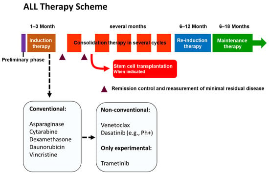
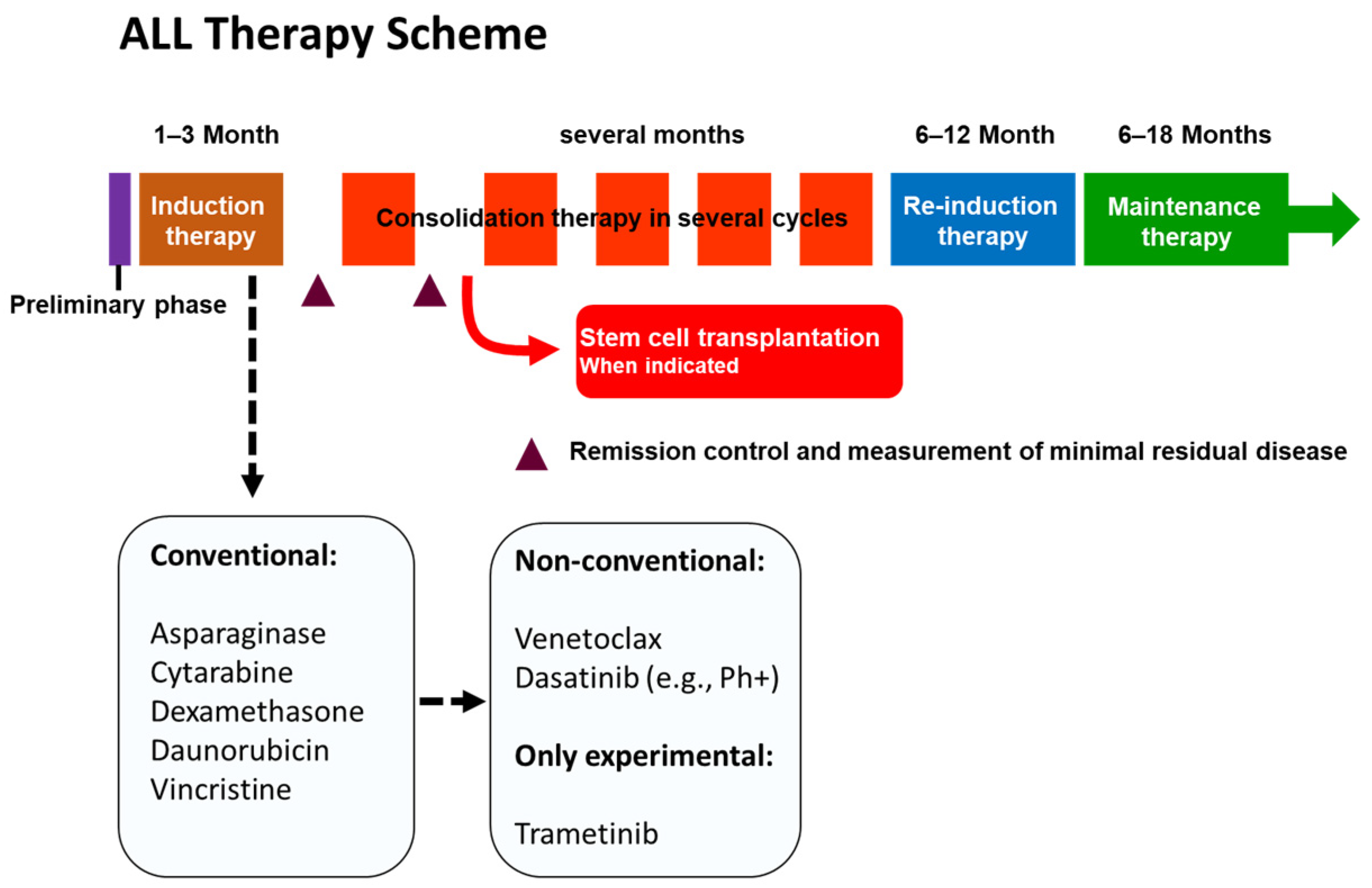
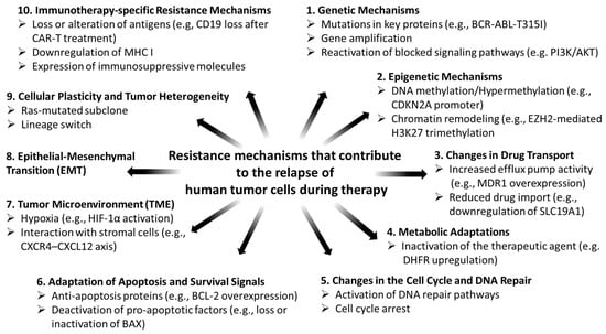
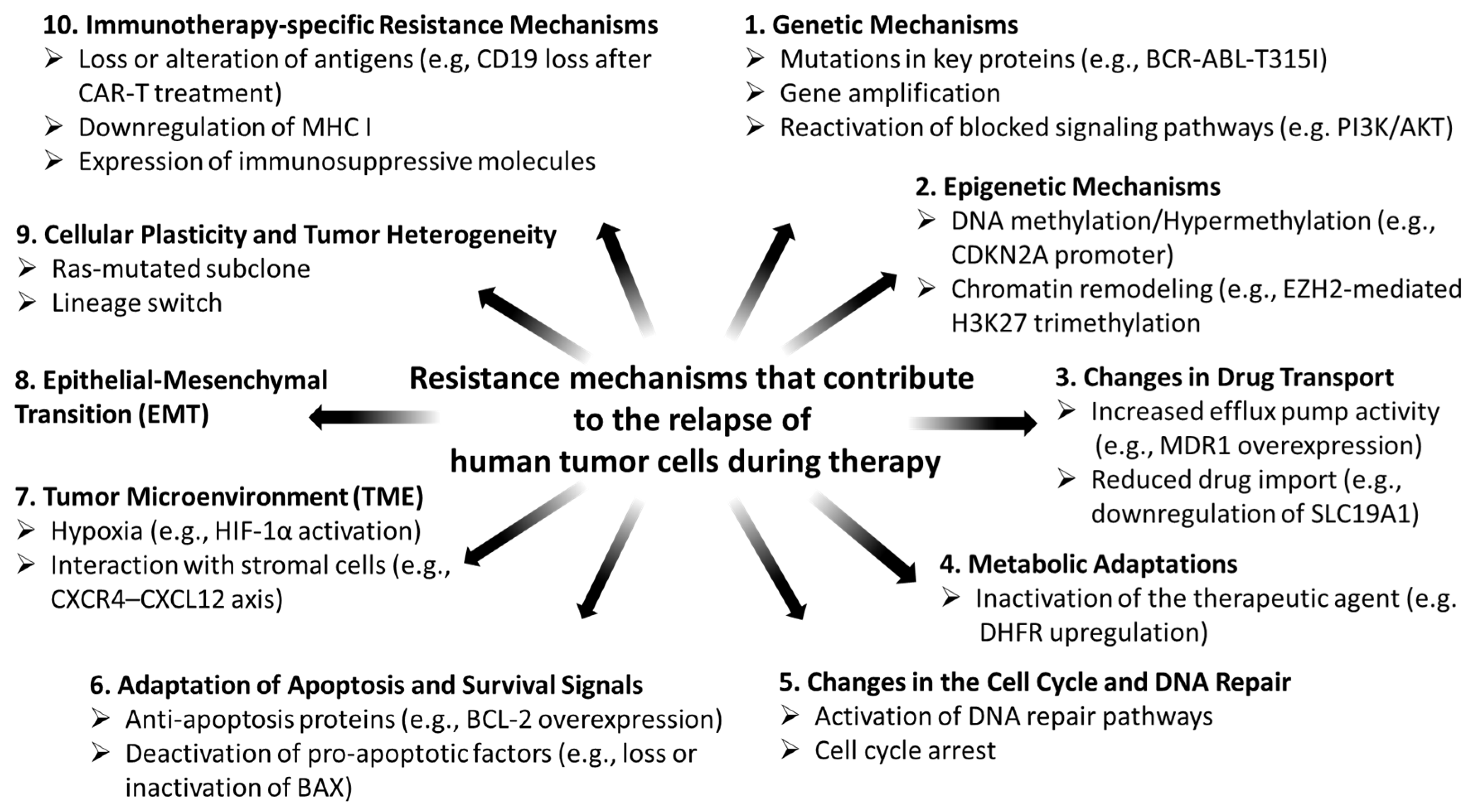
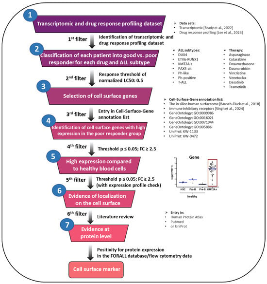
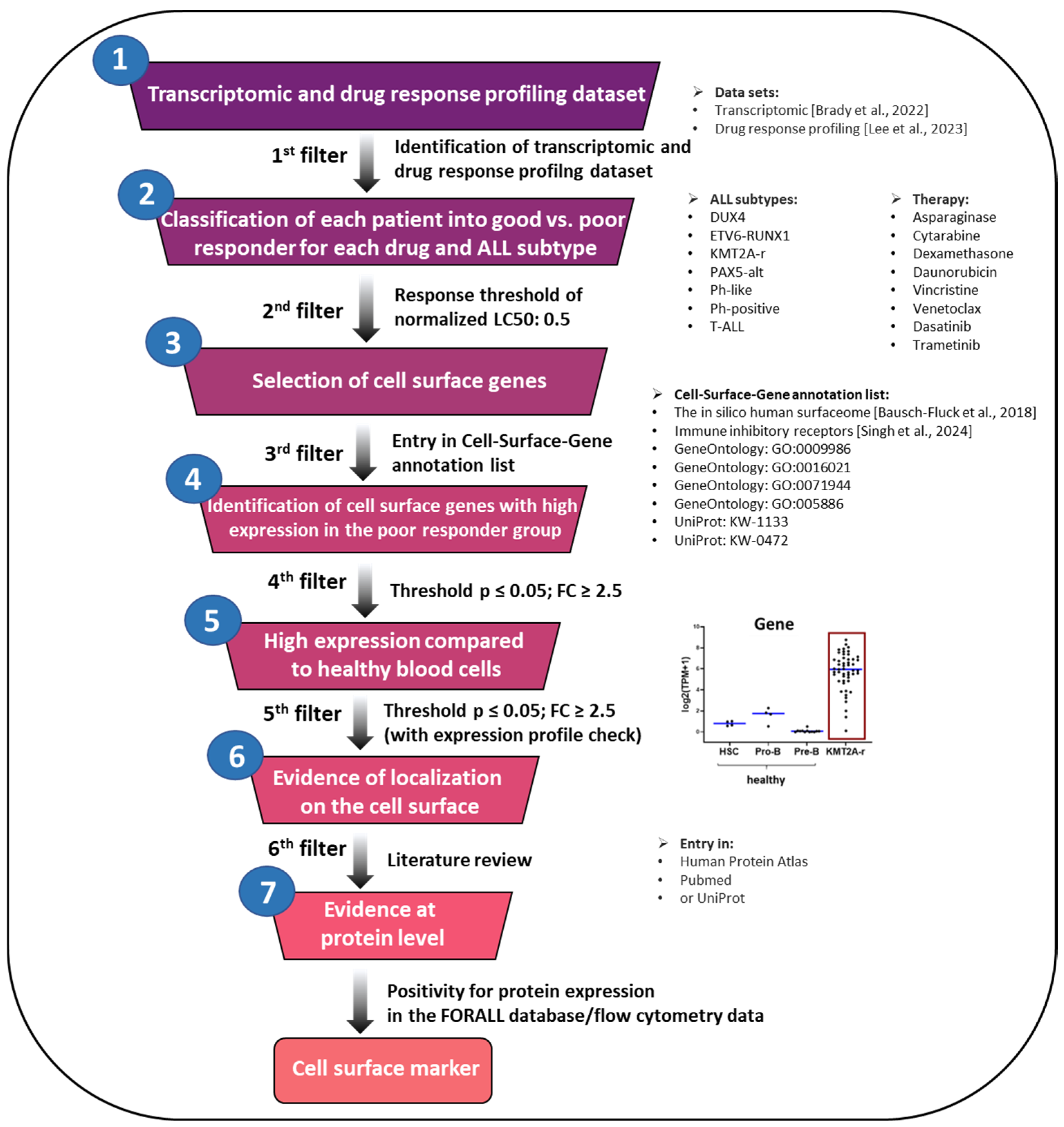
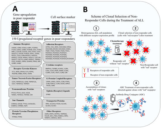
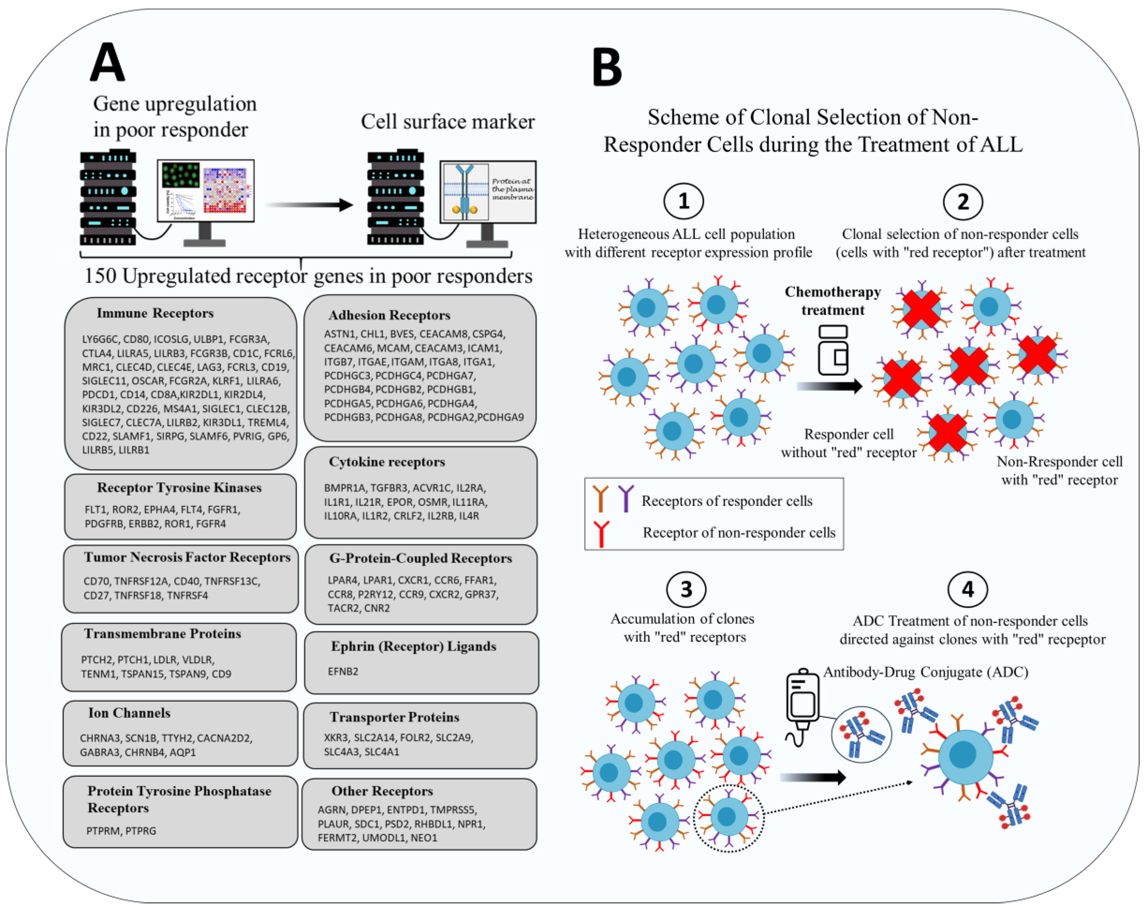

Disclaimer/Publisher’s Note: The statements, opinions and data contained in all publications are solely those of the individual author(s) and contributor(s) and not of MDPI and/or the editor(s). MDPI and/or the editor(s) disclaim responsibility for any injury to people or property resulting from any ideas, methods, instructions or products referred to in the content. |
© 2026 by the authors. Licensee MDPI, Basel, Switzerland. This article is an open access article distributed under the terms and conditions of the Creative Commons Attribution (CC BY) license.