


Abstract
This study focuses on the development and characterization of advanced composite materials based on poly(ε-caprolactone) (PCL) and poly(vinylidene fluoride) (PVDF), with or without silver nanoparticles (AgNPs), planned for peripheral nerve or bone regeneration. The complementary properties of PCL (biocompatibility and biodegradability) and PVDF (mechanical stability and piezoelectric functionality) were exploited by blending the polymers in different ratios, resulting in binary (PCL/PVDF) and ternary (PCL/PVDF/AgNPs) composites. Green-synthesized AgNPs were integrated to enhance antimicrobial activity and to support tissue repair through improved signal transmission. Functional thin films and electrospun fibres were obtained and subjected to advanced characterization techniques, including scanning electron microscopy (SEM), energy-dispersive X-ray spectroscopy (EDX), Fourier transform infrared spectroscopy (FTIR), X-ray diffraction (XRD), and thermal analysis. The results demonstrated appropriate morphology, chemical composition, structural stability, and favourable interactions with simulated physiological media. Preliminary biocompatibility assays confirmed good cell viability, supporting the biomedical applicability of the designed scaffolds. Overall, the obtained results highlight the potential of AgNPs-functionalized PCL/PVDF binary and ternary composites as promising candidates for flexible, durable, and bioactive implants in peripheral nerve or bone regeneration.
1. Introduction
The human nervous system is an intricate and highly coordinated network in which axons mediate rapid communication between the central nervous system (CNS) and the peripheral nervous system (PNS) [1]. These long-range connections form during embryonic development and continue to extend throughout growth to maintain functional integration across distant neural regions [2]. When such pathways are disrupted—through stroke, traumatic injuries, spinal cord lesions, peripheral nerve transections, or neurodegenerative diseases—the continuity of neural circuits is lost, resulting in impaired sensory and motor function. A major therapeutic challenge arises from the limited regenerative capacity of the nervous system: the adult CNS displays minimal axonal regrowth due to inhibitory cues and glial scarring, while PNS regeneration remains incomplete and frequently misdirected [2]. Pharmacological and biological interventions, including neurotrophic factors such as epidermal growth factor (EGF), erythropoietin (EPO), and glial cell line-derived neurotrophic factor (GDNF) or nanoparticle-based delivery systems, have shown potential, yet their efficacy is restricted by physiological barriers like the blood–brain and blood–spinal cord barriers, requiring high doses with systemic risks [3]. These limitations underscore the need for advanced tissue-engineering strategies capable of bridging nerve gaps, guiding axonal growth, and modulating the local microenvironment, positioning biomaterials-based approaches as promising candidates for next-generation nerve repair therapies [2,3].
Among polymeric materials, poly(ε-caprolactone) (PCL) and poly(vinylidene fluoride) (PVDF) are widely explored because of their complementary attributes. PCL is a biodegradable aliphatic polyester widely used in biomedical engineering owing to its excellent biocompatibility, high flexibility, easy processability, and adjustable semicrystalline structure [4]. Its low melting point, broad solubility window, and tunable mechanical properties—determined by molecular weight and crystallinity—make it particularly suitable for fabrication through solvent casting, molding, electrospinning, and 3D printing [5]. Although PCL degrades slowly through hydrolytic and enzymatic cleavage, this long-term stability is advantageous for applications that require sustained structural support, controlled degradation, or prolonged therapeutic release [6]. Beyond its physicochemical attributes, PCL has demonstrated strong potential in diverse biomedical applications, highlighting its relevance for tissue regeneration. Thin PCL films have been successfully used as long-acting drug-delivery reservoirs, enabling controlled release of small and large therapeutic agents in ocular and subcutaneous administration [7]. PCL-based nanocomposite films incorporating clay or metal oxides have shown antimicrobial and antibiofilm activity, even at low filler concentrations, supporting their use in protective wound interfaces [8]. Moreover, micro- and nanoporous PCL devices have enabled long-term cell encapsulation and vascular integration in vivo, demonstrating sustained cellular viability and functional stability over months [9]. PCL nanofiber scaffolds, particularly when combined with bioactive nanoparticles such as nanodiamonds, have further shown enhanced wound healing performance through improved cell adhesion and reduced microbial load [10]. Collectively, these case studies underscore PCL versatility as a structural and functional platform for biomedical scaffolds, drug delivery systems, and regenerative constructs—attributes that also justify its selection for nerve-regeneration approaches.
PVDF is a semi-crystalline fluoropolymer valued in biomedical engineering for its exceptional chemical stability, mechanical endurance, and electroactive behaviour [11,12,13]. Unlike conventional thermoplastics, PVDF combines high tensile strength, durability under cyclic loading, and resistance to solvents, radiation, and harsh physiological conditions, while also exhibiting multiple crystalline phases with distinct functional properties. Among them, the β-phase is particularly important due to its strong dipole alignment, which enables piezoelectric, pyroelectric, and ferroelectric responses—features that allow PVDF to convert mechanical stimuli into electrical cues relevant for interfacing with biological tissues [12,13,14]. Its processability into thin films, membranes, and electrospun fibres, together with good biocompatibility and moderate flexibility, has positioned PVDF as one of the most promising electroactive polymers for next-generation biomedical devices. Recent case studies highlight the versatility of PVDF in applications that demand electrical responsiveness, structural stability, or enhanced biological performance. When combined with silver nanoparticles (AgNPs), PVDF nanofibers electrospun at low nanoparticle loadings (PVDF/AgNPs) demonstrated improved β-phase content and amplified piezoelectric output, together with antibacterial activity and potential anticancer effects, illustrating a multifunctional platform suitable for therapeutic stimulation and infection control [15]. Electrospun PVDF scaffolds modified with self-assembling peptides promoted neural stem cell adhesion, alignment, and differentiation, confirming the material ability to reproduce key extracellular matrix cues for neural repair [16]. In peripheral nerve regeneration models, composite PVDF/PCL conduits and PVDF/graphene derivatives enhanced β-phase formation, hydrophilicity, and axonal guidance, leading to improved functional recovery in vivo [17,18,19]. Beyond neural interfaces, PVDF nanofiber systems with permanent hydrophobicity have supported sustained osteoblast stimulation through self-generated electromechanical cues [20]. Collectively, these findings show that PVDF electroactive nature and mechanical robustness make it a powerful candidate for scaffolds and biointerfaces that benefit from mechanically induced electrical signalling.
AgNPs are extensively integrated into biomedical systems due to their strong antibacterial activity, anti-inflammatory effects, and ability to regulate oxidative stress—properties that make them highly valuable for preventing infection in implantable biomaterials [21,22,23]. Through the sustained release of silver ions (Ag+), AgNPs disrupt bacterial membranes, increase permeability, and induce intracellular damage, offering broad-spectrum protection against both Gram-positive and Gram-negative pathogens. Recent biomedical studies underscore the multifunctionality of AgNPs in applications that require broad-spectrum antimicrobial action, modulation of inflammation, and enhanced tissue regeneration. AgNP-loaded hydrogels and composite dressings have accelerated the closure of chronic and infected wounds, particularly diabetic ulcers, by reducing bacterial load, suppressing biofilm formation, and improving the quality of the repair microenvironment [24,25,26]. In dental and implantable polymer coatings, low AgNP concentrations have effectively prevented early microbial adhesion and preserved material integrity, supporting long-term protection of orthodontic interfaces and resin-based devices [27,28,29,30]. AgNP-functionalized polymer matrices have also been explored as drug-delivery enhancers, where their high surface reactivity improves therapeutic loading, cellular uptake, and immunomodulatory effects. Together, these findings position AgNPs as a potent additive for regenerative systems requiring infection control, improved tissue response, or multifunctional therapeutic action.
In this study, solvent casting, electrospinning, and green synthesis were employed to fabricate multifunctional PCL/PVDF-based composites. Solvent casting enables the production of uniform polymer films by dissolving the polymer in a volatile solvent, casting the solution onto a substrate, and allowing controlled evaporation. This technique ensures precise thickness control, smooth surface morphology, and effective incorporation of fillers or bioactive agents, yet remains limited by slow solvent removal and challenges in producing films thicker than ~200 µm [31,32,33]. Electrospinning relies on a high-voltage electrostatic field that elongates a polymer jet into micro- and nanofibers, generating scaffolds with exceptionally high surface-area-to-volume ratios, interconnected porosity, and tuneable fibre alignment. These parameters—governed by solution composition, electric field strength, flow rate, and environmental conditions—allow precise modulation of fibre diameter, morphology, and network architecture, enabling structures optimized for cell attachment, directional axonal guidance, and β-phase enhancement in PVDF [34,35,36,37,38]. Green synthesis of AgNPs uses biological reducing and capping agents derived from plants, fungi, or microorganisms to convert Ag+ into stable Ag0 nanoparticles under mild, eco-friendly conditions. This method eliminates hazardous chemical reductants, enhances nanoparticle biocompatibility, and facilitates direct integration into polymer matrices for antimicrobial and regenerative applications [39,40]. Together, these fabrication routes allow the development of AgNP-functionalized PCL/PVDF systems as both solvent-cast films and electrospun fibre structures, combining mechanical integrity, electroactive functionality, and antimicrobial performance into a single multifunctional platform.
Similar case studies underscore the feasibility of combining degradable polyester matrices, electroactive fluoropolymers and metallic nanoparticles in regenerative medicine. Thin film PCL devices have already been used as long-acting drug reservoirs and macro-encapsulation systems, showing long-term in vivo stability and biocompatibility [7], while PCL/clay nanocomposites have demonstrated pronounced antimicrobial activity [8]. In parallel, PVDF/AgNPs electrospun mats exhibited increased β-phase content and enhanced piezoelectric output together with antibacterial effects [15], while PVDF scaffolds modified with self-assembling peptides improved neural stem-cell adhesion, alignment and differentiation by better mimicking the neural extracellular matrix [16]. Composite PVDF/PCL nerve conduits and PVDF structures functionalized with carbon nanostructures enhanced electroactivity, hydrophilicity and axonal guidance, enabling repair of sciatic nerve defects of up to 15 mm in rat models [18]. Moreover, PVDF nanofiber scaffolds with stable surface properties have been shown to deliver self-generated electromechanical stimulation to osteoblasts, illustrating the broader translational potential of piezoelectric fibrous platforms in tissue engineering [20].
In this work, we present the synthesis and multi-scale characterization of PCL/PVDF composite materials—produced as solvent-cast thin films and electrospun fibres—functionalized with green-synthesized AgNPs for tissue regeneration applications. While PCL and PVDF have been extensively studied, the novelty of this work resides in a multi-faceted synergistic approach. First, we engineered a functional triad system coupling a piezoelectric PVDF phase (for endogenous electromechanical stimulation) with a flexible, biodegradable PCL matrix, and integrated green-synthesized AgNPs to confer antimicrobial protection without residual chemical toxicity. Second, and most notably, we conducted a direct comparative evaluation across two distinct macroscopic dimensionalities: 2D solvent-cast dense films and 3D electrospun porous fibrous networks. By maintaining identical compositional ratios, this dual-morphology approach allows us to isolate how structural topography influences the physical properties and the preosteoblast cellular response. We hypothesize that this specific integration of mechanical support, piezoelectric signalling, and eco-friendly infection control provides a highly versatile, pro-regenerative microenvironment applicable to both peripheral nerve guidance and hard tissue (osteogenic) regeneration.
2. Materials and Methods
2.1. Materials
All reagents and solvents used in this study were of analytical grade and are listed in Table 1 together with their chemical formulas, purity and suppliers. Poly(ε-caprolactone) (PCL) and poly(vinylidene fluoride) (PVDF) served as the polymeric matrices for film and fibre fabrication, while silver nitrate (AgNO3), ammonia and the freshly prepared Zingiber officinale (ginger) plant extract were used for the green synthesis of silver nanoparticles (AgNPs). Organic solvents such as N,N-dimethylformamide (DMF), acetone and ethanol ensured complete polymer dissolution and processability during solvent casting and electrospinning. All chemicals were purchased from Sigma-Aldrich (Darmstadt, Germany). Distilled water was employed throughout all preparation and washing steps to avoid ionic contamination.
Table 1.
List of reagents used for composites synthesis, with abbreviations, chemical formula, purity, and supplier.
2.2. Synthesis Methods
2.2.1. AgNP Synthesis and Incorporation
PE was obtained by Soxhlet method using water as solvent. A 0.1 M AgNO3 solution was prepared by dissolving 8.49 g AgNO3 in 500 mL distilled water. A volume of 100 mL was heated at 50 °C (500 rpm, 10 min). Extract was added, temperature raised to 100 °C, and 15 drops of 25% NH4OH were introduced to adjust pH and initiate reduction. The suspension was centrifuged twice at 6000 rpm for 5 min, washed twice with distilled water, and the solid was dried. The full characterization dataset for the specific batch of AgNPs used in this study, which includes high-resolution SEM image, EDX spectrum and XRD pattern, is provided in Figures S1–S3 of the Supplementary Material. For films, AgNPs were incorporated at 2 wt% into 10% (w/v) polymer solutions with 5 min ultrasonication before casting. For fibres, AgNPs (2 wt%) were ultrasonically dispersed for 10 min in the solvent prior to polymer addition.
2.2.2. PCL/PVDF Film Preparation
Polymeric solutions were prepared at 10% (w/v) concentration by dissolving PCL and PVDF separately in a solvent mixture of DMF and Ac (DMF:Ac = 1:1, v/v). For each polymer, 2 g were weighed and dissolved in 20 mL of solvent under magnetic stirring at 65 °C until complete homogenization. The two stock solutions were then mixed at different volumetric ratios to obtain binary blends of PCL and PVDF, as detailed in Table 2. Each mixture was homogenized at 65 °C for 2 h under continuous stirring, cast onto glass substrates, and dried in an oven at 55 °C for 12 h. The films were subsequently immersed in ethanol to remove solvent residues and finally dried at room temperature for 24 h.
Table 2.
Composition of PCL/PVDF films. We have used the same color for each sample.
For AgNP-functionalized blends (Table 3), the total volume per formulation was 13 mL at 10% (w/v) polymer (1.3 g). AgNPs were added at 2 wt% (0.026 g) relative to the total polymer amount. Mixtures were ultrasonicated 5 min for dispersion, stirred at 65 °C to homogenize, cast, dried for 12 h at 55 °C, ethanol-washed, then dried for another 24 h.
Table 3.
Composition of PCL/PVDF films with AgNPs.
2.2.3. PCL/PVDF Fibre Preparation
Electrospinning solutions were prepared at 10% (w/v), with a total volume of 10 mL for each formulation. As summarized in Table 4, PCL was dissolved in DMF, while PVDF was dissolved in a DMF/acetone mixture (1:1, v/v). For AgNP-functionalized samples, the nanoparticles (2 wt% relative to the total polymer mass) were first dispersed in the corresponding solvent by ultrasonication for 10 min prior to polymer addition. All solutions were then completely homogenized by magnetic stirring until full dissolution was achieved prior to the electrospinning process.
Table 4.
Composition of PCL/PVDF fibres with and without AgNPs.
Since PCL and PVDF require fundamentally different electrospinning conditions, forming a stable single-blend solution was unfeasible. Furthermore, simultaneous electrospinning resulted in chaotic fibre deposition due to electrostatic repulsion between the polymer jets. Therefore, electrospinning was performed using two separate syringes, each containing one of the prepared polymer solutions (PCL-based or PVDF-based). These syringes were operated alternately during the process to obtain the final blended fibrous mats. Electrospinning was carried out under controlled conditions, using a flow rate of 1 mL/h, an applied voltage of 20 kV, a needle angle of 30° relative to the vertical axis, and a needle-to-collector distance of 13 cm. Fibres were collected on pre-cleaned metallic substrates.
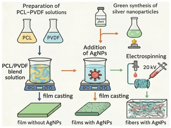
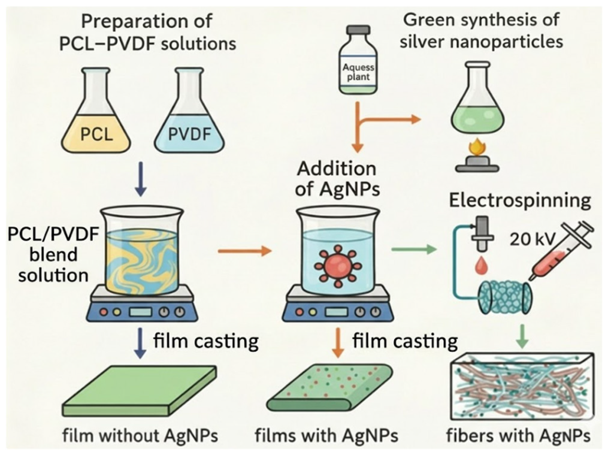
To illustrate the overall technological route, the main steps for polymer dissolution, nanoparticle synthesis, casting, and electrospinning are summarized in the general flowchart (Figure 1).
Figure 1.
General flowchart of the preparation steps for PCL/PVDF/AgNP films and electrospun fibres.
2.3. Characterization Methods
Scanning electron microscopy (SEM) was employed to examine the morphology of the films and fibres. Analyses were carried out using a FEI Quanta Inspect F50 (FEI Company, Hillsboro, OR, USA) equipped with an energy-dispersive X-ray (EDX) detector, operated at 30 kV. Prior to observation, the samples were sputter-coated with a thin gold layer by DC magnetron sputtering.
Fourier transform infrared spectroscopy (FTIR) was used to identify functional groups and assess molecular interactions in the polymer blends. Measurements were performed in ATR mode on a Nicolet iS50 FTIR spectrometer (Thermo Scientific, Waltham, MA, USA), in the wavenumber range of 4000–400 cm−1, with a resolution of 4 cm−1 and 32 scans per sample.
X-ray diffraction (XRD) was applied to determine crystalline phases and degree of crystallinity. Diffractograms were recorded with a Shimadzu XRD-6000 diffractometer (Shimadzu Corporation, Kyoto, Japan) using Cu Kα radiation (λ = 0.154 nm, Ni filter), over a 2θ range of 10–50°, with a step size of 0.02°.
Thermal behaviour was investigated by thermogravimetric analysis (TGA) and differential scanning calorimetry (DSC) using a STA 449 F3 Jupiter system (NETZSCH, Selb, Germany). The measurements were performed from room temperature to 900 °C at a heating rate of 10 °C/min in an air atmosphere.
Bioactivity in vitro was evaluated by immersion in Simulated Body Fluid (SBF) prepared according to the Kokubo protocol [41], with ion concentrations similar to human plasma. Samples were immersed at 37 °C for 28 days to evaluate the stability and degradation-related changes of the bioresorbable polymeric matrix under physiological-like conditions.
Biocompatibility was assessed using MC3T3-E1 pre-osteoblast cells (ATCC, Manassas, VA, USA). All samples were thermally treated at 120 °C for 1 h to remove residual solvents potentially affecting cytocompatibility, as well as to sterilize them, then cooled under sterile conditions. Cells were cultured in α-MEM supplemented with 10% fetal bovine serum and 1% penicillin–streptomycin, in standard cell culture conditions (37 °C, 5% CO2, 90% humidity). Samples were placed in 12-well plates and seeded at a concentration of 40,000 cells/100 µL/sample. After 1 h incubation in standard conditions, to allow cell adhesion, 1 mL of complete medium was added per well, and cultures were incubated for another 48 h in standard cell culture conditions. After this time, cell viability was quantified using the MTT tetrazolium salt assay. Medium was replaced with a MTT working solution (10% of a 5 mg/mL MTT stock in PBS diluted in complete α-MEM) and incubated for 2 h. Formazan crystals were dissolved in DMSO, and absorbance was read at 570 nm. The results were normalized to the negative control, set at 100%, and expressed as mean ± SEM (Standard Error of the Mean) (n = 3), with statistical significance indicated as * p < 0.05, ** p < 0.01, and *** p < 0.001.
3. Results and Discussion
3.1. PCL/PVDF Film Characterization
3.1.1. Scanning Electron Microscopy (SEM)
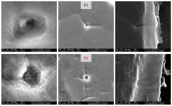
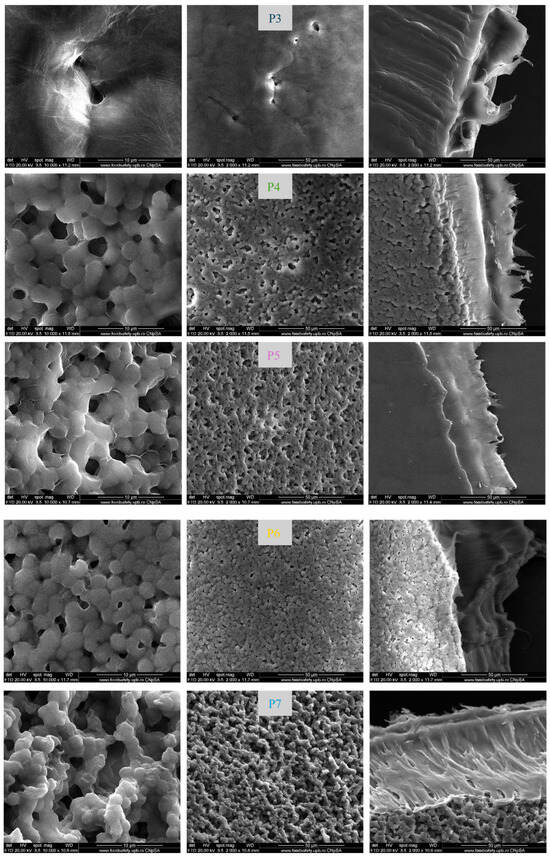
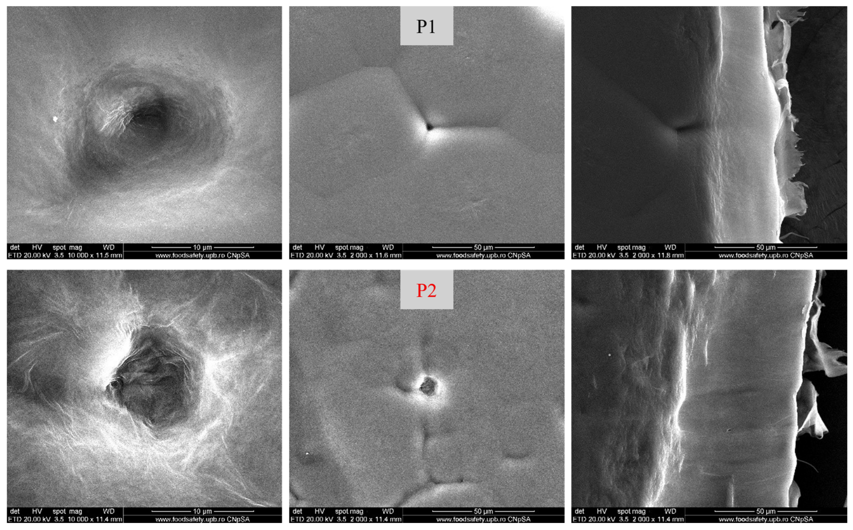
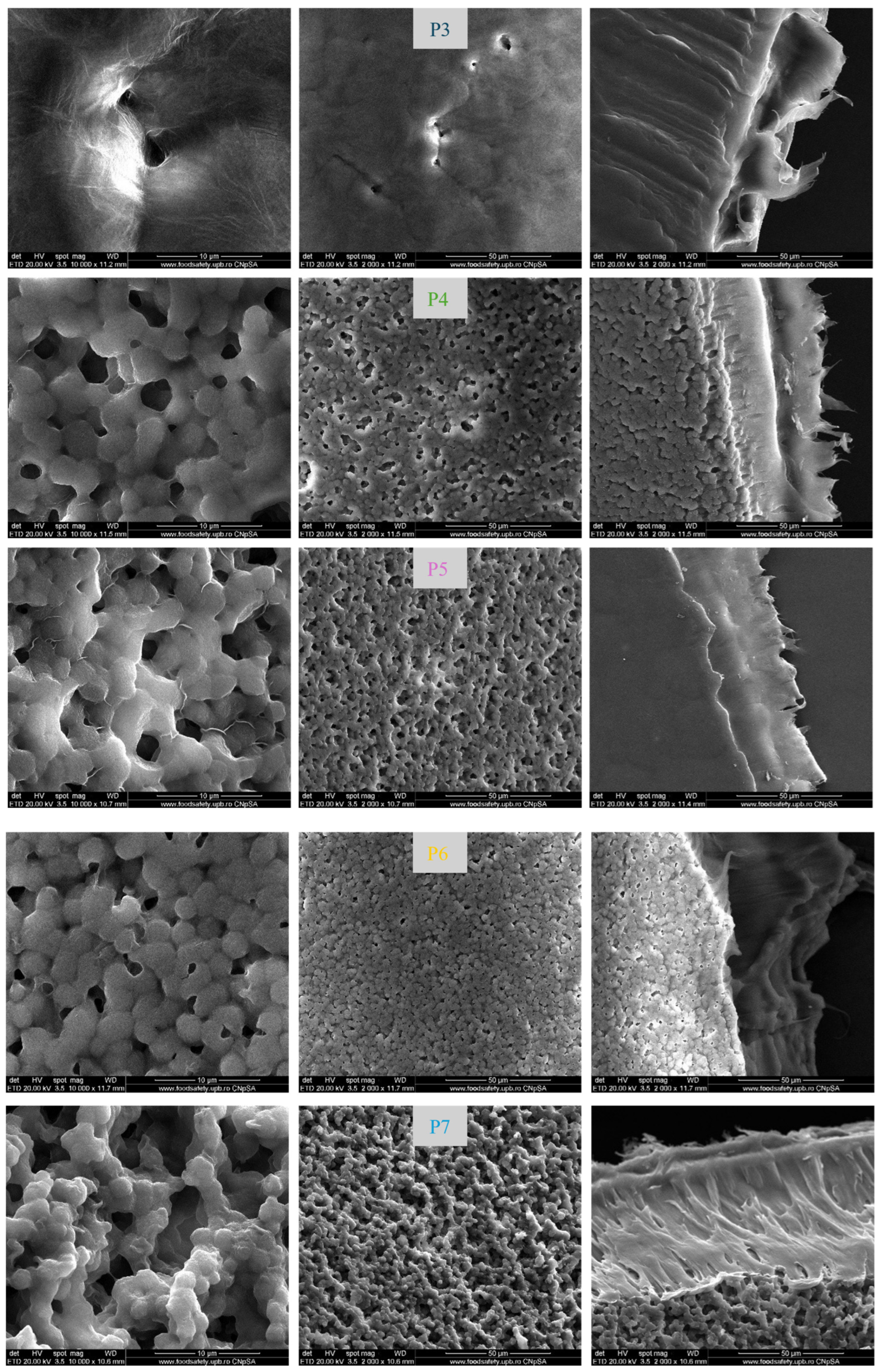
The surface and cross-sectional morphologies of the PCL/PVDF films are presented in Figure 2. Pure PCL (sample P1) exhibited a granular surface with large, non-uniform domains and limited interconnectivity of pores, features that may restrict fluid diffusion despite the relatively dense cross-section. With the addition of 10% PVDF (sample P2), the morphology became finer and more homogeneous, with slightly increased porosity and compact cross-sections, suggesting improved miscibility of the phases. At 20% PVDF (sample P3), phase separation became more pronounced, with layered structures and signs of exfoliation visible in cross-sections. The 30% PVDF composition (sample P4) showed interconnected pores but distinct phase boundaries, reflecting reduced compatibility between the two polymers. At the 50%/50% ratio (sample P5), granules appeared fragmented, and the porosity increased considerably, with weak interfacial adhesion and discontinuities in the structure. In the case of 70% PVDF (sample P6), PCL appeared as dispersed inclusions within a continuous PVDF matrix, with cross-sections confirming clear phase segregation. Pure PVDF (sample P7) presented the most homogeneous morphology, with well-bonded domains and a stratified structure, although significant basal porosity could still compromise mechanical stability. These results highlight that while intermediate blends enhance porosity, phase separation and reduced cohesion become more evident at higher PVDF contents.
Figure 2.
SEM images of PCL/PVDF composite films (P1–P7), showing surface morphology at magnifications of 10,000× (left column) and 2000× (middle column), together with cross-sectional views acquired at 2000× (right column).
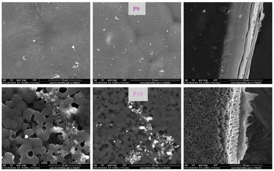
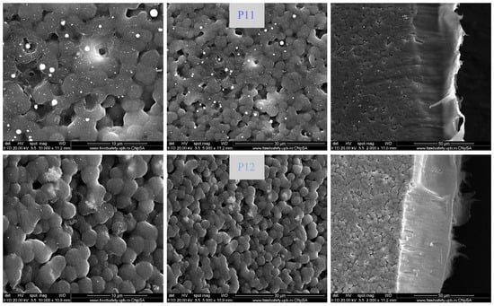
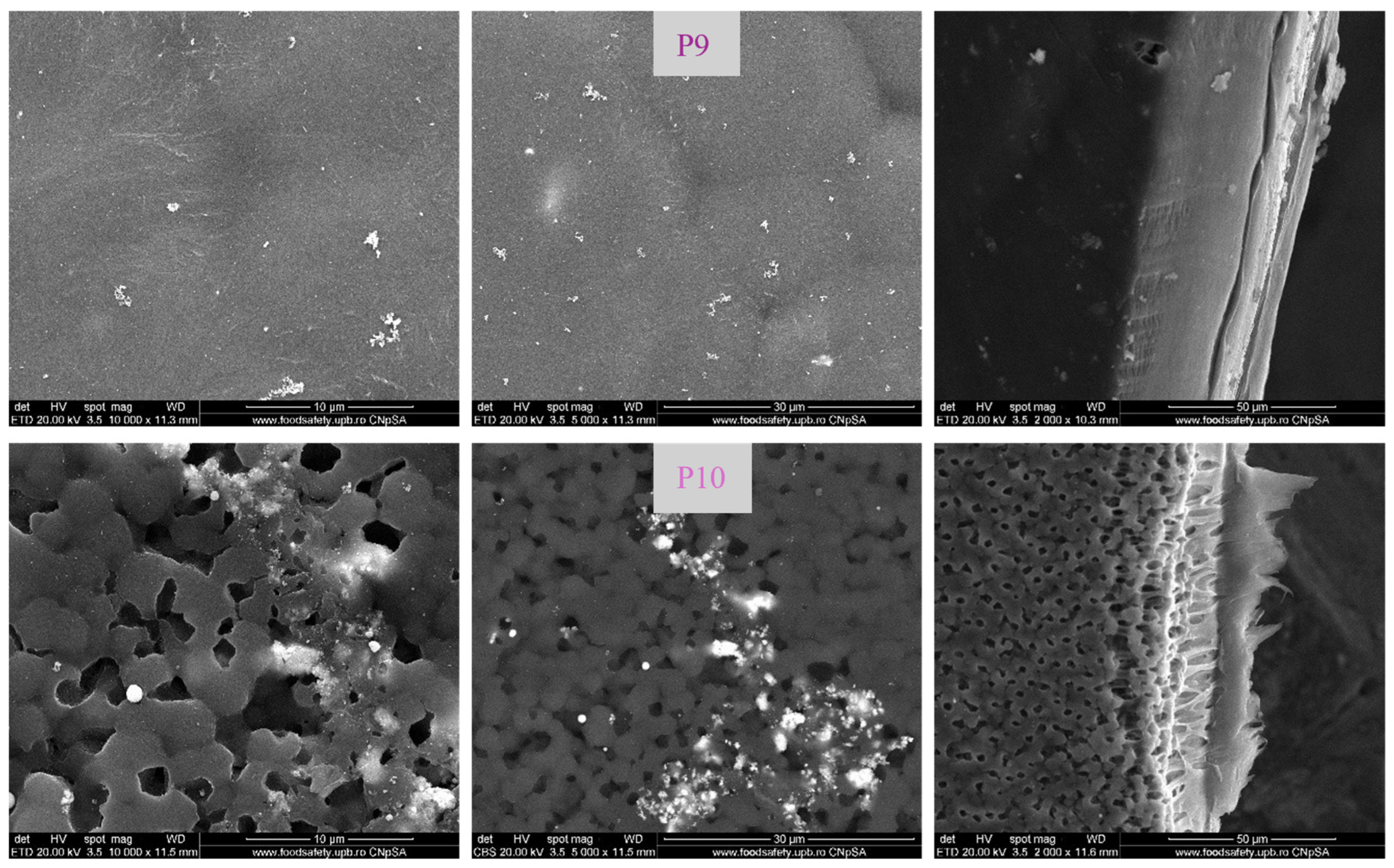
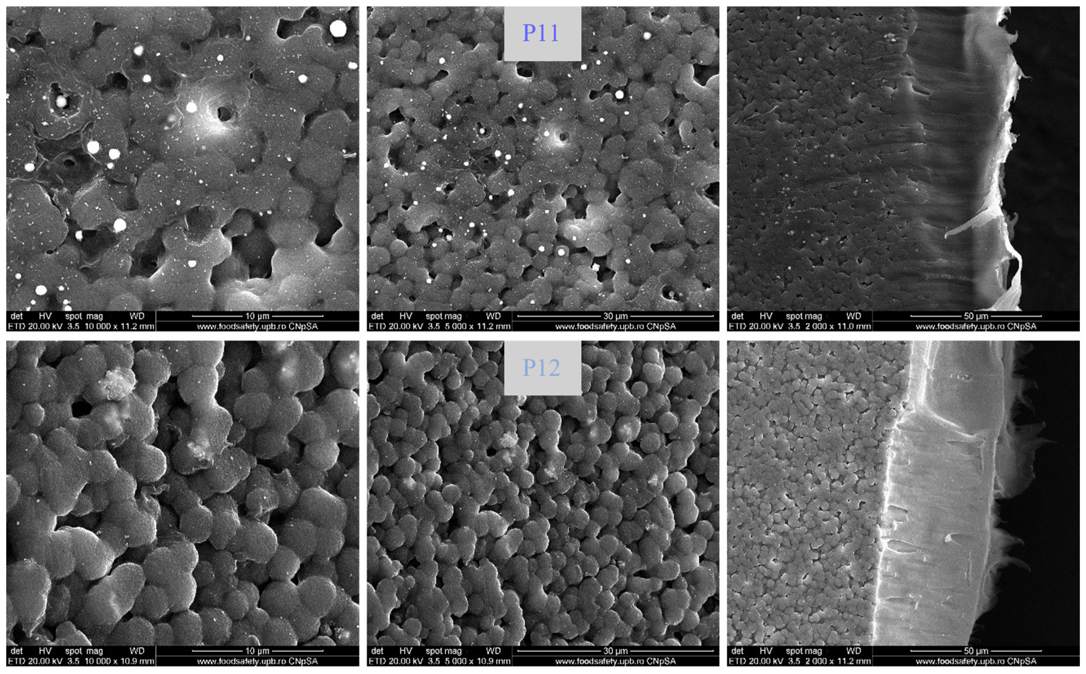
The morphology of AgNP-functionalized PCL/PVDF composites is illustrated in Figure 3. The SEM images of samples P9–P12, prepared with different polymer ratios and 2% AgNPs, together with their cross-sections, reveal distinct surface textures and nanoparticle distributions. In sample P9 (pure PCL), AgNPs appeared finely dispersed without visible agglomerates, while the cross-section showed a compact and continuous structure. Sample P10 (30% PVDF) exhibited a more porous and granular surface with uneven nanoparticle distribution, accompanied by a stratified internal architecture. Sample P11 (50% PVDF) presented a relatively uniform integration of AgNPs, with a balanced porosity and well-interacting phases, as observed in the cross-section. In the case of sample P12 (pure PVDF), the morphology appeared granular yet compact, with AgNPs homogenously distributed throughout the matrix. Taken together, the incorporation of AgNPs influenced surface roughness and porosity while maintaining the structural integrity of the films.
Figure 3.
SEM images of PCL/PVDF/AgNP composite films (P9–P12), showing surface morphology at magnifications of 10,000× (left column) and 5000× (middle column), together with cross-sectional views acquired at 2000× (right column).
3.1.2. Energy-Dispersive X-Ray Spectroscopy (EDX)
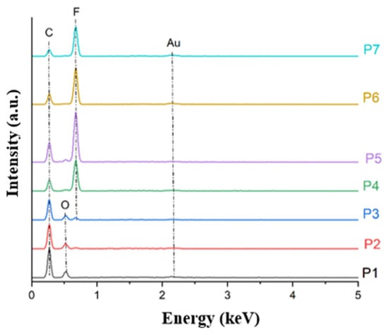
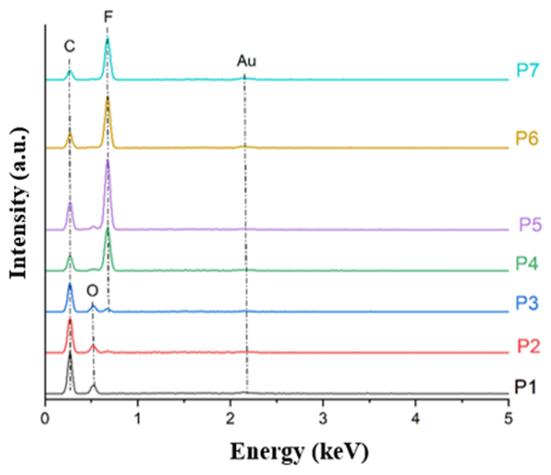
The elemental composition of the PCL/PVDF films was assessed by EDX, as shown in Figure 4. The spectrum of pure PCL (sample P1) was dominated by signals from carbon (C) and oxygen (O), consistent with its chemical structure. With increasing PVDF content in the blends (samples P2–P6), a progressive rise in the fluorine (F) peak was observed, accompanied by a gradual decrease in oxygen intensity, reflecting the substitution of PCL with PVDF in the mixtures. This compositional transition culminated in the case of pure PVDF (sample P7), where the spectrum was characterized predominantly by carbon and fluorine, with oxygen being nearly absent, confirming the exclusive presence of the fluoropolymer. In all spectra, the presence of gold (Au) was also detected, originating from the sputter-coating applied to improve electrical conductivity during SEM/EDX analysis. These results validate the controlled distribution of the two polymers across the prepared films, in agreement with the intended formulation ratios.
Figure 4.
EDX spectra of PCL/PVDF composite films with increasing PVDF content (P1–P7).
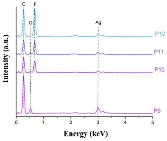
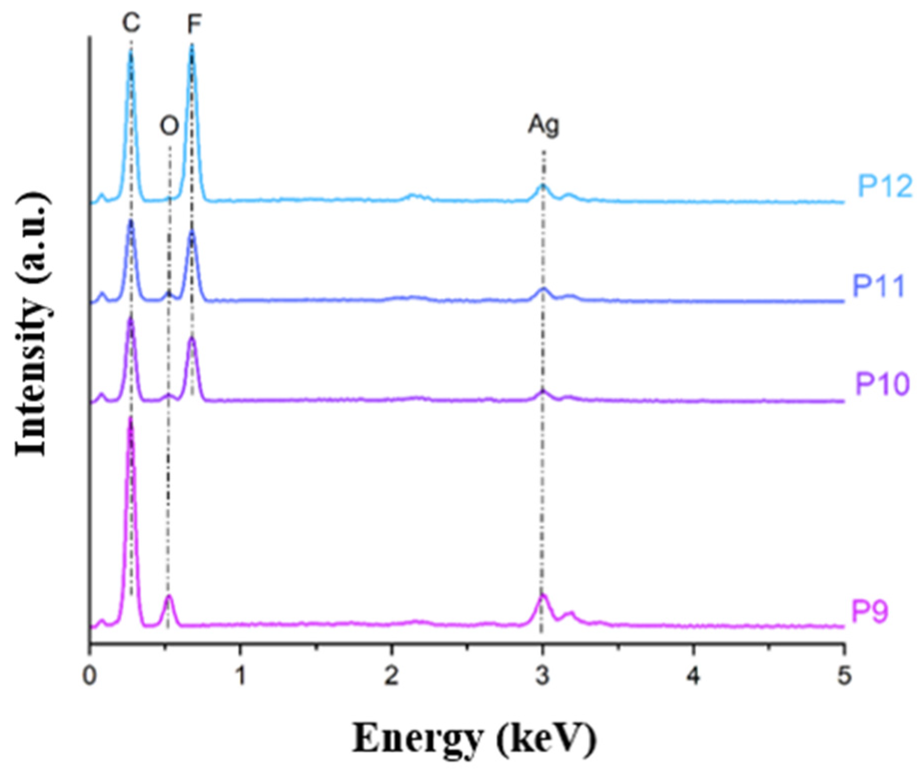
The elemental composition of AgNP-functionalized PCL/PVDF composite films (P9–P12) is presented in Figure 5. The EDX spectra confirmed the presence of carbon (C) and oxygen (O) associated with the PCL phase, fluorine (F) originating from PVDF, and a distinct silver (Ag) signal at ~3 keV in all AgNP-containing samples. As the PVDF content increased from P9 to P12, a progressive enhancement of the fluorine peak was observed, accompanied by a reduction in oxygen intensity, consistent with the gradual replacement of the PCL phase. The persistence of the silver signal across all compositions indicates successful incorporation and retention of AgNPs within both polymer matrices, independent of blend ratio. Similar EDX spectra confirmed AgNP integration for PCL-based composite systems, where AgNPs were homogeneously distributed within the polymer network, preserving antimicrobial functionality [29], as well as for PVDF-based electrospun membranes containing embedded AgNPs, where stable silver incorporation was achieved without phase segregation [42]. These findings support the compatibility of AgNPs with both PCL and PVDF and validate their uniform integration within the composite films developed in this study.
Figure 5.
EDX spectra of PCL/PVDF/AgNP composite films with varying composition (P9–P12).
3.1.3. Fourier-Transform Infrared Spectroscopy (FTIR)
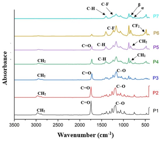
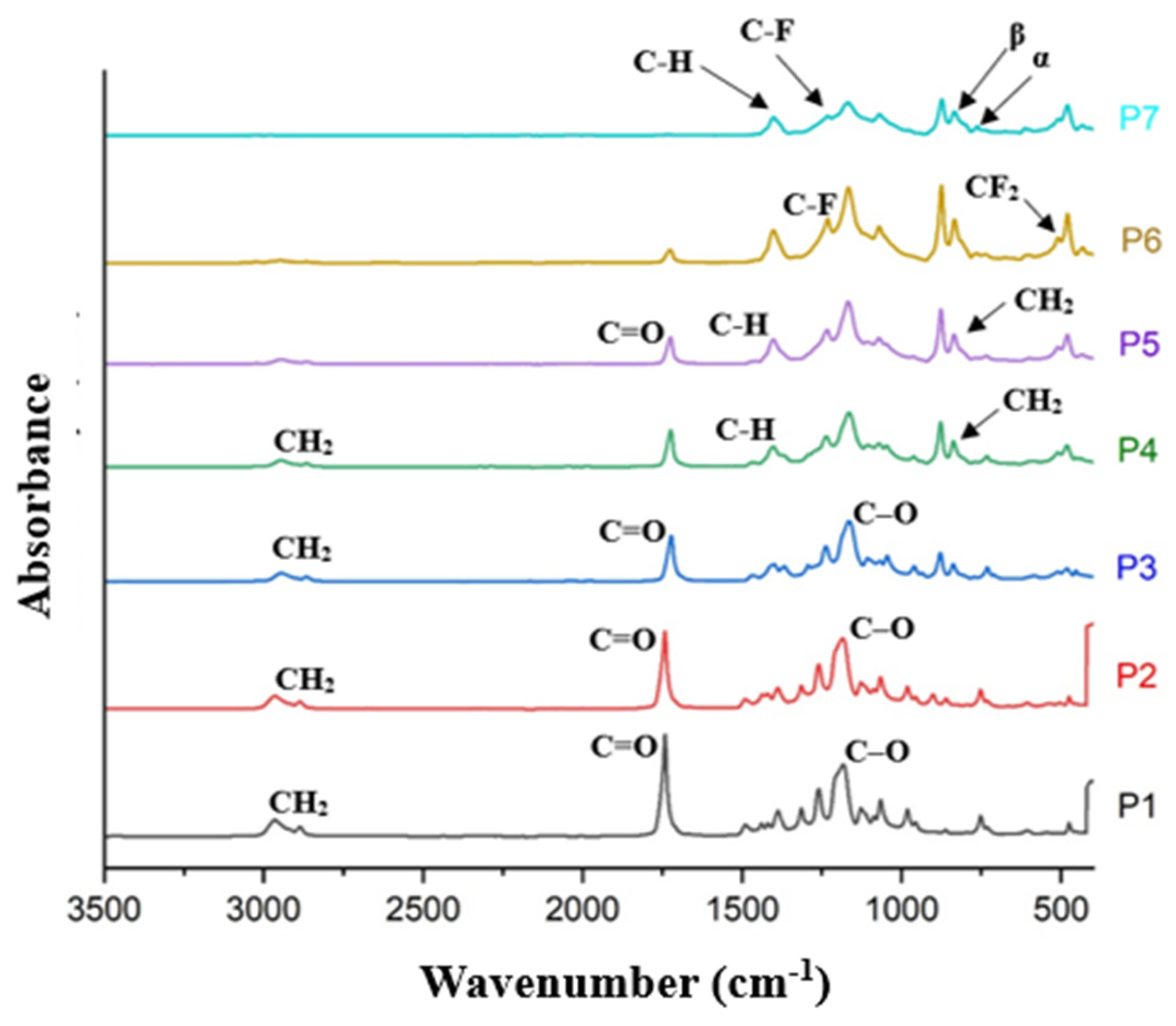
The FTIR spectra of PCL/PVDF composite films (Figure 6) confirm the successful formation of binary blends while preserving the characteristic functional groups of both polymers. Pure PCL (P1) exhibited the expected absorption bands corresponding to aliphatic CH2 stretching (~2940 and ~2860 cm−1) and a strong ester carbonyl (C=O) stretching band at ~1720 cm−1, which represents the dominant vibrational feature of the ester functionality rather than the entire ester grouping, in agreement with literature reports [43,44]. Pure PVDF (P7) showed characteristic absorptions associated with C–H and C–F vibrations, with intense bands in the fingerprint region (~1396 and ~1175 cm−1). The presence of the electroactive β-phase was evidenced by the strong absorption at ~840 cm−1, while the weak shoulder near ~763 cm−1 indicated minor contributions from the non-polar α-phase, consistent with previous studies on semi-crystalline PVDF films [45].
Figure 6.
FTIR spectra of PCL/PVDF composite films with increasing PVDF content (P1–P7).
For the blended systems (P2–P6), the spectra combined the signatures of both polymers without the appearance of new bands, indicating physical blending rather than chemical reaction. A slight shift of the PCL carbonyl band toward higher wavenumbers (~1725 cm−1) was observed with increasing PVDF content, suggesting weak intermolecular interactions, such as dipole–dipole or hydrogen-bond-like interactions between PCL carbonyl groups and PVDF fluorinated chains, as reported for similar PCL/PVDF systems [45,46]. Importantly, the β-phase-related bands of PVDF (notably at ~840 and ~510 cm−1) were retained in all blends, demonstrating that incorporation of PCL does not suppress the electroactive crystalline phase of PVDF. These results indicate partial molecular compatibility between the two polymers and support the suitability of the blends for applications requiring combined mechanical flexibility and electroactivity.
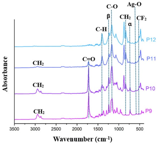
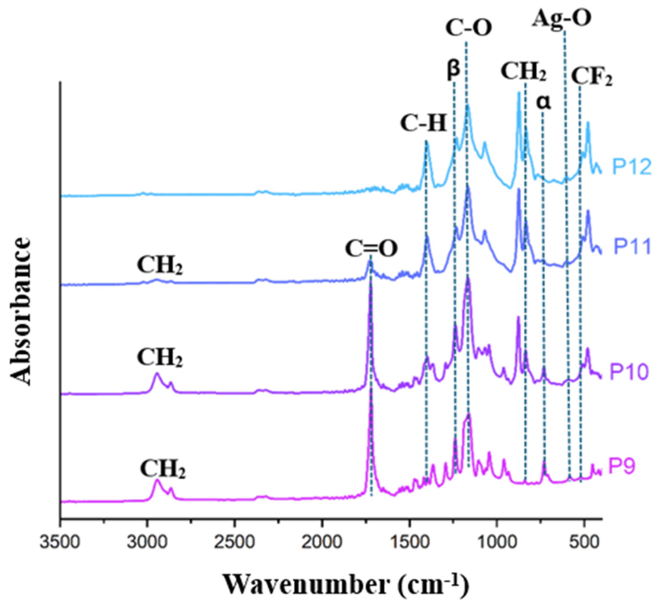
The FTIR spectra of AgNP-functionalized PCL/PVDF composite films (P9–P12), shown in Figure 7, revealed the functional groups of both polymers together with signatures of AgNP incorporation. In P9 (pure PCL), characteristic CH2 stretching bands appeared at ~2940 and ~2860 cm−1, while the C=O band at ~1720 cm−1 was slightly shifted to ~1764 cm−1, suggesting interactions between PCL carbonyl groups and AgNPs. A broad band near 540 cm−1 further confirmed Ag–O bonding [47]. In P10 (30% PVDF), features of both polymers were visible, with CH2 and C=O absorptions of PCL overlapping with C–F vibrations of PVDF around 1274–1170 cm−1. Samples P11 (50% PVDF) and P12 (pure PVDF) showed spectra dominated by PVDF, with bands at ~1400 cm−1 (CH2 wagging), ~1180 cm−1 (CF2 stretching), and the β-phase markers at 840 cm−1 and 510 cm−1. The α-phase signal at ~763 cm−1 was mainly observed in P12 but with reduced intensity, indicating mixed crystallinity with a predominance of the electroactive β-phase [16]. In conclusion, FTIR confirmed the coexistence of PCL and PVDF depending on blend ratio, while AgNP incorporation was validated by the Ag–O band.
Figure 7.
FTIR spectra of PCL/PVDF/AgNP composite films with varying composition (P9–P12).
3.1.4. X-Ray Diffraction (XRD)
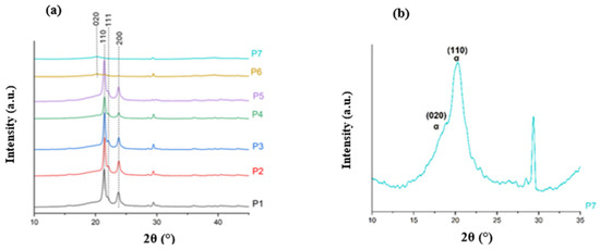
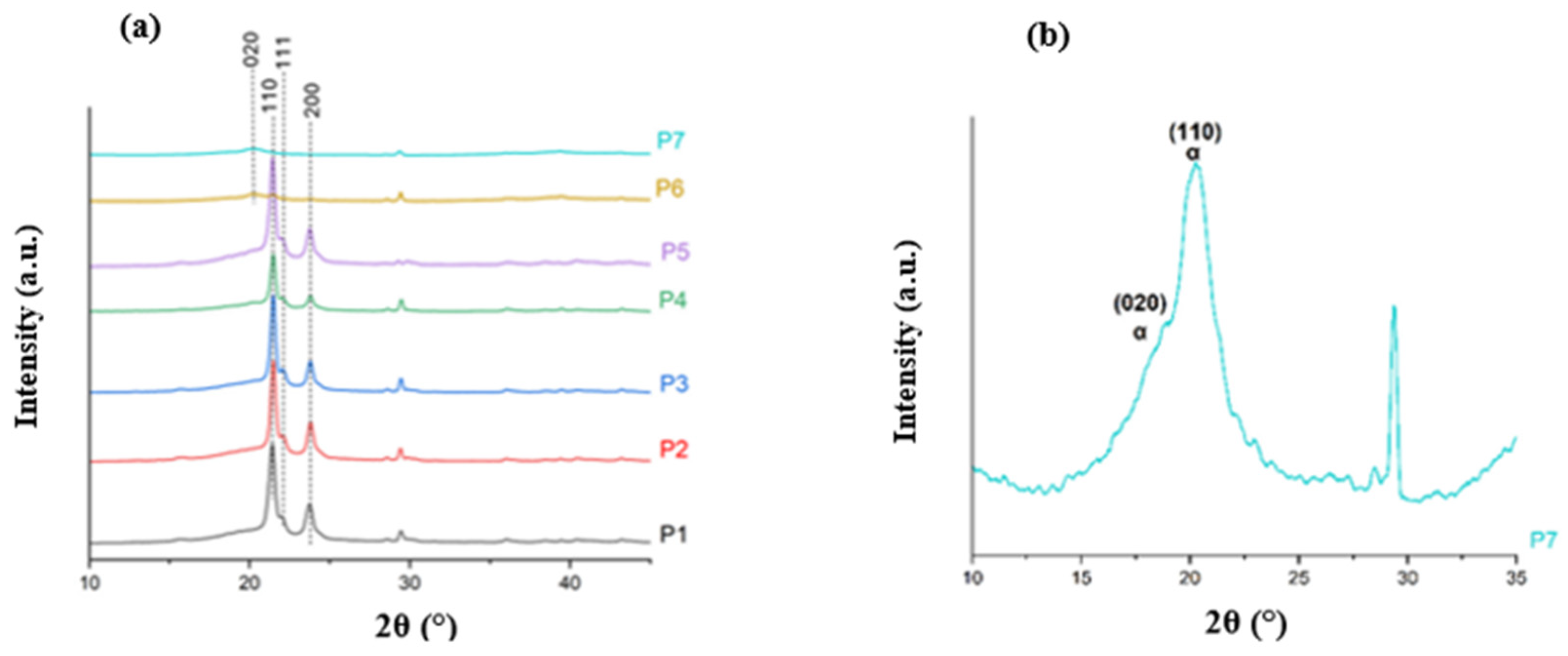
The XRD patterns of PCL/PVDF films (P1–P7) are shown in Figure 8a, highlighting the crystalline fingerprints of each polymer. Pure PCL (P1) exhibited the characteristic reflections at 2θ ≈ 21.1, 22.0 and 23.4°, assigned to the (110), (111) and (200) planes of orthorhombic PCL, confirming its semicrystalline nature [48]. Pure PVDF (P7) displayed a low-intensity reflection at 2θ ≈ 18.3°, commonly attributed to the (020) plane of the α-phase, which is the thermodynamically favoured form obtained by conventional crystallization (Figure 8b) [49].
Figure 8.
XRD patterns of (a) PCL/PVDF composite films with increasing PVDF content (P1–P7) and (b) a PVDF unitary film (P7) over a narrow range.
For the blend films (P2–P6), the diffractograms contained contributions from both components: the PCL reflections remained visible, while their relative intensities decreased with increasing PVDF content, and the α-PVDF reflection at ~18.3° was also detected, indicating coexistence of crystalline domains within the blends. Importantly, XRD did not provide an unambiguous, resolved diffraction feature that could be assigned to electroactive PVDF (β/γ) in these samples; any such contribution can be masked by the strong and partially overlapping PCL peaks in the 21–23° region and by the generally lower diffraction intensity of minor or less-ordered phases. Therefore, while FTIR suggests the presence of electroactive PVDF conformations, the present XRD data primarily support α-phase PVDF as the dominant crystalline form in P7 and in the blends, with possible β/γ contributions remaining below XRD detectability under these conditions [50,51].
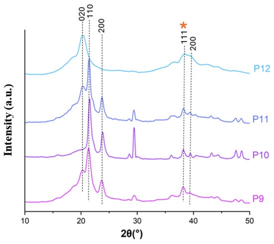
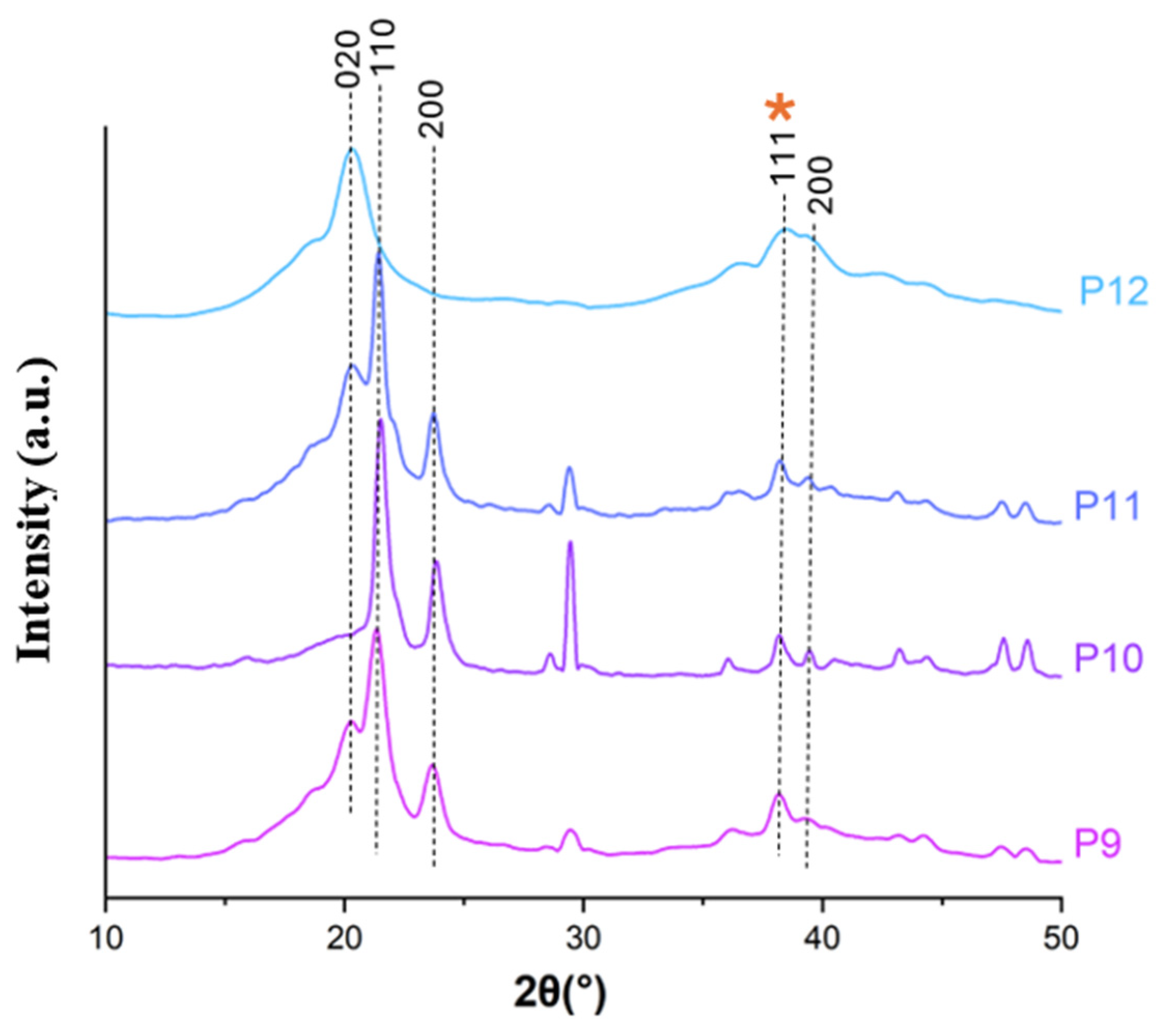
The XRD patterns of AgNP-functionalized PCL/PVDF films (P9–P12), presented in Figure 9, indicate that the crystalline structures of both polymers are preserved after AgNP incorporation. In P9 (pure PCL), the characteristic reflections at 2θ ≈ 21 and 23°, corresponding to the (110) and (200) planes of orthorhombic PCL, remained clearly visible, confirming that the semicrystalline nature of PCL was not affected [51]. Likewise, P12 (pure PVDF) retained the α-phase diffraction peak at 2θ ≈ 18.3°, assigned to the (020) plane, indicating unchanged PVDF crystallinity in the presence of AgNPs. The intermediate compositions (P10 and P11) exhibited diffraction features of both polymers, reflecting phase coexistence and relative composition. The characteristic reflection of metallic silver was detected at 2θ ≈ 38°, corresponding to the (111) plane (marked with an orange asterisk in Figure 9), further confirming the successful incorporation and presence of AgNPs within the polymer matrices [52].
Figure 9.
XRD patterns of PCL/PVDF/AgNP composite films with varying composition (P9–P12). The orange asterisk stands for silver (Ag).
3.1.5. Thermal Analysis
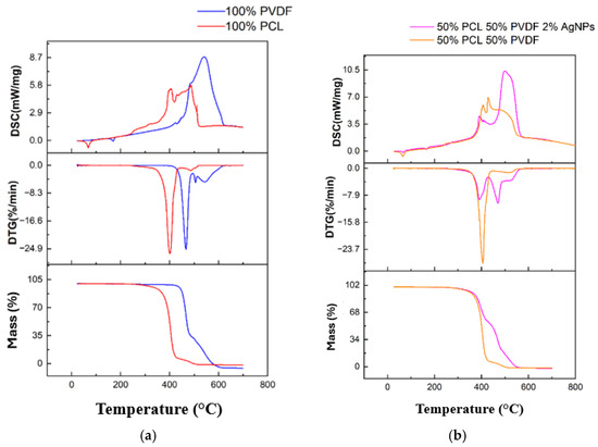
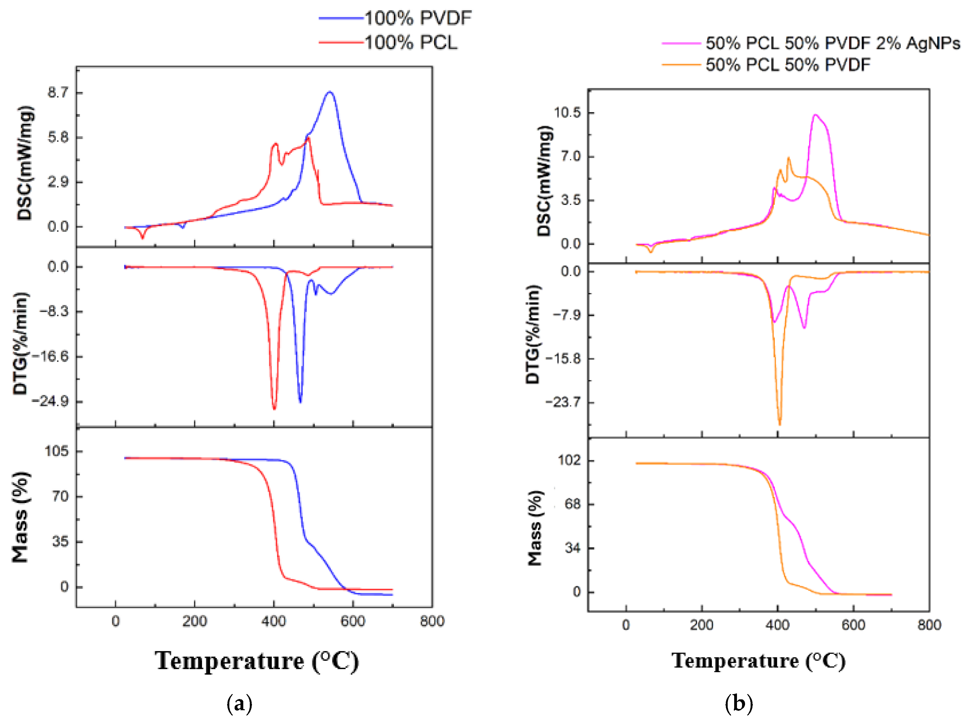
The thermal behaviour of the neat polymers (pure PCL and pure PVDF) was investigated by simultaneous TGA, DTG, and DSC, as shown in Figure 10a. Both materials exhibited good thermal stability at low temperatures, maintaining nearly constant mass up to ~300 °C for PCL and ~420 °C for PVDF. PCL underwent sharp mass loss in the range 300–420 °C, whereas PVDF degradation started above 420 °C and extended up to 520–550 °C, confirming the higher thermal resistance of PVDF. The DTG curves revealed distinct degradation peaks: PCL showed a single maximum rate of weight loss at ~400 °C, while PVDF displayed a shifted peak at ~480 °C, highlighting the superior stability of the fluoropolymer. The DSC curves further supported these observations. Minor endothermic events below 200 °C were attributed to solvent trace volatilization or phase transitions, while major exothermic peaks above 400 °C corresponded to polymer decomposition and combustion. The main exotherm of PCL appeared near 400 °C, whereas that of PVDF was shifted to ~480 °C, reflecting the stronger thermal robustness of PVDF. Overall, PVDF demonstrated superior thermal stability compared to PCL, both in terms of degradation onset and maximum degradation temperature.
Figure 10.
Thermal analysis curves (TGA, DTG, DSC) of (a) pure PCL (red) and pure PVDF (blue) films and (b) equimassic PCL/PVDF blends with (magenta) and without (orange) AgNPs.
The thermal behaviour of equimassic PCL/PVDF blends with and without AgNPs is presented in Figure 10b, with the analysis aimed at evaluating the relative thermal stability of each component and the effect of AgNP incorporation, rather than high-temperature application performance. Both samples remained thermally stable up to approximately 310–320 °C, after which significant mass loss occurred. The non-functionalized blend exhibited a rapid degradation between 320 and 520 °C, while the AgNP-containing blend showed a slower degradation profile extending up to ~550 °C, indicating a moderate improvement in thermal stability. DTG curves revealed two degradation maxima associated with PCL and PVDF decomposition. In the presence of AgNPs, these peaks were slightly shifted toward higher temperatures and showed reduced intensity, suggesting a decrease in degradation rate. This behaviour can be attributed to a barrier and heat-dissipation effect of AgNPs, which restricts polymer chain mobility and slows the diffusion of volatile degradation products. In addition, interfacial interactions between AgNPs and polymer chains may hinder bond scission during thermal decomposition. DSC traces displayed minor endothermic events below 200 °C, related to residual solvent removal or polymer transitions, followed by pronounced exothermic processes above 400 °C. The AgNP-functionalized blend exhibited a slightly shifted and more intense exothermic peak, consistent with delayed degradation and increased energy absorption. Overall, these results indicate that AgNPs contribute to stabilizing the PCL/PVDF composite by modulating degradation kinetics, enabling a clearer assessment of the individual and combined thermal responses of the system.
3.2. PCL-PVDF Fibre Characterization
3.2.1. Scanning Electron Microscopy (SEM)
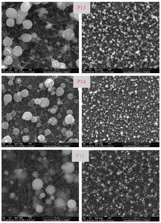
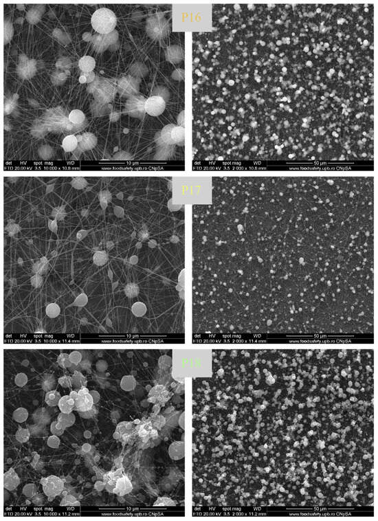
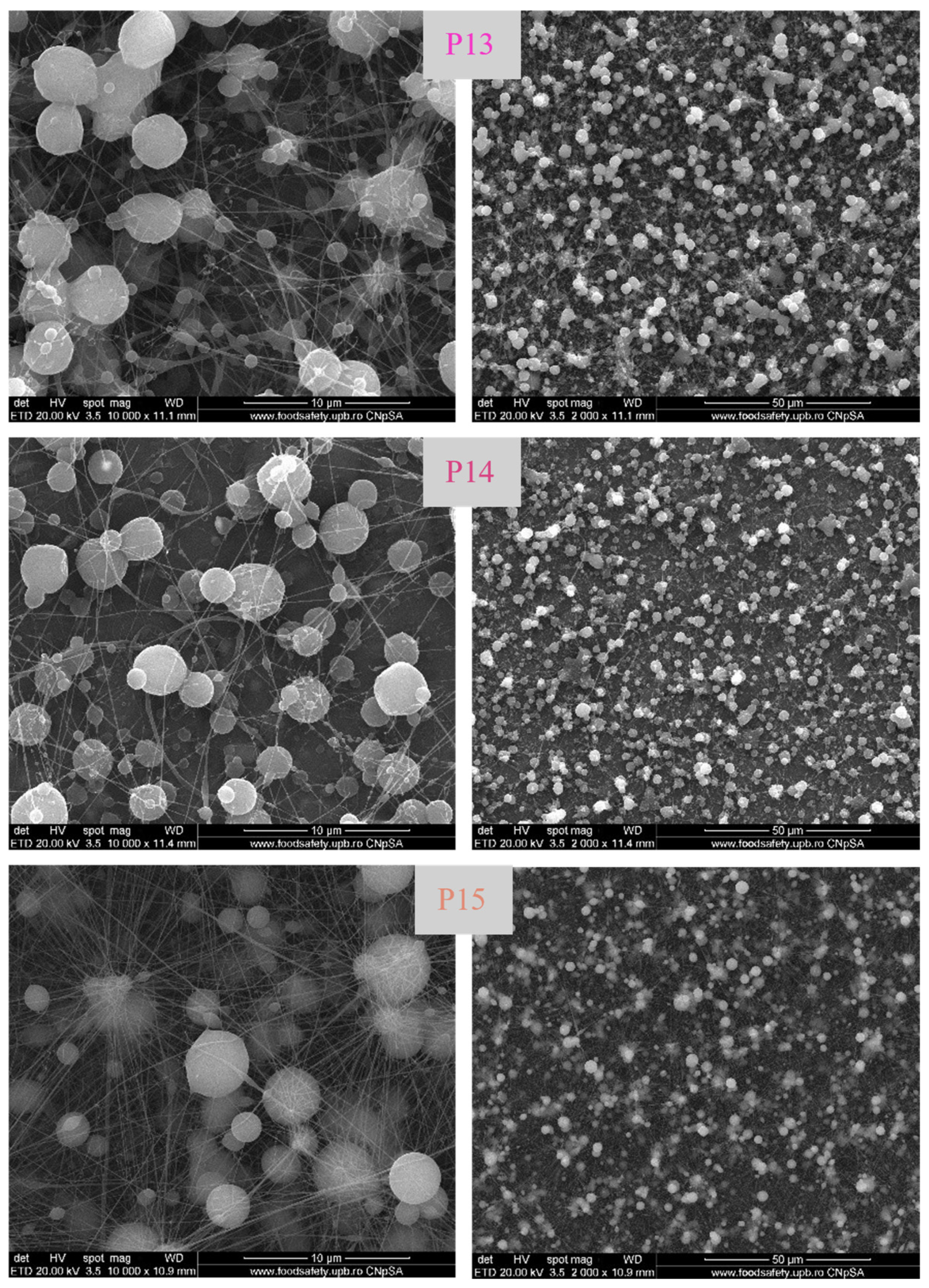
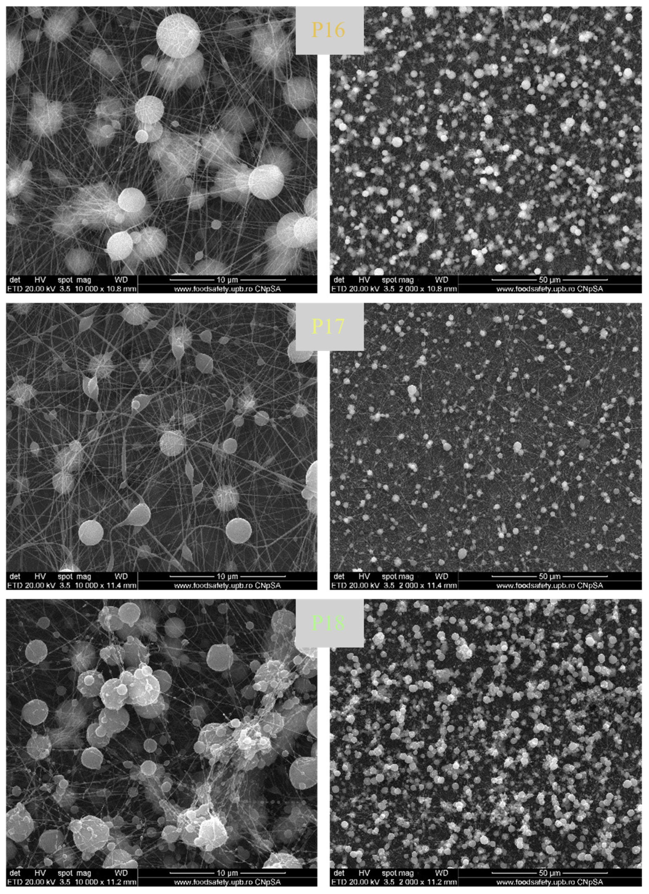
The morphology of electrospun fibres obtained from different PCL/PVDF formulations, with and without AgNPs, is shown in Figure 11. SEM analysis revealed that all electrospun PCL/PVDF-based fibrous systems formed continuous and interconnected networks, with fibre diameters predominantly in the nanometre range. Neat PCL fibres (sample P13) exhibited relatively uniform diameters, typically below 0.2 μm, indicative of stable jet formation and favourable viscoelastic properties of the polymer solution. The introduction of AgNPs (sample P14) did not disrupt fibre continuity; however, localized spherical or thickened regions appeared along the fibres, with characteristic dimensions slightly larger (below 1 μm) or much larger (several micrometres) than the fibre diameter.
Figure 11.
SEM images of PCL/PVDF composite fibres without AgNPs (P13, P15, P17) and with AgNPs (P14, P16, P18), showing surface morphology at magnifications of 10,000× (left column) and 2000× (right column).
These morphological features, also observed in PVDF-based fibres (samples P15 and P16) and in PCL/PVDF systems (samples P17 and P18), should not be regarded as structural defects. Instead, they reflect local mass accumulation and charge redistribution occurring during electrospinning, particularly in nanoparticle-loaded systems and architectures obtained by successive electrospinning from distinct polymer solutions. In the case of PCL/PVDF fibres, the polymers were processed from separate solutions, and therefore no molecular-level interactions are expected. However, incomplete solidification of previously deposited fibres and the presence of an already formed fibrous layer modify the local electrostatic field experienced by the subsequently electrospun jet, leading to controlled variations in fibre diameter and the formation of thickened regions.
From a functional perspective, these locally thickened zones may act as mechanical reinforcement points and regions with altered electrical response. In electroactive systems containing PVDF, such heterogeneities can promote spatially non-uniform electric fields and enhanced electromechanical stimulation at the microscale, which may be advantageous for cell–material interactions. Overall, the observed morphology reflects the complex electrohydrodynamic behaviour of multi-component and nanoparticle-functionalized systems and supports the formation of structurally continuous, mechanically robust, and electroactive fibrous scaffolds, rather than indicating processing-related defects.
3.2.2. Energy-Dispersive X-Ray Spectroscopy (EDX)
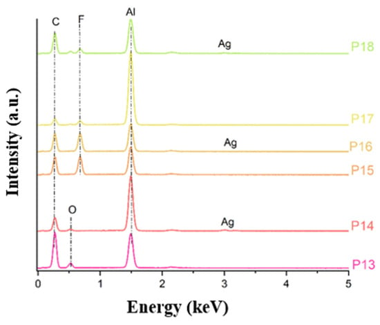
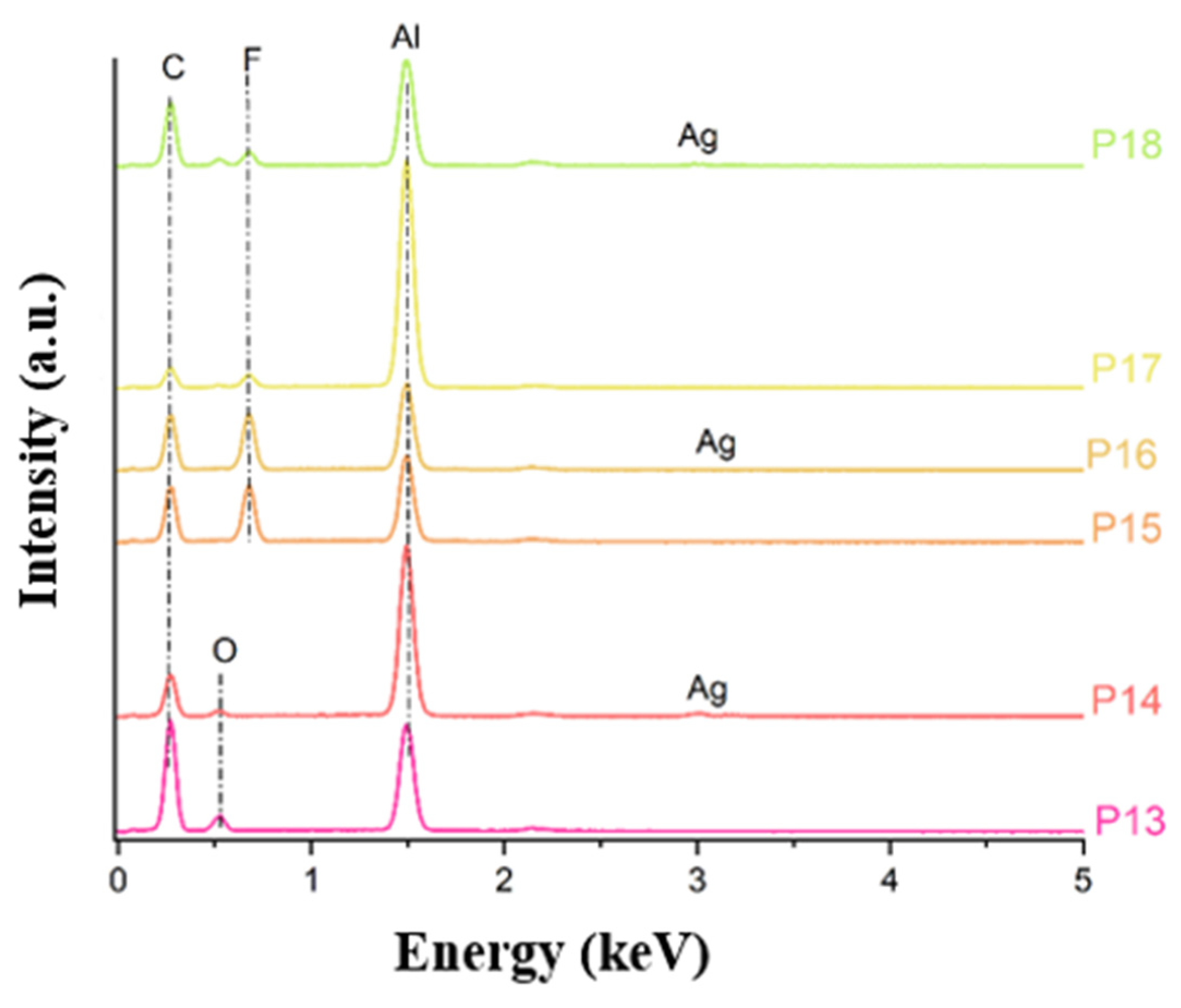
The EDX spectra of electrospun fibres (Figure 12) showed the expected elemental fingerprints of the polymer matrices and AgNP incorporation. PCL-based fibres (P13, P14) were dominated by carbon (C) and oxygen (O), whereas PVDF-containing fibres (P15–P18) additionally displayed fluorine (F) as a diagnostic PVDF marker. A distinct silver (Ag) signal (≈3 keV) was detected only in the functionalized samples (P14, P16 and P18), confirming successful AgNP incorporation into the fibres. All spectra exhibited an aluminium (Al) peak originating from the foil collector. Overall, EDX enables clear discrimination between PCL- and PVDF-containing fibres (via fluorine) and verifies AgNP presence selectively in the designed formulations (via silver).
Figure 12.
EDX spectra of PCL/PVDF fibres with and without AgNPs (P13–P18).
3.2.3. Fourier-Transform Infrared Spectroscopy (FTIR)
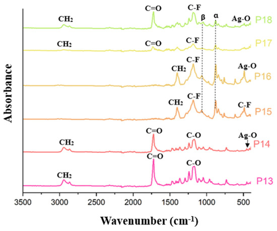
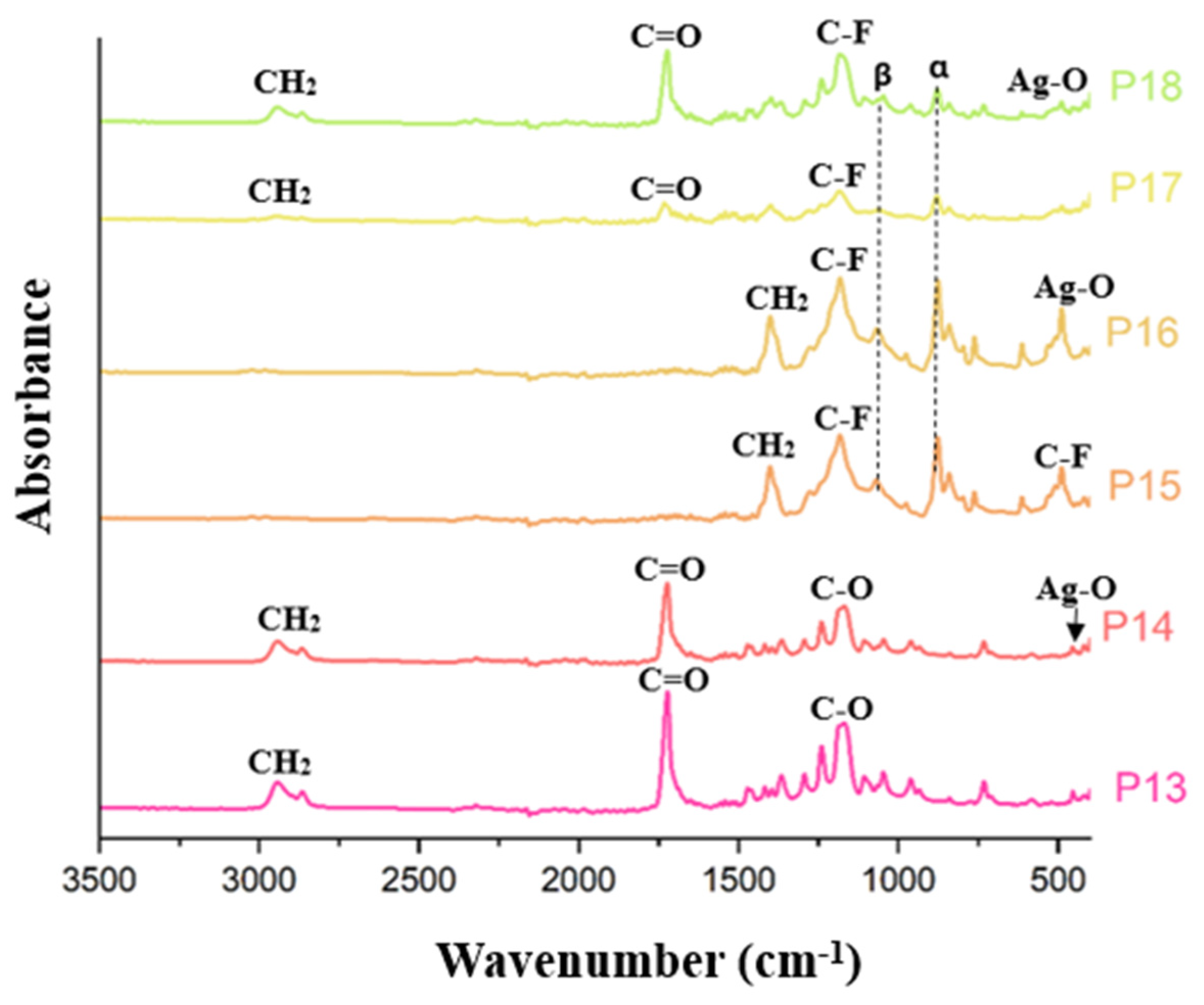
The FTIR spectra of electrospun fibres based on PCL, PVDF, and their blends, with and without AgNPs (Figure 13), confirm the preservation of the characteristic chemical structures of each component. Pure PCL fibres (P13) exhibited the typical ester-related absorptions, including CH2 stretching vibrations (~2940 and ~2860 cm−1), the carbonyl (C=O) band (~1720 cm−1), and C–O stretching modes (~1238–1170 cm−1) [53]. Upon AgNP incorporation (P14), these bands were retained, while a slight shift of the carbonyl band towards higher wavenumbers (~1760 cm−1), together with the appearance of a broad Ag–O band around ~540 cm−1, indicated interactions between PCL chains and AgNPs.
Figure 13.
FTIR spectra of PCL/PVDF fibres with and without AgNPs (P13–P18).
Pure PVDF fibres (P15) showed the expected absorptions associated with CH2 wagging (~1400 cm−1), CF2 stretching (~1274 cm−1), and the electroactive β-phase marker at ~840 cm−1, with a weaker contribution from the α-phase (~976 cm−1), consistent with mixed crystallinity [18]. These features were preserved after AgNP functionalization (P16), accompanied by an increased intensity of the Ag–O band, confirming successful nanoparticle incorporation.
The sequentially electrospun PCL/PVDF fibres (P17) displayed a superposition of the characteristic bands of both polymers, evidencing the coexistence of PCL and PVDF within the fibrous construct. Notably, despite being obtained by successive electrospinning from separate polymer solutions, no peak splitting, band duplication, or phase-specific segregation was observed, indicating chemical homogeneity at the FTIR detection scale. The AgNP-functionalized composite fibres (P18) similarly preserved all polymeric signatures together with the Ag–O contribution, confirming uniform chemical integration of both polymers and nanoparticles throughout the fibrous network [54].
3.3. Biological Evaluation
3.3.1. SBF Immersion Test
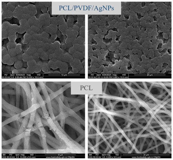
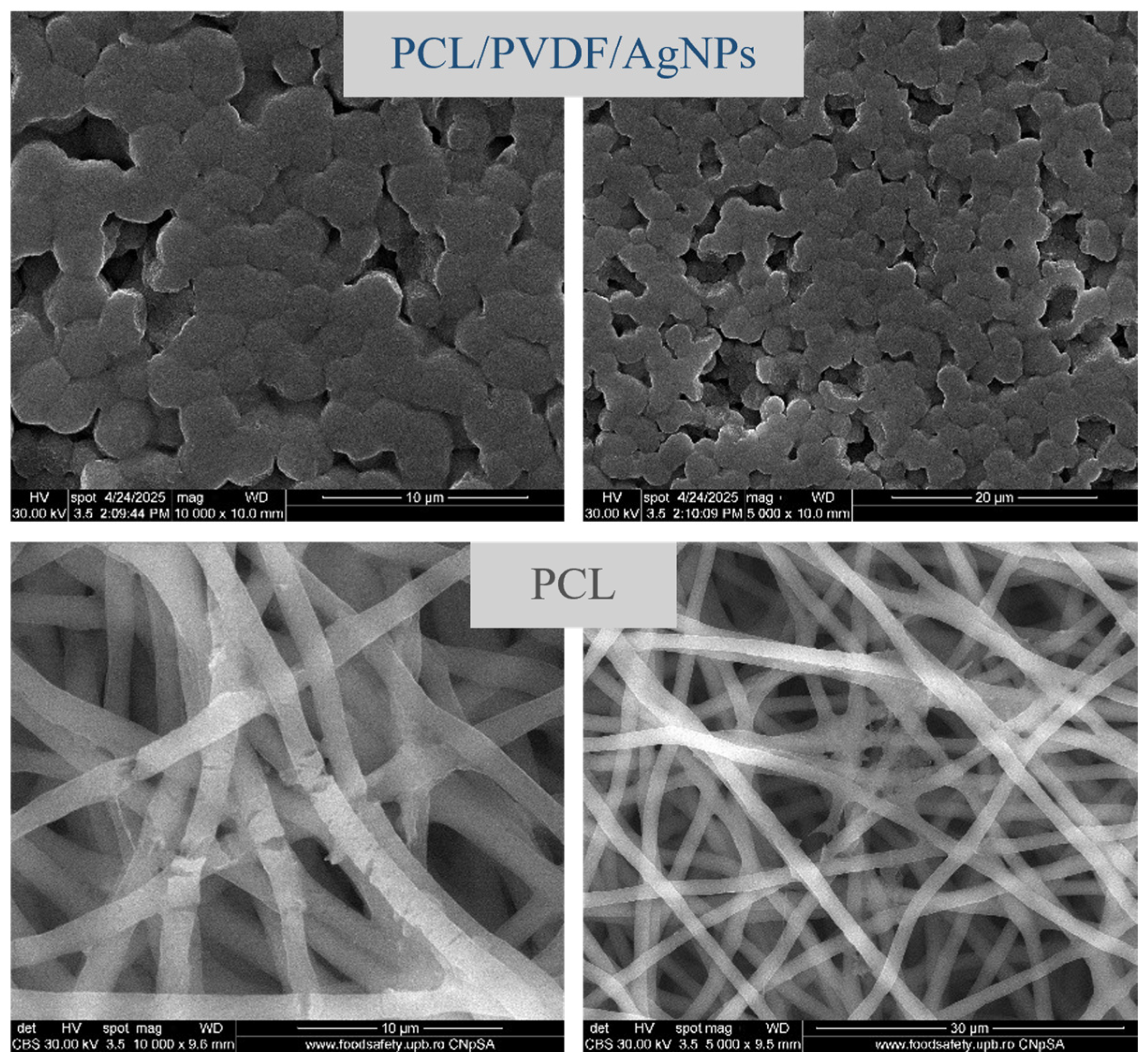
To evaluate the structural stability of the polymeric systems under physiologically relevant conditions, selected film and fibre samples were immersed in SBF for 28 days. The SEM images recorded after immersion (Figure 14) reveal distinct degradation behaviours depending on material composition. The equimassic PCL/PVDF composite film containing AgNPs preserved a rough and porous surface morphology, with no major cracks or collapse of the film structure. The absence of pronounced surface erosion indicates good structural stability of the composite matrix during prolonged exposure to SBF, which can be attributed to the presence of the PVDF phase and the reinforcing effect of AgNPs. In contrast, pure PCL fibres exhibited visible morphological changes after immersion, including local fibre thinning, short interruptions and partial loss of network continuity. These features are indicative of hydrolytic degradation of PCL, associated with the cleavage of ester bonds in aqueous environments. Despite these alterations, the fibrous architecture remained partially preserved, suggesting a gradual degradation process rather than abrupt structural failure.
Figure 14.
SEM images of equimassic PCL/PVDF composite films and PCL fibres after 28 days of immersion in SBF, showing surface morphology at magnifications of 10,000× (left column) and 5000× (right column).
3.3.2. Cell Viability and Adhesion
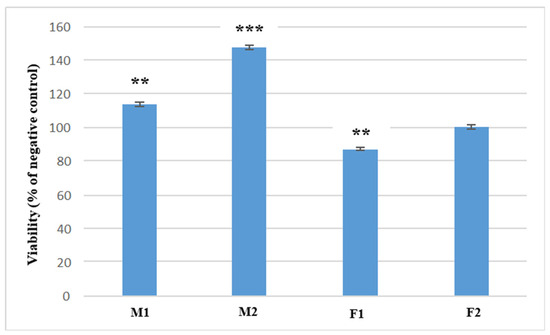
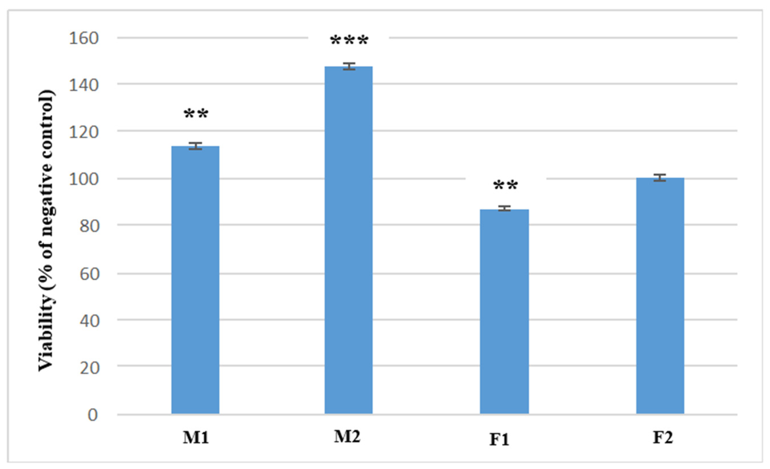
Cell viability of MC3T3 preosteoblasts incubated for 48 h in the presence of PCL/PVDF-based membranes and fibres was assessed by the MTT assay (Figure 15), which characterizes the total metabolism of the cells. Results obtained for the membrane-type samples (M1—equimassic PCL/PVDF; M2—equimassic PCL/PVDF with AgNPs) showed a statistically significant increase in the metabolic activity compared to the negative control (p < 0.01), indicating an enhanced stimulation of osteoblast cells by the substrates. In contrast, cells cultured on fibre-type samples (F1—PCL/PVDF; F2—PCL/PVDF/AgNPs) showcased biocompatible behaviour, as calculated viability values remained above 80%, fulfilling the ISO 10993-5:2009 standard [55]. Importantly, incorporation of AgNPs enhanced preosteoblast metabolism in both membranes and fibres, as confirmed by significant differences between AgNP-containing and non-functionalized counterparts (M1 vs. M2, p = 0.0001; F1 vs. F2, p = 0.002). This effect can be attributed to the release of silver ions, which may stimulate cellular activity under biological conditions.
Figure 15.
Viability of preosteoblast cells incubated for 2 days in the presence of PCL/PVDF membrane (M1—PCL/PVDF; M2—PCL/PVDF with AgNPs) and fibre (F1—PCL/PVDF; F2—PCL/PVDF/AgNPs) samples. Data is shown as mean ± SEM (n = 3); ** p < 0.01, *** p < 0.001.
The stimulatory effect of AgNP incorporation on preosteoblast metabolic activity observed in our PCL/PVDF membranes and fibres aligns with prior findings on polymeric scaffolds functionalized with silver. In a composite PLGA/PCL scaffold system, AgNP impregnation combined with collagen coating significantly enhanced MC3T3 cell proliferation and adhesion compared to control scaffolds, as measured by in vitro viability/proliferation assays and supported by increases in osteogenic marker expression [56]. These results indicate that AgNPs can do more than confer antimicrobial protection—at controlled levels they contribute to a more favourable cellular microenvironment, promoting osteoblastic activity on polymer matrices. The present MTT outcomes, showing enhanced metabolic activity for AgNP-functionalized PCL/PVDF membranes and fibres relative to non-functionalized counterparts, are consistent with this dual role of silver in scaffold design. Similar to our observations of enhanced osteoblastic activity on AgNP-functionalized PCL/PVDF composites, recent work on electrospun PCL/silk fibroin scaffolds with integrated AgNPs demonstrated improved in vitro cytocompatibility and enhanced osteoblast adhesion/mineralization compared to unmodified PCL constructs, further supporting the role of controlled AgNP incorporation in promoting favourable cell–material interactions [57]. These findings are consistent with reported studies on PVDF/AgNP-based electroactive biomaterials, where AgNP incorporation enhanced cellular activity while preserving cytocompatibility. PVDF/AgNP nanofibrous systems have been shown to support cell viability and promote tissue regeneration through the combined effect of piezoelectric stimulation and controlled Ag+ release [58]. In line with these reports, the present results demonstrate that AgNP-functionalized PCL/PVDF membranes and fibres improve preosteoblast metabolic activity while maintaining viability above the ISO 10993-5 threshold, supporting their suitability for regenerative applications requiring both biocompatibility and antimicrobial protection.
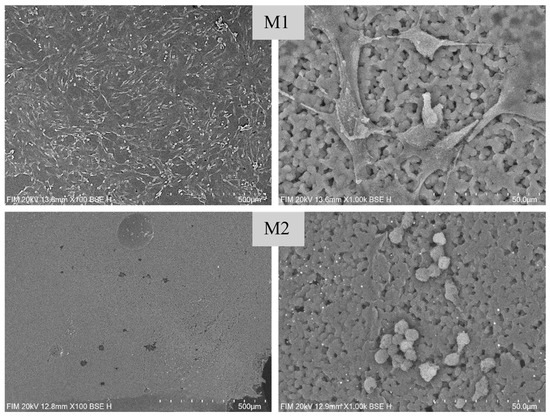
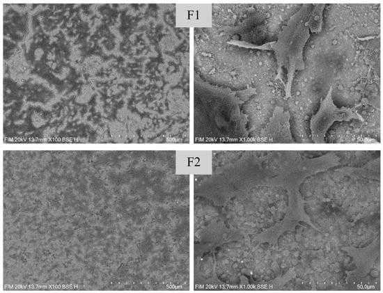
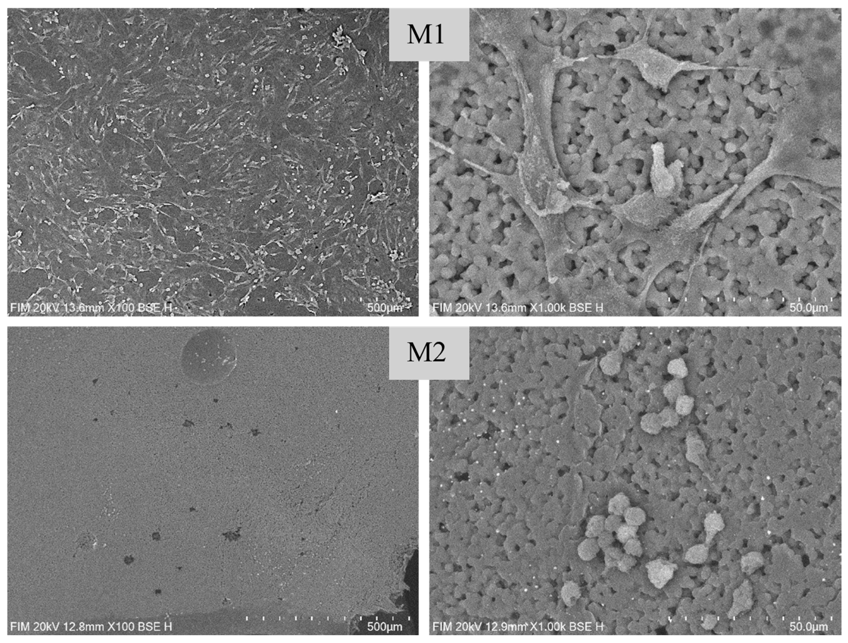
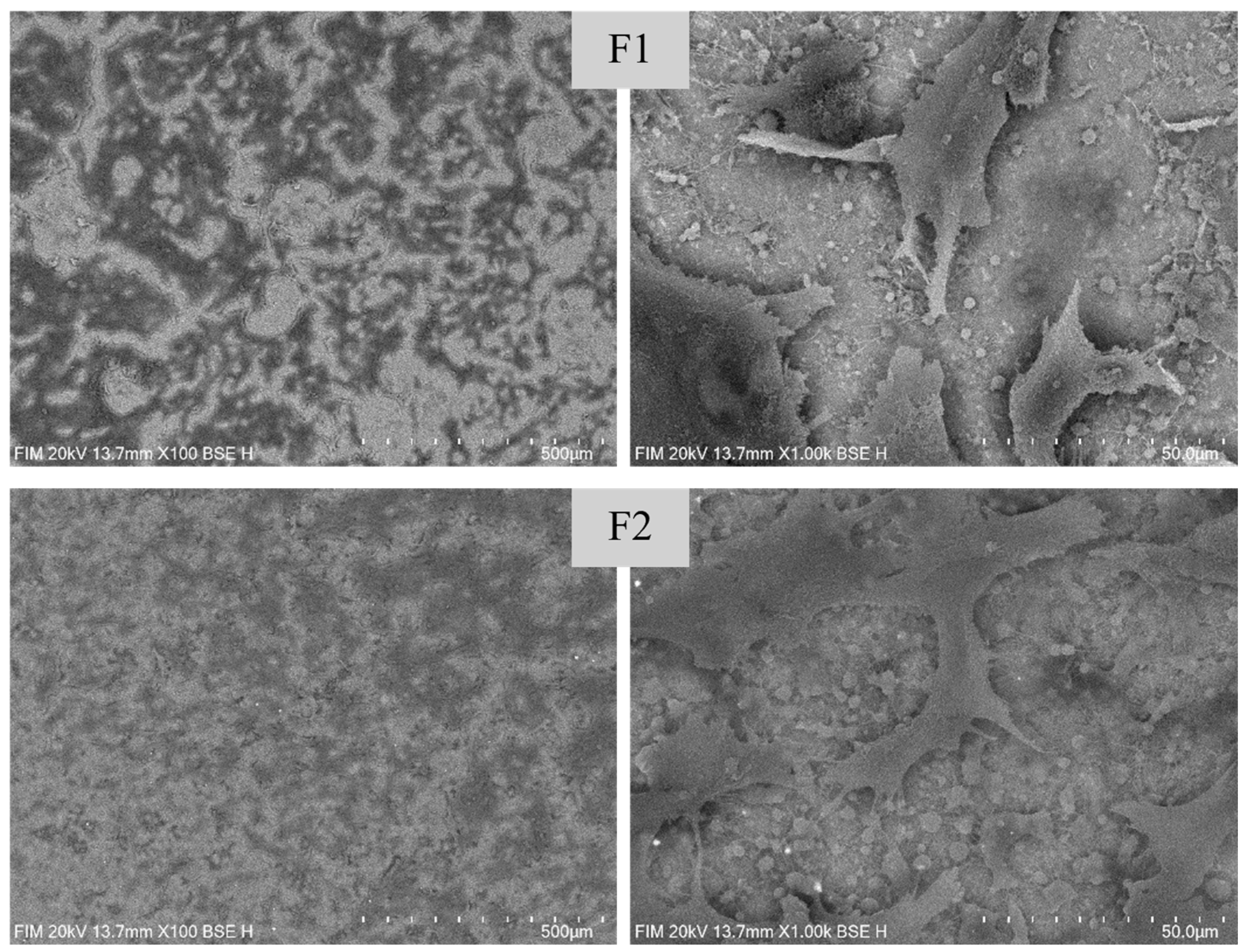
SEM observations of cell adhesion on the samples are shown in Figure 16. On membrane M1 (equimassic PCL/PVDF), cells displayed uniform coverage at low magnification (100×), while higher magnification (1000×) revealed flattened morphology and thin extensions, suggesting efficient adhesion on a continuous and solvent-free surface. For the AgNP-containing membrane (M2), the structure was preserved but some irregular surface features were visible. Cells appeared rounder, and discrete accumulations were observed on the surface, which can be associated with local surface heterogeneities induced by AgNP incorporation and related changes in surface chemistry and topography. For the fibrous scaffolds, enhanced adhesion was evident. Sample F1 (PCL/PVDF fibres) supported cell anchoring, with numerous filopodia extending across the fibrous network at 1000×. For F2 (PCL/PVDF/AgNP fibres), cells maintained similar morphology but with brighter extensions and more pronounced interactions with the substrate, suggesting that silver incorporation influenced both contrast and adhesion behaviour. Overall, fibrous architectures promoted stronger cell attachment compared to membranes, while AgNP functionalization further enhanced cell spreading and scaffold–cell interactions.
Figure 16.
SEM images of cells cultured on the surface of PCL/PVDF membrane (M1—PCL/PVDF; M2—PCL/PVDF with AgNPs) and fibre (F1—PCL/PVDF; F2—PCL/PVDF/AgNPs) samples. The first two rows display the membrane samples, while the following two rows correspond to the fibre samples. The images in both columns represent the same sample, shown at different magnifications: 100× (left column) and 1000× (right column).
Similar to the present observations, several studies on polymer composite scaffolds reported enhanced preosteoblast responses in vitro. Electrospun HA/PCL/gelatin nanofibre scaffolds demonstrated increased osteoblast proliferation and successful cell attachment in MTT and SEM analyses, confirming scaffold biocompatibility and suitability for bone tissue applications [59]. PCL surfaces modified with calcium phosphate nanostructures supported preosteoblast adhesion, proliferation, and differentiation, highlighting the importance of surface chemistry and nanostructures in modulating cell behaviour [60]. Additionally, hybrid PCL scaffolds enabled sustained MC3T3 preosteoblast proliferation and early osteogenic activity in vitro, reinforcing the potential of composite scaffolds to promote cell growth and differentiation [61]. Complementary findings have been reported for PVDF-based electrospun scaffolds, where fibrous architectures and electroactive β-phase content promoted cell adhesion and proliferation without inducing cytotoxic effects [61]. Also, PVDF and PVDF/AgNP systems maintained high cell viability and supported favourable cell–material interactions under physiological conditions, attributed to their surface properties and piezoelectric response [62]. Together, these literature data are in good agreement with the present results, confirming that both PCL- and PVDF-containing composite membranes and fibres can provide cytocompatible environments that support preosteoblast metabolic activity and adhesion.
4. Conclusions
This work demonstrated the successful development and advanced characterization of PCL/PVDF composite materials, both with and without AgNP functionalization, tailored for peripheral nerve regeneration. The synergistic properties of the two polymers were effectively combined: the biodegradability and cytocompatibility of PCL together with the mechanical stability and electroactive behaviour of PVDF. SEM, EDX, FTIR, XRD, and thermal analyses confirmed the structural integrity, semicrystalline character, and phase coexistence in both films and fibres, while SBF immersion and MTT assays highlighted bioactivity and cytocompatibility. Depending on the processing route, the developed materials can be employed either as planar membranes or as three-dimensional guidance structures. Solvent-cast films can be further rolled into tubular nerve guidance conduits, a configuration previously demonstrated for PCL/PVDF composites in peripheral nerve repair applications, enabling the bridging of nerve gaps and the guidance of axonal regrowth [18]. The incorporation of AgNPs, which is well documented in the literature for conferring antimicrobial functionality, also improved preosteoblast metabolic activity in the present study, suggesting that the metallic phase may support both protective and regenerative roles. Fibrous scaffolds provided better cellular adhesion compared to membranes, confirming the importance of architecture in guiding cell–material interactions. Overall, the findings support the feasibility of using PCL/PVDF/AgNP systems as multifunctional composites for neural repair. Moving forward, efforts should concentrate on refining nanoparticle dispersion to ensure uniform functionality, adapting fabrication processes for reproducibility and scalability, and validating the regenerative potential through in vivo experiments. These steps are essential to bridge the gap between laboratory-scale demonstrations and clinically relevant applications in peripheral nerve repair. Beyond peripheral nerve repair, the developed PCL/PVDF composites also hold promise for a wide range of biomedical applications. Their electroactive behaviour and antimicrobial functionality make them suitable candidates for antibacterial wound patches, coatings for implantable devices, and controlled drug delivery systems. The fibrous architecture mimicking the extracellular matrix may further support applications in guided bone regeneration or periodontal membranes, where both structural support and bioactivity are required. In addition, their flexibility and piezoelectric properties open opportunities for soft, electroactive patches designed for tissue stimulation and neuromuscular interfaces. These future directions highlight the versatility of PCL/PVDF/AgNP-based materials and their potential for translation into multifunctional platforms in regenerative medicine.
Supplementary Materials
The following supporting information can be downloaded at: https://www.mdpi.com/article/10.3390/macromol6020026/s1, Figure S1: SEM micrograph of the synthesized AgNPs at 100,000× magnification, showing their spherical morphology and size distribution; Figure S2: Energy-dispersive X-ray (EDX) spectrum of the synthesized AgNPs, confirming the elemental presence and purity of silver. Figure S3: X-ray diffraction (XRD) pattern of the synthesized AgNPs.
Author Contributions
Conceptualization, C.B.; Methodology, L.N., R.C.P. and C.B.; Investigation, L.N., R.C.P. and C.B.; Resources, R.C.P., S.I.J. and C.B.; Writing—original draft, L.N. and C.B.; Writing—review & editing, L.N. and C.B.; Supervision, S.I.J. and C.B. All authors have read and agreed to the published version of the manuscript.
Funding
This research received no external funding.
Data Availability Statement
The raw data supporting the conclusions of this article will be made available by the authors on request.
Acknowledgments
The authors are grateful to the Romanian Government for providing access to the research infrastructure of the National Center for Micro and Nanomaterials through the National Program titled “Installations and Strategic Objectives of National Interest”.
Conflicts of Interest
The authors declare no conflict of interest.
References
- Cleveland Clinic. Nervous System: What It Is, Parts, Function & Disorders, 16 Noiembrie 2023. Available online: https://my.clevelandclinic.org/health/body/21202-nervous-system (accessed on 25 April 2025).
- Struzyna, L.A.; Harris, J.P.; Katiyar, K.S.; Chen, H.I.; Cullen, D.K. Restoring nervous system structure and function using tissue engineered living scaffolds. Neural Regen. Res. 2015, 10, 679–685. [Google Scholar] [CrossRef] [Scilit]
- Tam, R.Y.; Fuehrmann, T.; Mitrousis, N.; Shoichet, M.S. Regenerative Therapies for Central Nervous System Diseases: A Biomaterials Approach. Neuropsychopharmacology 2014, 39, 169–188. [Google Scholar] [CrossRef] [Scilit]
- Kurakula, M.; Koteswara Rao, G.S.N.; Yadav, K.S. Fabrication and Characterization of Polycaprolactone-Based Green Materials for Drug Delivery. In Applications of Advanced Green Materials; Elsevier: Amsterdam, The Netherlands, 2021; pp. 395–423. [Google Scholar] [CrossRef] [Scilit]
- What Is Polycaprolactone? Structure, Density, and Applications. Available online: https://europlas.com.vn/en-US/blog-1/what-is-polycaprolactone-structure-density-and-applications (accessed on 8 November 2024).
- Heimowska, A.; Morawska, M.; Bocho-Janiszewska, A. Biodegradation of poly(ε-caprolactone) in natural water environments. Pol. J. Chem. Technol. 2017, 19, 120–126. [Google Scholar] [CrossRef] [Scilit]
- Schlesinger, E.; Ciaccio, N.; Desai, T.A. Polycaprolactone Thin-Film Drug Delivery Systems: Empirical and Predictive Models for Device Design. Mater. Sci. Eng. C 2015, 57, 232–239. [Google Scholar] [CrossRef] [Scilit] [PubMed]
- Holešová, S.; Barabaszová, K.Č.; Hundáková, M.; Ščuková, M.; Hrabovská, K.; Joszko, K.; Antonowicz, M.; Gzik-Zroska, B. Development of Novel Thin Polycaprolactone (PCL)/Clay Nanocomposite Films with Antimicrobial Activity Promoted by the Study of Mechanical, Thermal, and Surface Properties. Polymers 2021, 13, 3193. [Google Scholar] [CrossRef] [Scilit]
- Nyitray, C.E.; Chang, R.; Faleo, G.; Lance, K.D.; Bernards, D.A.; Tang, Q.; Desai, T.A. Polycaprolactone Thin-Film Micro- and Nanoporous Cell-Encapsulation Devices. ACS Nano 2015, 9, 5675–5682. [Google Scholar] [CrossRef] [Scilit] [PubMed]
- Ehterami, A.; Salehi, M.; Farzamfar, S.; Vaez, A.; Samadian, H.; Sahrapeyma, H.; Mirzaii, M.; Ghorbani, S.; Goodarzi, A. In vitro and in vivo study of PCL/COLL wound dressing loaded with insulin-chitosan nanoparticles on cutaneous wound healing in rats model. Int. J. Biol. Macromol. 2018, 117, 601–609. [Google Scholar] [CrossRef] [Scilit]
- Damdinov, B.B.; Danilova, V.A.; Minakov, A.V.; Pryaznhnikov, M.I. Rheological Properties of PVDF Solutions. J. Sib. Fed. Univ. Math. Phys. 2021, 14, 265–272. [Google Scholar] [CrossRef] [Scilit]
- Nishiyama, T.; Sumihara, T.; Sasaki, Y.; Sato, E.; Yamato, M.; Horibe, H. Crystalline structure control of poly(vinylidene fluoride) films with the antisolvent addition method. Polym. J. 2016, 48, 1035–1038. [Google Scholar] [CrossRef] [Scilit]
- Koseki, Y.; Aimi, K.; Ando, S. Crystalline structure and molecular mobility of PVDF chains in PVDF/PMMA blend films analyzed by solid-state 19F MAS NMR spectroscopy. Polym. J. 2012, 44, 757–763. [Google Scholar] [CrossRef] [Scilit]
- Ruan, L.; Yao, X.; Chang, Y.; Zhou, L.; Qin, G.; Zhang, X. Properties and Applications of the β Phase Poly(vinylidene fluoride). Polymers 2018, 10, 228. [Google Scholar] [CrossRef] [Scilit] [PubMed]
- Dallaev, R.; Pisarenko, T.; Sobola, D.; Orudzhev, F.; Ramazanov, S.; Trčka, T. Brief Review of PVDF Properties and Applications Potential. Polymers 2022, 14, 4793. [Google Scholar] [CrossRef] [Scilit] [PubMed]
- Milenković, S.; Virijević, K.; Živić, F.; Radojević, I.; Grujović, N. Composite Nanoarchitectonics of Electrospun Piezoelectric PVDF/AgNPs for Biomedical Applications, Including Breast Cancer Treatment. Materials 2024, 17, 3872. [Google Scholar] [CrossRef] [Scilit] [PubMed]
- Zhu, T.; Cui, Y.; Zhang, M.; Zhao, D.; Liu, G.; Ding, J. Engineered three-dimensional scaffolds for enhanced bone regeneration in osteonecrosis. Bioact. Mater. 2020, 5, 584–601. [Google Scholar] [CrossRef] [Scilit]
- Cheng, Y.; Xu, Y.; Qian, Y.; Chen, X.; Ouyang, Y.; Yuan, W.-E. 3D structured self-powered PVDF/PCL scaffolds for peripheral nerve regeneration. Nano Energy 2020, 69, 104411. [Google Scholar] [CrossRef] [Scilit]
- Abzan, N.; Kharaziha, M.; Labbaf, S. Development of three-dimensional piezoelectric polyvinylidene fluoride-graphene oxide scaffold by non-solvent induced phase separation method for nerve tissue engineering. Mater. Des. 2019, 167, 107636. [Google Scholar] [CrossRef] [Scilit]
- Kitsara, M.; Blanquer, A.; Murillo, G.; Humblot, V.; De Bragança Vieira, S.; Nogués, C.; Ibáñez, E.; Esteve, J.; Barrios, L. Permanently Hydrophilic, Piezoelectric PVDF Nanofibrous Scaffolds Promoting Unaided Electromechanical Stimulation on Osteoblasts. Nanoscale 2019, 11, 8906–8917. [Google Scholar] [CrossRef] [Scilit]
- Xu, L.; Wang, Y.-Y.; Huang, J.; Chen, C.-Y.; Wang, Z.-X.; Xie, H. Silver nanoparticles: Synthesis, medical applications and biosafety. Theranostics 2020, 10, 8996–9031. [Google Scholar] [CrossRef] [Scilit]
- Abbas, R.; Luo, J.; Qi, X.; Naz, A.; Khan, I.A.; Liu, H.; Yu, S.; Wei, J. Silver Nanoparticles: Synthesis, Structure, Properties and Applications. Nanomaterials 2024, 14, 1425. [Google Scholar] [CrossRef] [Scilit]
- Sudarman, F.; Shiddiq, M.; Armynah, B.; Tahir, D. Silver nanoparticles (AgNPs) synthesis methods as heavy-metal sensors: A review. Int. J. Environ. Sci. Technol. 2023, 20, 9351–9368. [Google Scholar] [CrossRef] [Scilit]
- Gherasim, O.; Puiu, R.A.; Bîrcă, A.C.; Burdușel, A.-C.; Grumezescu, A.M. An updated review on silver nanoparticles in biomedicine. Nanomaterials 2020, 10, 2318. [Google Scholar] [CrossRef] [Scilit]
- Kong, C.; Chen, S.; Ge, W.; Zhao, Y.; Xu, X.; Wang, S.; Zhang, J. Riclin-Capped silver nanoparticles as an antibacterial and anti-inflammatory wound dressing. Int. J. Nanomed. 2022, 17, 2629–2641. [Google Scholar] [CrossRef] [Scilit] [PubMed]
- Khalil, M.M.H.; Ismail, E.H.; El-Magdoub, F. Biosynthesis of Au nanoparticles using olive leaf extract. Arab. J. Chem. 2010, 4, 431–437. [Google Scholar] [CrossRef] [Scilit]
- Restrepo, M.; Bussaneli, D.G.; Jeremias, F.; Cordeiro, R.; Raveli, D.; Magalhães, A.; Candolo, C.; Santos-Pinto, L. Control of white spot lesions with use of fluoride varnish or chlorhexidine gel during orthodontic treatment: A randomized clinical trial. J. Clin. Pediatr. Dent. 2016, 40, 274–280. [Google Scholar] [CrossRef] [Scilit] [PubMed]
- Hernández-Gómora, A.E.; Lara-Carrillo, E.; Robles-Navarro, J.B.; Scougall-Vilchis, R.J.; Hernández-López, S.; Medina-Solís, C.E.; Morales-Luckie, R.A. Biosynthesis of silver nanoparticles on orthodontic elastomeric modules: Evaluation of mechanical and antibacterial properties. Molecules 2017, 22, 1407. [Google Scholar] [CrossRef] [Scilit] [PubMed]
- Yuan, Q.; Zhang, Q.; Xu, X.; Du, Y.; Xu, J.; Song, Y.; Wang, Y. Development and Characterization of Novel Orthodontic Adhesive Containing PCL–Gelatin–AgNPs Fibers. J. Funct. Biomater. 2022, 13, 303. [Google Scholar] [CrossRef] [Scilit]
- Abashkin, V.; Pędziwiatr-Werbicka, E.; Horodecka, K.; Zhogla, V.; Ulashchik, E.; Shmanai, V.; Shcharbin, D.; Bryszewska, M. Silver nanoparticles modified by carbosilane dendrons and PEG as delivery vectors of small interfering RNA. Int. J. Mol. Sci. 2023, 24, 840. [Google Scholar] [CrossRef] [Scilit]
- Carestream. Using Polymer Solution Casting to Deliver High-Quality Films. Available online: https://www.carestream.com/specials/contract-manufacturing/using-polymer-solution-casting-to-deliver-high-quality-films/index.html (accessed on 14 February 2025).
- Borbolla-Jiménez, F.V.; Peña-Corona, S.I.; Farah, S.J.; Jiménez-Valdés, M.T.; Pineda-Pérez, E.; Romero-Montero, A.; Del Prado-Audelo, M.L.; Bernal-Chávez, S.A.; Magaña, J.J.; Leyva-Gómez, G. Films for Wound Healing Fabricated Using a Solvent Casting Technique. Pharmaceutics 2023, 15, 1914. [Google Scholar] [CrossRef] [Scilit]
- Siemann, U. Solvent Film Casting—A Versatile Technology for Specialty Films. In 44th Annual Technical Conference Proceedings, Philadelphia, PA, USA, 21–26 April 2001; Society of Vacuum Coaters: Albuquerque, NM, USA, 2001; p. 253. Available online: https://www.svc.org/index.php?src=directory&view=resource_library&srctype=detail&refno=6392 (accessed on 14 February 2025).
- Ahmadi Bonakdar, M.; Rodrigue, D. Electrospinning: Processes, structures, and materials. Macromolecules 2024, 4, 58–103. [Google Scholar] [CrossRef] [Scilit]
- Nanoscience Instruments. What Is Electrospinning? Nanoscience, 6 December 2024. Available online: https://www.nanoscience.com/techniques/electrospinning/ (accessed on 15 February 2025).
- Keirouz, A.; Wang, Z.; Reddy, V.S.; Nagy, Z.K.; Vass, P.; Buzgo, M.; Ramakrishna, S.; Radacsi, N. The History of Electrospinning: Past, Present, and Future Developments. Adv. Mater. Technol. 2023, 8, 2201723. [Google Scholar] [CrossRef] [Scilit]
- Nanoscience Instruments. 10 Reasons to Use Electrospinning, Nanoscience, 14 February 2025. Available online: https://www.nanoscience.com/blogs/10-reasons-to-use-electrospinning/ (accessed on 16 February 2025).
- Electrospinning Disadvantages, Nanoscience Blog, February 2025. Available online: https://spin.digitalpartner.com.tr/guncel/electrospinning/electrospinning-disadvantages/ (accessed on 16 February 2025).
- Vishwanath, R.; Negi, B. Conventional and green methods of synthesis of silver nanoparticles and their antimicrobial properties. Curr. Res. Green Sustain. Chem. 2021, 4, 100205. [Google Scholar] [CrossRef] [Scilit]
- Sahayaraj, K.; Rajesh, S. Bionanoparticles: Synthesis and Antimicrobial Applications. In Science Against Microbial Pathogens: Communicating Current Research and Technological Advances; Méndez-Vilas, A., Ed.; Formatex Research Center: Badajoz, Spain, 2011; Volume 1, pp. 228–244. [Google Scholar]
- Taş, A.C. How to Prepare SBF Solution. Available online: https://www.cuneyttas.com/How-to-prepare-SBF-solution.htm (accessed on 2 June 2025).
- Wu, S.; Zhang, L.; Qiu, X.; Guo, Y.; Dong, L.; Guo, M.; Zhao, J. Preparation and Characterization of Highly Conductive PVDF/PAN Conjugate Electrospun Fibrous Membranes with Embedded Silver Nanoparticles. Polymers 2024, 16, 3540. [Google Scholar] [CrossRef] [Scilit] [PubMed]
- Chen, C.; Liu, F.; Zhang, X.; Zhao, Z.; Liu, S. Fabrication, characterization and adsorption properties of cucurbit[7]uril-functionalized polycaprolactone electrospun nanofibrous membranes. Beilstein J. Org. Chem. 2019, 15, 992–999. [Google Scholar] [CrossRef] [Scilit] [PubMed]
- Sadeghzadeh, H.; Mehdipour, A.; Dianat-Moghadam, H.; Salehi, R.; Khoshfetrat, A.B.; Hassani, A.; Mohammadnejad, D. PCL/Col I-based magnetic nanocomposite scaffold provides an osteoinductive environment for ADSCs in osteogenic cues-free media conditions. Stem Cell Res. Ther. 2022, 13, 143. [Google Scholar] [CrossRef] [Scilit] [PubMed]
- Sun, X.; Shiraz, H.; Wong, R.; Zhang, J.; Liu, J.; Lu, J.; Meng, N. Enhancing the Performance of PVDF/GO Ultrafiltration Membrane via Improving the Dispersion of GO with Homogeniser. Membranes 2022, 12, 1268. [Google Scholar] [CrossRef] [Scilit]
- Bagherzadeh, E.; Sherafat, Z.; Zebarjad, S.M.; Khodaei, A.; Yavari, S.A. Stimuli-responsive piezoelectricity in electrospun polycaprolactone (PCL)/Polyvinylidene fluoride (PVDF) fibrous scaffolds for bone regeneration. J. Mater. Res. Technol. 2023, 23, 379–390. [Google Scholar] [CrossRef] [Scilit]
- Ramírez-Cedillo, E.; Ortega-Lara, W.; Rocha-Pizaña, M.R.; Gutierrez-Uribe, J.A.; Elías-Zúñiga, A.; Rodríguez, C.A. Electrospun Polycaprolactone Fibrous Membranes Containing Ag, TiO2 and Na2Ti6O13 Particles for Potential Use in Bone Regeneration. Membranes 2019, 9, 12. [Google Scholar] [CrossRef] [Scilit]
- Balu, R.; Kumar, T.S.S.; Ramalingam, M.; Ramakrishna, S. Electrospun Polycaprolactone/Poly(1,4-butylene adipate-co-polycaprolactam) Blends: Potential Biodegradable Scaffold for Bone Tissue Regeneration. J. Biomater. Tissue Eng. 2011, 1, 30–39. [Google Scholar] [CrossRef] [Scilit]
- Popa, A.; Toloman, D.; Stan, M.; Stefan, M.; Radu, T.; Vlad, G.; Ulinici, S.; Baisan, G.; Macavei, S.; Barbu-Tudoran, L.; et al. Tailoring the RhB removal rate by modifying the PVDF membrane surface through ZnO particles deposition. J. Inorg. Organomet. Polym. Mater. 2021, 31, 1642–1652. [Google Scholar] [CrossRef] [Scilit]
- Tsonos, C.; Pandis, C.; Soin, N.; Sakellari, D.; Myrovali, E.; Kripotou, S.; Kanapitsas, A.; Siores, E. Multifunctional nanocomposites of poly(vinylidene fluoride) reinforced by carbon nanotubes and magnetite nanoparticles. Express Polym. Lett. 2015, 9, 1104–1118. [Google Scholar] [CrossRef] [Scilit]
- Benhacine, F.; Hadj-Hamou, A.S.; Habi, A. Development of long-term antimicrobial poly (ε-caprolactone)/silver exchanged montmorillonite nanocomposite films with silver ion release property for active packaging use. Polym. Bull. 2016, 73, 1207–1227. [Google Scholar] [CrossRef] [Scilit]
- Nthunya, L.N.; Derese, S.; Gutierrez, L.; Verliefde, A.R.; Mamba, B.B.; Barnard, T.G.; Mhlanga, S.D. Green synthesis of silver nanoparticles using one-pot and microwave-assisted methods and their subsequent embedment on PVDF nanofibre membranes for growth inhibition of mesophilic and thermophilic bacteria. New J. Chem. 2019, 43, 4168–4180. [Google Scholar] [CrossRef] [Scilit]
- Liverani, L.; Raffel, N.; Fattahi, A.; Preis, A.; Hoffmann, I.; Boccaccini, A.R.; Beckmann, M.W.; Dittrich, R. Electrospun patterned porous scaffolds for the support of ovarian follicles growth: A feasibility study. Sci. Rep. 2019, 9, 1150. [Google Scholar] [CrossRef] [Scilit] [PubMed]
- Shivalingappa, R.T.; Murthy, H.N.R.N.; Purushothaman, P.; Badiger, P.; Savarn, S.; Majumdar, U.; Angadi, G. Development of poly (vinylidene fluoride)/silver nanoparticle electrospun nanofibre mats for energy harvesting. Polym. Polym. Compos. 2021, 29, S1084–S1091. [Google Scholar] [CrossRef] [Scilit]
- ISO 10993-5:2009; Biological Evaluation of Medical Devices—Part 5: Tests for In Vitro Cytotoxicity. International Organization for Standardization: Geneva, Switzerland, 2009.
- Qian, Y.; Zhou, X.; Zhang, F.; Diekwisch, T.G.H.; Luan, X.; Yang, J. Triple PLGA/PCL scaffold modification including silver impregnation, collagen coating, and electrospinning significantly improve biocompatibility, antimicrobial, and osteogenic properties for orofacial tissue regeneration. ACS Appl. Mater. Interfaces 2019, 11, 37381–37396. [Google Scholar] [CrossRef] [Scilit] [PubMed]
- Suaza, M.L.M.; Rivera, J.C.L.; Acevedo, M.E.M.; Ossa-Orozco, C.P.; Yunda, D.U.; Triviño, D.G.Z. Polycaprolactone, silk fibroin and silver nanoparticles scaffolds as a substrate for bone tissue engineering. Mater. Technol. 2025, 40, 2529502. [Google Scholar] [CrossRef] [Scilit]
- Liu, T.; Xie, F.; Geng, L.; He, R.; Sun, M.; Ni, T.; Xu, P.; Xing, C.; Peng, Y.; Chen, K.; et al. Micro-Electro Nanofibrous Dressings Based on PVDF-AgNPs as Wound Healing Materials to Promote Healing in Active Areas. Int. J. Nanomed. 2025, 20, 771–789. [Google Scholar] [CrossRef] [Scilit]
- Aminatun; Aisyah Sujak, M.K.; Djony Izak, R.; Hadi, S.; Sari, Y.W.; Gunawarman; Cahyati, N.; Yusuf, Y.; Abdullah, C.A.C. Fabrication and biocompatibility evaluation of hydroxyapatite–polycaprolactone–gelatin composite nanofibers as a bone scaffold. RSC Adv. 2024, 14, 24815–24827. [Google Scholar] [CrossRef] [Scilit]
- Ganbaatar, S.E.; Kim, H.-K.; Kang, N.-U.; Kim, E.C.; U, H.J.; Cho, Y.-S.; Park, H.-H. Calcium Phosphate (CaP) Composite Nanostructures on Polycaprolactone (PCL): Synergistic Effects on Antibacterial Activity and Osteoblast Behavior. Polymers 2025, 17, 200. [Google Scholar] [CrossRef] [Scilit]
- Gonzalez-Pujana, A.; Carranza, T.; Santos-Vizcaino, E.; Igartua, M.; Guerrero, P.; Hernandez, R.M.; de la Caba, K. Hybrid 3D Printed and Electrospun Multi-Scale Hierarchical Polycaprolactone Scaffolds to Induce Bone Differentiation. Pharmaceutics 2022, 14, 2843. [Google Scholar] [CrossRef] [Scilit]
- Li, Y.; Liao, C.; Tjong, S.C. Electrospun Polyvinylidene Fluoride-Based Fibrous Scaffolds with Piezoelectric Characteristics for Bone and Neural Tissue Engineering. Nanomaterials 2019, 9, 952. [Google Scholar] [CrossRef] [Scilit]

Disclaimer/Publisher’s Note: The statements, opinions and data contained in all publications are solely those of the individual author(s) and contributor(s) and not of MDPI and/or the editor(s). MDPI and/or the editor(s) disclaim responsibility for any injury to people or property resulting from any ideas, methods, instructions or products referred to in the content. |
© 2026 by the authors. Licensee MDPI, Basel, Switzerland. This article is an open access article distributed under the terms and conditions of the Creative Commons Attribution (CC BY) license.