


Abstract
This study presents the first characterization of the globulin fraction from a newly registered chickpea cultivar, which represents the first desi-type cultivar (GB Cappuccino) released in Brazil. Although desi chickpeas are widely consumed in other countries, they have not been part of the Brazilian dietary pattern, and this introduction may represent an opportunity for changing this scenario. Characterizing its proteins is essential, given that legumes are recognized as important protein sources. In this study, globulins were confirmed as the predominant protein fraction, with the legumin-like fraction accounting for more than 80% of the total globulins. Its electrophoretic and amino acid profiles were highly distinctive and strongly influenced by this major fraction. In addition to the expected solubilization in saline solution, under in vitro pepsin–pancreatin digestion conditions designed to assess maximum hydrolysis potential, the globulin fraction was partially hydrolyzed, indicating a degree of protein digestibility while simultaneously releasing peptides that exhibited antioxidant activity and angiotensin-converting enzyme (ACE) inhibitory potential. Overall, these results highlight the nutritional relevance of this new cultivar and, based on the preliminary bioactivity screening performed, suggest that its globulin-rich protein composition may represent a promising source of bioactive peptides.
1. Introduction
Chickpea (Cicer arietinum L.) is one of the most important pulses worldwide, recognized for its high nutritional value and versatility in food applications. Its protein content, typically ranging from 18 to 25%, positions chickpea as a relevant plant-based protein source for human diets, particularly in the context of increasing demand for sustainable and health-promoting protein ingredients [1,2,3,4]. However, the chemical composition and functional attributes of chickpea proteins are strongly influenced by genetic background, grain type (desi or kabuli), and agronomic conditions, leading to significant variability among cultivars [1,3].
Globally, desi-type chickpeas account for the majority of production, whereas consumption in Brazil has historically been limited almost exclusively to kabuli cultivars. This scenario is expected to change with the recent registration of the first Brazilian desi-type cultivar, GB Cappuccino, by the National Cultivar Registry (RNC) in 2025 [5]. As a newly developed cultivar, scientific information regarding its compositional characteristics and potential health-related attributes remains scarce, particularly with respect to its protein fractions.
Proteins from chickpea seeds are predominantly composed of globulins, followed by albumins and glutelins, with legumin-type (11S-like) and vicilin-type (7S-like) globulins representing the major storage proteins [4]. Although globulins are generally reported to constitute approximately 50% of total chickpea proteins, their relative proportions, structural organization, and physicochemical behavior may vary considerably among cultivars. These variations are of particular relevance because they directly affect nutritional quality, digestibility, techno-functional performance, and the ability of proteins to generate bioactive peptides during digestion or processing [4,6].
Previous studies on chickpea proteins have largely focused on commercially established cultivars or on comparisons between desi and kabuli types from regions where chickpeas are traditionally consumed [6,7,8]. In contrast, there is currently a lack of information regarding the globulin fraction of newly developed Brazilian chickpea cultivars, including their compositional profile, structural features, susceptibility to gastrointestinal digestion, and potential to release peptides with biological activity. This gap is particularly relevant given emerging evidence that globulin structure and subunit interactions play a decisive role in enzymatic accessibility and bio-functional outcomes, such as antioxidant and angiotensin-converting enzyme (ACE) inhibitory activities [9,10,11].
Understanding these properties is essential not only from a nutritional perspective but also from a macromolecular standpoint, as globulin behavior in solution, response to ionic strength, and resistance to proteolysis reflect underlying structural and intermolecular interactions that govern protein functionality in food systems [4,6]. Therefore, cultivar-specific characterization is required to support the rational use of chickpea proteins as functional ingredients and as sources of health-promoting peptides.
In this context, the present study aims to provide the first comprehensive characterization of the globulin fraction from the newly registered Brazilian desi-type chickpea cultivar GB Cappuccino. Specifically, this work investigates (i) the compositional distribution of globulin subfractions, (ii) their electrophoretic and amino acid profiles, (iii) their behavior under simulated gastrointestinal digestion, and (iv) the generation of peptides with antioxidant and ACE-inhibitory potential. By addressing these aspects, this study contributes to filling an important knowledge gap regarding the macromolecular and bio-functional properties of proteins from emerging chickpea cultivars developed for the Brazilian agricultural and food sectors.
2. Materials and Methods
2.1. Materials
Chickpea (Cicer arietinum) var. GB Cappuccino was used, provided by AgroGarbanzo Produção Agrícola LTDA (Cristalina, GO/Brazil). Pepsin (from porcine gastric mucosa, P7012, 2188 units/mg solids), pancreatin (from porcine pancreas—P3292), ACE (Angiotensin-converting enzyme human, SAE0075), OPA (o-phthaldialdehyde, P1378), ABTS (2,2′-azino-bis(3-etilbenzotiazolina-6-sulfonato, A1888), and FAPGG (N-[3-(2-furyl) acryloyl]-L-phenylalanylglycyl-glycine, F7131) were purchased from Sigma Chemical Co., St. Louis, MO, USA. Other chemicals were reagent grade.
2.2. Extractions/Globulins Isolation
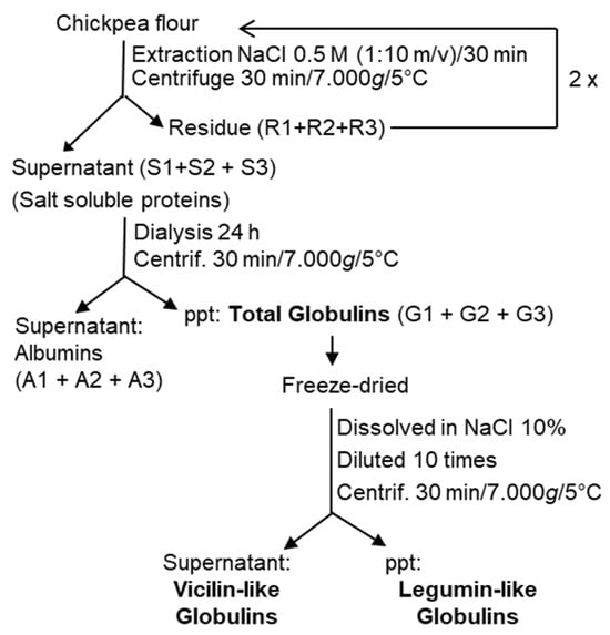
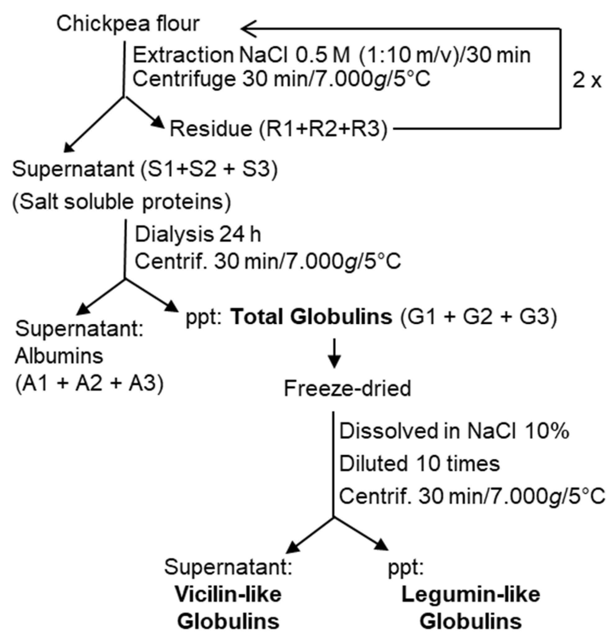
The solubility was observed via Osborne’s protein fractionation [7] to estimate the globulin content, as described by Tavano et al. [8], with some modifications as described in the general scheme presented in Figure 1.
Figure 1.
General scheme of globulin isolation.
Whole seeds were ground using a mill (Pulverisette 14, Fritsch, Germany) to pass through a 45-mesh sieve. The flour sample was extracted three times with 0.5 mol/L NaCl solution (1:10 w/v) by shaking for 30 min. The combined supernatants, containing salt soluble proteins, were saved and then dialyzed against distilled water for 24 h at 5 °C, with frequent water changes. Total globulins were obtained as a precipitate after centrifugation (7000× g for 30 min at 5 °C—Centrifuge FANEN Excelsa 4, MOD 280R, São Carlos, SP, Brazil) and freeze-dried (Freeze-dryer LioBrás Mod.L101, São Carlos, SP, Brazil). Any albumins that may have been solubilized in the saline solution remained in the supernatant and were subsequently discarded. The legumin-like and vicilin-like globulins were separated from total chickpea globulin as described by Kumar and Venkataraman [12]. The isolated freeze-dried total globulin was dissolved in 100 g/L NaCl, and this solution was diluted 10 times with distilled water. After centrifugation (7000× g/30 min), the supernatant containing vicilin-like globulin was freeze-dried, and major globulins, as precipitate, were freeze-dried.
2.3. Protein Determination
Nitrogen was determined according to the Kjeldahl method [13]. Protein content was calculated as nitrogen content × 6.25. In addition, a specific conversion factor of globulins was estimated from the amino acid composition data, as described in Section 2.6.
2.4. In Vitro Protein Digestibility
In vitro protein digestibility was determined as described by Akeson and Stahman [14] using a pepsin–pancreatin incubation sequence. Briefly: Fifty milligrams of protein from each sample were incubated with 7.5 mL of 0.1 M KCl–HCl buffer (pH 1.5) containing 0.75 mg of pepsin for 3 h at 37 °C in a shaking water bath (Cientec, Belo Horizonte, Brazil). After that, the pH was shifted by adding 7.5 mL of 0.2 M sodium phosphate buffer (pH 8.0) containing 2 mg of pancreatin. The mixture was then incubated for an additional 24 h at 37 °C. This extended intestinal digestion time was maintained as originally described by Akesson and Stahman [14] and was intentionally applied to maximize the hydrolysis potential of the globulin fraction. The enzymatic reaction was stopped by boiling for 5 min. An enzyme blank was prepared under identical conditions without protein samples, and the degrees of hydrolysis, antioxidant activity, and angiotensin-converting enzyme (ACE) inhibitory activity were calculated after subtraction of the enzyme blank values.
2.5. α-Amino Groups Determination
Free α-amino groups were determined spectrophotometrically (UV−Vis spectrophotometer BEL photonic UV-M51, made in PRC), using OPA reagent, as described by Church et al. [15]. Briefly, aliquots of the samples between 0 and 130 uL were added to 1 mL of OPA reagent, prepared every day (25 mL of 100 nmol/L sodium tetraborate, 2.5 mL of SDS 20%, 40 mg of OPA in 1 mL of methanol, 100 uL of α-mercaptoethanol, and adjusted to 50 mL with distilled water). After exactly 2 min of reaction, the absorbance was measured at 340 nm. Analytical reference curves were constructed by using L-leucine as the standard.
2.6. Amino Acid Determination
The total amino acids were quantified by pre-column derivatization, released after acid hydrolysis (6 mol/L), under heating (110 °C/20 h), followed by liquid chromatography, using a high-performance liquid chromatograph (Shimadzu, Kyoto, Japan), according to White, Hart and Fry [16]. The amino acids were dissolved in diluent and introduced in a column C18 Luna/Phenomenex (250 mm 4.6 mm, 5 μ; Phenomenex Inc., Torrence, CA, USA), with UV detector at 254 nm (Shimadzu Corporation, Tokyo, Japan). Quantification was performed by comparison with the amino acid standard Thermo Scientific (Rockford, IL, USA) and DL-2-aminobutyric acid (Sigma-Aldrich, St. Louis, MO, USA) was used as an internal standard, according to Hagen et al. [17]. Tryptophan residues were determined after pronase hydrolysis of samples and reaction with p-dimethylaminobenzaldehyde according to Spies [18]. Analyses were performed in duplicate, accepting differences of not more than 4%, and the data are presented as the means of the two determinations.
2.7. Potential Antioxidant Activity
The ABTS+ radical was generated by incubating 7 mmol/L ABTS with 2.4 mmol/L potassium persulfate for 16 h in the dark, following Shalaby and Shanab [19]. The working solution was diluted with distilled water to an absorbance of 0.700 at 734 nm (UV−Vis spectrophotometer BEL photonic UV-M51, BEL Engineering, Shanghai, China). For the assay, 250 µL of each sample (diluted to contain 20 µg of hydrolysate protein/mL in the reaction mixture), were mixed with 750 µL of ABTS+ solution. Absorbance was recorded after 30 min of incubation in the dark. Antioxidant activity was quantified using a TROLOX standard curve (1.0–7.0 nmol/mL) and expressed as nmol of TROLOX equivalents per mg of protein.
2.8. Angiotensin-Converting Enzyme Inhibition
Angiotensin-converting enzyme (ACE) inhibitory activity was determined according to the method described by Holmquist et al. [20], using FAPGG as substrate, with slight modifications. In a quartz cuvette, 750 µL of substrate solution (0.5 mM FAPGG in 50 mM Tris–HCl buffer, pH 7.5, containing 0.3 M NaCl) was added, followed by 40 µL of the sample (at varying concentrations). After the addition of 10 µL of ACE (0.5 U·mL−1), the reaction was monitored at 340 nm for 30 min (UV−Vis spectrophotometer BEL photonic UV-M51, made in PRC), with readings taken at zero time and every subsequent minute. For the non-inhibited ACE activity (100% activity), distilled water was used in place of the sample. The percentage of inhibitory activity/min for each sample concentration was used to construct a curve, based on at least three different concentrations. The equation of the resulting line was used to estimate the amount of sample required to inhibit 50% of the enzyme activity (IC50).
2.9. SDS-PAGE
SDS-PAGE was carried out as described by Laemmli [21], using a 12% polyacrylamide gel as separating gel and a 4% stacking gel. The samples were pre-mixed with sample buffer containing 0.5 mol/L Tris–HCl buffer pH 6.8, 1% bromophenol blue, 10% glycerol, and 2% SDS. Molecular weight standard mixture was used (SigmaMarker™ wide range—S8445, containing: α-Lactalbumin, bovine milk (14,200); Trypsin inhibitor, soybean (20,000); Trypsinogen, bovine pancreas (24,000); Carbonic anhydrase, bovine erythrocytes (29,000); Ovalbumin, chicken egg (45,000); Glutamic dehydrogenase, bovine liver (55,000); Albumin, bovine serum (66,000); Phosphorylase B, rabbit muscle (97,000); β-Galactosidase, E. coli (116,000); Myosin, porcine heart (200,000)). The gels were stained with Coomassie brilliant blue G-250 and destained using a methanol-acetic acid solution.
2.10. Statistical Analysis
Data were analyzed with JASP software (version 0.19.1, 2023). The Shapiro–Wilk test was employed to assess normality, Pearson’s test was used to examine correlations, and ANOVA with Tukey’s post hoc test was conducted for mean comparisons. A significance level of p < 0.05 was adopted.
3. Results and Discussion
Isolation of the total globulin fraction from chickpea grains (Table 1) confirmed that globulins constitute the most abundant protein class in chickpeas, accounting for approximately 43% of the total protein, which corresponds to about 9.5% of the total grain mass. The same table shows the proportion of legumin-type globulins within this fraction, which, as expected, represent the major component (approximately 82.5% of the total globulins).
Table 1.
Composition of globulins in chickpea protein (Cicer arietinum L. var. GB Cappuccino).
When the same methodology was used for the extraction and isolation of protein fractions from the IAC-Marrocos cultivar, which is also grown in Brazil, a very similar proportion of total protein composition was observed by Tavano et al. [8], with globulins accounting for 47.85% of the total protein, composed of approximately 88.9% legumin-like and 11% vicilin-like globulins.
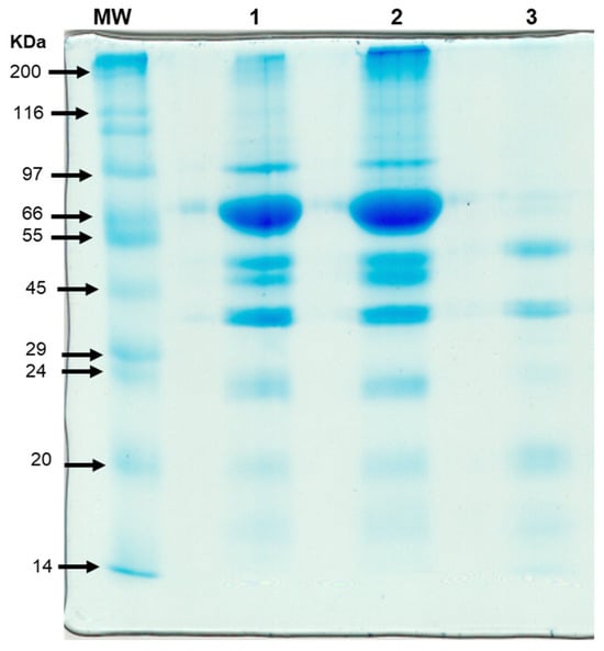
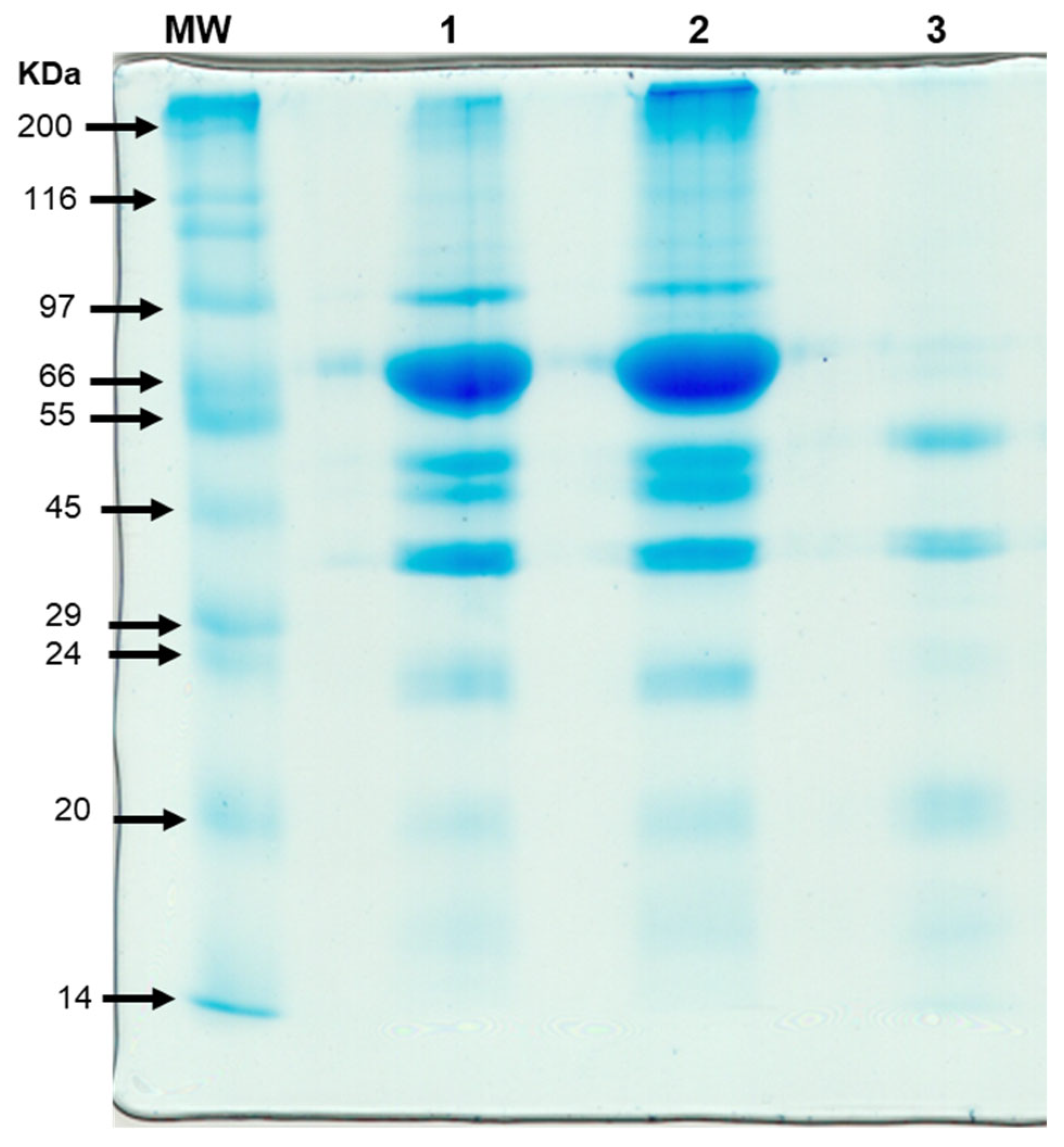
The electrophoretic profile of the isolated fractions (Figure 2) also confirmed the predominance of the legumin-like fraction, whose band pattern clearly overlaps that of the total globulin fraction. The molecular weight range between 29 and 97 kDa includes the most characteristic bands of these proteins, with a prominent band around 66 kDa. In contrast, the vicilin-like fraction displayed bands mainly below 55 kDa. These results are consistent with previous studies reporting similar patterns for globulins from different chickpea cultivars.
Figure 2.
SDS-PAGE of isolated globulin fractions of chickpea var. GB Cappuccino under non-reducing conditions. Lanes: MW—Molecular weight markers; 1—Total globulins; 2—Legumin-like (11S-like) globulins; 3—Vicilin-like (7S-like) globulins.
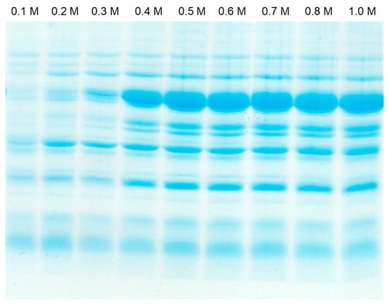
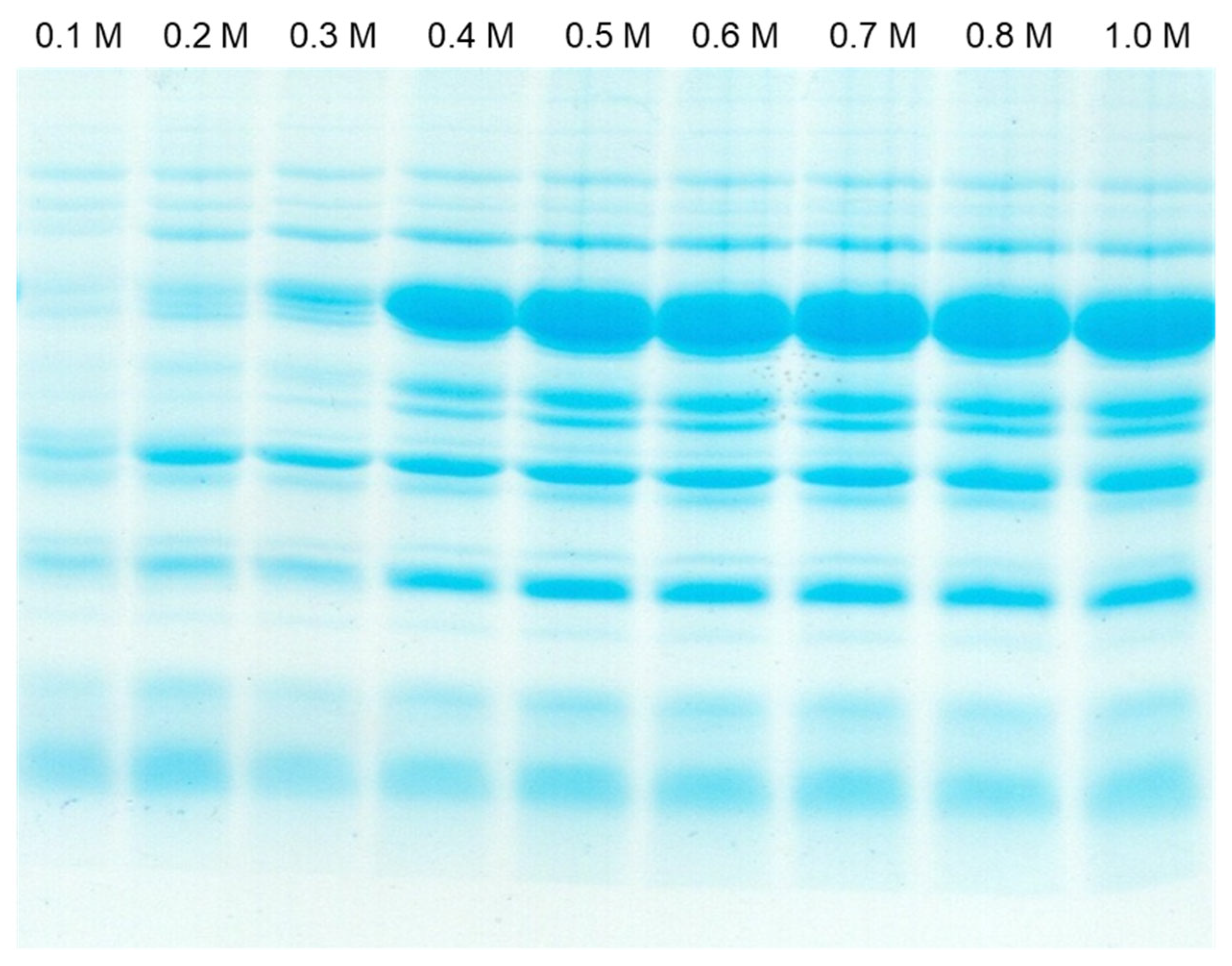
Although the legumin-like fraction is less water-soluble and typically requires higher NaCl concentrations for solubilization than the vicilin-like globulin, extraction with low salt concentrations already promoted the partial solubilization of proteins whose bands correspond to legumin-like globulins (Figure 3). During the separation of globulins into legumin- and vicilin-like fractions, total globulins were initially solubilized in NaCl 10%, and the solution was subsequently diluted to a final NaCl concentration of 1% (≈0.17 M). However, Figure 3 shows that bands similar to those of legumin-like proteins were already visible at 0.1 M NaCl.
Figure 3.
SDS-PAGE of salt soluble proteins of chickpea (var. GB Cappuccino), at different NaCl concentration (0.1 to 1.0 mol·L−1), on non-reducing condition.
This observation is further supported by Siqueira et al. [22], who reported that the percentage of proteins solubilized in water from different chickpea genotypes, including GB Cappuccino, was considerably higher than expected if only albumins were extracted. Their data showed that 57.17% of total proteins were solubilized, of which 32% corresponded to albumins. Since no salt was added to the extraction medium (only deionized water), the endogenous salts present in the grains may have facilitated partial globulin solubilization. According to Diniz et al. [23], this cultivar contains about 2.4% minerals, part of which may dissolve and act as salts, contributing to globulin solubilization even at initially low NaCl concentrations. Upon dialysis of the aqueous extracts, Siqueira et al. [22] observed protein precipitation corresponding to 25.05% of total protein, which could be attributed to globulins. Extrapolating this value to the present study corresponds to approximately 58% of the total globulin fraction. Here, before dialysis, the total soluble protein extracted in 0.5 M NaCl reached 73.12%. Subtracting the 42.83% attributed to globulins yields an estimated 30.29% of albumin-type proteins.
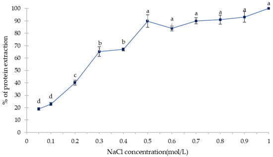
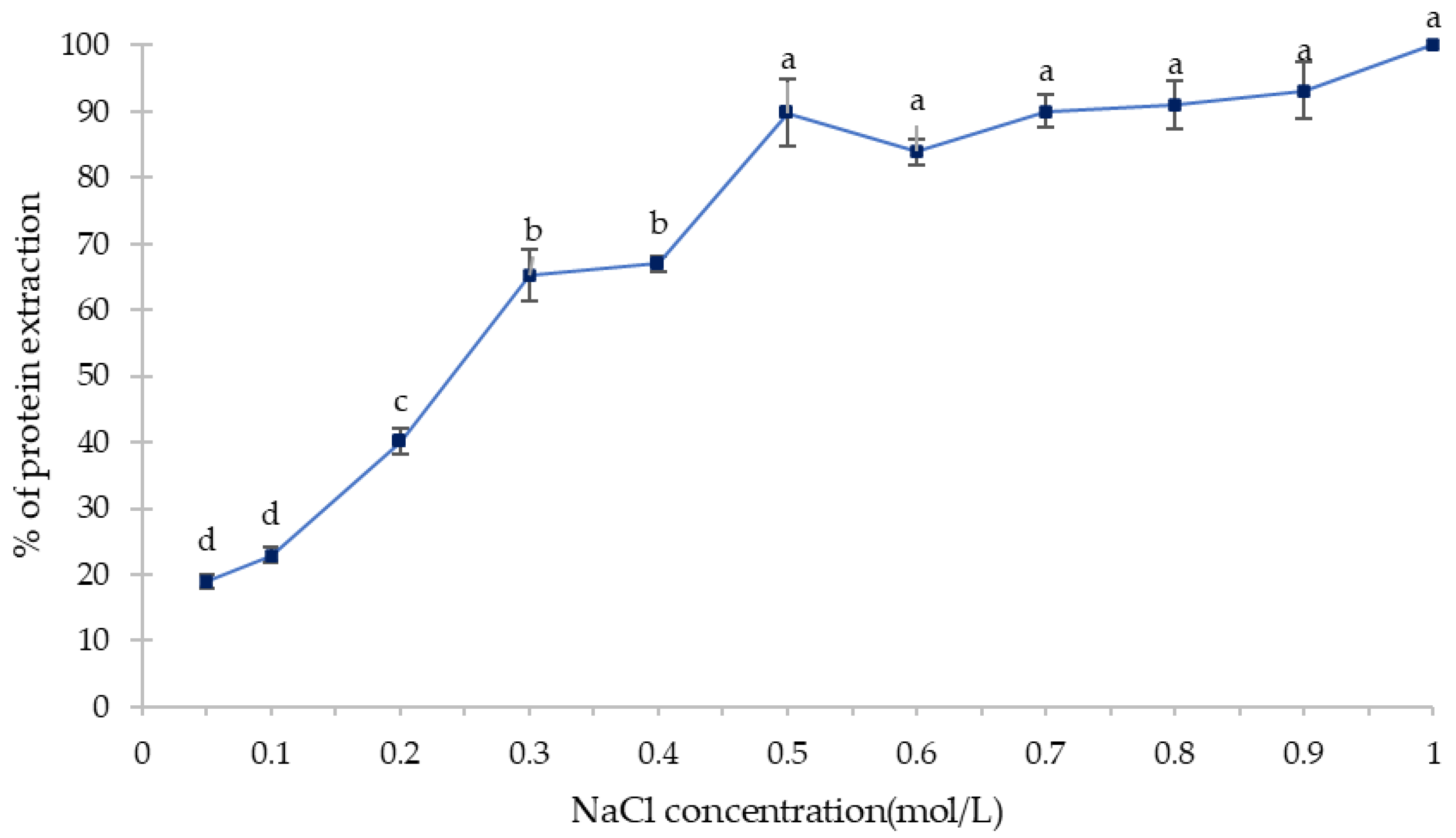
Although globulins showed partial solubilization at low salt concentrations, the results in Figure 4 emphasize the critical role of the NaCl concentration in the solubilization of these protein fractions. Based on these findings, 0.5 M NaCl was confirmed to be an appropriate concentration for the first step of total globulin isolation.
Figure 4.
Effect of NaCl concentration (0.005–1.0 mol/L) on the chickpea salt-soluble proteins extractability. The values are the mean of three determinations. Different letters above the standard deviation error bars indicate statistically significant differences between the samples (p < 0.05).
As demonstrated by Chang et al. [6], vicilin exhibits greater solubility in neutral pH conditions than legumin when no salt is present to assist solubilization. The authors also highlight an important observation from their data: the solubility of total globulin is not merely a reflection of the summed behavior of its two major fractions, vicilin and legumin. Instead, a distinct behavior may emerge when these fractions are studied together as total globulin, likely arising from conformational changes within each fraction in response to the extraction medium. These structural adjustments influence their interactions, resulting in a collective protein response that differs from the behavior of the isolated fractions—underscoring the strong influence of the overall balance of forces among the combined subunits.
The profiles observed here, under both reducing and non-reducing conditions, are highly consistent with those reported by Ye et al. [9] for desi-type chickpeas from Xinjiang, China.
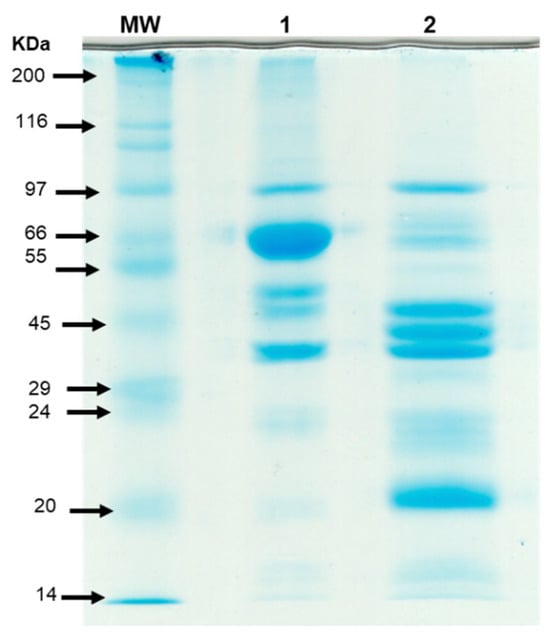
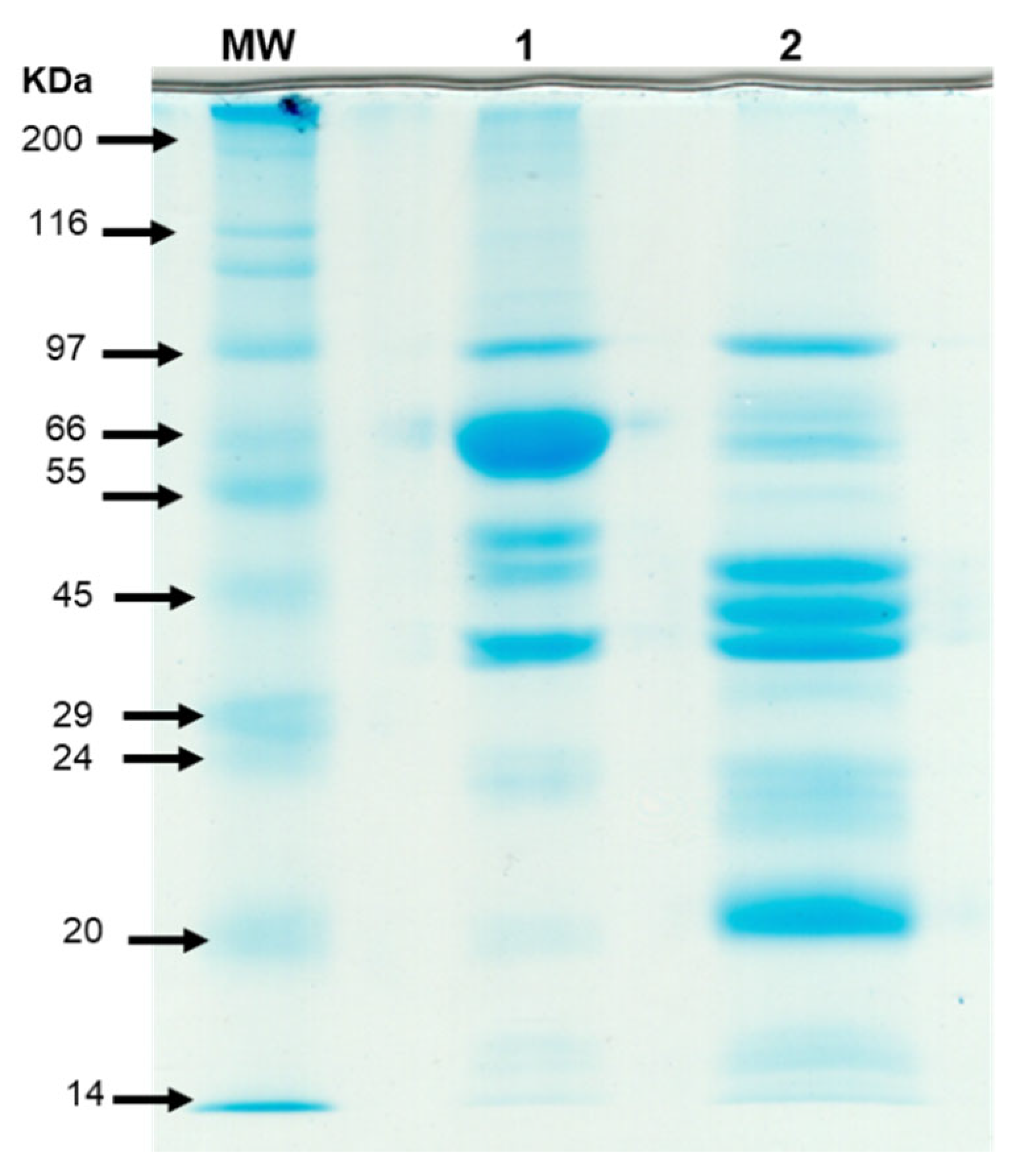
Figure 5 shows the electrophoretic profile of the total globulin fraction before and after treatment with β-mercaptoethanol. The post-treatment profile indicates that the globulin may be composed of monomeric proteins, showing no changes after exposure to reducing conditions, as well as multimeric proteins, since alterations in some bands are evident. Notably, the pronounced band around 66 kDa almost disappeared, while new bands appeared in the region of approximately 20 kDa. The presence of cysteine, as confirmed by the data in Table 2, is consistent with these results, indicating the presence of disulfide bonds. These data are expected, according to other authors [4], who attribute the presence of disulfide bonds particularly to those formed between subunits of the legumin-like fraction.
Figure 5.
SDS-PAGE of isolated total globulin of chickpea var. GB Cappuccino under non-reducing (1) and reducing conditions (2). MW: Molecular weight markers.
Table 2.
Amino acid pattern of globulins isolated from chickpea var. GB Cappuccino.
The isolated total globulin fraction was further evaluated for its nutritional characteristics in the native (non-heat-treated) state. Two key parameters defining protein quality were analyzed: the amino acid composition and digestibility. The total amino acid profile is presented in Table 2.
FAO reference amino acid requirement patterns [24] are also provided in Table 2, allowing for comparison between the amino acid composition and nutritional recommendations. Four amino acids were below the levels recommended for young children: sulfur-containing amino acids (methionine + cystine), tryptophan, threonine and valine. For adults, the primary limitations were sulfur amino acids and tryptophan. Gao et al. [25] reported methionine and cysteine contents of 0.82 and 0.51 g/100 g protein, respectively, consistent with our findings.
The similarities between our globulins and those described by Ye et al. [9] extend beyond the electrophoretic pattern. The amino acid composition was also remarkably similar across all amino acids, including methionine, with reported values of 1.31% compared with 1.06% in the present work. Other amino acids showed even closer correspondence, such as Ile (4.22% vs. 4.26%) and Gly (3.65% vs. 3.63% in this study).
For other chickpea cultivars also grown in Brazil, the results were quite similar, showing low concentrations of sulfur-containing amino acids, as previously reported by Tavano et al. [8,26].
Although sulfur amino acid limitation may be viewed as a nutritional drawback, recent evidence suggests that methionine-restricted diets may contribute to longevity promotion [27,28].
From the amino acid data, the specific nitrogen-to-protein conversion factor (for the Kjeldahl method) was calculated. The nitrogen content of the sample was 16.53%, corresponding to a conversion factor of 6.05. Using this conversion factor, it was estimated that the lyophilized globulin fraction obtained after precipitation contained 100% protein.
Mariotti, Tomé, and Mirand [29] have, since 2008, highlighted the importance of revisiting the universal nitrogen-to-protein conversion factor of 6.25. In their review, the authors emphasize that the use of this factor often leads to an overestimation of protein content for most foods. On average, they suggest a lower conversion factor of approximately 5.6 and report a range of values depending on the food matrix, including legumes. Because this factor is dependent on the amino acid composition of the proteins, and considering that different protein fractions within the same chickpea sample may exhibit distinct amino acid profiles [8], it is reasonable to assume that fraction-specific conversion factors may exist for chickpea proteins
The amino acid analysis also enabled an estimation of the molar amount of amino acids per gram of sample, providing insight into the number of peptide bonds potentially cleaved during hydrolysis. This allows a more accurate estimation of the degree of hydrolysis based on the measured α-amino group release. Although a mean amino acid molecular weight of 113 is commonly assumed, the value calculated for this sample, 114.44, was in close agreement.
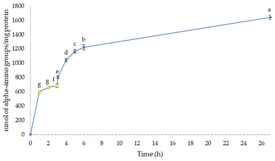
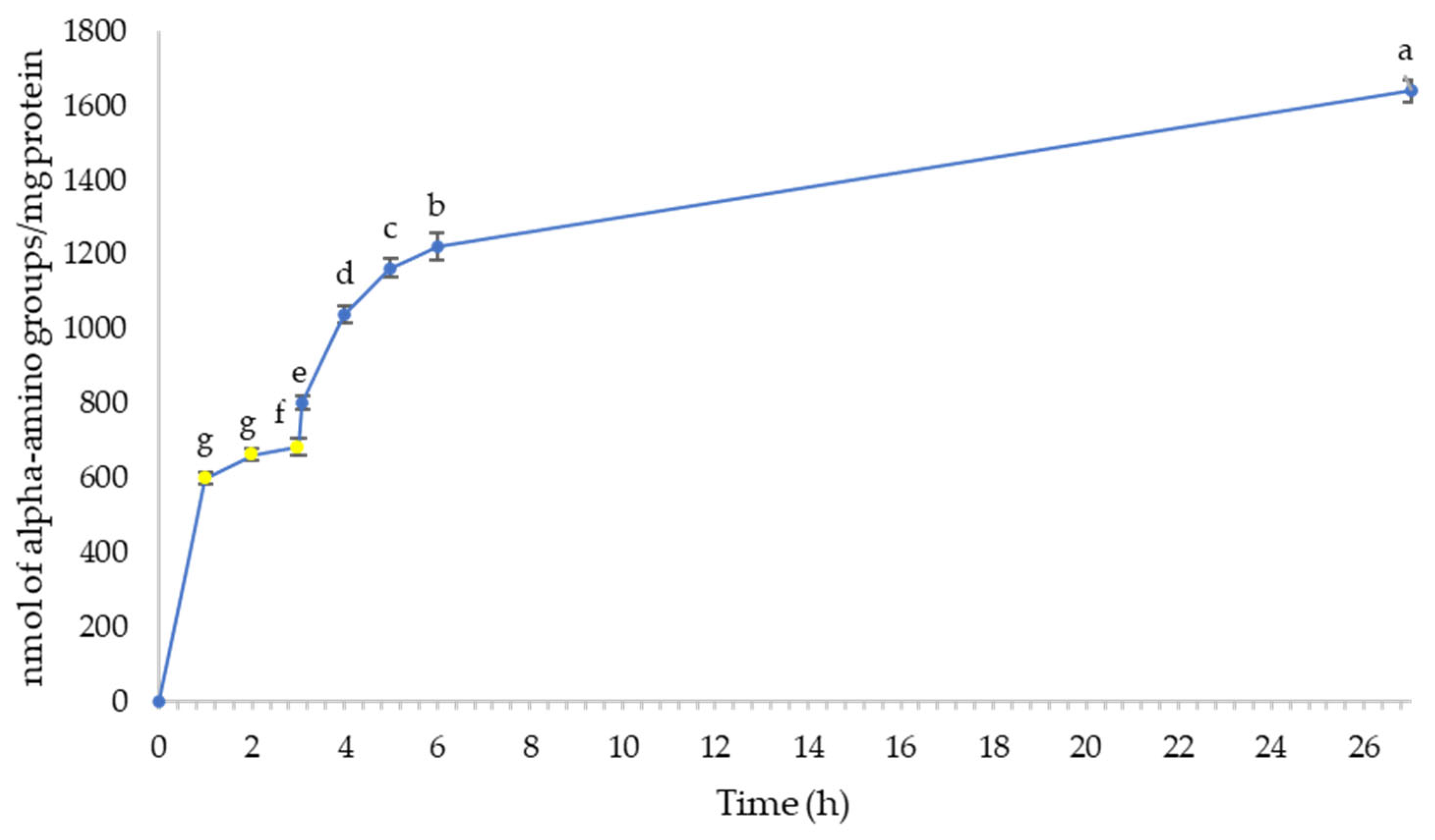
The hydrolysis profile of globulins subjected to digestive enzymes (pepsin and pancreatin) was also investigated. Figure 6 shows the in vitro simulated digestion curve, where limited hydrolysis was observed with pepsin alone, followed by a marked increase in peptide bond cleavage—reflected by the release of α-amino groups—after pancreatin addition.
Figure 6.
In vitro hydrolysis of chickpea globulin at different times using pepsin–pancreatin sequence (yellow points represent pepsin digestion, and blue points are results after pancreatin addiction). Results are expressed as mean of three determinations. Different letters above the standard deviation error bars indicate statistically significant differences between the samples (p < 0.05).
Although pepsin shows a strong preference for cleaving peptide bonds immediately following Phe and Leu residues, and these two amino acids together account for a considerable proportion of the globulin fraction (approximately 13.1%), the overall extent of hydrolysis achieved by pepsin alone was relatively low. Based on the total amount of α-amino groups released after the gastric digestion phase, a value of 683.08 nmol of α-amino groups per mg of protein was obtained (Figure 6). In contrast, considering the amino acid composition of this fraction, the estimated molar amount of Phe and Leu residues per mg of protein was 991.31 nmol/mg, as calculated from the data presented in Table 2. Thus, if only these residues are considered for estimating the number of potentially cleavable peptide bonds, the degree of hydrolysis would correspond to approximately 68.9% of the theoretically available sites.
Of course, not all amino acids are located in accessible peptide bonds, nor are Phe and Leu the only residues involved in sites susceptible to pepsin action. Nonetheless, this estimation provides an indication that a greater number of bonds might be hydrolyzed, both by pepsin and by other enzymes present in pancreatin.
Considering the total amino acid content of 7550.62 nmol per mg of protein, as calculated from the data presented in Table 2, pepsin hydrolysis corresponded to 9.05%. In contrast, following pancreatin treatment, the release of 1638.85 nmol of α-amino groups after 24 h of hydrolysis (Figure 7) resulted in an estimated degree of hydrolysis of 21.7% at the end of the process.
Figure 7.
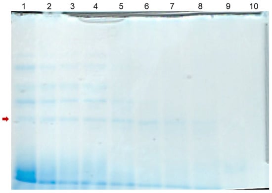
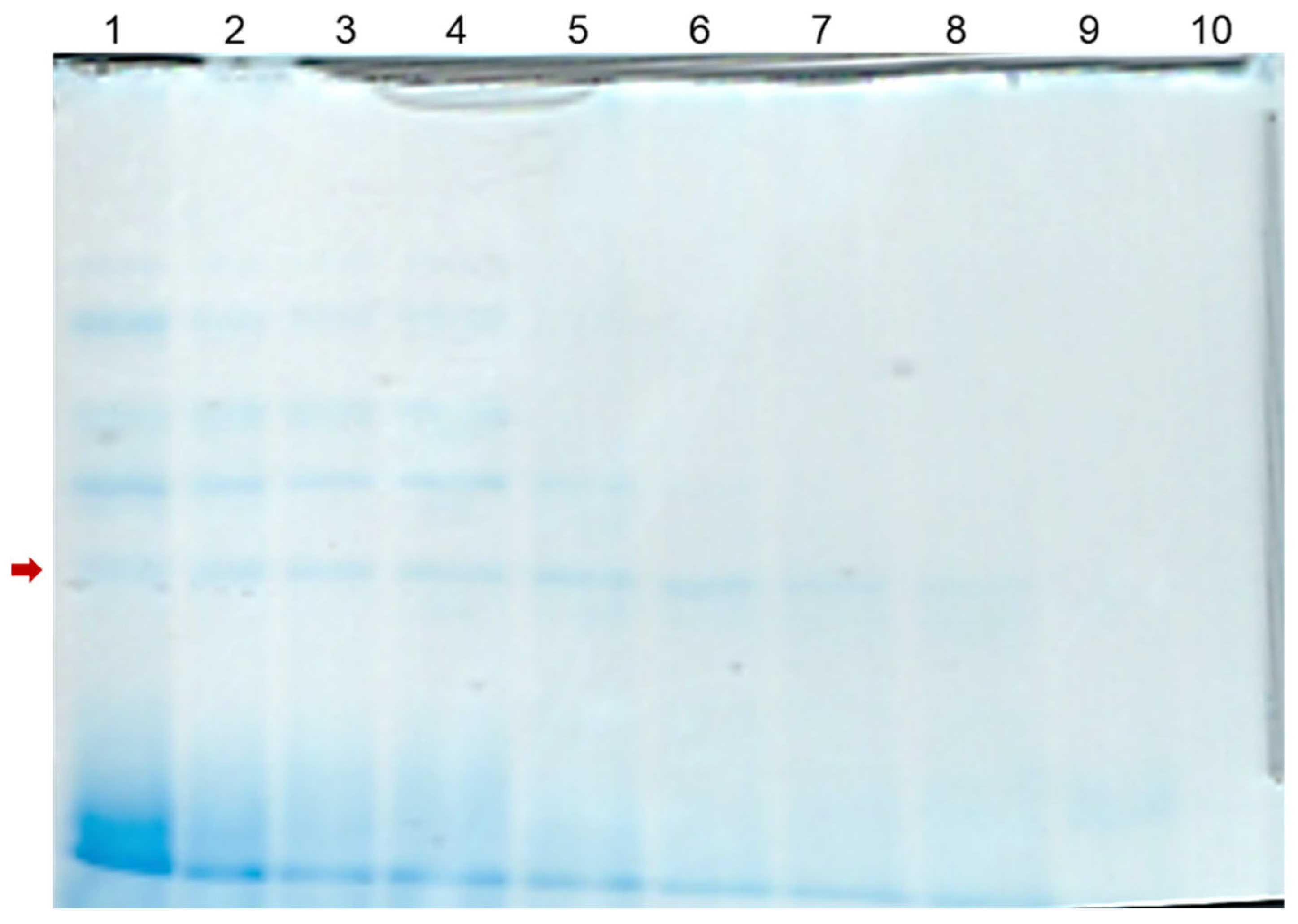
SDS-PAGE of isolated total globulin of chickpea var. GB Cappuccino under non-reducing condition. Lane 1: native total globulin; Lanes 2–4: globulin after pepsin digestion (1, 2 and 3 h); Lanes 5–9: pepsin + pancreatin digestion (5 min, 1 h, 2 h, 3 h and 24 h); Lane 10: only enzymes solution.
These results suggest that the native globulin fraction possesses a structural arrangement that limits the accessibility of digestive enzymes. Tavano et al. [8,26] observed a similar behavior in globulins isolated from chickpea (var. IAC-Marrocos), also cultivated in Brazil, reporting a clear increase in hydrolysis potential when the samples were subjected to heat treatment prior to digestion. Using the same experimental conditions of this paper, the same authors reported a degree of hydrolysis of 65.99% for casein following pepsin–pancreatin digestion.
Likewise, Contardo et al. [10] emphasized that the structural organization of globulin subfractions plays a key role in their susceptibility to enzymatic digestion. The authors highlighted that β-sheet conformations may hinder hydrolysis, as can the presence of disulfide bonds linking polypeptide chains. The presence of such bonds in the native structure likely contributes to the resistance to hydrolysis observed in the present study (Figure 6).
Figure 7, presenting the electrophoretic profiles of hydrolysates at different digestion times, supports this observation.
Pepsin hydrolysis had little effect on the globulin fraction, as several protein bands remained intact until pancreatin was added (Figure 7) and remained resistant for several hours even in the presence of pancreatin, disappearing only after 24 h of enzyme exposure, as indicated in Figure 7 by the red arrow.
Although the resistance to hydrolysis of specific regions of the protein chain may appear nutritionally unfavorable, it can be paradoxically advantageous when considering the potential bioactivity of the resulting peptides, as digestive resistance is a desirable property for bioactive peptide stability. Ahmed, Sun, and Udenigwe [11] discussed the importance of resistance to digestion for peptides to exert bioactive effects in the human body, particularly in light of the requirement for their absorption in intact form in order to reach their site of action.
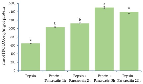
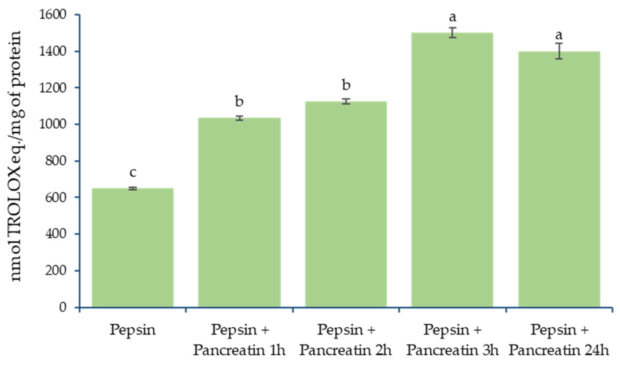
Figure 8 shows the antioxidant activity of hydrolysates obtained at different digestion times. The results indicate that digestion with pepsin followed by pancreatin can release peptides with antioxidant potential.
Figure 8.
Antioxidant activity (ABTS) of isolated total globulin of chickpea var. GB Cappuccino after pepsin (3 h) and additional pancreatin digestion (pepsin 3 h + pancreatin 1 h, 2 h, 3 h and 24 h). The values are the mean of three determinations. Different letters above the standard deviation error bars indicate statistically significant differences between the samples (p < 0.05).
As discussed by Xu et al. [30], peptides containing 2 to 10 amino acids—particularly those enriched in hydrophobic and aromatic residues—may be more effective in exerting antioxidant activity, whether in biological systems or as agents that mitigate oxidative processes within food matrices.
The results obtained in the present study indicate a relevant antioxidant activity of the hydrolysates (about 650 nmol TROLOX equivalents per mg of hydrolysate after pepsin hydrolysis and 1400 nmol TROLOX equivalents per mg of hydrolysate, after pepsin-pancreatin hydrolysis, as observed in Figure 8). Navarro-Leyva et al. [31] reported values of approximately 173 nmol TROLOX equivalents per mg of hydrolysate for a desi-type black chickpea (Cicer arietinum L.) genotype (ICC3761) from Mexico, using either pepsin–pancreatin digestion, as applied in the present study, or alcalase treatment. These data provide a useful reference for contextualizing the antioxidant capacity observed herein, despite methodological differences among studies. When Arcan and Yemenicioğlu [32] hydrolyzed total chickpea protein (from Turkey) using pepsin, they reported an antioxidant activity equivalent to 277 nmol Trolox equivalents per mg of hydrolysate powder.
Several authors have shown that peptides may exhibit multiple bioactivities. In the present study, in addition to antioxidant activity, ACE-inhibitory activity was also detected in pepsin + pancreatin digested (Table 3).
Table 3.
Angiotensin-Converting Enzyme (ACE) inhibition by globulin hydrolysates from chickpea (var. GB Cappuccino).
Although the mechanisms regulating hypertension are widely recognized as multifactorial, specific points within this cascade of events can serve as targets for bioactive compounds that support blood pressure regulation. For instance, modulation of the renin–angiotensin system through the inhibition of the angiotensin-converting enzyme (ACE) is a well-established and widely applied strategy [33]. Captopril, a highly potent and effective antihypertensive agent, is a classic example of an ACE inhibitor.
Although the hydrolysate exhibited a lower potency than captopril (Table 3), this difference is consistent with previous reports comparing peptide-based ACE inhibitors with pharmaceutical agents. Captopril was included solely to provide a reference point for readers, given the substantial difficulty in directly comparing data across the literature, where a wide diversity of assays, substrates, and experimental conditions is reported. These findings suggest that, while this hydrolysate does not have therapeutic potential as a drug, the chickpea globulin fraction may act as an adjuvant or enhancer of blood pressure-modulating effects.
It is important to note that the hydrolysate evaluated here comprised all protein fragments released from the globulin fraction. Further research is required to identify and characterize the individual peptides responsible for these bioactivities (especially considering LC-MS/MS and fractionation of <3 kDa peptides), which may lead to the discovery of more potent sequences.
Overall, the results obtained in this study indicate that the globulin fraction from the newly released desi-type chickpea cultivar GB Cappuccino exhibits relevant nutritional characteristics and a measurable bioactive potential under in vitro conditions. However, it is important to emphasize that this work represents a first approach to exploring the properties of these proteins. Further studies are warranted to more comprehensively assess their nutritional quality, digestive behavior under physiologically relevant conditions, and additional bioactivities. In particular, future investigations should focus on the identification of bioactive peptides and the evaluation of other biological effects commonly attributed to chickpea-derived peptides, such as anti-inflammatory and antidiabetic properties.
Author Contributions
Conceptualization, O.L.T.; formal analysis, L.C.B., S.C.d.O.D., T.d.F.L., B.d.C.G.S., A.T.d.M. and R.L.C.; data curation, O.L.T. and M.T.B.P.; writing—original draft preparation, L.C.B. and O.L.T.; writing—review and editing, O.L.T. and M.T.B.P.; visualization, M.T.B.P.; project administration, O.L.T.; funding acquisition, O.L.T. and M.T.B.P. All authors have read and agreed to the published version of the manuscript.
Funding
This work was supported by the Coordenação de Aperfeiçoamento de Pessoal de Nivél Superior-CAPES (Finance Code 001 and LCB Scholarship), the Fundação de Amparo à Pesquisa do Estado de Minas Gerais-FAPEMIG Brasil (APQ-01240-21/APQ-02644-23; TFL and RLC Scholarship), and the Conselho Nacional de Desenvolvimento Científico e Tecnológico-CNPq/MCTI (Grant Number 421595/2023-0 and 421375/2025-6; BCGS and ATM Scholarship).
Data Availability Statement
The raw data supporting the conclusions of this article will be made available by the authors on request.
Acknowledgments
The authors acknowledge FAPEMIG, CAPES, CNPq, and Osmar Artiaga from the AgroGarbanzo Produção Agrícola LTDA, Cristalina-GO/Brazil, for providing the chickpeas.
Conflicts of Interest
The authors declare that this study received the chickpeas from AgroGarbanzo Produção Agricola LTDA. The company was not involved in the study design, collection, analysis, interpretation of data, the writing of this article or the decision to submit it for publication.
References
- Koul, B.; Sharma, K.; Sehgal, V.; Yadav, D.; Mishra, M.; Bharadwaj, C. Chickpea (Cicer arietinum L.) biology and biotechnology: From domestication to biofortification and biopharming. Plants 2022, 11, 2926. [Google Scholar] [CrossRef] [Scilit] [PubMed]
- Boukid, F. Chickpea (Cicer arietinum L.) protein as a prospective plant-based ingredient: A review. Int. J. Food Sci. Technol. 2021, 56, 5435–5444. [Google Scholar] [CrossRef] [Scilit]
- Kaur, R.; Prasad, K. Technological, processing and nutritional aspects of chickpea (Cicer arietinum) A review. Trends Food Sci. Technol. 2021, 109, 448–463. [Google Scholar] [CrossRef] [Scilit]
- Grasso, N.; Lynch, N.L.; Arendt, E.K.; O’Mahony, J.A. Chickpea protein ingredients: A review of composition, functionality, and applications. Compr. Rev. Food Sci. Food Saf. 2022, 21, 435–452. [Google Scholar] [CrossRef] [Scilit]
- Brasil—Registro Nacional De Cultivares—RNC. 2025. Available online: https://sistemas.agricultura.gov.br/snpc/cultivarweb/cultivares_registradas.php (accessed on 17 November 2025).
- Chang, L.; Lan, Y.; Bandillo, N.; Ohm, J.B.; Chen, B.; Rao, J. Plant proteins from green pea and chickpea: Extraction, fractionation, structural characterization and functional properties. Food Hydrocoll. 2022, 123, 107165. [Google Scholar] [CrossRef] [Scilit]
- Osborne, T.B. The Vegetable Proteins; Longmans Green and Company: London, UK, 1924; Volume 2, p. 154. [Google Scholar]
- Tavano, O.L.; Silva Junior, S.I.; Demonte, A.; Neves, V.A. Nutritional responses of rats to diets based on chickpea (Cicer arietinum L.) seed meal or its protein fractions. J. Agric. Food Chem. 2008, 56, 11006–11010. [Google Scholar] [CrossRef] [Scilit]
- Ye, J.; Shi, N.; Rozi, P.; Kong, L.; Zhou, J.; Yang, H. A comparative study of the structural and functional properties of chickpea albumin and globulin protein fractions. Food Bioprocess Technol. 2024, 17, 3253–3266. [Google Scholar] [CrossRef] [Scilit]
- Contardo, I.; Gutiérrez, S.; Hurtado-Murillo, J.; Escobar, N. Understanding the structural differences in chickpea globulins and their relationship with in vitro protein digestibility. Food Res. Int. 2025, 202, 115702. [Google Scholar] [CrossRef] [Scilit]
- Ahmed, T.; Sun, X.; Udenigwe, C.C. Role of structural properties of bioactive peptides in their stability during simulated gastrointestinal digestion: A systematic review. Trends Food Sci. Technol. 2022, 120, 265–273. [Google Scholar] [CrossRef] [Scilit]
- Kumar, K.G.; Venkataraman, L.V. Chickpea seed proteins: Isolation and characterization of 10.3 S protein. J. Agric. Food Chem. 1980, 28, 524–529. [Google Scholar] [CrossRef] [Scilit]
- AOAC International. AOAC Official Methods of Analysis, 8th ed.; AOAC International: Arlington, VA, USA, 1995. [Google Scholar]
- Akeson, W.R.; Stahmann, M.A. A pepsin pancreatin digest index of protein quality evaluation. J. Nutr. 1964, 83, 257–261. [Google Scholar] [CrossRef] [Scilit] [PubMed]
- Church, F.C.; Swaisgood, H.E.; Porter, D.H.; Catignani, G.L. Spectrophotometric assay using o-phthaldialdehyde for determination of proteolysis in milk and isolated milk proteins. J. Dairy Sci. 1983, 66, 1219–1227. [Google Scholar] [CrossRef] [Scilit]
- White, J.A.; Hart, R.J.; Fry, J.C. An evaluation of the Waters Pico-Tag system for the amino-acid analysis of food materials. J. Autom. Chem. 1986, 8, 170–177. [Google Scholar] [CrossRef] [Scilit] [PubMed]
- Hagen, S.R.; Frost, B.; Augustin, J. Precolumn phenylisothiocyanate derivatization and liquid chromatography of amino acids in food. J. Assoc. Off. Anal. Chem. 1989, 72, 912–916. [Google Scholar] [CrossRef] [Scilit]
- Spies, J.R. Determination of tryptophan in proteins. Anal. Chem. 1967, 49, 1412–1415. [Google Scholar] [CrossRef] [Scilit]
- Shalaby, E.S.; Shanab, S.M.M. Comparison of DPPH and ABTS assays for determining antioxidant potential of water and methanol extracts of Spirulina patensis. Indian J. Geo-Mar. Sci. 2013, 42, 556–564. [Google Scholar]
- Holmquist, B.; Bünning, P.; Riordan, J.F. A continuous spectrophotometric assay for angiotensin converting enzyme. Anal. Biochem. 1979, 95, 540–548. [Google Scholar] [CrossRef] [Scilit]
- Laemmli, U.K. Cleavage of Structural Proteins during the Assembly of the Head of Bacteriophage T4. Nature 1970, 227, 680–685. [Google Scholar] [CrossRef] [Scilit]
- Siqueira, B.M.; Diniz, J.A.; Antonio, S.A.M.D.; da Silva Lemos, A.C.B.; de Paula, R.A.V.; da Silva Júnior, S.I.; Tavano, O.L. Water-soluble components of ten Brazilian desi and kabuli chickpeas and their potential for health-promoting plant-based beverages. Food Chem. Adv. 2025, 7, 100955. [Google Scholar] [CrossRef] [Scilit]
- Diniz, J.A.; Antonio, S.A.M.D.; da Silva Lemos, A.C.B.; Borim, L.C.; Monaco, M.; da Silva Júnior, S.I.; Tavano, O.L. Short-Time Germination Modifies Chickpea Grains and Enhances the Production of High-Quality Functional Beverages: Insights into the Soaking Stage in Transformation Processes. ACS Omega 2025, 10, 25518. [Google Scholar] [CrossRef] [Scilit]
- FAO; WHO. Dietary protein quality evaluation in human nutrition. Report of 593 and FAO Expert Consultation. FAO Food Nutr. Pap. 2013, 92, 67. [Google Scholar]
- Gao, Y.; Yin, A.; Feng, J.; Zhao, G.; Pang, J.; Yuan, T.; Shi, J. Structures, functional properties and simulated digestion in vitro of chickpea protein fractions. Eur. Food Res. Technol. 2025, 251, 3287–3303. [Google Scholar] [CrossRef] [Scilit]
- Tavano, O.L.; Neves, V.A.; Silva Junior, S.I. In vitro versus in vivo protein digestibility techniques for calculating PDCAAS (protein digestibility-corrected amino acid score) applied to chickpea fractions. Food Res. Int. 2016, 89, 756–763. [Google Scholar] [CrossRef] [Scilit] [PubMed]
- Fang, H.; Stone, K.P.; Wanders, D.; Forney, L.A.; Gettys, T.W. The origins, evolution, and future of dietary methionine restriction. Annu. Rev. Nutr. 2022, 42, 201–226. [Google Scholar] [CrossRef] [Scilit] [PubMed]
- Lee, B.C.; Kaya, A.; Gladyshev, V.N. Methionine restriction and life-span control. Ann. N. Y. Acad. Sci. 2016, 1363, 116–124. [Google Scholar] [CrossRef] [Scilit] [PubMed]
- Mariotti, F.; Tomé, D.; Mirand, P.P. Converting nitrogen into protein--beyond 6.25 and Jones’ factors. Crit. Rev. Food Sci. Nutr. 2008, 48, 177–184. [Google Scholar] [CrossRef] [Scilit] [PubMed]
- Xu, B.; Dong, Q.; Yu, C.; Chen, H.; Zhao, Y.; Zhang, B.; Chen, M. Advances in research on the activity evaluation, mechanism and structure-activity relationships of natural antioxidant peptides. Antioxidants 2024, 13, 479. [Google Scholar] [CrossRef] [Scilit]
- Navarro-Leyva, A.; López-Angulo, G.; Delgado-Vargas, F.; López-Valenzuela, J.Á. Antioxidant, anti-inflammatory, hypoglycemic, and anti-hyperglycemic activity of chickpea protein hydrolysates evaluated in BALB-c mice. J. Food Sci. 2023, 88, 4262–4274. [Google Scholar] [CrossRef] [Scilit]
- Arcan, I.; Yemenicioğlu, A. Effects of controlled pepsin hydrolysis on antioxidant potential and fractional changes of chickpea proteins. Food Res. Int. 2010, 43, 140–147. [Google Scholar] [CrossRef] [Scilit]
- Okagu, I.U.; Ezeorba, T.P.; Aham, E.C.; Aguchem, R.N.; Nechi, R.N. Recent findings on the cellular and molecular mechanisms of action of novel food-derived antihypertensive peptides. Food Chem. Mol. Sci. 2022, 4, 100078. [Google Scholar] [CrossRef] [Scilit]

Disclaimer/Publisher’s Note: The statements, opinions and data contained in all publications are solely those of the individual author(s) and contributor(s) and not of MDPI and/or the editor(s). MDPI and/or the editor(s) disclaim responsibility for any injury to people or property resulting from any ideas, methods, instructions or products referred to in the content. |
© 2026 by the authors. Licensee MDPI, Basel, Switzerland. This article is an open access article distributed under the terms and conditions of the Creative Commons Attribution (CC BY) license.