


Abstract
Background: Skin diseases represent a formidable global public health challenge, contributing significantly to the global burden of non-fatal morbidity. In resource-limited rural settings, these conditions often remain underdocumented, despite their profound impact on quality of life and their role as sentinels of systemic health. Objective: This study aimed to describe and compare the spectrum, relative frequency, and associated systemic comorbidities of dermatological conditions among paediatric and adult populations in rural Eastern Cape, South Africa. Methods: A retrospective, cross-sectional analysis was conducted using 532 outpatient records from the Nelson Mandela Academic Hospital in the Eastern Cape, collected across a 12-month period between 1 August 2020 and 31 July 2021. Data regarding demographics, primary and secondary clinical diagnoses, and documented systemic comorbidities were extracted and analyzed using descriptive and inferential statistics. Results: The study population was predominantly female (61.5%) and adult (70.7%), with an overall median age of 31 years. Inflammatory dermatoses accounted for the primary burden in both paediatric (71.8%) and adult (71.3%) cohorts. Atopic dermatitis was the leading diagnosis overall (27.8%; n = 148/532), followed by acne vulgaris (10.2%; n = 54/532). Paediatric patients had a significantly higher prevalence of infectious dermatoses than adults (17.9% vs. 10.9%, p = 0.039). Systemic comorbidities were present in 28.4% of the cohort (n = 151/532), occurring almost exclusively in adults (38.0% vs. 5.1% in children, p < 0.001). Among adults with comorbidities (n = 143), HIV/AIDS (34.3%; n = 49), hypertension (28.0%; n = 40), diabetes mellitus (11.9%; n = 17), and tuberculosis (11.9%; n = 17) were the most frequent conditions. In multivariable logistic regression, only adult age group (adjusted odds ratio [aOR] 23.4, 95% CI 8.8–62.0, p < 0.001) and male sex (aOR 2.2, 95% CI 1.3–3.7, p = 0.005) were independently associated with comorbidity. Conclusions: In this tertiary hospital cohort, inflammatory dermatoses predominated, reflecting a complex interplay between dermatological presentations and the regional burden of HIV, tuberculosis, and non-communicable diseases among hospital attendees. Because findings are derived from a single tertiary referral facility, they reflect a selected patient population and cannot be generalized to the broader community or general population of the Eastern Cape. Nevertheless, these data highlight the need to strengthen primary healthcare diagnostic capacity and integrate dermatological screening into existing chronic disease programs.
1. Introduction
Skin diseases represent a major public health issue worldwide, affecting individuals of all ages and contributing significantly to morbidity, psychosocial suffering, and impaired quality of life [1]. Epidemiologically, dermatological conditions are among the leading contributors to the global burden of non-fatal diseases, accounting for nearly 9.3 million disability-adjusted life years (DALYs) [2,3]. While their overall burden is now widely recognized, their distribution, clinical characteristics, and determinants vary considerably across countries, regions, and socio-environmental contexts, influenced by genetic, climatic, socioeconomic, and cultural factors [4,5]. In many settings, particularly in low- and middle-income countries, skin diseases remain underdiagnosed, undertreated, and rarely documented, despite their major impact on daily functioning, productivity, and health service utilization [6].
Environmental exposures, climatic variations, hygiene practices, and the local prevalence of infectious diseases shape the dermatological profile of a population [7,8,9]. At the same time, social determinants, including poverty, overcrowding, limited access to care, and disparities in the provision of specialized services, exacerbate the burden of skin conditions in resource-limited settings [6]. Additionally, dermatological manifestations can be the initial signs of systemic diseases, highlighting the central role of dermatology in integrated patient care [10]. In this context, understanding the epidemiology of skin diseases in specific environments is essential for improving prevention, early detection, and access to appropriate care.
South Africa is a particularly good example of these disparities, with a healthcare system marked by significant inequalities between a well-resourced private sector and an underfunded public sector serving the majority of the population [8]. These contrasts are particularly evident in rural provinces such as the Eastern Cape, where communities face persistent challenges of poverty, geographic isolation, and limited access to specialists [11]. In this context, tertiary hospitals play a key role in providing specialized dermatological care through a vast referral network that integrates primary and secondary care facilities.
The Nelson Mandela Academic Hospital (NMAH), the main tertiary referral center in the former Transkei region, receives a large volume of dermatological consultations each month (an average of 750). However, despite this sustained clinical activity, published data on the frequency, relative proportion, and diversity of skin conditions in rural tertiary facilities in South Africa remain extremely limited. The few existing reports remain inconsistent across regions or focus mainly on urban areas, leaving many gaps in our understanding of the dermatological needs of underserved rural populations. This highlights the urgent need for robust epidemiological data to inform resource allocation, strengthen clinical services, guide public health strategies, and support training in dermatology and primary care. This study aims to describe the spectrum and relative frequency of skin conditions and associated systemic comorbidities among paediatric and adult patients attending the outpatient dermatology department at NMAH over a one-year period. By evaluating this tertiary referral cohort, the study provides baseline epidemiological insights into specialized clinical attendance patterns in a rural, resource-limited setting, while recognizing that referral pathways select for specific disease severities and complexities.
2. Materials and Methods
2.1. Study Design, Study Setting, and Population
This retrospective cross-sectional study is based on the review of outpatient dermatology records from NMAH, collected over a 12-month period from 1 August 2020, to 31 July 2021. NMAH, a provincial government-funded tertiary teaching hospital located in Mthatha, is the only tertiary referral center for the northeastern region of the Eastern Cape province, serving a rural population of approximately three million [12]. With 736 beds, it is part of the Mthatha Hospital Complex and plays a central role in specialized dermatological care. The population studied is mainly composed of rural, black patients from low-income backgrounds, for whom the NMAH is the only specialized facility accessible for the management of skin conditions. Patients were categorized into paediatric (<13 years) and adult (≥13 years) cohorts in accordance with NMAH institutional clinical triage policies, under which patients aged 13 years and older are routinely evaluated in adult medical and dermatological outpatient services.
2.2. Eligibility Criteria
All medical records of unique individual patients who attended the dermatology outpatient department for one or more dermatological conditions between 1 August 2020, and 31 July 2021, were eligible for inclusion. To ensure data independence, repeat visits or duplicate entries for the same patient during the 12-month study period were merged, analyzing only the initial comprehensive consultation record. Records with missing demographic data, illegible clinical notes, or consultations occurring outside the study period were excluded.
2.3. Sample Size
The sample size calculation was based on Cochran’s formula for finite populations, assuming an expected proportion (p) of 62.7% for skin conditions based on regional point-prevalence literature [13], a 95% confidence level (α = 0.05), and a 4% margin of error (e = 0.05), and a source population of 5751. The baseline calculation yielded a minimum required sample of 512 records. To account for potential missing, incomplete, or illegible chart entries, a pragmatic 4% inflation factor was applied to the calculated sample size, resulting in a target sample size of 532 patient records. Sampling procedure and age allocation: To reflect age distribution patterns among clinic attendees, sampling was stratified by age category (paediatric: 0–12 years; adult: ≥13 years) based on historical department attendance proportions (approximately 30% paediatric and 70% adult). Simple random sampling was executed within each age stratum across the 12-month study period (August 2020 to July 2021). Records were selected using a computer-generated randomization procedure until the predetermined sample sizes (156 paediatric records and 376 adult records; total N = 532) were fulfilled.
2.4. Data Collection and Study Variables
Data collection consisted of extracting initial patient information from the dermatology department’s consultation registry, then reviewing all hospital records for adult and paediatric patients seen during the study period, from August 2020 to August 2021. A standardized data extraction form was used to systematically retrieve clinical variables from the outpatient dermatology registry and individual hospital records. The tool captured patient age, sex, primary and secondary dermatological diagnoses, and documented systemic comorbidities. Diagnoses were based on routine clinical assessments by dermatology medical officers, registrars, and consultant dermatologists at Nelson Mandela Academic Hospital (NMAH). Given the retrospective design, diagnostic criteria were not standardised, and ancillary investigations were considered only when documented in the medical records. No independent diagnostic verification was undertaken.
2.5. Data Management and Statistical Analysis
The data were verified to ensure accuracy and completeness, then entered into Microsoft Excel on a password-protected personal computer accessible only to the researcher. After cleaning, the entire dataset was imported into the Statistical Package for the Social Sciences version 30 (IBM SPSS, Armonk, NY, USA, 2024) [14] for analysis. Age was the only continuous variable and was described using the median and interquartile range because of its skewed distribution. It was further categorized into pediatric and adult dermatological age groups. Categorical variables were summarized as frequencies and relative proportions (percentages) alongside 95% confidence intervals (CIs). Group comparisons across demographic categories and dermatosis groups were evaluated using Pearson’s chi-square test or Fisher’s exact test, as appropriate for expected cell counts. Multivariable logistic regression was performed to identify factors independently associated with having comorbidity. Variables that were significant in the bivariate analysis, together with sex and related dermatological variables considered potential confounders, were included in the model. Adjusted odds ratios with 95% CIs were reported. The model demonstrated adequate fit (Hosmer–Lemeshow p = 0.392). Statistical significance was set at a two-tailed p < 0.05.
2.6. Ethical Considerations
Ethical approval to conduct this study was obtained from two regulatory bodies. The Eastern Cape Department of Health issued its authorization under number EC_202402_010 on 14 February 2024, while the Walter Sisulu University Health Sciences Research Ethics and Biosafety Committee approved the protocol under number 059/2022 on 12 February 2024. Patient data confidentiality was strictly maintained: all information was anonymized, stored in password-protected files, and accessible only to the principal investigator. Since this was a quality assessment that did not require direct participant identification, each file was assigned an anonymous serial code to facilitate follow-up while maintaining anonymity. Institutional approval for the study was also obtained from the hospital management.
3. Results
3.1. Demographic Characteristics of Patients with Various Skin Diseases Attending the Dermatology Outpatient Department
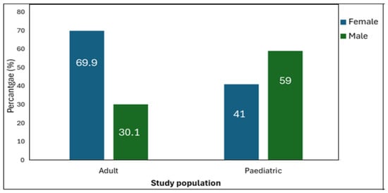
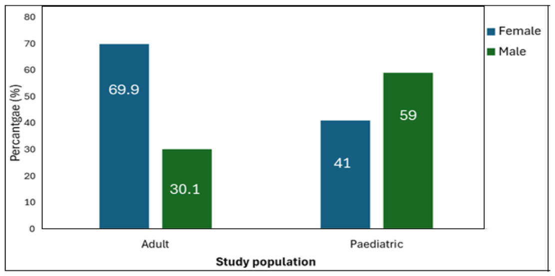
Table 1 presents the demographic profile of the participants with skin conditions. The median age of the 532 participants was 31 years (IQR: 10–51), ranging from 20 days (0.055 years) to 92 years. Females had a higher median age than males (34 years [IQR: 19–52] vs. 19 years [IQR: 6–47]). Overall, females accounted for 61.5% of the study population (n = 327; 95% CI: 57.3–65.5), yielding a female-to-male ratio of 1.6:1. Adults (≥13 years) represented 70.7% of the study population (n = 376; 95% CI: 66.7–74.4), whereas paediatric patients accounted for 29.3% (n = 156; 95% CI: 25.6–33.3). The median age was 5 years (IQR: 2–8) in the paediatric group and 41 years (IQR: 30–57) in the adult group. Within the paediatric population, median age was comparable between females (4 years [IQR: 2–8.5]) and males (5 years [IQR: 2–7.5]). Among adults, male participants had a slightly higher median age than females (44 years [IQR: 26–64] vs. 40 years [IQR: 30–55]). There were significantly (p < 0.001) more females in the adult population than in the paediatric population (69.9% vs. 41.0%), whereas males predominated in the paediatric population compared to the adult population (59.0% vs. 30.1%) (Figure 1).
Table 1.
Demographic distribution of patients admitted to dermatology consultations by age (n = 532).
Figure 1.
Distribution of demographic attributes across adult and paediatric studied populations.
3.2. Frequency and Distribution of Various Skin Diseases Among 532 Patients Attending the Dermatology Outpatient Department
Table 2 presents the distribution of dermatological conditions among the 532 patients stratified by age group. Overall, among the 532 unique patients evaluated, 517 (97.2%) presented with a single dermatosis, 13 (2.4%) presented with two concurrent dermatoses, and 2 (0.4%) presented with three concurrent dermatoses, yielding a total of 549 distinct clinical diagnoses (388 among adults, 161 among children). Across the entire cohort, atopic dermatitis was the most frequent primary diagnosis (27.8%; n = 148/532), followed by acne vulgaris (10.2%; n = 54/532; or 15.0%, n = 80/532 when combined with nodulocystic acne, n = 26). Among children aged 0–12 years (N = 156 patients; 161 total diagnoses), atopic dermatitis was the most common diagnosis (n = 87, affecting 55.8% of paediatric patients), followed by tinea capitis (n = 17; 10.9%), seborrheic dermatitis (n = 14; 9.0%), vitiligo (n = 7; 4.5%), papular urticaria (n = 6; 3.8%), and haemangioma (n = 5; 3.2%). Staphylococcal scalded skin syndrome was identified in one paediatric case (0.6%).
Table 2.
Frequency and distribution of dermatological conditions among paediatric (N = 156) and adult (N = 376) dermatology outpatient attendees.
Among patients aged ≥13 years, the spectrum of dermatological conditions was considerably more heterogeneous. Atopic dermatitis remained the most frequent diagnosis (16.2%), followed by acne vulgaris (14.1%), nodulocystic acne (6.9%), tinea capitis (5.9%), psoriasis (3.7%), photosensitive dermatitis (3.7%), other dermatoses (3.2%), lichen planus (2.9%), papular urticaria (2.9%), discoid eczema (2.7%), and seborrheic dermatitis (2.7%). Most of the remaining dermatological conditions, including autoimmune diseases, vascular disorders, infectious dermatoses, drug reactions, and cutaneous neoplasms, were observed in fewer than 2% of patients each. Several conditions, including Kaposi’s sarcoma, mycosis fungoides, melanoma, discoid lupus erythematosus, bullous disorders, and toxic epidermal necrolysis, were identified exclusively in the older age group, although each occurred at a low frequency.
3.3. Age- and Sex-Specific Distribution of Dermatosis Categories and Individual Dermatological Disorders
The distribution of major dermatosis categories according to age group and sex is presented in Table 3. Inflammatory dermatoses were the predominant diagnostic category across all demographic groups, affecting 71.8% of paediatric patients and 71.3% of adults (p = 0.988), and 73.7% of females and 67.8% of males (p = 0.172). Infectious dermatoses were significantly more frequent in paediatric patients than adults (17.9% vs. 10.9%, p = 0.039) and in males than females (21.5% vs. 7.6%, p < 0.001). Autoimmune dermatoses occurred significantly more often among females than males (8.9% vs. 3.4%, p = 0.024), whereas no age-related difference was observed (6.9% vs. 6.4%, p = 0.983). Neoplastic dermatoses were identified exclusively among adults (4.3%), resulting in a significant age-group difference (p = 0.005), but showed no sex-related variation (p = 0.861). Similarly, vascular dermatoses, drug reactions, and other dermatoses were infrequent and did not differ significantly by age or sex (all p > 0.05).
Table 3.
Distribution of major dermatosis categories according to age group and sex.
At the disease level, distinct age- and sex-specific patterns were observed (Supplementary Table S1). Atopic dermatitis predominated in paediatric patients (55.8%), whereas acne vulgaris (14.1%) and nodulocystic acne (6.9%) were characteristic of adults. Infectious disorders such as tinea capitis were more common in children (10.9% vs. 5.9%), while neoplastic and several chronic inflammatory disorders, including Kaposi’s sarcoma, mycosis fungoides, psoriasis, discoid lupus erythematosus, and pyoderma gangrenosum, were observed almost exclusively in adults. By sex, acne vulgaris was more frequent in females (16.2% vs. 0.5%), whereas atopic dermatitis (37.6% vs. 21.7%), tinea capitis (14.6% vs. 2.8%), nodulocystic acne (8.3% vs. 2.8%), and Kaposi’s sarcoma (2.4% vs. 0.3%) predominated in males.
3.4. Frequency and Distribution of Systemic Comorbidities Among Dermatology Attendees
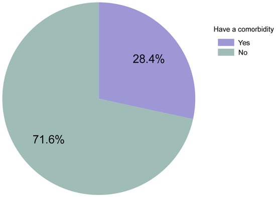
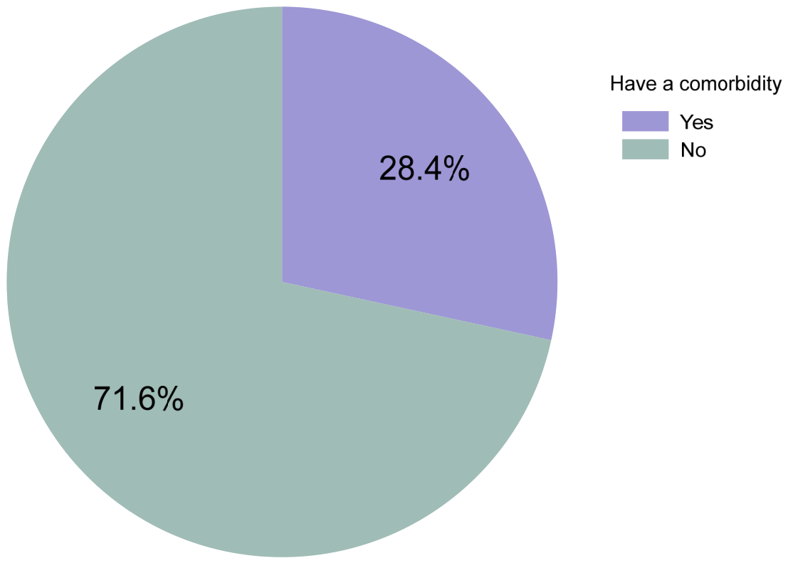
Figure 2 illustrates the overall distribution of systemic diseases among the study population. Overall, documented systemic comorbidities were present in 28.4% of the cohort (n = 151/532; 95% CI: 24.7–32.3). Systemic comorbidities were documented in 38.0% of adults (n = 143/376; 38.0% of adult cohort) compared with only 5.1% of children (n = 8/156; 5.1% of paediatric cohort; p < 0.001). Among the 143 adult patients with comorbidities, HIV/AIDS was the most frequent condition (34.3%, n = 49; representing 13.0% of all 376 adult patients), followed by hypertension (28.0%, n = 40; 10.6% of all adults), diabetes mellitus (11.9%, n = 17; 4.5% of all adults), tuberculosis (11.9%, n = 17; 4.5% of all adults), systemic lupus erythematosus (9.1%, n = 13; 3.5% of all adults), allergic rhino-conjunctivitis (7.0%, n = 10; 2.7% of all adults), and asthma (6.3%, n = 9; 2.4% of all adults). Combined with 4 paediatric cases, total tuberculosis cases equaled 21 (13.9% of all 151 comorbidity cases; 3.9% of the overall cohort). Among the eight paediatric attendees with systemic comorbidities (Table 4), tuberculosis was the most frequently reported condition (50.0%), followed by allergic rhino-conjunctivitis (37.5%) and systemic lupus erythematosus (12.5%).
Figure 2.
Proportion of participants with systemic diseases (N = 532).
Table 4.
Comorbidities observed in the paediatric and adult populations.
Additionally, in adults, breast fibroadenoma accounted for 2.8% of comorbidities, while bipolar mood disorder and cerebral palsy each represented 2.1%. The remaining systemic comorbidities, including allergic sinusitis, cataract, left eye keratosis, stroke, epilepsy, iron deficiency anemia, liver injury, end-stage renal disease, and rheumatoid arthritis, were each reported in 1.4% of adults with comorbidities. Several other conditions, namely dyslipidemia, visual impairment, varicose veins, deep vein thrombosis, lupus nephritis, chronic obstructive airway disease, ulcer, hypothyroidism, leucocytosis, arthritis, chronic hepatitis, psoriatic arthritis, and major depressive disorder, were each observed in 0.7% of adults with comorbidities.
3.5. Association of Comorbidity Status with Demographic Characteristics and Dermatosis Categories
Table 5 presents the distribution of comorbidity status (N = 151 Yes; N = 381 No; Total N = 532) across demographic characteristics and major dermatosis categories. Comorbidity frequency differed significantly by age group (p < 0.001), occurring in 38.0% of adults (n = 143/376) compared to 5.1% of children (n = 8/156). No significant difference was observed by sex (29.4% in females vs. 26.8% in males, p = 0.596). Among patients with infectious dermatoses (N = 69), 15.9% (n = 11) had a documented comorbidity compared to 30.2% (n = 140/463) among those without infectious dermatoses (p = 0.016). Patients presenting with neoplastic dermatoses (N = 16) had a significantly higher comorbidity frequency (56.3%; n = 9/16) than non-neoplastic cases (27.5%; n = 142/516; p = 0.017). No statistically significant bivariate associations were observed between comorbidity presence and inflammatory (p = 0.536), autoimmune (p = 0.686), vascular (p = 0.312), drug reaction (p = 0.441), or other dermatosis categories (p = 0.988).
Table 5.
Distribution of comorbidity status across demographic variables and dermatosis categories (N = 532).
Across systemic diseases, females comprised 63.6% (n = 96) of participants with systemic diseases (Supplementary Table S2). HIV/AIDS showed a similar sex distribution, whereas hypertension, diabetes mellitus, systemic lupus erythematosus, and asthma were more frequent in females, and tuberculosis predominated in males. Most remaining systemic diseases occurred infrequently and exhibited sex-specific distributions.
In the multivariable logistic regression analysis, the presence of comorbidity was significantly associated with age and sex (Table 6). Adult patients had increased odds of comorbidity compared with paediatric patients (adjusted odds ratio [aOR] = 23.4, 95% CI: 8.8–62.0; p < 0.001). Male patients had more than twice the odds of having a comorbidity compared with females (aOR = 2.2, 95% CI: 1.3–3.7; p = 0.005).
Table 6.
Factors independently associated with the presence of comorbidity.
4. Discussion
This study provides an in-depth overview of the dermatoses observed in a rural dermatology department in South Africa, highlighting the epidemiological characteristics of an underserved population. Skin diseases are one of the main contributors to the global burden of non-fatal diseases, accounting for approximately 9.3 million disability-adjusted life years (DALYs) [3]. Against this global background, the present study offers valuable insight into the pattern of skin diseases encountered among patients referred to a tertiary dermatology service in the Eastern Cape. Importantly, as a hospital-based study conducted at a single tertiary center, these observations reflect tertiary clinic attendance patterns rather than true population-level morbidity in the community.
Regarding the overall prevalence of dermatological conditions, inflammatory dermatoses predominated (71.43%) in this cohort, followed by infectious dermatoses (12.97%). The most common diagnoses were atopic dermatitis (35.9%) and acne vulgaris (14.9%). This pattern is consistent with trends reported in several regions worldwide, in both low- and high-income countries. In this cohort, inflammatory dermatoses predominated (71.4%), followed by infectious dermatoses (13.0%). Across the entire study population, atopic dermatitis was the single most common primary diagnosis (27.8%, n = 148/532), followed by acne vulgaris (10.2%, n = 54/532; expanding to 15.0%, n = 80/532 when including nodulocystic acne). This observed predominance of inflammatory skin disorders aligns with clinical patterns documented in sub-Saharan African tertiary settings [15,16,17], though the relative frequency of inflammatory disease in this tertiary cohort exceeds figures reported in community-based surveys in Turkey (23%) [18] and Cameroon (15.7%) [17]. Conversely, the relatively low prevalence of infectious dermatoses observed in the Eastern Cape contrasts sharply with the substantially higher rates reported in other settings, including 33.25% in Brazil by Pinheiro-Dantas et al. [19], 36.1% in Tanzania by Muki et al. [15], and 72.3% in Nigeria by Kalu et al. [20]. These differences could be explained by geoclimatic variations: drier, more temperate areas, such as the Eastern Cape, tend to favor inflammatory skin conditions (eczema), while tropical and humid regions, such as Cameroon or Tanzania, are more conducive to skin infections, promoting microbial proliferation and an increase in infectious dermatoses [21]. In addition, structural barriers to accessing care and delays in seeking consultation in rural areas can contribute to more advanced or chronic presentations of inflammatory diseases [22].
A marked heterogeneity of dermatological conditions was observed across age groups. Children mainly presented with inflammatory and infectious dermatoses, with a high prevalence of atopic dermatitis (55.8%). This observed distribution is consistent with biological hypotheses regarding skin barrier development, immune maturation, and environmental exposures in childhood [23]. The slight male predominance among paediatric attendees (59.0%) may reflect sex-related differences in the distribution of specific dermatoses (such as atopic dermatitis, tinea capitis, seborrheic dermatitis, and vitiligo) or healthcare-seeking patterns. Similar sex-related differences have been reported by Katibi et al. [24] in KwaZulu-Natal, South Africa, although the underlying mechanisms remain uncertain.
In contrast, adult patients exhibited a broader and more heterogeneous spectrum of dermatological disorders. In addition to inflammatory diseases (71.3%), adults frequently presented with infectious, autoimmune, neoplastic, and hormonal/drug reaction conditions, including atopic dermatitis (16.2%), acne vulgaris (14.1%), nodulocystic acne (6.9%), photosensitive dermatitis (3.7%), psoriasis (3.7%), and tinea capitis (5.9%). These observed patterns align with established clinical literature describing age-associated changes in disease spectrum, which may be influenced by underlying hormonal shifts and immune aging [25], as well as cumulative environmental or occupational exposures [26]. The persistence of atopic dermatitis into adulthood also confirms the documented chronicity of this disease [27]. Less frequent diagnoses included pityriasis rubra pilaris and pityriasis versicolor (each n = 1; 0.3%) (Table 2). Pityriasis rosea was not documented as a distinct diagnosis in this cohort. This acute, self-limiting papulosquamous eruption has been associated with human herpesvirus 6 and 7 reactivation, and recurrence, although uncommon, has been reported [28]. Its frequency in the present setting cannot be determined from the available records. More broadly, the retrospective design and reliance on recorded diagnoses may have limited the capture of transient or atypical dermatoses. These considerations highlight the importance of accurate clinical documentation and diagnostic differentiation among papulosquamous and superficial fungal disorders in dermatological surveillance.
The demographic characteristics of the study population showed notable differences by sex. Women accounted for 61.5% of the study population, a trend consistent with observations from dermatology departments internationally [29,30,31]. The predominance of women among adults (69.9%) may reflect greater healthcare-seeking behaviour, increased awareness of skin disorders with cosmetic implications, and the higher prevalence of autoimmune and hormone-related dermatoses in females [32]. Indeed, several dermatological conditions were significantly more common among women, including connective tissue diseases (particularly discoid lupus erythematosus), pigmentary disorders (melasma), cutaneous lymphomas (mycosis fungoides), neutrophilic dermatoses (pyoderma gangrenosum), papulosquamous disorders (psoriasis and lichen planus), hypersensitivity reactions (papular urticaria), and adnexal disorders (acne vulgaris). The female predominance among adult attendees may reflect differences in healthcare-seeking behavior and cosmetic awareness, as well as potential biological and hormonal influences on specific autoimmune, inflammatory, and adnexal disorders [33,34]. However, these mechanisms remain hypothetical, as the study did not directly assess hormonal factors, biological susceptibility, or healthcare-seeking patterns. Additionally, infectious dermatoses were more frequent among male than female patients (21.5% vs. 7.6%, p < 0.001). Potential explanations, including differences in occupational or environmental exposures, outdoor activities, hygiene practices, and skin physiology, warrant further investigation [35,36]. Prospective studies incorporating relevant exposure and behavioral data are needed to clarify these associations. Although females predominated overall, males exhibited significantly higher rates of infectious dermatoses (21.46%) than females (7.65%). This disparity may be explained by greater occupational and environmental exposures in rural settings, differences in hygiene practices, and sex-related variations in skin barrier function and immune responses [37]. Likewise, the slightly higher proportions of inflammatory, infectious, and neoplastic dermatoses observed among males may be associated with increased occupational hazards and cumulative ultraviolet radiation exposure [38], although these differences did not reach statistical significance except for infectious dermatosis (p < 0.001).
An important objective of this study was to describe the systemic diseases associated with dermatological conditions. Systemic comorbidities were documented in 28.4% (151/532) of participants and were substantially more frequent among adults than children (38.0% vs. 5.1%). Among the 143 adults with comorbidities, HIV was the most common condition (34.3%, n = 49), followed by hypertension (28.0%, n = 40), diabetes mellitus (11.9%, n = 17), and tuberculosis (11.9%, n = 17). Including four paediatric cases, tuberculosis affected 21 patients, accounting for 13.9% of those with documented comorbidities. This profile reflects the coexistence of HIV and tuberculosis alongside non-communicable diseases in the Eastern Cape [39,40,41]. Cutaneous manifestations such as Kaposi’s sarcoma and herpes zoster underscore the diagnostic importance of skin findings in patients with HIV-associated immune dysfunction [42,43]. The observed predominance of inflammatory dermatoses among patients with HIV may be related to chronic immune dysregulation and immune reconstitution during antiretroviral therapy [44,45], whereas tuberculosis-related skin manifestations may involve cell-mediated immune responses, hypersensitivity reactions, or adverse effects of treatment [41]. Diabetes-associated hyperglycemia may impair immune function and increase susceptibility to fungal infections [46], while vascular dysfunction, inflammation, and coexisting conditions may contribute to cutaneous morbidity among patients with diabetes or hypertension [47,48]. However, the observed associations between comorbidity and infectious or neoplastic dermatoses were largely confounded by age group and sex.
These findings underscore the need for strengthening integrated approaches that combine dermatology, HIV care, chronic disease management, and systematic screening for comorbidities. Developing dermatological skills in primary care, integrating skin screening into HIV care programs, and utilizing teledermatology are promising strategies for enhancing access to and continuity of care in rural areas [30,49]. In addition, although quality of life was not directly assessed, the prevalence of visible chronic skin conditions (acne, atopic dermatitis, psoriasis, pigmentation disorders) suggests a significant psychosocial burden. Previous data associate these conditions with anxiety, depression, impaired social functioning, and decreased self-esteem [22,50,51], effects that are likely exacerbated in rural areas where psychosocial resources are limited.
5. Limitations
However, several limitations must be acknowledged. First, the single-centre, tertiary-care setting introduces substantial referral bias. As a specialized referral facility, Nelson Mandela Academic Hospital may disproportionately receive patients with severe, chronic, treatment-refractory, or diagnostically complex dermatoses, while mild or self-limiting conditions managed in primary healthcare settings may be underrepresented. Accordingly, the reported findings reflect the frequency and distribution of diagnoses among dermatology attendees rather than population-level prevalence and should not be generalised to the broader Eastern Cape population. Second, the retrospective design relied on routine clinical records, without prospective standardization of diagnostic criteria or systematic laboratory confirmation of all diagnoses. Consequently, diagnostic misclassification and incomplete documentation of comorbidities may have affected the observed disease distribution. Third, multiple comparisons without adjustment increase the risk of type I error. These findings should therefore be interpreted with caution, and future analyses that appropriately account for multiple testing are warranted. Fourth, the study period (August 2020 to July 2021) coincided with the COVID-19 pandemic in South Africa. Pandemic-related lockdown measures, public transport disruptions, and patient reluctance to attend tertiary hospitals likely altered healthcare-seeking behavior, selecting for more severe or acute dermatological presentations while depressing routine outpatient visits. Furthermore, reliance on retrospective chart entries without prospective diagnostic standardization or mandatory laboratory confirmation (e.g., routine histopathology or fungal cultures for all cases) introduces potential misclassification bias. In addition, defining the paediatric cutoff at <13 years, while aligned with NMAH operational triage policies, shifts early adolescent pubertal cases (such as adolescent acne vulgaris) into the adult cohort, influencing age-stratified comparisons. Finally, the retrospective cross-sectional design precludes causal inference; proposed biological, behavioral, and environmental explanations remain hypothetical because the relevant factors were not directly assessed. Prospective, multicentre studies incorporating standardized diagnostic assessments and detailed exposure data are needed to validate these findings and improve their generalisability. Furthermore, the absence of standardized quality-of-life measures and detailed socioeconomic data limited the evaluation of psychosocial burden and the influence of social determinants on dermatological health.
6. Conclusions
This study provides a characterization of dermatological clinical attendance patterns and associated systemic comorbidities at a rural tertiary hospital in the Eastern Cape, South Africa. Inflammatory dermatoses predominated among clinic attendees, with atopic dermatitis and acne vulgaris representing the leading diagnoses, while HIV/AIDS was the most frequent systemic comorbidity among adults. The greater likelihood of comorbidity among male and adult patients highlights the importance of age- and sex-sensitive clinical assessment and screening for coexisting chronic conditions in dermatological practice. Because findings reflect a tertiary hospital cohort, they cannot be generalized to community prevalence in the Eastern Cape. Nevertheless, these findings highlight the need to strengthen integrated dermatological care, enhance primary care diagnostic capacity, and support chronic disease screening in rural healthcare networks. Future prospective studies incorporating longitudinal follow-up, quality-of-life outcomes, socioeconomic determinants, and implementation strategies such as teledermatology and task-shifting are warranted to optimize dermatological care and resource allocation.
Supplementary Materials
The following supporting information can be downloaded at https://www.mdpi.com/article/10.3390/dermato6040042/s1. Table S1: Summary distribution of specific skin conditions by age and sex. Table S2: Distribution of the different systemic diseases across the different types of dermatoses.
Author Contributions
Conceptualization, V.N.-Y., G.T.K., O.O., M.K.-K.N. and A.M.; methodology, V.N.-Y., G.T.K., O.O., M.K.-K.N. and A.M.; software, V.N.-Y., G.T.K., O.O., M.K.-K.N. and A.M.; validation, V.N.-Y., O.O. and A.M.; formal analysis, M.K.-K.N.; investigation, V.N.-Y.; resources, V.N.-Y. and A.M.; data curation, V.N.-Y. and A.M.; writing—original draft preparation, V.N.-Y. and G.T.K.; writing—review and editing, V.N.-Y., G.T.K., O.O., M.K.-K.N. and A.M.; visualization, M.K.-K.N.; supervision, O.O. and A.M.; project administration, V.N.-Y. All authors have read and agreed to the published version of the manuscript.
Funding
This research received no external funding.
Institutional Review Board Statement
The study was conducted in accordance with the Declaration of Helsinki and was approved by the Walter Sisulu University Health Sciences Research Ethics and Biosafety Committee (protocol code 059/2022; date of approval 12 February 2024).
Informed Consent Statement
Patient consent was waived because the study involved no direct contact with participants, and data were collected solely through a retrospective record review. Furthermore, all data were anonymized and contained no personal identifiers that could enable participants to be readily identified from the raw dataset.
Data Availability Statement
The dataset analyzed and presented in this study is available upon reasonable request from the corresponding authors.
Acknowledgments
We acknowledge the nursing and administrative staff of the Dermatology Outpatient Department for their assistance in retrieving and sorting patient medical records, which greatly facilitated data collection.
Conflicts of Interest
The authors declare no conflicts of interest.
Abbreviations
The following abbreviations are used in this manuscript:
| HIV | Human Immunodeficiency Virus |
| AIDS | Acquired Immunodeficiency Syndrome |
| WHO | World Health Organisation |
| NMAH | Nelson Mandela Academic Hospital |
| DALYs | Disability-adjusted life years |
References
- Mahfouz, M.S.; Alqassim, A.Y.; Hakami, F.A.; Alhazmi, A.K.; Ashiri, A.M.; Hakami, A.M.; Khormi, L.M.; Adawi, Y.M.; Jabrah, A.A. Common Skin Diseases and Their Psychosocial Impact among Jazan Population, Saudi Arabia: A Cross-Sectional Survey during 2023. Medicina 2023, 59, 1753. [Google Scholar] [CrossRef] [Scilit] [PubMed]
- Flohr, C.; Hay, R. Putting the burden of skin diseases on the global map. Br. J. Dermatol. 2021, 184, 189–190. [Google Scholar] [CrossRef] [Scilit] [PubMed]
- Seth, D.; Cheldize, K.; Brown, D.; Freeman, E.F. Global Burden of Skin Disease: Inequities and Innovations. Curr. Dermatol. Rep. 2017, 6, 204–210. [Google Scholar] [CrossRef] [Scilit] [PubMed]
- Ayalew, T.L.; Feleke, M.G.; Ashager, K.; Eshetu, A. Atopic dermatitis and its associated factors in Ethiopia: A systematic review and meta-analysis. BMC Infect. Dis. 2025, 25, 1651. [Google Scholar] [CrossRef] [Scilit] [PubMed]
- Christensen, E.M.M.; Jemec, G.; Saunte, D.M.L.; Mortensen, O.S. Introducing Social Dermatology. Acta Derm. Venereol. 2025, 105, adv42622. [Google Scholar] [CrossRef] [Scilit] [PubMed]
- WHO. Skin Diseases as a Global Public Health Priority; WHO: Geneva, Switzerland, 2025; p. 5. [Google Scholar]
- Junior, V.H.; Mendes, A.L.; Talhari, C.C.; Miot, H.A. Impact of environmental changes on Dermatology. An. Bras. Dermatol. 2021, 96, 210–223. [Google Scholar] [CrossRef] [Scilit] [PubMed]
- Rensburg, R. Healthcare in South Africa: How Inequity is Contributing to Inefficiency. Available online: https://www.wits.ac.za/news/latest-news/opinion/2021/2021-07/healthcare-in-south-africa-how-inequity-is-contributing-to-inefficiency.html (accessed on 2 February 2021).
- Parker, E.R.; Rosenbach, M.; Davis, M.D.P. The Voice of the American Dermatological Association: 2025 Official Policy Statement on Climate Change. J. Investig. Dermatol. 2025, 145, 1251–1256. [Google Scholar] [CrossRef] [Scilit] [PubMed]
- Sampaio, A.L.; Bressan, A.L.; Vasconcelos, B.N.; Gripp, A.C. Skin manifestations associated with systemic diseases—Part I. An. Bras. Dermatol. 2021, 96, 655–671. [Google Scholar] [CrossRef] [Scilit] [PubMed]
- Morris-Paxton, A.A.; Reid, S.; Ewing, R.G. Primary healthcare services in the rural Eastern Cape, South Africa: Evaluating a service-support project. Afr. J. Prim. Health Care Fam. Med. 2020, 12, e1–e7. [Google Scholar] [CrossRef] [Scilit] [PubMed]
- Clark, K.; Rao, A.; Chen, V.; Mda, P.; Piek, F.; Irudeo, J.; Maharaj, R.; Wallis, L.; Quinn, T.; Hansoti, B. We Need to Target Trauma: A Prospective Observational Study in Eastern Cape Province, South Africa. South Afr. Med. J. 2020, 110, 38–43. [Google Scholar] [CrossRef] [Scilit] [PubMed]
- Wright, A.K.; Swan, R.; Xu, J.; Lwin, S.M.; Azrielant, S.; Chateau, A.; Ede, C.; Sons, J.; Gxolo, Z.; Skenjana, A.; et al. Skin disease in the Eastern Cape (SKINSCAPE): A Global Psoriasis Atlas point prevalence study in rural South Africa. Br. J. Dermatol. 2026, 194, 125–134. [Google Scholar] [CrossRef] [Scilit] [PubMed]
- IBM-Corp. IBM SPSS Statistics for Windows, Version 30.0; [Computer Software]; IBM Corp.: Armonk, NY, USA, 2024.
- Muki, U.; Shayo, G.; Minja, E.; Bakari, M.; Kazema, A.; Lidenge, S.J. Prevalence and impact on quality of life of dermatological disorders and their associated factors among the elderly in urban areas of Dar es Salaam, Tanzania. Sci. Rep. 2025, 15, 26019. [Google Scholar] [CrossRef] [Scilit] [PubMed]
- Dlova, N.C.; Mankahla, A.; Madala, N.; Grobler, A.; Tsoka-Gwegweni, J.; Hift, R.J. The spectrum of skin diseases in a black population in Durban, KwaZulu-Natal, South Africa. Int. J. Dermatol. 2015, 54, 279–285. [Google Scholar] [CrossRef] [Scilit] [PubMed]
- Bissek, A.C.; Tabah, E.N.; Kouotou, E.; Sini, V.; Yepnjio, F.N.; Nditanchou, R.; Nchufor, R.N.; Defo, D.; Dema, F.; Fonsah, J.Y.; et al. The spectrum of skin diseases in a rural setting in Cameroon (sub-Saharan Africa). BMC Dermatol. 2012, 12, 7. [Google Scholar] [CrossRef] [Scilit] [PubMed]
- Bilgili, M.E.; Yildiz, H.; Sarici, G. Prevalence of skin diseases in a dermatology outpatient clinic in Turkey. A cross-sectional, retrospective study. J. Dermatol. Case Rep. 2013, 7, 108–112. [Google Scholar] [CrossRef] [Scilit] [PubMed]
- Dantas, L.D.; Bakos, L.; Balbinot, G.; Drechsler, C.E.; Eidt, L.M. Prevalence of dermatoses in dermatologic evaluation requests from patients admitted to a tertiary hospital for 10 years. An. Bras. Dermatol. 2015, 90, 762–764. [Google Scholar] [CrossRef] [Scilit] [PubMed]
- Kalu, E.I.; Wagbatsoma, V.; Ogbaini-Emovon, E.; Nwadike, V.U.; Ojide, C.K. Age and sex prevalence of infectious dermatoses among primary school children in a rural South-Eastern Nigerian community. Pan. Afr. Med. J. 2015, 20, 182. [Google Scholar] [CrossRef] [Scilit] [PubMed]
- Russo, F.; Zink, A.; Magnolo, N.; Scala, E.; Scala, E. Atopic Dermatitis and Climate: Environmental Stressors and Care Strategies. Dermatol. Ther. 2025, 15, 3479–3493. [Google Scholar] [CrossRef] [Scilit] [PubMed]
- Sanclemente, G.; Burgos, C.; Nova, J.; Hernández, F.; González, C.; Reyes, M.I.; Córdoba, N.; Arévalo, Á.; Meléndez, E.; Colmenares, J.; et al. The impact of skin diseases on quality of life: A multicenter study. Actas Dermosifiliogr. 2017, 108, 244–252. [Google Scholar] [CrossRef] [Scilit]
- Rajma, J.; Ashwath, A.; Nallasivan, S.; Arun, A.C.; Rubini, M.S. Clinico immunological profile of immune-mediated disorders in South Indian children- A prospective observational study. J. Fam. Med. Prim. Care 2025, 14, 2672–2679. [Google Scholar] [CrossRef] [Scilit] [PubMed]
- Katibi, O.S.; Dlova, N.C.; Chateau, A.V.; Mosam, A. The prevalence of paediatric skin conditions at a dermatology clinic in KwaZulu-Natal Province over a 3-month period: Research. South Afr. J. Child Health 2016, 10, 121–125. [Google Scholar] [CrossRef] [Scilit]
- Carmina, E.; Dreno, B.; Lucky, W.A.; Agak, W.G.; Dokras, A.; Kim, J.J.; Lobo, R.A.; Ramezani Tehrani, F.; Dumesic, D. Female Adult Acne and Androgen Excess: A Report from the Multidisciplinary Androgen Excess and PCOS Committee. J. Endocr. Soc. 2022, 6, bvac003. [Google Scholar] [CrossRef] [Scilit] [PubMed]
- Cooper, G.S.; Parks, C.G. Occupational and environmental exposures as risk factors for systemic lupus erythematosus. Curr. Rheumatol. Rep. 2004, 6, 367–374. [Google Scholar] [CrossRef] [Scilit] [PubMed]
- Sen, A.; Chowdhury, S.; Poddar, I.; Bandyopadhyay, D. Inpatient Dermatology: Characteristics of Patients and Admissions in a Tertiary Level Hospital in Eastern India. Indian J. Dermatol. 2016, 61, 561–564. [Google Scholar] [CrossRef] [Scilit] [PubMed]
- Drago, F.; Ciccarese, G.; Rebora, A.; Parodi, A. Relapsing pityriasis rosea. Dermatology 2014, 229, 316–318. [Google Scholar] [CrossRef] [Scilit] [PubMed]
- Sattar, M.; Sumsuzzoha, S.; Zaman, S.; Das, A. The Pattern of Skin Diseases in Patients Attending OPD of Dermatology and Venereology in a Tertiary Care Hospital in Bangladesh. Ann. Int. Med. Dent. Res. 2022, 8, 270–277. [Google Scholar]
- Alahi, M.; Hossain, M.; Mohammad, A. Pattern of Skin Diseases in Patients Attending OPD of Dermatology and Venereology in a Tertiary Care Hospital in Bangladesh. J. Natl. Inst. Neurosci. Bangladesh 2018, 4, 116–122. [Google Scholar] [CrossRef] [Scilit]
- Yousuf, A.K.M.; Joarder, Y.; Aktar, A.; Hoq, M.N.; Rahman, M.; Akter, F.; Begum, H.; Akhtar, M.R.; Rahman, A.K.M.S. Pattern of skin diseases in patients attended OPD of Dermatology and Venereology in a tertiary care teaching hospital in Bangladesh. Bangladesh J. Physiol. Pharmacol. 2014, 29, 8–11. [Google Scholar] [CrossRef] [Scilit]
- Fougerousse, A.C.; Alexandre, M.; Darrigade, A.S.; Merhand, S.; Marquié, A.; Hamza, M.; Le Fur, G.; Jachiet, M.; Bursztejn, A.C.; Taieb, C. Impact of Atopic Dermatitis on Adult Women’s Lives: A Survey of 1,009 French Women. Acta Derm. Venereol. 2024, 104, adv10321. [Google Scholar] [CrossRef] [Scilit] [PubMed]
- Aman, S.; Nadeem, M.; Mahmood, K.; Ghafoor, M.B. Pattern of skin diseases among patients attending a tertiary care hospital in Lahore, Pakistan. J. Taibah Univ. Med. Sci. 2017, 12, 392–396. [Google Scholar] [CrossRef] [Scilit] [PubMed]
- Pan, C.; Zhao, A.; Li, M. Atopic Dermatitis-like Genodermatosis: Disease Diagnosis and Management. Diagnostics 2022, 12, 2177. [Google Scholar] [CrossRef] [Scilit] [PubMed]
- Chen, W.; Mempel, M.; Traidl-Hofmann, C.; Al Khusaei, S.; Ring, J. Gender aspects in skin diseases. J. Eur. Acad. Dermatol. Venereol. 2010, 24, 1378–1385. [Google Scholar] [CrossRef] [Scilit] [PubMed]
- Lagacé, F.; D’Aguanno, K.; Prosty, C.; Laverde-Saad, A.; Cattelan, L.; Ouchene, L.; Oliel, S.; Genest, G.; Doiron, P.; Richer, V.; et al. The Role of Sex and Gender in Dermatology—From Pathogenesis to Clinical Implications. J. Cutan. Med. Surg. 2023, 27, Np1–Np36. [Google Scholar] [CrossRef] [Scilit] [PubMed]
- Chen, S.J.; Chen, S.; Wang, Y. Global Burden of Fungal Skin Diseases and Future Projections: An Analysis of the 2021 Global Burden of Disease Study. Mycoses 2025, 68, e70134. [Google Scholar] [CrossRef] [Scilit] [PubMed]
- Merin, K.A.; Shaji, M.; Kameswaran, R. A Review on Sun Exposure and Skin Diseases. Indian J. Dermatol. 2022, 67, 625. [Google Scholar] [CrossRef] [Scilit] [PubMed]
- Ugwu, C.L.J.; Ncayiyana, J.R. Spatial disparities of HIV prevalence in South Africa. Do sociodemographic, behavioral, and biological factors explain this spatial variability? Front. Public Health 2022, 10, 994277. [Google Scholar] [CrossRef] [Scilit] [PubMed]
- Ochonye, B.; Sanni, O.F.; Emmanuel, G.; Umoh, P.; Kalaiwo, A.; Abang, R.; Amechi, P.; Ahkigbe, M.; Akinpelumi, S.; Motilewa, O. A retrospective study of tuberculosis prevalence and associated factors among HIV-positive key populations in Nigeria. PLoS Glob. Public Health 2024, 4, e0003461. [Google Scholar] [CrossRef] [Scilit] [PubMed]
- Valderas, J.M.; Starfield, B.; Sibbald, B.; Salisbury, C.; Roland, M. Defining comorbidity: Implications for understanding health and health services. Ann. Fam. Med. 2009, 7, 357–363. [Google Scholar] [CrossRef] [Scilit] [PubMed]
- Mohseni Afshar, Z.; Goodarzi, A.; Emadi, S.N.; Miladi, R.; Shakoei, S.; Janbakhsh, A.; Aryanian, Z.; Hatami, P. A Comprehensive Review on HIV-Associated Dermatologic Manifestations: From Epidemiology to Clinical Management. Int. J. Microbiol. 2023, 2023, 6203193. [Google Scholar] [CrossRef] [Scilit] [PubMed]
- Mazibuko, M.; Dlova, N.C. The Clinical Spectrum and Outcome of Skin Conditions in Patients Admitted to Dermatology Wards at King Edward VIII Hospital, Durban. Master’s Thesis, University of KwaZulu-Natal, Durban, South Africa, 2016. [Google Scholar]
- Chimbetete, T.; Buck, C.; Choshi, P.; Selim, R.; Pedretti, S.; Divito, S.J.; Phillips, E.J.; Lehloenya, R.; Peter, J. HIV-Associated Immune Dysregulation in the Skin: A Crucible for Exaggerated Inflammation and Hypersensitivity. J. Investig. Dermatol. 2023, 143, 362–373. [Google Scholar] [CrossRef] [Scilit] [PubMed]
- Motswaledi, H.M. Common dermatological conditions in the HIV patient. South Afr. Fam. Pract. 2019, 61, S19–S24. [Google Scholar] [CrossRef] [Scilit]
- Dorf, N.; Maciejczyk, M. Skin manifestations in diabetes-what is new? Front. Med. 2025, 12, 1640144. [Google Scholar] [CrossRef] [Scilit] [PubMed]
- Linder, D.; Dreiher, J.; Zampetti, A.; Sampogna, F.; Cohen, A.D. Seborrheic dermatitis and hypertension in adults: A cross-sectional study. J. Eur. Acad. Dermatol. Venereol. JEADV 2013, 28, 1450–1455. [Google Scholar] [CrossRef] [Scilit] [PubMed]
- Nguyen, K.H.; Alcantara, C.A.; Glassman, I.; May, N.; Mundra, A.; Mukundan, A.; Urness, B.; Yoon, S.; Sakaki, R.; Dayal, S.; et al. Cutaneous Manifestations of Mycobacterium tuberculosis: A Literature Review. Pathogens 2023, 12, 920. [Google Scholar] [CrossRef] [Scilit] [PubMed]
- Hu, J.; McKoy, K.; Papier, A.; Klaus, S.; Ryan, T.; Grossman, H.; Masenga, E.J.; Sethi, A.; Craft, N. Dermatology and HIV/AIDS in Africa. J. Glob. Infect. Dis. 2011, 3, 275–280. [Google Scholar] [CrossRef] [Scilit] [PubMed]
- Yew, Y.W.; Kuan, A.H.Y.; Ge, L.; Yap, C.W.; Heng, B.H. Psychosocial impact of skin diseases: A population-based study. PLoS ONE 2020, 15, e0244765. [Google Scholar] [CrossRef] [Scilit] [PubMed]
- Costeris, C.; Petridou, M.; Ioannou, Y. Psychological Impact of Skin Disorders on Patients’ Self-esteem and Perceived Social Support. Dermatol. Skin. Sci. 2021, 3, 14–22. [Google Scholar]

Disclaimer/Publisher’s Note: The statements, opinions and data contained in all publications are solely those of the individual author(s) and contributor(s) and not of MDPI and/or the editor(s). MDPI and/or the editor(s) disclaim responsibility for any injury to people or property resulting from any ideas, methods, instructions or products referred to in the content. |
© 2026 by the authors. Licensee MDPI, Basel, Switzerland. This article is an open access article distributed under the terms and conditions of the Creative Commons Attribution (CC BY) license.