


Abstract
Phimosis, the inability to retract the prepuce over the glans penis, is one of the most common reasons for referral of boys to pediatric surgeons and urologists, yet the great majority of cases represent a normal developmental stage rather than true disease. Distinguishing physiological non-retractility, which resolves spontaneously in almost all boys by adolescence, from pathological phimosis caused by scarring or balanitis xerotica obliterans (lichen sclerosus) is fundamental to rational management. Over the past three decades, topical corticosteroid therapy has emerged as an effective, inexpensive, and low-risk first-line alternative to circumcision for symptomatic phimosis. Pooled data from randomized controlled trials and the 2024 Cochrane systematic review indicate that corticosteroids applied to the stenotic preputial ring for four to eight weeks probably increase complete or partial resolution of phimosis compared with placebo, although the overall certainty of this evidence is limited by heterogeneity in the age of participants, baseline severity, corticosteroid preparation, and treatment duration. Individual studies report resolution in approximately 65% to 95% of treated boys, and treatment avoids the costs, anesthetic exposure, and complications of surgery. Betamethasone 0.05–0.1% is the most extensively studied agent, but clobetasol, mometasone, fluticasone, triamcinolone, and hydrocortisone have all been used successfully. In the relatively small studies available, measurable systemic absorption and suppression of the hypothalamic–pituitary–adrenal axis have not been detected at the doses and durations used, and local adverse effects are rare and reversible, although rare systemic or local effects cannot be entirely excluded, particularly with ultrapotent preparations or prolonged use. Recurrence after successful treatment is uncommon and generally responds to a repeat course. This narrative review synthesizes the anatomy and natural history of the prepuce, the classification and etiology of phimosis, and the mechanism, efficacy, safety, dosing, and cost-effectiveness of topical corticosteroids, and situates them within current international guideline recommendations and the specific role of circumcision in scarred and lichen sclerosus-associated disease.
1. Introduction
Phimosis, derived from the Greek word for “muzzling,” denotes the inability to fully retract the prepuce (foreskin) over the glans penis [1,2]. It is among the most frequent reasons for the referral of boys to primary-care physicians, pediatric surgeons, and urologists, and it remains a leading indication for medically motivated circumcision [3,4]. Because the condition provokes considerable parental anxiety and generates a large volume of consultations, it carries a clinical and economic burden that is disproportionate to its usually benign nature [2,5].
The central concept in the rational management of phimosis is the distinction between physiological and pathological forms [6]. In the great majority of boys, a non-retractile foreskin in infancy and early childhood reflects a normal developmental stage in which the inner preputial epithelium remains fused to the glans, rather than any disease process, and it resolves spontaneously with age in almost all cases [6,7,8]. True pathological phimosis, characterized by a scarred, fibrotic, non-distensible preputial ring, is comparatively uncommon and is most often caused by balanitis xerotica obliterans (lichen sclerosus) [9,10]. Failure to recognize this distinction has historically led to a substantial number of unnecessary circumcisions in boys whose foreskins were developmentally normal [8,11].
Circumcision, one of the oldest and most commonly performed surgical procedures, has traditionally been regarded as the definitive treatment for persistent phimosis [1]. However, it requires anesthesia, incurs financial cost, and carries a measurable rate of complications, approximately 1.5% in neonates and up to 6% in older children, ranging from bleeding and infection to meatal stenosis and, rarely, catastrophic glans injury [12,13]. These considerations have motivated the search for less invasive, foreskin-preserving alternatives [14].
Since the early 1990s, the topical application of corticosteroid preparations to the stenotic preputial ring has emerged as an effective, inexpensive, and low-risk conservative treatment for phimosis in boys [15,16]. A growing body of randomized controlled trials, cohort studies, cost-effectiveness analyses, and systematic reviews now supports this approach, and contemporary international guidelines recommend topical corticosteroids as first-line therapy for symptomatic phimosis, reserving circumcision for scarred or lichen sclerosus-associated disease [1,17,18]. This narrative review synthesizes the anatomy and natural history of the prepuce, the classification and etiology of phimosis, and the mechanism of action, efficacy, dosing, safety, and cost-effectiveness of topical corticosteroid therapy, and situates these within current evidence-based practice [1,19].
2. Search Strategy and Evidence Selection
This work is a narrative review. Relevant literature was identified through searches of PubMed/MEDLINE, the Cochrane Library, Embase, and Scopus for articles published between 1990 and 2025. The principal search terms combined phimosis, prepuce, foreskin, and non-retractile foreskin with topical corticosteroid, betamethasone, clobetasol, mometasone, fluticasone, triamcinolone, and hydrocortisone, and with balanitis xerotica obliterans, lichen sclerosus, circumcision, and cost-effectiveness. Reference lists of key articles, systematic reviews, and current clinical guidelines were also screened for additional sources. Priority was given to randomized controlled trials, systematic reviews, and meta-analyses, in particular the 2014 and 2024 Cochrane reviews. Only articles available in English were considered. Because this is a narrative rather than a systematic review, no formal risk-of-bias assessment or quantitative pooling of data was undertaken.
3. Anatomy and Normal Development of the Prepuce
The prepuce is a specialized, double-layered fold of skin and mucosa that covers the glans penis. Its development begins around the eighth week of gestation and is essentially complete by the sixteenth week [7]. At birth, the inner surface of the prepuce and the epithelium of the glans share a common squamous lining, so that the two structures are physiologically fused. Separation occurs gradually over months to years through a process of epithelial desquamation and keratinization that forms epithelial cell nests, which coalesce into a continuous preputial space [6,7]. Consequently, almost all normal male neonates are born with a non-retractile foreskin, and preputial adhesions and a physiologically tight preputial orifice are the rule rather than the exception in this age group [7,8].
Longitudinal and cross-sectional studies have repeatedly documented the gradual, age-dependent acquisition of preputial retractability [6,8]. In his classic study, Gairdner observed that only about 4% of newborns had a fully retractable prepuce [7]. Kayaba and colleagues, examining 603 Japanese boys, found that the proportion with a completely retractable prepuce rose from 0% at six months to roughly 63% by 11–15 years, while the frequency of a tight preputial ring fell from about 84% to 9% over the same interval [8]. Similarly, Imamura reported that only 38% of three-year-old Japanese boys had a fully retractable foreskin, and Øster documented that the incidence of preputial adhesions declined from around 63–70% at 6–7 years to about 5% by 16–17 years, with physiological phimosis being uncommon in schoolboys and showing a strong tendency to regress spontaneously [6,11]. Age-specific prevalence data from other populations, such as the Taiwanese cohort reported by Ko and colleagues, are concordant with these findings [20].
These data establish that incomplete preputial separation and non-retractility are normal in neonates, infants, and young children, and that the prepuce becomes progressively retractable throughout childhood and adolescence, with the physiological process typically complete by the mid-to-late teenage years [6,8,11]. The reported incidence of persistent phimosis therefore falls with age, to approximately 1% by 16–17 years [6,17]. An important clinical corollary is that asymptomatic physiological adhesions and non-retractility require no intervention, and forcible retraction of the foreskin should be avoided, as it can cause pain, bleeding, and cicatricial scarring that produces iatrogenic (secondary) pathological phimosis [3,8].
4. Definitions and Classification
A recurring source of confusion in both the lay and medical literature is the indiscriminate use of the term “phimosis” to describe both the normal developmental non-retractility of childhood and genuine pathological narrowing of the prepuce [6]. To avoid this, the terms physiological phimosis and pathological phimosis are preferred (Table 1). Physiological phimosis describes a supple, non-scarred, developmentally non-retractile prepuce, frequently accompanied by balanopreputial adhesions, in a boy who has not yet completed the normal process of preputial separation [7,8]. Pathological (true) phimosis describes a prepuce with a distinct, scarred, indurated, and non-distensible fibrotic ring that will not resolve spontaneously and is most commonly the result of balanitis xerotica obliterans [9,10]. A further useful distinction is between primary phimosis, in which the foreskin has never been retractile, and secondary phimosis, in which a previously retractile foreskin becomes narrowed, typically following forcible retraction, recurrent inflammation, or scarring [21].
Table 1.
Distinguishing physiological from pathological phimosis.
Several grading systems have been proposed to standardize the assessment of preputial retractability and to permit comparison of treatment outcomes [8,15]. The scale introduced by Kikiros and colleagues, and its later modifications, grade the prepuce from full, easy retractability at one extreme to complete non-retractility with a pinpoint, cicatricial opening at the other, while Kayaba and colleagues classified preputial morphology into five types according to the extent of glans exposure achievable on gentle retraction [8,15]. Such objective grading is clinically useful because the pre-treatment severity of phimosis has been correlated, albeit inconsistently, with the likelihood of response to topical therapy, and because it allows the response to a course of treatment to be documented reproducibly [22,23].
Clinically, physiological phimosis and simple preputial adhesions must also be differentiated from one another and from a genuinely stenotic ring. When adhesions predominate, partial retraction is usually possible and the preputial orifice itself is of normal caliber, whereas a truly phimotic ring restricts retraction irrespective of adhesions [17]. Ballooning of the foreskin during micturition, intermittent dysuria, and recurrent balanoposthitis are common presenting complaints, but they are frequently manifestations of physiological phimosis and, in the absence of scarring, do not by themselves mandate surgery [8,17].
5. Etiology and Pathophysiology of Pathological Phimosis
The principal causes of acquired pathological phimosis in children are balanitis xerotica obliterans (lichen sclerosus), recurrent balanoposthitis, chronic inflammation, and cicatrization following forcible retraction of a developmentally fused foreskin [9,10,24]. Of these, balanitis xerotica obliterans (BXO) is by far the most important, being the single most common identifiable pathological cause of a scarred, non-retractile prepuce in boys [10,25].
BXO is the male genital variant of lichen sclerosus, a chronic, progressive, sclerosing inflammatory dermatosis of uncertain etiology first described by Stühmer in 1928. Proposed contributory mechanisms include autoimmunity, genetic predisposition, chronic irritation, and infection [24,25]. It characteristically produces whitish, atrophic, sclerotic plaques of the glans and distal prepuce with a firm, pale, cicatricial preputial ring, and it may extend to involve the urethral meatus and cause meatal stenosis [24,26]. Although the diagnosis can often be suspected clinically, histological confirmation remains the reference standard, the characteristic features being epidermal atrophy, hyperkeratosis, basal cell liquefactive degeneration, a homogenized (hyalinized) upper dermis, and a subjacent band of lymphocytic infiltrate [9,27]. Clinical examination alone underestimates its incidence, particularly in boys under five years of age, in whom clinical over-diagnosis is also common [28].
Histological examination of preputial specimens obtained at circumcision for medically indicated phimosis reveals evidence of BXO in a substantial proportion of cases, reported at approximately 35% or higher overall in some series, and around 17% in boys younger than ten years, with a peak incidence between about 9 and 11 years of age [10,17,25]. An association between BXO and childhood obesity has also been described [29]. Because genital lichen sclerosus is associated in adult life with an elevated risk of penile squamous cell carcinoma, and because some immunohistochemical studies in adults have raised the possibility of premalignant change, the accurate identification and follow-up of affected boys is clinically important, although whether the malignant risk described in adult men also applies to childhood disease is not established [27,30].
The pathophysiology by which the physiological preputial ring normally loosens involves a combination of progressive histological remodeling, hormonal influences, intermittent erections, and mechanical stretching, and interference with these processes, most notably repeated forcible retraction, can convert a normal developmental state into secondary cicatricial phimosis [17,21]. Structural analyses of phimotic prepuces have demonstrated alterations in the collagen and elastic-fiber architecture of the resistant preputial ring, providing a histological rationale for pharmacological approaches that modify connective-tissue composition and inflammation [22,31].
6. Clinical Presentation and Diagnosis
Most boys with a non-retractile foreskin are asymptomatic, and the finding is often noted incidentally by parents or at routine examination [6,8]. When symptoms occur, they commonly include ballooning of the prepuce during micturition, intermittent dysuria or spraying of the urinary stream, recurrent balanoposthitis and, in older boys, discomfort on erection [17,32]. Recurrent urinary tract infection may occur, particularly in infants and in boys with underlying urinary-tract abnormalities [17,33].
The diagnosis of phimosis is clinical and rests on inspection and gentle attempted retraction of the prepuce, with careful attention to whether a supple physiological orifice or a firm, pale, scarred ring is present [17,28]. The presence of a whitish, indurated, cicatricial preputial ring, or of atrophic white plaques on the glans and meatus, should raise suspicion of balanitis xerotica obliterans, although clinical examination alone lacks sensitivity and specificity, especially in younger children [24,28]. Where the foreskin is excised, histological examination of the specimen is recommended to confirm or exclude lichen sclerosus, given its distinct prognosis and its recognized association with later meatal pathology and, in adults, penile malignancy [25,27,30]. Circumstances that warrant active treatment rather than observation include symptomatic phimosis, suspected lichen sclerosus, recurrent urinary tract infection, and phimosis in boys with vesicoureteral reflux or posterior urethral valves [17].
7. The Treatment Landscape and Its Historical Evolution
Four broad management strategies are available for phimosis in boys, namely watchful waiting with reassurance and hygiene advice, topical medical therapy (principally with corticosteroids), foreskin-preserving surgery such as preputioplasty or dorsal slit, and circumcision [14,32]. For physiological phimosis and asymptomatic preputial adhesions, expectant management is appropriate and is strongly supported by the natural history of spontaneous resolution described above, so that active treatment is reserved for boys who are symptomatic or who have pathological disease [6,8,17].
Circumcision is among the oldest surgical procedures known and, in the English-speaking world, became a common medical intervention during the late nineteenth and twentieth centuries. For much of that period it was regarded as the standard remedy for persistent phimosis [1,13]. The recognition that most childhood non-retractility is physiological, together with growing awareness of the costs and complications of surgery, prompted a re-evaluation of routine circumcision and stimulated interest in conservative alternatives [12,32]. Foreskin-preserving operations such as preputioplasty can relieve a tight ring while retaining the prepuce and yield good outcomes in selected boys, but they still entail anesthesia and operative risk [14,32].
Against this background, topical corticosteroid therapy was introduced in the early 1990s [15]. Early reports by Kikiros and colleagues and by Golubović and colleagues demonstrated that application of a corticosteroid preparation to the phimotic ring could achieve retractability in a substantial majority of boys, and subsequent studies rapidly established topical corticosteroids as a genuine, foreskin-preserving alternative to surgery that can be offered as initial therapy in almost all cases of non-scarred symptomatic phimosis [1,15,16].
8. Mechanism of Action of Topical Corticosteroids on the Prepuce
Topical corticosteroids are lipophilic molecules that diffuse across the keratinocyte cell membrane and bind the cytoplasmic glucocorticoid receptor. The activated receptor complex then translocates to the nucleus, where it modulates gene transcription through both transactivation and transrepression, complemented by more rapid non-genomic effects [34,35]. The net cutaneous actions are conventionally grouped as anti-inflammatory, immunosuppressive, antiproliferative (antimitotic), and vasoconstrictive [34,36].
The anti-inflammatory effect is mediated in part through induction of annexin A1 (lipocortin-1), which inhibits phospholipase A2 and thereby reduces the generation of prostaglandins and leukotrienes, together with suppression of pro-inflammatory cytokine transcription and a reduction in vasodilatation, capillary permeability, and leukocyte infiltration [34,35]. Corticosteroids additionally suppress the maturation, proliferation, and function of immune cells, and, of particular relevance to the prepuce, exert an antiproliferative action that inhibits fibroblast activity and collagen synthesis [34,36].
It is precisely this last property that is therapeutically exploited in phimosis. The resistance of the phimotic ring reflects a combination of chronic low-grade inflammation and an altered collagen and elastic-fiber composition of the distal preputial skin [22,31]. By dampening inflammation and reducing dermal collagen deposition, topical corticosteroids thin the stenotic ring, increase the extensibility and elasticity of the skin and, when combined with gentle retraction, facilitate loosening of the ring and separation of residual balanopreputial adhesions [15,31,37]. In effect, the epidermal and dermal thinning that constitutes an unwanted adverse effect during long-term dermatological use is harnessed as the desired therapeutic mechanism during a short, localized course of treatment [36,38].
This mechanistic rationale also underlies the frequent recommendation to accompany corticosteroid application with a daily program of gentle preputial retraction or “stretching,” and it provides the basis for combination formulations in which hyaluronidase is added to degrade the hyaluronic acid of the dermal ground substance and thereby enhance tissue pliability [39,40,41].
9. Corticosteroid Agents, Potency, and Formulation
A wide range of topical corticosteroids has been used to treat phimosis, spanning the full spectrum of potency classes used in dermatology (Table 2) [36]. Betamethasone, most often as the valerate or dipropionate ester in concentrations from 0.025% to 0.2%, is by far the most extensively studied agent and is the de facto reference treatment in the literature [21,22,40]. The valerate and dipropionate esters should not be regarded as interchangeable, because the potency of a topical corticosteroid depends on the ester, concentration, vehicle, and the classification system used, all of which vary across formulations and jurisdictions. Other agents with published pediatric experience include mometasone furoate 0.1%, the superpotent clobetasol propionate 0.05%, triamcinolone acetonide 0.1%, fluticasone propionate 0.05%, and the low-potency agent hydrocortisone 1% to 2.5% [42,43,44,45].
Table 2.
Topical corticosteroids used in the treatment of phimosis, by potency class. Potency classification is approximate and is not fixed across esters, concentrations, vehicles, or national classification systems.
Comparative data indicate that both moderately potent and highly potent corticosteroids are effective, and there is no consistent evidence that any single molecule is clearly superior for uncomplicated phimosis [23,46]. In a prospective randomized study, highly potent and moderately potent preparations produced comparable resolution rates, and the classification of phimosis grade before treatment was more predictive of outcome than the specific agent chosen [23,47]. A randomized open-label trial found that inexpensive over-the-counter hydrocortisone 1% was not significantly inferior to prescription triamcinolone 0.1%, a finding with obvious implications for access and cost [44].
The choice of agent is more consequential when the underlying pathology is balanitis xerotica obliterans, for which ultrapotent corticosteroids such as clobetasol propionate 0.05% are generally preferred as first-line medical therapy, in keeping with the evidence and guidelines for genital lichen sclerosus rather than by extrapolation from studies of severe non-scarred phimosis [43,48]. When corticosteroids fail or are contraindicated in lichen sclerosus, topical calcineurin inhibitors (tacrolimus, pimecrolimus) have been used as steroid-sparing second-line agents, although comparative studies in genital lichen sclerosus generally favor potent corticosteroids over tacrolimus for efficacy [48,49].
Formulation also influences performance. Ointments are generally more occlusive and potent than creams of the same molecule, and the vehicle affects percutaneous penetration [36]. Finally, some investigators have added hyaluronidase to betamethasone in an attempt to increase tissue pliability, although randomized data suggest that the addition confers little or no advantage over betamethasone alone [40,41].
10. Efficacy and the Evidence Base
Since the first descriptions in the early 1990s, a large and consistent body of evidence has accumulated to support the efficacy of topical corticosteroids for phimosis in boys, comprising numerous prospective series and randomized controlled trials as well as several systematic reviews, meta-analyses, and a network meta-analysis [2,15,19,46]. Taken together, these studies establish that a short course of a topical corticosteroid resolves or improves non-scarred phimosis in the majority of treated boys and thereby avoids circumcision in most cases [1,38].
The most authoritative synthesis is the Cochrane systematic review, first published in 2014 and updated in 2024. The 2014 review pooled twelve randomized trials enrolling 1395 participants and found that, compared with placebo, corticosteroids significantly increased the rate of complete or partial clinical resolution of phimosis [2]. The 2024 update, applying GRADE methodology, concluded that topical corticosteroids probably increase the likelihood of resolution and of complete foreskin retraction relative to placebo and that treatment-site adverse effects are rare, while cautioning that the overall certainty of the evidence is limited by suboptimal reporting, clinical heterogeneity in the age of participants and severity of phimosis, and variation in the corticosteroid and treatment duration used [1].
Individual studies illustrate the consistency of the effect across agents, populations, and eras (Table 3). The seminal report of Kikiros and colleagues demonstrated that local steroid application produced retractability in most treated boys, and the placebo-controlled study of Golubović and colleagues confirmed a clear benefit over vehicle [15,16]. Orsola and colleagues treated 137 boys with 0.05% betamethasone twice daily for one month and reported that 90% had an easily retractable prepuce at six months, while the randomized trial of Monsour and colleagues similarly showed betamethasone to be superior to placebo [21,50]. Large series by Chu and colleagues and by Ashfield and colleagues (194 children) confirmed high success rates when topical steroids were used as primary therapy, and Zavras and colleagues reported successful conservative treatment in approximately 91% of 1185 boys treated with fluticasone propionate 0.05% [37,38,45].
Reported success rates for betamethasone-based regimens generally range from about 65% to over 90%. These reported figures combine different endpoints, including complete retractability, partial improvement, symptomatic relief, and avoidance of circumcision, which are not equivalent and should be interpreted with caution. The pre-treatment severity of phimosis remains the most consistent predictor of response [22,46,47]. Importantly, efficacy is not confined to mild disease. Prospective studies using potent agents such as clobetasol propionate, and long-term cohorts including boys with severe phimosis, have documented substantial rates of resolution even in high-grade cases, albeit with somewhat lower success than in milder phimosis [43,51,52]. Durability has also been examined. An 18-month randomized follow-up study confirmed that resolution achieved with topical steroids is largely maintained over time in most boys, provided that regular retraction and hygiene are continued [53].
Quantitative pooling reinforces these observations. A conventional meta-analysis by Liu and colleagues confirmed a significant benefit of corticosteroids over placebo in achieving resolution, and the network meta-analysis of Sridharan and Sivaramakrishnan compared multiple corticosteroid regimens and again supported their efficacy relative to placebo, while highlighting the limited head-to-head data available to rank individual agents definitively [19,46]. Randomized comparisons that included over-the-counter hydrocortisone further suggest that even low-potency, inexpensive preparations can achieve clinically meaningful improvement, expanding the options available to families and health systems [44].
Table 3.
Selected studies of topical corticosteroid therapy for phimosis in boys.
11. Dosing, Duration, and Application Technique
Although protocols vary, a broadly consistent regimen has emerged from the literature. A thin layer of corticosteroid is applied to the distal, stenotic portion of the prepuce once or twice daily for a course of four to eight weeks [1,21,38]. Comparative data suggest that the specific frequency is not critical, twice-daily and thrice-daily schedules have produced similar results, so that ease of adherence can reasonably guide the choice [55]. For boys with only a partial response at four weeks, extending treatment or repeating the course improves outcomes. A randomized trial found that response rates at 60 days exceeded those at 30 days, indicating that a longer course captures additional responders [40,56].
Application technique matters. Parents are typically instructed to apply the preparation to both the inner and outer aspects of the tight ring, sometimes with gentle massage, and to begin cautious daily retraction after approximately five to seven days of treatment, taking care never to force the foreskin or cause pain or bleeding [21,39]. Adjunctive manual retraction (“preputial stretching”), with or without corticosteroid, contributes to loosening of the ring, and continued gentle retraction and routine hygiene after the treatment course are widely recommended to consolidate and maintain the result [39,53,57].
12. Recurrence and Long-Term Outcomes
Re-stenosis after initially successful treatment is recognized but affects only a minority of boys, and reported recurrence rates are generally low [1,53]. Long-term follow-up studies indicate that most boys who achieve retractability maintain it, particularly when regular retraction during bathing and good hygiene are continued, and recurrences frequently respond to a further course of topical corticosteroid rather than requiring surgery [51,52,53].
The principal exception is phimosis caused by balanitis xerotica obliterans. Because lichen sclerosus is a chronic, progressive, scarring dermatosis, corticosteroids may arrest inflammation and delay or occasionally avert surgery, but scarred disease is substantially less likely to resolve durably with topical therapy alone and commonly progresses to require circumcision [25,43]. Moreover, boys circumcised for lichen sclerosus remain at risk of later meatal stenosis, underscoring the importance of accurate diagnosis, histological confirmation, and continued follow-up in this subgroup [24,26].
13. Special Populations and Clinical Scenarios
Several clinical situations modify the threshold for, and expectations of, topical corticosteroid therapy. In boys with recurrent urinary tract infection or with upper-tract abnormalities such as vesicoureteral reflux or posterior urethral valves, guidelines recommend active treatment of phimosis, and topical corticosteroids offer a non-surgical means of rendering the foreskin retractile and improving preputial hygiene [17,58]. This rationale rests on the well-documented, if debated, association between an uncircumcised state and a higher incidence of infantile urinary tract infection, attributed to periurethral colonization by uropathogens beneath the foreskin [33,59,60,61].
In boys with recurrent balanoposthitis, achieving retractability and improved hygiene can reduce the frequency of inflammatory episodes. However, recurrent balanoposthitis or phimosis refractory to adequate topical therapy should raise suspicion of underlying balanitis xerotica obliterans [17,24].
Boys with balanitis xerotica obliterans constitute a distinct group in whom potent or ultrapotent corticosteroids are used first-line, durable resolution with medical therapy alone is less likely, and close follow-up with histological confirmation is advised [25,43,48]. This vigilance is justified by the recognized, albeit long-delayed, association between genital lichen sclerosus and penile squamous cell carcinoma in adult life, with malignant transformation reported in the region of a few to several percent of affected adults in various series, and by limited evidence, derived largely from adult men, that timely treatment and circumcision may reduce this risk. The magnitude of any malignant risk arising from childhood disease remains undefined [27,62,63,64].
Age also influences management. Although topical corticosteroids are effective even in boys younger than three years, the great majority of non-retractility at this age is physiological and destined to resolve spontaneously, so that treatment should be reserved for boys who are genuinely symptomatic or who have pathological disease, and forcible retraction must be avoided [8,11,65].
14. Safety and Adverse Effects
A consistent theme across the literature is that short, localized courses of topical corticosteroid for phimosis are very well tolerated. Treatment-site adverse effects were reported only rarely in the trials included in the Cochrane review, and large prospective series have documented few if any local complications [1,38].
The theoretical local adverse effects familiar from dermatological practice, epidermal and dermal atrophy, telangiectasia, striae, hypopigmentation, folliculitis, and secondary infection, are a function of steroid potency, quantity, duration, occlusion, and cumulative dose, and are therefore unlikely to develop during the small-area, time-limited courses used for phimosis. Nonetheless, they justify limiting potency and duration to what is necessary [36,66].
Concern about percutaneous absorption and suppression of the hypothalamic–pituitary–adrenal (HPA) axis, which is legitimate for potent steroids applied to large areas under occlusion, has been specifically addressed in the phimosis setting. A dedicated study found no significant suppression of the HPA axis during clinical treatment of phimosis, and a randomized trial using salivary cortisol as a biomarker detected no measurable systemic absorption of topical betamethasone [41,67]. These data provide reassurance that, at the doses and durations used, clinically important systemic toxicity is unlikely in otherwise healthy boys, although the available studies are small and rare systemic effects cannot be entirely excluded, particularly with ultrapotent preparations, prolonged or repeated courses, or incorrect application [1,67].
15. Cost-Effectiveness and Comparison with Circumcision
A further argument in favor of topical corticosteroids is economic. Circumcision is resource-intensive, typically requiring an operating room, general or regional anesthesia, and surgical and nursing time, whereas a course of topical corticosteroid costs only a few units of an inexpensive, widely available medication [5,38].
The formal cost-effectiveness analysis of Berdeu and colleagues compared surgical and medical approaches to phimosis and concluded that initial treatment with a topical corticosteroid is markedly less expensive than primary circumcision, such that a strategy of offering medical treatment first was economically dominant in that analysis [5]. This analysis is now relatively old and was conducted in a single health system, so its cost estimates may not transfer directly to contemporary or other settings. This advantage persists even when the costs of the subset of boys who ultimately require surgery after failed medical therapy are taken into account, because a successful first-line course of corticosteroid averts circumcision in the majority of cases [5,21]. The demonstration that inexpensive over-the-counter hydrocortisone can achieve clinically meaningful improvement offers the prospect of reducing costs still further and of improving access in resource-limited settings [44].
The comparison with circumcision is not, however, purely financial. Conservative treatment preserves the prepuce and its potential functional roles, avoids the operative and anesthetic risks of surgery, including bleeding, infection, meatal stenosis, and, rarely, serious glans injury, and spares the young child the potential psychological impact of genital surgery [12,68]. These considerations, taken together with comparable resolution of symptoms in non-scarred phimosis, underlie the now-widespread recommendation that a trial of topical corticosteroid should precede any decision to circumcise for phimosis alone [1,5].
16. Guidelines and Recommendations
Contemporary guidelines reflect this evidence. The European Association of Urology—European Society for Paediatric Urology (EAU–ESPU) guidelines on paediatric urology make a strong recommendation to offer a corticosteroid ointment or cream as first-line treatment for primary symptomatic phimosis, noting that circumcision will also resolve the problem where surgery is preferred [17,18]. The same guidelines recommend circumcision in the presence of lichen sclerosus or a scarred phimosis, advise treatment of phimosis in boys with recurrent urinary tract infection or with urinary-tract abnormalities such as vesicoureteral reflux or posterior urethral valves, counsel against retraction of asymptomatic preputial adhesions, and recommend awaiting their spontaneous resolution before puberty [17].
Guidance on circumcision itself has likewise become more nuanced. The 2012 American Academy of Pediatrics policy statement concluded that the health benefits of newborn male circumcision outweigh the risks and justify access for families who choose it, while stopping short of recommending routine circumcision [13,69]. That statement addresses elective neonatal circumcision rather than the treatment of established phimosis, so its direct relevance to the present review is limited, and current paediatric urology guidance remains the principal clinical reference [1,70].
17. Circumcision and Foreskin-Preserving Surgery
Because topical corticosteroids are usually discussed as an alternative to surgery, the surgical options warrant brief consideration. Circumcision is among the most commonly performed operations worldwide, with an estimated 37–39% of males circumcised globally, most often for religious or cultural rather than medical reasons [1,71]. Widely accepted medical indications in boys are now largely confined to pathological (scarred) phimosis and balanitis xerotica obliterans, recurrent balanoposthitis, and phimosis complicating recurrent urinary tract infection or high-risk urinary-tract anomalies [3,17,72].
The best-established medical benefit of circumcision in childhood is a reduction in urinary tract infection. Early work by Wiswell and colleagues and by Rushton and Majd demonstrated a markedly higher incidence of urinary tract infection in uncircumcised male infants, and subsequent meta-analyses confirmed the association [58,59,60]. A systematic review by Singh-Grewal and colleagues found an approximately eight- to ten-fold reduction in the odds of urinary tract infection after circumcision, a large cohort study reached similar conclusions, and a lifetime meta-analysis estimated a cumulative risk of about 32% in uncircumcised versus 9% in circumcised males [33,73,74]. However, because the absolute risk of urinary tract infection in most boys is low, the number needed to treat is high, and the benefit is concentrated in high-risk groups such as those with previous infection or high-grade vesicoureteral reflux. The balance of benefit against the risks and irreversibility of surgery remains debated [61,73,75,76].
Circumcision is not without complications. A systematic review reported a median complication rate of approximately 1.5% for neonatal and infant circumcision, rising to around 6% for procedures performed in older children, with bleeding, infection, excessive or insufficient skin removal, meatal stenosis, and, rarely, serious glans injury among the recognized adverse events [12,77]. These considerations, together with the loss of the prepuce and its debated protective and sensory functions, provide much of the rationale for preferring conservative therapy where it is likely to succeed [12,78].
Where surgery is required but preservation of the foreskin is desired, foreskin-preserving procedures offer a middle path. Preputioplasty, typically a dorsal slit or triradiate incision of the tight ring closed transversely, relieves the constriction while retaining the prepuce and yields good functional and cosmetic results provided the foreskin is mobilized regularly afterward [14,79]. Even in balanitis xerotica obliterans, foreskin-preserving preputioplasty combined with intralesional triamcinolone has been reported to achieve retractability in about 81% of boys, with a lower rate of meatal stenosis than circumcision, albeit with a small risk of recurrent disease [26,80].
For established balanitis xerotica obliterans, however, complete circumcision remains the definitive surgical treatment and is often regarded as curative when disease is confined to the foreskin. It also permits mandatory histological examination and may reduce the long-term risk of malignant transformation, although this inferred benefit rests largely on adult data [25,62,81,82].
18. Practical Application, Prognostic Factors, and Management of Lichen Sclerosus
The reproducibility of this simple approach across widely different settings and agents is striking. Prospective series using betamethasone, clobetasol, or mometasone, by Wright, Iken and colleagues, Webster and Leonard, Makhija and colleagues, Jørgensen and Svensson, and others, have all reported resolution in a large majority of treated boys [83,84,85,86,87]. Comparisons of dosing schedules suggest that twice-daily and thrice-daily regimens are similarly effective, so that the least burdensome effective schedule can reasonably be chosen [55].
Several factors influence the likelihood of success. A higher pre-treatment phimosis grade, an altered or scarred appearance of the preputial skin, and underlying lichen sclerosus are each associated with a poorer response, whereas non-scarred physiological phimosis responds most reliably [22,31,47]. Randomized comparisons against placebo and against circumcision suggest that much of the benefit is attributable to the corticosteroid rather than to retraction or natural history alone, although protocols and control interventions vary considerably and the independent contribution of the steroid, stretching, adherence, and natural maturation is not equally established across studies, and that a trial of medical therapy does not compromise subsequent surgery in the minority who require it [54,65].
It is equally important to recognize when treatment is unnecessary. Preputial ballooning during micturition, a frequent trigger for referral, is usually a benign feature of physiological phimosis without objective evidence of obstructed voiding, and pathological phimosis is genuinely uncommon, with one large study estimating a cumulative incidence of only about 0.6% by 15 years of age [88,89]. Reassurance about the natural history, avoidance of forcible retraction, and simple hygiene are therefore appropriate for most non-retractile foreskins, and unnecessary intervention should be avoided [90,91].
When the underlying diagnosis is lichen sclerosus, medical management follows dermatological principles. Ultrapotent topical corticosteroids such as clobetasol propionate are appropriate first-line medical therapy for suspected early or inflammatory disease, and a placebo-controlled study in children specifically documented the response of balanitis xerotica obliterans to local steroid application [92,93,94]. When the disease is already established, with a firm cicatricial preputial ring or meatal involvement, or when it fails to respond to adequate topical therapy, circumcision with histological examination is the definitive treatment. Nonetheless, lichen sclerosus is a chronic, potentially progressive dermatosis that frequently relapses after topical therapy, so that steroids often control rather than cure the disease and close follow-up is required. The condition occurs even in boys under five years of age, and histological confirmation of excised tissue is essential [95,96,97,98,99]. Novel clinical and structural observations continue to refine patient selection and expected outcomes for topical therapy in this group [31,56].
19. Limitations of the Current Evidence and Future Directions
Despite the consistency of the overall message, the evidence base has important limitations. The randomized trials vary widely in the corticosteroid and concentration used, the treatment duration, the definition of success, and the length of follow-up, and many are small and incompletely reported, so that the certainty of the pooled evidence has been rated as low to moderate [1,2,46]. The absence of a universally accepted grading system and of standardized, patient-centered outcome measures hampers comparison across studies, few trials directly compare individual agents or regimens head-to-head, data on long-term recurrence are limited, and most studies are underpowered to detect uncommon adverse events [19,47,100].
Uncertainty is greatest for balanitis xerotica obliterans. Its true incidence in boys is probably underestimated, its optimal medical versus surgical management is not firmly established, and, although genital lichen sclerosus is associated in adult men with penile squamous cell carcinoma, the magnitude of any malignant risk arising from childhood disease, and hence the appropriate intensity and duration of surveillance, remain undefined [27,63,64,101].
Future research should therefore prioritize adequately powered randomized trials using standardized grading and outcome definitions, direct comparisons between corticosteroid regimens and between topical therapy and foreskin-preserving surgery, identification of clinical, histological, and molecular predictors of response and recurrence, and prospective, long-term evaluation of boys with lichen sclerosus that incorporates quality-of-life and health-economic endpoints [28,30,52,80].
20. Practical Algorithm for the Management of Phimosis in Boys
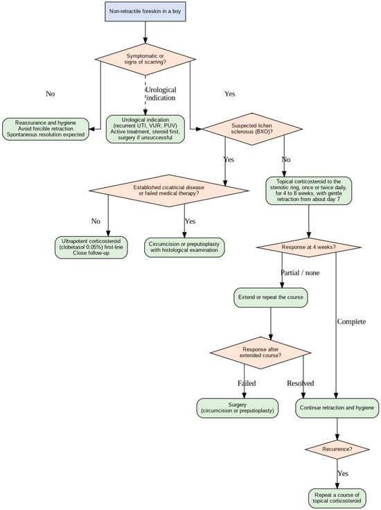
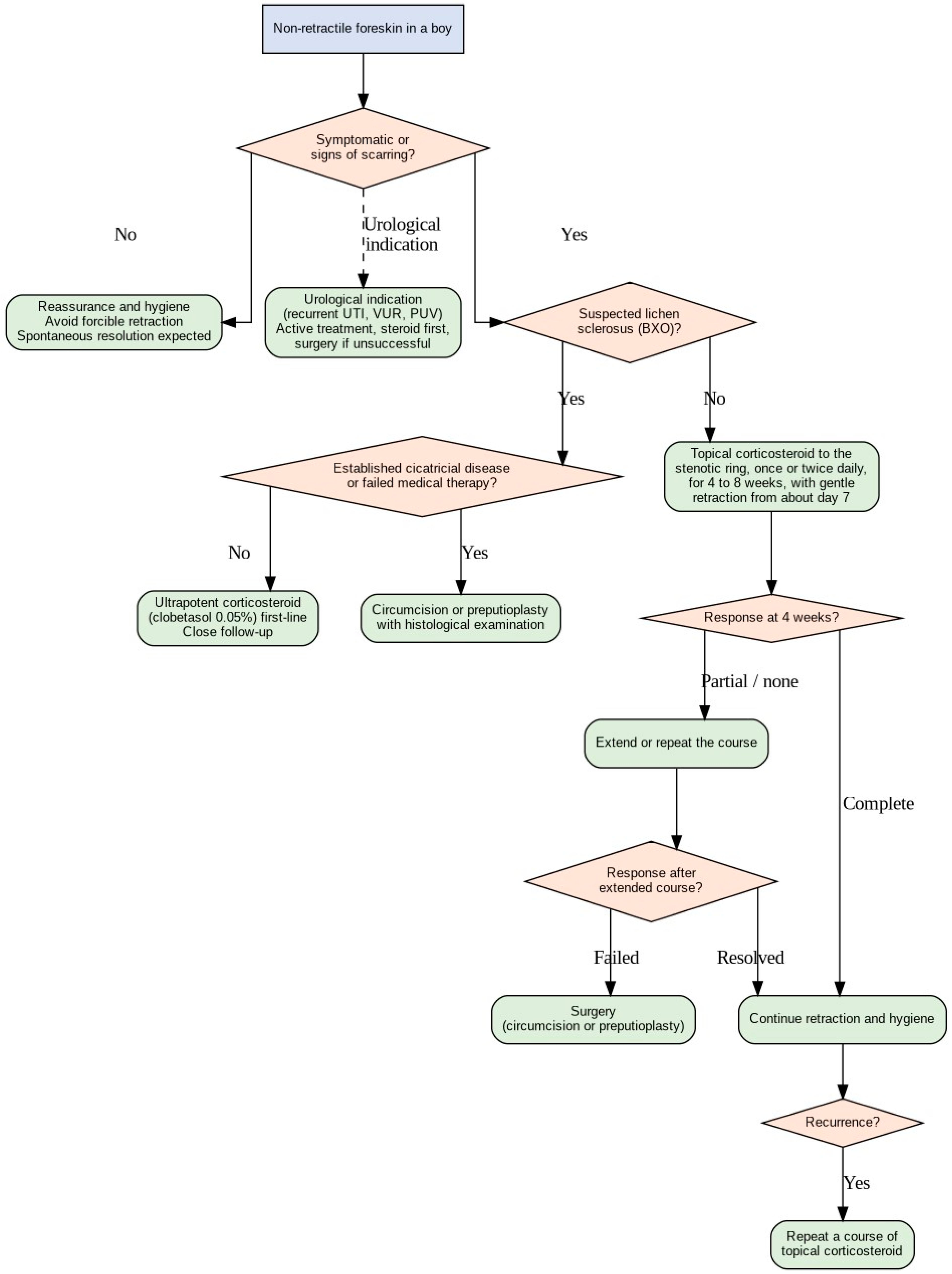
The following algorithm (Figure 1) summarizes the management approach described in this review and is intended as a practical guide rather than a rigid protocol. It follows the boy from initial assessment, through the distinction between developmental non-retractility and disease, to the choice between conservative treatment and surgery.
Figure 1.
Practical algorithm for the management of phimosis in boys.
21. Conclusions
Most non-retractility of the foreskin in childhood is physiological and resolves spontaneously, and should be managed with reassurance rather than intervention. For boys who genuinely require treatment for symptomatic phimosis, topical corticosteroids are an effective first-line option. They probably improve or resolve phimosis in the majority of non-scarred cases, appear safe when used in short, localized courses, are inexpensive, and are well accepted, and they allow most boys to avoid circumcision and retain the prepuce. Accordingly, international guidelines now recommend a topical corticosteroid as first-line therapy for primary symptomatic phimosis, reserving surgery for pathological (scarred) phimosis and balanitis xerotica obliterans, for treatment failures, and for boys with specific urological indications. On current evidence, a trial of topical corticosteroid is therefore a reasonable first-line treatment for symptomatic phimosis in boys, with careful clinical vigilance for the minority whose disease reflects underlying lichen sclerosus.
Author Contributions
Conceptualization, M.B.; investigation, M.B., J.B., B.D., D.J., K.J., K.P., S.V. and B.Ž.; data curation, M.B., J.B., B.D., D.J., K.J., K.P., S.V. and B.Ž.; writing—original draft preparation, M.B., J.B., B.D., D.J., K.J., K.P., S.V. and B.Ž.; writing—review and editing, M.B., J.B., B.D., D.J., K.J., K.P., S.V. and B.Ž.; visualization, M.B., J.B., B.D., D.J., K.J., K.P., S.V. and B.Ž.; supervision, M.B. and B.Ž.; project administration, M.B.; funding acquisition, M.B. All authors have read and agreed to the published version of the manuscript.
Funding
This research received no external funding.
Institutional Review Board Statement
Not applicable.
Informed Consent Statement
Not applicable.
Data Availability Statement
No new data were created or analyzed in this study. Data sharing is not applicable to this article.
Conflicts of Interest
The authors declare no conflicts of interest.
Abbreviations
The following abbreviations are used in this manuscript:
| HPA | Hypothalamic–pituitary–adrenal axis |
| BXO | Balanitis xerotica obliterans |
| RCT | Randomized controlled trial |
| BID | Bis in die |
| TID | Ter in die |
| OTC | Over-the-counter |
References
- Moreno, G.; Ramirez, C.; Corbalán, J.; Peñaloza, B.; Morel Marambio, M.; Pantoja, T. Topical corticosteroids for treating phimosis in boys. Cochrane Database Syst. Rev. 2024, 1, CD008973. [Google Scholar] [CrossRef] [Scilit] [PubMed]
- Moreno, G.; Corbalán, J.; Peñaloza, B.; Pantoja, T. Topical corticosteroids for treating phimosis in boys. Cochrane Database Syst. Rev. 2014, 2014, CD008973. [Google Scholar] [CrossRef] [Scilit] [PubMed]
- Rickwood, A.M. Medical indications for circumcision. BJU Int. 1999, 83, 45–51. [Google Scholar] [CrossRef] [Scilit] [PubMed]
- Herzog, L.W.; Alvarez, S.R. The frequency of foreskin problems in uncircumcised children. Am. J. Dis. Child. 1986, 140, 254–256. [Google Scholar] [CrossRef] [Scilit] [PubMed]
- Berdeu, D.; Sauze, L.; Ha-Vinh, P.; Blum-Boisgard, C. Cost-effectiveness analysis of treatments for phimosis: A comparison of surgical and medicinal approaches and their economic effect. BJU Int. 2001, 87, 239–244. [Google Scholar] [CrossRef] [Scilit] [PubMed]
- Oster, J. Further fate of the foreskin. Incidence of preputial adhesions, phimosis, and smegma among Danish schoolboys. Arch. Dis. Child. 1968, 43, 200–203. [Google Scholar] [CrossRef] [Scilit] [PubMed]
- Gairdner, D. The fate of the foreskin, a study of circumcision. Br. Med. J. 1949, 2, 1433–1437. [Google Scholar] [PubMed]
- Kayaba, H.; Tamura, H.; Kitajima, S.; Fujiwara, Y.; Kato, T.; Kato, T. Analysis of shape and retractability of the prepuce in 603 Japanese boys. J. Urol. 1996, 156, 1813–1815. [Google Scholar] [CrossRef] [Scilit]
- Clemmensen, O.J.; Krogh, J.; Petri, M. The histologic spectrum of prepuces from patients with phimosis. Am. J. Dermatopathol. 1988, 10, 104–108. [Google Scholar] [CrossRef] [Scilit] [PubMed]
- Kiss, A.; Király, L.; Kutasy, B.; Merksz, M. High incidence of balanitis xerotica obliterans in boys with phimosis: Prospective 10-year study. Pediatr. Dermatol. 2005, 22, 305–308. [Google Scholar] [CrossRef] [Scilit] [PubMed]
- Imamura, E. Phimosis of infants and young children in Japan. Acta Paediatr. Jpn. 1997, 39, 403–405. [Google Scholar] [CrossRef] [Scilit] [PubMed]
- Weiss, H.A.; Larke, N.; Halperin, D.; Schenker, I. Complications of circumcision in male neonates, infants and children: A systematic review. BMC Urol. 2010, 10, 2. [Google Scholar] [CrossRef] [Scilit] [PubMed]
- Task Force on Circumcision; Blank, S.; Brady, M.; Buerk, E.; Carlo, W.; Diekema, D.; Freedman, A.; Maxwell, L.; Wegner, S. Circumcision policy statement. Pediatrics 2012, 130, 585–586. [Google Scholar] [CrossRef] [Scilit] [PubMed]
- Hotonu, S.; Mohamed, A.; Rajimwale, A.; Gopal, M. Save the foreskin: Outcomes of preputioplasty in the treatment of childhood phimosis. Surgeon 2020, 18, 150–153. [Google Scholar] [CrossRef] [Scilit] [PubMed]
- Kikiros, C.S.; Beasley, S.W.; Woodward, A.A. The response of phimosis to local steroid application. Pediatr. Surg. Int. 1993, 8, 329–332. [Google Scholar] [CrossRef] [Scilit]
- Golubovic, Z.; Milanovic, D.; Vukadinovic, V.; Rakic, I.; Perovic, S. The conservative treatment of phimosis in boys. Br. J. Urol. 1996, 78, 786–788. [Google Scholar] [CrossRef] [Scilit] [PubMed]
- Radmayr, C.; Bogaert, G.; Burgu, B.; Bujons, A.; Castagnetti, M.S.; O’Kelly, F.; Pakkasjärvi, N.A.; Quaedackers, J.; Rawashdeh, Y.F.H.; Silay, M.S.; et al. EAU Guidelines on Paediatric Urology; European Association of Urology: Arnhem, The Netherlands, 2024. [Google Scholar]
- Skott, M.; Kennedy, U.; Gnech, M.; van Uitert, A.; Bujons, A.; ’t Hoen, L.; Rawashdeh, Y.F.; Silay, M.S.; O’Kelly, F.; Quaedackers, J.; et al. European Association of Urology-European Society of Paediatric Urology Guidelines on Paediatric Urology: Summary of 2024 Updates. Part II. Eur. Urol. 2025, 88, 190–203. [Google Scholar] [CrossRef] [Scilit] [PubMed]
- Sridharan, K.; Sivaramakrishnan, G. Topical corticosteroids for phimosis in children: A network meta-analysis of randomized clinical trials. Pediatr. Surg. Int. 2021, 37, 1117–1125. [Google Scholar] [CrossRef] [Scilit] [PubMed]
- Ko, M.C.; Liu, C.K.; Lee, W.K.; Jeng, H.S.; Chiang, H.S.; Li, C.Y. Age-specific prevalence rates of phimosis and circumcision in Taiwanese boys. J. Formos. Med. Assoc. 2007, 106, 302–307. [Google Scholar] [CrossRef] [Scilit] [PubMed]
- Orsola, A.; Caffaratti, J.; Garat, J.M. Conservative treatment of phimosis in children using a topical steroid. Urology 2000, 56, 307–310. [Google Scholar] [CrossRef] [Scilit] [PubMed]
- Marques, T.C.; Sampaio, F.J.; Favorito, L.A. Treatment of phimosis with topical steroids and foreskin anatomy. Int. Braz. J. Urol. 2005, 31, 370–374. [Google Scholar] [CrossRef] [Scilit] [PubMed]
- Yang, S.S.; Tsai, Y.C.; Wu, C.C.; Liu, S.P.; Wang, C.C. Highly potent and moderately potent topical steroids are effective in treating phimosis: A prospective randomized study. J. Urol. 2005, 173, 1361–1363. [Google Scholar] [CrossRef] [Scilit] [PubMed]
- Nguyen, A.T.M.; Holland, A.J.A. Balanitis xerotica obliterans: An update for clinicians. Eur. J. Pediatr. 2020, 179, 9–16. [Google Scholar] [CrossRef] [Scilit] [PubMed]
- Celis, S.; Reed, F.; Murphy, F.; Adams, S.; Gillick, J.; Abdelhafeez, A.H.; Lopez, P.J. Balanitis xerotica obliterans in children and adolescents: A literature review and clinical series. J. Pediatr. Urol. 2014, 10, 34–39. [Google Scholar] [CrossRef] [Scilit] [PubMed]
- Homer, L.; Buchanan, K.J.; Nasr, B.; Losty, P.D.; Corbett, H.J. Meatal stenosis in boys following circumcision for lichen sclerosus (balanitis xerotica obliterans). J. Urol. 2014, 192, 1784–1788. [Google Scholar] [CrossRef] [Scilit] [PubMed]
- Arena, S.; Ieni, A.; Currò, M.; Vaccaro, M.; Di Fabrizio, D.; Cassaro, F.; Bonfiglio, R.; Montalto, A.S.; Tuccari, G.; Alibrandi, A.; et al. Immunohistological Analysis of Lichen Sclerosus of the Foreskin in Pediatric Age: Could It Be Considered a Premalignant Lesion? Biomedicines 2023, 11, 1986. [Google Scholar] [CrossRef] [Scilit] [PubMed]
- Ghidini, F.; Virgone, C.; Pulvirenti, R.; Trovalusci, E.; Gamba, P. Could a careful clinical examination distinguish physiologic phimosis from balanitis xerotica obliterans in children? Eur. J. Pediatr. 2021, 180, 591–595. [Google Scholar] [CrossRef] [Scilit] [PubMed]
- Fuchs, M.E.; Beecroft, N.; Dajusta, D.G.; McLeod, D.J. The Association Between BXO and Obesity in Boys Undergoing Circumcision. Glob. Pediatr. Health 2017, 4, 2333794X17742749. [Google Scholar] [CrossRef] [Scilit] [PubMed]
- Edmonds, E.V.; Hunt, S.; Hawkins, D.; Dinneen, M.; Francis, N.; Bunker, C.B. Clinical parameters in male genital lichen sclerosus: A case series of 329 patients. J. Eur. Acad. Dermatol. Venereol. 2012, 26, 730–737. [Google Scholar] [CrossRef] [Scilit] [PubMed]
- Favorito, L.A.; Balassiano, C.M.; Rosado, J.P.; Cardoso, L.E.; Costa, W.S.; Sampaio, F.J. Structural analysis of the phimotic prepuce in patients with failed topical treatment compared with untreated phimosis. Int. Braz. J. Urol. 2012, 38, 802–808. [Google Scholar] [CrossRef] [Scilit] [PubMed]
- Dewan, P.A.; Tieu, H.C.; Chieng, B.S. Phimosis: Is circumcision necessary? J. Paediatr. Child Health 1996, 32, 285–289. [Google Scholar] [CrossRef] [Scilit] [PubMed]
- Morris, B.J.; Wiswell, T.E. Circumcision and lifetime risk of urinary tract infection: A systematic review and meta-analysis. J. Urol. 2013, 189, 2118–2124. [Google Scholar] [CrossRef] [Scilit] [PubMed]
- Uva, L.; Miguel, D.; Pinheiro, C.; Antunes, J.; Cruz, D.; Ferreira, J.; Filipe, P. Mechanisms of action of topical corticosteroids in psoriasis. Int. J. Endocrinol. 2012, 2012, 561018. [Google Scholar] [CrossRef] [Scilit] [PubMed]
- Ahluwalia, A. Topical glucocorticoids and the skin--mechanisms of action: An update. Mediat. Inflamm. 1998, 7, 183–193. [Google Scholar] [CrossRef] [Scilit] [PubMed]
- Coondoo, A.; Phiske, M.; Verma, S.; Lahiri, K. Side-effects of topical steroids: A long overdue revisit. Indian Dermatol. Online J. 2014, 5, 416–425. [Google Scholar] [CrossRef] [Scilit] [PubMed]
- Chu, C.C.; Chen, K.C.; Diau, G.Y. Topical steroid treatment of phimosis in boys. J. Urol. 1999, 162, 861–863. [Google Scholar] [CrossRef] [Scilit] [PubMed]
- Ashfield, J.E.; Nickel, K.R.; Siemens, D.R.; MacNeily, A.E.; Nickel, J.C. Treatment of phimosis with topical steroids in 194 children. J. Urol. 2003, 169, 1106–1108. [Google Scholar] [CrossRef] [Scilit] [PubMed]
- Zampieri, N.; Corroppolo, M.; Camoglio, F.S.; Giacomello, L.; Ottolenghi, A. Phimosis: Stretching methods with or without application of topical steroids? J. Pediatr. 2005, 147, 705–706. [Google Scholar] [CrossRef] [Scilit] [PubMed]
- Nascimento, F.J.; Pereira, R.F.; Silva, J.L., 2nd; Tavares, A.; Pompeo, A.C. Topical betamethasone and hyaluronidase in the treatment of phimosis in boys: A double-blind, randomized, placebo-controlled trial. Int. Braz. J. Urol. 2011, 37, 314–319. [Google Scholar] [CrossRef] [Scilit] [PubMed]
- Nunes E Santos, R.M.S.; Figueiredo, A.A.; de Bessa Junior, J.; Bastos, A.N.; Gouvêa, B.F.; Masson, A.V.; Delgado, C.S.; Esquerdo, M.; Massuqueto, E.; Netto, J.M.B. Does the addition of hyaluronidase to betamethasone in topical treatment of phimosis improves results?—A randomized double-blind clinical trial. J. Pediatr. Urol. 2024, 20, 898–903. [Google Scholar] [CrossRef] [Scilit] [PubMed]
- Pileggi Fde, O.; Vicente, Y.A. Phimotic ring topical corticoid cream (0.1% mometasone furoate) treatment in children. J. Pediatr. Surg. 2007, 42, 1749–1752. [Google Scholar] [CrossRef] [Scilit] [PubMed]
- Lee, C.H.; Lee, S.D. Effect of topical steroid (0.05% clobetasol propionate) treatment in children with severe phimosis. Korean J. Urol. 2013, 54, 624–630. [Google Scholar] [CrossRef] [Scilit] [PubMed]
- Chamberlin, J.D.; Dorgalli, C.; Abdelhalim, A.; Davis-Dao, C.A.; Chalmers, C.L.; Kelly, M.S.; Wang, Z.T.; Chuang, K.W.; McAleer, I.M.; Stephany, H.A.; et al. Randomized open-label trial comparing topical prescription triamcinolone to over-the-counter hydrocortisone for the treatment of phimosis. J. Pediatr. Urol. 2019, 15, 388.e1–388.e5. [Google Scholar] [CrossRef] [Scilit] [PubMed]
- Zavras, N.; Christianakis, E.; Mpourikas, D.; Ereikat, K. Conservative treatment of phimosis with fluticasone proprionate 0.05%: A clinical study in 1185 boys. J. Pediatr. Urol. 2009, 5, 181–185. [Google Scholar] [CrossRef] [Scilit] [PubMed]
- Liu, J.; Yang, J.; Chen, Y.; Cheng, S.; Xia, C.; Deng, T. Is steroids therapy effective in treating phimosis? A meta-analysis. Int. Urol. Nephrol. 2016, 48, 335–342. [Google Scholar] [CrossRef] [Scilit] [PubMed]
- Kuehhas, F.E.; Miernik, A.; Sevcenco, S.; Tosev, G.; Weibl, P.; Schoenthaler, M.; Lassmann, J. Predictive power of objectivation of phimosis grade on outcomes of topical 0.1% betamethasone treatment of phimosis. Urology 2012, 80, 412–416. [Google Scholar] [CrossRef] [Scilit] [PubMed]
- Ebert, A.K.; Vogt, T.; Rösch, W.H. Topical therapy of balanitis xerotica obliterans in childhood. Long-term clinical results and an overview. Urologe A 2007, 46, 1682–1686. [Google Scholar]
- Funaro, D.; Lovett, A.; Leroux, N.; Powell, J. A double-blind, randomized prospective study evaluating topical clobetasol propionate 0.05% versus topical tacrolimus 0.1% in patients with vulvar lichen sclerosus. J. Am. Acad. Dermatol. 2014, 71, 84–91. [Google Scholar] [CrossRef] [Scilit] [PubMed]
- Monsour, M.A.; Rabinovitch, H.H.; Dean, G.E. Medical management of phimosis in children: Our experience with topical steroids. J. Urol. 1999, 162, 1162–1164. [Google Scholar] [CrossRef] [Scilit] [PubMed]
- Zhou, G.; Jiang, M.; Yang, Z.; Xu, W.; Li, S. Efficacy of topical steroid treatment in children with severe phimosis in China: A long-term single centre prospective study. J. Paediatr. Child Health 2021, 57, 1960–1965. [Google Scholar] [CrossRef] [Scilit] [PubMed]
- Reddy, S.; Jain, V.; Dubey, M.; Deshpande, P.; Singal, A.K. Local steroid therapy as the first-line treatment for boys with symptomatic phimosis—A long-term prospective study. Acta Paediatr. 2012, 101, e130–e133. [Google Scholar] [CrossRef] [Scilit] [PubMed]
- Lund, L.; Wai, K.H.; Mui, L.M.; Yeung, C.K. An 18-month follow-up study after randomized treatment of phimosis in boys with topical steroid versus placebo. Scand. J. Urol. Nephrol. 2005, 39, 78–81. [Google Scholar] [CrossRef] [Scilit] [PubMed]
- Esposito, C.; Centonze, A.; Alicchio, F.; Savanelli, A.; Settimi, A. Topical steroid application versus circumcision in pediatric patients with phimosis: A prospective randomized placebo-controlled clinical trial. World J. Urol. 2008, 26, 187–190. [Google Scholar] [CrossRef] [Scilit] [PubMed]
- Palmer, L.S.; Palmer, J.S. The efficacy of topical betamethasone for treating phimosis: A comparison of two treatment regimens. Urology 2008, 72, 68–71. [Google Scholar] [CrossRef] [Scilit] [PubMed]
- Zampieri, N.; Corroppolo, M.; Zuin, V.; Bianchi, S.; Camoglio, F.S. Phimosis and topical steroids: New clinical findings. Pediatr. Surg. Int. 2007, 23, 331–335. [Google Scholar] [CrossRef] [Scilit] [PubMed]
- Ghysel, C.; Vander Eeckt, K.; Bogaert, G.A. Long-term efficiency of skin stretching and a topical corticoid cream application for unretractable foreskin and phimosis in prepubertal boys. Urol. Int. 2009, 82, 81–88. [Google Scholar] [CrossRef] [Scilit] [PubMed]
- Rushton, H.G.; Majd, M. Pyelonephritis in male infants: How important is the foreskin? J. Urol. 1992, 148, 733–736. [Google Scholar] [CrossRef] [Scilit] [PubMed]
- Wiswell, T.E.; Smith, F.R.; Bass, J.W. Decreased incidence of urinary tract infections in circumcised male infants. Pediatrics 1985, 75, 901–903. [Google Scholar] [CrossRef] [Scilit]
- Wiswell, T.E.; Hachey, W.E. Urinary tract infections and the uncircumcised state: An update. Clin. Pediatr. (Phila.) 1993, 32, 130–134. [Google Scholar] [CrossRef] [Scilit] [PubMed]
- Van Howe, R.S. Effect of confounding in the association between circumcision status and urinary tract infection. J. Infect. 2005, 51, 59–68. [Google Scholar] [CrossRef] [Scilit] [PubMed]
- Depasquale, I.; Park, A.J.; Bracka, A. The treatment of balanitis xerotica obliterans. BJU Int. 2000, 86, 459–465. [Google Scholar] [CrossRef] [Scilit] [PubMed]
- Nasca, M.R.; Innocenzi, D.; Micali, G. Penile cancer among patients with genital lichen sclerosus. J. Am. Acad. Dermatol. 1999, 41, 911–914. [Google Scholar] [CrossRef] [Scilit] [PubMed]
- Pietrzak, P.; Hadway, P.; Corbishley, C.M.; Watkin, N.A. Is the association between balanitis xerotica obliterans and penile carcinoma underestimated? BJU Int. 2006, 98, 74–76. [Google Scholar] [CrossRef] [Scilit] [PubMed]
- Elmore, J.M.; Baker, L.A.; Snodgrass, W.T. Topical steroid therapy as an alternative to circumcision for phimosis in boys younger than 3 years. J. Urol. 2002, 168, 1746–1747. [Google Scholar] [CrossRef] [Scilit]
- Abraham, A.; Roga, G. Topical steroid-damaged skin. Indian J. Dermatol. 2014, 59, 456–459. [Google Scholar] [CrossRef] [Scilit] [PubMed]
- Pileggi, F.O.; Martinelli, C.E., Jr.; Tazima, M.F.; Daneluzzi, J.C.; Vicente, Y.A. Is suppression of hypothalamic-pituitary-adrenal axis significant during clinical treatment of phimosis? J. Urol. 2010, 183, 2327–2331. [Google Scholar] [CrossRef] [Scilit] [PubMed]
- Yilmaz, E.; Batislam, E.; Basar, M.M.; Basar, H. Psychological trauma of circumcision in the phallic period could be avoided by using topical steroids. Int. J. Urol. 2003, 10, 651–656. [Google Scholar] [CrossRef] [Scilit] [PubMed]
- Task Force on Circumcision; Blank, S.; Brady, M.; Buerk, E.; Carlo, W.; Diekema, D.; Freedman, A.; Maxwell, L.; Wegner, S. Male circumcision. Pediatrics 2012, 130, e756–e785. [Google Scholar] [CrossRef] [Scilit] [PubMed]
- Shahid, S.K. Phimosis in children. ISRN Urol. 2012, 2012, 707329. [Google Scholar] [CrossRef] [Scilit] [PubMed]
- Morris, B.J.; Wamai, R.G.; Henebeng, E.B.; Tobian, A.A.; Klausner, J.D.; Banerjee, J.; Hankins, C.A. Estimation of country-specific and global prevalence of male circumcision. Popul. Health Metr. 2016, 14, 4. [Google Scholar] [CrossRef] [Scilit] [PubMed]
- Edwards, S.K.; Bunker, C.B.; Ziller, F.; van der Meijden, W.I. 2013 European guideline for the management of balanoposthitis. Int. J. STD AIDS 2014, 25, 615–626. [Google Scholar] [CrossRef] [Scilit] [PubMed]
- Singh-Grewal, D.; Macdessi, J.; Craig, J. Circumcision for the prevention of urinary tract infection in boys: A systematic review of randomised trials and observational studies. Arch. Dis. Child. 2005, 90, 853–858. [Google Scholar] [CrossRef] [Scilit] [PubMed]
- To, T.; Agha, M.; Dick, P.T.; Feldman, W. Cohort study on circumcision of newborn boys and subsequent risk of urinary-tract infection. Lancet 1998, 352, 1813–1816. [Google Scholar] [CrossRef] [Scilit] [PubMed]
- Shaikh, N.; Morone, N.E.; Bost, J.E.; Farrell, M.H. Prevalence of urinary tract infection in childhood: A meta-analysis. Pediatr. Infect. Dis. J. 2008, 27, 302–308. [Google Scholar] [CrossRef] [Scilit] [PubMed]
- Hayashi, Y.; Kohri, K. Circumcision related to urinary tract infections, sexually transmitted infections, human immunodeficiency virus infections, and penile and cervical cancer. Int. J. Urol. 2013, 20, 769–775. [Google Scholar] [CrossRef] [Scilit] [PubMed]
- Siroosbakht, S.; Rezakhaniha, B. A comprehensive comparison of the early and late complications of surgical circumcision in neonates and children: A cohort study. Health Sci. Rep. 2022, 5, e939. [Google Scholar] [CrossRef] [Scilit] [PubMed]
- Cold, C.J.; Taylor, J.R. The prepuce. BJU Int. 1999, 83, 34–44. [Google Scholar] [CrossRef] [Scilit] [PubMed]
- Cuckow, P.M.; Rix, G.; Mouriquand, P.D. Preputial plasty: A good alternative to circumcision. J. Pediatr. Surg. 1994, 29, 561–563. [Google Scholar] [CrossRef] [Scilit] [PubMed]
- Wilkinson, D.J.; Lansdale, N.; Everitt, L.H.; Marven, S.S.; Walker, J.; Shawis, R.N.; Roberts, J.P.; Mackinnon, A.E.; Godbole, P.P. Foreskin preputioplasty and intralesional triamcinolone: A valid alternative to circumcision for balanitis xerotica obliterans. J. Pediatr. Surg. 2012, 47, 756–759. [Google Scholar] [CrossRef] [Scilit] [PubMed]
- Meuli, M.; Briner, J.; Hanimann, B.; Sacher, P. Lichen sclerosus et atrophicus causing phimosis in boys: A prospective study with 5-year follow-up after complete circumcision. J. Urol. 1994, 152, 987–989. [Google Scholar] [CrossRef] [Scilit] [PubMed]
- Bunker, C.B.; Shim, T.N. Male genital lichen sclerosus. Indian J. Dermatol. 2015, 60, 111–117. [Google Scholar] [CrossRef] [Scilit] [PubMed]
- Wright, J.E. The treatment of childhood phimosis with topical steroid. Aust. N. Z. J. Surg. 1994, 64, 327–328. [Google Scholar] [CrossRef] [Scilit] [PubMed]
- Iken, A.; Ben Mouelli, S.; Fontaine, E.; Quenneville, V.; Carpentier, X.; Chartier-Kastler, E.; Beurton, D. Treatment of phimosis with locally applied 0.05% clobetasol propionate. Prospective study with 108 children. Prog. Urol. 2002, 12, 1268–1271. [Google Scholar] [PubMed]
- Webster, T.M.; Leonard, M.P. Topical steroid therapy for phimosis. Can. J. Urol. 2002, 9, 1492–1495. [Google Scholar] [CrossRef] [Scilit]
- Makhija, D.; Shah, H.; Tiwari, C.; Dwiwedi, P.; Gandhi, S. Outcome of topical steroid application in children with non-retractile prepuce. Dev. Period Med. 2018, 22, 71–74. [Google Scholar] [CrossRef] [Scilit] [PubMed]
- Jørgensen, E.T.; Svensson, A. The treatment of phimosis in boys, with a potent topical steroid (clobetasol propionate 0.05%) cream. Acta Derm. Venereol. 1993, 73, 55–56. [Google Scholar] [CrossRef] [Scilit] [PubMed]
- Babu, R.; Harrison, S.K.; Hutton, K.A. Ballooning of the foreskin and physiological phimosis: Is there any objective evidence of obstructed voiding? BJU Int. 2004, 94, 384–387. [Google Scholar] [CrossRef] [Scilit] [PubMed]
- Shankar, K.R.; Rickwood, A.M. The incidence of phimosis in boys. BJU Int. 1999, 84, 101–102. [Google Scholar] [CrossRef] [Scilit] [PubMed]
- Thorvaldsen, M.A.; Meyhoff, H.H. Pathological or physiological phimosis? Ugeskr. Laeger 2005, 167, 1858–1862. [Google Scholar] [PubMed]
- Deibert, G.A. The separation of the prepuce in the human penis. Anat. Rec. 1933, 57, 387–399. [Google Scholar] [CrossRef] [Scilit]
- Dahlman-Ghozlan, K.; Hedblad, M.A.; von Krogh, G. Penile lichen sclerosus et atrophicus treated with clobetasol dipropionate 0.05% cream: A retrospective clinical and histopathological study. J. Am. Acad. Dermatol. 1999, 40, 451–457. [Google Scholar] [CrossRef] [Scilit] [PubMed]
- Kiss, A.; Csontai, A.; Pirót, L.; Nyirády, P.; Merksz, M.; Király, L. The response of balanitis xerotica obliterans to local steroid application compared with placebo in children. J. Urol. 2001, 165, 219–220. [Google Scholar] [CrossRef] [Scilit] [PubMed]
- Chi, C.C.; Kirtschig, G.; Baldo, M.; Brackenbury, F.; Lewis, F.; Wojnarowska, F. Topical interventions for genital lichen sclerosus. Cochrane Database Syst. Rev. 2011, 2011, CD008240. [Google Scholar] [CrossRef] [Scilit] [PubMed]
- Powell, J.J.; Wojnarowska, F. Lichen sclerosus. Lancet 1999, 353, 1777–1783. [Google Scholar] [CrossRef] [Scilit] [PubMed]
- Meffert, J.J.; Davis, B.M.; Grimwood, R.E. Lichen sclerosus. J. Am. Acad. Dermatol. 1995, 32, 393–416. [Google Scholar] [CrossRef] [Scilit] [PubMed]
- Clouston, D.; Hall, A.; Lawrentschuk, N. Penile lichen sclerosus (balanitis xerotica obliterans). BJU Int. 2011, 108, 14–19. [Google Scholar] [CrossRef] [Scilit] [PubMed]
- Jayakumar, S.; Antao, B.; Bevington, O.; Furness, P.; Ninan, G.K. Balanitis xerotica obliterans in children and its incidence under the age of 5 years. J. Pediatr. Urol. 2012, 8, 272–275. [Google Scholar] [CrossRef] [Scilit] [PubMed]
- Yardley, I.E.; Cosgrove, C.; Lambert, A.W. Paediatric preputial pathology: Are we circumcising enough? Ann. R. Coll. Surg. Engl. 2007, 89, 62–65. [Google Scholar] [CrossRef] [Scilit] [PubMed]
- Morris, B.J.; Matthews, J.G.; Krieger, J.N. Prevalence of Phimosis in Males of All Ages: Systematic Review. Urology 2020, 135, 124–132. [Google Scholar] [CrossRef] [Scilit] [PubMed]
- Kizer, W.S.; Prarie, T.; Morey, A.F. Balanitis xerotica obliterans: Epidemiologic distribution in an equal access health care system. South. Med. J. 2003, 96, 9–11. [Google Scholar] [CrossRef] [Scilit] [PubMed]

Disclaimer/Publisher’s Note: The statements, opinions and data contained in all publications are solely those of the individual author(s) and contributor(s) and not of MDPI and/or the editor(s). MDPI and/or the editor(s) disclaim responsibility for any injury to people or property resulting from any ideas, methods, instructions or products referred to in the content. |
© 2026 by the authors. Licensee MDPI, Basel, Switzerland. This article is an open access article distributed under the terms and conditions of the Creative Commons Attribution (CC BY) license.