


Simple Summary
After surgery for endometrial cancer, many patients receive brachytherapy to reduce the risk of cancer returning at the top of the vagina. This treatment is commonly planned using computed tomography, whereas magnetic resonance imaging provides better soft-tissue visualization but is more complex and less widely available. In this exploratory study of 25 patients, magnetic resonance imaging produced smaller target volumes, while the evaluated target-coverage and organ-at-risk dose parameters were broadly similar between the imaging modalities. Because the study was not designed to establish clinical equivalence or safety, these findings should be considered hypothesis-generating. Computed tomography-based planning appeared adequate for most patients in this cohort, whereas magnetic resonance imaging may offer additional value in selected, anatomically complex cases. Larger multicenter studies are needed to confirm these observations.
Abstract
Objectives: To assess the impact of magnetic resonance imaging (MRI) versus computed tomography (CT) on target delineation and dosimetry in adjuvant vaginal cuff brachytherapy (VBT) for endometrial cancer. Methods: Twenty-five consecutive patients receiving adjuvant high-dose-rate VBT after hysterectomy were prospectively included. All underwent standardized treatment of 4 × 5 Gy in 5 mm tissue depth using a vaginal cylinder with a fixed loading pattern; individualized optimization was permitted for suboptimal coverage or proximity of organs at risk (OARs). For each patient, clinical target volume (CTV) and OARs (bladder, rectum, sigmoid, bowel) were contoured on both CT and MRI of the first fraction. Dosimetric parameters (CTV D90, D98, D50; OAR D2cc) were compared. Treatment characteristics and outcomes were analyzed retrospectively. Results: Median CTV was smaller on MRI (9.8 cm3) than on CT (13.6 cm3). CTV coverage was comparable between modalities, with mean EQD2 D90 of 5.32 Gy per fraction on MRI versus 5.10 Gy per fraction on CT (p = 0.433) and mean EQD2 D98 of 3.95 Gy per fraction on MRI versus 3.80 Gy per fraction on CT (p = 0.491). In contrast, EQD2 D50 was significantly higher on MRI-based plans than on CT-based plans (9.12 vs. 8.62 Gy per fraction, p = 0.012). OAR D2cc values showed no significant differences between modalities. Most patients (80%) were treated with the institutional standard plan, and 20% required individualized optimization. After a median follow-up of 25 months (range 0–72), five patients (20%) recurred (one local, one regional, three distant), and one (4%) died. No grade ≥ 3 gastrointestinal or genitourinary toxicities occurred among patients with available data. Conclusions: In this small exploratory cohort, MRI-based planning delineated smaller CTVs, whereas the evaluated target-coverage and OAR-dose parameters were broadly similar between modalities. These findings suggest that CT-based planning may be adequate for many routine cases, with MRI potentially useful for selected, anatomically complex patients. Confirmation in larger multicenter studies is required.
1. Introduction
Endometrial cancer is the most common female pelvic malignancy in developed countries, with an incidence of about 140,000 per year or 15.5 per 100,000 women in Europe, according to GLOBOCAN 2022 [1]. Standard primary treatment of localized tumors consists of total hysterectomy with bilateral salpingo-oophorectomy, with or without lymph node staging, followed by adjuvant radiotherapy in selected patients based on risk factors [2,3]. Postoperative brachytherapy has been shown to significantly reduce local recurrence rates. PORTEC-2 demonstrated excellent vaginal control with VBT, with less gastrointestinal toxicity than pelvic EBRT [4,5].
Traditionally, intracavitary VBT was planned using two-dimensional X-ray imaging after insertion of a vaginal cylinder and a rectal probe for in vivo dosimetry. The standard approach involved a prescribing dose at 5 mm tissue depth, assuming a uniform vaginal wall thickness of 5 mm. This conventional approach was used in earlier multicenter trials [6], but contemporary planning increasingly relies on cross-sectional imaging to assess target anatomy and dose distribution.
With the advent of modern cross-sectional imaging, computed tomography has largely replaced X-ray-based planning, allowing for improved visualization of applicator placement and adjacent organs at risk. However, CT’s limited soft tissue contrast remains a drawback. Evidence from cervical cancer brachytherapy shows that CT-based target volumes tend to overestimate tumor extent compared with MRI, potentially resulting in unnecessary irradiation of organs at risk (OARs) [7].
Previous studies have investigated the influence of different cylinder sizes [8], angles [9], applicator types [10], and treatment durations [11,12] on dose distribution. Yet, the specific dosimetric differences between CT- and MRI-based planning have mostly been explored in phantom models [13] or in the setting of primary radiotherapy with an intact uterus [14,15]. Importantly, a retrospective MRI-based evaluation of previously CT-planned VBT demonstrated underdosage of the clinical target volume (CTV) in over half of patients [16].
Given MRI’s superior soft tissue contrast [17], it may offer significant advantages for precise target delineation and optimized sparing of OARs in adjuvant VBT. Despite its increasing use in gynecologic brachytherapy, particularly for cervical cancer [18], MRI guidance has not yet become standard for endometrial cancer.
The integration of MRI into adjuvant brachytherapy planning could potentially improve treatment accuracy, reduce toxicities, and pave the way for dose escalation or adaptive strategies in selected patients. However, data on its implementation, benefits, and clinical impact remain limited.
The aim of this study is to evaluate the potential role of MRI-guided planning in adjuvant vaginal cuff brachytherapy for endometrial cancer. Specifically, we compare MRI- with CT-based planning in terms of target volume, coverage and OAR dose exposure as well as clinical outcome.
2. Materials and Methods
2.1. Patients and Treatment
This is a prospective single-center cohort study, which was conducted in accordance with the Declaration of Helsinki and approved by the Institutional Ethics Committee of Medical University of Vienna (protocol code 1397/2018). Informed consent was obtained from all patients involved in the study. Between 2018 and 2021, patients with histologically confirmed endometrial carcinoma were included after undergoing total hysterectomy with bilateral salpingo-oophorectomy with or without lymph node sampling. The following parameters were collected: age, histology, TNM stage, grading, margin status, lymphovascular invasion, use of chemotherapy or external beam radiotherapy, number of fractions of VBT, cylinder size, recurrences and side effects. All patients underwent postoperative vaginal cuff brachytherapy. Adjuvant treatment decisions were based on the contemporary ESMO–ESGO–ESTRO recommendations [19] applicable during the study period. In selected small non-endometrioid tumors with surgical lymph-node staging, VBT combined with systemic chemotherapy was also used according to institutional practice. Additional oncological treatments, including chemotherapy and external beam radiotherapy, were recorded. Adjuvant treatment decisions, including the use or omission of EBRT and chemotherapy, were made independently of participation in this imaging study. The study protocol did not prescribe omission of EBRT or treatment de-escalation.
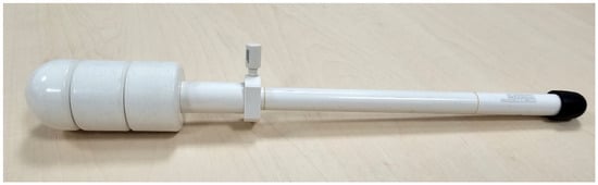
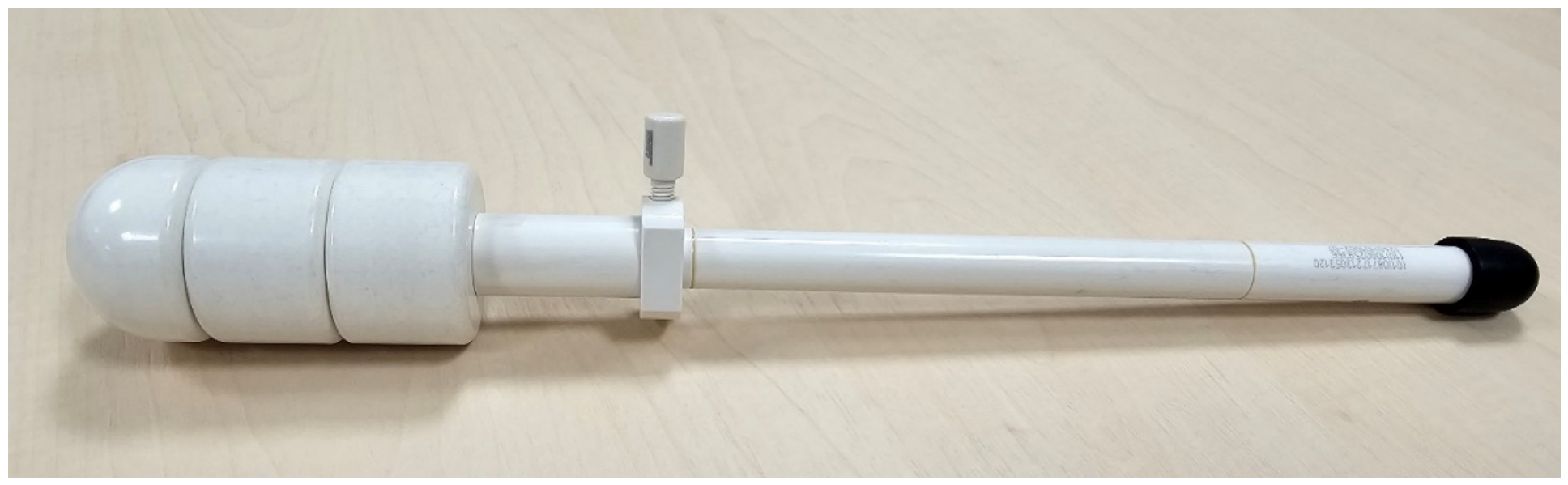
A vaginal cylinder applicator commercialized by Elekta AB with diameters between 25 and 40 mm was used (Figure 1). Applicator size was selected individually after clinical examination and measurement of vaginal length to ensure optimal fit and mucosal contact.
Figure 1.
Example of a cylinder applicator with a diameter of 35 mm and length of 7.5 cm.
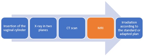
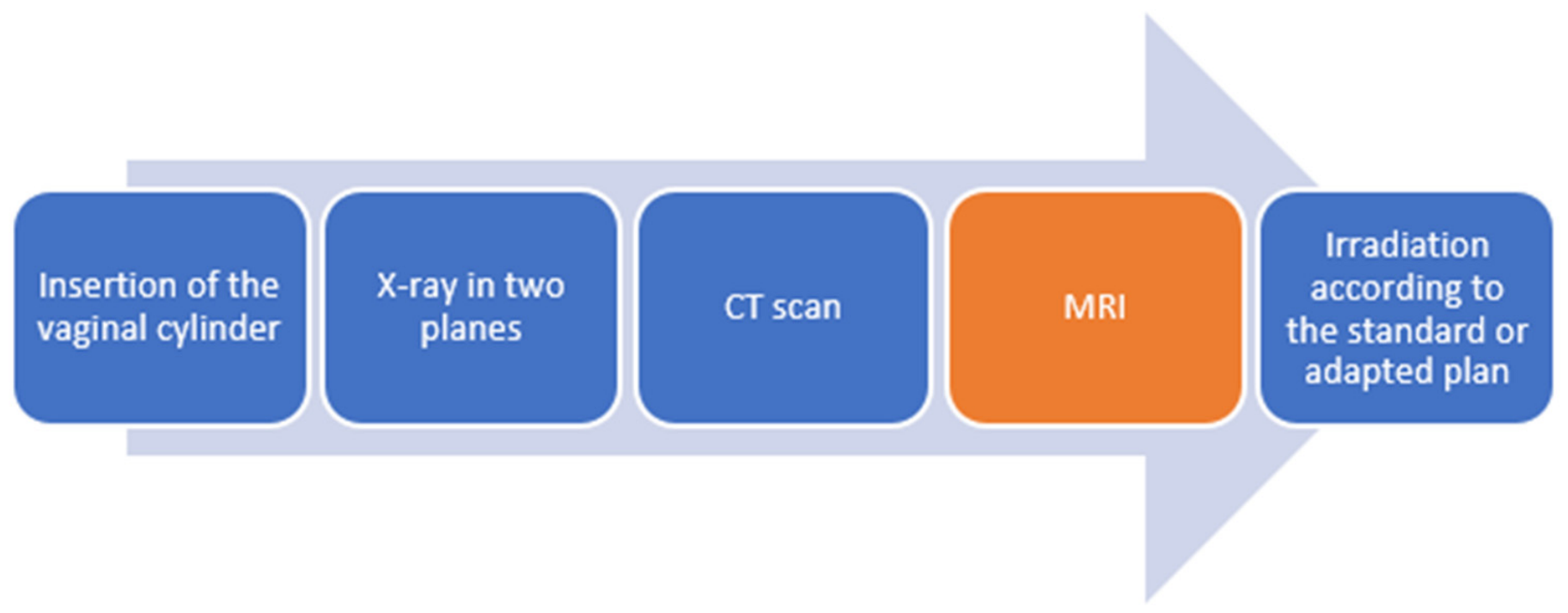
Patients were positioned in lithotomy, and a Foley catheter was placed at the first fraction to allow bladder filling control. The bladder filling protocol consisted of emptying the bladder completely and retrogradely filling it with 50 mL of sodium solution before MRI and again before CT. The cylinder was assembled and inserted under direct visualization. For the study, CT and MRI were acquired only at the first fraction with the applicator in situ. At all four fractions, orthogonal X-rays were obtained solely for routine verification of applicator position and geometry; fractions 2–4 were, therefore, performed without additional cross-sectional imaging. The clinical workflow is exemplified in Figure 2.
Figure 2.
Clinical workflow.
The institutional standard consisted of prescribing 5 Gy at 5 mm tissue depth in the proximal third of the vagina, delivered in four fractions of 5 Gy, applied over 2 weeks. The institutional regimen of 20 Gy in four fractions has been reported in previous clinical series [20] and was used during the study period as a well-tolerated postoperative schedule; contemporaneous European recommendations did not mandate a single VBT fractionation regimen [19].
The objective of this study is to compare CT-based and MRI-based planning in adjuvant vaginal cuff brachytherapy by analyzing CTV and dose parameters for both target and OARs. Specifically, we aim to
- −
- Quantify differences in CTV volume delineated on CT versus MRI;
- −
- Evaluate dose to 90% (D90) and other dose parameters for the CTV on each modality;
- −
- Compare dose to most exposed 2 cc (D2cc) of OARs (bladder, rectum, sigmoid colon);
- −
- Evaluate in how many patients imaging-based planning (CT or MRI) would have led to modifications of the standard X-ray-based prescription (5 Gy at 5 mm);
- −
- Report treatment outcomes and patients’ demographics in this cohort.
Imaging
X-ray—Orthogonal X-ray images were obtained in anteroposterior and lateral planes with the vaginal cylinder in place to verify applicator geometry and anatomical position.
CT—CT imaging was performed using a Somatom Definition AS Siemens with a slice thickness of 2 mm. Patients were positioned in a supine position, without intravenous or oral contrast. CT images were used for contouring the CTV and OARs on axial images and for generating CT-based treatment plans.
MRI—MRI was performed using an open, low-field Magnetom C system (Siemens) with 0.35 T field strength. Image acquisition followed the general MRI principles described by the GYN GEC-ESTRO Working Group for image-guided gynecological brachytherapy [21]. T2-weighted sequences were acquired in axial, para-axial, coronal, and sagittal planes covering the vaginal cuff and adjacent pelvic organs with the applicator in place. The orientation of the axial plane was based on the cylinder position during the acquisition of the sagittal plane and tilted in the cylinder axis. Slice thickness was 5 mm. MRI images were used for CTV and OAR delineation and for the generation of MRI-based treatment plans.
2.2. Contouring and Planning
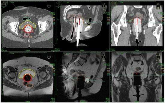
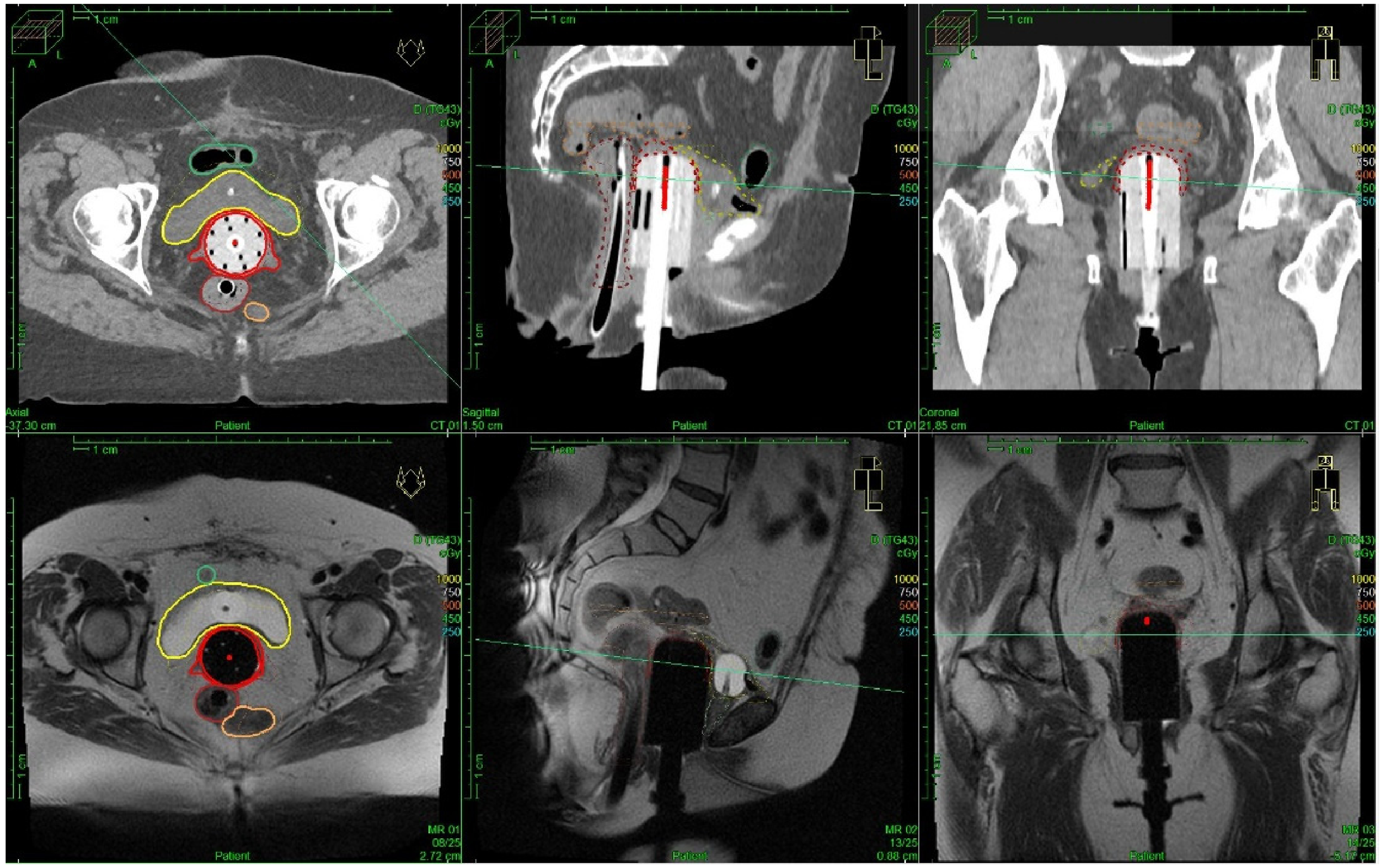
All CT- and MRI-based CTV and OAR contours were generated by a single radiation oncologist with more than three years of experience in brachytherapy. CT-based contouring was performed first, followed by MRI-based contouring approximately one week later. The contours were generated separately on each mod ality without simultaneous display of the contours from the other modality (Figure 3). The postoperative CTV encompassed the vaginal cuff and the vaginal wall over the proximal third of the vagina, with the longitudinal extent based on the vaginal length measured during clinical examination. On both CT and T2-weighted MRI, the vaginal wall was delineated according to its visible anatomical thickness, following the inner and outer wall boundaries identifiable on the respective modality. Thus, CTV thickness was patient-specific and could vary along the vaginal wall and at the cuff. Paravaginal tissues were not routinely included. The cranial extent comprised the visible postoperative vaginal cuff, while the caudal extent corresponded to the proximal third of the clinically measured vaginal length. No specific published postoperative VBT contouring atlas was used; contours were based on the visible anatomical boundaries on each imaging modality.
Figure 3.
CT and MRI contouring: axial, sagittal, and coronal CT images (top row) and corresponding MRI images (bottom row) of a representative patient.
For each patient, three plans were generated:
- −
- X-ray-based standard plan (5 Gy at 5 mm tissue depth in the proximal third of the vagina, determined during clinical examination prior to the first application by measuring the vaginal length). The X-ray-based plan represented the institutional reference workflow during the study period and was included to determine whether cross-sectional imaging would have prompted treatment adaptation; it was not included in the primary dosimetric comparison between CT- and MRI-based planning.
- −
- CT-based plan.
- −
- MRI-based plan.
Plans were created in Oncentra Treatment Planning System (The Netherlands), version 4.6.1, using points-distance optimization, geometrical-distance optimization or manual optimization. The cylinder-based rigid registration was performed in Oncentra Brachy planning system between CT and respective MRI. The delivered brachytherapy plan was created on the MRI and was evaluated on the CT. That was a key in keeping the same dwell positions, prescription depth, treated length, and optimization constraints for both imaging modalities. Dose–volume histogram (DVH) parameters were calculated for the CTV (D90, D98, D50) and OARs (D2cc for bladder, rectum, sigmoid, small bowel, urethra). Absorbed dose was reported in Gy and converted to biologically equieffective dose in 2 Gy fractions (EQD2) using α/β ratios of 10 Gy for the CTV and 3 Gy for OARs. The paired dosimetric analysis evaluated the VBT component only; EBRT dose was not included in the reported dose parameters.
2.3. Plan Adaptation
For each patient, we assessed whether imaging-based planning (CT or MRI) would have led to a modification of the institutional X-ray-based standard plan (5 Gy prescribed at 5 mm tissue depth over the proximal third of the vagina). For individualized plans, the fraction dose remained 5 Gy. Manual dwell-time optimization and, when required, adjustment of the prescription depth or treated length were performed to improve coverage of the contoured vaginal cuff while limiting dose to adjacent OARs. Any deviations from the standard plan were documented, including adjustments in prescription depth (e.g., increased to 7 mm to ensure adequate target coverage in cases with a thicker vaginal wall or reduced to 3 mm to limit dose to adjacent bowel loops), modifications of the treated length, or changes in channel activation patterns and dwell-time optimization. These adaptations were used to classify each case as either treated with the standard plan or requiring individualized, image-guided optimization.
2.4. Statistical Analysis
Descriptive statistics were reported as median, mean ± standard deviation, and interquartile ranges. Normality of the within-patient differences between MRI- and CT-based measurements was assessed using the Shapiro–Wilk test. Comparisons between CT- and MRI-based plans were performed using paired t-tests. A two-sided p-value < 0.05 was considered statistically significant. Analyses were carried out using SPSS statistics (version 24, IBM, Armonk, NY, USA).
3. Results
3.1. Patient Characteristics
A total of 25 consecutive patients were included in the study. Patient and treatment characteristics are presented in Table 1.
Table 1.
Patient and treatment characteristics (n = 25). Abbreviations: LND = lymph node dissection; SLNB = sentinel lymph node biopsy; G = tumor grade; L = lymphatic invasion; V = vascular invasion; R0 = microscopically negative resection margin; VBT = vaginal cuff brachytherapy.
The median age at diagnosis was 65 years (range: 51–80 years).
Most patients presented with early-stage disease: T1a (n = 7, 28%), T1b (n = 9, 36%), and T2 (n = 7, 28%), while two patients (8%) had T3a disease. Nodal status was predominantly negative (N0, n = 23, 92%), with 1 patient N1 (4%) and 1 patient Nx (4%).
Lymph node assessment consisted of lymphadenectomy in 12 patients (48%) and sentinel lymph node biopsy in 11 patients (44%). Two patients (8%) did not undergo surgical lymph node assessment.
Histology was mainly endometrioid adenocarcinoma (n = 16, 64%), followed by serous/papillary carcinoma (n = 3, 12%), mixed histology (n = 2, 8%), carcinosarcoma (n = 3, 12%), and clear cell carcinoma (n = 1, 4%). Tumor grading showed low grade in 8 patients (32%), high grade in 13 (52%), and unknown in 4 patients (16%). Molecular pathology analysis was not standard at the time of the trial execution and was, therefore, not performed on the included patients at diagnosis.
Significant lymphovascular invasion was present in 2 patients (8%) (L1), while vascular invasion was documented in 1 patient (4%). Resection margins were negative (R0) in 20 patients (80%); in 5 cases, margin status was not explicitly reported.
3.2. Treatment Characteristics
All 25 patients completed adjuvant vaginal cuff brachytherapy as prescribed. All patients received 20 Gy in four fractions of 5 Gy, with 2 fractions per week. The median interval from surgery to the first brachytherapy fraction was 55 days (range 28–168 days). Vaginal cylinder diameter ranged from 25 to 40 mm. The most frequently used applicator size was 30 mm (48%) followed by 35 mm (36%), 25 mm (12%), and 40 mm (4%). Chemotherapy was recorded in 8 patients (32%), whereas 17 patients (68%) had no recorded chemotherapy. One patient (4%), who had T2N1 grade 3 serous/papillary carcinoma, received adjuvant EBRT in addition to VBT and chemotherapy.
3.2.1. Target Volume and Dosimetric Analysis
The median CTV volume was 9.8 cm3 (range: 4.5–31.2) on MRI compared with 13.6 cm3 (range: 5.9–28.0) on CT. The mean CTV volume was 10.9 cm3 on MRI vs. 13.9 cm3 on CT, corresponding to an average relative reduction of approximately 21% with MRI-based delineation (Table 2). This difference was statistically significant (paired t-test, p = 0.00035).
Table 2.
CTV volumes and dosimetric parameters for MRI- and CT-based planning (n = 25 patients; EQD2 dose values are reported per fraction).
Dose coverage of the CTV on MRI and CT is summarized in Table 2. The mean EQD2 D90 was 5.32 Gy per fraction for MRI vs. 5.10 Gy per fraction for CT (p = 0.433), and the mean EQD2 D98 was 3.95 Gy per fraction for MRI vs. 3.80 Gy per fraction for CT (p = 0.491), with no significant differences. In contrast, the EQD2 D50 was significantly higher on MRI-based plans (9.12 Gy vs. 8.62 Gy per fraction, p = 0.012). The mean paired difference in CTV volume (MRI minus CT) was −2.98 cm3 (95% CI, −4.46 to −1.50). For the per-fraction EQD2 parameters, the mean paired differences were 0.22 Gy for D90 (95% CI, −0.35 to 0.78), 0.15 Gy for D98 (95% CI, −0.29 to 0.59), and 0.51 Gy for D50 (95% CI, 0.12 to 0.89).
3.2.2. Organs at Risk Dosimetry
The volume and the dose exposure to organs at risk (D2cc, EQD2) was similar between CT- and MRI-based planning (Table 3).
Table 3.
OAR D2cc doses for MRI- and CT-based planning (n = 25 patients; EQD2 dose values are reported per fraction). Mean Δ was calculated as MRI minus CT using unrounded paired patient-level values.
For the bladder, the mean D2cc in EQD2 was 7.58 Gy per fraction on MRI and 7.40 Gy per fraction on CT, with a mean difference of +0.18 Gy. The difference was not statistically significant (p = 0.44).
For the rectum, mean D2cc in EQD2 was 6.34 Gy per fraction on MRI compared with 6.30 Gy per fraction on CT, with a negligible difference of +0.04 Gy (p = 0.87).
For the sigmoid, mean D2cc in EQD2 was 3.23 Gy per fraction on MRI and 3.22 Gy per fraction on CT, with a mean difference of +0.01 Gy (p = 0.97).
For the bowel, mean D2cc in EQD2 was 4.01 Gy per fraction on MRI versus 3.95 Gy per fraction on CT, with a difference of +0.06 Gy (p = 0.81).
For the urethra, mean D2cc in EQD2 was 2.27 Gy per fraction on MRI compared with 2.46 Gy per fraction on CT, with a difference of −0.20 Gy. This trend toward lower urethral dose on MRI was not statistically significant (p = 0.11).
The mean paired differences in per-fraction EQD2 D2cc (MRI minus CT) were 0.17 Gy for the bladder (95% CI, −0.29 to 0.63), 0.04 Gy for the rectum (95% CI, −0.47 to 0.55), 0.01 Gy for the sigmoid (95% CI, −0.56 to 0.58), and 0.06 Gy for the bowel (95% CI, −0.44 to 0.56).
3.2.3. Treatment Plan Adaptations
Of the 25 patients, 20 (80%) were treated with the institutional standard plan (prescription 5 Gy at 5 mm tissue depth), while 5 (20%) required an individualized plan, all of which were manually optimized to cover the asymmetrical edges of the vaginal cuff.
3.3. Clinical Outcomes
With a median follow-up of 25 months (range 0–72 months), 5 patients (20%) developed recurrences: one local, one regional, and three distant. At the time of analysis, 24 patients (96%) were alive, while 1 patient (4%) had died of metastatic disease. One patient had a recorded follow-up of zero months because the last documented contact coincided with the final VBT fraction; no subsequent follow-up was available.
Treatment was well tolerated. Toxicity data were available for 21 patients (84%), none of whom had recorded grade ≥ 3 toxicity. Detailed grade 1–2 toxicity data were unavailable.
The patient with local vaginal recurrence had a T2N0 grade 2 endometrioid carcinoma with lymphovascular invasion and had received adjuvant carboplatin/paclitaxel; recurrence involved the lower third of the vagina. The patient with regional recurrence had a T3aN0 carcinosarcoma and had received chemotherapy; grade and lymphovascular invasion status were not recorded. The three patients with distant recurrence had, respectively, a T2N1 grade 3 serous/papillary carcinoma with documented lymphatic invasion and chemotherapy, a T2N0 grade 2 endometrioid carcinoma without lymphovascular invasion or chemotherapy, and a T2N0 grade 3 endometrioid carcinoma without lymphovascular invasion or chemotherapy.
4. Discussion
In this prospective single-institution analysis of 25 patients undergoing adjuvant vaginal cuff brachytherapy for endometrial cancer, we directly compared CT- and MRI-based planning. Our results demonstrate that MRI systematically delineated smaller CTV volumes than CT, yet target coverage (D90, D98) was preserved, and only D50 differed significantly, being higher on MRI-based plans. OAR doses (D2cc for bladder, rectum, sigmoid, bowel, and urethra) were comparable between modalities.
From a planning perspective, most patients (80%) were treated according to the institutional standard plan, consisting of 5 Gy prescribed at 5 mm tissue depth in the proximal third of the vagina. The inclusion of the X-ray-based standard plan reflects the institutional reference workflow during the study period and should not be interpreted as supporting two-dimensional planning alone in contemporary practice. Cross-sectional imaging, at least with CT, is preferable to verify applicator position, assess the vaginal cuff and adjacent OARs, and identify patients requiring individualized optimization. In five patients (20%), individualized plans were required, reflecting the added value of image-based workflows for selected cases. Adaptations were mainly performed to account for patient-specific anatomy. These individualized adjustments highlight the potential of cross-sectional imaging to guide more personalized planning beyond a uniform depth prescription.
Clinically, treatment was well tolerated, with no grade ≥ 3 toxicities. Only one patient experienced a local vaginal recurrence, while three developed distant metastases. Given the small and clinically heterogeneous cohort, recurrence outcomes remain descriptive and do not establish the adequacy or comparative effectiveness of CT- versus MRI-based vaginal cuff planning.
Together, these findings show that MRI-based planning is feasible and modifies target delineation compared with CT. However, the absence of statistically significant differences in D90, D98, or OAR D2cc cannot be interpreted as proof of equivalence, particularly given the small sample size. The modest increase in D50 on MRI-based plans should also be interpreted cautiously, because its clinical relevance in postoperative VBT is uncertain. D50 reflects the dose received by 50% of the CTV and does not directly represent near-minimum target coverage. Because D90, D98, and OAR D2cc values were not significantly different, the higher D50 cannot be assumed to improve tumor control or reduce toxicity and should be considered an exploratory dosimetric finding.
Evidence on MRI in postoperative VBT remains limited. Chapman et al. retrospectively evaluated 32 patients using MRI-defined vaginal cuff contours and plans prescribing 7 Gy at 5 mm depth. At least 1 cc of the cuff received less than 75% of the prescription dose in 69% of patients and less than 50% in 50% [16]. These findings highlight potential undercoverage of undistended vaginal cuff tissue. In our study, MRI delineation yielded smaller CTVs without statistically significant differences in D90 or D98 compared with CT. However, our paired comparison of imaging modalities addresses a different question from evaluating cuff coverage under a fixed prescription, limiting direct comparison.
Owrangi et al. demonstrated the feasibility of MRI-based vaginal cylinder reconstruction using solid applicator models [13]. The AAPM Task Group 303 report provides guidance on MRI commissioning, clinical workflows, and quality assurance [22]. Studies in medically inoperable endometrial cancer [14,15] and the MR–CT comparison of interstitial brachytherapy for vaginal recurrence by Kamran et al. [23] address different clinical settings. Their target definitions and treatment techniques limit direct extrapolation to postoperative vaginal cuff brachytherapy.
CT-based postoperative VBT also has supporting clinical evidence. De Sanctis et al. treated 108 patients with stage I endometrioid carcinoma using CT-based planning and 21 Gy in three fractions over one week, prescribed at 5 mm depth. At a median follow-up of 44 months, two patients had vaginal involvement at recurrence, and no grade 3–4 bladder, vaginal, or gastrointestinal toxicity was reported [24]. The American Brachytherapy Task Group review describes the value of CT for assessing applicator–vaginal contact and air gaps [25]. Richardson et al. demonstrated that air pockets between the cylinder and vaginal mucosa can reduce mucosal dose, supporting the importance of this assessment [26]. These findings provide clinical context for CT-based planning. Other studies have evaluated CT-based postoperative VBT, including a comparison of conventional and anatomy-based three-dimensional planning by Gultekin et al. and a clinical series of image-guided VBTs by Muangwong et al. These studies provide further context for individualized CT-based planning [27,28].
Unlike cervical cancer, where MRI has become central to brachytherapy planning [18], its incremental value may be less pronounced in postoperative vaginal cuff brachytherapy. Under the conditions evaluated in this study, CT-based planning achieved target-coverage and OAR-dose parameters like those obtained with MRI-based planning. Nevertheless, the small single-center cohort precludes definitive conclusions regarding equivalence, safety, or comparative effectiveness, and MRI may remain valuable in patients with anatomically complex vaginal cuffs or proximity of OARs. Routine MRI-based planning may increase imaging time, resource use, scheduling complexity, and costs compared with CT-based planning. Although a formal cost-effectiveness analysis was beyond the scope of this study, these practical considerations may limit routine MRI implementation. A selective approach, reserving MRI for patients with irregular vaginal cuff anatomy, uncertain target definition on CT, proximity of OARs, or suspected residual disease, may represent a pragmatic clinical strategy.
The role of adjuvant VBT in endometrial cancer has undergone significant changes in recent years. Traditionally, VBT was a widely applied standard in the postoperative management of intermediate- and high-intermediate-risk EC, supported by trials such as PORTEC-2, which demonstrated excellent local control with reduced toxicity compared with EBRT [4,29]. However, the advent of molecular pathology and the results of PORTEC-4a are shifting treatment algorithms toward a more individualized risk stratification [30]. Integration of The Cancer Genome Atlas (TCGA)-based molecular classification [31] enables more refined patient selection, and many low-risk subgroups are now considered for observation alone, reducing the number of patients receiving adjuvant VBT. Therefore, the current and future role of VBT is becoming more selective, focusing on those patients who are at genuine risk of local relapse despite otherwise favorable features [32].
Despite excellent local control rates with adjuvant vaginal brachytherapy [5], isolated local recurrences may still occur in patients with adverse pathological or molecular features. In the patient who developed a local recurrence, these features included extensive lymphovascular invasion at initial diagnosis and MMR deficiency documented at the second recurrence; however, MMR status had not been assessed at initial diagnosis.
Molecular risk stratification provides an important context for interpreting the contemporary role of adjuvant VBT [30,33]. However, systematic molecular classification was unavailable in this cohort, limiting its generalizability to patients selected using current molecularly informed criteria. The molecular findings obtained later in a single patient do not establish the mechanism of recurrence or identify patients who would benefit from MRI-based planning.
The vaginal recurrence occurred in a patient with extensive lymphovascular invasion, which may have contributed to the pattern of relapse. However, involvement of the lower third of the vagina, beyond the intended proximal treatment length, does not demonstrate inadequate vaginal cuff delineation or insufficient coverage at the 5 mm prescription depth. The contribution of tumor biology and treatment extent cannot be determined from this single case. Chapman et al. demonstrated that conventional prescription depth frequently results in underdosage at the vaginal apex on MRI, which may contribute to local failure in selected cases [16].
These observations support a more individualized approach to adjuvant treatment, integrating molecular classification with optimized image-guided planning. MRI-based planning could allow tailored prescription depths or coverage in anatomically complex cases, while molecular profiling can identify patients who may benefit from intensified or combined adjuvant strategies beyond standard VBT.
The main strengths of this study are its prospective imaging-based design with systematic comparison of CT- and MRI-based planning in a cohort of consecutive postoperative EC patients and the detailed dosimetric analysis of both target coverage and organ-at-risk doses. Furthermore, clinical outcomes with a median follow-up of more than two years were available, adding translational relevance.
However, several limitations must be acknowledged. First, the study included only 25 patients from a single institution, limiting statistical power and generalizability. Consequently, nonsignificant findings, particularly for OAR dose parameters, may reflect type II error and should not be interpreted as evidence of equivalence between CT- and MRI-based planning. The study also was not powered to assess recurrence, survival, or late toxicity. The median follow-up of 25 months may be insufficient to capture delayed genitourinary and gastrointestinal toxicities; therefore, the absence of grade ≥ 3 toxicity should be interpreted cautiously. Furthermore, treatment planning was based on single-fraction imaging without repeated MRI during subsequent fractions, which may not account for intrapatient anatomical variation. MRI was acquired on a 0.35 T system with 5 mm slice thickness, whereas CT used 2 mm slices. Differences in spatial resolution, image contrast, and partial-volume effects may, therefore, have contributed to the observed CTV differences, and the magnitude of these differences may not be directly generalizable to contemporary higher-field MRI protocols. All contours were generated by a single observer; therefore, interobserver variability could not be assessed. In addition, CT contouring was consistently performed before MRI contouring. Although the two contouring sessions were separated by approximately one week, and the previous CT contours were not displayed during MRI contouring, recall or carry-over bias cannot be completely excluded.
The cohort was heterogeneous in tumor stage, histological subtype and grade, and chemotherapy use varied between patients. This limits the interpretation of aggregate oncological outcomes and their generalizability to a more narrowly defined postoperative VBT population. Although the within-patient design supports the paired CT–MRI dosimetric comparison, it does not establish the appropriateness of VBT for each clinical risk group. Systematic molecular classification was unavailable in this cohort, limiting the generalizability of our findings to patients selected using contemporary molecularly informed criteria.
5. Conclusions
MRI-based planning yielded smaller CTVs and higher D50s than CT-based planning, without statistically significant differences in D90, D98, or OAR D2cc. These exploratory findings suggest that CT may be adequate for routine postoperative VBT planning, while MRI may provide additional information in anatomically complex cases. Larger studies are needed to establish whether these differences translate into clinical benefits.
Author Contributions
Conceptualization, I.-M.P. and J.K.; methodology, J.K.; validation, I.-M.P., A.S. and J.K.; formal analysis, I.-M.P. and A.W.; investigation, I.-M.P. and J.K.; data curation, V.D., A.W. and C.G.; writing—original draft, I.-M.P.; writing—review and editing, I.-M.P., V.D. and J.K.; visualization, A.S. and J.K.; supervision, A.S. All authors have read and agreed to the published version of the manuscript.
Funding
This research received no external funding.
Institutional Review Board Statement
The study was conducted in accordance with the Declaration of Helsinki and approved by the Institutional Ethics Committee of Medical University of Vienna (protocol code 1397/2018 and date of approval 8 May 2018).
Informed Consent Statement
Informed consent was obtained from all patients involved in the study.
Data Availability Statement
The original contributions presented in this study are included in the article. Further inquiries can be directed to the corresponding author.
Conflicts of Interest
The authors declare no conflicts of interest.
Abbreviations
The following abbreviations are used in this manuscript:
| LND | Lymph node dissection |
| SLNB | Sentinel lymph node biopsy |
| VBT | Vaginal cuff brachytherapy |
| MRI | Magnetic resonance imaging |
| CT | Computed tomography |
| OARs | Organs at risk |
| CTV | Clinical target volume |
References
- WHO. GLOBOCAN 2022 (2022) Estimated Cancer Incidence, Mortality and Prevalence Worldwide in 2022. Available online: https://gco.iarc.fr/today/en/dataviz/bars?mode=population&cancers=24&populations=903_904_905_908_909_935 (accessed on 1 June 2026).
- Concin, N.; Matias-Guiu, X.; Cibula, D.; Colombo, N.; Creutzberg, C.L.; Ledermann, J.; Mirza, M.R.; Vergote, I.; Abu-Rustum, N.R.; Bosse, T.; et al. ESGO–ESTRO–ESP guidelines for the management of patients with endometrial carcinoma: Update 2025. Lancet Oncol. 2025, 26, e423–e435. [Google Scholar] [CrossRef] [Scilit]
- Harkenrider, M.M.; Abu-Rustum, N.; Albuquerque, K.; Bradfield, L.; Bradley, K.; Dolinar, E.; Doll, C.M.; Elshaikh, M.; Frick, M.A.; Gehrig, P.A.; et al. Radiation Therapy for Endometrial Cancer: An American Society for Radiation Oncology Clinical Practice Guideline. Pract. Radiat. Oncol. 2023, 13, 41–65. [Google Scholar] [CrossRef] [Scilit]
- Nout, R.; Smit, V.; Putter, H.; Jürgenliemk-Schulz, I.; Jobsen, J.; Lutgens, L.; van der Steen-Banasik, E.; Mens, J.; Slot, A.; Kroese, M.S.; et al. Vaginal brachytherapy versus pelvic external beam radiotherapy for patients with endometrial cancer of high-intermediate risk (PORTEC-2): An open-label, non-inferiority, randomised trial. Lancet 2010, 375, 816–823. [Google Scholar] [CrossRef] [Scilit]
- For the PORTEC Study Group; Wortman, B.G.; Creutzberg, C.L.; Putter, H.; Jürgenliemk-Schulz, I.M.; Jobsen, J.J.; Lutgens, L.C.H.W.; van der Steen-Banasik, E.M.; Mens, J.W.M.; Slot, A.; et al. Ten-year results of the PORTEC-2 trial for high-intermediate risk endometrial carcinoma: Improving patient selection for adjuvant therapy. Br. J. Cancer 2018, 119, 1067–1074. [Google Scholar] [CrossRef] [Scilit]
- De Boer, S.M.; Powell, M.E.; Mileshkin, L.; Katsaros, D.; Bessette, P.; Haie-Meder, C.; Ottevanger, P.B.; Ledermann, J.A.; Khaw, P.; Colombo, A.; et al. Adjuvant chemoradiotherapy versus radiotherapy alone for women with high-risk endometrial cancer (PORTEC-3): Final results of an international, open-label, multicentre, randomised, phase 3 trial. Lancet Oncol. 2018, 19, 295–309. [Google Scholar] [CrossRef] [Scilit]
- Viswanathan, A.N.; Erickson, B.; Gaffney, D.K.; Beriwal, S.; Bhatia, S.K.; Lee Burnett, O.; D’Souza, D.P.; Patil, N.; Haddock, M.G.; Jhingran, A.; et al. Comparison and Consensus Guidelines for Delineation of Clinical Target Volume for CT- and MR-Based Brachytherapy in Locally Advanced Cervical Cancer. Int. J. Radiat. Oncol. Biol. Phys. 2014, 90, 320–328. [Google Scholar] [CrossRef] [Scilit]
- Rakhra, S.S.; Weaver, C.; Donnelly, E.D.; Helenowski, I.; Prescott, A.E.; Strauss, J.B. The effect of pelvic radiotherapy on vaginal brachytherapy cylinder diameter: Implications for optimal treatment order. Brachytherapy 2016, 15, 549–553. [Google Scholar] [CrossRef] [Scilit]
- Ozdemir, Y.; Dolek, Y.; Onal, C. Effects of vaginal cylinder position on dose distribution in patients with endometrial carcinoma in treatment of vaginal cuff brachytherapy. J. Contemp. Brachytherapy 2017, 3, 230–235. [Google Scholar] [CrossRef] [Scilit]
- Bahadur, Y.A.; Constantinescu, C.; Hassouna, A.H.; Eltaher, M.M.; Ghassal, N.M.; Awad, N.A. Single versus multichannel applicator in high-dose-rate vaginal brachytherapy optimized by inverse treatment planning. J. Contemp. Brachytherapy 2014, 4, 362–370. [Google Scholar] [CrossRef] [Scilit]
- Rovirosa, A.; Ascaso, C.; Arenas, M.; Sabater, S.; Herreros, A.; Camarasa, A.; Rios, I.; Holub, K.; Pahisa, J.; Biete, A. Can we shorten the overall treatment time in postoperative brachytherapy of endometrial carcinoma? Comparison of two brachytherapy schedules. Radiother. Oncol. 2015, 116, 143–148. [Google Scholar] [CrossRef] [Scilit]
- Albuquerque, K.; Hrycushko, B.A.; Harkenrider, M.M.; Mayadev, J.; Klopp, A.; Beriwal, S.; Petereit, D.G.; Scanderbeg, D.J.; Yashar, C. Compendium of fractionation choices for gynecologic HDR brachytherapy—An American Brachytherapy Society Task Group Report. Brachytherapy 2019, 18, 429–436. [Google Scholar] [CrossRef] [Scilit]
- Owrangi, A.M.; Jolly, S.; Balter, J.M.; Cao, Y.; Maturen, K.E.; Young, L.; Zhu, T.; Prisciandaro, J.I. Clinical implementation of MR-guided vaginal cylinder brachytherapy. J. Appl. Clin. Med. Phys. 2015, 16, 490–500. [Google Scholar] [CrossRef] [Scilit]
- Gebhardt, B.; Gill, B.; Glaser, S.; Kim, H.; Houser, C.; Kelley, J.; Sukumvanich, P.; Edwards, R.; Comerci, J.; Olawaiye, A.; et al. Image-guided tandem and cylinder brachytherapy as monotherapy for definitive treatment of inoperable endometrial carcinoma. Gynecol. Oncol. 2017, 147, 302–308. [Google Scholar] [CrossRef] [Scilit]
- Jordan, S.E.; Micaily, I.; Hernandez, E.; Ferriss, J.S.; Miyamoto, C.T.; Li, S.; Micaily, B. Image-guided high-dose-rate intracavitary brachytherapy in the treatment of medically inoperable early-stage endometrioid type endometrial adenocarcinoma. Brachytherapy 2017, 16, 1144–1151. [Google Scholar] [CrossRef] [Scilit]
- Chapman, C.H.; Prisciandaro, J.I.; Maturen, K.E.; Cao, Y.; Balter, J.M.; McLean, K.; Jolly, S. MRI-Based Evaluation of the Vaginal Cuff in Brachytherapy Planning: Are We Missing the Target? Int. J. Radiat. Oncol. Biol. Phys. 2016, 95, 743–750. [Google Scholar] [CrossRef] [Scilit]
- Allen, B.C.; Hosseinzadeh, K.; Qasem, S.A.; Varner, A.; Leyendecker, J.R. Practical Approach to MRI of Female Pelvic Masses. Am. J. Roentgenol. 2014, 202, 1366–1375. [Google Scholar] [CrossRef] [Scilit]
- Pötter, R.; Tanderup, K.; Kirisits, C.; De Leeuw, A.; Kirchheiner, K.; Nout, R.; Tan, L.T.; Haie-Meder, C.; Mahantshetty, U.; Segedin, B.; et al. The EMBRACE II study: The outcome and prospect of two decades of evolution within the GEC-ESTRO GYN working group and the EMBRACE studies. Clin. Transl. Radiat. Oncol. 2018, 9, 48–60. [Google Scholar] [CrossRef] [Scilit]
- Colombo, N.; Creutzberg, C.; Amant, F.; Bosse, T.; González-Martín, A.; Ledermann, J.; Marth, C.; Nout, R.; Querleu, D.; Mirza, M.R.; et al. ESMO-ESGO-ESTRO Consensus Conference on Endometrial Cancer: Diagnosis, treatment and follow-up. Ann. Oncol. 2016, 27, 16–41. [Google Scholar] [CrossRef] [Scilit]
- Ríos, I.; Rovirosa, A.; Ascaso, C.; Valduvieco, I.; Herreros, A.; Castilla, L.; Sabater, S.; Holub, K.; Pahisa, J.; Biete, A.; et al. Vaginal-cuff control and toxicity results of a daily HDR brachytherapy schedule in endometrial cancer patients. Clin. Transl. Oncol. 2016, 18, 925–930. [Google Scholar] [CrossRef] [Scilit]
- Dimopoulos, J.C.A.; Petrow, P.; Tanderup, K.; Petric, P.; Berger, D.; Kirisits, C.; Pedersen, E.M.; van Limbergen, E.; Haie-Meder, C.; Pötter, R. Recommendations from Gynaecological (GYN) GEC-ESTRO Working Group (IV): Basic principles and parameters for MR imaging within the frame of image based adaptive cervix cancer brachytherapy. Radiother. Oncol. 2012, 103, 113–122. [Google Scholar] [CrossRef] [Scilit]
- Prisciandaro, J.; Zoberi, J.; Cohen, G.; Kim, Y.; Johnson, P.; Paulson, E.; Song, W.; Hwang, K.; Erickson, B.; Beriwal, S.; et al. AAPM task group report 303 endorsed by the ABS: MRI implementation in HDR brachytherapy—Considerations from simulation to treatment. Med. Phys. 2022, 49, e983–e1023. [Google Scholar] [CrossRef] [Scilit]
- Kamran, S.C.; Manuel, M.M.; Catalano, P.; Cho, L.; Damato, A.L.; Lee, L.J.; Schmidt, E.J.; Viswanathan, A.N. MR- versus CT-based high-dose-rate interstitial brachytherapy for vaginal recurrence of endometrial cancer. Brachytherapy 2017, 16, 1159–1168. [Google Scholar] [CrossRef] [Scilit]
- De Sanctis, V.; Musio, D.; Felice, F.; Marampon, F.; Valeriani, M.; Bonome, P.; Anzellini, D.; Facondo, G.; Vullo, G.; Massaro, M.; et al. One-week vaginal brachytherapy schedule as exclusive adjuvant post-operative treatment in intermediate- and high-intermediate-risk endometrial cancer patients. J. Contemp. Brachytherapy 2020, 12, 124–130. [Google Scholar] [CrossRef] [Scilit]
- Harkenrider, M.M.; Block, A.M.; Alektiar, K.M.; Gaffney, D.K.; Jones, E.; Klopp, A.; Viswanathan, A.N.; Small, W. American Brachytherapy Task Group Report: Adjuvant vaginal brachytherapy for early-stage endometrial cancer: A comprehensive review. Brachytherapy 2017, 16, 95–108. [Google Scholar] [CrossRef] [Scilit]
- Richardson, S.; Palaniswaamy, G.; Grigsby, P.W. Dosimetric Effects of Air Pockets Around High–Dose Rate Brachytherapy Vaginal Cylinders. Int. J. Radiat. Oncol. Biol. Phys. 2010, 78, 276–279. [Google Scholar] [CrossRef] [Scilit]
- Gultekin, M.; Yilmaz, M.; Biltekin, F.; Yuce, D.; Sari, S.; Akyol, F.; Yildiz, F. Adjuvant vaginal cuff brachytherapy: Dosimetric comparison of conventional versus 3-dimensional planning in endometrial cancer. J. Contemp. Brachytherapy 2020, 12, 601–605. [Google Scholar] [CrossRef] [Scilit]
- Muangwong, P.; Tharavichitkul, E.; Chakrabandhu, S.; Klunklin, P.; Onchan, W.; Jia-Mahasap, B.; Toapichattrakul, P.; Nobnop, W.; Watcharawipha, A.; Galalae, R.M.; et al. Preliminary results of adjuvant image-guided vaginal brachytherapy alone for early stage endometrial carcinoma. Gynecol. Oncol. Rep. 2024, 54, 101423. [Google Scholar] [CrossRef] [Scilit]
- Nout, R.A.; Putter, H.; Jürgenliemk-Schulz, I.M.; Jobsen, J.J.; Lutgens, L.C.H.W.; Van Der Steen-Banasik, E.M.; Mens, J.W.M.; Slot, A.; Kroese, M.C.S.; Nijman, H.W.; et al. Five-year quality of life of endometrial cancer patients treated in the randomised Post Operative Radiation Therapy in Endometrial Cancer (PORTEC-2) trial and comparison with norm data. Eur. J. Cancer 2012, 48, 1638–1648. [Google Scholar] [CrossRef] [Scilit]
- Van Den Heerik, A.S.V.M.; Horeweg, N.; Haverkort, M.A.D.; Kuijsters, N.; Kommoss, S.; Koppe, F.L.A.; E Nowee, M.; Westerveld, H.; A A De Jong, M.; Frühauf, F.; et al. Molecular profile-based adjuvant treatment for women with high-intermediate risk endometrial cancer (PORTEC-4a): Results of a randomised, open-label, phase 3, multicentre, non-inferiority trial. Lancet Oncol. 2026, 27, 23–35. [Google Scholar] [CrossRef] [Scilit]
- The Cancer Genome Atlas Research Network; Levine, D.A. Integrated genomic characterization of endometrial carcinoma. Nature 2013, 497, 67–73. [Google Scholar] [CrossRef] [Scilit]
- León-Castillo, A.; Horeweg, N.; Peters, E.E.M.; Ter Haar, N.; Smit, V.T.H.B.M.; De Kroon, C.D.; Boennelycke, M.; Hogdall, E.; Hogdall, C.; Nout, R.R.; et al. Pattern of recurrence of the molecular subgroups in stage I high-grade endometrial cancer. Gynecol. Oncol. 2025, 197, 43–50. [Google Scholar] [CrossRef] [Scilit]
- Horeweg, N.; Nout, R.A.; Jürgenliemk-Schulz, I.M.; Lutgens, L.C.H.W.; Jobsen, J.J.; Haverkort, M.A.D.; Mens, J.W.M.; Slot, A.; Wortman, B.G.; de Boer, S.M.; et al. Molecular Classification Predicts Response to Radiotherapy in the Randomized PORTEC-1 and PORTEC-2 Trials for Early-Stage Endometrioid Endometrial Cancer. J. Clin. Oncol. 2023, 41, 4369–4380. [Google Scholar] [CrossRef] [Scilit]

Disclaimer/Publisher’s Note: The statements, opinions and data contained in all publications are solely those of the individual author(s) and contributor(s) and not of MDPI and/or the editor(s). MDPI and/or the editor(s) disclaim responsibility for any injury to people or property resulting from any ideas, methods, instructions or products referred to in the content. |
© 2026 by the authors. Licensee MDPI, Basel, Switzerland. This article is an open access article distributed under the terms and conditions of the Creative Commons Attribution (CC BY) license.