


Abstract
Although cryoablation of tumor tissue is known to induce systemic antitumor immune responses, the underlying mechanisms remain poorly understood, and therapeutic efficacy is often unpredictable. During cryoablation, the cryoprobe creates a temperature gradient, resulting in a central zone of lethal tissue destruction and a periphery characterized by sub-lethal injury. In this peripheral zone, cells undergo programmed cell death triggered by thermal shock and ischemia. A widely cited, yet debated, hypothesis suggests that these apoptotic events in the tumor periphery may suppress the antitumor immune response. The purpose of this study was to experimentally test this hypothesis. We utilized the murine breast cancer cell line (4T1-Luc) and a panel of human cell lines (MCF-7, BT-20, BT-474, MDA-MB-231, MDA-MB-453, HBL-100). Following validation via flow cytometry using Annexin V-AF488, Rhodamine 123, TMRE, PI, and 7-AAD, we established −7 °C and −80 °C as temperature conditions that predominantly induce apoptosis and necrosis, respectively. These conditions were used to prepare cryo-treated 4T1-Luc cells for the vaccination of syngeneic mice. Ten days post-immunization, the efficacy of the treatment was evaluated in subcutaneous solid tumor and lung metastasis models by monitoring tumor growth, quantifying tumor-specific antibodies, performing histological analysis of tumor and lung tissues, and quantifying lung metastases via PCR and luciferase assays. Contrary to the initial hypothesis, our findings demonstrate that tumor cells undergoing sub-lethal cryoablation do not acquire immunosuppressive characteristics. Instead, they promote systemic antitumor immunity. Although antitumor immunity induced by apoptotic and necrotic tumor cells was comparable in a subcutaneous tumor model, vaccination with necrotic tumor cells proved more effective than vaccination with apoptotic cells in controlling lung metastasis formation.
Keywords:
cancer; cryotherapy; cryosurgery; apoptosis; necrosis; antitumor immunity; cryo-immunotherapy 1. Introduction
Cryoablation (CA) is emerging as a promising, minimally invasive alternative to surgical resection for the treatment of malignant tumors [1]. In addition to reduced operative morbidity and favorable economic outcomes, CA is increasingly acknowledged for its capacity to elicit systemic immune responses. Nevertheless, the precise mechanisms underlying these effects remain poorly understood, rendering CA-induced immune modulation difficult to predict or manage. Successful development of modern approaches to combining CA with antitumor immunotherapy [2] requires a clear understanding of the immune effects induced by the ablation procedure itself and/or by the ablated tumor tissue.
According to the generally accepted theory, the effect of cryoablation (CA) is explained by the biophysiological characteristics of the temperature gradient generated during the procedure. The cryosurgical site is subdivided into two distinct regions: a central zone of total coagulative necrosis and a peripheral zone exhibiting varying degrees of sub-lethal cellular injury. The mechanisms of direct cell death in the central zone are associated with intracellular ice crystal formation and osmotic dehydration [3,4]. Cells in the peripheral zone survive the initial injury but undergo secondary damage due to freezing and ischemia. This phenomenon of secondary, or delayed, injury remains insufficiently characterized. Previous investigations have evaluated the role of programmed cell death (apoptosis) associated with caspase-3-like protease activation [5], mitochondrial dysfunction [6], and TRAIL pathway sensitization [7]. More recent studies have linked CA-induced effects with apoptotic pathways involving altered expression of programmed cell death regulators (PD-1, PD-L1) and metabolism-related proteins (GLUT-1, HIF-1α, PK-M2) [8], the activation of the pro-apoptotic Bax/caspase-3 pathway, and the downregulation of TGF-β/Smad signaling [9]. A recent study revealed apoptosis-related signaling across different molecular subtypes of breast cancer subjected to CA [10]. This comprehensive analysis identified key pro-apoptotic genes (e.g., BIK, BMF, and TXNIP), stress-associated or cytoprotective genes (e.g., HSPB1 and PPT1), and specific miRNAs (e.g., miR-582-5p, miR-421, and miR-106b-5p) as critical mediators of apoptosis following CA-induced tissue injury. As cells dying by apoptosis may cause immunosuppression in various circumstances [11], altogether these findings give rise to the hypothesis that the ratio of necrotic to apoptotic tissue volumes determines the magnitude of the subsequent antitumor immune response. This hypothesis has been discussed in several comprehensive reviews [2,12,13]. However, the theoretical assumption that apoptotic cells in the CA zone exert an immune-tolerant or even immunosuppressive effect has not yet received experimental confirmation. Our study was designed to address this issue.
In order to model the temperature gradient formed in tumors during the CA procedure in vivo, we treated cancer with a range of sub-zero temperatures. Although this rather mechanistic model did not reflect the structural and functional characteristics of cryoablated tissues, it can partially mimic the state of tumor cells subjected to freezing. We analyzed the profile of apoptosis and cell damage markers to identify conditions that predominantly induce apoptosis or necrosis in breast cancer cells in vitro. Subsequently, we utilized these differentially treated cells to immunize syngeneic mice. The development of antitumor immunity was evaluated following the implantation of viable tumor cells in models of subcutaneous solid tumors and lung metastasis. Our results revealed that apoptotic tumor cells do not suppress, but rather elicit, an antitumor immune response. Antitumor immunity induced by apoptotic and necrotic tumor cells was comparable in a subcutaneous tumor model; however, lung metastasis growth was suppressed more strongly by vaccination with necrotic tumor cells than by vaccination with apoptotic cells.
2. Materials and Methods
2.1. Cell Culturing and Treatment
4T1-Luc, a genetically engineered variant of the murine 4T1 mammary carcinoma cell line, was stably transduced to express a luciferase reporter gene; the human breast cancer cell lines MCF7, BT20, BT474, MDAMB231, MDAMB453 and a cell line derived from the milk of an apparently healthy woman (HBL100) were used in the study. All cell lines were incubated in RPMI 1640 medium supplemented with 10% heat-inactivated fetal bovine serum (PanEco, Moscow, Russia) in tissue-culture-treated plastic flasks at 37 °C with 5% CO2.
Prior to each experiment, cells were seeded in 75 cm2 culture flasks and cultured until a confluence of 60–70% was reached. Immediately before the experiment, the culture medium was replaced with 2 mL of fetal bovine serum (FBS). Thermostating was performed using the LOIP FT-311-80 cryostat (Loip Ltd., Saint-Petersburg, Russia). The culture flasks were sealed with plastic film and immersed in cooled isopropanol. Temperature control was achieved using the cryostat’s internal thermal sensor and an immersion thermocouple placed at the bottom of the culture flask. The difference in readings between the two sensors did not exceed 1.5 °C, and the cooling time to the desired temperature was less than 120 s. After low-temperature exposure, the cells were thawed to room temperature, and the plates were supplemented with 10 mL of serum-free medium. Cells were either immediately used for necrosis assessment or incubated for 24 h under standard conditions for subsequent assessment of apoptosis.
2.2. Flow Cytometry
Cells were washed with PBS and detached from the surface by trypsinization. Collected cells were washed with 5% FBS in PBS and re-suspended in a staining solution (1 million cells in 500 µL). Reagents used for staining included Annexin V, AF 488 conjugate (cat. 21515), LumiTracker® Mito TMRE (cat. 2274), LumiTracker® Mito Rhodamine 123 (cat. 2958), Propidium Iodide (cat. 39010), and 7-AAD (cat. 4569). All reagents were purchased from Lumiprobe Ltd. (Moscow, Russia) and used in accordance with the manufacturer’s recommendations. Measurements were performed on a CytoFLEX analyzer, and data were processed using CytExpert software version 2.7 (Beckman Coulter, Brea, CA, USA) and FlowJo software vervion 11 (BD Biosciences, Milpitas, CA, USA).
2.3. Animal Models and Treatment
Female BALB/C mice were purchased from the “Rappolovo” laboratory animal nursery, a branch of the Kurchatov Institute National Research Centre (Rappolovo, Russia), and housed in a specific pathogen-free (SPF) environment with free access to food and water. Mice were used for experiments no earlier than three weeks after arrival.
Before each cryo-vaccination experiment, the mice were randomized into three groups of six animals each. Vaccination was performed by the subcutaneous administration of a suspension of 104 4T1-Luc cells that had been pre-incubated for 20 min at a sub-lethal (−7 °C) or lethal (−80 °C) temperature. Ten days after vaccination, solid tumors were established by the subcutaneous inoculation of a 4T1 cell suspension (106 cells) into each flank. Mice were humanely euthanized after 21 days; tumor tissues and blood were collected for analysis. In separate experiments, mice were vaccinated in the same way and a cell suspension (0.5 × 10⁵ cells in 100 µL) was slowly injected into the tail vein. Mice were euthanized after 28 days; lung tissues were separated into three portions and used for histology assessment, DNA isolation and luciferase assay.
2.4. Tumor Graft Assessment
Subcutaneous tumor size was measured weekly using external calipers. Tumor volume was calculated using the modified ellipsoidal formula: Tumor volume = 1/2(length × width2). Following sacrifice, subcutaneous tumor tissue and portions of the lungs were harvested and subjected to standard formalin fixation, paraffin embedding, and H&E staining. The quantification of lung metastases was performed using two independent methods: a luciferase assay and qPCR. For the luciferase assay, tissue samples were weighed, placed in lysis buffer (25 mM Tris-phosphate (pH 7.8), 2 mM CDTA, 10% glycerol, and 1% Triton X-100), homogenized, and incubated for 10 min at room temperature. The lysates were then centrifuged, and the supernatants were analyzed using the TransDetect® Double-Luciferase Reporter Assay Kit (TransGen Biotech, Beijing, China). Luciferase activity was measured immediately using a Varioskan LUX Multimode Microplate Reader (Thermo Fisher Scientific, Waltham, MA, USA), and results were normalized to the weight of the lung tissue (per gram). To quantify the firefly Luciferase gene copies, the samples of lung tissue were placed in a lysis buffer (0.1 M Tris Cl, pH 8.0, 7 M Urea, 0.35 M NaCl, 10 mM EDTA, 2%(w/v) SDS), homogenized, and DNA was isolated by standard phenol–chloroform extraction followed by ethanol precipitation. The PCR was carried out to quantify the luciferase gene (FW primer: ACCATGGAAGATGCCAAAAAC, RV primer: TTACACGGCGATCTTGCC) and mouse b-2-microglobin genes (FW primer: TTGGTTGAGAAGCAGAAACA, RV primer: CACACAGTCAAGTTCCCAAA) in parallel, as described previously [14]. PCR was performed using BioMaster HS-qPCR SYBR Blue 2× (cat. MHC030-400, BioLabMix Ltd., Novosibirsk, Russia) according to the manufacturer’s protocol with 250 ng of total genomic DNA. PCR conditions were: 95 °C for 1 min, 56 °C for 45 s and 72 °C for 45 s for 30 cycles, with the first cycle at 95 °C for 10 min in a CFX96 Touch Real-Time PCR Detection System (Bio-Rad Laboratories, Inc., Hercules, CA, USA). Luciferase gene quantity was normalized versus mouse b-2-microglobin using the standard approach for calculation: d(Ct) = 2 (Ct Luc−Ct B2M).
2.5. Enzyme-Linked Immunosorbent Assay (ELISA) with Mouse Plasma
4T1-Luc cells were cultured in 96-well plates to 60–70% confluence under standard conditions and washed twice with warm PBS. Blood was collected from mice immediately following euthanasia; plasma was isolated by centrifugation and diluted 1:1 with PBS. Subsequently, 100 µL of diluted plasma was added to the cells and incubated for 4 h at 37 °C, with each sample analyzed in triplicate. Following incubation, the plasma was removed, and the cells were washed twice with warm PBS. Cells were then incubated for 1 h at room temperature with a secondary HRP-labeled antibody (rabbit anti-mouse (Abcam, cat. ab6728; Waltham, MA, USA) diluted 1:1000 in PBS. Staining was performed using the Mouse and Rabbit-Specific HRP/DAB (ABC) Detection IHC kit (Abcam, cat. ab64264; Waltham, MA, USA). Luminometric signals were measured immediately using a Varioskan LUX Multimode Microplate Reader (Thermo Fisher Scientific, Waltham, MA, USA).
2.6. Statistics Used for Experimental Groups Comparison
Three independent series of in vitro experiments were performed using 4T1-Luc cells in the context of each of the five staining assays (Annexin V/AF 488, LumiTracker® Mito TMRE, LumiTracker® Mito Rhodamine 123, Propidium Iodide, and 7-AAD). Flow cytometry results are expressed as median fluorescence intensity (MFI), analyzed across a range of temperatures. Data represent the mean of three independent experiments ± standard deviation (SD). Additionally, six human cell lines were analyzed by flow cytometry at selected conditions; statistical analysis was not performed for this dataset. For in vivo experiments, data normality was assessed using the Shapiro–Wilk test. Comparisons between two groups were performed using the Mann–Whitney U test, while differences between multiple groups were evaluated using the Kruskal–Wallis test.
3. Results
3.1. Profiling of Apoptotic and Necrotic Markers in Mouse Breast Cancer Cell 4T1-Luc
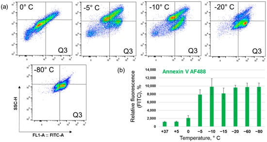
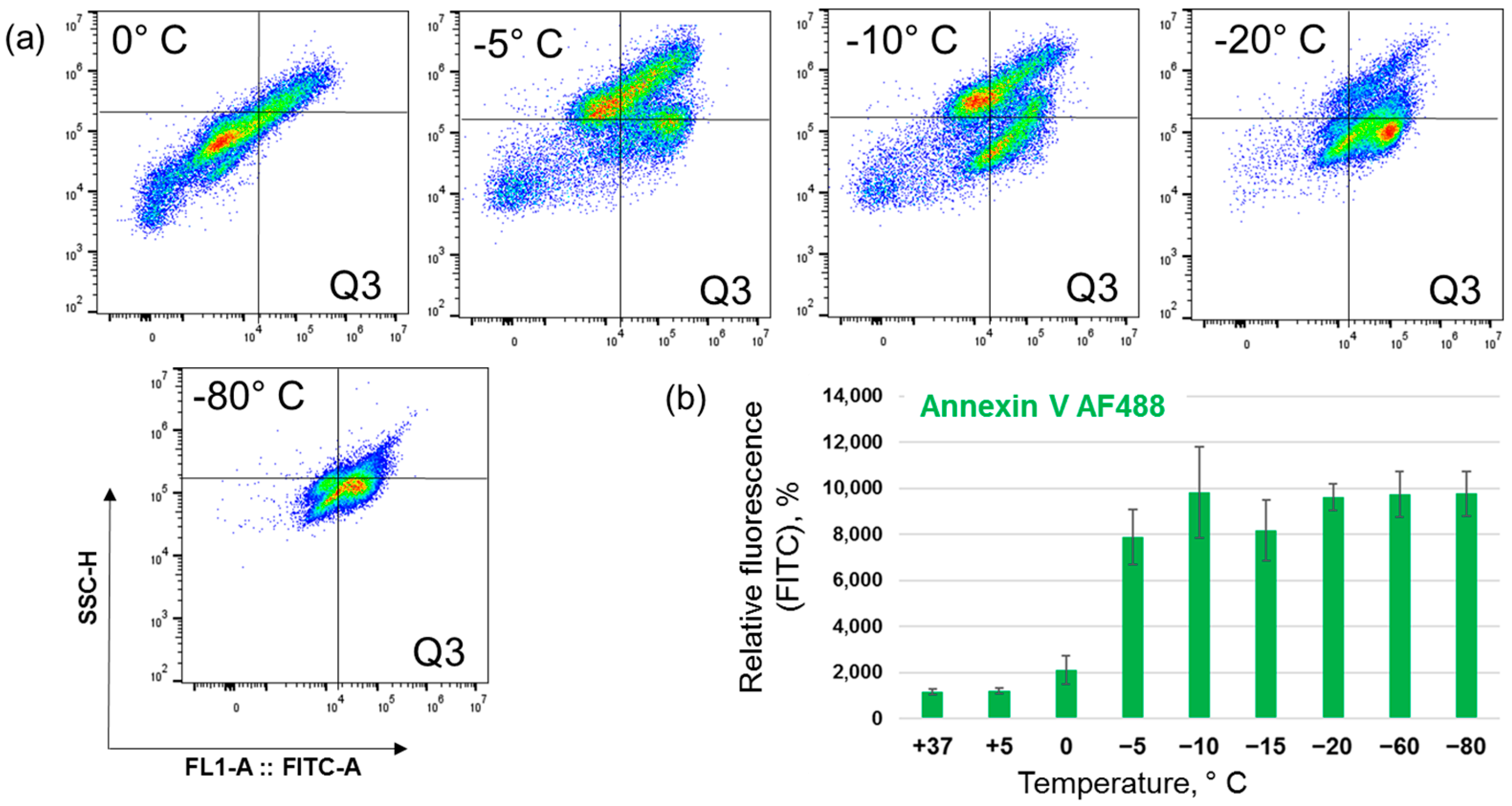
A monolayer of 4T1-Luc triple-negative breast cancer cells was placed in fetal bovine serum and incubated at various temperatures (+37, +5, 0, −5, −10, −15, −20, −50, and −80 °C) for 20 min. Following treatment, the cells were returned to standard culture conditions and incubated for 24 h. Cells were then trypsinized, washed, stained for markers of apoptosis and analyzed via flow cytometry. Each experimental series was performed in triplicate; relative fluorescence intensity values were averaged across three independent experiments. First, apoptosis was assessed by detecting phosphatidylserine (PS) exposure using Annexin V-AF488 staining. Representative flow cytometry dot plots for cells incubated at 0 °C, −5 °C, −10 °C, −20 °C, and −80 °C are presented in Figure 1a. The relative fluorescence signals from the Q3 quadrant, representing apoptotic events across all experimental groups (n = 3 for each), are summarized in Figure 1b.
Figure 1.
Quantification of apoptotic 4T1-Luc cells with flow cytometry after Annexin V-AF488 staining. Cells were exposed to various temperatures for 20 min, returned to standard culture conditions for 24 h, trypsinized, stained with Annexin V, and analyzed by flow cytometry. (a) Representative flow cytometry dot plots for cells treated at 0 °C, −5 °C, −10 °C, −15 °C, −20 °C, and −80 °C. The color coding is utilized to indicate the local density of events: blue/cool colors represent areas of low event density, while red/hot colors denote areas of high event density. (b) Median fluorescence intensity (MFI) in the Q3 quadrant quantified across all experimental conditions. Values are expressed as the mean of results of independent experiments (n = 3) ± SD.
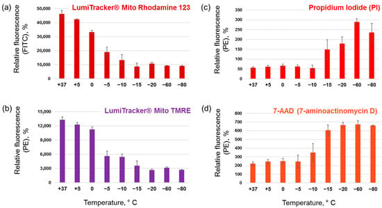
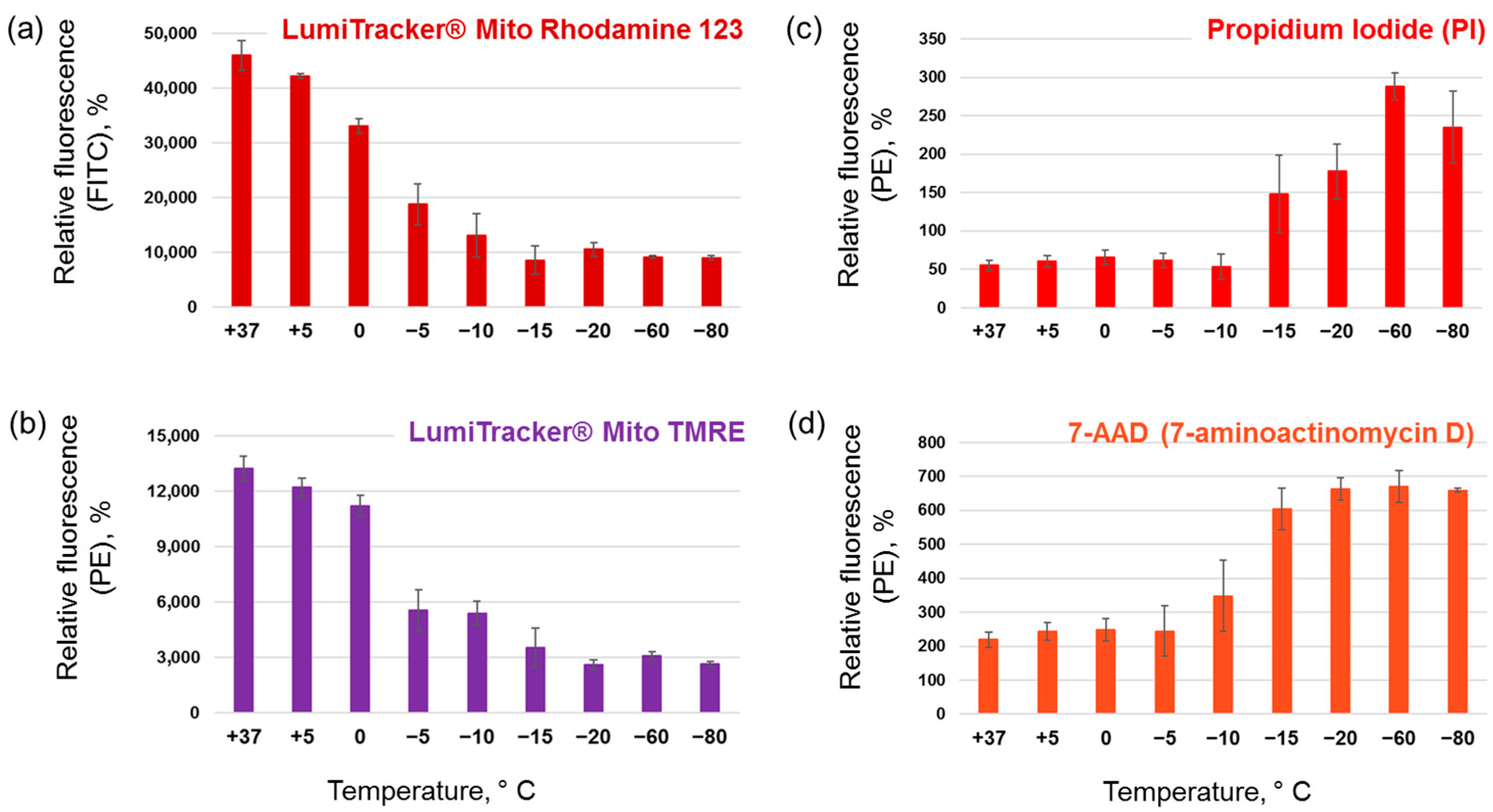
A 20 min exposure at −5 °C was sufficient to induce apoptosis-associated changes in membrane composition in a subpopulation of cells, whereas temperatures of −10 °C and below resulted in widespread alterations across the entire cell population. To further investigate these changes, we assessed mitochondrial membrane potential using Rhodamine 123 and TMRE staining. Representative dot plots are provided in the Supplementary Information (Supplementary Figures S1 and S2), and quantitative results are shown in Figure 2a,b. Both assays revealed that even exposure to +5 °C triggered mitochondrial outer membrane permeabilization. Progressive decreases in temperature gradually reduced mitochondrial functionality, with temperatures of −15 °C and below leading to uniform mitochondrial membrane depolarization.
Figure 2.
Profiling of mitochondrial membrane potential and cell membrane integrity in mouse cells. (a,b) Mitochondrial membrane potential was assessed in 4T1-Luc cells exposed to various temperatures for 20 min, followed by 24 h of recovery, using (a) Rhodamine 123 and (b) TMRE staining. (c,d) Cell membrane integrity was evaluated in 4T1-Luc cells immediately following a 20 min temperature exposure by staining with (c) Propidium Iodide (PI) and (d) 7-AAD. Graphs represent the median fluorescence intensity (MFI) quantified across all experimental conditions. Values are expressed as the mean of results of independent experiments (n = 3) ± SD.
It is well accepted that freezing induces intracellular ice crystal formation and osmotic dehydration, leading to the disintegration of the cell surface membrane and immediate cell death. To evaluate the correlation between freezing depth and these phenomena, cells were treated with a range of temperatures, immediately trypsinized, and stained with the nucleotropic dyes Propidium Iodide (PI) and 7-aminoactinomycin D (7-AAD). Raw flow cytometry data are presented in the Supplementary Information (Supplementary Figures S3 and S4), and quantitative results are shown in Figure 2c,d. Our findings indicate that cell surface and nuclear membrane barrier function began to be compromised after exposure to −10 °C (7-AAD) or −15 °C (PI), with complete disintegration observed after exposure at −60 °C. These observations are consistent with direct cryoablation-induced cell destruction, leading to necrosis.
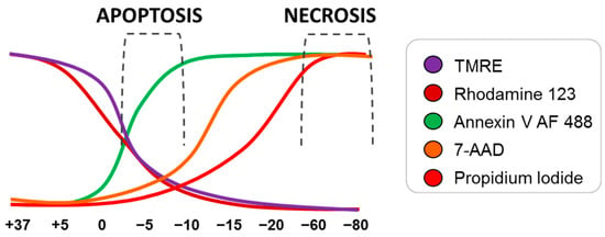
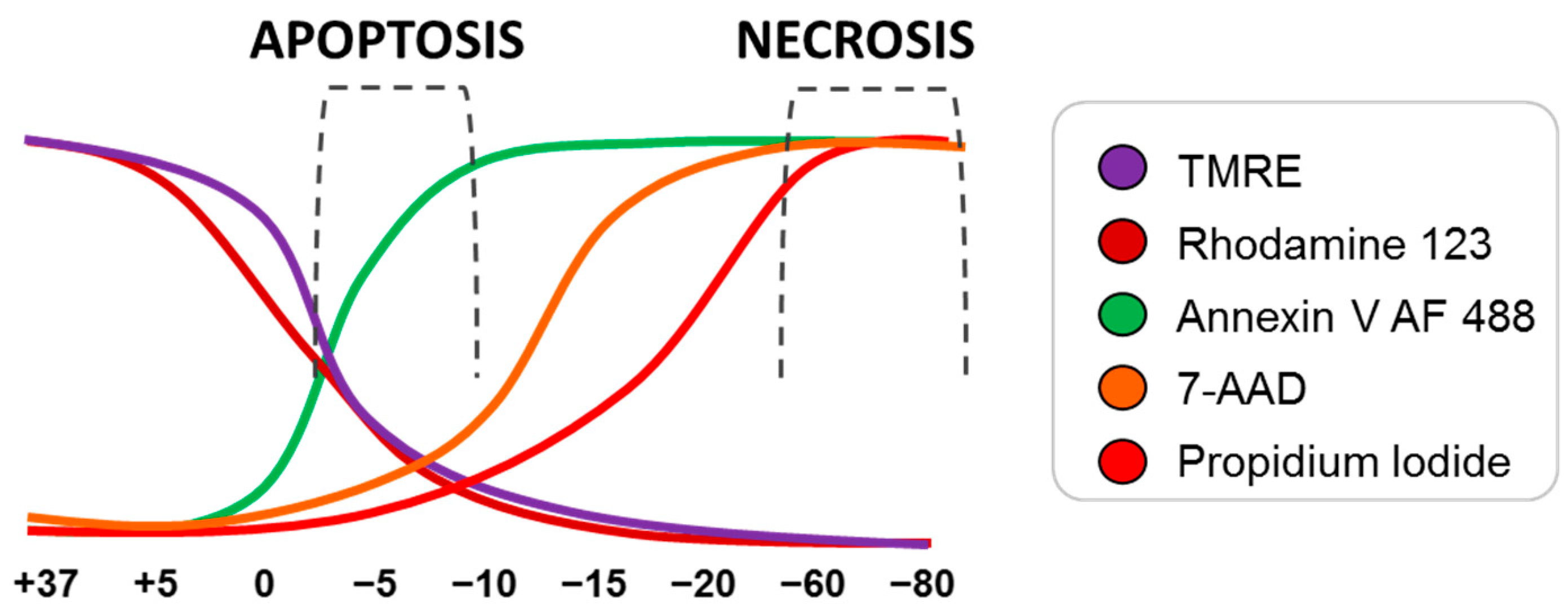
The findings are summarized in Figure 3, which illustrates the transition of cellular characteristics from apoptosis-related to necrosis-related across the range of exposure temperatures. Due to the natural heterogeneity of the cell population and the complex mechanisms of cell death, it is impossible to define temperature conditions that exclusively induce apoptosis or necrosis. It is evident that exposure at a specific temperature resulted in the formation of a mixture of cells in states of apoptosis and necrosis, in varying proportions. Nevertheless, based on the obtained results, we identified −7 °C (which predominantly induced apoptosis) and −80 °C (which predominantly induced necrosis) as the target temperatures for subsequent experiments.
Figure 3.
Profile of apoptosis- and necrosis-related alterations induced by freezing of 4T1-Luc cells (scheme). Intervals of temperature associated with mostly apoptosis or necrosis are indicated.
3.2. Reaction of Human Breast Cancer Cells to Exposure at −7 °C and −80 °C
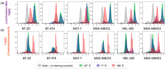
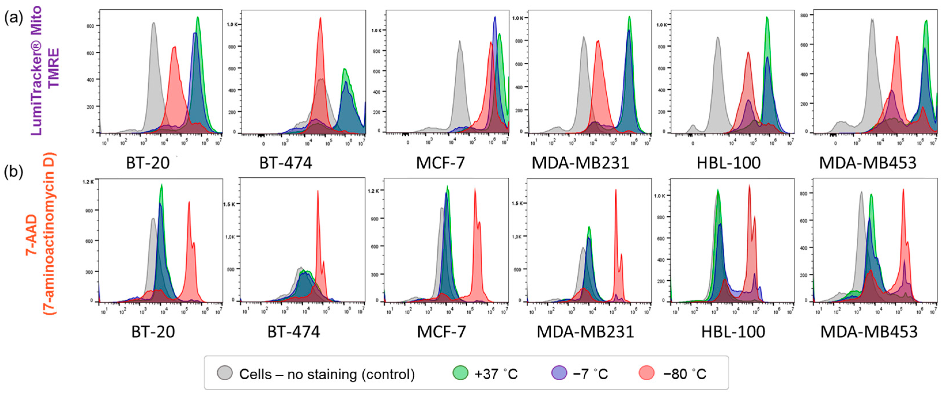
To confirm the generality of observed phenomena, five human breast cancer cell lines with varying phenotypes and malignant potentials (BT-20, BT-474, MCF-7, MDA-MB-231, and MDA-MB-453) were tested, alongside the HBL-100 cell line, derived from the breast milk of a healthy donor. These experiments were conducted as a confirmatory validation set to ensure the generalizability of our findings observed in the murine model. Cells were subjected to a 20 min exposure at either sub-lethal (−7 °C) or lethal (−80 °C) temperatures, using the previously described conditions. Mitochondrial membrane potential and cell membrane integrity were subsequently assessed via TMRE and 7-AAD staining, respectively. As shown in Figure 4a, the electrical charge across the inner mitochondrial membrane was moderately reduced in all tested cell lines following sub-lethal freezing (−7 °C), although the extent of this reduction varied. In contrast, lethal freezing (−80 °C) resulted in a substantial decline in mitochondrial functionality across all cases. Notably, in all cell lines except BT-474, TMRE staining intensity remained higher than that of non-stained control cells following lethal freezing, suggesting either a basal level of background TMRE staining or residual mitochondrial stability despite lethal thermal injury.
Figure 4.
Assessment of mitochondrial membrane potential and cell membrane integrity in human cells. Mitochondrial membrane potential (a) was evaluated using the potentiometric dye tetramethylrhodamine ethyl ester (TMRE). Cell membrane integrity (b) was assessed after staining with 7-aminoactinomycin D (7-AAD). Flow cytometry was used to quantify the fluorescence signal distribution in the PE channel. Histograms depict fluorescence intensity from control cells (no staining), cells cultured under normal conditions (staining baseline), cells subjected to sub-lethal temperature exposure (−7 °C), and cells exposed to lethal temperature (−80 °C).
Figure 4b illustrates the results of the cell surface membrane permeability assay. Exposure to the sub-lethal temperature (−7 °C) did not induce detectable membrane disintegration. Conversely, exposure to the lethal temperature (−80 °C) resulted in membrane rupture and significant nuclear staining in all six cell lines. These results indicate that sub-lethal freezing induces programmed cell death, whereas lethal freezing leads to direct cellular breakdown—a phenomenon that is consistently reproduced across diverse breast cancer cell lines
3.3. Effect of Apoptotic and Necrotic Cryo-Vaccine on s.c. Tumor Growth
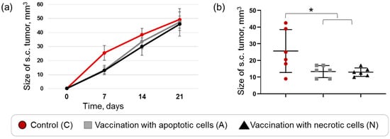
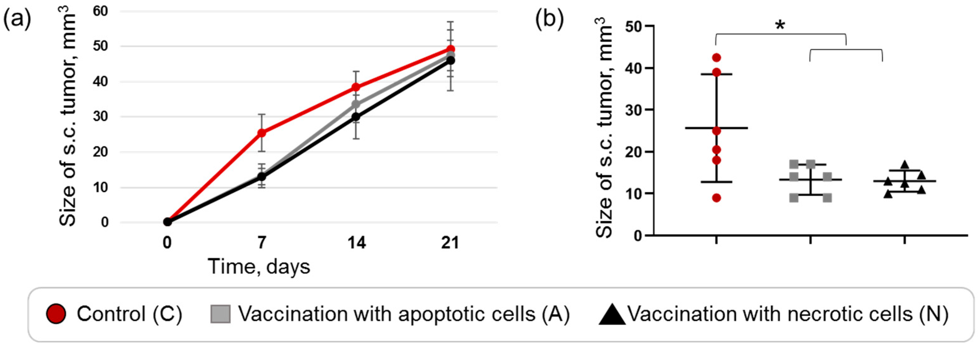
To investigate the ability of cryoablated cells to stimulate a specific antitumor immune response, we utilized a syngeneic murine model with 4T1-Luc cells. Mice were divided into three experimental groups: the first group was vaccinated with sub-lethally frozen cells (−7 °C), the second with lethally frozen cells (−80 °C), and the third served as an unvaccinated control group. Vaccination consisted of a single subcutaneous injection of a cryo-treated cell suspension. Ten days post-vaccination, tumor growth was not detected in any of the mice; consequently, all animals were challenged with a subcutaneous injection of viable 4T1-Luc cells into each flank. Tumor progression was monitored weekly (Figure 5a).
Figure 5.
Effect of mice vaccination by tumor cells subjected to either sub-lethal or lethal cryoablation. To evaluate the immunogenicity of cryoablated cells, mice were immunized via subcutaneous injection with a suspension of either sub-lethally (−7 °C) or lethally (−80 °C) frozen tumor cells. Ten days post-vaccination, mice were challenged with a subcutaneous injection of viable tumor cells in both flanks. The results for each experimental group are represented by the colors defined in the figure legend. (a) Tumor progression was monitored weekly. Data represent the mean tumor volume for groups of six animals. (b) Tumor volume assessment at 7 days post-challenge. Each data point represents the mean volume of two subcutaneous (s.c.) tumors per animal. Statistical significance between the control group (n = 6) and the combined treatment group (n = 12) was determined using the Mann–Whitney U test. Significant differences are indicated by asterisks * indicating p < 0.0227.
Immunization with either apoptotic or necrotic tumor cells resulted in an initial suppression of tumor growth. This antitumor effect was observable at 7 days post-subcutaneous tumor challenge, with statistically significant differences between both immunized groups and the non-immunized control group (Figure 5b). However, this protective effect was transient; by day 21, tumor sizes were comparable across all experimental groups.
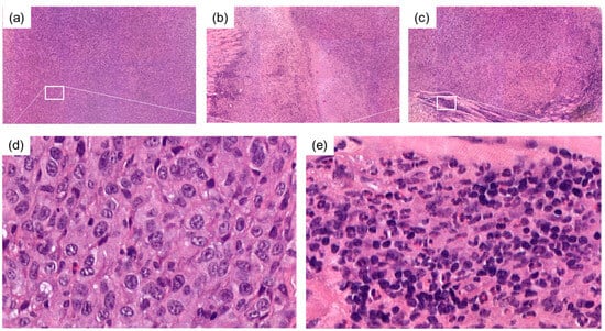
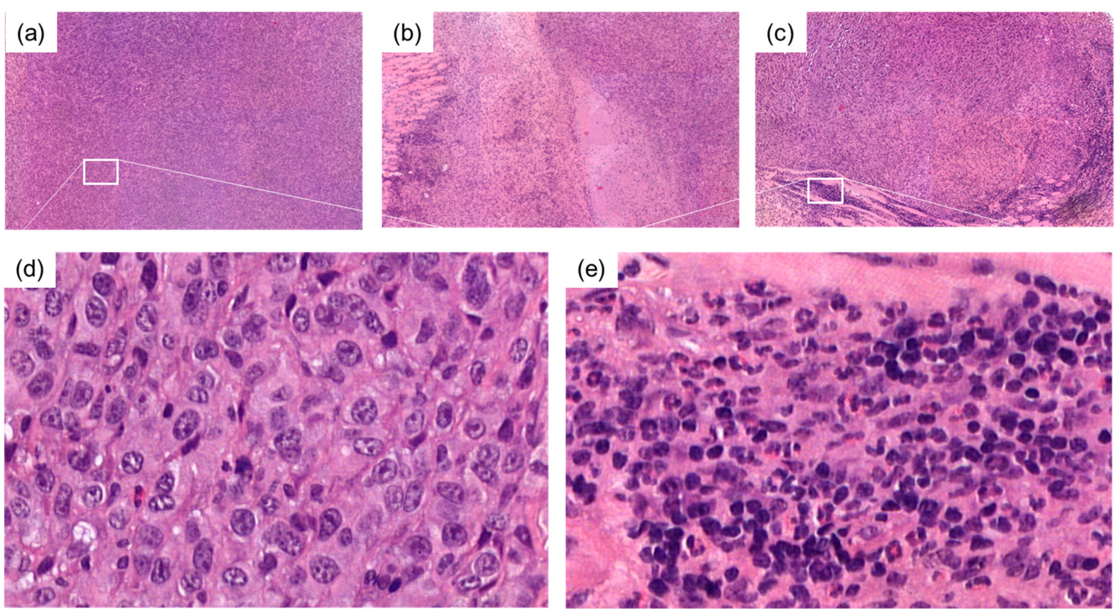
To gain insight into the mechanisms underlying the observed antitumor immune response, we performed hematoxylin and eosin (H&E) staining of tumor tissues (representative images are shown in Figure 6).
Figure 6.
Histological assessment of 4T1-Luc syngeneic subcutaneous tumors. Hematoxylin and Eosin (H&E) staining of tumor tissue harvested from mice. (a) Representative image from the control (non-immunized) group. (b,c) Representative images from mice immunized with suspension of sub-lethally (−7 °C) and lethally (−80 °C) frozen tumor cells, respectively. Panels (a–c) were acquired at 5× magnification. Panels (d,e) represent high-magnification (80×) views of the regions indicated by boxes in the corresponding low-magnification images, illustrating local tissue architecture and inflammatory infiltration.
Low-magnification images (5× objective; Figure 6a–c) show the overall tissue architecture. Tumors from non-immunized mice (Figure 6a) displayed a uniform morphology, whereas tumors from both immunized groups exhibited distinct regions of lymphocytic infiltration and necrosis (Figure 6b,c). High-magnification images of these regions (80× objective; Figure 6d,e) confirm signs of immune cell activation in the tumors of mice vaccinated with apoptotic cells (Figure 6e) compared to the non-immunized controls (Figure 6d). The presented images serve as a general illustration of the immune response within the tumor tissue of vaccinated animals. While these qualitative data are informative, a quantitative assessment of immune cell infiltration and inflammatory markers would be required to fully elucidate the underlying mechanisms and confirm these observations.
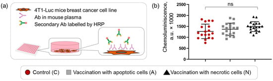
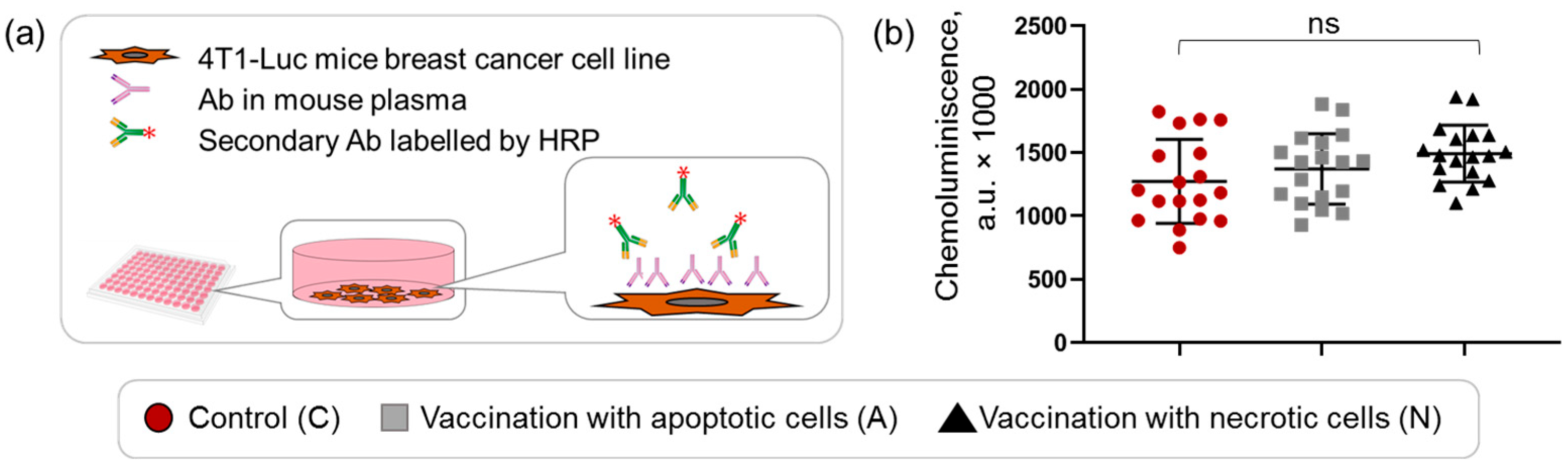
To assess the involvement of humoral immunity, we quantified tumor-specific antibodies using a modified enzyme-linked immunosorbent assay (ELISA). The principle of the assay is outlined in Figure 7a. 4T1-Luc cells were seeded in 96-well plates and incubated with diluted mouse plasma. Following washing, tumor-specific antibodies were detected using HRP-labeled secondary rabbit anti-mouse antibodies. Each assay was performed in triplicate, and the results are presented in Figure 7b. While we observed an upward trend in antibody levels in the immunized mice, these differences did not reach statistical significance. It should be noted that blood samples were collected at the end of the experiment; by this time, all mice—including the non-immunized controls—had been exposed to the tumor challenge, likely resulting in a robust systemic humoral response in all groups that may have masked the initial vaccine-induced antibody production.
Figure 7.
Quantification of tumor-specific antibodies in mouse plasma by ELISA. (a) Schematic representation of the ELISA protocol. (b) Chemiluminescence intensity was measured following incubation with an HRP-conjugated secondary antibody and substrate. Data are presented as triplicate measurements for each plasma sample (animal). The results for each experimental group are represented by the colors defined in the figure legend. Difference between the groups was statistically not significant (ns).
3.4. Effect of Apoptotic and Necrotic Cryo-Vaccine on Lung Metastasis Formation
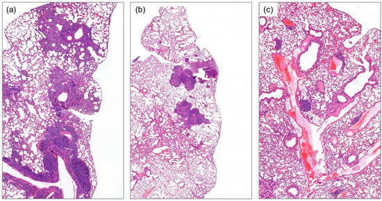
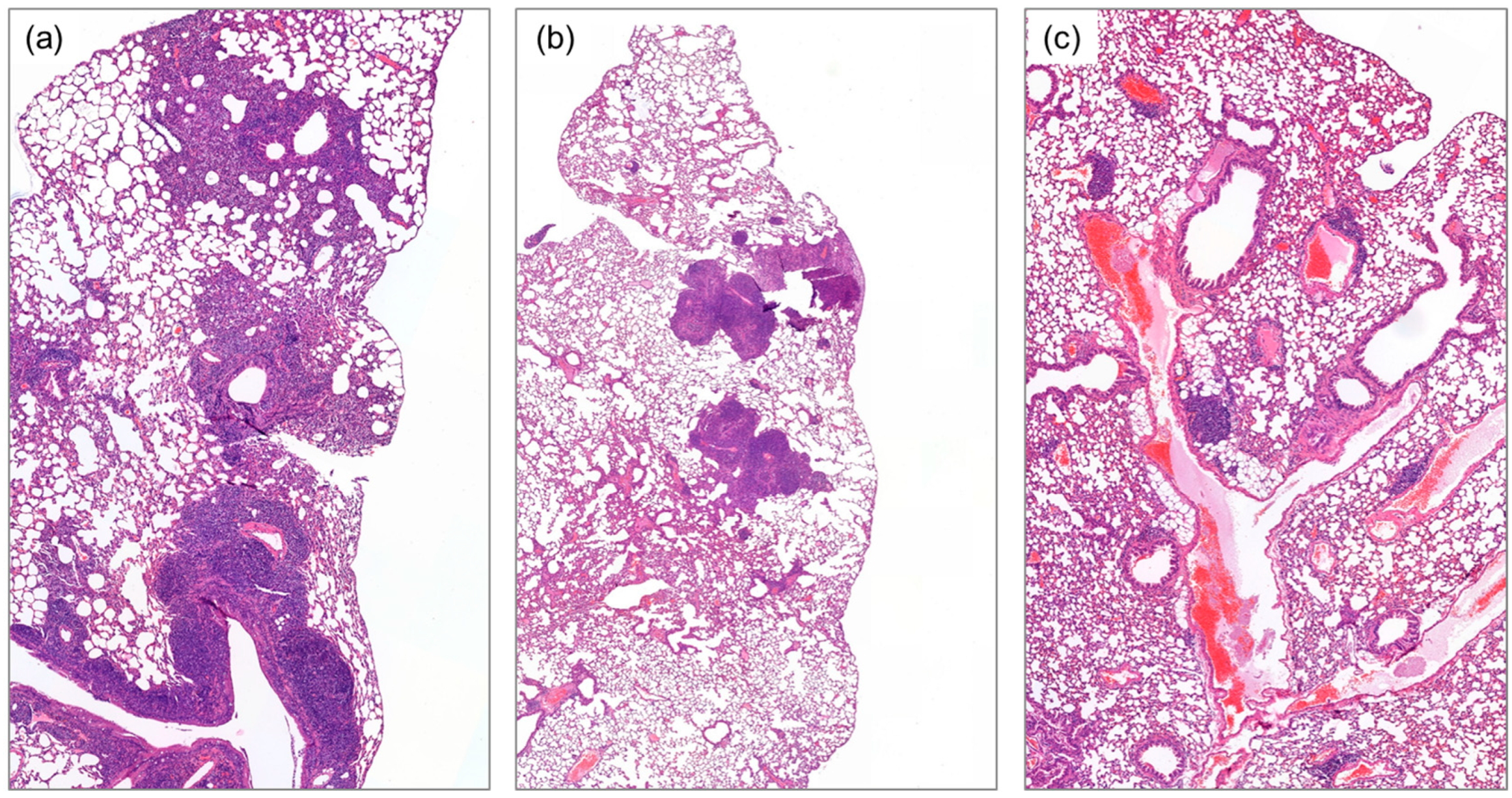
The final experiment was aimed at assaying the effect of “cryo-vaccination” on the ability of circulating tumor cells to form metastasis. Injection of tumor cell suspension in the tail vein provides a simplified model of lung metastasis development, whereas stable transfection with the luciferase gene provides us with the option of performing accurate quantification of metastatic 4T1-Luc cell borders. Mice were subdivided into three groups and vaccinated the same way as described above. After ten days, 4T1-Luc cell suspensions were slowly injected into the tail vein. Samples were harvested after 28 days, and lung “metastases” were visualized in all animals (Figure 8).
Figure 8.
Histological assessment of lung metastases formed by 4T1-Luc after intravenous injection. Hematoxylin and eosin (H&E) staining of lung tissue harvested from mice; images were acquired at 5× magnification. (a) Representative image from the control (non-immunized) group. (b,c) Representative images from mice immunized with suspension of sub-lethally (−7 °C) and lethally (−80 °C) frozen tumor cells, respectively.
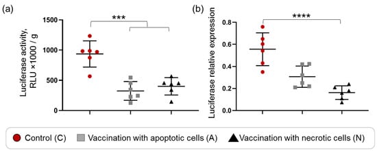
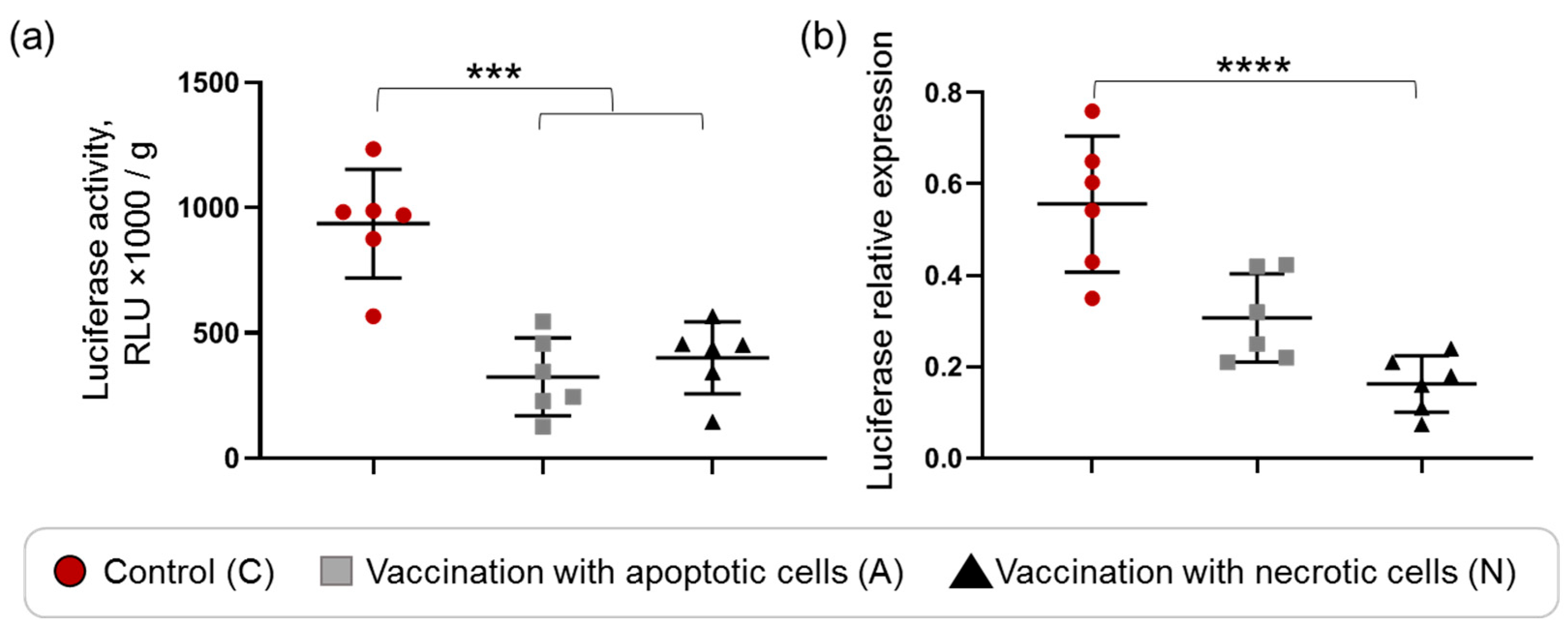
Moreover, lung tissue from each animal was analyzed using two independent approaches. First, luciferase activity was measured via the bioluminescence reaction; these results, normalized to lung tissue weight, are presented in Figure 9a. This assay revealed a statistically significant difference between the control group and the combined cryo-vaccinated groups. While the difference between the apoptotic and necrotic vaccination groups was observable, it did not reach statistical significance (Figure 9a).
Figure 9.
Quantification of 4T1-Luc metastatic cells in lungs of mice. The results for each experimental group are represented by the colors defined in the figure legend. (a) Results of luciferase activity assay of lysed lung tissue. Normalized data versus assayed tissue weight. The statistical significance of differences between the control group (n = 6) and the combined treatment group (n = 12) was determined using the Mann–Whitney U test, with *** indicating p < 0.0002. (b) Results of luciferase gene quantification with PCR normalized versus mouse b-2-microglobin genes with parallel quantification. The statistical significance of differences between the three groups (n = 6 of each) was determined using the Kruskal–Wallis test, with **** indicating p < 0.00001.
Second, PCR-based quantification of the luciferase gene DNA generally corroborated the luciferase activity results; however, this more sensitive method revealed statistically significant differences between all three groups (Figure 9b). Specifically, vaccination with necrotic cells was more effective than vaccination with apoptotic cells in preventing the formation of lung metastases. Thus, assessment of lung metastasis formation performed by two independent approaches revealed higher immunogenic potency of lethally cryoablated necrotic tumor cells compared to apoptotic cells, which may reflect the involvement of the same molecular mechanism but at different degrees.
4. Discussion
4.1. Study Limitations
The present study has several limitations that must be acknowledged to ensure the correct interpretation of the results. First, the in vitro cell freezing model was designed to mimic the peripheral and central zones of clinical cryoablation (CA). However, this experimental system cannot fully recapitulate the complex and dynamic nature of clinical CA, which involves stromal interactions, vascular injury, and systemic inflammatory responses. Consequently, our findings establish a mechanistic baseline that provides a rationale for planning next experiments and for optimizing thermal protocols in future pre-clinical and clinical investigations.
Second, we acknowledge that freezing-induced cell death is a complex process involving both apoptotic and necrotic pathways, depending on the status and microenvironment of each cell. Assessing cell populations using a limited set of markers provides only a generalized estimation of prevalent cellular states. The specific thermal conditions (−7 °C and −80 °C) were selected to represent “predominantly apoptotic” and “predominantly necrotic” states, respectively, as a formalization necessary for the design of subsequent experiments.
Third, our study utilized a specific tumor model (4T1 murine breast cancer cells) and set of human breast cancer cell lines; therefore, the translatability of these results to other cancer types warrants further investigation. Future studies incorporating larger experimental cohorts, dynamic analysis of humoral immune responses, and a deeper characterization of tumor-infiltrating immune cells are essential to fully elucidate the nature of CA-induced antitumor immunity. These follow-up studies will be necessary to validate the preliminary findings of our current work.
4.2. Revisiting the Generally Accepted Hypothesis
In this study, we utilized two in vivo models to test the hypothesis that sub-lethal cryoablation induces an immunosuppressive effect via programmed cell death. Our results provided no evidence of suppressed antitumor immunity following vaccination with apoptotic tumor cells, thereby contradicting the prevailing hypothesis [2,12,13]. While the present study demonstrated the immunomodulatory potential of cryo-treated tumor cells, we did not evaluate specific molecular markers of immunogenic cell death. Future investigations into the role of canonical DAMPs (such as surface calreticulin exposure, HMGB1 release, and ATP secretion) as well as molecular mechanisms of immune cell activation will be essential to fully characterize the nature of the observed phenomenon. Although vaccination with apoptotic cells elicited a less robust antitumor response compared to necrotic cell vaccination in the lung metastases model, this may be attributed to a lower release of intracellular antigens and damage-associated molecular patterns (DAMPs) from sub-lethally injured cells. While our findings refute the aforementioned hypothesis, they are consistent with experimental evidence outside the field of cryoablation. For instance, the in vitro study demonstrated that apoptotic tumor cells are effectively phagocytosed by dendritic cells, which subsequently prime CD8+ cytotoxic T-lymphocytes more efficiently than either tumor cells alone or dendritic cells pulsed with crude tumor lysates [15].
4.3. Practical Relevance to Local Effect
For in vitro experiments, exposure to −7 °C and −80 °C was selected to model conditions predominantly inducing apoptosis-associated and necrosis-associated markers, respectively. However, in vivo results showed that vaccination with cells displaying either apoptotic or necrotic features elicited similar protective effects, albeit to varying degrees in a lung metastasis model. These findings suggest that, in the context of CA, apoptosis may not exert the high level of immune suppression that has historically been assumed.
These results hold significant practical implications for cryo-immunotherapy. Proceeding from the premise that apoptotic cells in the CA periphery are inherently immunosuppressive, many researchers have developed sophisticated strategies to circumvent this perceived limitation. For example, thermosensitive nanoparticles capable of releasing irinotecan and PD-L1-silencing siRNA at 4 °C have been proposed to stimulate antitumor responses in primary cryoablated tumors and promote long-term immunological memory [16]. Furthermore, pH-responsive gallium-based metal–organic framework (Ga-MOF) nanoplatforms have been engineered to co-deliver gallium ions and baicalein, simultaneously triggering ferroptosis and endoplasmic reticulum stress within the ablation zone [17]. In line with our findings, these studies demonstrate that sub-lethally cryoablated tumor tissue—which is not solely characterized by apoptotic events—possesses significant therapeutic potential that can be further enhanced by targeted local interventions.
4.4. Practical Relevance to Systemic Antitumor Immune Reaction
The observation that vaccination with both lethally and sub-lethally cryoablated tumor cells induces a transient antitumor effect is particularly significant. This highlights the potential for systemic immunomodulation to effectively enhance host responses to cryoablated tumor tissue. Among these strategies, the combination of cryoablation (CA) with the systemic administration of immune checkpoint inhibitors (CPIs) appears to be a particularly promising approach for overcoming the transient nature of this immunity.
For instance, Waitz et al. (2012) demonstrated the efficacy of combining CA with CTLA-4 blockade in a murine model of prostate adenocarcinoma [18]. In their study, the growth of distant, non-ablated tumors was significantly delayed or prevented following the combined CA with CPI treatment—an effect not observed with CA monotherapy. Furthermore, these distant tumors exhibited robust infiltration of CD4+ and CD8+ T cells, alongside an increased ratio of effector to regulatory T lymphocytes, signifying the induction of a potent, systemic antitumor immune response. Zhu et al. (2019) further evaluated the efficacy of combining CA with CPI blockade in a murine model of renal cell carcinoma [19]. In this study, mice bearing bilateral tumors were treated with CA applied to the primary tumor. In the contralateral (control) tumor, the authors observed a transient increase in CD8+ T-cell infiltration, concomitant with upregulated expression of IFN-γ and PD-L1. This indicates the formation of an adaptive regulatory loop: while CA effectively stimulates an antitumor immune response, the tumor subsequently counteracts this activation by upregulating PD-L1, which suppresses effector T-cell function. Comparing three treatment regimens (CA monotherapy, CA plus CPI, and CA plus isotype control), the authors found that only the combination of CA and CPI yielded a significant therapeutic effect. Similarly, Yu et al. (2023) utilized a murine breast cancer model to demonstrate a causal sequence of events [20]. CA induces a distinct but transient antitumor immune response in distant tumor tissue, which is subsequently dampened by the PD-1/PD-L1 checkpoint axis. Disrupting this axis with anti-PD-1 antibodies reactivates and sustains the immune response, resulting in a pronounced therapeutic outcome. More recently, Kao et al. (2026) employed a syngeneic mouse model of hepatocellular carcinoma to confirm earlier findings and elucidated two critical aspects of the CA and CPI combination concept [21]. Firstly, the authors optimized the combination therapy regimen: after establishing an orthotopic tumor, animals received neoadjuvant ICI therapy (anti-PD-1), with CA subsequently performed at the “peak” of the PD-1 blockade. Secondly, they demonstrated that partial CA was sufficient to achieve the desired therapeutic effect. These findings collectively established for the first time that CA can function not only as an “elegant” surgical technique but also as a pivotal component of systemic antitumor therapy.
4.5. Conclusions and Future Perspectives
Our findings reveal that complete and lethal cryoablation (CA) is not a prerequisite for stimulating an effective antitumor immune response. Rather, a strategy prioritizing the systemic enhancement of immune competence and reactivity appears to be a more efficacious approach. This paradigm shift is currently being validated in an expanding array of clinical trials investigating the combination of CA and CPIs for the treatment of triple-negative breast cancer (NCT05806385), melanoma (NCT05779423), non-small-cell lung cancer (NCT03290677), advanced biliary tract cancer (NCT05781074), gastric cancer (NCT06957977), and prostate cancer (NCT02423928). Consequently, future research should focus on further elucidating the molecular and cellular mechanisms of CA-induced antitumor immunity and developing methods for its systematic and controlled enhancement.
Supplementary Materials
The following supporting information can be downloaded at https://www.mdpi.com/article/10.3390/immuno6030056/s1. Figure S1: Mitochondrial membrane potential assessment by staining with Rhodamine123 and quantification by flow cytometry. Cells were exposed to indicated temperatures for 20 minutes, returned to standard culture conditions for 24 hours, trypsinized, stained with a potentiometric dye Rhodamine123 and analyzed by flow cytometry. Median fluorescence intensity (MFI) in the Q1 quadrant was quantified across all experiments and summarized in Figure 2a. Figure S2: Mitochondrial membrane potential assessment by staining with TMRE and quantification by flow cytometry. Cells were exposed to indicated temperatures for 20 minutes, returned to standard culture conditions for 24 hours, trypsinized, stained with a potentiometric dye tetramethylrhodamine ethyl ester (TMRE) and analyzed by flow cytometry. Median fluorescence intensity (MFI) in the Q1 quadrant was quantified across all experiments and summarized in Figure 2b. Figure S3: Cell surface membrane assessment by staining with Propidium Iodide (PI) and quantification by flow cytometry. Cells were exposed to indicated temperatures for 20 minutes, trypsinized, stained with a nucleotropic dye PI and analyzed by flow cytometry. Median fluorescence intensity (MFI) in the Q3 quadrant was quantified across all experiments and summarized in Figure 2c. Figure S4: Cell surface membrane assessment by staining with 7 aminoactinomycin D (7 AAD) and quantification by flow cytometry. Cells were exposed to indicated temperatures for 20 minutes, trypsinized, stained with a nucleotropic dye 7 AAD and analyzed by flow cytometry. Median fluorescence intensity (MFI) in the Q3 quadrant was quantified across all experiments and summarized in Figure 2d.
Author Contributions
Conceptualization, G.P. and A.M.; methodology, A.M., L.Z., V.E., A.G., and T.S.; morphology, A.K.; formal analysis, L.Z.; resources, G.P. and V.E.; data curation, A.M.; writing—original draft preparation, A.M.; writing—review and editing, G.P.; visualization, L.Z. and A.K.; supervision, G.P.; project administration, A.M.; funding acquisition, G.P. All authors have read and agreed to the published version of the manuscript.
Funding
This research was supported by a grant (24021600040-2) from the Ministry of Health of the Russian Federation for V.E.
Institutional Review Board Statement
The study was conducted in accordance with the Declaration of Helsinki. The animal study protocol was approved by the Ethic Committee of Petrov’s National Medical Research Center of oncology (protocol 22/47-№2 from 2 March 2020).
Data Availability Statement
Data are available upon request to the corresponding author.
Conflicts of Interest
The authors declare no conflicts of interest.
Abbreviations
| CA | Cryoablation |
| FBS | Fetal bovine serum |
| PBS | Phosphate-buffered saline |
| 7-AAD | 7-Aminoactinomycin D |
| TMRE | Tetramethylrhodamine ethylester |
| PI | Propidium iodide |
| CPI | Check point inhibitor |
References
- Xu, K.; Korpan, N.N.; Niu, L. Modern Cryosurgery for Cancer; World Scientific: Singapore, 2012; ISBN 978-981-4329-65-1. [Google Scholar]
- Yakkala, C.; Chiang, C.L.-L.; Kandalaft, L.; Denys, A.; Duran, R. Cryoablation and Immunotherapy: An Enthralling Synergy to Confront the Tumors. Front. Immunol. 2019, 10, 2283. [Google Scholar] [CrossRef] [Scilit] [PubMed]
- Maccini, M.; Sehrt, D.; Pompeo, A.; Chicoli, F.A.; Molina, W.R.; Kim, F.J. Biophysiologic considerations in cryoablation: A practical mechanistic molecular review. Int. Braz. J. Urol. 2011, 37, 693–696. [Google Scholar] [CrossRef] [Scilit] [PubMed][Green Version]
- Yiu, W.; Basco, M.T.; Aruny, J.E.; Cheng, S.W.; Sumpio, B.E. Cryosurgery: A review. Int. J. Angiol. 2007, 16, 1–6. [Google Scholar] [CrossRef] [Scilit] [PubMed]
- Yagi, T.; Hardin, J.A.; Valenzuela, Y.M.; Miyoshi, H.; Gores, G.J.; Nyberg, S.L. Caspase Inhibition Reduces Apoptotic Death of Cryopreserved Porcine Hepatocytes. Hepatology 2001, 33, 1432–1440. [Google Scholar] [CrossRef] [Scilit] [PubMed]
- Yang, W.; Addona, T.; Nair, D.G.; Qi, L.; Ravikumar, T.S. Apoptosis induced by cryo-injury in human colorectal cancer cells is associated with mitochondrial dysfunction. Int. J. Cancer 2003, 103, 360–369. [Google Scholar] [CrossRef] [Scilit] [PubMed]
- Ismail, M.; Morgan, R.; Harrington, K.; Davies, J.; Pandha, H. Enhancing prostate cancer cryotherapy using tumour necrosis factor related apoptosis-inducing ligand (TRAIL) sensitisation in an in vitro cryotherapy model. Cryobiology 2009, 59, 207–213. [Google Scholar] [CrossRef] [Scilit] [PubMed]
- You, Z.; Zhang, T.; Dai, L.; Wen, J.; Liu, M.; Zhao, T.; Yin, G.; Wu, Y.; Zhang, S. Effectiveness and Mechanism of Cryoablation in the Treatment of Oral Mucosal Melanoma. Cancer Med. 2026, 15, e71577. [Google Scholar] [CrossRef] [Scilit] [PubMed]
- Cao, Y.; Li, X.; Gong, W.; Tan, J.; Shen, J.; Zhou, X. Sequential cryoablation enhances chemotherapeutic efficacy in pancreatic cancer by downregulating TGF-β mediated fibrosis in the sub-lethal zone. J. Therm. Biol. 2026, 139, 104503. [Google Scholar] [CrossRef] [Scilit] [PubMed]
- Panfil, A.; Boroń, K.; Sirek, T.; Sirek, A.; Zmarzły, N.; Wróbel, M.; Wróbel, Z.; Boroń, D.; Ossowski, P.; Stefaniak, M.; et al. Apoptotic Signaling Across Breast Cancer Subtypes and Cryoablation-Induced Tissue Injury. Int. J. Mol. Sci. 2026, 27, 5174. [Google Scholar] [CrossRef] [Scilit] [PubMed]
- Sun, E.W.; Shi, Y.F. Apoptosis: The quiet death silences the immune system. Pharmacol. Ther. 2001, 92, 135–145. [Google Scholar] [CrossRef] [Scilit] [PubMed]
- Aarts, B.M.; Klompenhouwer, E.G.; Rice, S.L.; Imani, F.; Baetens, T.; Bex, A.; Horenblas, S.; Kok, M.; Haanen, J.B.A.G.; Beets-Tan, R.G.H.; et al. Cryoablation and immunotherapy: An overview of evidence on its synergy. Insights Imaging 2019, 10, 53. [Google Scholar] [CrossRef] [Scilit] [PubMed]
- Liu, Q.; Zhang, C.; Chen, X.; Han, Z. Modern cancer therapy: Cryoablation meets immune checkpoint blockade. Front. Oncol. 2024, 14, 1323070. [Google Scholar] [CrossRef] [Scilit] [PubMed]
- Malek, A.; Catapano, C.V.; Czubayko, F.; Aigner, A. A sensitive polymerase chain reaction-based method for detection and quantification of metastasis in human xenograft mouse models. Clin. Exp. Metastasis 2010, 27, 261–271. [Google Scholar] [CrossRef] [Scilit] [PubMed]
- Hoffmann, T.K.; Meidenbauer, N.; Dworacki, G.; Kanaya, H.; Whiteside, T.L. Generation of tumor-specific T-lymphocytes by cross-priming with human dendritic cells ingesting apoptotic tumor cells. Cancer Res. 2000, 60, 3542–3549. [Google Scholar] [PubMed]
- Ou, W.; Stewart, S.; White, A.; Kwizera, E.A.; Xu, J.; Fang, Y.; Shamul, J.G.; Xie, C.; Nurudeen, S.; Tirada, N.P.; et al. In-situ cryo-immune engineering of tumor microenvironment with cold-responsive nanotechnology for cancer immunotherapy. Nat. Commun. 2023, 14, 392. [Google Scholar] [CrossRef] [Scilit] [PubMed]
- Chen, Y.; Zhang, C.; Wang, X.; Yu, Z.; Hu, K.; Rao, W.; Zhou, T. Self-amplifying cryo-immunotherapy enabled by Ga-MOF nanoplatform for metastatic lung cancer. Chem. Eng. J. 2025, 525, 170334. [Google Scholar] [CrossRef] [Scilit]
- Waitz, R.; Solomon, S.B.; Petre, E.N.; Trumble, A.E.; Fassò, M.; Norton, L.; Allison, J.P. Potent Induction of Tumor Immunity by Combining Tumor Cryoablation with Anti–CTLA-4 Therapy. Cancer Res. 2012, 72, 430–439. [Google Scholar] [CrossRef] [Scilit] [PubMed]
- Zhu, C.; Lin, S.; Liang, J.; Zhu, Y. PD-1 blockade enhances the anti-tumor immune response induced by cryoablation in a murine model of renal cell carcinoma. Cryobiology 2019, 87, 86–90. [Google Scholar] [CrossRef] [Scilit] [PubMed]
- Yu, Z.-P.; Sun, X.-W.; He, Y.-P.; Gu, J.; Jin, Y. PD-1 monoclonal antibodies enhance the cryoablation-induced antitumor immune response: A breast cancer murine model research. Int. J. Hyperth. 2023, 40, 2164625. [Google Scholar] [CrossRef] [Scilit] [PubMed]
- Kao, T.; Santana, J.G.; Meister, E.; Shewarega, A.; Israel, J.; Peschke, L.M.H.; Tefera, J.; Matuschewski, N.; Sobirey, R.; Zhang, X.; et al. Neoadjuvant PD-1 Inhibition prior to Partial Cryoablation of Murine Hepatocellular Carcinoma Modulates the Tumor Microenvironment toward Favorable Immunological Profiles. J. Vasc. Interv. Radiol. 2026, 37, 108585. [Google Scholar] [CrossRef] [Scilit] [PubMed]
Disclaimer/Publisher’s Note: The statements, opinions and data contained in all publications are solely those of the individual author(s) and contributor(s) and not of MDPI and/or the editor(s). MDPI and/or the editor(s) disclaim responsibility for any injury to people or property resulting from any ideas, methods, instructions or products referred to in the content. |
© 2026 by the authors. Licensee MDPI, Basel, Switzerland. This article is an open access article distributed under the terms and conditions of the Creative Commons Attribution (CC BY) license.