


Abstract
Chronic nasopharyngeal inflammation represents a state of persistent mucosal immune dysregulation within the nasopharynx, a key inductive site of upper airway immunity. In this review, we propose a hypothesis-generating conceptual framework in which disease progression reflects a sequential shift in dominant immune pathways—from early CD4+ T-cell-driven activation, through to T helper 17 (Th17)-mediated epithelial barrier dysfunction, and ultimately to CD8+ T-cell-mediated cytotoxic injury. These transitions are characterized by dynamic alterations in CD4/CD8 and Th17/Treg balance, contributing to epithelial fragility and sustained inflammatory activity. Beyond local pathology, prolonged inflammation may engage neuro-immune circuits, including vagal and neuroendocrine pathways, potentially linking mucosal immune activation to systemic physiological dysregulation. While several elements of this model are supported by existing knowledge in mucosal immunology, the integration of local and systemic immune processes remains partly hypothetical and requires further experimental validation. Epipharyngeal Abrasive Therapy (EAT) may represent a potential intervention within this framework, although its immunological mechanisms remain incompletely understood. However, the underlying mechanisms and clinical efficacy of EAT remain to be fully elucidated. This framework integrates local mucosal immune dynamics with potential systemic consequences and provides a conceptual basis for future investigations into chronic mucosal inflammation. It is intended as a hypothesis-generating conceptual review that integrates existing observations and proposes a conceptual framework to guide future mechanistic and clinical studies.
1. Introduction
Chronic nasopharyngeal inflammation represents a persistent state of mucosal immune activation within the nasopharynx, an essential inductive site of upper airway immunity. In this review, “chronic nasopharyngeal inflammation” is used as the primary immunological term, while “chronic epipharyngitis” refers to the corresponding clinical condition commonly used in clinical practice. The nasopharynx contains specialized lymphoid structures and resident immune cell populations that orchestrate early immune responses to inhaled pathogens and environmental antigens. Recent studies have highlighted the nasopharynx-associated lymphoid tissue (NALT) as a critical hub for T-cell activation, cytokine production, and the establishment of localized immune memory, including CD4+, CD8+, and tissue-resident memory T cells (T_RM) [1,2,3].
Persistent inflammation in this region is increasingly recognized as a driver of epithelial remodeling, barrier dysfunction, and chronic disease. Histopathological analyses of chronic nasopharyngeal disorders demonstrate epithelial disruption, immune cell infiltration, and dysregulated cytokine signaling, suggesting that chronic inflammation is maintained by both structural and immunological alterations [4,5,6,7]. These findings align with broader concepts in mucosal immunology, where T helper 17 (Th17)-mediated inflammation, impaired regulatory T-cell (Tregs) activity, and cytotoxic CD8+ T-cell responses contribute to chronicity and tissue fragility.
Despite its clinical relevance, the immunological progression of chronic nasopharyngeal inflammation has not been systematically conceptualized. Building on established principles of mucosal immune regulation, we propose a multi-layered model describing a sequential shift in dominant immune pathways—from early CD4+ T-cell-driven activation, through to Th17-associated epithelial barrier dysfunction, and ultimately to CD8+ T-cell-mediated cytotoxic injury. This framework integrates cellular dynamics with structural changes and provides a mechanistic basis for understanding disease persistence.
Furthermore, emerging evidence suggests that nasopharyngeal inflammation may influence systemic physiology through neuro-immune pathways, including vagal and neuroendocrine signaling [8]. Although these systemic effects remain hypothetical, they offer a conceptual link between localized mucosal inflammation and broader homeostatic disturbances. Despite growing interest in chronic nasopharyngeal inflammation, important knowledge gaps remain. Longitudinal immunological studies are scarce, mucosal immune findings have not been systematically integrated with clinical observations, and no unifying framework currently links epithelial barrier dysfunction, T-cell dynamics, and potential systemic manifestations. The applicability of mechanisms established in other mucosal diseases to chronic nasopharyngeal inflammation remains uncertain and requires disease-specific validation.
This review proposes a conceptual framework to describe the immunological progression of chronic nasopharyngeal inflammation. While the model is grounded in established principles of mucosal immunology, several components—particularly those related to neuro-immune and systemic interactions—remain speculative and require further experimental validation. By integrating current knowledge with clinical observations, this framework aims to guide future mechanistic and translational research. To our knowledge, this is the first framework to integrate sequential immune dynamics, mucosal barrier dysfunction, and neuro-immune interactions specifically in the context of the nasopharynx.
2. Mucosal Immunity of the Nasopharynx
2.1. The Nasopharynx as an Immune Interface
The nasopharynx represents a critical anatomical and immunological interface between the external environment and the host immune system. Positioned at the crossroads of the respiratory and digestive tracts, this region is continuously exposed to airborne pathogens, environmental particles, and commensal microorganisms. The nasopharyngeal mucosa integrates structural and immunological components that collectively contribute to frontline defense and immune homeostasis [7,9].
Unlike peripheral tissues, the nasopharynx combines epithelial barrier function with organized and diffuse immune structures. The mucosal surface is lined by specialized epithelial cells that regulate pathogen entry while simultaneously sensing environmental stimuli through pattern recognition receptors. These epithelial cells actively participate in immune responses by producing cytokines, chemokines, and antimicrobial peptides, thereby shaping both innate and adaptive immunity [5,6].
In addition to epithelial components, the nasopharyngeal mucosa contains lymphoid aggregates and immune cell populations that support antigen sampling and presentation. These structures facilitate the activation of T and B lymphocytes and contribute to the induction of both local and systemic immune responses [1,3]. Together, these elements establish a dynamic immunological environment that balances effective pathogen defense with the prevention of excessive inflammation. These structural and immunological characteristics provide the biological basis for the sequential immune dynamics proposed in this review, in which dysregulated interactions between epithelial barriers and immune cell populations drive the progression of chronic nasopharyngeal inflammation.
2.2. Tissue-Resident Immunity and Local Immune Regulation
Recent advances in immunology have emphasized the importance of tissue-resident immune populations in mucosal defense. Among these, tissue-resident memory T cells (T_RM) constitute a key component of long-term immunity at barrier surfaces. In contrast to circulating memory T cells, T_RM cells remain localized within tissues, allowing rapid and site-specific responses upon antigen re-exposure [10,11].
T_RM cells are strategically positioned within epithelial and subepithelial compartments, where they function as sentinels of mucosal immunity. Upon activation, these cells rapidly produce effector cytokines and recruit additional immune cells, thereby amplifying local immune responses. In addition, subsets of CD8+ T_RM cells exert direct cytotoxic activity, enabling the immediate elimination of infected or damaged epithelial cells [10,12].
The activity of tissue-resident immune cells is tightly regulated to maintain immune equilibrium. Interactions between effector T cells and Tregs are essential to prevent excessive inflammation and tissue damage. This balance between activation and suppression is particularly critical in the nasopharynx, where continuous antigen exposure necessitates fine-tuned immune control [13,14].
Beyond cellular immunity, the epithelial barrier itself contributes to immune regulation. Epithelial cells respond dynamically to microbial and inflammatory stimuli, modulating immune cell recruitment and activation. This coordinated interaction between epithelial cells and immune populations forms a multilayered defense system that maintains mucosal integrity while preventing chronic inflammatory responses [6,7].
These structural and cellular features define the immunological landscape of the nasopharynx and provide the physiological basis for understanding the dysregulated immune processes described in subsequent sections. These regulatory interactions between tissue-resident immune cells, epithelial barriers, and local cytokine environments provide a critical foundation for the dynamic shifts in immune balance described in this review, particularly the transition from regulated mucosal homeostasis to chronic inflammatory dysregulation.
3. Immunological Features of Chronic Nasopharyngeal Inflammation
3.1. CD4+ T-Cell-Dominant Immune Activation
Chronic inflammation of the nasopharyngeal mucosa may involve early immune activation characterized by increased CD4+ T-cell responses, which play a central role in adaptive immunity. Rather than representing a uniform initiating event, CD4+ T-cell predominance may reflect one of the early immunological patterns associated with persistent antigen exposure [15].
In the nasopharyngeal mucosa, CD4+ T cells are abundantly distributed near the epithelial surface and can directly interact with antigens penetrating the mucosal barrier. Clinical evidence has demonstrated that patients with chronic nasopharyngeal inflammation exhibit a significant increase in CD4+ cells within nasopharyngeal abrasive samples, which decrease following repeated Epipharyngeal Abrasive Therapy (EAT) in parallel with symptomatic improvement [15].
This early phase is characterized by CD4+ T-cell predominance with relatively limited epithelial damage, suggesting a potentially reversible stage of inflammation. Regulatory mechanisms such as Treg induction may suppress excessive activation and restore immune homeostasis [16,17].
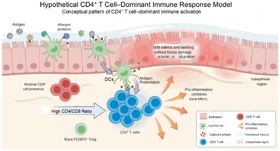
Persistent antigenic stimulation or impaired regulation may contribute to a more pro-inflammatory state, representing one possible pattern within the proposed model (Figure 1). However, direct evidence defining CD4+ T cells as the primary driver of disease initiation remains limited, and their role should be interpreted within a broader, context-dependent immune network [16,17].
Figure 1.
This figure illustrates a hypothesis-generating conceptual model and should not be interpreted as an experimentally validated mechanism. CD4+ T-cell-dominant immune activation pattern. Schematic representation of a possible immune activation pattern in chronic nasopharyngeal inflammation characterized by CD4+ T-cell-mediated immune activation. This model may involve increased CD4+ T-cell infiltration and cytokine production with minimal epithelial damage and may represent a potentially reversible inflammatory state.
3.2. Th17-Mediated Barrier Dysfunction
As inflammation persists, the immune response may shift toward a Th17-dominant state, representing a common pro-inflammatory pattern observed in chronic mucosal diseases [18].
Interleukin (IL)-17 promotes neutrophil recruitment and induces epithelial cells to release pro-inflammatory cytokines. Sustained activation results in epithelial barrier disruption and increased permeability, amplifying inflammatory signaling cascades [14].
Importantly, IL-6 plays a central role in Th17 differentiation and inflammatory amplification. Histological studies have demonstrated that IL-6 expression is highly upregulated in the nasopharyngeal submucosa in chronic nasopharyngeal inflammation and is significantly reduced following EAT [19].
Furthermore, spatial transcriptomic analysis has revealed that persistent inflammatory signaling, including the IL-6 and Tumor Necrosis Factor-alpha (TNF)–α pathways, remains activated in both epithelial and immune cell compartments of the nasopharynx in chronic inflammatory states, supporting the presence of sustained immune activation within the tissue microenvironment [20].
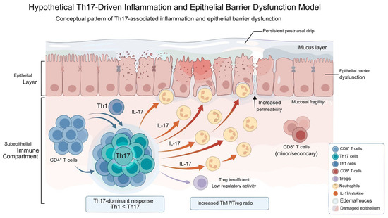
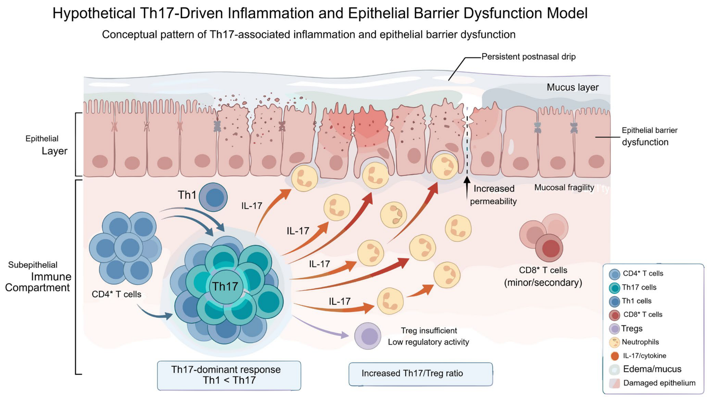
This hypothetical stage represents one possible pattern in which immune dysregulation may impair epithelial integrity and contribute to chronic inflammation (Figure 2). Although altered Th17/Treg balance has been reported in chronic epipharyngitis, longitudinal changes and the effects of EAT on this balance remain unknown. However, this transition is not universal and may vary depending on the underlying immune context and environmental factors [18].
Figure 2.
This figure illustrates a hypothesis-generating conceptual model and should not be interpreted as an experimentally validated mechanism. Th17-mediated epithelial barrier dysfunction. Illustration of a possible Th17-associated immune pattern. Production of IL-17 may promote neutrophil recruitment and inflammatory amplification and may be associated with the disruption of epithelial barrier integrity, potentially contributing to chronic inflammation. Blue arrows indicate immune cell interactions and activation pathways. Red arrows indicate IL-17-mediated inflammatory effects and epithelial barrier damage. Purple arrows indicate regula-tory pathways associated with Treg activity. * indicates epithelial tight junctions. Disruption of these structures may contribute to epithelial barrier dysfunction.
3.3. CD8+ T-Cell-Mediated Cytotoxic Injury
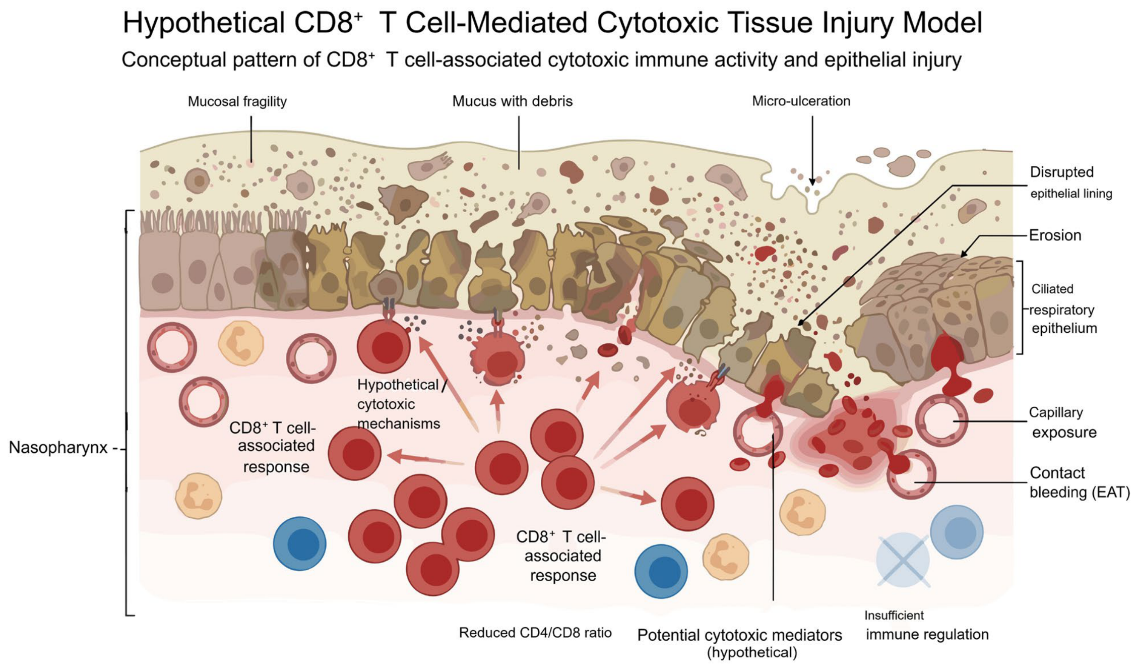
In one hypothetical stage of the proposed model, CD8+ cytotoxic T cells may become increasingly prominent, representing one of several effector pathways associated with tissue injury [21,22].
While essential for pathogen clearance, excessive activation may contribute to epithelial injury and mucosal damage. This may be associated with micro-ulceration, vascular exposure, and further amplification of inflammatory cascades [22].
Spatial transcriptomic data demonstrate that chronic inflammatory states are associated with persistent activation of immune signaling pathways, including T-cell receptor (TCR) signaling and cytotoxic immune responses, suggesting that prolonged immune activation contributes to tissue damage [20].
Notably, this stage represents a shift in dominant mechanisms rather than replacement; CD4+, Th17, and CD8+ responses coexist and interact dynamically within the inflammatory microenvironment (Figure 3) [18].
Figure 3.
This figure illustrates a hypothesis-generating conceptual model and should not be interpreted as an experimentally validated mechanism. CD8+ T-cell-mediated cytotoxic injury. Possible immune pattern associated with CD8+ T-cell accumulation. Potential cytotoxic mechanisms may contribute to epithelial injury; however, direct evidence for perforin- or granzyme-mediated tissue damage in chronic nasopharyngeal inflammation is currently unavailable. Arrows indicate the direction of CD8+ T cell-associated immune responses and potential tissue in-jury.
However, the extent to which CD8+ T-cell-mediated cytotoxicity drives disease progression in the nasopharynx remains incompletely defined and likely reflects context-dependent interactions with other immune cell populations. Direct evidence demonstrating perforin- or granzyme-mediated epithelial injury by CD8+ T cells in chronic nasopharyngeal inflammation is currently unavailable, and this proposed mechanism should therefore be regarded as hypothesis-generating [22].
3.4. Integrated Model of Sequential Immune Dynamics
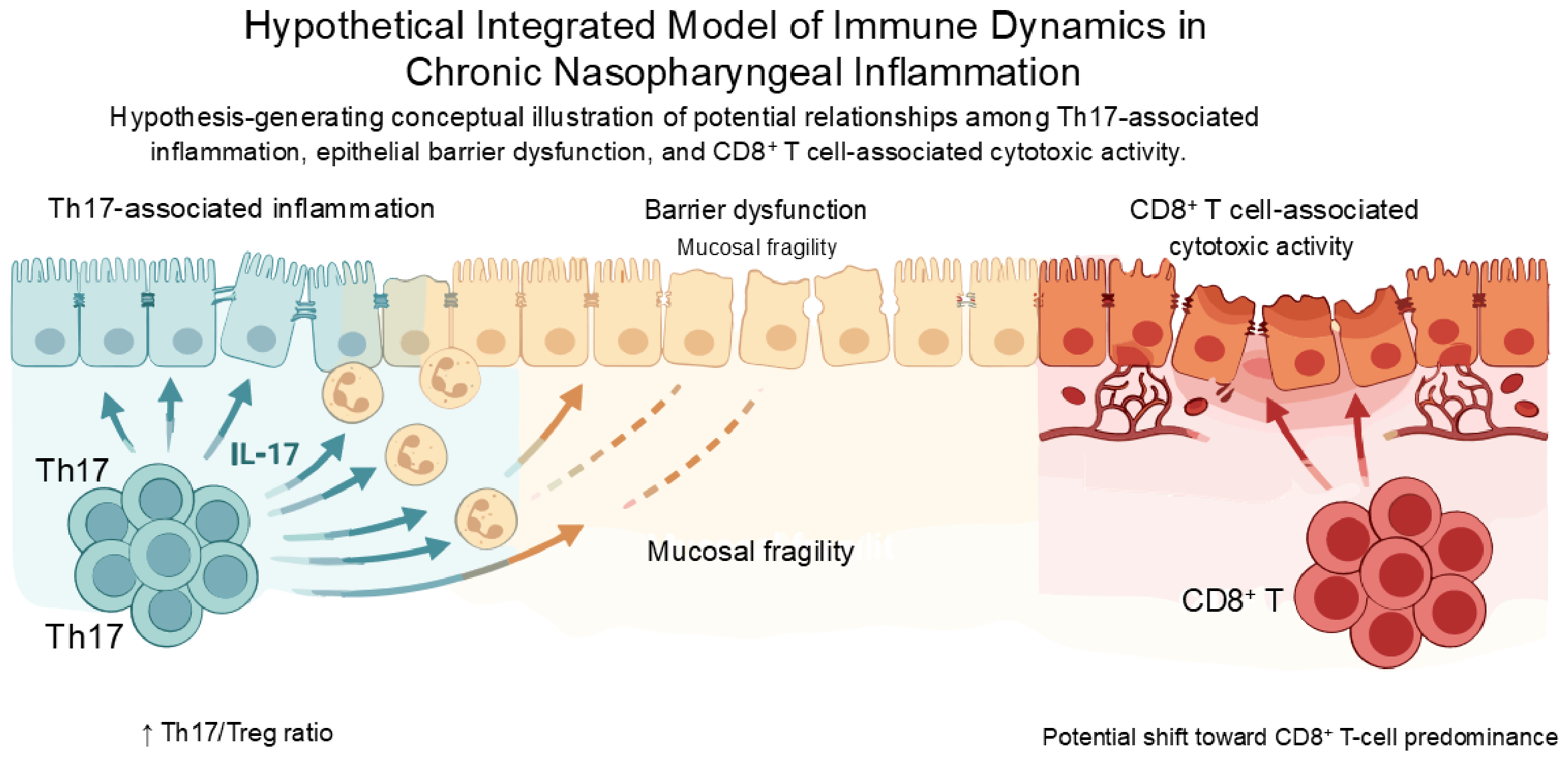
Based on these observations, we propose a conceptual and hypothesis-generating model in which chronic nasopharyngeal inflammation may involve sequential shifts in dominant immune mechanisms:
- CD4+ T-cell-driven activation;
- Th17-mediated epithelial barrier dysfunction;
- CD8+ cytotoxic tissue injury [18].
This framework is intended to describe a representative pattern of immune dynamics rather than a fixed or universal progression.
No longitudinal studies have directly demonstrated this temporal sequence in chronic nasopharyngeal inflammation, and the proposed ordering should therefore be interpreted as a conceptual framework rather than an established biological progression. Several components of this model are extrapolated from findings in other mucosal inflammatory diseases and should not be interpreted as disease-specific mechanisms of chronic nasopharyngeal inflammation.
These immune patterns may coexist and interact within a complex inflammatory microenvironment. Persistent immune activation, together with impaired regulation, drives progression from reversible inflammation to structural damage [18].
Recent spatial transcriptomic analyses further support this model by demonstrating that chronic nasopharyngeal inflammation involves the coordinated activation of epithelial cells, B cells, and T cells, along with sustained cytokine signaling and dysregulated immune pathways [20]. This integrated framework provides a mechanistic basis for understanding chronic mucosal inflammation and its progression and is summarized schematically in Figure 4.
Figure 4.
This figure illustrates a hypothesis-generating conceptual model and should not be interpreted as an experimentally validated mechanism. Conceptual relationships among proposed immune patterns in chronic nasopharyngeal inflammation. The figure summarizes hypothetical associations among CD4+ T-cell activation, Th17-associated epithelial barrier dysfunction, and CD8+ T-cell-associated cytotoxic responses. These immune patterns may coexist and interact within the inflammatory microenvironment and should not be interpreted as evidence of a fixed temporal progression.
4. Link Between Local and Systemic Immunity
The following section describes a hypothetical framework linking local nasopharyngeal inflammation to systemic immune regulation. No direct evidence currently demonstrates that chronic nasopharyngeal inflammation causes hypothalamic dysfunction, autonomic imbalance, or related systemic symptoms. While supported in part by general principles of neuro-immune interactions, direct evidence specific to the nasopharynx remains limited [8,23].
Chronic inflammation of the nasopharyngeal mucosa has been proposed as a potential contributor to systemic immune dysregulation. Given its anatomical and immunological position within Waldeyer’s ring, the nasopharynx functions as a key mucosal immune interface, continuously exposed to inhaled pathogens and environmental antigens [1,3].
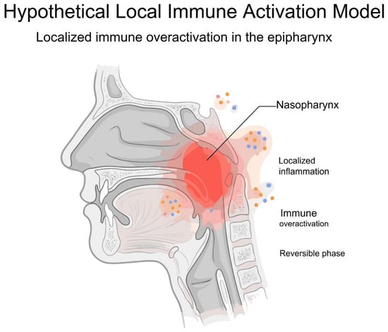
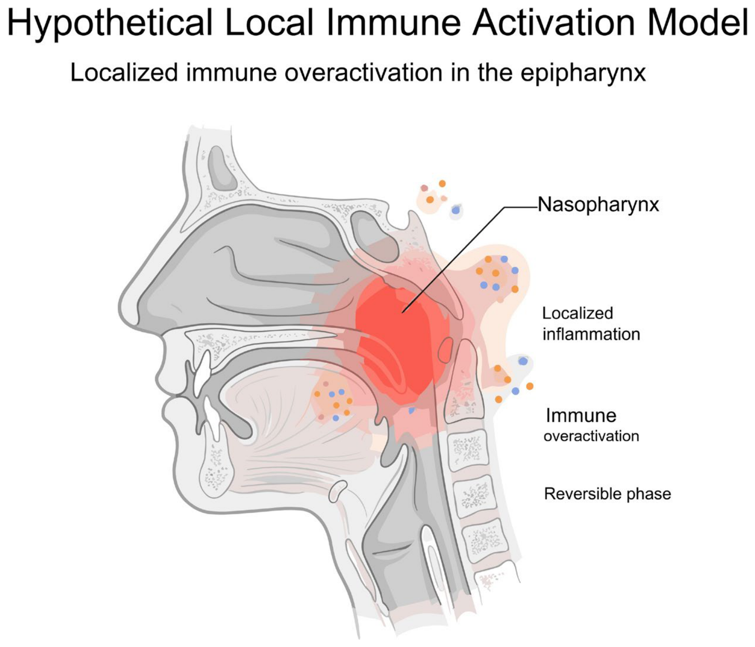
Persistent inflammation in this region may lead to sustained activation of local immune cells, including lymphocytes and antigen-presenting cells, which can subsequently influence systemic immune responses. This initial phase of localized mucosal immune activation is illustrated in Figure 5. In this phase, persistent antigen exposure and chronic stimulation result in localized immune overactivation within the nasopharyngeal mucosa, representing a potentially reversible stage of disease progression. Previous studies have suggested that chronic nasopharyngeal inflammation is associated with increased expression of pro-inflammatory cytokines and alterations in T-cell populations, indicating a shift toward a pro-inflammatory immune state [19,20].
Figure 5.
This figure illustrates a hypothesis-generating conceptual model and should not be interpreted as an experimentally validated mechanism. Localized mucosal immune activation in the nasopharynx. Schematic illustration of localized immune activation within the nasopharyngeal mucosa. Persistent antigen exposure may lead to sustained activation of immune cells, including lymphocytes and antigen-presenting cells, and may represent an initial and potentially reversible inflammatory pattern.
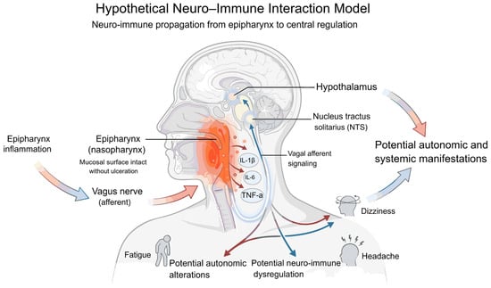
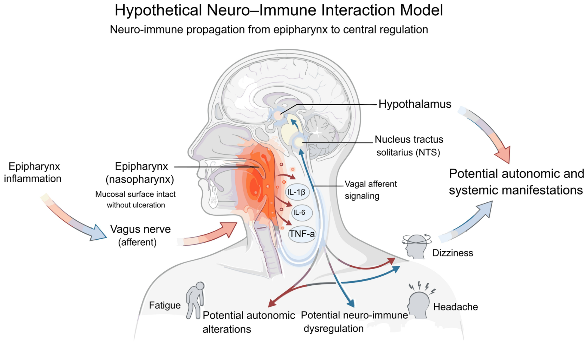
One possible mechanism underlying this local-to-systemic linkage involves neural–immune interactions. The nasopharyngeal mucosa is densely innervated by trigeminal and vagal afferent fibers, enabling inflammatory signals to be transmitted to central regulatory networks [8,23]. This neuro-immune propagation from the nasopharynx to central autonomic and regulatory systems is illustrated in Figure 6. Through these pathways, local mucosal inflammation may influence central autonomic circuits, including the nucleus tractus solitarius and hypothalamic regulation, ultimately contributing to systemic immune modulation. Through these pathways, local mucosal inflammation may influence systemic immune regulation, including cytokine production and neuroendocrine responses [24]. These relationships remain hypothetical and have not been directly demonstrated in chronic nasopharyngeal inflammation [8,23].
Figure 6.
This figure illustrates a hypothesis-generating conceptual model and should not be interpreted as an experimentally validated mechanism. Neuro-immune propagation from the nasopharynx to central regulatory systems. Diagram depicting hypothetical neural–immune interactions linking the nasopharynx to central autonomic and regulatory systems. Inflammatory signals may potentially be conveyed via trigeminal and vagal afferent pathways to the brainstem and hypothalamus and may be associated with systemic immune modulation. These relationships remain speculative and have not been directly demonstrated in chronic nasopharyngeal inflammation.
In addition, neurogenic inflammation mediated by trigeminal pathways may further amplify local immune activation. Release of neuropeptides such as calcitonin gene-related peptide can enhance vascular permeability and inflammatory signaling, potentially contributing to the maintenance and propagation of chronic inflammation [23]. Such mechanisms may create a feedback loop linking local mucosal inflammation with systemic immune dysregulation [8,23].
When this neuro-immune interaction becomes persistently dysregulated, the effects may extend beyond local and central processes, resulting in broader disruption of systemic immune homeostasis [25]. However, this proposed relationship remains speculative and requires direct experimental validation [23].
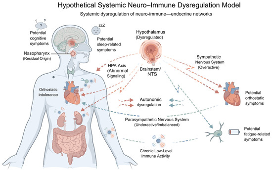
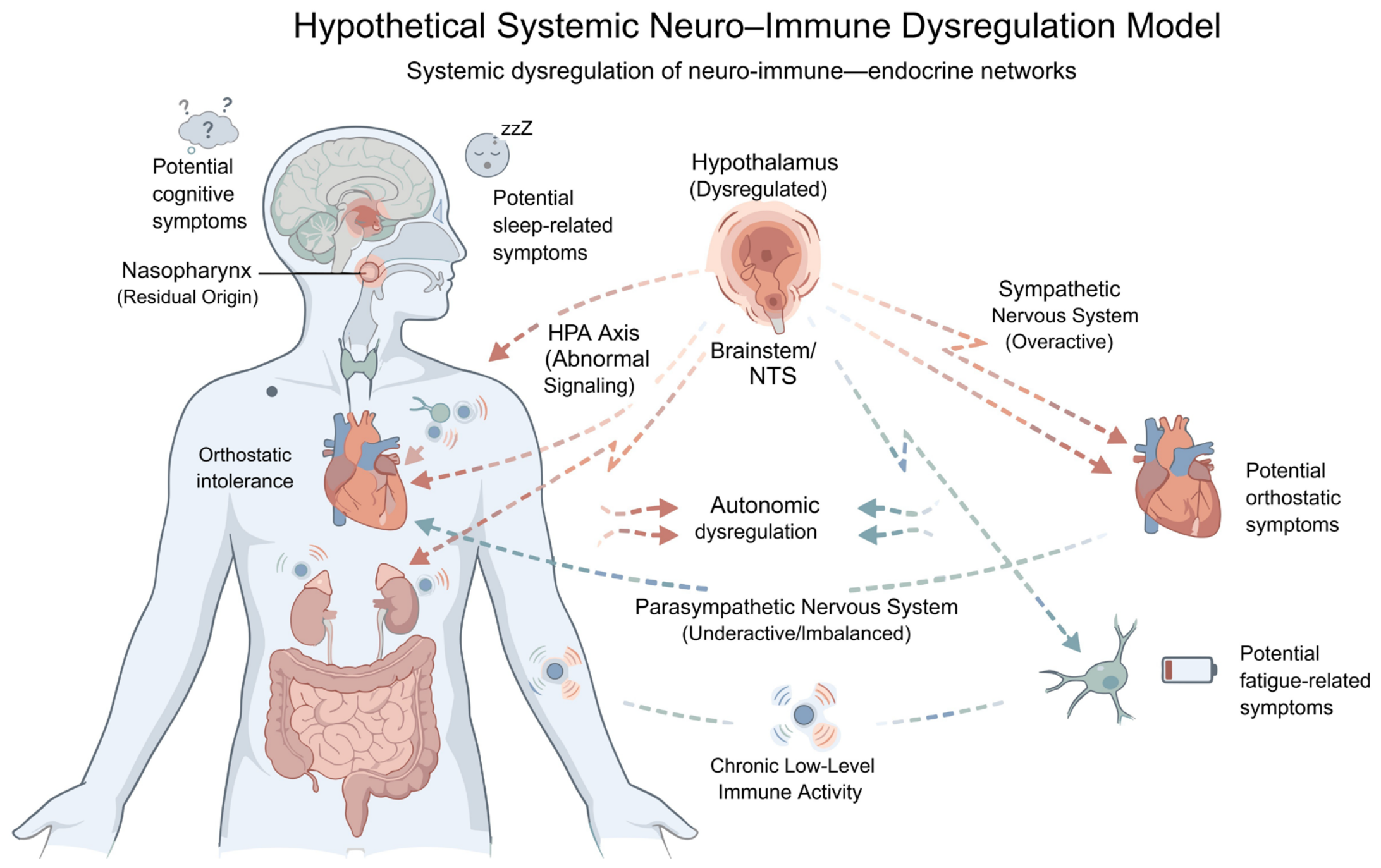
The downstream consequences of this process, including autonomic imbalance and dysregulation of neuro-immune–endocrine networks, are illustrated in Figure 7 [23].
Figure 7.
This figure illustrates a hypothesis-generating conceptual model and should not be interpreted as an experimentally validated mechanism. Systemic neuro-immune dysregulation and homeostatic breakdown. Representation of possible systemic manifestations associated with persistent neuro-immune dysregulation. Alterations in autonomic balance, HPA axis signaling, and neuro-immune networks may potentially be associated with chronic low-grade inflammation and disturbances of systemic homeostasis. These relationships remain speculative and have not been directly demonstrated in chronic nasopharyngeal inflammation.
Within this context, EAT may be hypothesized to influence this pathological linkage. By mechanically stimulating and modulating the inflamed mucosa, EAT may reduce local inflammatory activity and, consequently, attenuate downstream systemic immune responses. Although the precise mechanisms remain to be fully elucidated, this framework provides a plausible immunological basis for the systemic effects associated with EAT [26,27].
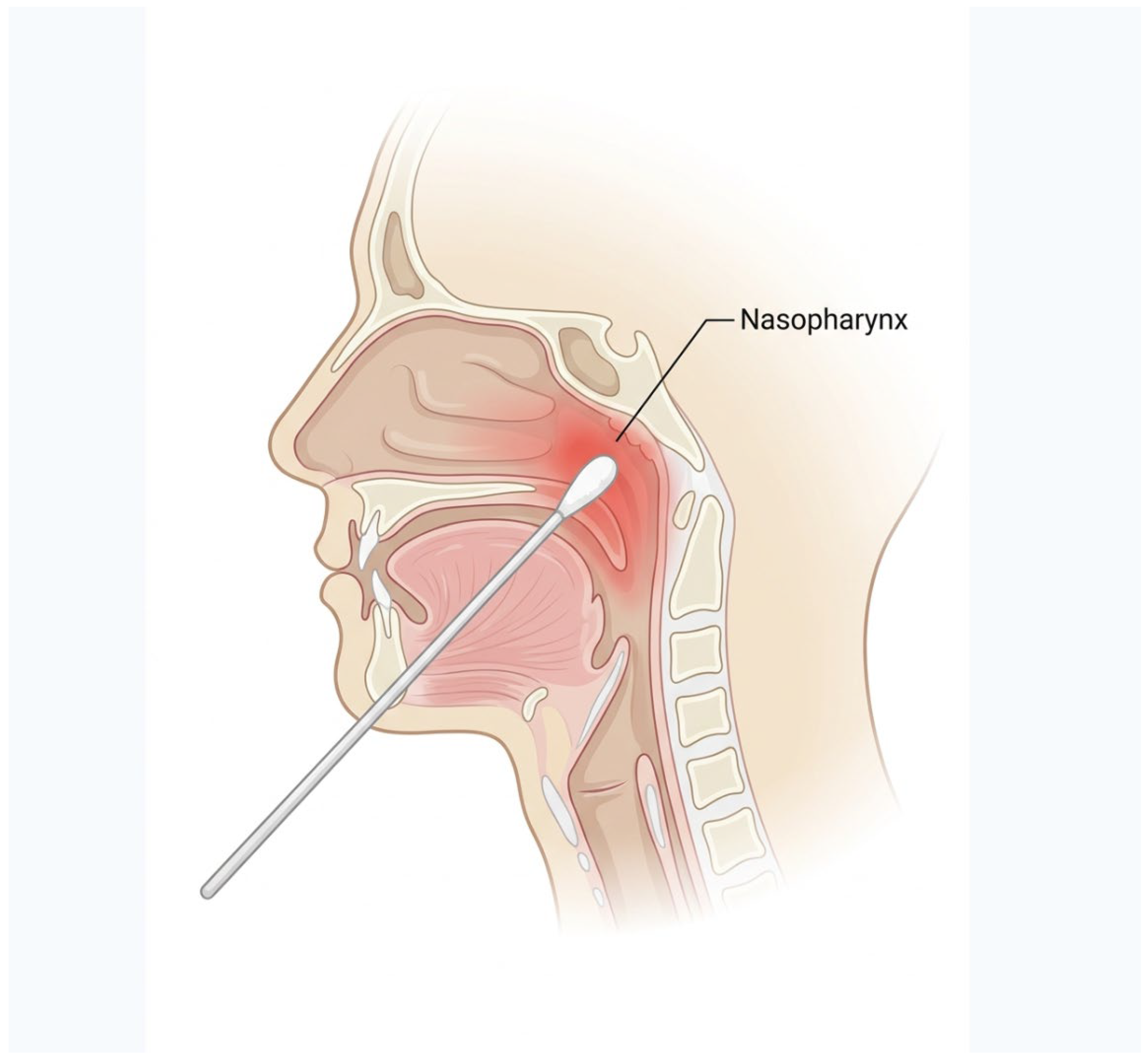
Taken together, these observations suggest that the nasopharynx may serve as a functional bridge between local mucosal immunity and systemic immune homeostasis. Interventions targeting this region may therefore have broader implications beyond local disease, potentially contributing to the regulation of systemic inflammatory processes. The EAT procedure is illustrated in Figure 8.
Figure 8.
Epipharyngeal abrasive therapy (EAT) procedure. Illustration of the epipharyngeal abrasive therapy technique. Mechanical stimulation of the nasopharyngeal mucosa is performed using a cotton applicator to modulate local inflammation.
5. Implications for Immune Regulation and Disease Concept
The findings presented in this study suggest that chronic inflammation of the nasopharyngeal mucosa may represent a biologically relevant source of systemic immune perturbation. The concept that focal mucosal inflammation contributes to broader immune dysregulation has long been proposed, yet its mechanistic and anatomical basis remains incompletely defined, particularly in the context of the nasopharynx [24,28]. However, these associations remain largely observational, and causal relationships between chronic nasopharyngeal inflammation and systemic disease have not been established.
Within this framework, the nasopharynx can be conceptualized as a dynamic immunological interface, where persistent antigen exposure and chronic inflammation may alter local immune cell activation and cytokine signaling, thereby influencing systemic immune homeostasis. Clinical observations supporting associations between chronic nasopharyngeal inflammation and diverse systemic conditions should be interpreted cautiously; however, they are consistent with a model in which sustained mucosal immune activation contributes to downstream immune modulation [24,28].
Recent advances in endoscopic imaging have enabled more precise visualization of mucosal structural alterations associated with chronic inflammation. Findings such as black spots and granular changes, observed under band-limited light endoscopy, may represent structural correlates of underlying mucosal immune activity, including submucosal congestion, epithelial disruption, and inflammatory cell infiltration [29,30]. As such, these features may serve as surrogate markers reflecting localized immune activation rather than merely descriptive morphological findings [25,29].
Importantly, the integration of these features into structured assessment frameworks has improved reproducibility and discrimination of inflammatory states [25,29]. From an immunological perspective, such approaches may contribute to the standardization of mucosal phenotyping, facilitating more robust investigation of the relationship between local inflammatory states and systemic immune responses [31].
Within this context, EAT may be interpreted as an intervention that modulates local immune activity at the mucosal interface. However, several important limitations should be acknowledged. Evidence from randomized controlled trials remains limited, treatment protocols vary among institutions, and the reproducibility of reported outcomes has not been fully established. In addition, mechanistic studies directly demonstrating systemic immune modulation by EAT remain scarce. At present, the strongest evidence for EAT relates to improvements in local symptoms and inflammatory findings, whereas systemic immunological effects remain unproven [32,33]. Rather than being considered solely a physical treatment, EAT may represent a potential modulator of immune dynamics by altering the local inflammatory milieu. Observed reductions in inflammatory features following repeated stimulation are consistent with the attenuation of mucosal immune activation [34,35].
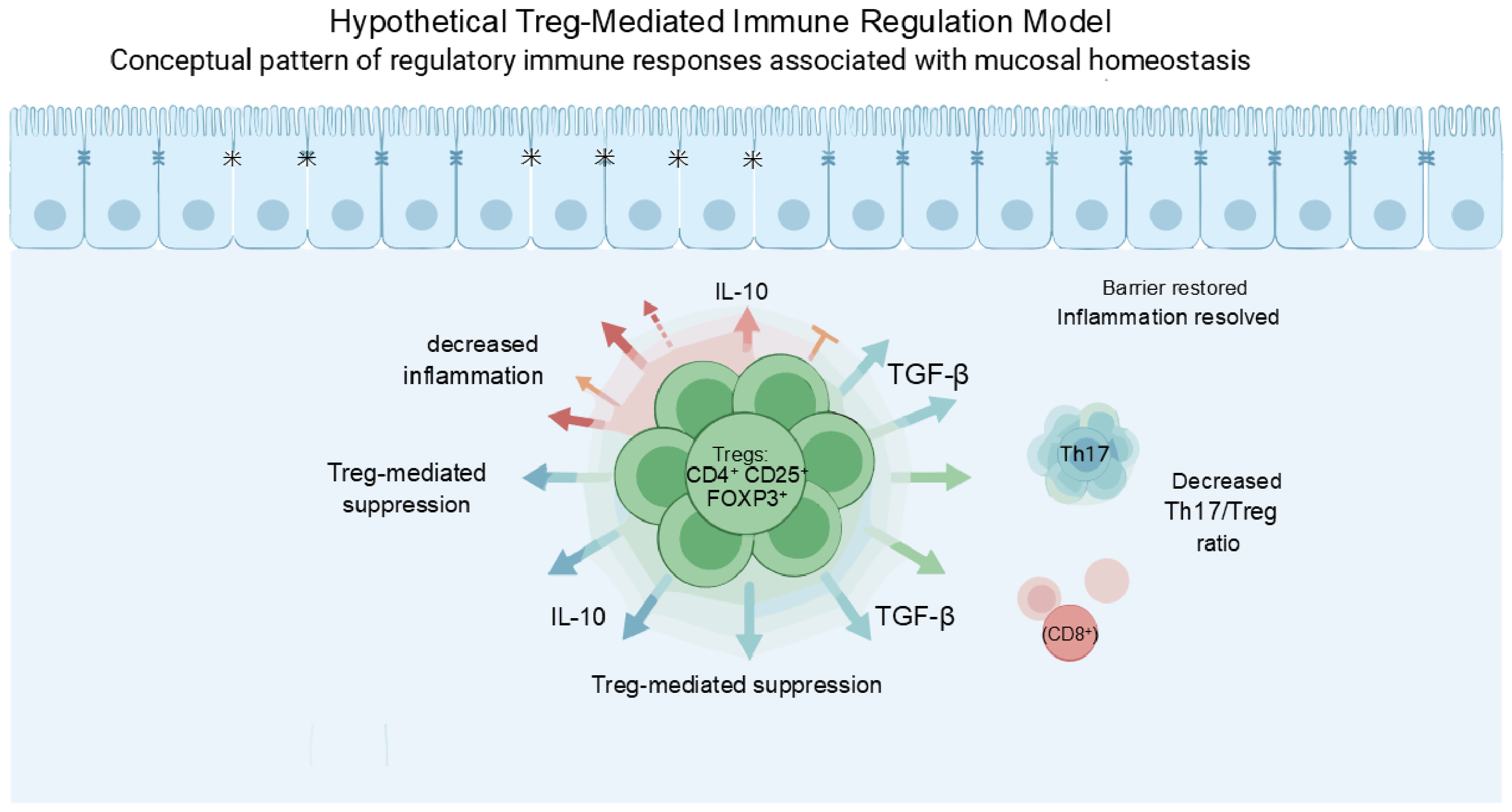
Although direct evidence linking these changes to defined immunological pathways remains limited, this interpretation aligns with the broader concept of mucosal interventions influencing systemic immune regulation. Altered Th17/Treg balance has been reported in chronic epipharyngitis; however, longitudinal changes and the effects of EAT on this balance remain unknown. The resolution phase of mucosal inflammation, characterized by regulatory immune responses including Treg-mediated suppression and restoration of epithelial barrier integrity, is illustrated in Figure 9 [31].
Figure 9.
This figure illustrates a hypothesis-generating conceptual model and should not be interpreted as an experimentally validated mechanism. Treg-mediated resolution of mucosal inflammation. Schematic representation of a possible regulatory phase of mucosal inflammation. Expansion of Tregs (CD4+CD25+FOXP3+) and production of anti-inflammatory cytokines such as IL-10 and TGF-β may contribute to suppression of excessive immune activation and may be associated with restoration of epithelial barrier integrity and immune homeostasis. * indicates epithelial tight junctions associated with restoration of barrier integrity.
Taken together, these considerations support a conceptual model in which the nasopharynx functions as a regulatory node linking mucosal and systemic immunity. The evidence supporting this proposed immune dynamics model is summarized in Table 1. Persistent inflammation at this site may contribute to immune dysregulation, whereas targeted local modulation may influence broader immune homeostasis. Future studies incorporating quantitative immune profiling, including cytokine analyses and immune cell characterization, will be essential to validate this model and to establish causal relationships within neuro-immune and mucosal immune networks.
Table 1.
Evidence supporting the proposed immune dynamics model of chronic nasopharyngeal in-flammation.
6. Broader Implications for Mucosal Immunity
The conceptual framework proposed in this study may have broader relevance for understanding mucosal immune regulation beyond the nasopharynx and provide broader insights into mucosal immunity. Mucosal tissues, including the upper airway, gastrointestinal tract, and respiratory system, serve as critical interfaces between host immunity and environmental exposure. These sites require tightly regulated immune responses to balance pathogen defense and tissue integrity [28,35].
A defining feature of mucosal immunity is the integration of local and systemic immune processes. Mucosa-associated immune compartments are interconnected through cellular trafficking and cytokine signaling, allowing localized inflammation to influence systemic immune homeostasis [24,28]. In this context, persistent inflammation at a single mucosal site may propagate beyond local boundaries and contribute to broader immune dysregulation [24].
This principle is consistent with the model described in this study, in which chronic nasopharyngeal inflammation may act as a localized driver of systemic immune perturbation. Similar mechanisms have been described in other mucosal systems, including the gut–lung axis, where compartmentalized immune activation influences distal inflammatory responses [24].
From an immunological perspective, structural and functional changes observed at mucosal surfaces may reflect underlying immune activity. Such features can serve as indirect but practical markers of immune status, enabling linkage between localized inflammation and systemic immune outcomes [35].
Furthermore, increasing evidence suggests that the localized modulation of mucosal environments can influence systemic immunity. Interventions targeting mucosal or neuro-immune interfaces have demonstrated the capacity to alter immune responses beyond the site of intervention. Within this broader framework, localized therapies may be interpreted as modulators of immune networks rather than purely site-specific treatments [23].
Collectively, these findings support the view that mucosal surfaces function as distributed regulatory nodes within an integrated immune system. Understanding how localized inflammation influences systemic immunity may provide new insights into chronic inflammatory diseases and guide the development of targeted immunomodulatory strategies. Collectively, these observations suggest that mucosal immune dysregulation may represent a shared immunological principle across multiple barrier tissues.
7. Limitations
This hypothesis-generating conceptual framework has several important limitations. First, direct longitudinal immunological studies evaluating sequential changes in CD4+ T cells, Th17-associated inflammation, and CD8+ T-cell-associated cytotoxic activity in chronic nasopharyngeal inflammation are currently lacking. Second, several components of the proposed model are extrapolated from observations in other mucosal inflammatory diseases and therefore require disease-specific validation. Third, the proposed neuro-immune interactions and potential systemic consequences remain speculative and have not been directly demonstrated in chronic nasopharyngeal inflammation. Finally, the immunological mechanisms underlying Epipharyngeal Abrasive Therapy (EAT) remain incompletely understood, and controlled mechanistic studies are currently limited.
8. Future Perspectives
Future research should aim to validate the conceptual framework proposed in this study through direct immunological and mechanistic investigations. Although the present model integrates current knowledge of mucosal immunity and clinical observations, it remains largely hypothetical and requires experimental confirmation.
One important direction will be the application of quantitative immune profiling in patients with chronic nasopharyngeal inflammation. Detailed characterization of immune cell subsets, including CD4+ T cells, Th17 cells, Tregs, and CD8+ T cells, combined with cytokine analysis, may help to define immunological states corresponding to different stages of disease progression.
In addition, advances in spatial and single-cell technologies are expected to provide further insight into mucosal immune dynamics. Techniques such as spatial transcriptomics and single-cell RNA sequencing may allow precise mapping of immune activation, epithelial remodeling, and cell–cell interactions within the nasopharyngeal microenvironment. These approaches could help to validate the proposed sequential immune model and clarify the transition between inflammatory and regulatory phases.
Another important area for investigation is the interaction between mucosal immunity and neuro-immune pathways. Elucidating how neural signaling pathways, including vagal afferent circuits, influence immune regulation will be essential for understanding the mechanisms underlying systemic effects of localized inflammation.
Furthermore, longitudinal studies will be required to determine whether modulation of local mucosal inflammation leads to measurable changes in systemic immune parameters [36,37]. Establishing such causal relationships will be critical for translating the present conceptual framework into clinically relevant applications.
Finally, the development of standardized, non-invasive biomarkers reflecting mucosal immune activity represents an important future goal. Such biomarkers may facilitate patient stratification, disease monitoring, and evaluation of therapeutic responses, thereby bridging the gap between mechanistic immunology and clinical practice.
9. Conclusions
Chronic nasopharyngeal inflammation represents a dynamic process of mucosal immune dysregulation characterized by sequential shifts in dominant immune pathways and disruption of immune homeostasis.
The framework proposed in this study integrates local immune activation, neuro-immune propagation, and systemic immune modulation into a unified conceptual model, positioning the nasopharynx as a potential regulatory interface linking mucosal and systemic immunity.
Chronic nasopharyngeal inflammation may provide one potential clinical context for exploring mucosal immune dysregulation, although numerous other chronic inflammatory diseases have been extensively studied in this field.
Funding
This research received no external funding. The APC was funded by the author.
Data Availability Statement
No new data were created or analyzed in this study. Data sharing is not applicable to this article.
Acknowledgments
The author used Microsoft Copilot for English language editing and FigureAI (FigureLabs) for figure preparation. All outputs were reviewed and approved by the author, who takes full responsibility for the content of this manuscript.
Conflicts of Interest
The author declares no conflict of interest.
References
- Brandtzaeg, P. Potential of nasopharynx-associated lymphoid tissue for vaccine responses in the airways. Am. J. Respir. Crit. Care Med. 2011, 183, 1595–1604. [Google Scholar] [CrossRef] [Scilit] [PubMed]
- Zhou, X.; Wu, Y.; Zhu, Z.; Lu, C.; Zhang, C.; Zeng, L.; Xie, F.; Zhang, L.; Zhou, F. Mucosal immune response in biology, disease prevention and treatment. Signal Transduct. Target. Ther. 2025, 10, 7. [Google Scholar] [CrossRef] [Scilit] [PubMed]
- Kiyono, H.; Fukuyama, S. NALT- versus Peyer’s-patch-mediated mucosal immunity. Nat. Rev. Immunol. 2004, 4, 699–710. [Google Scholar] [CrossRef] [Scilit] [PubMed]
- Brandtzaeg, P. Function of mucosa-associated lymphoid tissue in antibody formation. Immunol. Investig. 2010, 39, 303–355. [Google Scholar] [CrossRef] [Scilit] [PubMed]
- van Woudenbergh, E.; Lakerveld, A.J.; van Heerbeek, N.; Honings, J.; van Kasteren, P.B.; de Jonge, M.I.; Hartog, G.D. Primary human nasal, nasopharyngeal, and bronchial epithelia show distinct immune responses to pathogens. Pathog. Dis. 2025, 83, ftaf011. [Google Scholar] [CrossRef] [Scilit] [PubMed]
- Whitsett, J.A.; Alenghat, T. Respiratory epithelial cells orchestrate pulmonary innate immunity. Nat. Immunol. 2015, 16, 27–35. [Google Scholar] [CrossRef] [Scilit] [PubMed]
- Ganesan, S.; Comstock, A.T.; Sajjan, U.S. Barrier function of airway epithelium. Tissue Barriers 2013, 1, e24997. [Google Scholar] [CrossRef] [Scilit] [PubMed]
- Pavlov, V.A.; Tracey, K.J. Neural regulation of immunity: Molecular mechanisms and clinical translation. Nat. Neurosci. 2017, 20, 156–166. [Google Scholar] [CrossRef] [Scilit] [PubMed]
- Parker, D.; Prince, A. Innate immunity in the respiratory epithelium. Am. J. Respir. Cell Mol. Biol. 2011, 45, 189–201. [Google Scholar] [CrossRef] [Scilit] [PubMed]
- Schenkel, J.M.; Masopust, D. Tissue-resident memory T cells. Immunity 2014, 41, 886–897. [Google Scholar] [CrossRef] [Scilit] [PubMed]
- Lange, J.; Rivera-Ballesteros, O.; Buggert, M. Human mucosal tissue-resident memory T cells in health and disease. Mucosal Immunol. 2022, 15, 389–397. [Google Scholar] [CrossRef] [Scilit] [PubMed]
- Longet, S.; Paul, S. Pivotal role of tissue-resident memory lymphocytes in mucosal infections. Front. Immunol. 2023, 14, 1216402. [Google Scholar] [CrossRef] [Scilit] [PubMed]
- Pandiyan, P.; Bhaskaran, N.; Zou, M.; Schneider, E.; Jayaraman, S.; Huehn, J. Microbiome-dependent regulation of Tregs and Th17 cells in mucosa. Front. Immunol. 2019, 10, 426. [Google Scholar] [CrossRef] [Scilit] [PubMed]
- Hu, Y.; Yang, Y.; Li, Y.; Zhang, Q.; Zhang, W.; Jia, J.; Han, Z.; Wang, J. Th17/Treg imbalance in inflammatory diseases. Front. Immunol. 2025, 16, 1651063. [Google Scholar] [CrossRef] [Scilit] [PubMed]
- Mogitate, M. Epipharyngeal abrasive therapy downregulates the number of epipharyngeal abrasive CD4 cells with symptomatic recovery. Cureus 2023, 15, e50288. [Google Scholar] [CrossRef] [Scilit] [PubMed]
- Sakaguchi, S.; Yamaguchi, T.; Nomura, T.; Ono, M. Regulatory T cells and immune tolerance. Cell 2008, 133, 775–787. [Google Scholar] [CrossRef] [Scilit] [PubMed]
- Josefowicz, S.Z.; Lu, L.F.; Rudensky, A.Y. Regulatory T cells: Mechanisms of differentiation and function. Annu. Rev. Immunol. 2012, 30, 531–564. [Google Scholar] [CrossRef] [Scilit] [PubMed]
- Giudice, A.; Brescia, C.; Morano, D.; Viglietto, G.; Luzza, F.; Amato, R.; Spagnuolo, R. Th17/Treg balance in mucosal inflammation. Front. Cell Dev. Biol. 2026, 14, 1774790. [Google Scholar] [PubMed]
- Nishi, K.; Yoshimoto, S.; Nishi, S.; Tsunoda, T.; Ohno, J.; Yoshimura, M.; Hiromatsu, K.; Yamano, T. Epipharyngeal abrasive therapy down-regulates SARS-CoV-2 entry factors ACE2 and TMPRSS2. In Vivo 2022, 36, 371–374. [Google Scholar] [CrossRef] [Scilit] [PubMed]
- Nishi, K.; Yoshimoto, S.; Tanaka, T.; Kimura, S.; Tsunoda, T.; Watanabe, A.; Teranaka, K.; Oguma, Y.; Ogawa, H.; Kumai, T.; et al. Spatial transcriptomic analysis of chronic epipharyngitis and its improvement after epipharyngeal abrasive therapy. Sci. Rep. 2023, 15, 8618. [Google Scholar]
- Giles, J.R.; Globig, A.M.; Kaech, S.M.; Wherry, E.J. CD8 T cells in the cancer-immunity cycle. Immunity 2023, 56, 2231–2253. [Google Scholar] [CrossRef] [Scilit] [PubMed]
- Kumar, V. Cytotoxic T cells: Kill, memorize, and mask to maintain immune homeostasis. Int. J. Mol. Sci. 2025, 26, 8788. [Google Scholar] [CrossRef] [Scilit] [PubMed]
- Chavan, S.S.; Pavlov, V.A.; Tracey, K.J. Mechanisms and Therapeutic Relevance of Neuro-immune Communication. Immunity 2017, 46, 927–942. [Google Scholar] [CrossRef] [Scilit] [PubMed]
- Budden, K.F.; Gellatly, S.L.; Wood, D.L.A.; Cooper, M.A.; Morrison, M.; Hugenholtz, P.; Hansbro, P.M. Emerging pathogenic links between microbiota and the gut–lung axis. Nat. Rev. Microbiol. 2017, 15, 55–63. [Google Scholar] [CrossRef] [Scilit] [PubMed]
- Mogitate, M.; Ito, H.; Ohno, Y.; Nishiwaki, N.; Yamaguchi, Y.; Fujikawa, M.; Fukuo, A.; Sasaki, Y.; Watanabe, Y.; Wada, K. Deep learning-based objective quantification of nasopharyngeal endoscopic findings for standardized assessment of inflammation. Diagnostics 2026, 16, 2015. [Google Scholar] [CrossRef] [Scilit] [PubMed]
- Ito, H. The effect of epipharyngeal abrasive therapy (EAT) on the baroreceptor reflex (BR). Cureus 2023, 15, e45080. [Google Scholar] [CrossRef] [Scilit] [PubMed]
- Ito, H. Autonomic stimulation action of epipharyngeal abrasive therapy (EAT) on chronic epipharyngitis. Cureus 2024, 16, e63182. [Google Scholar] [CrossRef] [Scilit] [PubMed]
- Mowat, A.M.; Agace, W.W. Regional specialization within the intestinal immune system. Nat. Rev. Immunol. 2014, 14, 667–685. [Google Scholar] [CrossRef] [Scilit] [PubMed]
- Mogitate, M.; Yamaguchi, Y.; Fujikawa, M.; Fukuo, A.; Sasaki, Y.; Nishiwaki, N.; Ohno, Y.; Ito, H.; Watanabe, Y.; Wada, K. Diagnostic value of black spots and granular changes in pretreatment endoscopic evaluation of chronic epipharyngitis. Auris Nasus Larynx 2026, 53, 589–595. [Google Scholar] [CrossRef] [Scilit] [PubMed]
- Tanaka, A. Band-limited light endoscopic diagnosis and endoscopic epipharyngeal abrasive therapy in chronic epipharyngitis. Jibi Inkoka 2018, 31, 57–67. (In Japanese) [Google Scholar]
- Mogitate, M. Th17/Treg imbalance reveals local–systemic immune dissociation in nasopharyngeal inflammation and its association with endoscopic severity. Medicina 2026, 62, 1490. [Google Scholar] [CrossRef] [Scilit]
- Mogitate, M. Effectiveness of epipharyngeal abrasive therapy on chronic epipharyngitis and the exhaled nitric oxide levels. Intern. Med. 2022, 62, 1139–1144. [Google Scholar] [CrossRef] [Scilit] [PubMed]
- Mogitate, M. Exhaled nitric oxide levels are associated with the severity of chronic epipharyngitis and decreased via epipharyngeal abrasion. Auris Nasus Larynx 2023, 50, 534–539. [Google Scholar] [CrossRef] [Scilit] [PubMed]
- Ohno, Y. Effectiveness of epipharyngeal abrasive therapy for chronic epipharyngitis. Cureus 2019, 11, e6422. [Google Scholar]
- Mogitate, M.; Sasaki, Y.; Komiyama, A. Outcome of an outpatient specialty clinic for chronic epipharyngitis. Auris Nasus Larynx 2021, 48, 451–456. [Google Scholar] [CrossRef] [Scilit] [PubMed]
- Ito, H. Autonomic reflexes with epipharyngeal abrasive therapy (EAT). Cureus 2025, 17, e77575. [Google Scholar] [CrossRef] [Scilit] [PubMed]
- Ito, H. Treatment response trajectories of epipharyngeal abrasive therapy (EAT) in chronic nasopharyngitis: Phenotypic classification into early, delayed, and non-responders based on ΔEAT with outcome stratification. Cureus 2026, 18, e109478. [Google Scholar] [CrossRef] [Scilit] [PubMed]

Disclaimer/Publisher’s Note: The statements, opinions and data contained in all publications are solely those of the individual author(s) and contributor(s) and not of MDPI and/or the editor(s). MDPI and/or the editor(s) disclaim responsibility for any injury to people or property resulting from any ideas, methods, instructions or products referred to in the content. |
© 2026 by the author. Licensee MDPI, Basel, Switzerland. This article is an open access article distributed under the terms and conditions of the Creative Commons Attribution (CC BY) license.