


Abstract
The endometrial microbiome has been implicated in implantation success, and intrauterine peripheral blood mononuclear cell (PBMC) therapy has been proposed to improve endometrial receptivity; however, its effects on the endometrial microbial environment remain poorly understood. This prospective study evaluated changes in endometrial microbiome composition following PBMC therapy in 26 in vitro fertilization (IVF) patients. Paired endometrial biopsies were collected before and after intrauterine administration of autologous PBMCs, and 16S rRNA gene sequencing (V4–V5 region) was performed. Microbiome composition was analyzed at genus and species levels, and alpha and beta diversity metrics were compared between paired samples. No significant changes in microbial composition or diversity were observed across the entire cohort following treatment. However, patients with lower baseline Lactobacillus abundance exhibited greater microbiome responsiveness, showing larger increases in Lactobacillus after treatment. This association remained significant after adjustment for age and BMI (ρ = −0.457, p = 0.019; β = −0.367, p = 0.047). Moreover, subgroup analysis identified two distinct response patterns based on Lactobacillus dynamics: a Lactobacillus-increase group (n = 14) and a no-increase group (n = 12). Baseline Lactobacillus dominance (LD: >90% relative abundance) was less frequent in the increase group (14%) than in the no-increase group (50%). In Lactobacillus-increase patients, the median relative abundance rose from 17.6% to 95.5% (p < 0.001), 64% achieved LD, and Shannon and Simpson diversity indices decreased significantly (p = 0.04 and p = 0.02, respectively). In the no-increase group, Lactobacilli remained the most abundant taxon, and the diversity indices were unchanged following treatment. These findings suggest that baseline endometrial microbiome composition may influence responsiveness to PBMC therapy and could support future patient stratification strategies in IVF treatment. Whether these microbiome changes translate into improved clinical outcomes requires evaluation in future studies.
1. Introduction
Successful embryo implantation in both natural and assisted reproduction depends on a complex interplay between embryo competence and endometrial receptivity. Implantation failure remains a major limiting factor in assisted reproductive technology (ART), accounting for a substantial proportion of unsuccessful in vitro fertilization (IVF) cycles despite advances in embryo selection and laboratory techniques [1,2]. While embryo quality is a critical determinant of reproductive success, increasing attention has focused on the role of the endometrial microenvironment, including its immunological, cellular, and molecular components, in establishing optimal conditions for implantation [1,3,4]. Although the uterus was historically considered sterile, sequencing-based studies have demonstrated the presence of a low-biomass but functionally relevant microbial community within the endometrial cavity. Accumulating evidence indicates that the endometrial microbiome is an important component of this microenvironment, capable of modulating local immune responses and influencing implantation outcomes [5,6,7,8].
Among this microbial community, the relative abundance of Lactobacillus species has emerged as a key determinant of reproductive success. A Lactobacillus-dominant microbiome, commonly defined as ≥90% Lactobacillus spp., has been consistently associated with higher implantation, pregnancy, and live birth rates in IVF cycles, whereas dysbiotic profiles enriched with non-Lactobacillus taxa correlate with poorer outcomes [9,10,11,12,13,14]. Mechanistically, Lactobacillus species are thought to contribute to an optimal uterine environment through lactic acid production, maintenance of acidic pH, suppression of pathogenic bacteria, and modulation of inflammatory signaling pathways critical for embryo implantation [13,14].
In parallel, immunological factors are central to implantation success, and therapeutic strategies targeting the maternal immune response have gained increasing attention [15,16]. Intrauterine peripheral blood mononuclear cell (PBMC) therapy has been proposed as an immunomodulatory approach to enhance endometrial receptivity. PBMC administration is thought to promote a favorable cytokine environment, regulate immune cell activity, and facilitate trophoblast invasion, with several clinical studies and meta-analyses reporting improved implantation and pregnancy rates in selected patient populations [17,18,19].
In addition to those immunomodulatory effects, PBMCs may influence the endometrial microbial ecosystem by altering the local inflammatory environment, epithelial barrier function, and antimicrobial defense pathways within the endometrium. Because host immune responses are recognized as major determinants of microbial colonization and community stability at mucosal surfaces [12], PBMC-induced changes in the uterine immune environment could potentially affect the composition and diversity of the endometrial microbiome. However, whether intrauterine PBMC administration is associated with measurable changes in the endometrial microbiome has not yet been investigated.
Therefore, the present exploratory study aimed to evaluate the effect of intrauterine PBMC therapy on endometrial microbial composition and diversity by analyzing paired pre- and post-treatment endometrial biopsies. We hypothesized that PBMC administration would induce changes in the endometrial microbial ecosystem, reflected by alterations in microbial diversity or taxonomic composition, potentially favoring a microbiological profile associated with enhanced endometrial receptivity. By characterizing microbiome changes following PBMC treatment, this study sought to explore a previously unexamined mechanism through which PBMC-based immunotherapy may influence the uterine environment.
2. Materials and Methods
2.1. Study Design and Patient Cohort
This prospective paired-sample study included 26 women undergoing in vitro fertilization (IVF) treatment at a single fertility centre. Eligible participants were of reproductive age with regular cycles. Patients were excluded if they had received antibiotics within 3 months before sample collection, had evidence of active or chronic genital tract infection, or presented with known uterine pathology, including chronic endometritis, endometrial polyps, or cavity-distorting fibroids.
Each participant underwent two endometrial biopsies collected during the mid-luteal phase of two consecutive unstimulated menstrual cycles. The first biopsy was obtained before intrauterine peripheral blood mononuclear cell (PBMC) administration (pre-treatment sample), and the second biopsy was collected the following month, 2 days after PBMC treatment (post-treatment sample). This enabled intra-individual assessment of microbiome changes associated with the intervention while minimizing interindividual variability.
The study was conducted in accordance with the Declaration of Helsinki and approved by Nadezhda Women’s Health Hospital Ethics Committee (Approval No. 56/29.12.2020). Written informed consent was obtained from all participants prior to enrollment.
2.2. PBMC Isolation and Intrauterine Administration
Autologous PBMCs were isolated from peripheral venous blood using density-gradient centrifugation. Briefly, 12 mL whole blood was collected in sodium heparin Vacutainer® tubes (Cat. No. 367869, BD, Erembodegem, Belgium), diluted 1:1 with phosphate-buffered saline (PBS, Cat. No. P04-36500, PAN-Biotech, Aidenbach, Germany), carefully layered onto Pancoll separation medium (Cat. No. P04-60100, PAN-Biotech) and centrifuged at 400× g 25 min. The mononuclear cell layer was collected, washed twice in sterile PBS, and resuspended in culture medium (RPMI1640 (Cat. No. P04-22100, PAN-Biotech) supplemented with 1 mg/mL human serum albumin (Cat. No. A1A029AA, Takeda, Zurich, Switzerland). PBMCs were counted using a hemocytometer, and viability was assessed by trypan blue exclusion. Cells were resuspended at a concentration of 1 × 10⁷ cells/mL and incubated for 24 h under standard cell culture conditions (37 °C, 5% CO2). Following incubation, the cell suspension (0.5 mL; viability ≥ 95%) was administered intrauterinely using a sterile embryo-transfer catheter under ultrasound guidance. The procedure was performed during the mid-luteal phase, on day 3 post-ovulation, 2 days before the second biopsy, according to standard clinical protocols. No procedure-related adverse events were recorded.
2.3. Endometrial Sampling and DNA Extraction
Endometrial tissue samples were obtained using a sterile Pipelle catheter under aseptic conditions during the mid-secretory phase of two consecutive natural menstrual cycles (5 days post-ovulation). In the second cycle, PBMCs were administered intrauterinely on post-ovulation day 3, and the second biopsy was collected 2 days later, corresponding to the same menstrual phase as the baseline sampling. To minimize contamination from the lower genital tract, all instruments were sterile and sampling was performed following standard gynecological procedures. Care was taken to prevent contact with the vaginal walls and aspiration was discontinued before the catheter reached the internal cervical os. Immediately after collection, the outer surface of each catheter was wiped with sterile gauze, after which the biopsy specimens were transferred into sterile DNA-free collection tubes and stored at −80 °C until further processing.
Total microbial DNA was isolated from endometrial biopsy specimens using the Quick-DNA Fecal/Soil Microbe Microprep Kit (Cat. No. D6010, Zymo Research, Irvine, CA, USA) following the manufacturer’s protocol. The extraction procedure was optimized for low-biomass tissue samples and incorporated cell lysis steps to maximize bacterial DNA recovery. DNA quantity and quality were evaluated using a NanoDrop™ 2000c spectrophotometer (Cat. No. ND-2000, Thermo Scientific, Waltham, MA, USA). To assess possible contamination introduced during sample processing, extraction blanks were included and carried through all laboratory procedures alongside study samples.
2.4. 16S rRNA Gene Amplification and Sequencing
Bacterial community composition was characterized through sequencing of the V4-V5 hypervariable region of the 16S rRNA gene. Amplification was performed using barcoded universal primer pair 515F (5′-GTGCCAGCMGCCGCGGTAA-3′) and 907R (5′-CCGTCAATTCCTTTGAGTTT-3′). PCR reactions were conducted using a high-fidelity DNA polymerase.
Amplicons were verified by agarose gel electrophoresis, purified, and quantified before being combined in equimolar concentrations. Sequencing libraries were generated using standard Illumina preparation protocols, including end repair, adenylation, and adapter ligation. Library quality control was performed using Qubit fluorometry and quantitative PCR analysis.
Sequencing was carried out by Novogene Co., Ltd. (Cambridge, UK) on the Illumina HiSeq platform using reads of 250 bp from each end, with a target sequencing depth of approximately 30,000 reads per sample.
Due to the low microbial biomass of endometrial samples, contamination control measures were implemented throughout laboratory processing. Both extraction blanks and no-template PCR controls were included alongside study samples throughout DNA extraction, amplification and sequencing workflows to identify potential background contaminants. Any taxa detected in negative controls were evaluated as potential contaminants and their prevalence and relative abundance were assessed in relation to the biological samples. Taxa detected exclusively in biological samples and absent from negative controls were considered unlikely to represent reagent- or laboratory-derived contamination. Positive PCR controls were included to verify successful amplification and sequencing performance.
2.5. Bioinformatic Processing
Sequence data were processed using the QIIME software package (version 1.7.0). Forward and reverse reads were merged and subjected to quality-control filtering. Chimeric sequences were identified and removed prior to downstream analyses. High-quality sequences were clustered into operational taxonomic units (OTUs) at a 97% sequence similarity threshold using UPARSE (v7.0.1090) [20]. Representative sequences from each OTU were subsequently selected for taxonomic classification.
Taxonomic assignments were generated within QIIME using the Mothur classifier and the SILVA SSU rRNA reference database (release 138) [21]. Classifications were accepted using confidence values ranging from 0.8 to 1.0 and were reported across multiple taxonomic ranks, including phylum, class, order, family, genus, and species when possible.
To reduce bias associated with unequal sequencing depth, OTU counts were normalized to the sample with the lowest read number before downstream analyses. Sequences identified in negative controls were reviewed during downstream analyses and considered when evaluating potential contaminant taxa.
2.6. Microbiome Analysis
2.6.1. Taxonomic Profiling
Relative abundances of bacterial taxa were determined at both the genus and species levels. Particular emphasis was placed on Lactobacillus spp. because of their recognized role in reproductive health and embryo implantation. Samples were classified as Lactobacillus dominant (LD) when the relative abundance of Lactobacillus accounted for 90% or more of the total bacterial community. Data are reported for the top 10 most prevalent genera, which were identified exclusively in the endometrial samples and absent in controls.
2.6.2. Alpha Diversity
Within-sample microbial diversity was assessed using multiple alpha-diversity metrics, including the Shannon diversity index, Chao1 richness estimator, Simpson diversity index, observed features, Pielou’s evenness, dominance index, and Good’s coverage. These complementary indices were used to evaluate microbial richness, diversity and community structure within individual samples.
2.6.3. Beta Diversity
Differences in microbial composition between samples were quantified using Bray–Curtis dissimilarity and unweighted UniFrac distance metrics. Principal coordinates analysis (PCoA) was used to visualize patterns of community similarity and separation among samples.
2.7. Statistical Analysis
Statistical analyses and figures were prepared using Python (version 3.12) and SPSS (version 21.0; IBM Corp., Armonk, NY, USA). Because each participant contributed baseline and post-treatment samples, paired analyses using Wilcoxon signed-rank test were performed to assess differences in alpha-diversity metrics and relative taxon abundances. Comparisons of overall microbial community structure were evaluated using permutational multivariate analysis of variance (PERMANOVA) based on beta-diversity distance matrices. The change in Lactobacillus abundance following PBMC treatment was calculated as post-treatment abundance minus baseline abundance. To assess the relationship between baseline Lactobacillus abundance and treatment-induced change, Spearman rank correlation analysis was performed. To determine whether baseline Lactobacillus abundance independently predicted treatment response, a multivariable linear regression model was constructed with change in Lactobacillus abundance as the dependent variable. Baseline Lactobacillus abundance was included as the primary predictor, while age and body mass index (BMI) were included as covariates to account for potential confounding effects. Additional subgroup analyses were conducted according to post-treatment changes in Lactobacillus abundance. Patients were categorized into either a Lactobacillus-increase group or a no-increase group. Statistical significance was defined as a two-sided p-value < 0.05. Where appropriate, p-values were adjusted for multiple testing using the Benjamini–Hochberg false discovery rate (FDR) correction.
3. Results
3.1. Participants
A total of 26 patients (aged 30–48, mean 37.7) for whom paired endometrial samples (pre- and post-intervention) were obtained were included in the study. Baseline characteristics of the cohort were comparable, with no major differences observed in key demographic or clinical variables across the study population (Table 1).
Table 1.
Baseline patient characteristics of the study cohort.
3.2. Overall Microbiome Shift After PBMC
When considering the cohort as a whole, no statistically significant changes were observed following PBMC intervention. Specifically, relative taxonomic abundances remained stable, and no significant differences were detected in measures of alpha diversity (including Shannon and Simpson indices) or beta diversity. These findings suggest that, at the population level, PBMC administration did not induce a uniform shift in the uterine microbiome composition (Table 2).
Table 2.
Mean relative abundance of the top 10 most prevalent genera identified in paired pre- and post-treatment with intrauterine PBMC.
3.3. Baseline Lactobacillus Abundance Predicts Microbiome Response Following PBMC Treatment
To investigate whether baseline microbiome composition influenced responsiveness to PBMC therapy, we examined the relationship between baseline Lactobacillus relative abundance and the magnitude of Lactobacillus change following treatment.
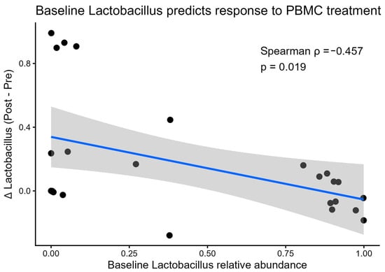
Correlation analysis revealed a significant inverse association between baseline Lactobacillus abundance and subsequent change after PBMC administration. Patients with lower baseline Lactobacillus abundance demonstrated greater increases following treatment, whereas those with higher baseline abundance exhibited smaller changes. This relationship was significant using Spearman (ρ = −0.457, p = 0.019) correlation analysis (Figure 1).
Figure 1.
Association between baseline Lactobacillus abundance and microbiome response to PBMC therapy. Scatter plot showing the relationship between baseline endometrial Lactobacillus relative abundance and the subsequent change in Lactobacillus abundance following PBMC administration. Each point represents one patient. The x-axis indicates baseline Lactobacillus relative abundance before treatment, whereas the y-axis represents the change in abundance after treatment (Post–Pre). The solid line represents the fitted linear regression model, and the shaded area indicates the 95% confidence interval.
To further evaluate whether this association remained independent of potential confounding factors, a multivariable linear regression model was constructed including baseline Lactobacillus abundance as the predictor variable, and age and body mass index (BMI) as covariates. Baseline Lactobacillus abundance remained a significant independent predictor of microbiome response following PBMC treatment (β = −0.367, 95% CI −0.729 to −0.006, p = 0.047), whereas age and BMI were not significantly associated with treatment response (p > 0.05 for both variables).
These findings indicate that baseline microbiome composition influences the magnitude of microbiome remodeling following PBMC administration, with patients exhibiting lower initial Lactobacillus abundance demonstrating the greatest responsiveness to treatment.
3.4. Identification of Two Response Patterns
Despite the absence of global effects, exploratory stratified analysis revealed two distinct response patterns based on changes in Lactobacillus abundance. Fourteen subjects demonstrated an increase in Lactobacillus following intervention (Lactobacillus-increase group), whereas twelve subjects showed no such increase (no-increase group).
3.5. Baseline Differences Between Groups
At baseline, the prevalence of Lactobacillus dominance (LD) differed between groups. Only 2 of 14 individuals in the Lactobacillus-increase group exhibited LD, compared to 6 of 12 in the no-increase group (Table 1). This suggests that individuals who later exhibited an increase in Lactobacillus were less likely to be Lactobacillus-dominant at baseline, indicating a potential ceiling effect or differing ecological starting points between groups.
3.6. Microbiome Changes Post-Treatment
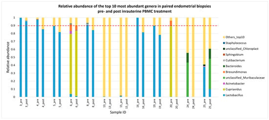
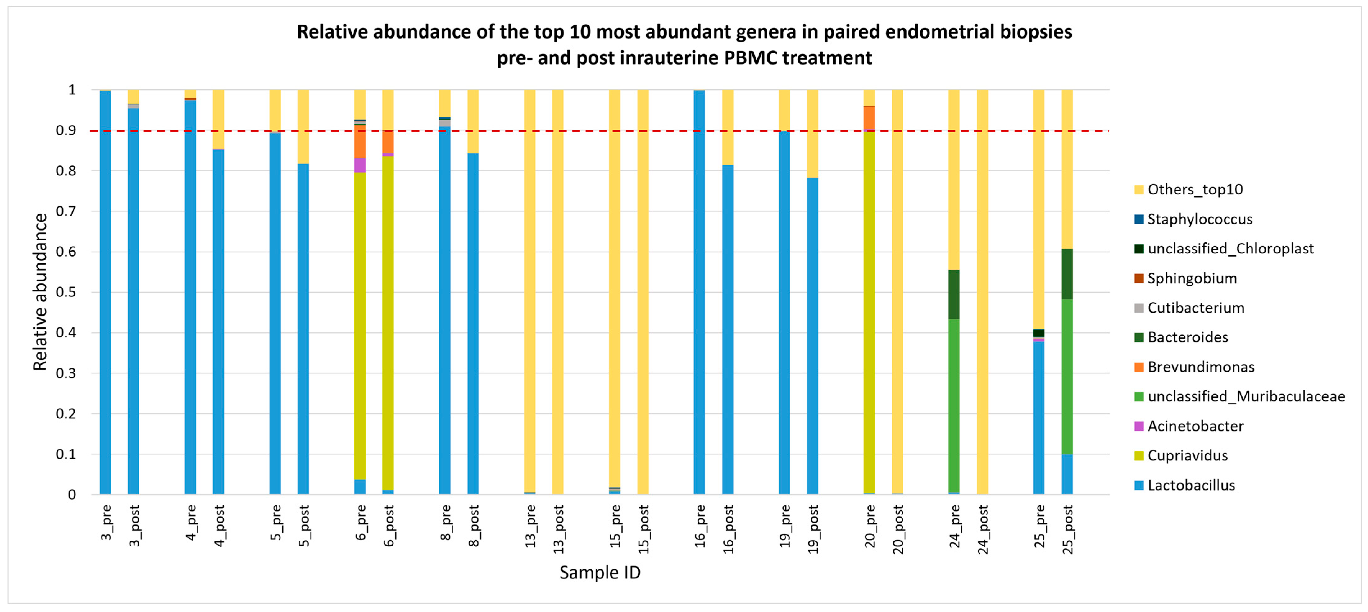
Across patients, microbial profiles varied substantially, with some samples showing strong Lactobacillus predominance and others displaying increased contributions from genera such as Cupriavidus, Acinetobacter, Brevundimonas, Bacteroides, and Staphylococcus. Comparison of paired samples highlights inter-individual variability in response to intrauterine PBMC treatment, including shifts toward or away from Lactobacillus dominance and changes in the relative contribution of non-Lactobacillus taxa.
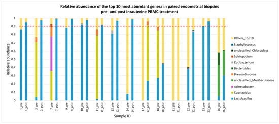
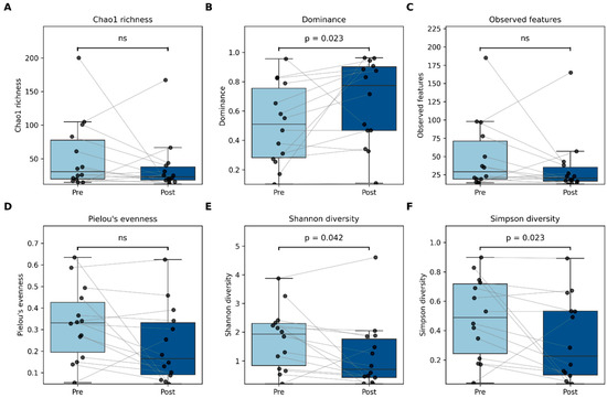
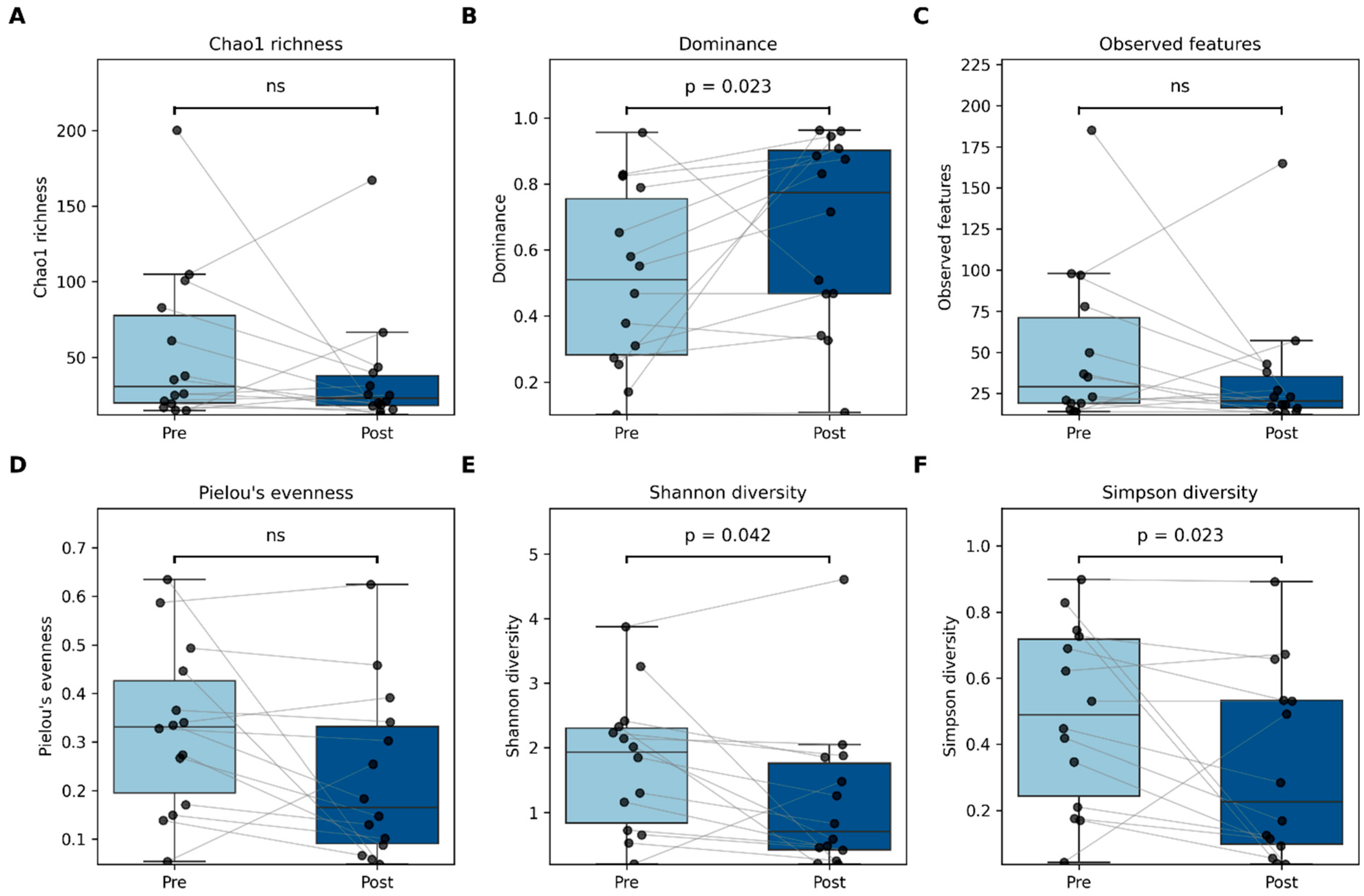
In this Lactobacillus increase group, post-treatment samples showed a higher relative abundance of Lactobacillus (mean 75.1 ± 34.6% vs. 37.2 ± 40.3%, p = 0.0006), with several individuals (9/14 patients) shifting toward or surpassing the dominance threshold (Figure 2). Correspondingly, reductions in the relative abundance of other genera—including Cupriavidus, Acinetobacter, Brevundimonas, and Staphylococcus—were observed in individual paired comparisons (patients 2, 7, 11, 18, 26; Figure 2). These patterns highlight a treatment-associated shift toward Lactobacillus-enriched microbial communities. In this group, no significant differences were observed for richness (Chao1), observed features, or evenness (Pielou’s). In contrast, dominance increased significantly following treatment (p = 0.023), while both Shannon diversity (p = 0.042) and Simpson diversity (p = 0.023) decreased (Figure 3). Collectively, these changes are consistent with a shift toward less diverse, more compositionally dominated microbial community, consistent with increased prominence of Lactobacillus.
Figure 2.
Relative abundance of the top 10 bacterial genera in paired endometrial biopsies from the Lactobacillus increase group before and after intrauterine PBMC treatment. Stacked bar plots show genus-level microbial composition for individual patients who exhibited an increase in Lactobacillus relative abundance following treatment. Each pair of bars represents pre-treatment (“_pre”) and post-treatment (“_post”) samples from the same subject (x-axis). The y-axis indicates relative abundance (proportion from 0 to 1). Colors denote the ten most abundant genera across all samples, with remaining low-abundance taxa fraction grouped as “Others_top10”. A red dotted horizontal line at 0.9 marks the threshold for Lactobacillus dominance. Samples with ≥90% relative abundance of Lactobacillus are classified as Lactobacillus-dominant, while those below this threshold are considered non-dominant or polymicrobial.
Figure 3.
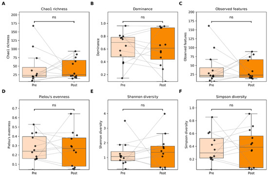
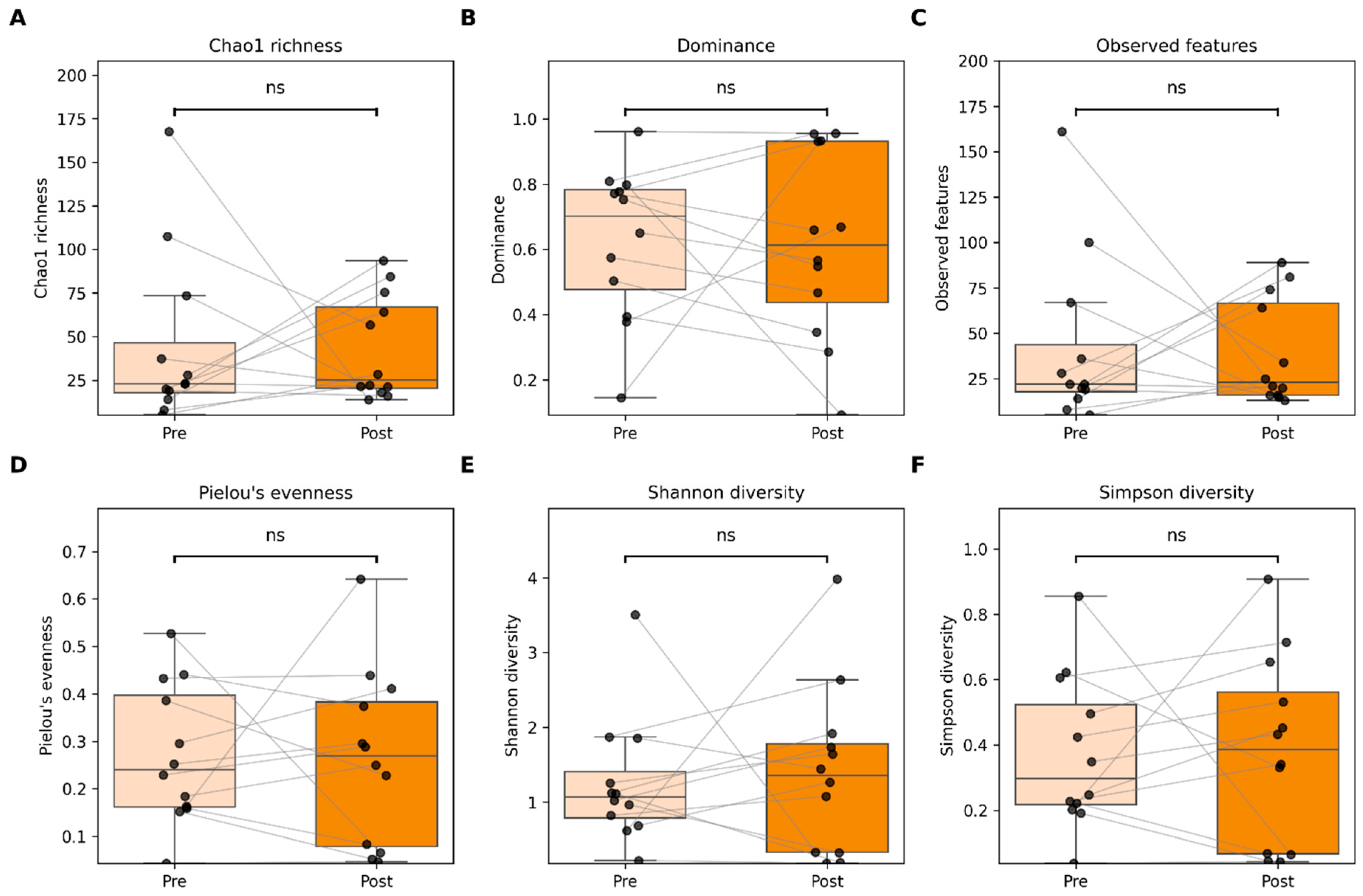
Alpha diversity metrics in the Lactobacillus increase group following intrauterine PBMC treatment. Boxplots with paired observations illustrate changes in alpha diversity between pre-treatment (“Pre”) and post-treatment (“Post”) endometrial biopsy samples from individuals who exhibited an increase in Lactobacillus relative abundance after treatment (n = 14). Each dot represents a single sample, and gray connecting lines indicate paired measurements from the same subject. Boxes represent the interquartile range (IQR), with the central line indicating the median and whiskers extending to 1.5× IQR. (A) Chao1 richness, (B) dominance, (C) observed features, (D) Pielou’s evenness, (E) Shannon diversity, and (F) Simpson diversity are shown. Statistical comparisons between paired samples were performed using a paired test (Wilcoxon signed-rank test), and p-values are indicated above each panel; “ns” denotes not significant.
Within the no-increase group, while the relative abundance of Lactobacillus decreased post-intervention (mean 43.1 ± 43.3% vs. 50.9 ± 46.8%, p = 0.01), it remained the most prevalent taxon (Figure 4). In addition, no significant differences were observed for any of the alpha diversity metrics after intrauterine PBMC treatment (Figure 5). The microbiome in this subgroup remained relatively stable, suggesting either resistance to perturbation or a baseline state that was already compositionally stable or “optimized.”
Figure 4.
Relative abundance of the top 10 bacterial genera in paired endometrial biopsies from the no-increase group before and after intrauterine PBMC treatment. Stacked bar plots show genus-level microbial composition for individual patients who did not exhibit an increase in Lactobacillus relative abundance following treatment. Each pair of bars represents pre-treatment (“_pre”) and post-treatment (“_post”) samples from the same subject (x-axis). The y-axis indicates relative abundance (proportion from 0 to 1). Colors denote the ten most abundant genera across all samples, with remaining low-abundance taxa fraction grouped as “Others_top10”. A red dotted horizontal line at 0.9 marks the threshold for Lactobacillus dominance. Samples with ≥90% relative abundance of Lactobacillus are classified as Lactobacillus-dominant, while those below this threshold are considered non-dominant or polymicrobial.
Figure 5.
Alpha diversity metrics of endometrial microbiota in paired biopsies in the no-increase group before and after intrauterine PBMC treatment. Boxplots with paired observations illustrate changes in alpha diversity between pre-treatment (“Pre”) and post-treatment (“Post”) endometrial biopsy samples from individuals who did not exhibit an increase in Lactobacillus relative abundance after treatment (n = 12). Each dot represents a single sample, and gray connecting lines indicate paired measurements from the same subject. Boxes represent the interquartile range (IQR), with the central line indicating the median and whiskers extending to 1.5× IQR. (A) Chao1 richness, (B) dominance, (C) observed features, (D) Pielou’s evenness, (E) Shannon diversity, and (F) Simpson diversity are shown. Statistical comparisons between paired samples were performed using a paired test (Wilcoxon signed-rank test), and p-values are indicated above each panel; “ns” denotes not significant.
3.7. Beta Diversity and Community Structure
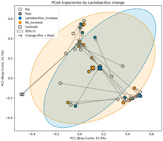
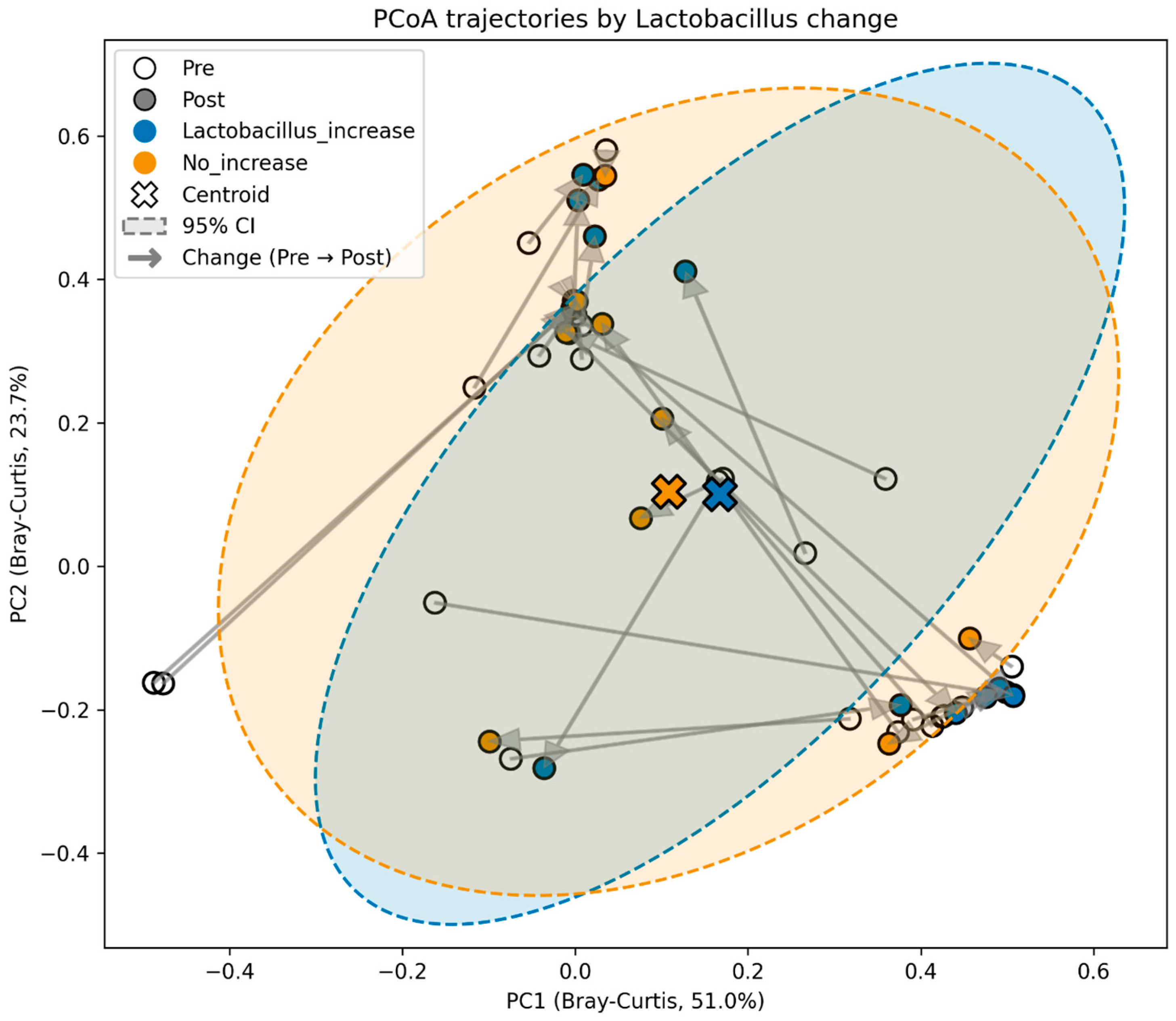
Principal coordinates analysis (PCoA) based on Bray–Curtis dissimilarities was used to evaluate changes in overall microbial community composition following PBMC treatment. No significant shift in overall microbial community composition was observed between pre- and post-treatment samples (PERMANOVA, pseudo-F = 1.16, R2 = 0.0008, p = 0.286, 999 permutations). Subjects in the Lactobacillus-increase group demonstrated more noticeable changes in community composition following treatment, whereas those in the no-increase group generally exhibited smaller or more variable shifts (Figure 6). Nevertheless, the two response groups did not differ significantly in overall community structure (PERMANOVA, pseudo-F = 0.80, R2 = 0.0002, p = 0.480), also visualised by the closely positioned centroids and largely overlapping 95% confidence intervals (Figure 6).
Figure 6.
Principal coordinates analysis (PCoA) of microbial community trajectories categorized by Lactobacillus abundance change. The plot illustrates the compositional shifts of microbial communities before (Pre) and after (Post) treatment, based on Bray–Curtis dissimilarity. Each pair of points connected by an arrow represents an individual sample’s trajectory from pre- to post-treatment. Empty circles indicate pre-treatment samples, while filled circles represent post-treatment samples. Samples are colored according to Lactobacillus abundance change status: blue for samples with an increase in Lactobacillus and orange for those without an increase. Group centroids for pre- and post-treatment samples are marked by crosses (×) with corresponding colors. Ellipses denote the 95% confidence intervals (CI) around group centroids, illustrating the variability within each group. Gray arrows connect paired pre- and post-treatment points to visualize the direction and magnitude of microbial community change for each subject. The axes display the first two principal coordinates (PC1 and PC2) derived from Bray–Curtis distances, with the percentage of variance explained by each coordinate indicated in parentheses.
Collectively, these findings suggest that PBMC treatment was not associated with large-scale restructuring of the endometrial microbiome. Instead, the observed treatment-related effects appear to involve changes in the relative abundance of specific taxa, particularly Lactobacillus, rather than broad alterations in overall community composition.
4. Discussion
In this study, PBMC administration was not associated with a uniform shift in the endometrial microbiome when the cohort was analyzed as a whole. However, stratified analyses revealed a clear subgroup-specific response, with approximately half of the participants demonstrating a marked increase in Lactobacillus abundance. These findings indicate that PBMC therapy does not exert a universal microbiome-modifying effect. Rather, its influence appears to depend on the pre-existing ecological state of the endometrium, suggesting that baseline microbiome composition is an important determinant of responsiveness.
The observation that only women with lower baseline Lactobacillus abundance responded suggests that PBMC therapy is unlikely to induce a new microbial state but instead may promote a shift toward a pre-existing physiological balance. This implies that the therapy acts by modifying the endometrial environment in a manner that allows endogenous microbial communities to reorganize and move toward a more Lactobacillus-dominant profile, rather than directly shaping microbial composition.
These findings suggest that PBMC therapy may exert indirect effects on the endometrial microbiome, potentially mediated through immune modulation rather than direct microbial targeting [22,23,24,25]. PBMCs are known to influence local cytokine levels and immune cell recruitment, which in turn may create ecological conditions that favour the expansion of specific taxa such as Lactobacillus [17,23,24,26]. One potential mechanism involves PBMC-induced modulation of the local inflammatory environment. Previous studies have demonstrated that activated PBMCs induce the expression of cytokines and growth factors, including interleukin (IL)-1β, IL-6, IL-10, leukemia inhibitory factor (LIF), and vascular endothelial growth factor (VEGF), all of which have recognized roles in endometrial receptivity and tissue remodeling [17,25,27,28]. Alterations in cytokine signaling may modify epithelial barrier function, nutrient availability, antimicrobial peptide production, and mucosal immune surveillance, thereby influencing microbial colonization dynamics, as studied extensively in the vagina [12,23,24,26,29,30]. Under such conditions, Lactobacillus species may gain a competitive advantage over less adapted taxa, resulting in the shift toward Lactobacillus dominance observed in a subset of patients.
An alternative explanation is that PBMC therapy does not directly promote Lactobacillus growth but instead facilitates restoration of microbial homeostasis specifically in women with pre-existing dysbiosis [11,22,23,31]. Our data are more consistent with this explanation rather than with a direct antimicrobial effect because microbiome changes occurred predominantly in participants with lower baseline Lactobacillus abundance, whereas women with already Lactobacillus-dominant microbiomes exhibited little change. In ecological terms, dysbiotic microbial communities may exist in a less stable state and therefore be more susceptible to perturbation, whereas Lactobacillus-dominant communities may already represent a relatively stable equilibrium [30,32]. PBMC-induced immune modulation may shift the local environment toward conditions that favor the re-establishment of a Lactobacillus-dominated ecosystem, while producing little detectable effect in individuals whose microbiome is already optimized.
While in many body sites, decreased microbial diversity is considered unfavourable, the endometrium is an exception. A growing body of evidence indicates that a low-diversity, Lactobacillus-dominated microbiome is associated with reproductive health and improved implantation potential [7,9,10,12,13]. Thus, the shift toward lower diversity observed here may reflect a transition toward a more physiologically favourable state rather than dysbiosis. Moreno et al. (2016) first highlighted that an LD endometrial microbiome correlates with higher implantation and live birth rates [6]. More recent work has reinforced the concept that microbiome composition is a critical determinant of endometrial receptivity [10,12,13,14,33,34]. Although the mechanisms underlying this association remain incompletely understood, emerging evidence suggests that metabolic regulation is an important component of endometrial receptivity. It is therefore conceivable that Lactobacillus-dominated communities contribute to a microenvironment conducive to implantation through effects on metabolite production, nutrient availability, pH regulation, and local tissue homeostasis [29,30,35]. At the same time, the literature emphasises the relative stability of the endometrial microbiome compared to other mucosal sites, with some studies suggesting limited modifiability [8,31,36]. Our findings refine this perspective by demonstrating that modifiability may be conditionally dependent on baseline ecological state, rather than universally constrained. This suggests that the apparent stability of the endometrial microbiome reported in previous studies may partly reflect differences in baseline microbial composition rather than an inherent resistance to change across all patients.
In some samples, we detected high relative abundance of Cupriavidus and Acinetobacter, consistent with findings from other authors [9,10,11]. The detection of unclassified_Chloroplast sequences reflects amplification of chloroplast-derived 16S rRNA genes, which can occur in microbiome studies because chloroplasts are of bacterial ancestry. Notably, these sequences were identified in two pre-treatment endometrial samples but were not detected in extraction or negative controls, arguing against reagent contamination as their source. Chloroplast DNA may originate from plant-derived material introduced through dietary or environmental exposure; however, its biological relevance within the endometrial microbiome remains unclear.
An important finding of this study is the marked heterogeneity in microbiome response following PBMC administration. Averaging responses across the entire cohort obscured biologically meaningful changes that became apparent only after stratification according to baseline microbiome composition. This observation suggests that the absence of an overall treatment effect should not necessarily be interpreted as a lack of biological activity. Instead, it highlights that PBMC therapy may benefit a biologically distinct subgroup of patients. Such heterogeneity may explain why previous studies evaluating immunomodulatory interventions have reported inconsistent outcomes and underscores the importance of patient stratification in future clinical trials. Specifically, PBMC therapy may be more beneficial in individuals with non-Lactobacillus-dominant microbiomes at baseline, who appear more “modifiable” and capable of transitioning toward an LD state. In contrast, patients with pre-existing LD microbiomes may derive limited microbiome-related benefit, possibly due to a ceiling effect. This raises the possibility of using baseline microbiome profiling to guide therapeutic decision-making and optimise the selection of patients most likely to benefit from PBMC intervention. An endometrial-based stratification guide for immunotherapy has already shown promise in improving reproductive outcomes [37].
Several limitations should be acknowledged. First, although the sample size was comparable to those of other exploratory studies investigating the endometrial microbiome, the relatively small number of participants, particularly within the subgroup analyses, may have limited the ability to detect more subtle effects and may affect the generalizability of the findings. Second, the study did not include correlations with clinical outcomes such as implantation or pregnancy rates, preventing direct assessment of functional relevance. Lastly, subgroup classification was performed post hoc, raising the possibility of bias and necessitating validation in independent cohorts.
Future studies should aim to identify predictive biomarkers of microbiome responsiveness to PBMC therapy, potentially integrating microbial, immunological, and clinical parameters. Combining microbiome profiling with immune characterization such as cytokine signatures, immune cell phenotyping, and transcriptomic analyses of the endometrium may provide mechanistic insight into the observed effects. Prospective, adequately powered studies are needed to validate these findings and determine whether microbiome-guided PBMC therapy can improve reproductive outcomes. Interventional trials incorporating stratified designs based on baseline microbiome composition could be particularly informative.
5. Conclusions
PBMC administration does not uniformly alter the endometrial microbiome at the population level. However, it may promote Lactobacillus dominance in a biologically distinct subset of patients, particularly those with low Lactobacillus abundance. These findings highlight the importance of baseline microbiome composition and reveal two distinct response patterns that are obscured in aggregate analyses, underscoring the need for personalised approaches in reproductive immunotherapy.
From a clinical perspective, our results suggest that the endometrial microbiome may influence individual responses to PBMC treatment and could potentially serve as a biomarker for patient stratification. The identification of patients who exhibit a favourable microbiome shift following PBMC administration may contribute to the development of more targeted immunomodulatory fertility treatments. More broadly, these findings support the integration of microbiome-informed strategies into reproductive medicine. Future studies with larger cohorts and clinical outcome data are warranted to determine whether these microbiome changes translate into improved reproductive outcomes and to further refine personalised therapeutic approaches.
Author Contributions
Conceptualization, M.R., D.P. and S.H.; methodology, M.R., T.T., D.M., B.R. and S.H.; software, M.R., D.P. and R.G.; validation, M.R., D.P., R.G. and M.H.; formal analysis, M.R., T.T. and D.P.; investigation, M.R., M.H., J.S., S.K. and I.P.; resources, T.T., D.M., G.S. and S.H.; data curation, M.R., M.H., T.T., J.S., S.K. and I.P.; writing—original draft preparation, M.R.; writing—review and editing, D.P., R.G. and I.P.; visualization, M.R. and D.P.; supervision, D.P., G.S. and S.H.; project administration, B.R. and S.H.; funding acquisition, G.S. and S.H. All authors have read and agreed to the published version of the manuscript.
Funding
This research was funded by the National Science Fund, Ministry of Education, Bulgaria, Contract No. KP-06-N53/14/16.11.2021.
Institutional Review Board Statement
The study was conducted in accordance with the Declaration of Helsinki, and approved by Nadezhda Women’s Health Hospital Ethics Committee (protocol code 56/29.12.2020).
Informed Consent Statement
Informed consent was obtained from all subjects involved in the study.
Data Availability Statement
The original data presented in the study are openly available in zenodo.org at DOI: 10.5281/zenodo.20665446.
Conflicts of Interest
The authors declare no conflicts of interest. The funders had no role in the design of the study; in the collection, analyses, or interpretation of data; in the writing of the manuscript; or in the decision to publish the results.
Abbreviations
The following abbreviations are used in this manuscript:
| ART | Assisted Reproductive Technology |
| BMI | Body Mass Index |
| CI | Confidence Interval |
| DNA | Deoxyribonucleic Acid |
| FDR | False Discovery Rate |
| HiSeq | High-Throughput Sequencing Platform |
| IL | Interleukin |
| IQR | Interquartile Range |
| IVF | In Vitro Fertilization |
| LD | Lactobacillus-Dominant |
| LIF | Leukemia Inhibitory Factor |
| OTU | Operational Taxonomic Unit |
| PBS | Phosphate-Buffered Saline |
| PBMC | Peripheral Blood Mononuclear Cell(s) |
| PCoA | Principal Coordinates Analysis |
| PCR | Polymerase Chain Reaction |
| PERMANOVA | Permutational Multivariate Analysis of Variance |
| QIIME | Quantitative Insights Into Microbial Ecology |
| rRNA | Ribosomal Ribonucleic Acid |
| RPMI | Roswell Park Memorial Institute Medium |
| SD | Standard Deviation |
| SILVA | Silva Ribosomal RNA Database |
| UniFrac | Unique Fraction Metric |
| VEGF | Vascular Endothelial Growth Factor |
References
- Bashiri, A.; Halper, K.I.; Orvieto, R. Recurrent implantation failure: Update overview on etiology, diagnosis, treatment and future directions. Reprod. Biol. Endocrinol. 2018, 16, 121. [Google Scholar] [CrossRef] [Scilit] [PubMed]
- Cimadomo, D.; Rienzi, L.; Conforti, A.; Forman, E.; Canosa, S.; Innocenti, F.; Poli, M.; Hynes, J.; Gemmell, L.; Vaiarelli, A.; et al. Opening the black box: Why do euploid blastocysts fail to implant? A systematic review and meta-analysis. Hum. Reprod. Update 2023, 29, 570–633. [Google Scholar] [CrossRef] [Scilit] [PubMed]
- Ma, J.; Gao, W.; Li, D. Recurrent Implantation Failure: A Comprehensive Summary from Etiology to Treatment. Front. Endocrinol. 2023, 13, 1061766. [Google Scholar] [CrossRef] [Scilit] [PubMed]
- Robertson, S.A.; Moldenhauer, L.M.; Green, E.S.; Care, A.S.; Hull, M.L. Immune determinants of endometrial receptivity: A biological perspective. Fertil. Steril. 2022, 117, 1107–1120. [Google Scholar] [CrossRef] [Scilit] [PubMed]
- Franasiak, J.M.; Werner, M.D.; Juneau, C.R.; Tao, X.; Landis, J.; Zhan, Y.; Treff, N.R.; Scott, R.T. Endometrial microbiome at the time of embryo transfer: Next-generation sequencing of the 16S ribosomal subunit. J. Assist. Reprod. Genet. 2016, 33, 129–136. [Google Scholar] [CrossRef] [Scilit] [PubMed]
- Moreno, I.; Codoñer, F.M.; Vilella, F.; Valbuena, D.; Martinez-Blanch, J.F.; Jimenez-Almazán, J.; Alonso, R.; Alamá, P.; Remohí, J.; Pellicer, A.; et al. Evidence that the endometrial microbiota has an effect on implantation success or failure. Am. J. Obstet. Gynecol. 2016, 215, 684–703. [Google Scholar] [CrossRef] [Scilit] [PubMed]
- Benner, M.; Ferwerda, G.; Joosten, I.; van der Molen, R.G. How uterine microbiota might be responsible for a receptive, fertile endometrium. Hum. Reprod. Update 2018, 24, 393–415. [Google Scholar] [CrossRef] [Scilit] [PubMed]
- Reschini, M.; Benaglia, L.; Ceriotti, F.; Borroni, R.; Ferrari, S.; Castiglioni, M.; Guarneri, D.; Porcaro, L.; Vigano’, P.; Somigliana, E.; et al. Endometrial microbiome: Sampling, assessment, and possible impact on embryo implantation. Sci. Rep. 2022, 12, 8467. [Google Scholar] [CrossRef] [Scilit] [PubMed]
- Moreno, I.; Garcia-Grau, I.; Perez-Villaroya, D.; Gonzalez-Monfort, M.; Bahçeci, M.; Barrionuevo, M.J.; Taguchi, S.; Puente, E.; Dimattina, M.; Lim, M.W.; et al. Endometrial microbiota composition is associated with reproductive outcome in infertile patients. Microbiome 2022, 10, 1. [Google Scholar] [CrossRef] [Scilit] [PubMed]
- Chen, L.; Ke, Y.; Guo, H.; Wu, B.; Liu, L. The relationship between vaginal and endometrial microbiota and the outcome of Frozen-Thawed embryo transfer during hormone replacement cycles. BMC Microbiol. 2025, 25, 494. [Google Scholar] [CrossRef] [Scilit] [PubMed]
- Chen, C.; Song, X.; Wei, W.; Zhong, H.; Dai, J.; Lan, Z.; Li, F.; Yu, X.; Feng, Q.; Wang, Z.; et al. The microbiota continuum along the female reproductive tract and its relation to uterine-related diseases. Nat. Commun. 2017, 8, 875. [Google Scholar] [CrossRef] [Scilit] [PubMed]
- Gao, X.; Louwers, Y.V.; Laven, J.S.E.; Schoenmakers, S. Clinical Relevance of Vaginal and Endometrial Microbiome Investigation in Women with Repeated Implantation Failure and Recurrent Pregnancy Loss. Int. J. Mol. Sci. 2024, 25, 622. [Google Scholar] [CrossRef] [Scilit] [PubMed]
- Hiratsuka, D.; Matsuo, M.; Hirota, Y. Endometrial Microbiome and Implantation: From Basic Knowledge to Clinical Medicine. Reprod. Med. Biol. 2026, 25, e70040. [Google Scholar] [CrossRef] [Scilit] [PubMed]
- Hanaoka, M.; Hanaoka, K.; Yamada, M. Effect of improvement in the endometrial microbiome on in vitro fertilization outcomes. J. Assist. Reprod. Genet. 2026, 43, 623–637. [Google Scholar] [CrossRef] [Scilit] [PubMed]
- Busnelli, A.; Somigliana, E.; Cirillo, F.; Baggiani, A.; Levi-Setti, P.E. Efficacy of therapies and interventions for repeated embryo implantation failure: A systematic review and meta-analysis. Sci. Rep. 2021, 11, 1747. [Google Scholar] [CrossRef] [Scilit] [PubMed]
- Liu, M.; Yuan, Y.; Qiao, Y.; Tang, Y.; Sui, X.; Yin, P.; Yang, D. The effectiveness of immunomodulatory therapies for patients with repeated implantation failure: A systematic review and network meta-analysis. Sci. Rep. 2022, 12, 18434. [Google Scholar] [CrossRef] [Scilit] [PubMed]
- Yu, N.; Zhang, B.; Xu, M.; Wang, S.; Liu, R.; Wu, J.; Yang, J.; Feng, L. Intrauterine administration of autologous peripheral blood mononuclear cells (PBMCs) activated by HCG improves the implantation and pregnancy rates in patients with repeated implantation failure: A prospective randomized study. Am. J. Reprod. Immunol. 2016, 76, 212–216. [Google Scholar] [CrossRef] [Scilit] [PubMed]
- Yakin, K.; Oktem, O.; Urman, B. Intrauterine administration of peripheral mononuclear cells in recurrent implantation failure: A systematic review and meta-analysis. Sci. Rep. 2019, 9, 3897. [Google Scholar] [CrossRef] [Scilit] [PubMed]
- Yang, D.N.; Wu, J.H.; Geng, L.; Cao, L.J.; Zhang, Q.J.; Luo, J.Q.; Kallen, A.; Hou, Z.H.; Qian, W.P.; Shi, Y.; et al. Efficacy of intrauterine perfusion of peripheral blood mononuclear cells (PBMC) for infertile women before embryo transfer: Meta-analysis. J. Obstet. Gynaecol. 2020, 40, 961–968. [Google Scholar] [CrossRef] [Scilit] [PubMed]
- Edgar, R.C. UPARSE: Highly accurate OTU sequences from microbial amplicon reads. Nat. Methods 2013, 10, 996–998. [Google Scholar] [CrossRef] [Scilit] [PubMed]
- Wang, Q.; Garrity, G.M.; Tiedje, J.M.; Cole, J.R. Naive Bayesian classifier for rapid assignment of rRNA sequences into the new bacterial taxonomy. Appl. Environ. Microbiol. 2007, 73, 5261–5267. [Google Scholar] [CrossRef] [Scilit] [PubMed]
- Agostinis, C.; Mangogna, A.; Bossi, F.; Ricci, G.; Kishore, U.; Bulla, R. Uterine Immunity and Microbiota: A Shifting Paradigm. Front. Immunol. 2019, 10, 2387. [Google Scholar] [CrossRef] [Scilit] [PubMed]
- Al-Nasiry, S.; Ambrosino, E.; Schlaepfer, M.; Morré, S.A.; Wieten, L.; Voncken, J.W.; Spinelli, M.; Mueller, M.; Kramer, B.W. The interplay between reproductive tract microbiota and immunological system in human reproduction. Front. Immunol. 2020, 11, 378. [Google Scholar] [CrossRef] [Scilit] [PubMed]
- Zhu, N.; Yang, X.; Liu, Q.; Chen, Y.; Wang, X.; Li, H.; Gao, H. “Iron triangle” of regulating the uterine microecology: Endometrial microbiota, immunity and endometrium. Front. Immunol. 2022, 13, 928475. [Google Scholar] [CrossRef] [Scilit] [PubMed]
- Blazheva, S.; Pachkova, S.; Bodurska, T.; Ivanov, P.; Blazhev, A.; Lukanov, T.; Konova, E. Unlocking the uterine code: Microbiota, immune cells, and therapy for recurrent reproductive failure. Microorganisms 2024, 12, 547. [Google Scholar] [CrossRef] [Scilit] [PubMed]
- Wang, W.; Feng, D.; Ling, B. Biologia Futura: Endometrial microbiome affects endometrial receptivity from the perspective of the endometrial immune microenvironment. Biol. Futur. 2022, 73, 291–300. [Google Scholar] [CrossRef] [Scilit] [PubMed]
- Fan, L.; Sha, M.; Li, W.; Kang, Q.; Wu, J.; Chen, S.; Yu, N. Intrauterine administration of peripheral blood mononuclear cells (PBMCs) improves embryo implantation in mice by regulating local Treg/Th17 cell balance. J. Reprod. Dev. 2021, 67, 359–368. [Google Scholar] [CrossRef] [Scilit] [PubMed]
- Rezaee, D.; Bandehpour, M.; Kazemi, B.; Bastaminejad, S.; Najafi, S.; Salehi, M. Cytokine changes and embryo attachment in mouse endometrial cells following treated with peripheral blood mononuclear cells (PBMCs) expressing ectopic hCG, and hCG-activated PBMCs. Asian Pac. J. Reprod. 2023, 12, 90–96. [Google Scholar] [CrossRef] [Scilit]
- Miller, E.A.; Beasley, D.E.; Dunn, R.R.; Archie, E.A. Lactobacilli Dominance and Vaginal pH: Why Is the Human Vaginal Microbiome Unique? Front. Microbiol. 2016, 7, 1936. [Google Scholar] [CrossRef] [Scilit] [PubMed]
- Chopra, C.; Bhushan, I.; Mehta, M.; Koushal, T.; Gupta, A.; Sharma, S.; Kumar, M.; Al Khodor, S.; Sharma, S. Vaginal microbiome: Considerations for reproductive health. Future Microbiol. 2022, 17, 1501–1513. [Google Scholar] [CrossRef] [Scilit] [PubMed]
- Kyono, K.; Hashimoto, T.; Kikuchi, S.; Nagai, Y.; Sakuraba, Y. A pilot study and case reports on endometrial microbiota and pregnancy outcome: An analysis using 16S rRNA gene sequencing among IVF patients, and trial therapeutic intervention for dysbiotic endometrium. Reprod. Med. Biol. 2018, 18, 72–82. [Google Scholar] [CrossRef] [Scilit] [PubMed]
- Lee, C.Y.; Diegel, J.; France, M.T.; Ravel, J.; Arnold, K.B. Evaluation of vaginal microbiome equilibrium states identifies microbial parameters linked to resilience after menses and antibiotic therapy. PLoS Comput. Biol. 2023, 19, e1011295. [Google Scholar] [CrossRef] [Scilit] [PubMed]
- Lozano, F.M.; Lledó, B.; Morales, R.; Cascales, A.; Hortal, M.; Bernabeu, A.; Bernabeu, R. Characterization of the Endometrial Microbiome in Patients with Recurrent Implantation Failure. Microorganisms 2023, 11, 741. [Google Scholar] [CrossRef] [Scilit] [PubMed]
- Bui, B.N.; van Hoogenhuijze, N.; Viveen, M.; Mol, F.; Teklenburg, G.; de Bruin, J.-P.; Besselink, D.; Brentjens, L.S.; Mackens, S.; Rogers, M.R.C.; et al. The endometrial microbiota of women with or without a live birth within 12 months after a first failed IVF/ICSI cycle. Sci. Rep. 2023, 13, 3444. [Google Scholar] [CrossRef] [Scilit] [PubMed]
- Zhang, X.; Zhu, Q.; Nie, W.; Yan, X.; Yuan, Z.; Tian, L. Linking the Warburg effect to endometrial receptivity: Metabolic parallels in embryo implantation. Front. Cell Dev. Biol. 2025, 13, 1683790. [Google Scholar] [CrossRef] [Scilit] [PubMed]
- Molina, N.M.; Sola-Leyva, A.; Saez-Lara, M.J.; Plaza-Diaz, J.; Tubić-Pavlović, A.; Romero, B.; Clavero, A.; Mozas-Moreno, J.; Fontes, J.; Altmäe, S. New Opportunities for Endometrial Health by Modifying Uterine Microbial Composition: Present or Future? Biomolecules 2020, 10, 593. [Google Scholar] [CrossRef] [Scilit] [PubMed]
- Lédée, N.; Petitbarat, M.; Dray, G.; Chevrier, L.; Kazhalawi, A.; Rahmati, M.; Vicaut, E.; Diallo, A.; Cassuto, N.G.; Ruoso, L.; et al. Endometrial immune profiling and precision therapy increase live birth rate after embryo transfer: A randomised controlled trial. Front. Immunol. 2025, 16, 1523871. [Google Scholar] [CrossRef] [Scilit] [PubMed]

Disclaimer/Publisher’s Note: The statements, opinions and data contained in all publications are solely those of the individual author(s) and contributor(s) and not of MDPI and/or the editor(s). MDPI and/or the editor(s) disclaim responsibility for any injury to people or property resulting from any ideas, methods, instructions or products referred to in the content. |
© 2026 by the authors. Licensee MDPI, Basel, Switzerland. This article is an open access article distributed under the terms and conditions of the Creative Commons Attribution (CC BY) license.