Review

From Metastatic Gateways to Immune Reservoirs: Reframing Tumor-Draining Lymph Nodes in Perioperative Cancer Immunotherapy


Department of Immunology, Kindai University Faculty of Medicine, Sakai 590-0197, Osaka, Japan
*
Author to whom correspondence should be addressed.
Immuno 2026, 6(3), 47; https://doi.org/10.3390/immuno6030047
Submission received: 5 June 2026 / Revised: 16 July 2026 / Accepted: 21 July 2026 / Published: 22 July 2026
(This article belongs to the Section Cancer Immunology and Immunotherapy)

Abstract

Immune checkpoint inhibitors (ICIs) are now being introduced into perioperative treatment for several solid tumors. This strategy is usually explained by tumor reduction before surgery or by the elimination of minimal residual disease (MRD) after surgery. However, these explanations may not be sufficient to understand why the timing of ICI treatment, especially before lymph node (LN) removal, is important. In this review, we discuss tumor-draining lymph nodes (tdLNs) from two different aspects. tdLNs are anatomical routes for regional and distant metastasis, but they are also sites where tumor antigens are presented and tumor-specific T cell responses are generated. In particular, preclinical and translational studies suggest that tdLNs may maintain stem-like or progenitor-exhausted T cells (TPEX) that can respond to PD-1 blockade and supply more differentiated exhausted T cells to tumor sites. However, current clinical trials of perioperative ICIs demonstrate therapeutic benefit in specific diseases and regimens, but do not directly establish tdLN preservation or tdLN-resident TPEX maintenance as the decisive mechanism of efficacy. We therefore present the tdLN-reservoir model as a hypothesis-generating framework rather than as a clinically validated basis for modifying lymph node management. We also discuss the possible roles of neoadjuvant and adjuvant ICI in relation to antigen flow, minimal residual disease, metastatic-site draining LNs, postoperative lymphatic dysfunction, and future immune-guided clinical trials. Importantly, current evidence does not support altering standard lymph node surgery or radiotherapy solely to preserve putative tdLN immune-reservoir function.

1. Introduction: Perioperative Immunotherapy Across Solid Tumors

Immune checkpoint inhibitors (ICIs) have become an important component of cancer treatment, initially in advanced or metastatic disease and more recently in earlier-stage, potentially curable tumors. This shift has led to growing interest in neoadjuvant and perioperative immunotherapy across several solid tumors. Neoadjuvant therapy refers to systemic or local treatment administered before definitive surgery. In the context of ICI, neoadjuvant treatment refers to immune checkpoint blockade given before tumor resection, with the conventional clinical objectives of reducing tumor burden, facilitating complete surgical removal, and enabling early pathological response assessment. In addition to these clinical objectives, neoadjuvant ICI may have an immunological role because it is delivered while tumor-derived antigen flow, lymphatic drainage, and regional immune architecture are still at least partially preserved [1,2,3,4].
Clinical evidence for perioperative ICI has increased in multiple tumor types. In melanoma, the SWOG S1801 trial showed that pembrolizumab given before and after surgery improved event-free survival compared with the same drug given only in the adjuvant setting, indicating that treatment timing itself can affect clinical outcome [5]. The NADINA trial further supported the value of a neoadjuvant approach by showing favorable outcomes with neoadjuvant ipilimumab plus nivolumab followed by surgery and response-driven adjuvant therapy in resectable stage III melanoma [6]. In non-small cell lung cancer (NSCLC), KEYNOTE-671 demonstrated clinical benefit from perioperative pembrolizumab combined with chemotherapy in resectable disease [7]. Together, these findings suggest that perioperative ICI is not a disease-specific strategy, but an emerging therapeutic framework for solid tumors [4].
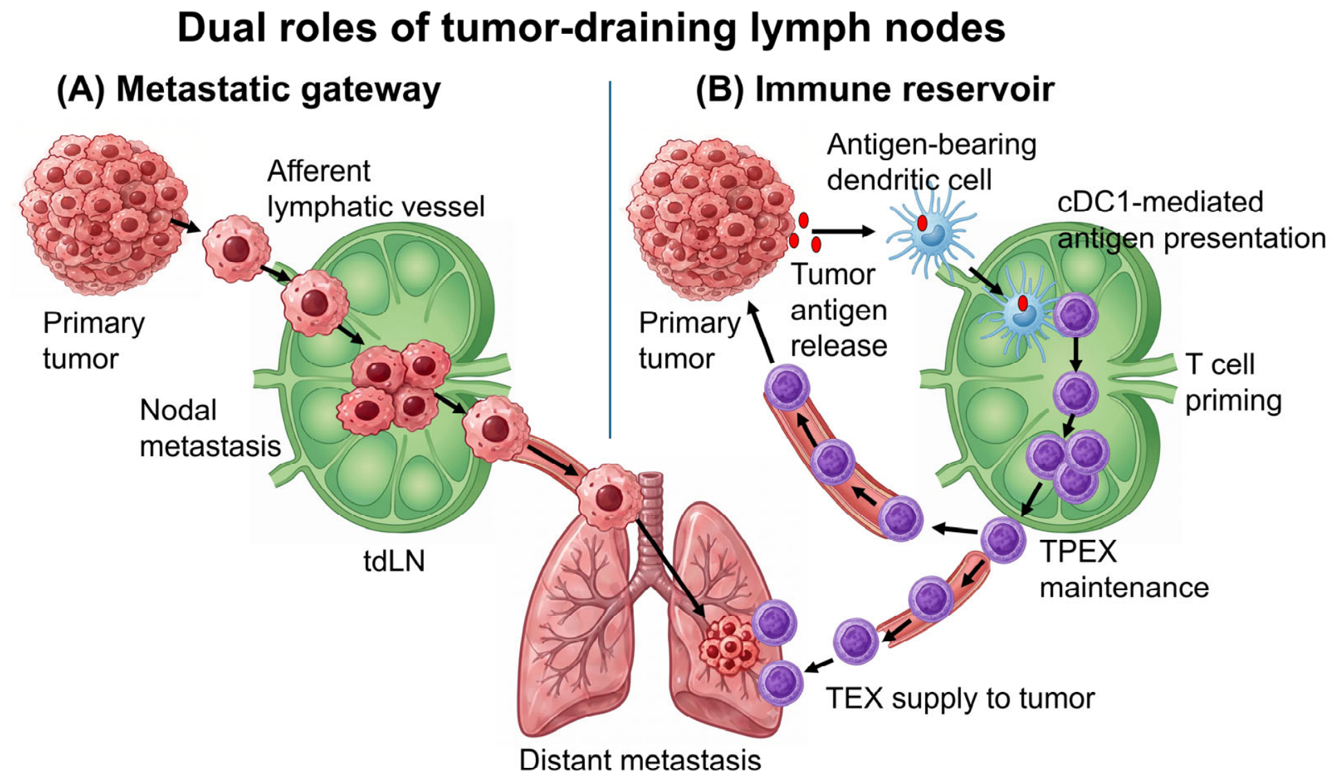
These clinical results raise an important mechanistic question: why should the administration of ICIs before the removal of the primary tumor and regional lymph nodes (LNs) be advantageous? One possible explanation is that the primary tumor and tumor-draining lymph nodes (tdLNs) are not merely surgical targets, but also active sites involved in the generation and regulation of antitumor immunity [8,9,10]. tdLNs are traditionally regarded as sites of lymphatic metastasis, and experimental studies have shown that tumor cells within LNs can enter nodal blood vessels and disseminate to distant organs [11,12]. At the same time, tdLNs are sites where tumor antigens are presented and tumor-specific T cell responses are initiated or maintained. This dual role of tdLNs as metastatic gateways and immune reservoirs is illustrated in Figure 1. In particular, studies of PD-1/PD-L1 blockade have shown that tdLNs can be critical for therapeutic efficacy and that checkpoint interactions within tdLNs regulate antitumor T cell immunity [8,9]. Therefore, perioperative immunotherapy should be considered not only as systemic drug treatment given around surgery, but also as a timing-dependent immune intervention that is influenced by whether the primary tumor and tdLN network remain intact.

2. Scope and Levels of Evidence

2.1. Novelty and Scope of This Review

Prior reviews have established the dual role of tdLNs as sites of metastatic dissemination and systemic immune surveillance. Therefore, the novelty of this review is not to reintroduce tdLN biology itself, but to integrate tdLN immune-reservoir concepts with the timing of perioperative ICI, nodal surgery, radiotherapy, postoperative lymphatic dysfunction, MRD control, and response-adapted trial design. In this sense, we use the term “reframing” in a limited and translational manner: tdLNs are discussed as clinically accessible immune tissues that may help interpret perioperative treatment timing, rather than as a newly discovered mechanism or as a validated basis for changing current nodal management.

2.2. Literature Search Strategy

This article is a narrative and hypothesis-generating review rather than a systematic review, bibliometric analysis, or meta-analysis. Relevant publications were identified through targeted searches of PubMed using combinations of terms related to perioperative, neoadjuvant, and adjuvant immune checkpoint inhibition; tumor-draining lymph nodes; T-cell exhaustion; progenitor-exhausted T cells; PD-1/PD-L1 blockade; and individual tumor types. The search covered publications indexed in PubMed through 15 June 2026. Reference lists of key publications were also manually examined to identify additional relevant studies.
For the clinical sections, we prioritized pivotal or practice-informing prospective phase II and phase III trials representing major neoadjuvant, adjuvant, or perioperative ICI strategies across different tumor types. For the mechanistic sections, we selected seminal and influential primary studies that established or substantially advanced the concepts of tdLN-dependent antitumor immunity, PD-1-responsive progenitor-exhausted T cells, and the TPEX/TEX continuum, together with recent translational studies directly relevant to perioperative immunotherapy. Studies were selected on the basis of their direct relevance and conceptual contribution to the questions addressed in this review, rather than with the aim of providing an exhaustive enumeration of all published studies. Duplicate publications, conference abstracts without a corresponding peer-reviewed full-text article, studies without direct relevance to the scope of this review, and reports focusing exclusively on advanced unresectable disease without clear relevance to perioperative treatment were generally excluded. No formal Preferred Reporting Items for Systematic Reviews and Meta-Analyses (PRISMA)-based study-selection procedure, risk-of-bias assessment, or quantitative meta-analysis was performed.
Generative AI tools, including ChatGPT and OpenAI image-generation tools, were used to assist with language editing and the development and refinement of conceptual figure illustrations. These tools were not used to determine study eligibility, select the cited evidence, interpret the scientific findings, or generate the conclusions of this review. All scientific content, the literature selection, figure labels, and final figure compositions were critically reviewed, revised, and approved by the authors, who take full responsibility for the manuscript.

2.3. Evidence Framework

Perioperative ICI is now supported by clinical evidence in selected solid tumors, but the mechanistic basis of its benefit remains incompletely defined. In particular, most clinical trials were designed to test treatment efficacy, not to determine whether tdLN preservation, tdLN-resident progenitor-exhausted T cells, or nodal immune reservoirs are required for therapeutic response. Therefore, it is important not to conflate clinical efficacy with mechanistic proof.
In this review, we use the tdLN immune-reservoir model as a hypothesis-generating framework to interpret perioperative immunotherapy. To avoid overstating the current evidence, we distinguish four levels of support: established clinical evidence, preclinical mechanistic evidence, translational inference, and speculative future clinical application. This distinction is particularly important because the concept may have implications for lymph node surgery, radiotherapy, and perioperative treatment design. The major claims discussed in this review are therefore categorized according to their current level of evidence in Table 1.

2.4. Translational Limitations and Clinical Caution

This evidence framework should be kept in mind throughout the review. The clinical efficacy of perioperative ICI in selected cancers is supported by prospective trials, whereas the tdLN–TPEX/TEX framework remains a mechanistic model derived mainly from preclinical and translational studies. Thus, concepts such as tdLN preservation, nodal immune reservoirs, and immune-guided lymph node management should be interpreted as research hypotheses rather than as clinical recommendations.
More specifically, tdLN-resident TPEX-like populations have not been consistently validated across all human cancer types, and longitudinal clinical datasets directly linking preserved tdLN immune activity to improved perioperative ICI outcomes remain limited. Thus, the tdLN–TPEX/TEX framework should be viewed as a mechanistically plausible model that requires prospective human validation using paired tumor, LN, blood, and MRD-based analyses.

2.5. Terminology Used in This Review

Terminology related to exhausted CD8+ T-cell states varies across studies. In this review, the terms “stem-like,” “progenitor-like exhausted,” and “TPEX” are used to describe related but not always identical populations of less differentiated tumor-reactive CD8+ T cells that retain proliferative potential and commonly express TCF1. “TCF1+ CD8+ T cells” is used as a marker-based description, whereas “TPEX” refers to a functional and differentiation state within the exhaustion continuum. “TEX” refers broadly to exhausted T cells, including intermediate, effector-like, and terminally exhausted states. These terms are not interchangeable in all experimental systems, and their precise meaning depends on tissue context, antigen specificity, marker panels, and functional assays.

3. tdLNs as Metastatic Gateways

Lymphatic spread is a common route of dissemination in many solid tumors. Tumor cells can access lymphatic vessels in the primary tumor or peritumoral tissue and are transported by lymphatic flow to regional draining LNs. This process is not only a passive movement of cancer cells. Tumor-derived signals, including lymphangiogenic factors, can remodel lymphatic vessels and alter the premetastatic environment of draining LNs. As a result, tdLNs may provide a specialized niche in which disseminated tumor cells (DTCs) are retained, adapt to local stromal and immune conditions, and develop into nodal metastases [13,14].
In clinical oncology, LN metastasis has long been used as an indicator of disease progression, prognosis, and treatment selection. Regional LNs are therefore important targets for surgical dissection and radiotherapy. However, this conventional view does not fully capture the biological role of nodal metastases. Recent experimental studies suggest that tumor cells growing in LNs may acquire access to the nodal vasculature and subsequently contribute to distant organ seeding [11]. In a related study, LN blood vessels, including high endothelial venules (HEVs), were shown to serve as a vascular route by which tumor cells can leave the LN and enter the systemic circulation [12]. These findings indicate that nodal metastases may function not only as regional disease deposits, but also as intermediates between lymphatic and hematogenous dissemination. Achen and Stacker integrated these observations into a model in which tumor cells enter LNs through afferent lymphatics, move through the nodal tissue, approach blood vessels within the node, and then disseminate systemically [15]. This model is useful for understanding tdLNs as anatomical structures that connect local tumor drainage, regional metastasis, and distant spread. Although the relative contribution of LN-derived dissemination may vary among tumor types and clinical settings, the concept broadens the role of tdLNs beyond that of passive filters or staging markers.
This metastatic function of tdLNs has direct relevance to perioperative immunotherapy. The same lymphatic route that allows tumor cells to reach regional LNs also carries tumor antigens and antigen-presenting cells to immune priming sites. Thus, tdLNs have a dual role (Figure 1). They can support tumor progression as metastatic intermediates, but they can also participate in the induction and maintenance of antitumor immunity. This dual nature creates an important therapeutic dilemma in the ICI era. Removal of metastatic LNs remains necessary for oncologic control, but the timing and extent of LN removal may influence the immune responses generated during neoadjuvant treatment. Understanding tdLNs as metastatic gateways is therefore essential before considering their role as immune reservoirs.

4. tdLNs as Immune Reservoirs

The role of tdLNs is not limited to metastatic spread. The lymphatic route that permits tumor cells to reach regional LNs also delivers soluble tumor antigens, cell debris, and antigen-bearing dendritic cells (DCs) from the tumor to the draining LN. In this anatomical setting, tdLNs provide a structured environment in which antigen-presenting cells interact with T cells and initiate tumor-specific immune responses. Therefore, tdLNs should be considered not only as possible sites of tumor cell deposition, but also as important sites for the generation and maintenance of systemic antitumor immunity [10,16].
DCs are central to this process. Among them, conventional type 1 DCs (cDC1s) are particularly important for CD8+ T cell immunity because they efficiently present tumor-derived antigens through the MHC class I pathway. Tumor antigens may be acquired in the tumor microenvironment and then carried to tdLNs by migratory DCs, or they may be transferred to LN-resident antigen-presenting cells. Within tdLNs, cDC1-dependent antigen presentation supports the activation and expansion of tumor-reactive CD8+ T cells. In addition to their role in initial priming, cDC1s also help preserve a population of proliferative, tumor antigen-specific TCF1+ CD8+ T cells in tdLNs, suggesting that DC–T cell interactions in LNs contribute to the durability of antitumor immunity [17,18].
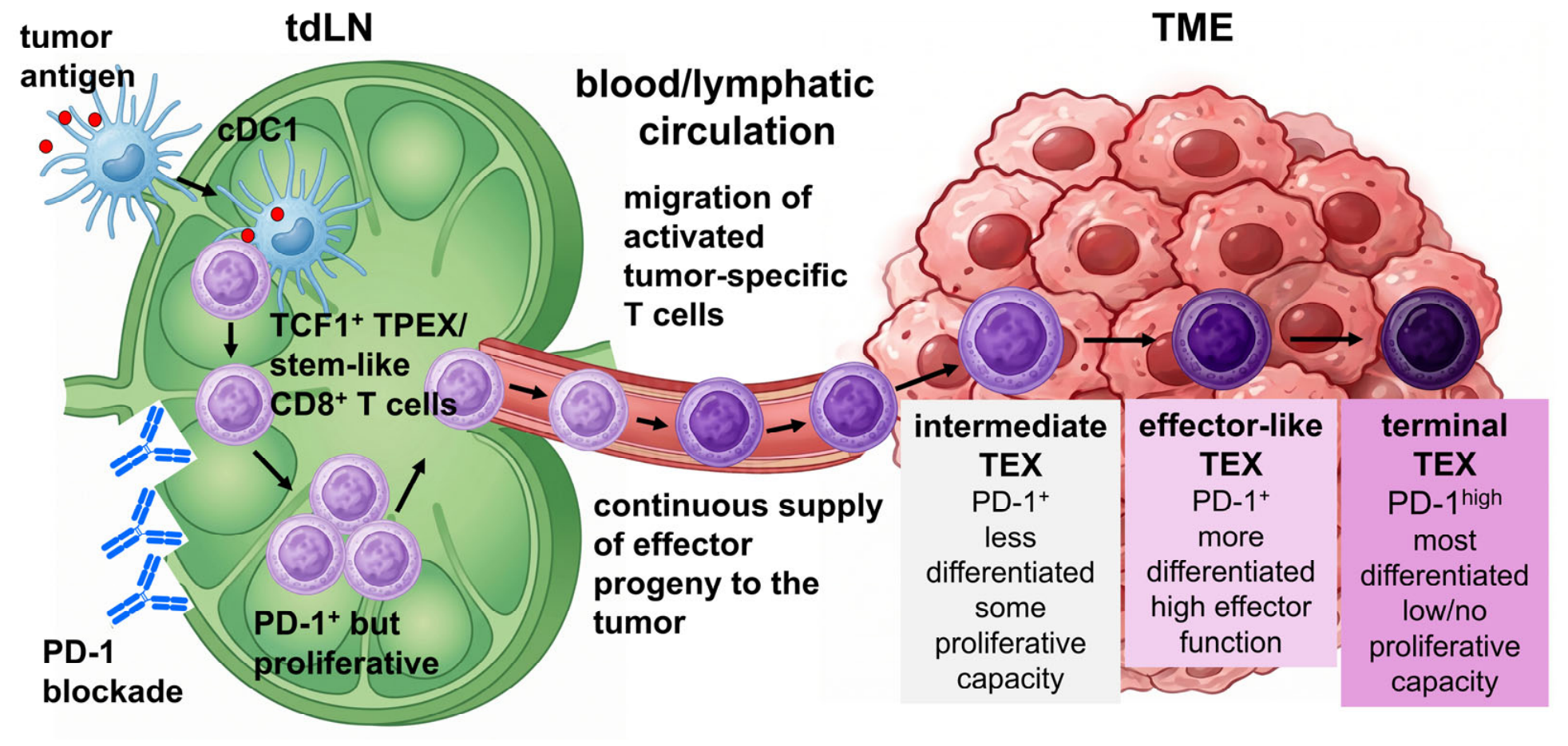
Recent studies have shown that tdLNs contain CD8+ T cell populations exhibiting stem-like features, known as progenitor-exhausted T cells (TPEX). These cells retain proliferative potential and differ from more differentiated exhausted T cells (TEX) that accumulate within tumors. They are important because they can serve as a source of newly generated effector-like progeny during an ongoing antitumor response. Thus, tdLNs are not simply places where T cell priming begins. They also provide a niche in which less differentiated tumor-specific T cells can be maintained before giving rise to more differentiated T cells that migrate to tumor sites [19].
This function is closely linked to the activity of PD-1/PD-L1 blockade. Studies in experimental tumor models have shown that intact tdLNs are required for optimal therapeutic effects of PD-1/PD-L1 blockade [8]. Other work has demonstrated that checkpoint interactions within tdLNs can suppress tumor-specific T cell responses, indicating that the draining LN itself is an important site of action for immune checkpoint therapy [9]. In addition, tumor-specific memory-like CD8+ T cells in draining LNs, now often interpreted as TPEX, have been shown to respond to PD-1/PD-L1 blockade and contribute to antitumor immunity [20].
Taken together, these findings support the possibility that tdLNs can function as one important immune reservoir within a broader antitumor immune network. They receive tumor antigens and DCs, support cDC1-dependent CD8+ T-cell priming and may help maintain less differentiated TCF1+ tumor-specific CD8+ T cells. However, tdLNs should not be viewed as the only anatomical site in which TPEX cells are maintained. Tertiary lymphoid structures (TLS), selected intratumoral niches, perivascular regions, and other lymphoid compartments may also contribute to the reservoir of PD-1-responsive tumor-reactive T cells. This concept provides a basis for considering why the timing of ICI relative to tumor resection and nodal surgery may be immunologically relevant.

5. The TPEX/TEX Continuum and Context-Dependent PD-1 Signaling

T cell exhaustion was originally described as a state induced by persistent antigen stimulation, particularly in chronic viral infection and cancer. It is characterized by sustained expression of inhibitory receptors, altered transcriptional regulation, reduced cytokine production, impaired proliferative capacity, and metabolic adaptation. However, exhaustion should not be understood simply as irreversible T cell failure. Rather, it represents a heterogeneous differentiation program that allows antigen-experienced T cells to persist under conditions of chronic stimulation, although their function and developmental potential change over time [21,22,23].
In this context, PD-1 should not be viewed simply as a dysfunctional marker or as a brake that should always be removed. PD-1 signaling can restrain excessive TCR and costimulatory signaling under conditions of persistent antigen exposure [24]. Although this inhibitory function can limit effector activity and tumor control, it may also protect antigen-specific T cells from overstimulation, terminal differentiation, metabolic stress, or deletion. Thus, the biological effect of PD-1 blockade depends on the differentiation state of the T cell, antigen load, inflammatory context, metabolic condition, and the timing and duration of pathway inhibition.
A key advance in this field has been the recognition that exhausted CD8+ T cells are not a uniform population. Less differentiated TPEX cells retain self-renewal capacity and can generate more differentiated progeny. These cells commonly express transcriptional regulators such as TCF1 and show greater proliferative potential than terminally TEX cells. In contrast, more differentiated TEX cells are enriched within the tumor microenvironment, display stronger cytotoxic or effector-associated features, and are positioned closer to direct tumor cell killing. However, they have limited ability to expand and are more prone to terminal dysfunction under persistent antigen exposure [23,25,26,27]. This differentiation hierarchy and its relationship to PD-1 blockade are summarized in Figure 2.
Although the TPEX/TEX framework is conceptually useful, it should not be interpreted as a simple linear pathway in which tdLN antigen presentation generates TPEX cells, PD-1 blockade expands this pool, and TPEX-derived TEX cells then mediate tumor control. The biology of exhausted T-cell differentiation is more complex and context-dependent. TCF1+ TPEX cells may be maintained in multiple anatomical compartments, including tdLNs, TLSs, selected intratumoral niches, perivascular regions, and possibly non-draining lymphoid tissues. Their differentiation, migration, and contribution to PD-1 blockade depend on antigen load, TCR affinity, inflammatory context, checkpoint signaling, metabolic state, dendritic-cell subsets, and local tissue architecture.
This developmental organization is important for understanding the mechanism of PD-1 blockade. Earlier interpretations often emphasized the functional recovery of TEX cells already present in tumors. This view is partly correct, but it is incomplete. Studies in chronic antigen models and cancer models have shown that the cells most capable of expanding after PD-1 pathway inhibition are not necessarily the most terminally differentiated TEX cells. Instead, an important component of the response may be supported by TPEX cells or less differentiated exhausted T cells with preserved proliferative potential [25,26,28]. These cells can expand after checkpoint blockade and supply more differentiated effector-like progeny that participate in tumor control.
At the same time, PD-1 blockade does not fully reset terminally differentiated TEX cells to a naïve or memory-like state. TEX cells acquire transcriptional and epigenetic features that are only partially reversible, and this limits the durability of functional restoration after PD-1 inhibition [21,29]. Therefore, the therapeutic effect of PD-1 blockade should be viewed less as a complete rejuvenation of terminal TEX cells and more as a shift in the balance of the TEX compartment. By reducing inhibitory signaling, PD-1 blockade may increase the proliferation or effector differentiation of TPEX cells in some settings. However, this effect should not be interpreted as simple removal of an always harmful brake. In chronically stimulated T cells, PD-1 signaling may also restrain excessive activation, limit terminal differentiation, and protect progenitor-like populations from overstimulation or metabolic collapse. Therefore, the outcome of PD-1 pathway inhibition is likely to depend on the timing of treatment, antigen burden, TCR signal strength, inflammatory milieu, metabolic state, and the anatomical niche in which tumor-reactive T cells reside.
This interpretation has direct relevance to perioperative immunotherapy. If the antitumor response depends on the continued presence of a TPEX pool, then the anatomical sites that maintain these cells become important. tdLNs, TLS, and selected intratumoral niches may all contribute to the preservation and differentiation of less exhausted tumor-specific CD8+ T cells. In this model, the efficacy of PD-1 blockade depends not only on the number of TEX cells within the tumor, but also on whether a renewable source of tumor-reactive T cells is available. The TPEX/TEX framework therefore helps explain why immune modulation before removal of the primary tumor and tdLN network may be clinically important.
This perspective also suggests that the goal of PD-1 pathway modulation is not simply maximal release of inhibition. Rather, effective therapy may require an appropriate balance between restoring effector function and preserving a renewable TPEX compartment. Excessive or poorly timed activation could theoretically accelerate terminal differentiation, whereas insufficient pathway inhibition may fail to generate effective antitumor responses. This balance is likely to differ among tumor types, treatment regimens, anatomical compartments, and stages of the immune response.

6. Potential Immunological Rationale for Neoadjuvant ICI: Immune Modulation Before Tumor and tdLN Removal

The clinical advantage of neoadjuvant ICI observed in selected settings may not be fully explained by reduction in tumor volume before surgery alone. A more important feature may be the timing of immune modulation. Before resection, the primary tumor remains as a source of tumor antigens, lymphatic drainage from the tumor bed is still preserved, DCs can traffic to tdLNs, and regional LNs can still support tumor-specific T cell priming. Therefore, neoadjuvant ICI is administered at a stage when the primary tumor, draining LNs, and lymphatic connections that support systemic antitumor immunity remain anatomically and immunologically intact [1,3].
This timing may explain why neoadjuvant ICI can generate broader immune effects than treatment given only after surgery. In preclinical models, immunotherapy before tumor removal showed superior control of distant disease compared with the same treatment given after surgery, suggesting that the presence of the tumor during treatment can be immunologically useful rather than merely detrimental [30]. Clinical and translational studies also support this concept. In melanoma, neoadjuvant checkpoint blockade induced stronger tumor-infiltrating T cell responses and greater TCR clonal expansion than adjuvant treatment alone [31]. In resectable NSCLC, neoadjuvant PD-1 blockade was associated with early expansion of tumor mutation-associated T cell clones in peripheral blood, indicating that local treatment of the tumor-bearing host can produce systemic immune changes [32]. Importantly, these observations should not be interpreted simply as evidence that stronger release of inhibitory signaling is always beneficial. Rather, neoadjuvant PD-1 blockade may reshape the balance among progenitor-like, effector-like, and terminally exhausted T-cell states while antigen flow and multiple immune niches remain at least partially preserved.
tdLNs may be one important component of this context. If PD-1/PD-L1 blockade acts, at least in part, by modulating immune reactions within tdLNs, then the timing of nodal surgery could be a relevant variable for mechanistic study. However, this possibility remains a biological hypothesis and should not be taken to imply that indicated lymph node dissection or radiotherapy impairs clinical benefit from neoadjuvant ICI. Experimental studies have shown that tdLNs are required for optimal responses to PD-1/PD-L1 pathway inhibition [8]. In addition, PD-1/PD-L1 interactions within tdLNs can restrain antitumor T cell immunity, supporting the idea that the draining LN is not simply a metastatic site but also a site where checkpoint blockade can act [9].
From this perspective, neoadjuvant ICI may be viewed as an intervention that acts while the tumor–lymphatic–tdLN axis and other immune niches remain at least partially intact. This does not mean that tdLNs are the exclusive site of immune activation or that all regional LNs should be preserved when they contain clinically significant metastases. Rather, it suggests that the timing of ICI relative to tumor resection and LN dissection may influence the quality and magnitude of antitumor immune responses across multiple anatomical compartments. The main value of neoadjuvant ICI may therefore lie not only in preoperative tumor shrinkage, but also in the generation of systemic immune pressure against residual tumor cells, micrometastases, and early metastatic niches.
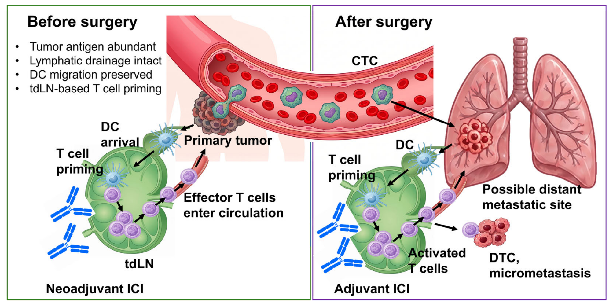
The immunological rationale for neoadjuvant ICI is therefore related to the presence of the primary tumor, preserved lymphatic drainage, DC migration, tdLN-based T-cell priming, and other immune niches before surgery. In contrast, the role of adjuvant ICI must be understood in a different postoperative context, in which macroscopic disease has been removed but MRD, dormant tumor cells, and micrometastatic lesions may persist. These distinct roles of neoadjuvant and adjuvant ICI are summarized in Figure 3.

7. Potential Immunological Rationale for Adjuvant ICI: Immune Surveillance Against MRD and Metastatic Niches

As summarized in Figure 3, adjuvant immune checkpoint inhibition acts in a different anatomical and immunological context from neoadjuvant treatment. After curative-intent surgery, visible tumor lesions and regional tdLNs may have been removed. However, surgical clearance of macroscopic disease does not necessarily mean biological eradication of cancer. Minimal residual disease (MRD) may persist as circulating tumor cells (CTCs), DTCs, or small micrometastatic deposits that remain below the limit of clinical detection. These residual cancer cells provide the substrate for later relapse and distant metastasis, but their interaction with the immune system is likely to be variable and highly dependent on anatomical site, antigen availability, and local inflammatory context [33].
A very small number of transformed tumor cells or DTCs may not immediately induce a productive adaptive immune response. For T cell immunity to be initiated, tumor antigens must be released, captured by antigen-presenting cells, transported to an appropriate lymphoid site, and presented in a context that supports T cell priming rather than tolerance. Experimental studies have shown that insufficient antigen availability can result in immunological ignorance, whereas increased antigen expression can overcome this barrier [34]. In addition, sporadic tumors may induce tolerance even when they express recognizable antigens [35]. These observations are consistent with the cancer-immunity cycle and cancer immunoediting models, in which antigen release, LN priming, effector T cell trafficking, immune equilibrium, and immune escape occur as dynamic and sequential processes rather than as a single event [36,37].
Tumor dormancy further complicates the timing of adjuvant immunotherapy. DTCs can remain clinically silent for long periods, either as non-proliferating cells, as small lesions restricted by the microenvironment, or as tumor cell populations held in partial check by immune pressure [38,39]. Recent work has also suggested that dormant DTCs may avoid immune attack in part because they are rare and therefore infrequently encountered by effector immune cells [40]. From this perspective, adjuvant ICI may not act at one defined moment after surgery. Instead, it may provide sustained immune pressure during a period in which dormant or slowly expanding tumor cells intermittently become immunologically visible.
This concept is also relevant to metastatic-site draining LNs. If micrometastatic lesions grow in distant organs, they may establish new routes of antigen drainage to LNs located outside the original surgical field. These LNs could then become sites where tumor antigens are presented and new or recalled T cell responses are generated. Because T cell state and function are strongly influenced by anatomical location and tissue context, the immune response to postoperative micrometastases may depend on where and when these metastatic-site immune niches develop [41]. Thus, adjuvant ICI can be viewed as a strategy to maintain or redirect antitumor immunity against evolving metastatic foci, rather than only as a drug given to eliminate residual cells immediately after surgery.
Clinical trial designs also reflect this uncertainty in timing, although they do not directly define the optimal biological duration of treatment. In resected melanoma, adjuvant pembrolizumab in KEYNOTE-054 was administered for a fixed period and improved recurrence-free survival [42]. In renal cell carcinoma, KEYNOTE-564 showed that adjuvant pembrolizumab after nephrectomy improved survival outcomes in high-risk patients [43]. Perioperative trials such as KEYNOTE-671 in NSCLC and KEYNOTE-689 in head and neck squamous cell carcinoma (HNSCC) also include postoperative ICI phases after surgery [7,44]. These regimens suggest that continued checkpoint blockade after surgery can be clinically beneficial, but the duration of treatment has largely been determined by trial design rather than by direct measurement of MRD activity, metastatic-site tdLN priming, or T cell reactivation.
Therefore, the rationale for adjuvant ICI should be framed more broadly than immediate killing of residual tumor cells. It may function as an immune surveillance strategy during a period when MRD, dormant disseminated cells, and early micrometastases are biologically unstable and not continuously visible to the immune system. The key unresolved question is when and where adjuvant ICI exerts its most important effect: on residual cells at the surgical site, on occult micrometastases, or within newly established draining LNs at distant anatomical sites. Defining biomarkers that capture MRD, tumor dormancy, antigen release, and metastatic-site immune priming will be necessary to determine which patients need prolonged adjuvant treatment and when treatment can be safely discontinued.

8. Cancer-Type-Specific Clinical Evidence

The clinical development of perioperative immune checkpoint inhibition has progressed across several solid tumors, but the biological meaning of this strategy is not identical among cancer types. Differences in tumor antigenicity, lymphatic anatomy, timing of surgery, use of chemotherapy, pattern of relapse, and dependence on regional LN management may influence how the tdLN–TPEX/TEX framework contributes to therapeutic efficacy.
Perioperative ICI trials differ substantially in tumor type, treatment regimen, comparator, timing of surgery, postoperative treatment, and clinical endpoint. Therefore, this review does not attempt a quantitative synthesis across these heterogeneous studies. Instead, we discuss pivotal or representative prospective phase II and phase III trials to illustrate cancer-type-specific patterns of neoadjuvant, adjuvant, and perioperative ICI strategies, as well as the limitations of applying a uniform tdLN-centered mechanism across solid tumors. Representative clinical examples are summarized in Table 2.
In melanoma, neoadjuvant ICI provides one of the clearest examples of how pathological response can guide postoperative treatment. The NADINA trial evaluated neoadjuvant ipilimumab plus nivolumab followed by surgery and response-driven adjuvant therapy in resectable stage III melanoma [6]. This approach is particularly informative because melanoma is generally highly immunogenic and frequently contains pre-existing tumor-reactive T cells. In this setting, preserved tdLNs may support rapid immune activation before surgery, and pathological response can be used as an early indicator of whether systemic antitumor immunity has been effectively induced.
In NSCLC, perioperative chemoimmunotherapy has been evaluated in trials such as KEYNOTE-671 and CheckMate 77T [7,45]. These studies combine neoadjuvant chemotherapy with PD-1 blockade, followed by surgery and postoperative ICI. The immunological interpretation may differ from melanoma because chemotherapy can contribute to tumor cell death, antigen release, and remodeling of the inflammatory environment. In addition, mediastinal LNs may provide an anatomical site for antigen presentation and T cell priming before resection. Thus, perioperative ICI in NSCLC may act through both tumor debulking and systemic immune induction before surgery, followed by continued immune pressure against MRD after surgery.
In HNSCC, including oral squamous cell carcinoma (OSCC), the role of regional LNs is particularly important. KEYNOTE-689 showed the clinical benefit of adding neoadjuvant and adjuvant pembrolizumab to surgery and standard postoperative therapy in resectable locally advanced disease [44]. From an immunological viewpoint, cervical tdLNs may be especially relevant because they are directly connected to the primary tumor through lymphatic drainage and are frequently included in surgical management. These nodes may function not only as metastatic sites but also as one component of a broader immune-reservoir network. However, TPEX-like cells and other PD-1-responsive tumor-reactive T cells may also be maintained in TLS, intratumoral niches, perivascular regions, and other lymphoid compartments. Therefore, HNSCC provides a clinically important model for considering how neck dissection, nodal response, and perioperative ICI should be integrated.
Triple-negative breast cancer (TNBC) represents another pattern of perioperative immunotherapy. In KEYNOTE-522, pembrolizumab was combined with neoadjuvant chemotherapy and continued after surgery [46]. In this setting, chemotherapy is not only cytotoxic but may also enhance antitumor immunity by increasing antigen release, inducing inflammatory signals, and altering the tumor microenvironment. PD-1 blockade may then amplify T cell responses generated during this period. The tdLN–TPEX/TEX framework in TNBC may therefore operate in close association with chemotherapy-induced immune remodeling rather than through checkpoint blockade alone.
In muscle-invasive bladder cancer, the NIAGARA trial evaluated durvalumab with neoadjuvant gemcitabine/cisplatin followed by radical cystectomy and adjuvant durvalumab [47]. This strategy illustrates how perioperative PD-L1 blockade can be combined with standard cisplatin-based chemotherapy. Before cystectomy, the primary tumor and pelvic lymphatic drainage remain present, allowing the possibility of treatment-induced antigen release and immune activation. After surgery, continued durvalumab may help maintain immune surveillance against residual disease. Thus, bladder cancer provides an example in which neoadjuvant chemoimmunotherapy and adjuvant immune maintenance are combined within one perioperative strategy.
Mismatch repair-deficient (dMMR) colon cancer provides a distinct model. In NICHE-2, short-course neoadjuvant nivolumab plus ipilimumab induced marked pathological responses in locally advanced dMMR colon cancer [48]. The strong response in this setting is likely related to high neoantigen burden and pre-existing immune recognition. Because dMMR tumors may already contain active tumor-reactive immune responses, a short period of checkpoint blockade before surgery may be sufficient to rapidly amplify antitumor immunity. This tumor type therefore highlights how intrinsic tumor antigenicity can strongly influence the magnitude and speed of response to neoadjuvant ICI.
Renal cell carcinoma differs from these examples. KEYNOTE-564 supports the clinical value of adjuvant pembrolizumab after nephrectomy in high-risk disease, whereas PROSPER RCC did not show a clear benefit for perioperative nivolumab [43,49]. These results suggest that the optimal timing of ICI may vary according to tumor biology and clinical context. In renal cell carcinoma, current evidence more strongly supports postoperative immune surveillance against MRD than neoadjuvant immune priming before nephrectomy. This contrast is important because it shows that the perioperative ICI paradigm should not be applied uniformly across all tumors.
Negative or mixed perioperative ICI trials are particularly important for avoiding confirmation bias. The lack of recurrence-free survival benefit in PROSPER RCC, despite perioperative nivolumab exposure, indicates that the presence of the primary tumor and regional immune structures does not automatically translate into superior clinical benefit. Such results suggest that neoadjuvant immune priming, postoperative MRD control, tumor antigenicity, treatment regimen, and anatomical context may differ substantially among tumor types. Therefore, positive perioperative trials should not be interpreted as uniform support for a single tdLN-centered mechanism.
Primary brain tumors, including glioblastoma, represent an important boundary case for the perioperative ICI framework. Unlike many extracranial solid tumors, gliomas rarely involve conventional regional LN metastasis, and lymph node surgery is not a central component of their management. Instead, antigen drainage and immune communication may involve distinct anatomical routes, including meningeal lymphatic pathways and cervical lymphoid tissues. Small neoadjuvant anti-PD-1 studies in recurrent glioblastoma have shown pharmacodynamic and immune changes within the tumor and peripheral immune compartments [50,51], but perioperative ICI has not become an established standard strategy for gliomas. Therefore, brain tumors should be considered separately from tumors in which regional LN surgery is a central component of curative-intent treatment. They highlight both the broader relevance and the limitations of applying a tdLN-centered framework across all solid tumors.
Taken together, these clinical examples suggest that the tdLN–TPEX/TEX framework may operate differently depending on tumor type. In highly immunogenic tumors such as melanoma and dMMR colon cancer, neoadjuvant ICI may rapidly amplify pre-existing tumor-reactive immunity. In tumors treated with chemoimmunotherapy, such as NSCLC, TNBC, and bladder cancer, chemotherapy-induced antigen release and inflammatory remodeling may cooperate with checkpoint blockade. In HNSCC, the close relationship between the primary tumor and cervical tdLNs makes LN management a central issue. In renal cell carcinoma, the current evidence emphasizes postoperative MRD control rather than a clear neoadjuvant advantage. In glioblastoma and other primary brain tumors, distinct lymphatic anatomy and the limited role of conventional nodal surgery make direct application of a tdLN-centered model inappropriate. These differences support the need for cancer-type-specific perioperative strategies rather than a single model of ICI timing for all solid tumors.

9. Reconsidering LN Surgery in the ICI Era

LN surgery has traditionally had three major purposes in solid tumors: pathological staging, regional disease control, and removal of clinically or microscopically involved LNs. This principle remains essential in the era of immunotherapy. Standard LN surgery and radiotherapy are based on disease-specific evidence for staging, regional control, and, in some settings, survival benefit. At present, there is no clinical evidence that standard LN surgery or radiotherapy should be reduced solely to preserve tdLN immune-reservoir function. The tdLN-reservoir hypothesis should therefore not be used as an independent justification for omitting indicated nodal dissection, reducing radiation fields, or altering established perioperative treatment pathways. Clinically metastatic LNs should not be left untreated simply because tdLNs may have immunological functions, and any modification of nodal management must be evaluated prospectively within disease-specific trials that preserve oncologic safety.
With this clinical caveat in mind, the relevant question is not whether established nodal treatment should be abandoned, but whether the timing, extent, and purpose of LN management should be studied as immunologically relevant variables when ICIs are delivered before surgery. Specifically, when ICIs are given in the neoadjuvant setting, could the timing and extent of LN removal influence the antitumor immune response induced by treatment?
This question arises because tdLNs are not passive anatomical structures. Experimental studies have shown that tdLNs can be required for optimal responses to PD-1/PD-L1 blockade, and that checkpoint interactions within tdLNs can regulate tumor-specific T cell immunity [8,9]. Recent reviews have therefore emphasized the clinical dilemma that tdLNs can act both as metastatic sites and as sites where antitumor immunity is initiated or maintained [52,53]. From this viewpoint, removal or irradiation of uninvolved or minimally involved lymphatic basins before immune activation could theoretically reduce the lymphoid sites available for antigen presentation, T cell priming, and maintenance of less differentiated tumor-reactive T cells. However, this idea remains a biological hypothesis and must be balanced against established oncologic indications for nodal treatment.
Melanoma provides the clearest clinical example of how LN surgery has already been de-escalated in selected settings. In MSLT-II, immediate completion of LN dissection after a positive sentinel LN improved regional control and provided additional staging information, but did not improve melanoma-specific survival compared with nodal observation [54]. This result supported a more selective approach to complete LN dissection in patients with limited sentinel node involvement. In the immunotherapy era, melanoma has moved further toward response-adapted surgery. In the PRADO study, pathological response after neoadjuvant ipilimumab plus nivolumab was used to guide subsequent surgery and adjuvant therapy, allowing treatment de-escalation in major responders and escalation in poor responders [55]. The phase 3 NADINA trial further supported neoadjuvant ipilimumab plus nivolumab followed by response-driven adjuvant therapy in resectable stage III melanoma [6]. These studies suggest that pathological response after neoadjuvant ICI can become a decision-making tool for tailoring the extent of subsequent treatment. However, this approach is best established in melanoma and should not be directly extrapolated to other tumor types without disease-specific evidence.
OSCC and other head and neck cancers require a more cautious interpretation. In early node-negative oral cancer, elective neck dissection has been shown to improve overall and disease-free survival compared with therapeutic neck dissection performed after nodal relapse [56]. This finding is important because it shows that occult nodal disease can be clinically meaningful and that elective nodal treatment should not be abandoned without strong evidence. At the same time, sentinel node biopsy has been evaluated as a less invasive staging strategy in selected patients with early oral cancer. The SENT trial showed that sentinel node biopsy can provide reliable staging of the clinically N0 neck in T1–T2 oral cancer, with high sentinel node identification and negative predictive value [57]. These data indicate that LN management in oral cancer can be individualized in some settings, but always within a framework that preserves oncologic safety.
The introduction of perioperative ICI in HNSCC adds another layer to this discussion. KEYNOTE-689 showed that neoadjuvant and adjuvant pembrolizumab added to surgery and standard postoperative therapy improved event-free survival in resectable locally advanced disease [44]. This trial does not by itself define how neck dissection should be modified after neoadjuvant ICI. However, it creates a new clinical context in which cervical LNs are exposed to checkpoint blockade before surgical removal. In this setting, the neck specimen may provide information not only about residual metastatic disease, but also about immune activation, pathological regression, T cell states, DC function, and the presence or loss of tumor-reactive T cell reservoirs. Thus, neck dissection after neoadjuvant ICI may become both a therapeutic procedure and a translational window for understanding nodal immune responses.
A practical framework is therefore needed. First, clinically involved nodes should remain targets for adequate surgical or radiation-based control. Second, elective treatment of clinically uninvolved nodal basins should be guided by tumor type, stage, anatomical risk, and available evidence, rather than relying solely on immunological speculation. Third, when neoadjuvant ICI is used, pathological response in both the primary tumor and LNs should be evaluated systematically. Fourth, immune monitoring of resected LNs may help distinguish nodes that were mainly metastatic deposits from nodes that retained active immune function. Such monitoring could include spatial analysis of DCs, TPEX-like CD8+ T cells, TCR clonality, TLS-like structure, and checkpoint ligand expression.
In summary, the ICI era does not eliminate the need for LN surgery. Instead, it changes the questions that LN surgery must answer. The goal is no longer only to determine whether nodal metastasis is present or absent, but also to understand how nodal disease, nodal immune activity, and treatment-induced pathological response should guide subsequent therapy. Future strategies may integrate sentinel node approaches, response-adapted surgery, pathological regression scoring, and immune profiling. The central challenge is to preserve oncologic safety while prospectively studying how nodal disease, nodal immune activity, and lymphoid tissue disruption influence systemic antitumor immunity.

10. Postoperative Lymphatic Dysfunction, Lymphedema, and Immune Surveillance

10.1. Established Clinical Consequences of Lymphatic Injury

Postoperative lymphatic dysfunction is usually discussed as a complication that affects quality of life, physical function, and long-term survivorship. In head and neck cancer, lymphedema can involve both external soft tissues and internal structures, and may contribute to discomfort, dysphagia, speech problems, cosmetic changes, and functional impairment [58,59]. LN dissection and radiotherapy are major contributors to this condition. Radiotherapy can damage lymphatic vessels, promote tissue fibrosis, and impair lymphatic drainage, whereas surgery can physically interrupt lymphatic channels and remove regional LNs [60]. These clinical consequences are well recognized and represent established adverse effects of local cancer therapy.
Fibrosis is another important component of postoperative lymphatic dysfunction. In lymphedema, chronic tissue swelling is frequently accompanied by extracellular matrix remodeling, soft tissue thickening, and fibrotic change. Together, lymphedema and fibrosis can produce long-lasting structural and functional changes in postoperative tissues. Thus, postoperative lymphatic dysfunction is an established survivorship issue after LN dissection and radiotherapy.

10.2. Plausible Immunological Consequences

The possible immunological implications of postoperative lymphatic dysfunction in the era of immune checkpoint inhibition have been less extensively defined. The lymphatic system is not only a drainage pathway for interstitial fluid. It also provides an anatomical route for antigen transport, DC migration, and immune cell trafficking. Afferent lymphatic vessels carry tissue-derived antigens and antigen-bearing DCs to draining LNs, where adaptive immune responses can be initiated or regulated [61]. In cancer, lymphatic vessels can therefore influence both metastasis and antitumor immunity. Recent reviews have emphasized that lymphatic transport shapes the delivery of tumor antigens, the movement of immune cells, and the local conditions under which immunotherapy acts [62,63].
From this perspective, postoperative disruption of lymphatic flow may alter not only fluid balance, but also the immune communication between peripheral tissues, residual tumor sites, and regional lymphoid structures. Lymphatic dysfunction can change the local immune environment by affecting leukocyte trafficking, antigen clearance, inflammatory cell accumulation, and immune regulation [64,65]. Experimental models have shown that CD4+ T cells are activated after lymphatic injury and can contribute to the initiation of lymphedema [66]. Regulatory T cells (Tregs) may also accumulate in lymphedematous tissues and participate in local immune suppression [67]. These observations suggest that lymphatic failure is not a purely mechanical disorder, but a condition in which persistent immune activation and local immune regulation may coexist.
Fibrosis may further modify the postoperative immune environment. Th2-skewed immune responses have been implicated in the development of tissue fibrosis and impaired lymphatic function after lymphatic injury [68]. In addition, lymph leakage or impaired lymphatic integrity may influence macrophage differentiation and favor anti-inflammatory or tissue-remodeling phenotypes [69]. These changes could theoretically affect local immune surveillance by modifying antigen availability, stromal architecture, immune cell access, and the balance between inflammatory and suppressive immune pathways.

10.3. Speculative Relevance to Adjuvant ICI and Recurrence

The relevance of these processes to cancer recurrence remains uncertain. At present, there is limited direct clinical evidence showing that postoperative lymphedema itself increases the risk of local recurrence or distant metastasis. Therefore, postoperative lymphatic dysfunction should not be presented as an established cause of cancer relapse. A more appropriate interpretation is that lymphatic injury, chronic inflammation, fibrosis, altered macrophage polarization, and impaired immune cell trafficking may create a postoperative tissue environment that could influence immune surveillance.
This possibility may be particularly relevant in patients treated with perioperative or adjuvant ICI, because the efficacy of immunotherapy depends on antigen transport, DC function, T cell priming, and T cell access to sites of residual disease. However, whether postoperative lymphatic dysfunction actually modifies the response to adjuvant ICI remains speculative and requires prospective validation. Thus, in the ICI era, lymphedema and fibrosis should be viewed not only as survivorship problems, but also as potential modifiers of local and regional immune surveillance, rather than as proven determinants of recurrence or treatment failure.
Future studies should examine postoperative lymphatic dysfunction using immunological endpoints. These may include spatial analysis of lymphatic vessels, DCs, macrophage subsets, T cell infiltration, Treg accumulation, fibrosis-associated stromal changes, and checkpoint ligand expression in postoperative tissues. Clinical studies should also evaluate whether the severity of lymphedema or fibrosis correlates with immune-related biomarkers, response to adjuvant ICI, or patterns of recurrence. Such studies would help determine whether postoperative lymphatic dysfunction is only a consequence of local therapy or whether it also modifies the immune environment in which perioperative immunotherapy must operate.

11. Future Directions: Immune-Guided Surgery and Response-Adapted Perioperative Therapy

The next step in perioperative immunotherapy is to move from uniform treatment schedules toward strategies guided by immune status, pathological response, and residual disease risk. Neoadjuvant ICI provides a unique opportunity because tumor tissues, draining LNs, and blood samples can be analyzed before treatment, during treatment, and at surgery. These paired samples may help identify where immune modulation occurs, which T cell populations expand, and whether the response is maintained after tumor removal [3].
In addition to tumor-intrinsic and blood-based biomarkers, tdLN immune signatures themselves may become candidate predictors of neoadjuvant ICI benefit. Features such as TCF1+ or TPEX-like CD8+ T-cell abundance, cDC1 density, TCR clonal expansion, PD-1/PD-L1 spatial organization, and preservation of lymphoid architecture could be evaluated together with pathological response, ctDNA clearance, event-free survival, and recurrence patterns. Such analyses may help determine whether tdLNs are merely sites of metastatic staging or whether they also contain immune states associated with effective perioperative antitumor responses.
First, immune monitoring of tdLNs and other immune niches should be developed as a routine research component of neoadjuvant ICI trials. Current assessment of LNs is mainly focused on the presence or absence of metastatic tumor cells. In the ICI era, this may be insufficient. tdLNs should also be evaluated together with TLS, intratumoral niches, perivascular regions, and blood for immune activity, including TCF1+ or TPEX-like CD8+ T cells, DC subsets, TCR clonality, B cell-rich lymphoid aggregates, and spatial organization of immune cells. Studies showing stem-like CD8+ T cell populations and cDC1-dependent maintenance of tumor-reactive T cells in tdLNs provide a rationale for this approach [17,19]. TCR sequencing may also help determine whether tumor-reactive clones are expanded locally, recruited from outside the tumor, or replaced after PD-1 blockade [70]. In addition, TLS and B cell-rich immune niches should be incorporated into spatial immune profiling because they have been associated with immunotherapy response in several cancer settings [71,72].
Second, pathological response should be integrated with immune biomarkers to guide postoperative treatment. In melanoma, pathological response after neoadjuvant therapy is strongly associated with clinical outcome and has already been used to adapt subsequent surgery and adjuvant therapy [55,73]. The NADINA trial further supports the clinical relevance of a neoadjuvant approach followed by response-driven postoperative management [6]. These studies suggest that major responders may not require the same intensity or duration of adjuvant therapy as poor responders. However, pathological regression alone may not fully reflect the quality of systemic immunity. Future response-adapted strategies should therefore combine pathological response with immune features such as tdLN T cell states, TCR clonal expansion, DC activation, circulating immune markers, and MRD status.
Third, LN surgery should be reconsidered within an immune-guided framework. This does not mean that metastatic LNs should be preserved when oncologic control requires their removal. Rather, it means that the timing, extent, and purpose of LN surgery should be discussed together with the immunological role of tdLNs. Recent discussions have emphasized that tdLNs can be both sites of tumor spread and sites of antitumor immune activation [52,53]. Therefore, future trials should ask whether LN dissection before, during, or after ICI affects immune priming, T cell expansion, pathological response, and recurrence patterns. In selected settings, sentinel node strategies, response-based nodal surgery, or limited nodal dissection may be evaluated, but only when oncologic safety is maintained.
Fourth, the duration of adjuvant ICI should be refined using biomarkers rather than fixed schedules alone. At present, many adjuvant or perioperative ICI regimens use predefined treatment periods, often based on clinical trial design. However, the biological window during which residual tumor cells become immunologically visible may differ among patients. MRD detected by circulating tumor DNA may provide one approach to identifying patients with persistent microscopic disease or early molecular relapse [74]. In the future, MRD-guided strategies could help determine whether adjuvant ICI should be continued, stopped, intensified, or combined with other therapies. Such decisions may be particularly important for patients with dormant micrometastases or evolving metastatic-site draining LNs, in whom immune activation may occur only after a variable period of tumor growth or antigen release.
Finally, perioperative immunotherapy should be studied as an integrated anatomical and immunological process. The primary tumor, tdLNs, metastatic sites, blood, bone marrow, and postoperative lymphatic environment should not be analyzed separately. Instead, they should be considered as connected compartments that shape antigen flow, DC migration, T cell priming, T cell exhaustion, and immune surveillance. Future trials should therefore include coordinated sampling of tumors, LNs, blood, MRD markers, and postoperative tissues. Key immune monitoring targets for each anatomical compartment are summarized in Table 3. Such studies may allow perioperative ICI to evolve from a fixed drug schedule into a personalized strategy that combines immune monitoring, response-adapted therapy, MRD-guided treatment duration, and immune-guided surgery.
Several clinically important questions remain unresolved and should be addressed before tdLN biology can inform perioperative treatment design. These questions are summarized in Table 4. Importantly, they are proposed as research priorities rather than as grounds for changing current surgical or radiotherapeutic standards.

12. Conclusions

Perioperative immunotherapy is changing the way solid tumors are treated, but its full significance cannot be understood only by focusing on tumor shrinkage or postoperative eradication of residual disease. The timing of immune checkpoint inhibition relative to surgery is immunologically important because the primary tumor, lymphatic drainage, DC migration, and tdLNs remain connected before resection. In this setting, tdLNs are not only anatomical sites of metastatic spread, but also immune organs that may support antigen presentation, T cell priming, and the maintenance of less differentiated tumor-reactive CD8+ T cells.
This dual role creates both an opportunity and a dilemma. On one hand, tdLNs may act as metastatic gateways and must be managed according to established oncologic principles. On the other hand, they may serve as immune reservoirs that contribute to the response to PD-1/PD-L1 blockade. Therefore, perioperative immunotherapy should be considered as a strategy that may act through interactions among the primary tumor, lymphatic drainage, tdLNs, TLS, intratumoral niches, and systemic immune compartments, rather than only within the tumor microenvironment. Importantly, this framework does not support modification of standard lymph node surgery or radiotherapy outside prospective, disease-specific trials. Rather, it identifies tdLNs as clinically accessible tissues in which the mechanisms of perioperative ICI can be tested.
Future treatment strategies should integrate pathological response, immune monitoring of tdLNs, TCR clonality, DC function, MRD assessment, and postoperative lymphatic status. Such an approach may help identify patients who require intensified adjuvant therapy, define candidates for treatment de-escalation in prospective trials, and inform the design of immune-guided nodal management strategies that preserve oncologic safety. Reframing tdLNs as metastatic gateways and putative immune-reservoir compartments, while placing the TPEX/TEX continuum within a broader multi-compartment immune network, may provide a useful framework for optimizing perioperative immunotherapy across solid tumors.

Author Contributions

Conceptualization, K.K.; literature review, K.K., Y.K. and K.N.; writing—original draft preparation, K.K.; writing—review and editing, K.K., Y.K. and K.N.; visualization, K.K.; supervision, K.K.; project administration, K.K. All authors have read and agreed to the published version of the manuscript.

Funding

This research received no external funding.

Institutional Review Board Statement

Not applicable.

Informed Consent Statement

Not applicable.

Data Availability Statement

No new data were created or analyzed in this study. Data sharing is not applicable to this article.

Acknowledgments

The authors thank colleagues and collaborators for helpful discussions on tumor immunology and perioperative cancer immunotherapy. During the preparation of this manuscript, the authors used ChatGPT (OpenAI, GPT-5.5) for assistance with language editing, manuscript structuring, and draft conceptual figure layouts. All AI-assisted outputs were reviewed, edited, and approved by the authors, who take full responsibility for the final content. All conceptual figures were designed by the authors to summarize the concepts discussed in this review and were not reproduced or adapted from previously published figures. AI-assisted tools were used only to generate preliminary visual layouts, which were subsequently revised and finalized by the authors.

Conflicts of Interest

The authors declare no conflicts of interest.

Abbreviations

The following abbreviations are used in this manuscript:
ICIimmune checkpoint inhibitor
MRDminimal residual disease
LNlymph node
tdLNtumor-draining lymph node
CD8cluster of differentiation 8
PD-1programmed cell death protein 1
TPEXprogenitor-exhausted T cell
TEXexhausted T cell
NSCLCnon-small cell lung cancer
PD-L1programmed death-ligand 1
HEVhigh endothelial venule
DCdendritic cell
cDC1conventional type 1 dendritic cell
PRISMAPreferred Reporting Items for Systematic Reviews and Meta-Analyses
DTCdisseminated tumor cell
MHCmajor histocompatibility complex
TCF1T cell factor 1
TLStertiary lymphoid structure
TCRT cell receptor
CTCcirculating tumor cell
HNSCChead and neck squamous cell carcinoma
OSCCoral squamous cell carcinoma
TNBCtriple-negative breast cancer
dMMRmismatch repair-deficient
RCCrenal cell carcinoma
Tregregulatory T cell
CD4cluster of differentiation 4
Th2T helper 2
AIartificial intelligence
TMEtumor microenvironment
ctDNAcirculating tumor DNA
EFSevent-free survival
APCarticle processing charge

References

  1. Topalian, S.L.; Taube, J.M.; Pardoll, D.M. Neoadjuvant checkpoint blockade for cancer immunotherapy. Science 2020, 367, eaax0182. [Google Scholar] [CrossRef] [Scilit] [PubMed]
  2. Versluis, J.M.; Long, G.V.; Blank, C.U. Learning from clinical trials of neoadjuvant checkpoint blockade. Nat. Med. 2020, 26, 475–484. [Google Scholar] [CrossRef] [Scilit] [PubMed]
  3. Topalian, S.L.; Forde, P.M.; Emens, L.A.; Yarchoan, M.; Smith, K.N.; Pardoll, D.M. Neoadjuvant immune checkpoint blockade: A window of opportunity to advance cancer immunotherapy. Cancer Cell 2023, 41, 1551–1566. [Google Scholar] [CrossRef] [Scilit] [PubMed]
  4. Awada, G.; Cascone, T.; van der Heijden, M.S.; Blank, C.U.; Kok, M.; Chalabi, M. The rapidly evolving paradigm of neoadjuvant immunotherapy across cancer types. Nat. Cancer 2025, 6, 967–987. [Google Scholar] [CrossRef] [Scilit] [PubMed]
  5. Patel, S.P.; Othus, M.; Chen, Y.; Wright, G.P., Jr.; Yost, K.J.; Hyngstrom, J.R.; Hu-Lieskovan, S.; Lao, C.D.; Fecher, L.A.; Truong, T.G.; et al. Neoadjuvant-Adjuvant or Adjuvant-Only Pembrolizumab in Advanced Melanoma. N. Engl. J. Med. 2023, 388, 813–823. [Google Scholar] [CrossRef] [Scilit] [PubMed]
  6. Blank, C.U.; Lucas, M.W.; Scolyer, R.A.; van de Wiel, B.A.; Menzies, A.M.; Lopez-Yurda, M.; Hoeijmakers, L.L.; Saw, R.P.M.; Lijnsvelt, J.M.; Maher, N.G.; et al. Neoadjuvant Nivolumab and Ipilimumab in Resectable Stage III Melanoma. N. Engl. J. Med. 2024, 391, 1696–1708. [Google Scholar] [CrossRef] [Scilit] [PubMed]
  7. Wakelee, H.; Liberman, M.; Kato, T.; Tsuboi, M.; Lee, S.H.; Gao, S.; Chen, K.N.; Dooms, C.; Majem, M.; Eigendorff, E.; et al. Perioperative Pembrolizumab for Early-Stage Non-Small-Cell Lung Cancer. N. Engl. J. Med. 2023, 389, 491–503. [Google Scholar] [CrossRef] [Scilit] [PubMed]
  8. Fransen, M.F.; Schoonderwoerd, M.; Knopf, P.; Camps, M.G.; Hawinkels, L.J.; Kneilling, M.; van Hall, T.; Ossendorp, F. Tumor-draining lymph nodes are pivotal in PD-1/PD-L1 checkpoint therapy. JCI Insight 2018, 3, e124507. [Google Scholar] [CrossRef] [Scilit] [PubMed]
  9. Dammeijer, F.; van Gulijk, M.; Mulder, E.E.; Lukkes, M.; Klaase, L.; van den Bosch, T.; van Nimwegen, M.; Lau, S.P.; Latupeirissa, K.; Schetters, S.; et al. The PD-1/PD-L1-Checkpoint Restrains T cell Immunity in Tumor-Draining Lymph Nodes. Cancer Cell 2020, 38, 685–700.e8. [Google Scholar] [CrossRef] [Scilit] [PubMed]
  10. Delclaux, I.; Ventre, K.S.; Jones, D.; Lund, A.W. The tumor-draining lymph node as a reservoir for systemic immune surveillance. Trends Cancer 2024, 10, 28–37. [Google Scholar] [CrossRef] [Scilit] [PubMed]
  11. Pereira, E.R.; Kedrin, D.; Seano, G.; Gautier, O.; Meijer, E.F.J.; Jones, D.; Chin, S.M.; Kitahara, S.; Bouta, E.M.; Chang, J.; et al. Lymph node metastases can invade local blood vessels, exit the node, and colonize distant organs in mice. Science 2018, 359, 1403–1407. [Google Scholar] [CrossRef] [Scilit] [PubMed]
  12. Brown, M.; Assen, F.P.; Leithner, A.; Abe, J.; Schachner, H.; Asfour, G.; Bago-Horvath, Z.; Stein, J.V.; Uhrin, P.; Sixt, M.; et al. Lymph node blood vessels provide exit routes for metastatic tumor cell dissemination in mice. Science 2018, 359, 1408–1411. [Google Scholar] [CrossRef] [Scilit] [PubMed]
  13. Karaman, S.; Detmar, M. Mechanisms of lymphatic metastasis. J. Clin. Investig. 2014, 124, 922–928. [Google Scholar] [CrossRef] [Scilit] [PubMed]
  14. Ji, H.; Hu, C.; Yang, X.; Liu, Y.; Ji, G.; Ge, S.; Wang, X.; Wang, M. Lymph node metastasis in cancer progression: Molecular mechanisms, clinical significance and therapeutic interventions. Signal Transduct. Target. Ther. 2023, 8, 367. [Google Scholar] [CrossRef] [Scilit] [PubMed]
  15. Achen, M.G.; Stacker, S.A. Exit Stage Left: A Tumor Cell’s Journey from Lymph Node to Beyond. Trends Cancer 2018, 4, 519–522. [Google Scholar] [CrossRef] [Scilit] [PubMed]
  16. du Bois, H.; Heim, T.A.; Lund, A.W. Tumor-draining lymph nodes: At the crossroads of metastasis and immunity. Sci. Immunol. 2021, 6, eabg3551. [Google Scholar] [CrossRef] [Scilit] [PubMed]
  17. Schenkel, J.M.; Herbst, R.H.; Canner, D.; Li, A.; Hillman, M.; Shanahan, S.L.; Gibbons, G.; Smith, O.C.; Kim, J.Y.; Westcott, P.; et al. Conventional type I dendritic cells maintain a reservoir of proliferative tumor-antigen specific TCF-1(+) CD8(+) T cells in tumor-draining lymph nodes. Immunity 2021, 54, 2338–2353.e6. [Google Scholar] [CrossRef] [Scilit] [PubMed]
  18. Pittet, M.J.; Di Pilato, M.; Garris, C.; Mempel, T.R. Dendritic cells as shepherds of T cell immunity in cancer. Immunity 2023, 56, 2218–2230. [Google Scholar] [CrossRef] [Scilit] [PubMed]
  19. Connolly, K.A.; Kuchroo, M.; Venkat, A.; Khatun, A.; Wang, J.; William, I.; Hornick, N.I.; Fitzgerald, B.L.; Damo, M.; Kasmani, M.Y.; et al. A reservoir of stem-like CD8(+) T cells in the tumor-draining lymph node preserves the ongoing antitumor immune response. Sci. Immunol. 2021, 6, eabg7836. [Google Scholar] [CrossRef] [Scilit] [PubMed]
  20. Huang, Q.; Wu, X.; Wang, Z.; Chen, X.; Wang, L.; Lu, Y.; Xiong, D.; Liu, Q.; Tian, Y.; Lin, H.; et al. The primordial differentiation of tumor-specific memory CD8(+) T cells as bona fide responders to PD-1/PD-L1 blockade in draining lymph nodes. Cell 2022, 185, 4049–4066.e25. [Google Scholar] [CrossRef] [Scilit] [PubMed]
  21. Wherry, E.J.; Kurachi, M. Molecular and cellular insights into T cell exhaustion. Nat. Rev. Immunol. 2015, 15, 486–499. [Google Scholar] [CrossRef] [Scilit] [PubMed]
  22. van der Leun, A.M.; Thommen, D.S.; Schumacher, T.N. CD8(+) T cell states in human cancer: Insights from single-cell analysis. Nat. Rev. Cancer 2020, 20, 218–232. [Google Scholar] [CrossRef] [Scilit] [PubMed]
  23. Chow, A.; Perica, K.; Klebanoff, C.A.; Wolchok, J.D. Clinical implications of T cell exhaustion for cancer immunotherapy. Nat. Rev. Clin. Oncol. 2022, 19, 775–790. [Google Scholar] [CrossRef] [Scilit] [PubMed]
  24. Hor, J.L.; Schrom, E.C.; Wong-Rolle, A.; Vistain, L.; Shang, W.; Dong, Q.; Zhao, C.; Jin, C.; Germain, R.N. Inhibitory PD-1 axis maintains high-avidity stem-like CD8(+) T cells. Nature 2026, 649, 194–204. [Google Scholar] [CrossRef] [Scilit] [PubMed]
  25. Miller, B.C.; Sen, D.R.; Al Abosy, R.; Bi, K.; Virkud, Y.V.; LaFleur, M.W.; Yates, K.B.; Lako, A.; Felt, K.; Naik, G.S.; et al. Subsets of exhausted CD8(+) T cells differentially mediate tumor control and respond to checkpoint blockade. Nat. Immunol. 2019, 20, 326–336. [Google Scholar] [CrossRef] [Scilit] [PubMed]
  26. Siddiqui, I.; Schaeuble, K.; Chennupati, V.; Fuertes Marraco, S.A.; Calderon-Copete, S.; Pais Ferreira, D.; Carmona, S.J.; Scarpellino, L.; Gfeller, D.; Pradervand, S.; et al. Intratumoral Tcf1(+)PD-1(+)CD8(+) T Cells with Stem-like Properties Promote Tumor Control in Response to Vaccination and Checkpoint Blockade Immunotherapy. Immunity 2019, 50, 195–211.e10. [Google Scholar] [CrossRef] [Scilit] [PubMed]
  27. Beltra, J.C.; Manne, S.; Abdel-Hakeem, M.S.; Kurachi, M.; Giles, J.R.; Chen, Z.; Casella, V.; Ngiow, S.F.; Khan, O.; Huang, Y.J.; et al. Developmental Relationships of Four Exhausted CD8(+) T Cell Subsets Reveals Underlying Transcriptional and Epigenetic Landscape Control Mechanisms. Immunity 2020, 52, 825–841.e8. [Google Scholar] [CrossRef] [Scilit] [PubMed]
  28. Im, S.J.; Hashimoto, M.; Gerner, M.Y.; Lee, J.; Kissick, H.T.; Burger, M.C.; Shan, Q.; Hale, J.S.; Lee, J.; Nasti, T.H.; et al. Defining CD8+ T cells that provide the proliferative burst after PD-1 therapy. Nature 2016, 537, 417–421. [Google Scholar] [CrossRef] [Scilit] [PubMed]
  29. Pauken, K.E.; Sammons, M.A.; Odorizzi, P.M.; Manne, S.; Godec, J.; Khan, O.; Drake, A.M.; Chen, Z.; Sen, D.R.; Kurachi, M.; et al. Epigenetic stability of exhausted T cells limits durability of reinvigoration by PD-1 blockade. Science 2016, 354, 1160–1165. [Google Scholar] [CrossRef] [Scilit] [PubMed]
  30. Liu, J.; Blake, S.J.; Yong, M.C.; Harjunpää, H.; Ngiow, S.F.; Takeda, K.; Young, A.; O’Donnell, J.S.; Allen, S.; Smyth, M.J.; et al. Improved Efficacy of Neoadjuvant Compared to Adjuvant Immunotherapy to Eradicate Metastatic Disease. Cancer Discov. 2016, 6, 1382–1399. [Google Scholar] [CrossRef] [Scilit] [PubMed]
  31. Blank, C.U.; Rozeman, E.A.; Fanchi, L.F.; Sikorska, K.; van de Wiel, B.; Kvistborg, P.; Krijgsman, O.; van den Braber, M.; Philips, D.; Broeks, A.; et al. Neoadjuvant versus adjuvant ipilimumab plus nivolumab in macroscopic stage III melanoma. Nat. Med. 2018, 24, 1655–1661. [Google Scholar] [CrossRef] [Scilit] [PubMed]
  32. Forde, P.M.; Chaft, J.E.; Smith, K.N.; Anagnostou, V.; Cottrell, T.R.; Hellmann, M.D.; Zahurak, M.; Yang, S.C.; Jones, D.R.; Broderick, S.; et al. Neoadjuvant PD-1 Blockade in Resectable Lung Cancer. N. Engl. J. Med. 2018, 378, 1976–1986. [Google Scholar] [CrossRef] [Scilit] [PubMed]
  33. Badia-Ramentol, J.; Linares, J.; Gómez-Llonin, A.; Calon, A. Minimal Residual Disease, Metastasis and Immunity. Biomolecules 2021, 11, 130. [Google Scholar] [CrossRef] [Scilit] [PubMed]
  34. Spiotto, M.T.; Yu, P.; Rowley, D.A.; Nishimura, M.I.; Meredith, S.C.; Gajewski, T.F.; Fu, Y.-X.; Schreiber, H. Increasing Tumor Antigen Expression Overcomes “Ignorance” to Solid Tumors via Crosspresentation by Bone Marrow-Derived Stromal Cells. Immunity 2002, 17, 737–747. [Google Scholar] [CrossRef] [Scilit] [PubMed]
  35. Willimsky, G.; Blankenstein, T. Sporadic immunogenic tumours avoid destruction by inducing T-cell tolerance. Nature 2005, 437, 141–146. [Google Scholar] [CrossRef] [Scilit] [PubMed]
  36. Chen, D.S.; Mellman, I. Oncology meets immunology: The cancer-immunity cycle. Immunity 2013, 39, 1–10. [Google Scholar] [CrossRef] [Scilit] [PubMed]
  37. Schreiber, R.D.; Old, L.J.; Smyth, M.J. Cancer immunoediting: Integrating immunity’s roles in cancer suppression and promotion. Science 2011, 331, 1565–1570. [Google Scholar] [CrossRef] [Scilit] [PubMed]
  38. Aguirre-Ghiso, J.A. Models, mechanisms and clinical evidence for cancer dormancy. Nat. Rev. Cancer 2007, 7, 834–846. [Google Scholar] [CrossRef] [Scilit] [PubMed]
  39. Sosa, M.S.; Bragado, P.; Aguirre-Ghiso, J.A. Mechanisms of disseminated cancer cell dormancy: An awakening field. Nat. Rev. Cancer 2014, 14, 611–622. [Google Scholar] [CrossRef] [Scilit] [PubMed]
  40. Goddard, E.T.; Linde, M.H.; Srivastava, S.; Klug, G.; Shabaneh, T.B.; Iannone, S.; Grzelak, C.A.; Marsh, S.; Riggio, A.I.; Shor, R.E.; et al. Immune evasion of dormant disseminated tumor cells is due to their scarcity and can be overcome by T cell immunotherapies. Cancer Cell 2024, 42, 119–134.e12. [Google Scholar] [CrossRef] [Scilit] [PubMed]
  41. Schenkel, J.M.; Pauken, K.E. Localization, tissue biology and T cell state—Implications for cancer immunotherapy. Nat. Rev. Immunol. 2023, 23, 807–823. [Google Scholar] [CrossRef] [Scilit] [PubMed]
  42. Eggermont, A.M.M.; Blank, C.U.; Mandala, M.; Long, G.V.; Atkinson, V.; Dalle, S.; Haydon, A.; Lichinitser, M.; Khattak, A.; Carlino, M.S.; et al. Adjuvant Pembrolizumab versus Placebo in Resected Stage III Melanoma. N. Engl. J. Med. 2018, 378, 1789–1801. [Google Scholar] [CrossRef] [Scilit] [PubMed]
  43. Choueiri, T.K.; Tomczak, P.; Park, S.H.; Venugopal, B.; Ferguson, T.; Symeonides, S.N.; Hajek, J.; Chang, Y.H.; Lee, J.L.; Sarwar, N.; et al. Overall Survival with Adjuvant Pembrolizumab in Renal-Cell Carcinoma. N. Engl. J. Med. 2024, 390, 1359–1371. [Google Scholar] [CrossRef] [Scilit] [PubMed]
  44. Uppaluri, R.; Haddad, R.I.; Tao, Y.; Le Tourneau, C.; Lee, N.Y.; Westra, W.; Chernock, R.; Tahara, M.; Harrington, K.J.; Klochikhin, A.L.; et al. Neoadjuvant and Adjuvant Pembrolizumab in Locally Advanced Head and Neck Cancer. N. Engl. J. Med. 2025, 393, 37–50. [Google Scholar] [CrossRef] [Scilit] [PubMed]
  45. Cascone, T.; Awad, M.M.; Spicer, J.D.; He, J.; Lu, S.; Sepesi, B.; Tanaka, F.; Taube, J.M.; Cornelissen, R.; Havel, L.; et al. Perioperative Nivolumab in Resectable Lung Cancer. N. Engl. J. Med. 2024, 390, 1756–1769. [Google Scholar] [CrossRef] [Scilit] [PubMed]
  46. Schmid, P.; Cortes, J.; Dent, R.; McArthur, H.; Pusztai, L.; Kummel, S.; Denkert, C.; Park, Y.H.; Hui, R.; Harbeck, N.; et al. Overall Survival with Pembrolizumab in Early-Stage Triple-Negative Breast Cancer. N. Engl. J. Med. 2024, 391, 1981–1991. [Google Scholar] [CrossRef] [Scilit] [PubMed]
  47. Powles, T.; Catto, J.W.F.; Galsky, M.D.; Al-Ahmadie, H.; Meeks, J.J.; Nishiyama, H.; Vu, T.Q.; Antonuzzo, L.; Wiechno, P.; Atduev, V.; et al. Perioperative Durvalumab with Neoadjuvant Chemotherapy in Operable Bladder Cancer. N. Engl. J. Med. 2024, 391, 1773–1786. [Google Scholar] [CrossRef] [Scilit] [PubMed]
  48. Chalabi, M.; Verschoor, Y.L.; Tan, P.B.; Balduzzi, S.; Van Lent, A.U.; Grootscholten, C.; Dokter, S.; Buller, N.V.; Grotenhuis, B.A.; Kuhlmann, K.; et al. Neoadjuvant Immunotherapy in Locally Advanced Mismatch Repair-Deficient Colon Cancer. N. Engl. J. Med. 2024, 390, 1949–1958. [Google Scholar] [CrossRef] [Scilit] [PubMed]
  49. Allaf, M.E.; Kim, S.E.; Master, V.; McDermott, D.F.; Harshman, L.C.; Cole, S.M.; Drake, C.G.; Signoretti, S.; Akgul, M.; Baniak, N.; et al. Perioperative nivolumab versus observation in patients with renal cell carcinoma undergoing nephrectomy (PROSPER ECOG-ACRIN EA8143): An open-label, randomised, phase 3 study. Lancet Oncol. 2024, 25, 1038–1052. [Google Scholar] [CrossRef] [Scilit] [PubMed]
  50. Cloughesy, T.F.; Mochizuki, A.Y.; Orpilla, J.R.; Hugo, W.; Lee, A.H.; Davidson, T.B.; Wang, A.C.; Ellingson, B.M.; Rytlewski, J.A.; Sanders, C.M.; et al. Neoadjuvant anti-PD-1 immunotherapy promotes a survival benefit with intratumoral and systemic immune responses in recurrent glioblastoma. Nat. Med. 2019, 25, 477–486. [Google Scholar] [CrossRef] [Scilit] [PubMed]
  51. McFaline-Figueroa, J.R.; Sun, L.; Youssef, G.C.; Huang, R.; Li, G.; Kim, J.; Lee, E.Q.; Nayak, L.; Chukwueke, U.; Beroukhim, R.; et al. Neoadjuvant anti-PD1 immunotherapy for surgically accessible recurrent glioblastoma: Clinical and molecular outcomes of a stage 2 single-arm expansion cohort. Nat. Commun. 2024, 15, 10757. [Google Scholar] [CrossRef] [Scilit] [PubMed]
  52. Saddawi-Konefka, R.; Schokrpur, S.; Gutkind, J.S. Let it be: Preserving tumor-draining lymph nodes in the era of immuno-oncology. Cancer Cell 2024, 42, 930–933. [Google Scholar] [CrossRef] [Scilit] [PubMed]
  53. von Renesse, J.; Lin, M.-C.; Ho, P.-C. Tumor-draining lymph nodes—Friend or foe during immune checkpoint therapy? Trends Cancer 2025, 11, 676–690. [Google Scholar] [CrossRef] [Scilit] [PubMed]
  54. Faries, M.B.; Thompson, J.F.; Cochran, A.J.; Andtbacka, R.H.; Mozzillo, N.; Zager, J.S.; Jahkola, T.; Bowles, T.L.; Testori, A.; Beitsch, P.D.; et al. Completion Dissection or Observation for Sentinel-Node Metastasis in Melanoma. N. Engl. J. Med. 2017, 376, 2211–2222. [Google Scholar] [CrossRef] [Scilit] [PubMed]
  55. Reijers, I.L.M.; Menzies, A.M.; van Akkooi, A.C.J.; Versluis, J.M.; van den Heuvel, N.M.J.; Saw, R.P.M.; Pennington, T.E.; Kapiteijn, E.; van der Veldt, A.A.M.; Suijkerbuijk, K.P.M.; et al. Personalized response-directed surgery and adjuvant therapy after neoadjuvant ipilimumab and nivolumab in high-risk stage III melanoma: The PRADO trial. Nat. Med. 2022, 28, 1178–1188. [Google Scholar] [CrossRef] [Scilit] [PubMed]
  56. D’Cruz, A.K.; Vaish, R.; Kapre, N.; Dandekar, M.; Gupta, S.; Hawaldar, R.; Agarwal, J.P.; Pantvaidya, G.; Chaukar, D.; Deshmukh, A.; et al. Elective versus Therapeutic Neck Dissection in Node-Negative Oral Cancer. N. Engl. J. Med. 2015, 373, 521–529. [Google Scholar] [CrossRef] [Scilit] [PubMed]
  57. Schilling, C.; Stoeckli, S.J.; Haerle, S.K.; Broglie, M.A.; Huber, G.F.; Sorensen, J.A.; Bakholdt, V.; Krogdahl, A.; von Buchwald, C.; Bilde, A.; et al. Sentinel European Node Trial (SENT): 3-year results of sentinel node biopsy in oral cancer. Eur. J. Cancer 2015, 51, 2777–2784. [Google Scholar] [CrossRef] [Scilit] [PubMed]
  58. Deng, J.; Ridner, S.H.; Dietrich, M.S.; Wells, N.; Wallston, K.A.; Sinard, R.J.; Cmelak, A.J.; Murphy, B.A. Prevalence of secondary lymphedema in patients with head and neck cancer. J. Pain. Symptom Manag. 2012, 43, 244–252. [Google Scholar] [CrossRef] [Scilit] [PubMed]
  59. Deng, J.; Wulff-Burchfield, E.M.; Murphy, B.A. Late Soft Tissue Complications of Head and Neck Cancer Therapy: Lymphedema and Fibrosis. J. Natl. Cancer Inst. Monogr. 2019, 2019, lgz005. [Google Scholar] [CrossRef] [Scilit] [PubMed]
  60. Allam, O.; Park, K.E.; Chandler, L.; Mozaffari, M.A.; Ahmad, M.; Lu, X.; Alperovich, M. The impact of radiation on lymphedema: A review of the literature. Gland. Surg. 2020, 9, 596–602. [Google Scholar] [CrossRef] [Scilit] [PubMed]
  61. Arasa, J.; Collado-Diaz, V.; Halin, C. Structure and Immune Function of Afferent Lymphatics and Their Mechanistic Contribution to Dendritic Cell and T Cell Trafficking. Cells 2021, 10, 1269. [Google Scholar] [CrossRef] [Scilit] [PubMed]
  62. Karakousi, T.; Mudianto, T.; Lund, A.W. Lymphatic vessels in the age of cancer immunotherapy. Nat. Rev. Cancer 2024, 24, 363–381. [Google Scholar] [CrossRef] [Scilit] [PubMed]
  63. Sun, M.; Angelillo, J.; Hugues, S. Lymphatic transport in anti-tumor immunity and metastasis. J. Exp. Med. 2025, 222, e20231954. [Google Scholar] [CrossRef] [Scilit] [PubMed]
  64. Yuan, Y.; Arcucci, V.; Levy, S.M.; Achen, M.G. Modulation of Immunity by Lymphatic Dysfunction in Lymphedema. Front. Immunol. 2019, 10, 76. [Google Scholar] [CrossRef] [Scilit] [PubMed]
  65. Kataru, R.P.; Baik, J.E.; Park, H.J.; Wiser, I.; Rehal, S.; Shin, J.Y.; Mehrara, B.J. Regulation of Immune Function by the Lymphatic System in Lymphedema. Front. Immunol. 2019, 10, 470. [Google Scholar] [CrossRef] [Scilit] [PubMed]
  66. García Nores, G.D.; Ly, C.L.; Cuzzone, D.A.; Kataru, R.P.; Hespe, G.E.; Torrisi, J.S.; Huang, J.J.; Gardenier, J.C.; Savetsky, I.L.; Nitti, M.D.; et al. CD4+ T cells are activated in regional lymph nodes and migrate to skin to initiate lymphedema. Nat. Commun. 2018, 9, 1970. [Google Scholar] [CrossRef] [Scilit] [PubMed]
  67. García Nores, G.D.; Ly, C.L.; Savetsky, I.L.; Kataru, R.P.; Ghanta, S.; Hespe, G.E.; Rockson, S.G.; Mehrara, B.J. Regulatory T Cells Mediate Local Immunosuppression in Lymphedema. J. Investig. Dermatol. 2018, 138, 325–335. [Google Scholar] [CrossRef] [Scilit] [PubMed]
  68. Avraham, T.; Zampell, J.C.; Yan, A.; Elhadad, S.; Weitman, E.S.; Rockson, S.G.; Bromberg, J.; Mehrara, B.J. Th2 differentiation is necessary for soft tissue fibrosis and lymphatic dysfunction resulting from lymphedema. FASEB J. 2013, 27, 1114–1126. [Google Scholar] [CrossRef] [Scilit] [PubMed]
  69. Herrada, A.A.; Olate-Briones, A.; Lazo-Amador, R.; Liu, C.; Hernández-Rojas, B.; Riadi, G.; Escobedo, N. Lymph Leakage Promotes Immunosuppression by Enhancing Anti-Inflammatory Macrophage Polarization. Front. Immunol. 2022, 13, 841641. [Google Scholar] [CrossRef] [Scilit] [PubMed]
  70. Yost, K.E.; Satpathy, A.T.; Wells, D.K.; Qi, Y.; Wang, C.; Kageyama, R.; McNamara, K.L.; Granja, J.M.; Sarin, K.Y.; Brown, R.A.; et al. Clonal replacement of tumor-specific T cells following PD-1 blockade. Nat. Med. 2019, 25, 1251–1259. [Google Scholar] [CrossRef] [Scilit] [PubMed]
  71. Cabrita, R.; Lauss, M.; Sanna, A.; Donia, M.; Skaarup Larsen, M.; Mitra, S.; Johansson, I.; Phung, B.; Harbst, K.; Vallon-Christersson, J.; et al. Tertiary lymphoid structures improve immunotherapy and survival in melanoma. Nature 2020, 577, 561–565. [Google Scholar] [CrossRef] [Scilit] [PubMed]
  72. Helmink, B.A.; Reddy, S.M.; Gao, J.; Zhang, S.; Basar, R.; Thakur, R.; Yizhak, K.; Sade-Feldman, M.; Blando, J.; Han, G.; et al. B cells and tertiary lymphoid structures promote immunotherapy response. Nature 2020, 577, 549–555. [Google Scholar] [CrossRef] [Scilit] [PubMed]
  73. Menzies, A.M.; Amaria, R.N.; Rozeman, E.A.; Huang, A.C.; Tetzlaff, M.T.; van de Wiel, B.A.; Lo, S.; Tarhini, A.A.; Burton, E.M.; Pennington, T.E.; et al. Pathological response and survival with neoadjuvant therapy in melanoma: A pooled analysis from the International Neoadjuvant Melanoma Consortium (INMC). Nat. Med. 2021, 27, 301–309. [Google Scholar] [CrossRef] [Scilit] [PubMed]
  74. Zheng, J.; Qin, C.; Wang, Q.; Tian, D.; Chen, Z. Circulating tumour DNA-Based molecular residual disease detection in resectable cancers: A systematic review and meta-analysis. EBioMedicine 2024, 103, 105109. [Google Scholar] [CrossRef] [Scilit] [PubMed]
Figure 1. Dual roles of tumor-draining lymph nodes as metastatic gateways and immune reservoirs. Tumor-draining lymph nodes (tdLNs) have two opposing but closely connected biological roles in cancer. (A) As metastatic gateways, tumor cells from the primary lesion can enter afferent lymphatic vessels and migrate to regional tdLNs, where they may survive and form nodal metastases. Tumor cells within metastatic LNs can further gain access to nodal blood vessels, including high endothelial venules (HEVs), and disseminate to distant organs. Thus, tdLNs may serve not only as regional metastatic sites but also as anatomical intermediates linking lymphatic spread to systemic dissemination. (B) As immune reservoirs, the same lymphatic route transports tumor antigens and antigen-bearing dendritic cells (DCs) from the tumor bed to tdLNs. Within tdLNs, conventional type 1 DC (cDC1)-mediated antigen presentation promotes tumor-specific T cell priming and supports the maintenance of progenitor-exhausted T cells (TPEX). These cells can exit tdLNs and give rise to more differentiated exhausted T cells (TEX) that traffic to primary tumors and micrometastatic lesions. This dual role provides a conceptual basis for understanding why the timing of immune checkpoint inhibition relative to tumor and LN removal may influence antitumor immunity.
Immuno 06 00047 g001
Figure 2. Proposed contribution of tdLNs to the TPEX/TEX continuum during PD-1 blockade. This schematic illustrates a tdLN-focused model in which tumor-draining lymph nodes (tdLNs) may contribute to the maintenance and mobilization of tumor-reactive CD8+ T cells with progenitor-exhausted features. Within tdLNs, cDC1-mediated presentation of tumor antigens may support the generation or maintenance of TCF1+ progenitor-exhausted T cells (TPEX), a less differentiated CD8+ T-cell population that expresses PD-1 but retains proliferative capacity. During PD-1 blockade, these cells may expand, differentiate, or contribute to effector-like exhausted T-cell progeny that traffic to tumor sites. However, this model should not be interpreted as a deterministic or exclusive tdLN-to-tumor pathway. TPEX-like or PD-1-responsive tumor-reactive T cells may also be maintained in other anatomical niches, including tertiary lymphoid structures (TLS), selected intratumoral niches, perivascular regions, circulating compartments, and non-draining lymphoid tissues. The balance among TPEX, intermediate TEX, effector-like TEX, and terminal TEX states is likely shaped by antigen load, TCR affinity, inflammatory context, metabolic state, dendritic-cell subsets, checkpoint signaling, and local tissue architecture. Thus, tdLNs should be viewed as one important component of a broader multi-compartment immune network that may influence the response to PD-1 blockade.
Immuno 06 00047 g002
Figure 3. Hypothesized immunological roles of neoadjuvant and adjuvant immune checkpoint inhibition. Neoadjuvant and adjuvant immune checkpoint inhibition (ICI) have different immunological roles during perioperative treatment. Before surgery, the primary tumor, tumor antigens, lymphatic drainage, dendritic cell migration, and tumor-draining lymph node (tdLN)-based T cell priming remain intact. Neoadjuvant ICI may therefore activate systemic antitumor immunity while the tumor–lymphatic–tdLN axis and other immune niches remain at least partially preserved. Surgery removes the primary tumor and may partially or extensively remove regional tdLNs, while also altering lymphatic architecture and allowing pathological response assessment. After surgery, minimal residual disease (MRD), circulating tumor cells (CTCs), disseminated tumor cells (DTCs), and micrometastases may remain. Some dormant or residual tumor cells may become immunologically visible only later, and emerging metastatic lesions may establish new drainage to metastatic-site lymph nodes. Adjuvant ICI may therefore maintain immune surveillance and immune pressure against MRD and developing metastatic niches during this uncertain postoperative period.
Immuno 06 00047 g003
Table 1. Levels of evidence supporting major claims related to the tdLN immune-reservoir hypothesis in perioperative ICI.
ClaimEvidence LevelCurrent SupportLimitation
Perioperative ICI improves outcomes in selected cancersClinical evidencePhase II/III trialsCancer-type and regimen dependent
tdLNs support antitumor T-cell primingPreclinical/mechanistic evidenceMouse and translational studiesHuman longitudinal validation limited
tdLN TPEX reservoirs contribute to PD-1 responseTranslational inferenceSupported by TCF1+ CD8 biologyNot consistently proven across human cancers
Nodal surgery timing may influence ICI efficacySpeculative future applicationBiologically plausibleShould not alter standard care outside trials
Table 2. Cancer-type-specific patterns of perioperative or adjuvant immune checkpoint inhibitor strategies viewed through the tdLN immune-reservoir and TPEX/TEX-continuum framework.
Cancer TypeRepresentative StrategyMain Clinical ImplicationImmunological InterpretationEvidence Level and Key Limitation
MelanomaNADINA: neoadjuvant ipilimumab plus nivolumab followed by surgery and response-driven adjuvant therapy [6] Neoadjuvant ICI enables pathological response-guided reduction or escalation of adjuvant therapyHigh tumor antigenicity and preserved tdLNs may allow strong systemic T cell priming before surgery. Pathological response can serve as an early readout of effective T cell activation.Strong clinical support for neoadjuvant/response-adapted ICI; tdLN/TPEX contribution remains inferential and may not generalize to less immunogenic tumors.
NSCLCKEYNOTE-671: perioperative pembrolizumab plus chemotherapy [7]; CheckMate 77T: perioperative nivolumab plus chemotherapy [45]Perioperative chemoimmunotherapy improves event-free survival and may reduce systemic relapseThe primary tumor and mediastinal tdLNs may support systemic antitumor T cell induction before resection, while postoperative ICI may maintain immune pressure against MRD.Clinical support for perioperative chemoimmunotherapy; tdLN-specific effects are confounded by chemotherapy, nodal heterogeneity, and surgical practice.
HNSCCKEYNOTE-689: neoadjuvant and adjuvant pembrolizumab added to surgery and standard adjuvant therapy [44]Perioperative pembrolizumab improves event-free survival in resectable locally advanced HNSCCCervical tdLNs may represent one immune-reservoir compartment connected to the primary tumor, but TLS, intratumoral niches, and circulating tumor-reactive T cells may also contribute to PD-1-responsive immunity.Clinical support for perioperative pembrolizumab; evidence does not yet justify modifying neck dissection or radiotherapy to preserve tdLNs.
TNBCKEYNOTE-522: neoadjuvant pembrolizumab plus chemotherapy followed by adjuvant pembrolizumab [46] Perioperative pembrolizumab improves pathological complete response, event-free survival, and overall survival in high-risk early-stage TNBCChemotherapy-induced tumor cell death, antigen release, and inflammatory remodeling may cooperate with PD-1 blockade to enhance T cell priming and systemic antitumor immunity.Strong clinical support for chemoimmunotherapy; the tdLN contribution is indirect and difficult to separate from chemotherapy-induced immune remodeling.
Muscle-invasive bladder cancerNIAGARA: neoadjuvant durvalumab plus gemcitabine/cisplatin followed by radical cystectomy and adjuvant durvalumab [47]Perioperative PD-L1 blockade improves event-free survival and overall survival in cisplatin-eligible muscle-invasive bladder cancerNeoadjuvant chemoimmunotherapy may promote antigen release and immune activation before cystectomy, whereas adjuvant durvalumab may sustain immune surveillance against MRD.Clinical support for perioperative durvalumab plus chemotherapy; the role of pelvic tdLNs remains untested.
dMMR colon cancerNICHE-2: short-course neoadjuvant nivolumab plus ipilimumab before surgery [48]Short-course neoadjuvant dual checkpoint blockade induces marked pathological responses in locally advanced dMMR colon cancerHigh neoantigen burden and pre-existing immune recognition in dMMR tumors may permit rapid expansion of tumor-specific T cells when checkpoint inhibition is delivered before tumor removal.Strong pathological response evidence; high neoantigen burden limits generalizability to less immunogenic tumors.
Renal cell carcinomaKEYNOTE-564: adjuvant pembrolizumab after nephrectomy [43]; PROSPER RCC: perioperative nivolumab before and after nephrectomy [49]Adjuvant pembrolizumab improves disease-free and overall survival, whereas perioperative nivolumab has not shown recurrence-free survival benefitIn RCC, the current evidence supports postoperative MRD control more strongly than neoadjuvant immune priming. The negative perioperative nivolumab result suggests that optimal timing, regimen, and immune context may differ from other solid tumors.Evidence is stronger for adjuvant pembrolizumab than perioperative ICI; this argues against a universal neoadjuvant or tdLN-centered model.
Glioblastoma/primary brain tumorsSmall neoadjuvant anti-PD-1 studies before resection in recurrent glioblastoma [50,51]Neoadjuvant PD-1 blockade can induce pharmacodynamic and immune changes, but is not established as standard perioperative therapyBrain tumors differ anatomically from extracranial cancers; antigen drainage and lymphoid interaction may involve meningeal and cervical lymphatic pathways rather than conventional regional tdLNsEarly clinical/translational evidence; limited survival benefit and unclear applicability to nodal surgery questions
Table 3. Key immune monitoring targets for perioperative immunotherapy.
CompartmentSuggested AnalysesBiological Meaning
Primary tumorCD8+ T cells, TEX, TLS, PD-L1, antigen presentation machineryLocal immune activation and tumor immune contexture
tdLNTPEX/TCF1+ CD8+ T cells, cDC1, TCR clonality, PD-1/PD-L1 axisImmune reservoir and priming capacity
BloodTCR expansion, Ki-67+ CD8+ T cells, ctDNA, cytokinesSystemic immune activation and MRD
Metastatic siteTumor antigen expression, local draining LN response, T cell infiltrationMetastatic-site immune reactivation
Postoperative tissuelymphatic vessels, fibrosis, macrophages, Tregs, T cell traffickingPostoperative immune ecology
Table 4. Key unresolved questions for tdLN biology in perioperative immunotherapy.
Unresolved QuestionWhy It MattersPossible Approach
Are metastatic tdLNs immunologically functional or exhausted?Metastatic nodes may differ from non-metastatic draining nodes in their ability to support priming or maintain TPEX-like cells.Spatial profiling of metastatic and non-metastatic LNs after neoadjuvant ICI.
Can heavily tumor-infiltrated nodes still support productive T-cell priming?Tumor burden may disrupt LN architecture, antigen presentation, and T-cell niches.Compare immune architecture, DC subsets, TCF1+ CD8+ T cells, and TCR clonality according to nodal tumor burden.
What is the optimal timing of LN dissection relative to neoadjuvant ICI?Timing may affect immune monitoring and possibly immune priming, but clinical modification is not justified without prospective evidence.Disease-specific trials with paired pre-treatment, on-treatment, and surgical LN sampling.
Which anatomical compartment provides the dominant PD-1-responsive T-cell pool?TPEX-like cells may reside in tdLNs, TLS, intratumoral niches, blood, or non-draining lymphoid tissues.Integrated tumor–LN–blood–TLS single-cell, spatial, and TCR analyses.
Can tdLN immune signatures predict neoadjuvant ICI benefit?tdLN biomarkers may complement tumor and blood biomarkers.Prospective correlation of tdLN immune states with pathological response, MRD clearance, EFS, and recurrence pattern.
Disclaimer/Publisher’s Note: The statements, opinions and data contained in all publications are solely those of the individual author(s) and contributor(s) and not of MDPI and/or the editor(s). MDPI and/or the editor(s) disclaim responsibility for any injury to people or property resulting from any ideas, methods, instructions or products referred to in the content.