


Abstract
Inflammatory bowel disease (IBD) is a chronic, immune-mediated gastrointestinal disorder. Although genetic susceptibility contributes to disease risk, it cannot explain the rapidly rising incidence, implicating environmental drivers. Diet has emerged as a key factor, especially the growing global consumption of ultra-processed foods (UPFs), characterised by extensive use of industrial food additives. While epidemiological studies increasingly associate UPF consumption with IBD risk, the underlying biological mechanisms remain insufficiently integrated. This review examines how UPF consumption contributes to IBD pathogenesis and whether dietary modification offers a more comprehensive therapeutic approach than immune inhibition alone. The evidence demonstrates that UPFs, particularly their additive components, disrupt gut homeostasis through converging mechanisms that closely align with IBD pathogenesis. By promoting dysbiosis, impairing mucus and epithelial barrier integrity, inducing endoplasmic reticulum stress, and activating inflammatory pathways such as the NLRP3 inflammasome, food additives sustain the self-perpetuating cycle of intestinal inflammation characteristic of IBD. Although immune dysregulation is central to disease expression, evidence indicates that it is largely driven by upstream disturbances in the gut environment. Current immune-targeted therapies control symptoms but do not address these upstream drivers. In contrast, dietary modulation, particularly reducing UPF exposure, offers a system-level strategy to restore gut homeostasis and complement existing treatments. Together, these findings support a shift in IBD research and management toward targeting upstream drivers of inflammation to improve long-term disease control and ultimately mitigate the growing global burden of IBD.
1. Introduction
Inflammatory bowel disease (IBD) is a chronic, immune-mediated disorder of the gastrointestinal (GI) tract that affects more than 3.8 million individuals worldwide, with prevalence continuing to rise globally [1]. IBD encompasses two main clinical subgroups: Crohn’s disease (CD) and ulcerative colitis (UC). CD is characterised by discontinuous “skip” lesions, ulceration, and strictures that may occur throughout the entire GI tract, with inflammation extending across all layers of the intestinal wall [2]. In contrast, UC is restricted to the colon and rectum and presents with continuous inflammation limited primarily to the mucosal and submucosal layers [3]. Both conditions follow a relapsing–remitting disease course, with alternating periods of remission and active inflammation, and commonly present with symptoms such as abdominal pain, diarrhoea, weight loss, and fatigue [3,4]. The aetiology of IBD is multifactorial and incompletely understood. Current models propose that disease arises from a dysregulated immune response to intestinal microbes in genetically susceptible individuals, shaped by interactions between host genetics, immune regulation, gut microbiota, and environmental exposures. Among the innate immune pathways involved in IBD, the NOD-like receptor family pyrin domain–containing 3 (NLRP3) inflammasome has emerged as a central regulator of inflammatory signalling at the gut–microbiota interface, responding to microbial products, epithelial stress, and barrier disruption [5]. Genome-wide association studies (GWAS) have identified more than 240 genetic susceptibility loci associated with IBD, many of which are involved in innate and adaptive immune signalling, autophagy, microbial sensing, and maintenance of epithelial barrier integrity [6,7]. While these findings highlight a clear genetic contribution, they are insufficient to explain the rapid increase in IBD incidence observed over recent decades. Between 1990 and 2021, the global prevalence of IBD increased by 88.3% [1]. Historically, IBD was most prevalent in Western countries such as those in Europe and North America. However, incidence is now rising rapidly in newly industrialised regions adopting Westernised lifestyles and dietary patterns, including parts of Asia where IBD was previously rare [8,9]. Migration studies further support the role of environmental influences: individuals relocating to Western countries often exhibit marked changes in gut microbiome composition and function, including reduced microbial diversity and enrichment of the pro-inflammatory taxa [10,11]. These observations strongly suggest that environmental and lifestyle factors, rather than genetics alone, are key drivers of the expanding global IBD burden.
Diet represents one of the most influential and modifiable environmental factors implicated in IBD risk. Multiple epidemiological studies indicate that dietary patterns significantly affect disease development. In a large European prospective cohort of 413,590 participants followed for 14 years, 179 incident cases of CD and 431 cases of UC were identified. Higher consumption of unprocessed and minimally processed foods was associated with a reduced risk of CD, whereas no significant association was observed for UC [12]. Complementary evidence from a case–control study in Iran reported that adherence to an “unhealthy” dietary pattern, characterised by high intake of processed and red meats, high-fat and processed dairy products, animal fats, refined sugars, and processed desserts, was associated with an increased risk of UC. In contrast, adherence to a “healthy” dietary pattern rich in fruits, vegetables, nuts, fish, low-fat dairy products, and vegetable oils, all minimally processed, exerted a protective effect against UC [13]. These dietary patterns are closely aligned with the Western diet, which is characterised by high consumption of ultra-processed foods (UPFs). UPFs account for approximately half of total caloric intake in many high-income countries, with consumption rapidly increasing worldwide [14]. This dietary shift parallels the rising incidence of chronic non-communicable inflammatory diseases, including IBD, prompting growing interest in a potential causal link [15,16]. Large prospective cohort studies have reported associations between high UPF intake and increased risk of IBD, particularly CD [17]. However, observational data alone cannot establish causality, highlighting the need for mechanistic investigation.
A defining feature of UPFs is their extensive use of industrial food additives, including emulsifiers, thickeners, artificial sweeteners, and colourants. Although these additives are approved as safe based on traditional toxicological assessments, emerging evidence suggests that several may adversely affect gut homeostasis [18]. In recent years, attention to UPF consumption has increased substantially; however, the specific role of individual food additives and their potential effects on intestinal barrier function and inflammation remain relatively underexplored [19]. UPFs are often primarily characterised by their high salt, sugar, and fat content, while it is frequently overlooked that food additives are increasingly used to reduce these components and create the perception of a healthier product. This emphasis risks framing UPFs as a dietary problem that can be addressed mainly through nutrient reformulation, thereby overlooking the possibility that food additives themselves may independently and substantially contribute to disease pathogenesis [19]. Consistent with this perspective, current public health dietary guidelines predominantly emphasise reductions in salt, sugar, and fat intake, yet they seldom address UPF consumption or the widespread use of industrial food additives. Consequently, the specific contribution of UPFs, and particularly their additive components, to intestinal inflammation remains poorly defined. In light of these knowledge gaps, this review focuses specifically on these food additives as potential contributors to IBD pathogenesis. The additives discussed were selected based on their widespread use in food processing and the availability of mechanistic and experimental evidence. The central research questions addressed are: (i) how does UPF consumption contribute to IBD pathogenesis through disruption of gut homeostasis and activation of the NLRP3 inflammasome; and (ii) can dietary modification provide a more comprehensive therapeutic approach to IBD than immune inhibition alone? This review first outlines the mechanistic framework underlying intestinal homeostasis and IBD pathogenesis, with particular emphasis on host–microbiota–immune interactions. It then synthesises current evidence on the effects of common food additives on gut microbiota, epithelial integrity, and inflammatory signalling pathways. Finally, it discusses the therapeutic implications of these findings, arguing that diet represents a critical and modifiable factor that may complement existing pharmacological strategies in the management of IBD.
2. Mechanistic Framework of IBD Pathogenesis
IBD arises from a complex interplay between the gut microbiota, the intestinal epithelial barrier, and the mucosal immune system. Disruption of this tightly regulated network leads to loss of intestinal homeostasis and sustained inflammatory signalling. This chapter outlines the mechanistic framework underlying IBD pathogenesis, focusing on how dysbiosis, barrier dysfunction, and immune dysregulation interact to drive chronic intestinal inflammation. Particular attention is given to the role of the NLRP3 inflammasome as a central integrator of microbial, epithelial, and stress-derived signals that perpetuate ongoing intestinal inflammation.
2.1. Role of the Gut Microbiota in IBD
In a healthy intestine, the gut microbiota exists in a stable and diverse ecosystem in which commensal species coexist with potentially pathogenic microbes in a balanced manner [20]. This relationship is fundamentally mutualistic: the human gut provides a nutrient-rich environment for microbial communities, while the microbiota contributes essential functions that support host physiology. These include nutrient metabolism, regulation of mucosal immune responses, and reinforcement of epithelial barrier integrity [21,22]. In addition, commensal microbes protect the host against enteric pathogens by competing for nutrients and ecological niches [23]. Diet plays a key role in maintaining this balanced microbial ecosystem. Fibre-rich diets promote the growth of beneficial bacterial species that ferment dietary fibre into short-chain fatty acids (SCFAs), such as acetate, propionate, and butyrate. Among these, butyrate is particularly important for intestinal homeostasis, acting through multiple mechanisms by which the microbiota supports epithelial barrier integrity and limits inappropriate immune activation [24].
One of the central hallmarks of IBD is a pronounced and persistent disruption of this microbial balance, commonly referred to as dysbiosis. Both human and animal studies consistently demonstrate long-lasting alterations in the gut microbiome of individuals with IBD [25,26,27,28]. In a longitudinal multi-omics study of 132 patients, Lloyd-Price et al. [29] identified functional dysbiosis throughout the disease course, characterised by disrupted microbial activity during inflammatory flares and impaired microbial transcription. Importantly, emerging evidence suggests that dysbiosis may precede clinical disease onset [29] and may predict relapse and remission dynamics in IBD [30], highlighting its potential role as both a driver and biomarker of disease. Dysbiosis in IBD is marked by a reduction in overall microbial diversity, typically reflected by decreased α-diversity [21], alongside a taxonomic shift from beneficial commensals toward pro-inflammatory and pathogenic taxa [31]. Several health-associated bacterial groups are consistently depleted, including members of the Firmicutes and Bacteroidetes phyla and genera such as Lactobacillus, Bifidobacterium, and Eubacterium [32,33]. In parallel, there is an expansion of inflammation-associated taxa, particularly within the Enterobacteriaceae family, notably Escherichia coli (E. coli), as well as Fusobacterium and Ruminococcus gnavus [31,34]. Of particular relevance to IBD pathogenesis are adherent-invasive E. coli (AIEC) strains. These bacteria possess the ability to adhere to the intestinal epithelium, invade and survive within host cells, and persist intracellularly, thereby sustaining chronic inflammatory signalling. The expansion of such pathobionts further links dysbiosis to epithelial barrier disruption and immune activation, reinforcing the self-perpetuating inflammatory loop characteristic of IBD.
2.2. Barrier Dysfunction and Intestinal Permeability in IBD
The intestinal epithelial barrier constitutes the first line of defence against luminal pathogens and harmful antigens. Its integrity depends on both epithelial tight junctions, comprising proteins such as occludin, claudins, and zonula occludens-1, and an intact mucus layer that physically separates the microbiota from the epithelial surface [35]. Together, these structures regulate paracellular permeability while preventing inappropriate immune activation. In the colon, the mucus layer is organised into two distinct layers composed primarily of MUC2 mucin secreted by goblet cells. The inner mucus layer forms a dense, bacteria-free barrier that prevents microbial contact with the epithelial surface, whereas the outer layer is more porous and serves as a niche for microbial colonisation by providing adhesion sites and mucin-derived nutrients [36,37]. Microorganisms inhabiting this niche constitute the mucosa-associated microbiota. Due to their close proximity to the epithelium, these microbes exert a strong influence on immune signalling, metabolic processes, and barrier maintenance, in part by stimulating mucus production [38,39]. Under healthy conditions, the gut microbiota actively supports barrier integrity through the production of SCFAs, particularly butyrate. Butyrate strengthens epithelial tight junctions, enhances mucus production, and exerts anti-inflammatory effects that collectively promote intestinal homeostasis, through both its role as the primary energy source for colonocytes and its signalling and epigenetic functions [40,41]. Clinical studies further indicate that preservation of SCFA production, especially butyrate, is associated with prolonged remission in patients with IBD [40].
Dysbiosis profoundly disrupts these protective mechanisms. Loss of microbial diversity is accompanied by a reduction in SCFA-producing bacteria and an increase in pro-inflammatory microbial metabolites, both of which compromise epithelial barrier integrity and promote immune dysregulation. Consequently, dysbiosis contributes to thinning of the mucus layer, impaired tight junction assembly, leading to increased intestinal permeability, features that are characteristic of IBD. Accumulating evidence indicates that impaired intestinal barrier function and increased permeability play a central role in IBD pathogenesis [42,43], leading some authors to describe IBD as a disease fundamentally rooted in barrier dysfunction [44]. Clinical studies have consistently reported increased paracellular permeability in IBD patients [45], accompanied by structural abnormalities in tight junctions and reduced expression of key junctional proteins [46]. In UC, barrier defects are further compounded by goblet cell depletion, thinning of the mucus layer, and altered mucin composition [47]. The importance of barrier integrity is further supported by genetic studies identifying variants in genes involved in the epithelial barrier that have been associated with increased IBD susceptibility [48]. Although it remains debated whether barrier dysfunction is a primary driver or a consequence of inflammation [49], experimental and clinical evidence suggest it may precede overt disease. In animal models of CD, including interleukin-10 (IL-10)–deficient and SAMP1/YitFc mice, increased intestinal permeability occurs before the onset of mucosal inflammation, supporting a potential causal role [49]. In line with these findings, clinical studies report that elevated permeability can be detected early in CD and is associated with an increased risk of disease relapse [50].
Disruption of the epithelial barrier has major immunological consequences. Increased permeability permits translocation of microbial-derived molecules, including lipopolysaccharide (LPS) and flagellin, across the epithelium, leading to activation of innate immune receptors and downstream inflammatory signalling pathways, including the NLRP3 inflammasome [51]. Sustained exposure to these microbial antigens drives chronic immune hyperactivation and excessive production of pro-inflammatory cytokines such as tumour necrosis factor-α (TNF-α), interleukin-6 (IL-6) and interleukin-1β (IL-1β), which are central mediators of intestinal inflammation and disease progression in IBD [52].
2.3. Immune Dysregulation in IBD
Under healthy conditions, the intestinal immune system maintains a finely tuned balance between immune surveillance and tolerance. Although the mucosal immune system is continuously exposed to luminal antigens, excessive adaptive immune activation is normally prevented by regulatory mechanisms that are largely shaped by the gut microbiota [53]. Through constant interaction with host immune cells, commensal microbes play a central role in maintaining intestinal homeostasis and restraining inappropriate inflammatory responses [20]. Immune regulation in the gut is orchestrated through the gut-associated lymphoid tissue (GALT), which includes Peyer’s patches. Within these structures, dendritic cells extend trans-epithelial dendrites to sample luminal antigens and present them to T cells in a controlled manner, thereby promoting immune tolerance rather than inflammation [54]. A key component of this regulatory network is the regulatory T cell (Treg) compartment. Tregs suppress effector T-cell differentiation and proliferation through the production of anti-inflammatory cytokines, particularly IL-10 and transforming growth factor-β (TGF-β). Suppression of effector T-cell activity is essential in the intestinal environment to prevent chronic immune activation against harmless dietary components and commensal microbes [55]. At the innate immune level, alternatively activated (M2-like) macrophages in the intestinal lamina propria further contribute to immune tolerance. These cells exert anti-inflammatory effects by suppressing effector T-cell responses via IL-10 secretion and by promoting Treg differentiation and suppressive function through TGF-β production [55]. Together, Tregs and M2 macrophages form a critical regulatory axis that restrains excessive inflammation and supports intestinal homeostasis. In parallel, epithelial and immune cells detect microbe-associated molecular patterns (MAMPs), including signals derived from commensal bacteria, through pattern-recognition receptors (PRRs). Continuous low-grade stimulation of these receptors by the resident microbiota reinforces immune tolerance while preserving the capacity to mount effective responses against pathogens [56]. In a healthy gut, this dynamic interaction between the microbiota, epithelial cells, and mucosal immune cells establishes a well-regulated immune environment that supports immune tolerance [57].
In IBD, this regulatory balance is disrupted. Compromised intestinal barrier integrity and increased permeability allow microbial products to access the lamina propria, resulting in sustained and amplified PRR signalling and heightened immune activation. In genetically susceptible individuals or those with an intrinsically hyperresponsive immune system, this loss of compartmentalisation permits commensal microbes to trigger inappropriate inflammatory responses, thereby disrupting intestinal homeostasis [20]. As a consequence, innate immune responses become exaggerated. Neutrophils are rapidly recruited to the inflamed mucosa, where they release reactive oxygen species (ROS) and proteolytic enzymes that further damage the epithelial barrier and perpetuate inflammation [58]. In parallel, macrophages and dendritic cells adopt altered cytokine profiles that favour pro-inflammatory T-helper cell differentiation while impairing regulatory pathways that normally restrain immune activation [59,60]. This dysregulated cytokine milieu is characterised by elevated levels of TNF-α, IL-1β, and IL-6, which collectively amplify inflammation and inhibit Treg development [61]. Consequently, IBD is marked by a profound imbalance between pro-inflammatory and regulatory immune responses. Tregs are reduced, whereas effector T-cell populations are expanded, resulting in sustained immune activation and impaired mucosal healing. Microbiota-derived SCFAs are important drivers of Treg expansion and function [62]. However, the reduced SCFA production associated with dysbiosis in IBD weakens this regulatory axis. Together, these findings highlight how barrier dysfunction, microbiota alterations, and immune dysregulation converge to sustain chronic intestinal inflammation.
2.4. The NLRP3 Inflammasome in IBD Pathogenesis
Together, these immune alterations create a microenvironment characterised by sustained innate immune activation, increased exposure to microbial- and damage-derived signals, and elevated pro-inflammatory cytokine production. The combination of increased intestinal permeability, persistent PRR engagement, and cellular stress provides the conditions required for activation of intracellular inflammatory pathways. One such pathway is the NLRP3 inflammasome, a cytosolic sensor that integrates microbial and danger signals to drive IL-1β and interleukin-18 (IL-18) maturation. Given its central role in linking innate immune sensing, cytokine release, and epithelial injury, dysregulated NLRP3 inflammasome activity has emerged as a key mechanistic link between immune dysregulation and chronic intestinal inflammation in IBD.
PRRs are germline-encoded sensors expressed by epithelial and immune cells that enable rapid detection of microbial invasion or tissue damage [63]. Major PRR families include Toll-like receptors (TLRs), such as TLR4, and NOD-like receptors (NLRs), including NLRP3 [64]. PRRs recognise pathogen-associated molecular patterns (PAMPs), such as LPS and flagellin, as well as damage-associated molecular patterns (DAMPs), including extracellular ATP and uric acid crystals released during cellular stress or injury [63]. Among NLRs, NLRP3 is of particular importance in intestinal inflammation. In contrast to TLRs, which detect ligands at the cell surface or within endosomes, NLRP3 resides in the cytoplasm and senses intracellular danger signals that reflect cellular stress and disrupted homeostasis. These include mitochondrial ROS, potassium efflux, and lysosomal damage [65,66]. Upon activation, NLRP3 assembles into the NLRP3 inflammasome. The NLRP3 inflammasome is a multiprotein complex consisting of NLRP3, the adaptor protein apoptosis-associated speck-like protein containing a CARD (ASC), and the effector protease caspase-1 [67]. Its activation requires two sequential signals. Signal 1 (priming) is initiated by PRR-mediated recognition of microbial ligands such as LPS, leading to NF-κB activation and transcriptional upregulation of NLRP3, as well as the cytokine precursors pro–IL-1β and pro–IL-18 [68]. Signal 2 (activation) is triggered by secondary danger signals, including extracellular ATP, mitochondrial ROS, or lysosomal disruption, which promote inflammasome assembly and caspase-1 activation [69]. Activated caspase-1 cleaves pro–IL-1β and pro–IL-18 into their mature, biologically active forms. IL-1β amplifies local inflammation, while IL-18 enhances antimicrobial immunity by stimulating IFN-γ production by NK and T cells [70]. Caspase-1 also cleaves gasdermin D, inducing pyroptosis, which is an inflammatory form of programmed cell death that eliminates damaged cells and releases DAMPs that further propagate immune activation [71]. While NLRP3 inflammasome activation is essential for host defence and tissue repair, excessive or dysregulated activation contributes to chronic inflammatory and metabolic diseases.
The role of the NLRP3 inflammasome in IBD is complex and context-dependent and is not yet completely understood. On one hand, excessive activation can amplify inflammatory signalling and contribute to mucosal injury, linking NLRP3 activity to IBD pathogenesis. On the other hand, under certain conditions, NLRP3 signalling may exert protective effects by limiting excessive inflammation and promoting tissue repair [72,73,74]. This dual role has led to apparently paradoxical findings across experimental studies. Evidence from human genetics supports involvement of the NLRP3 pathway in IBD susceptibility. Several GWAS have identified single-nucleotide polymorphisms (SNPs) in the NLRP3 gene linked to disease risk. The rs10754558 polymorphism has been associated with UC, with the GG genotype enriched in patients [75], while rs10733113 has been linked to CD in some cohorts [76], though not consistently replicated [77]. These findings suggest that NLRP3 variants may influence inflammasome activity, albeit with variable effects. Additional genetic evidence implicates downstream inflammasome signalling. GWAS meta-analyses have identified SNPs in genes encoding IL-1 and IL-18 receptors, including IL18R1, IL1R1, IL1RL1, IL1RL2, and IL1R2, as contributors to IBD susceptibility [78]. Moreover, polymorphisms within IL18 itself have been associated with increased risk of CD [79]. Consistent with these findings, IL-18 levels are elevated in CD and promote pathogenic immune responses [80], while increased IL-1β production in intestinal tissue correlates with disease severity in IBD patients [81,82]. Together, these findings indicate that not only the NLRP3 inflammasome but also its downstream signalling pathways contribute to genetic susceptibility to IBD. Regulation of NLRP3 activity is further influenced by CARD8, an inhibitory protein that restrains inflammasome assembly by limiting NLRP3–ASC interactions. Loss-of-function CARD8 variants lead to exaggerated NLRP3 activation and increased IL-1β release, thereby promoting intestinal inflammation and CD susceptibility [83]. Experimental evidence from animal models reinforces the pathogenic potential of excessive inflammasome activation. In DSS-induced colitis, caspase-1 activity is required for disease development, as caspase-1–deficient mice exhibit reduced inflammation and lower IL-1β and IL-18 levels compared with wild-type controls [73,84]. Together, these findings indicate that dysregulated NLRP3 inflammasome signalling contributes to IBD pathogenesis.
3. Impact of Food Additives on IBD Pathogenesis
3.1. UPF and Food Additives: An Overview
According to Monteiro’s NOVA classification, UPFs are not simply modified whole foods but industrial formulations composed primarily of refined energy sources, extracted nutrients, and multiple additives, subjected to extensive processing steps [14]. These products are typically energy-dense and high in refined starches, unhealthy fats, added sugars, and salt, while being low in dietary fibre, protein quality, and micronutrients [14,85]. As Chris van Tulleken has argued, many UPFs more closely resemble “edible substances” than food, reflecting their heavy reliance on industrial formulation. Their engineered palatability, long shelf life, and convenience promote frequent consumption across diverse settings, contributing to excessive energy intake. Common examples include sugar-sweetened beverages, industrial baked goods, processed meats, imitation dairy products, and ready-to-eat meals. A defining characteristic of UPFs is their extensive use of food additives. These substances are incorporated to enhance flavour, texture, appearance, and shelf stability, and to function as emulsifiers, stabilisers, fillers, or anti-caking agents. Globally, food additive authorisation varies substantially: approximately 4000 additives are permitted for use in the United States (U.S.), whereas around 300–400 additives are authorised within the European Union (EU) [86,87]. Unlike minimally processed foods, UPFs typically contain multiple additives in combination. A large analysis of over 126,000 food products in France revealed that more than half contained at least one additive, with over 10% containing five or more [88]. This widespread exposure raises concerns about the biological effects of chronic, combined additive intake.
Within the EU, food additives are authorised following risk assessment by the European Food Safety Authority (EFSA), after which they are assigned an E-number indicating permitted use [89,90]. Despite this regulatory framework, growing evidence suggests that certain additives may negatively affect health, particularly gut health. Many additives currently in use were approved in the 1970s and 1980s, at a time when they appeared harmless based on the available data. Yet these early evaluations relied on limited in vitro and in vivo testing and were conducted before current understanding of the gut microbiota and its central role in immune regulation and metabolic homeostasis, rendering many safety evaluations potentially outdated [91]. In response, Regulation (EU) No 257/2010 mandated the systematic re-evaluation of all additives authorised before 20 January 2009. However, to date only around 70% of these additives have been reassessed. Delays beyond the original 2020 deadline reflect both the scale of this effort and the recognition that some earlier safety assessments were insufficient by current standards [92,93]. Current EFSA risk assessment derives acceptable daily intakes (ADIs) from animal studies by identifying a no-observed-adverse-effect level (NOAEL), defined as the highest dose at which no treatment-related adverse effects are observed [92]. To derive a human health–protective intake level, the NOAEL is divided by a composite uncertainty factor (UF), typically set at 100 [94]. This default UF comprises a 10-fold factor accounting for interspecies differences between experimental animals and humans, and a further 10-fold factor to capture variability within the human population. Additional uncertainty factors may be applied when toxicological datasets are incomplete or where only short-term exposure studies are available [94]. This approach is therefore precautionary by design, incorporating conservative safety margins to account for uncertainty. Although this NOAEL–ADI framework effectively prevents overt toxicity, it was not designed to capture the subtle, chronic effects relevant to IBD. Regulatory testing assumes clear dose thresholds and evaluates individual additives in isolation, whereas ultra-processed diets involve long-term, cumulative exposure to multiple additives that may affect gut barrier function, immune regulation, and the intestinal microbiome at doses below established NOAELs [95,96]. Consequently, additives considered safe within this framework may still contribute to low-grade inflammation relevant to IBD pathogenesis.
Emerging experimental and epidemiological evidence summarised below indicates that several widely used additives can adversely affect the gut microbiome, intestinal barrier integrity, and inflammatory responses. Together, these findings underscore the need for updated safety evaluations that better reflect contemporary dietary patterns and consider vulnerable populations, including individuals with chronic inflammatory conditions such as IBD. In recent years, this topic has received considerable scientific attention, and an increasing body of evidence links food additives and UPFs more broadly to adverse effects on gut health, including IBD. The 2021 International Prospective Urban Rural Epidemiology (PURE) study reported a significant association between high UPF intake and increased risk of CD, with a similar but non-significant trend for UC [97]. A separate global prospective cohort found that individuals consuming ≥5 servings of UPFs per day had a substantially higher risk of developing IBD [17]. Additional studies have replicated this association [98]. Based on these findings, Narula and colleagues argue that the risk of developing IBD appears to be driven more by the degree of processing, particularly ultra-processing, than by the specific food items themselves [17]. To move beyond associative evidence, the following sections examine specific food additives commonly found in UPFs and evaluate their mechanistic effects on gut microbiota composition, epithelial barrier integrity, and immune signalling pathways relevant to IBD pathogenesis, with particular attention to NLRP3 inflammasome activation. An overview is provided in Table 1.
Table 1.
Major categories of additives commonly found in ultra-processed foods, representative sources, and pro-inflammatory mechanisms relevant to IBD pathogenesis.
3.2. CMC—Carboxymethylcellulose (E466) & P80—Polysorbate 80 (E433): Emulsifiers
Emulsifiers are widely used in food processing to stabilise fat–water mixtures and improve texture and shelf life. Among the most extensively studied are polysorbate-80 (P80) and carboxymethylcellulose (CMC), which are common components of UPFs such as ice cream, cake mixes, icing, and chocolate syrups.
Although chemically distinct, both compounds share amphiphilic, detergent-like properties that underlie their biological effects. Due to their surfactant nature, CMC and P80 can interact with lipid-based structures in the gastrointestinal tract. Experimental and theoretical models suggest that these emulsifiers partially insert into the mucus layer, reducing its hydrophobicity and structural integrity. This results in mucus thinning, increased bacterial proximity to the epithelium, and compromised gut barrier integrity [99,100], thereby facilitating epithelial contact with luminal microbes. Consistent with this mechanism, Chassaing et al. demonstrated that long-term exposure to low doses of CMC or P80 induces colitis in genetically susceptible mice deficient in IL-10 or TLR5 [101]. This was accompanied by marked alterations in the gut microbiota, including a reduction in health-associated Bacteroidales and an expansion of mucolytic bacteria. The resulting thinning of the mucus layer increased epithelial contact with bacteria, leading to enhanced gut permeability. This increased permeability correlated with elevated serum antibodies against flagellin and LPS, reflecting greater translocation of microbial products. Notably, germ-free mice that received faecal microbiota transplants from emulsifier-treated donors developed similar shifts in microbial composition and exhibited low-grade inflammation, demonstrating that CMC- and P80-induced pathology is microbiota-driven [101]. This was further supported by a follow-up study, where the mucosal simulator of the human intestinal microbial ecosystem (M-SHIME), a system that maintains a human microbiota in the absence of a live host, was used to assess direct effects of emulsifiers on human gut microbiota [102]. Both P80 and CMC altered the microbiota composition, driving expansion of pro-inflammatory, pathogenic taxa. When germ-free mice were colonised with these emulsifier-treated M-SHIME microbiotas, they developed low-grade intestinal inflammation, recapitulating many of the host and microbial changes observed in mice directly exposed to the emulsifiers [102]. Longer exposure studies further reinforce these findings. Mice receiving CMC or P80 for 13 weeks developed features of chronic low-grade inflammation, including colon shortening, splenomegaly, and elevated faecal lipocalin-2 (Lcn2), a validated marker of intestinal inflammation [101]. These inflammatory changes were accompanied by reduced microbial diversity, with decreased Clostridiales, including loss of several Firmicutes taxa such as Lactobacillus. Because Firmicutes contribute to gut homeostasis by producing lactic acid, lowering luminal pH, and suppressing pathogen overgrowth, their depletion likely exacerbates barrier dysfunction and inflammatory susceptibility. Based on these findings, the authors proposed that the increased use of dietary emulsifiers may have played a role in the rise of IBD, metabolic syndrome, and potentially other chronic inflammatory conditions since the mid-twentieth century [101].
At the immune level, emulsifier exposure also increased serum levels of the neutrophil-recruiting chemokines CXCL1 and CXCL2 [103]. This is highly relevant to IBD, as CXCL1 is significantly elevated in patients and plays a central role in neutrophil-driven mucosal injury [104]. Notably, both CXCL1 and CXCL2 have been shown to promote activation of the NLRP3 inflammasome [105], linking emulsifier-induced dysbiosis directly to inflammasome-mediated inflammatory signalling. Because excessive NLRP3 activation amplifies IL-1β and IL-18 release, key cytokines implicated in IBD pathogenesis, these chemokine-driven effects may contribute to chronic intestinal inflammation seen in IBD.
Multiple studies demonstrate that P80 induces pronounced dysbiosis that closely resembles microbial alterations observed in IBD. P80 consumption consistently reduces beneficial taxa such as Bifidobacterium and depletes key SCFA–producing genera, including Faecalibacterium, Subdoligranulum, and Clostridium leptum [106]. SCFAs, particularly butyrate, support epithelial energy supply, tighten junction integrity, and promote immune tolerance by facilitating Treg differentiation. Loss of these SCFA producers is strongly associated with IBD and contributes to impaired barrier function and heightened immune activation.
Consistent with this dysbiotic shift, P80 supplementation exacerbates intestinal inflammation in murine models of indomethacin-induced ileitis, leading to a marked reduction in microbial α-diversity and expansion of sulfide-producing organisms, including members of the Enterobacteriaceae family and Proteus mirabilis (P. mirabilis) [107]. Notably, P. mirabilis abundance is significantly increased in the stool of CD patients compared with healthy controls, and experimental colitis mouse models identify it as a pathobiont capable of promoting intestinal inflammation and are strongly associated with dysbiosis in these settings [108]. Mechanistic insight into P. mirabilis–driven pathology has been provided by germ-free mouse studies, which demonstrate robust induction of pro-inflammatory gene programs following colonisation. Exposure to P. mirabilis upregulated NF-κB–related genes (Nfkb1, Tnf, Tnfsf14) and neutrophil-associated chemotactic pathways (Cxcl5, Cxcr2), which are typically elevated in individuals with IBD. This was accompanied by increased protein levels of NF-κB, IL-18, and IL-1α. Pathway analyses further revealed activation of multiple inflammation-related pathways, including Toll-like receptor signalling, NOD-like receptor signalling, and the MAPK and NF-κB pathways. Together, these findings show that P. mirabilis drives a strongly pro-inflammatory transcriptional and cytokine profile, supporting its role as a potential pathobiont contributing to CD development [108]. Importantly, this inflammatory profile induced by P. mirabilis strongly suggests engagement of the NLRP3 inflammasome. NF-κB activation supplies the priming signal necessary for upregulation of inflammasome components, while microbial stress signals downstream of TLR and NOD engagement provide the activation signal. Elevated IL-18 protein levels further support inflammasome-related cytokine release. Together, these findings imply NLRP3 as a plausible mediator of P. mirabilis–driven inflammation in CD.
Beyond reshaping microbial composition, P80 may further exacerbate inflammation by facilitating bacterial translocation across the intestinal barrier. Membranous (M) cells in Peyer’s patches mediate the transport of luminal antigens to underlying immune cells to initiate intestinal immune responses. In vitro exposure of M cells to P80 resulted in a 59-fold increase in translocation of E. coli, including strains associated with CD, suggesting that P80 can enhance microbial penetration and immune activation [109]. Taken together, these findings indicate that P80 contributes to intestinal inflammation through multiple converging mechanisms: depletion of SCFA-producing commensals, expansion of pro-inflammatory pathobionts such as P. mirabilis, facilitation of NLRP3 inflammasome activation, and increased bacterial translocation. Collectively, these effects create a dysbiotic, barrier-disruptive, and pro-inflammatory gut environment that may contribute to the progression of IBD.
3.3. Carrageenan (E407/E407a): Thickener, Gelling Agent, Stabiliser
Carrageenans are sulphated polysaccharides commonly used as food additives in products such as flavoured milk, iced coffee, ice cream, and other dairy-based desserts. Accumulating evidence indicates that carrageenan exposure can disrupt gut microbial composition and promote intestinal dysfunction, raising concerns about its relevance to IBD.
Carrageenan occurs in three main isomeric forms, κ (kappa), ι (iota), and λ (lambda), which differ in sulfation patterns and polysaccharide structure, resulting in distinct biological effects. Ex vivo studies using the MiniBioReactor Array (MBRA) demonstrate that all three isomers reduce microbial α-diversity and increase the pro-inflammatory potential of the gut microbiota, although some effects appear context dependent [110]. At the taxonomic level, individual isomers exert specific dysbiotic effects. λ-carrageenan promotes expansion of Bacteroides, whereas ι-carrageenan reduces Faecalibacterium, a key butyrate-producing genus associated with mucosal health. κ-carrageenan increases Bacteroides and the potentially pathogenic Proteobacteria member Shigella while decreasing Bifidobacterium, an important SCFA producer [110]. Complementary mouse studies show that κ-carrageenan further enriches inflammation-associated taxa such as Alistipes finegoldii and Bacteroides acidifaciens [111]. Collectively, these carrageen-induced microbial shifts closely resemble dysbiosis patterns observed in IBD. Notably, similar shifts have been observed in larger animal models, including pigs, which are considered physiologically closer to humans than most non-primate species, further strengthening the translational relevance of these findings [112]. Importantly, the relevance of these experimental findings is supported by human intervention studies. In a randomised controlled trial involving UC patients in remission, individuals following a carrageenan-free diet were randomised to receive either carrageenan capsules (200 mg/day) or a placebo. Relapse occurred in 3 of 5 participants in the carrageenan group compared with 1 of 7 in the placebo group. Additionally, in the carrageen group, there was an increase in faecal calprotectin, although not significant, while in the placebo group, the faecal calprotectin levels remained stable [113]. A second randomised controlled trial reported comparable results, showing that UC patients adhering to a carrageenan-free diet demonstrated lower rates of symptom relapse, accompanied by reductions in IL-6 and faecal calprotectin levels [114]. Together, evidence from microbial, animal, and human studies indicates that carrageenan exposure promotes dysbiosis, reduces SCFA-producing taxa, and enhances inflammatory activity. These effects provide a plausible mechanistic link between carrageenan consumption and increased disease activity or relapse risk in UC.
3.4. Maltodextrin (MDX) (E1400): Bulking Agent, Filler, Carrier
Maltodextrin (MDX) is widely used in ultra-processed foods, including sports drinks, protein powders, instant foods, snacks, artificial sweeteners, and infant formula, where it functions as a bulking agent, texture modifier, and carrier for flavours and additives. Accumulating evidence indicates that it can impair gut homeostasis through converging microbiota- and barrier-dependent mechanisms.
The effects of MDX were investigated in IL-10 knockout mice, an inflammation-prone model, colonised with faecal microbiota from NOD2-deficient donors. As NOD2 mutations impair microbial sensing and promote a dysbiotic microbiome [115], this combination creates a highly sensitised intestinal environment. When fed a diet containing 1% MDX, these mice showed significant alterations in gut microbial composition, evidenced by shifts in both α- and β-diversity. These microbial changes were accompanied by pronounced disruption of the mucus barrier, including goblet cell mucin depletion and reduced intracellular mucin granule content [116]. Thymann et al. demonstrated that oral administration of MDX to preterm pigs led to the development of necrotising enterocolitis (NEC), characterised by significant body weight loss, villus erosion, mucosal inflammation, and dysbiosis [117]. In related work, the same group showed that MDX promotes pathogenic bacterial behaviours relevant to IBD. In vitro, MDX enhanced biofilm formation in AIEC, a pathotype strongly associated with CD and commonly found in the microbiota of CD patients but not healthy individuals. MDX exposure increased type I pili expression, thereby strengthening AIEC adhesion to intestinal epithelial monolayers in a pili-dependent manner. The study also reported a higher prevalence of the malX gene, required for MDX metabolism, in AIEC strains. This together suggests that MDX metabolism may facilitate AIEC colonisation advantage in the terminal ileum [117]. These findings are consistent with studies by Nickerson et al., who likewise proposed that MDX metabolism facilitates E. coli colonisation in the ileal environment [118]. Their subsequent research indicates that MDX disrupts intestinal antimicrobial protection, potentially predisposing the gut to chronic inflammation and serving as an environmental risk modifier [119].
In addition to microbiota-mediated effects, MDX directly compromises epithelial barrier function by inducing endoplasmic reticulum (ER) stress in goblet cells. In vitro studies in goblet-like intestinal epithelial cells demonstrated that MDX activates the p38 MAPK–dependent IRE1β pathway, leading to ER stress and a consequent reduction in MUC2 expression, the principal structural component of colonic mucus. Using in vivo experiments with mice, pharmacological inhibition of ER stress with tauroursodeoxycholic acid (TUDCA) prevented MDX-induced MUC2 depletion and attenuated colitis severity, establishing a causal role for ER stress in MDX-driven barrier dysfunction [120]. Prolonged MDX exposure further resulted in low-grade intestinal inflammation, characterised by altered colonic morphology, and increased expression of inflammatory markers such as IL-1β and Lcn-2. The pathological relevance of MDX-induced MUC2 depletion lies in its capacity to weaken the mucus-dependent barrier, thereby permitting increased microbial–epithelial contact. Given that loss of the MUC2-rich inner mucus layer is sufficient to trigger spontaneous colonic inflammation in Muc2-deficient models [36], MDX-mediated reductions in MUC2 provide a plausible mechanism by which this additive lowers the threshold for inflammation and exacerbates susceptibility to intestinal inflammation. The importance of MUC2 is further supported by studies showing that mutations in the MUC2 gene that impair mucin secretion or oligomerisation result in spontaneous colitis and heightened susceptibility to environmentally induced inflammation, indicating that mucin depletion is not merely a consequence of inflammation but can act as a primary driver of colitis development [121]. Taken together, these findings indicate that MDX contributes to intestinal inflammation through a dual mechanism: promoting dysbiosis and pathogenic bacterial behaviour while directly impairing mucus barrier integrity via ER stress–mediated suppression of MUC2. In susceptible hosts, these effects converge to increase microbial translocation, immune activation, and sustained inflammation, positioning MDX as a potential contributor to both the initiation and exacerbation of IBD.
3.5. Saccharin (E954) & Sucralose (E955): Artificial Sweeteners
Most artificial sweeteners used in food manufacturing are non-caloric artificial sweeteners (NAS), which provide sweetness without energy and are widely incorporated into soft drinks, snack foods, and low-calorie products. Increasing evidence indicates that NAS can disrupt gut homeostasis through combined effects on epithelial integrity, microbial behaviour, and host metabolism.
At the epithelial level, NAS such as saccharin and sucralose directly impair barrier function. In vitro studies demonstrate that these sweeteners induce intestinal epithelial cell death and disrupt tight junction integrity by decreasing expression of claudin-3, a key tight-junction sealing protein, while increasing claudin-15, which promotes pore formation and paracellular leakage, thereby enhancing paracellular permeability [122]. In a follow-up study, the same group showed that NAS exposure enhances the pathogenic behaviour of E. coli by increasing biofilm formation, epithelial adhesion, invasion, and cytotoxicity [122]. These effects are particularly relevant to IBD, as increased E. coli adhesion and translocation are hallmark features of CD. Beyond direct effects on the intestinal epithelium, NAS exert significant microbiota-dependent metabolic effects. In mice, long-term saccharin consumption induced pronounced dysbiosis and was associated with the development of glucose intolerance. Notably, this metabolic disruption was abolished when mice were treated with antibiotics, demonstrating that the metabolic disturbance was mediated by changes in the gut microbiota. In other words, saccharin does not directly impair glucose metabolism; its effects arise from the microbiome alterations it induces [123].
In long-term mouse studies of six months, sucralose consumption at doses equivalent to the human ADI reshapes the gut microbiota toward an IBD-like profile [124]. This includes expansion of Ruminococcus, a genus increased in CD, alongside depletion of anti-inflammatory taxa such as Anaerostipes, Lachnospiraceae, Dehalobacterium, and Streptococcus. Functional gene analysis further showed that sucralose exposure enriched microbial genes associated with pro-inflammatory pathways, particularly those involved in LPS and flagellar biosynthesis [124]. These products act as PAMPs that activate host immune responses: LPS induces IL-6 and TNF-α secretion, while excessive flagellin has been linked to impaired epithelial barrier integrity [125,126]. Accordingly, prolonged sucralose consumption increased the microbiome’s capacity to generate inflammatory stimuli, potentially promoting systemic inflammation. Metabolomic profiling reinforced this pro-inflammatory shift. Reductions in tyrosine and its downstream metabolites, p-hydroxyphenylacetic acid and cinnamic acid, were observed. These metabolites normally constrain neutrophil-derived ROS, so a reduction may indicate heightened oxidative stress [124,127]. Because ROS amplify inflammatory signalling and cytokine release, these metabolic alterations are likely to further exacerbate inflammation.
3.6. Red 40 (E129), Yellow 6 (E110) & Titanium Dioxide (TiO2) (E171): Food Colourants
Food colourants constitute a major class of additives widely used in meat products, sauces, dairy items, and baked goods. Two prominent categories are azo dyes and titanium dioxide (TiO2) nanoparticles, which differ markedly in chemical structure and biological behaviour. Azo dyes are synthetic, water-soluble compounds characterised by an azo bond (–N=N–) that confers intense colour [128]. Common examples include Allura Red (Red 40, E129) and Sunset Yellow (Yellow 6, E110). In contrast, TiO2 is an inorganic white pigment used to increase brightness and opacity. When used as a food additive (E171), TiO2 often consists of nanoparticles (<100 nm) and is not water-soluble. Notably, TiO2 was banned from food products in the EU in 2022 due to safety concerns, although it remains approved by the U.S. Food and Drug Administration [129].
3.6.1. Azo Dyes
In colitis-prone mouse models, Red 40 exacerbates intestinal inflammation without significantly altering overall faecal microbial composition. However, this pro-inflammatory effect is strictly microbiota-dependent as Red 40 fails to induce colitis in germ-free mice unless they are colonised with Bacteroides ovatus [130]. Further work showed that both Red 40 and Yellow 6 can be metabolised by commensal bacteria into 1-amino-2-naphthol-6-sulfonate sodium salt (ANSA-Na). Administration of ANSA-Na induced colitis in wild-type mice but not in germ-free animals. These findings collectively show that both the dyes and their bacterial metabolites exert pro-inflammatory effects only in the presence of specific gut microbes, underscoring the microbiota-dependence of dye-induced colitis [130].
3.6.2. TiO2 Microparticles
TiO2 nanoparticles exert multiple effects on gut homeostasis, spanning microbial, epithelial, and immune pathways. In DSS-induced colitis models, oral TiO2 exposure significantly exacerbated acute colonic inflammation in wild-type mice, whereas NLRP3-deficient animals were largely protected, indicating a critical role for NLRP3 inflammasome activation [131]. Complementary in vitro studies in human intestinal epithelial cells and macrophages demonstrated that TiO2 particles are readily internalised, stimulate ROS production, compromise epithelial barrier integrity, and activate the NLRP3–ASC–caspase-1 pathway, resulting in increased secretion of IL-1β and IL-18 [131]. Together, these findings suggest that TiO2 amplifies intestinal inflammation through inflammasome-dependent mechanisms, potentially increasing vulnerability in individuals with underlying inflammatory conditions such as IBD. TiO2 nanoparticles frequently co-occur with other food-grade nanoparticles, such as nano-structured synthetic amorphous silica (SAS), which are widely used as thickening agents or anti-caking/foaming agents [132]. The combined effects of these nanoparticles have been examined in immune cells. In bone marrow–derived dendritic cells primed with LPS to initiate NLRP3 readiness, subsequent exposure to TiO2 or SAS nanoparticles acted as a secondary stimulus that fully activated the NLRP3 inflammasome. This activation led to caspase-1–dependent maturation and secretion of IL-1β in dendritic cells. Mechanistically, these nanoparticles induce cellular injury and subsequent stress responses, including ROS generation and release of DAMPs, which together promote NLRP3 inflammasome activation [133].
In vivo studies further demonstrate that after short-term exposure, TiO2 nanoparticles can cross the intestinal epithelial barrier and accumulate within Peyer’s patches [134]. This accumulation was associated with an increased number of resident dendritic cells and a reduction in Tregs, suggesting impaired mucosal immune tolerance. Prolonged exposure further promoted persistent low-grade intestinal inflammation [134]. These findings are particularly relevant to IBD, as one of the early pathological hallmarks of CD is the alteration and activation of lymphoid aggregates in the gut, especially within the Peyer’s patches. Despite strong mechanistic and preclinical evidence, clinical findings remain inconclusive. While an initial pilot study reported reduced Crohn’s Disease Activity Index (CDAI) scores following a low-microparticle diet [135], larger prospective trials failed to demonstrate significant effects on disease activity or remission rates [136]. Thus, although TiO2 nanoparticles can activate inflammatory pathways central to IBD pathogenesis, their clinical relevance remains uncertain.
4. Inflammatory Consequences of Food Additive Consumption
The preceding sections established that several food additives commonly found in UPFs can disrupt gut homeostasis. A recurring theme across these findings is that additives do not act through a single pathway. Instead, they converge on microbial dysbiosis, barrier dysfunction, and immune activation. Together, these effects create conditions that favour chronic intestinal inflammation and resemble key pathological features of IBD.
4.1. Bacterial Translocation and E. coli
A recurring consequence of additive exposure is enhanced bacterial translocation across the intestinal barrier. Bacterial translocation refers to the passage of bacteria or their products from the GI tract into lymphatic or systemic circulation [137]. Under normal conditions, the intestinal epithelium acts as a barrier that limits this process. However, disruption of this barrier allows microbial components to cross, promoting sustained inflammation and contributing to disease progression. Multiple studies indicate that the translocation of bacterial products contributes to excessive inflammatory responses in CD patients [137]. Furthermore, the systemic presence of these bacterial components has been associated with an increased risk of disease relapse. Multiple additives, including P80, MDX, and artificial sweeteners, have been shown to increase the translocation of E. coli. This is of particular relevance to IBD, as AIEC are strongly implicated in CD. AIEC strains adhere to epithelial cells, invade macrophages and persist intracellularly [138,139,140]. Once internalised, E. coli can trigger macrophage secretion of IL-1β through an NLRP3-dependent mechanism [141]. Thus, additives that facilitate E. coli translocation effectively increase immune–microbial contact while simultaneously priming inflammatory pathways, creating conditions that amplify mucosal inflammation.
4.2. Microbiota Dysregulation
Many additives induce profound shifts in the gut microbiota that closely resemble the dysbiotic patterns observed in IBD patients. Additives such as P80, CMC, carrageenan, artificial sweeteners, and TiO2 consistently reduce beneficial taxa while promoting the expansion of opportunistic and pro-inflammatory taxa. These shifts recapitulate hallmark features of IBD-associated dysbiosis, including reduced microbial diversity, reduced levels of health-associated bacteria and enrichment of inflammation-associated taxa. One of the most reproducible findings is a loss of SCFA–producing, fibre-fermenting bacteria. SCFAs, particularly butyrate, are essential for epithelial integrity, as they strengthen tight junctions, fuel colonocytes, and support epithelial repair. In this way, SCFAs help prevent the “leaky gut” phenotype that is frequently exacerbated in IBD. Beyond barrier maintenance, SCFAs exert potent immunoregulatory effects by promoting Treg differentiation and suppressing pro-inflammatory cytokines such as TNF-α, IL-6, and IL-1β. Consequently, additive-induced reductions in SCFA production simultaneously weaken barrier function and impair immune tolerance, thereby lowering the threshold for chronic inflammation. At the same time, many additives promote the expansion of opportunistic, pathogenic, and pro-inflammatory bacteria. These organisms thrive in environments where beneficial species are depleted, and dysbiosis is established. Their overgrowth disturbs microbiome–immune communication, impairs mucosal tolerance, and further amplifies immune activation. The resulting increase in inflammatory cytokines and microbial translocation leads to additional epithelial injury, increased permeability, reinforcing a self-perpetuating inflammatory loop. Taken together, these additive-induced microbial changes, loss of protective species, reduced SCFA production, and enrichment of inflammatory pathobionts create conditions that closely mirror the dysbiotic state of IBD.
4.3. Barrier Dysfunction and Increased Intestinal Permeability
These microbial alterations are tightly linked to defects in intestinal barrier function. Multiple food additives, including P80, CMC, MDX, artificial sweeteners, and TiO2, have been shown to disrupt epithelial integrity and increase intestinal permeability. As outlined earlier, the intestinal barrier plays a central role in regulating host–microbe interactions, and its dysfunction is a well-established feature of IBD pathogenesis. Barrier disruption facilitates translocation of luminal bacteria and microbial products across the epithelium, leading to immune activation, amplified inflammatory responses, and progressive tissue injury. Through this mechanism, additive-induced barrier damage contributes to the self-perpetuating inflammatory cycle that characterises IBD. A key early event in this process is the disruption of the mucus layer. In IBD, thinning or dysfunction of the mucus barrier allows bacteria to directly contact epithelial cells, triggering cellular stress and inflammation. Inflammatory conditions increase the demand for MUC2 production by goblet cells, which can overwhelm ER folding capacity. This promotes activation of the unfolded protein response (UPR), leading to misfolded or improperly secreted mucins and further compromising mucus integrity. As mucus defects exacerbate inflammation, and inflammation further impairs mucus production, a vicious cycle emerges that hinders durable mucosal healing [142].
4.4. Cellular Stress Responses
Several food additives, including MDX, artificial sweeteners, and TiO2, have been shown to induce stress. Under stress conditions, misfolded proteins accumulate in the ER, triggering UPR activation to restore protein-folding homeostasis. While this response is initially adaptive, sustained ER stress becomes pathogenic and is increasingly recognised as a driver of chronic inflammation [143]. A key mechanistic link between ER stress and inflammation is activation of NF-κB, which provides the priming signal for NLRP3 inflammasome assembly by upregulating NLRP3 and pro-IL-1β expression [144]. Beyond priming, ER stress also generates the activation signals required for full inflammasome engagement. Protein misfolding elevates ROS production and promotes calcium release into the cytosol. Excess mitochondrial calcium uptake disrupts mitochondrial function, further amplifying ROS generation. When oxidative stress exceeds cellular antioxidant capacity, mitochondrial and cellular structures are damaged. Excessive ROS can also induce lipid peroxidation, in which radicals attack polyunsaturated fatty acids in cellular membranes [145]. This generates reactive products such as malondialdehyde (MDA) and 4-hydroxynonenal (4-HNE) that disrupt membrane integrity, impair tight junctions, thereby increasing epithelial permeability and facilitating bacterial translocation. In addition, lipid peroxidation products can activate inflammatory signalling pathways, further amplifying mucosal inflammation. Elevated levels of these lipid peroxidation markers have been reported in patients with IBD and correlate with disease activity [145]. These stress-induced disturbances create potent DAMPs that activate the NLRP3 inflammasome [146,147,148]. Importantly, oxidative stress can in turn exacerbate ER stress, creating a self-reinforcing cycle that sustains inflammasome activation and inflammatory signalling [149]. Consequently, food additives that induce ER stress or excessive ROS production can promote NLRP3-dependent inflammatory responses in the gut. By perpetuating ER stress–driven inflammation and disrupting epithelial homeostasis, these additives have the capacity to exacerbate IBD pathology and increase disease severity.
4.5. Low-Grade Inflammation
Consistent with these mechanistic insights, multiple additives exacerbate intestinal inflammation in experimental models. Emulsifiers (P80, CMC), azo dyes (Red 40, Yellow 6), MDX, artificial sweeteners, and TiO2 nanoparticles worsen colitis in genetically susceptible or epithelial-injury models. Moreover, several additives induce chronic low-grade inflammation even in otherwise healthy systems. Together, these findings suggest that additives can both initiate subclinical inflammatory states and exacerbate established disease. For individuals with genetic, microbial, or immunological susceptibility, such additive-induced disturbances may lower the threshold for disease onset, intensify flares, and perpetuate chronic inflammation.
4.6. Implications for IBD Pathogenesis
Collectively, the evidence presented in this chapter demonstrates that food additives commonly found in UPFs can disrupt gut homeostasis through converging effects on the microbiota, epithelial barrier, and immune system. By promoting dysbiosis, weakening barrier integrity, inducing cellular stress responses, and facilitating NLRP3-dependent immune activation, these additives create a permissive environment for chronic, self-sustaining intestinal inflammation, as illustrated in Figure 1. Importantly, such disturbances may not only sustain and exacerbate established IBD pathology but may also act as upstream triggers that initiate disease development. Taken together, the mechanistic data support the view that food additives represent a plausible environmental factor capable of lowering the threshold for IBD onset in susceptible individuals, thereby contributing to the contemporary rise in disease incidence.
Figure 1.
Self-perpetuating cycle of chronic intestinal inflammation associated with ultra-processed food consumption. Consumption of ultra-processed foods (UPFs) may initiate and sustain a self-perpetuating cycle of chronic intestinal inflammation through several interconnected mechanisms. UPF intake can promote gut microbial dysbiosis, characterised by loss of commensal and short-chain fatty acid (SCFA)–producing taxa and expansion of pathobionts, leads to altered microbial metabolite profiles, including reduced butyrate and increased production of pro-inflammatory metabolites. These metabolic shifts contribute to intestinal barrier disruption through mucus thinning, impaired tight junction integrity, and increased epithelial permeability. Barrier dysfunction permits translocation of microbes and microbial-derived pathogen-associated molecular patterns (PAMPs) and endogenous damage-associated molecular patterns (DAMPs), including lipopolysaccharide (LPS) and flagellin, into the lamina propria. Persistent exposure to these signals drives innate immune activation via pattern-recognition receptors (PRRs), resulting in NF-κB–dependent inflammatory gene priming. Subsequent NOD-like receptor family pyrin domain–containing 3 (NLRP3) inflammasome activation promotes caspase-1–dependent maturation of interleukin-1β (IL-1β) and interleukin-18 (IL-18), pyroptotic cell death, and recruitment of neutrophils. Sustained inflammatory signalling induces epithelial injury and disrupts the intestinal microenvironment, further exacerbating dysbiosis and barrier dysfunction and thereby sustaining a self-perpetuating cycle of chronic intestinal inflammation. ultra-processed foods (UPFs) and their additive components can disrupt intestinal homeostasis through multiple, converging mechanisms, thereby feeding into and amplifying the inflammatory loop depicted here. ↑ increase; ↓ decrease. Created with BioRender.com.
5. Rethinking Therapeutic Approaches to IBD
5.1. Current Therapies in IBD Management
Conventional treatment strategies for IBD have largely focused on suppressing immune-mediated inflammation. Traditional first-line therapies include aminosalicylates, corticosteroids, and immunosuppressants. In recent decades, biologics have transformed the management of moderate-to-severe disease by offering targeted inhibition of specific inflammatory pathways, such as TNF-α, integrins (α4β7), and cytokines including IL-12 and IL-23 [150]. While these agents provide more precise immune modulation than earlier therapies, they still carry risks of off-target systemic effects. Prolonged use increases susceptibility to opportunistic infections and other immunosuppression-related complications [151]. Moreover, the therapeutic efficacy of biologics is limited: approximately 30–40% of patients exhibit primary non-response, and a substantial proportion develop secondary loss of response over time [150]. These limitations reflect a broader therapeutic ceiling inherent to immune-focused interventions. IBD is not a single-pathway disorder but a heterogeneous and multifactorial disease shaped by the interplay between multiple biological and environmental processes. Targeting one inflammatory mediator or immune cell population is therefore unlikely to provide durable disease control for many patients. Indeed, non-immune drivers, such as dysbiosis and epithelial barrier dysfunction, can perpetuate inflammation independently of immune activation. As a result, substantial unmet needs remain, including high relapse rates, treatment-related toxicity, and insufficient improvement in long-term functional outcomes and quality of life [152].
5.2. IBD as a Systems-Level Disease
IBD is best understood as a systems-level disorder arising from dynamic interactions between immune dysregulation, genetic susceptibility, microbiome composition, epithelial barrier integrity, and environmental exposures. Among these, lifestyle-related exposures, and diet in particular, represent a central and modifiable component of disease risk and progression. Given this complexity, effective disease management requires an approach that addresses multiple modifiable drivers simultaneously rather than focusing narrowly on one aspect. This perspective aligns with systems biology and systems thinking, which emphasise the interconnectedness of biological networks and recognise that perturbations in one domain can propagate throughout the system [153]. Despite this, current therapeutic strategies in clinical practice often lack a systems-level perspective, overlooking environmental and microbial contributors to disease activity. Although inflammation and excessive immune activation are central features of IBD, they are increasingly understood as downstream consequences of a disrupted gut environment rather than primary initiating events. Dysbiosis and impaired epithelial barrier integrity increase intestinal permeability, facilitating microbial translocation and heightened immune exposure to luminal antigens. This triggers immune activation and inflammation, which in turn further damages the gut barrier and microbial ecosystem, establishing the self-perpetuating inflammatory cycle characteristic of IBD. Current therapeutic strategies largely intervene at this downstream inflammatory stage, leaving upstream drivers of pathogenesis unaddressed. As a result, inflammation is treated as the primary problem rather than as a manifestation of broader environmental dysfunction. This reactive approach limits long-term efficacy and fails to restore intestinal homeostasis. A more effective strategy should therefore be restorative, intervening earlier in the disease cascade by targeting the gut environment itself. Restoring microbial balance, strengthening barrier integrity, and supporting mucosal healing may indirectly recalibrate immune responses and offer greater potential for sustained disease control.
5.3. UPFs as Drivers of IBD Pathogenesis
This review demonstrates that UPFs, and particularly their additive components, exert detrimental effects on gut homeostasis through multiple interconnected mechanisms. The abundant presence of food additives in UPFs promotes dysbiosis, reduces SCFA-producing bacteria, impairs epithelial and mucus barrier integrity, induce ER stress, and activates inflammatory pathways, including NF-κB and the NLRP3 inflammasome. These disturbances mirror the key pathological features of IBD, including increased intestinal permeability, microbial translocation, immune hyperactivation, and chronic low-grade inflammation. The effects of UPFs, therefore, fit squarely within the established pathogenic framework of IBD. While it is clear that UPF consumption can exacerbate disease progression and sustain the self-perpetuating inflammatory loop characteristic of IBD, the evidence presented in this review also supports a broader hypothesis that UPFs may act not only as disease modifiers but also as potential initiators of IBD by lowering the threshold for disease onset, particularly in genetically and immunologically susceptible individuals.
5.4. The Rise of UPFs as a Public Health Concern
While the mechanistic evidence presented above demonstrates how UPF can drive intestinal inflammation, these findings gain additional significance when viewed in the context of population-level disease trends and public health data, including the recent Lancet Series. In November 2025, The Lancet published a three-paper Series examining the global rise in UPF consumption and its association with a broad range of chronic non-communicable diseases, including obesity, type 2 diabetes, cardiovascular disease, chronic kidney disease, and IBD. The first paper demonstrates that ultra-processed dietary patterns are consistently associated with increased risk of multiple chronic diseases, with effect sizes comparable in magnitude, but opposite in direction, to the protective effects of the Mediterranean diet [154]. It concludes that UPFs are a major driver of the global chronic disease burden and provides the scientific foundation for the Series’ policy and public health recommendations. Framing this dietary shift as a major public health concern, the authors argue that reducing UPF consumption requires coordinated policy action rather than reliance on individual choice [154]. The remaining two papers translate this evidence into action: one outlines a policy roadmap for governments to reduce UPF intake and expand access to whole and minimally processed foods [155], while the other analyses the political and commercial forces sustaining UPF dominance and identifies strategies to reshape food systems in favour of public health [156]. Collectively, the Series positions UPF consumption as a central determinant of chronic disease and illustrates the growing scientific and policy attention devoted to this issue, underscoring the urgency of addressing UPFs at the level of food systems and public health governance.
The rise in UPF consumption and its association with chronic non-communicable diseases, as highlighted by the Lancet Series, may also be mechanistically linked to NLRP3 inflammasome activation. As demonstrated in this current review, UPF consumption contributes to IBD pathogenesis through multiple converging pathways, many of which are capable of triggering inflammation via the NLRP3 inflammasome. Supporting this broader relevance, a review by Ramachandran and colleagues identifies NLRP3 inflammasome activation as both a marker and a key player of several lifestyle-associated diseases, including obesity, hyperlipidaemia, diabetes, chronic respiratory disease, oral disease, and cardiovascular disease [157]. Lifestyle factors such as diet, physical inactivity, and exposure to environmental pollutants are increasingly recognised as contributors to NLRP3 dysregulation, linking inflammasome activation to the shared pathophysiology of these chronic disorders. Together, these findings suggest that inflammasome-mediated pathways may underpin the broad disease burden associated with UPF consumption.
5.5. Diet, the Microbiota–Immune Axis, and Treatment Response
The convergence of mechanistic and epidemiological evidence highlights diet as a key determinant of gut homeostasis and inflammatory susceptibility. Diet is among the most influential and modifiable environmental factors in IBD. Following diagnosis, more than 80% of patients report making dietary changes, and the majority experience symptomatic improvement [158]. Nevertheless, dietary interventions remain underrepresented in routine clinical care and research. When lifestyle factors are considered, attention is often limited to smoking, while dietary intake is insufficiently assessed or excluded altogether [159]. This gap persists despite growing evidence that diet profoundly shapes gut microbiota composition, microbial metabolism, and immune function. Growing evidence highlights the importance of the gut microbiota in shaping disease courses and treatment outcomes. The gut microbiota plays a central role in maintaining immune tolerance and epithelial barrier integrity. Commensal bacteria and their metabolites, particularly SCFAs, support Treg differentiation, suppress excessive inflammatory responses, and reinforce tight junction integrity. Disruption of this microbiota–immune axis leads to loss of immune homeostasis and increased susceptibility to inflammation, making gut microbiota a key contributor to disease pathogenesis. Importantly, microbial composition has also been shown to predict responsiveness to biologic therapies. Patients with pronounced dysbiosis exhibit reduced response rates to biologic treatments such as vedolizumab, an integrin receptor antagonist [160], and stool-based models suggest that many non-responders to one biologic are unlikely to benefit from alternative anti-inflammatory agents. As diet is a primary determinant of microbial composition and function, it represents a key modulator of disease heterogeneity and therapeutic outcomes. Yet, many microbiome studies fail to adequately control for dietary intake, limiting their interpretability and clinical relevance [158].
5.6. Toward Diet-Centred and Restorative IBD Therapy
Given these insights, it is increasingly evident that addressing a single element of the IBD inflammatory cascade is insufficient. Diet modulation represents a uniquely powerful tool in this regard. By limiting UPF exposure and promoting whole and minimally processed foods, dietary interventions can reduce dysbiosis, enhance SCFA production, strengthen the epithelial barrier, and indirectly modulate immune activity. A growing number of dietary approaches have been shown to improve clinical outcomes and reduce inflammatory burden in IBD [22,161,162,163]. Common dietary interventions include exclusive and partial enteral nutrition, whole-food and exclusion diets, the specific carbohydrate diet, low-FODMAP diets, Mediterranean-style diets, and anti-inflammatory dietary patterns [22,161]. Although these dietary strategies differ in their specific composition, they share a common underlying principle: a strong reliance on whole foods and avoidance of UPF. This overlap highlights the importance of limiting UPF exposure in improving IBD progression. While current research increasingly supports the role of therapeutic diets in IBD management, dietary interventions are still not routinely integrated into standard patient care. Diet also offers direct anti-inflammatory potential. Nutrients such as omega-3 fatty acids have been shown to inhibit NLRP3 inflammasome activation, caspase-1 activity, and IL-1β secretion, while phytochemicals including curcumin, sulforaphane, and resveratrol exert similar inhibitory effects in experimental models [164,165]. In parallel, microbiota-directed strategies, such as prebiotics and probiotics, may further support restoration of gut homeostasis by enhancing beneficial microbial populations, strengthening barrier integrity, and promoting immune tolerance [151,163].
5.7. Final Perspective
IBD is not merely an immune disorder but a disease of a disrupted gut environment. Therapeutic strategies that focus exclusively on immune suppression are inherently limited and primarily address symptoms rather than underlying causes. In contrast, a restorative approach that targets the gut environment, by correcting dysbiosis, repairing the epithelial barrier, and reducing pro-inflammatory dietary exposures, offers the potential for more durable disease control. While diet alone is unlikely to fully prevent IBD, given the contribution of genetic susceptibility, it may significantly reduce disease risk in vulnerable individuals and attenuate disease severity in those already affected. Future IBD management should therefore move beyond reactive symptom control toward comprehensive, system-level interventions that prioritise gut healing and long-term homeostasis.
6. Discussion
This review examined how UPFs, and particularly their additive components, may contribute to IBD pathogenesis through disruption of gut homeostasis and activation of inflammatory pathways such as the NLRP3 inflammasome. The evidence reviewed supports the view that food additives can promote dysbiosis, impair epithelial and mucus barrier integrity, induce ER stress, and activate inflammatory pathways such as NF-κB and the NLRP3 inflammasome, collectively reinforcing the chronic inflammatory loop characteristic of IBD. Importantly, these mechanisms align closely with established pathogenic features of IBD, suggesting that UPFs may play a meaningful role not only in disease exacerbation but potentially also in disease initiation in susceptible individuals.
Despite the strength of this mechanistic framework, several important limitations must be considered when interpreting the available evidence. A key limitation of the current evidence base is that much of the mechanistic data derives from animal models, particularly murine models of chemically or genetically induced colitis. Models such as DSS-induced colitis, IL-10–deficient mice, and SAMP1/YitFc mice reproduce many hallmarks of human IBD, including epithelial barrier disruption, immune activation, and microbial dysbiosis. However, they do not fully capture the complexity, chronicity, and heterogeneity of human disease. Differences in microbiome composition, immune system development, metabolism of food additives, and exposure patterns limit direct translation to clinical populations. At the same time, these limitations do not negate the value of experimental models. Animal models remain indispensable for establishing causality and elucidating biological mechanisms that cannot be ethically or practically investigated in humans. When interpreted alongside epidemiological data and limited human intervention studies, the findings from these models provide converging evidence rather than isolated observations. Importantly, the consistency with which similar pathways, barrier dysfunction, dysbiosis, and inflammasome activation are observed across multiple models strengthens the biological plausibility of additive-driven effects on gut inflammation. This convergence is particularly relevant when viewed in the context of population-level observations. Although epidemiological studies consistently associate high UPF consumption with increased IBD risk, these studies cannot establish causality. However, the parallel rise in UPF intake and IBD incidence, migration studies linking Western dietary adoption to disease emergence, and limited human intervention studies, together with mechanistic evidence from experimental animal models, collectively support a causal inference framework. Together, these lines of evidence suggest that UPFs and their additives represent a plausible environmental driver contributing to the modern increase in IBD prevalence.
Another important limitation is that most experimental studies investigate the effects of individual food additives in isolation. In contrast, real-world UPFs typically contain multiple additives simultaneously, often combined with nutritional profiles characterised by high fat, sugar, and salt content and low dietary fibre. These components may interact synergistically, producing effects that differ qualitatively and quantitatively from those observed in single-additive studies. Additives that independently disrupt the mucus layer, induce ER stress, or alter microbial composition may collectively amplify barrier dysfunction and immune activation when consumed together. This suggests that current experimental models may underestimate, rather than overestimate, the true impact of UPFs on gut health, particularly under conditions that resemble habitual human consumption. The relevance of dose and exposure duration represents an additional consideration. While several studies use doses equivalent to the acceptable daily intake, exposure is often limited to weeks or months. In contrast, humans are exposed to UPFs and their additives chronically, often from early childhood and across multiple food sources daily. Chronic low-dose exposure may not induce overt inflammation but could gradually erode gut homeostasis, leading to low-grade inflammation that predisposes individuals to disease onset or relapse. This slow, cumulative disruption aligns with the concept of IBD as a disease that develops over time rather than arising from a single inflammatory insult. Importantly, the consequences of prolonged additive exposure may vary substantially between individuals. Not all individuals consuming UPFs develop IBD, highlighting the importance of host susceptibility. The effects of additives appear to be context-dependent, with more pronounced consequences in dysbiotic or genetically susceptible hosts. Factors such as genetic background, baseline microbiome composition, epithelial barrier integrity, and immunological environment likely determine whether additive-induced perturbations translate into pathology. This context dependency reinforces the systems-disease framework of IBD, in which environmental factors interact with host predisposition to drive pathology.
From a clinical perspective, dietary modification represents a feasible and potentially impactful intervention. Most patients with IBD already alter their diet following diagnosis, often reporting symptomatic benefits. However, dietary guidance remains inconsistently incorporated into standard care, and nutritional exposures are frequently overlooked in research. Integrating diet-based strategies does not imply abandoning pharmacological treatment but rather complementing it by addressing upstream drivers of disease activity. Such an approach may improve long-term outcomes, reduce relapse rates, and enhance quality of life. Taken together, these considerations highlight several priorities for future research, particularly the need for long-term human intervention studies that emphasise reductions in UPF consumption rather than isolated nutrients. Studies examining combined additive exposure, with analyses accounting for differences in host genetic background and baseline gut microbiota, are particularly needed. Additionally, dietary assessment should be routinely integrated into microbiome and treatment-response studies to improve interpretability and clinical relevance. Understanding how diet interacts with immune-targeted therapies may ultimately enable more personalised and effective IBD management strategies.
7. Conclusions
This review demonstrates that UPFs, and particularly their additive components, can disrupt gut homeostasis through multiple converging mechanisms that align closely with the pathogenesis of IBD. While immune dysregulation remains a central feature of disease expression, the evidence presented supports the view that it is largely driven by upstream disturbances in the gut environment. Current immune-targeted therapies, although effective for symptom control in many patients, do not address these foundational drivers of disease. In contrast, dietary modulation, particularly the reduction in UPF exposure, offers a promising, system-level strategy to restore gut homeostasis and complement existing treatments. Taken together, these findings support a shift in IBD research and management toward approaches that target the source of inflammation rather than its downstream manifestations. Addressing diet as a key modifiable environmental factor may be essential for improving long-term disease control, reducing relapse risk, and ultimately mitigating the growing global burden of IBD.
Author Contributions
Conceptualization, J.M.I.R. and A.A.t.V.; literature search, J.M.I.R.; writing—original draft preparation, J.M.I.R.; writing—review and editing, J.M.I.R. and A.A.t.V.; visualization, J.M.I.R.; supervision, A.A.t.V. All authors have read and agreed to the published version of the manuscript.
Funding
This research received no external funding.
Data Availability Statement
No new data were created or analysed in this study.
Conflicts of Interest
The authors declare no conflicts of interest.
Abbreviations
The following abbreviations are used in this article:
| IBD | Inflammatory bowel disease |
| GI | Gastrointestinal |
| CD | Crohn’s disease |
| UC | Ulcerative colitis |
| NLRP3 | NOD-like receptor family pyrin domain–containing 3 |
| GWAS | Genome-wide association studies |
| SCFA | Short-chain fatty acid |
| E. coli | Escherichia coli |
| AIEC | Adherent-invasive E. coli |
| IL-10 | Interleukin-10 |
| LPS | Lipopolysaccharide |
| TNF-α | Tumour necrosis factor-α |
| IL-6 | Interleukin-6 |
| IL-1β | Interleukin-1β |
| GALT | Gut-associated lymphoid tissue |
| Treg | Regulatory T cell |
| TGF-β | Transforming growth factor-β |
| MAMPs | Microbe-associated molecular patterns |
| PRRs | Pattern-recognition receptors |
| ROS | Reactive oxygen species |
| IL-18 | Interleukin-18 |
| TLRs | Toll-like receptors |
| NLRs | NOD-like receptors |
| PAMPs | Pathogen-associated molecular patterns |
| DAMPs | Damage-associated molecular patterns |
| ASC | Apoptosis-associated speck-like protein containing a CARD |
| SNPs | Single-nucleotide polymorphisms |
| U.S. | United States |
| EU | European Union |
| EFSA | European Food Safety Authority |
| ADIs | Acceptable daily intakes |
| NOAEL | No-observed-adverse-effect level |
| UF | Uncertainty factor |
| CMC | Carboxymethylcellulose |
| P-80 | Polysorbate-80 |
| M-SHIME | Mucosal simulator of the human intestinal microbial ecosystem |
| Lcn2 | Lipocalin-2 |
| P. mirabilis | Proteus mirabilis |
| Κ | Kappa |
| ι | Iota |
| Λ | Lamba |
| MBRA | MiniBioReactor Array |
| MDX | Maltodextrin |
| ER | Endoplasmic reticulum |
| TUDCA | Tauroursodeoxycholic acid |
| NAS | Non-caloric artificial sweeteners |
| TiO2 | Titanium dioxide |
| ANSA-Na | 1-amino-2-naphthol-6-sulfonate sodium salt |
| CDAI | Crohn’s Disease Activity Index |
| UPR | Unfolded protein response |
| SAS | Synthetic amorphous silica |
References
- Yang, K.; Zhang, C.; Gong, R.; Jiang, W.; Ding, Y.; Yu, Y.; Chen, J.; Zhu, M.; Zuo, J.; Huang, X.; et al. From west to east: Dissecting the global shift in inflammatory bowel disease burden and projecting future scenarios. BMC Public Health 2025, 25, 2696. [Google Scholar] [CrossRef] [Scilit]
- Levine, J.S.; Burakoff, R. Extraintestinal Manifestations of Inflammatory Bowel Disease. Gastroenterol. Hepatol. 2011, 7, 235–241. [Google Scholar] [PubMed] [PubMed Central]
- Bruner, L.P.; White, A.M.; Proksell, S. Inflammatory Bowel Disease. Prim. Care Clin. Off. Pract. 2023, 50, 411–427. [Google Scholar] [CrossRef] [Scilit] [PubMed]
- Mekhjian, H.S.; Switz, D.M.; Melnyk, C.S.; Rankin, G.B.; Brooks, R.K. Clinical features and natural history of Crohn’s disease. Gastroenterology 1979, 77, 898–906. [Google Scholar] [CrossRef] [Scilit]
- Zhen, Y.; Zhang, H. NLRP3 Inflammasome and Inflammatory Bowel Disease. Front. Immunol. 2019, 10, 276. [Google Scholar] [CrossRef] [Scilit] [PubMed] [PubMed Central]
- Liu, J.Z.; van Sommeren, S.; Huang, H.; Ng, S.C.; Alberts, R.; Takahashi, A.; Ripke, S.; Lee, J.C.; Jostins, L.; Shah, T.; et al. Association analyses identify 38 susceptibility loci for inflammatory bowel disease and highlight shared genetic risk across populations. Nat. Genet. 2015, 47, 979–986. [Google Scholar] [CrossRef] [Scilit] [PubMed] [PubMed Central]
- Vebr, M.; Pomahačová, R.; Sýkora, J.; Schwarz, J. A Narrative Review of Cytokine Networks: Pathophysiological and Therapeutic Implications for Inflammatory Bowel Disease Pathogenesis. Biomedicines 2023, 11, 3229. [Google Scholar] [CrossRef] [Scilit] [PubMed] [PubMed Central]
- Ceballos, D.; Hernández-Camba, A.; Ramos, L. Diet and microbiome in the beginning of the sequence of gut inflammation. World J. Clin. Cases 2021, 9, 11122–11147. [Google Scholar] [CrossRef] [Scilit] [PubMed] [PubMed Central]
- Christ, A.; Lauterbach, M.; Latz, E. Western Diet and the Immune System: An Inflammatory Connection. Immunity 2019, 51, 794–811. [Google Scholar] [CrossRef] [Scilit] [PubMed]
- Gorvitovskaia, A.; Holmes, S.P.; Huse, S.M. Interpreting Prevotella and Bacteroides as biomarkers of diet and lifestyle. Microbiome 2016, 4, 15. [Google Scholar] [CrossRef] [Scilit] [PubMed] [PubMed Central]
- Vangay, P.; Johnson, A.J.; Ward, T.L.; Al-Ghalith, G.A.; Shields-Cutler, R.R.; Hillmann, B.M.; Lucas, S.K.; Beura, L.K.; Thompson, E.A.; Till, L.M.; et al. US Immigration Westernizes the Human Gut Microbiome. Cell 2018, 175, 962–972.e10. [Google Scholar] [CrossRef] [Scilit] [PubMed] [PubMed Central]
- Meyer, A.; Dong, C.; Casagrande, C.; Chan, S.S.M.; Huybrechts, I.; Nicolas, G.; Rauber, F.; Levy, R.B.; Millett, C.; Oldenburg, B.; et al. Food Processing and Risk of Crohn’s Disease and Ulcerative Colitis: A European Prospective Cohort Study. Clin. Gastroenterol. Hepatol. 2023, 21, 1607–1616.e6. [Google Scholar] [CrossRef] [Scilit] [PubMed]
- Rashvand, S.; Behrooz, M.; Samsamikor, M.; Jacobson, K.; Hekmatdoost, A. Dietary patterns and risk of ulcerative colitis: A case-control study. J. Hum. Nutr. Diet. 2018, 31, 408–412. [Google Scholar] [CrossRef] [Scilit] [PubMed]
- Monteiro, C.A.; Cannon, G.; Levy, R.B.; Moubarac, J.-C.; Louzada, M.L.; Rauber, F.; Khandpur, N.; Cediel, G.; Neri, D.; Martinez-Steele, E.; et al. Ultra-processed foods: What they are and how to identify them. Public Health Nutr. 2019, 22, 936–941. [Google Scholar] [CrossRef] [Scilit] [PubMed] [PubMed Central]
- Srour, B.; Kordahi, M.C.; Bonazzi, E.; Deschasaux-Tanguy, M.; Touvier, M.; Chassaing, B. Ultra-processed foods and human health: From epidemiological evidence to mechanistic insights. Lancet Gastroenterol. Hepatol. 2022, 7, 1128–1140. [Google Scholar] [CrossRef] [Scilit] [PubMed]
- Popkin, B.M.; Ng, S.W. The nutrition transition to a stage of high obesity and noncommunicable disease prevalence dominated by ultra-processed foods is not inevitable. Obes. Rev. 2022, 23, e13366. [Google Scholar] [CrossRef] [Scilit] [PubMed] [PubMed Central]
- Narula, N.; Wong, E.C.L.; Dehghan, M.; Mente, A.; Rangarajan, S.; Lanas, F.; Lopez-Jaramillo, P.; Rohatgi, P.; Lakshmi, P.V.M.; Varma, R.P.; et al. Association of ultra-processed food intake with risk of inflammatory bowel disease: Prospective cohort study. BMJ 2021, 374, n1554. [Google Scholar] [CrossRef] [Scilit] [PubMed] [PubMed Central]
- Paula Neto, H.A.; Ausina, P.; Gomez, L.S.; Leandro, J.G.B.; Zancan, P.; Sola-Penna, M. Effects of Food Additives on Immune Cells As Contributors to Body Weight Gain and Immune-Mediated Metabolic Dysregulation. Front. Immunol. 2017, 8, 1478. [Google Scholar] [CrossRef] [Scilit] [PubMed] [PubMed Central]
- Vanderstappen, J.; Hoekx, S.; Vissers, E.; Wellens, J.; Sabino, J. Ultra-processed foods in inflammatory bowel disease: An emerging global health challenge. Best Pract. Res. Clin. Gastroenterol. 2025, 79, 102072. [Google Scholar] [CrossRef] [Scilit] [PubMed]
- Konjar, Š.; Benedik, E.; Šestan, M.; Veldhoen, M.; Županič, A. Systems biology to unravel Western diet-associated triggers in inflammatory bowel disease. Front. Immunol. 2025, 16, 1621334. [Google Scholar] [CrossRef] [Scilit] [PubMed] [PubMed Central]
- Lane, E.R.; Zisman, T.L.; Suskind, D.L. The microbiota in inflammatory bowel disease: Current and therapeutic insights. J. Inflamm. Res. 2017, 10, 63–73. [Google Scholar] [CrossRef] [Scilit] [PubMed] [PubMed Central]
- Mentella, M.C.; Scaldaferri, F.; Pizzoferrato, M.; Gasbarrini, A.; Miggiano, G.A.D. Nutrition, IBD and Gut Microbiota: A Review. Nutrients 2020, 12, 944. [Google Scholar] [CrossRef] [Scilit] [PubMed] [PubMed Central]
- Guarner, F.; Malagelada, J.R. Gut flora in health and disease. Lancet 2003, 361, 512–519. [Google Scholar] [CrossRef] [Scilit] [PubMed]
- Yan, H.; Ajuwon, K.M. Butyrate modifies intestinal barrier function in IPEC-J2 cells through a selective upregulation of tight junction proteins and activation of the Akt signaling pathway. PLoS ONE 2017, 12, e0179586. [Google Scholar] [CrossRef] [Scilit] [PubMed] [PubMed Central]
- Kleessen, B.; Kroesen, A.J.; Buhr, H.J.; Blaut, M. Mucosal and invading bacteria in patients with inflammatory bowel disease compared with controls. Scand. J. Gastroenterol. 2002, 37, 1034–1041. [Google Scholar] [CrossRef] [Scilit] [PubMed]
- Pedamallu, C.S.; Bhatt, A.S.; Bullman, S.; Fowler, S.; Freeman, S.S.; Durand, J.; Jung, J.; Duke, F.; Manzo, V.; Cai, D.; et al. Metagenomic Characterization of Microbial Communities In Situ Within the Deeper Layers of the Ileum in Crohn’s Disease. Cell. Mol. Gastroenterol. Hepatol. 2016, 2, 563–566.e5. [Google Scholar] [CrossRef] [Scilit] [PubMed] [PubMed Central]
- Pittayanon, R.; Lau, J.T.; Leontiadis, G.I.; Tse, F.; Yuan, Y.; Surette, M.; Moayyedi, P. Differences in Gut Microbiota in Patients with vs. Without Inflammatory Bowel Diseases: A Systematic Review. Gastroenterology 2020, 158, 930–946.e1. [Google Scholar] [CrossRef] [Scilit] [PubMed]
- Khan, I.; Ullah, N.; Zha, L.; Bai, Y.; Khan, A.; Zhao, T.; Che, T.; Zhang, C. Alteration of Gut Microbiota in Inflammatory Bowel Disease (IBD): Cause or Consequence? IBD Treatment Targeting the Gut Microbiome. Pathogens 2019, 8, 126. [Google Scholar] [CrossRef] [Scilit] [PubMed] [PubMed Central]
- Lloyd-Price, J.; Arze, C.; Ananthakrishnan, A.N.; Schirmer, M.; Avila-Pacheco, J.; Poon, T.W.; Andrews, E.; Ajami, N.J.; Bonham, K.S.; Brislawn, C.J.; et al. Multi-omics of the gut microbial ecosystem in inflammatory bowel diseases. Nature 2019, 569, 655–662. [Google Scholar] [CrossRef] [Scilit] [PubMed] [PubMed Central]
- Pascal, V.; Pozuelo, M.; Borruel, N.; Casellas, F.; Campos, D.; Santiago, A.; Martinez, X.; Varela, E.; Sarrabayrouse, G.; Machiels, K.; et al. A microbial signature for Crohn’s disease. Gut 2017, 66, 813–822. [Google Scholar] [CrossRef] [Scilit] [PubMed] [PubMed Central]
- Lupp, C.; Robertson, M.L.; Wickham, M.E.; Sekirov, I.; Champion, O.L.; Gaynor, E.C.; Finlay, B.B. Host-mediated inflammation disrupts the intestinal microbiota and promotes the overgrowth of Enterobacteriaceae. Cell Host Microbe 2007, 2, 119–129. [Google Scholar] [CrossRef] [Scilit] [PubMed]
- Frank, D.N.; St. Amand, A.L.; Feldman, R.A.; Boedeker, E.C.; Harpaz, N.; Pace, N.R. Molecular-phylogenetic characterization of microbial community imbalances in human inflammatory bowel diseases. Proc. Natl. Acad. Sci. USA 2007, 104, 13780–13785. [Google Scholar] [CrossRef] [Scilit] [PubMed] [PubMed Central]
- Joossens, M.; Huys, G.; Cnockaert, M.; De Preter, V.; Verbeke, K.; Rutgeerts, P.; Vandamme, P.; Vermeire, S. Dysbiosis of the faecal microbiota in patients with Crohn’s disease and their unaffected relatives. Gut 2011, 60, 631–637. [Google Scholar] [CrossRef] [Scilit] [PubMed]
- Tan, C.; Zheng, Z.; Wan, X.; Cao, J.; Wei, R.; Duan, J. The role of gut microbiota and amino metabolism in the effects of improvement of islet β-cell function after modified jejunoileal bypass. Sci. Rep. 2021, 11, 4809. [Google Scholar] [CrossRef] [Scilit]
- Ulluwishewa, D.; Anderson, R.C.; McNabb, W.C.; Moughan, P.J.; Wells, J.M.; Roy, N.C. Regulation of Tight Junction Permeability by Intestinal Bacteria and Dietary Components. J. Nutr. 2011, 141, 769–776. [Google Scholar] [CrossRef] [Scilit] [PubMed]
- Johansson, M.E.V.; Phillipson, M.; Petersson, J.; Velcich, A.; Holm, L.; Hansson, G.C. The inner of the two Muc2 mucin-dependent mucus layers in colon is devoid of bacteria. Proc. Natl. Acad. Sci. USA 2008, 105, 15064–15069. [Google Scholar] [CrossRef] [Scilit] [PubMed] [PubMed Central]
- Johansson, M.E.V.; Larsson, J.M.H.; Hansson, G.C. The two mucus layers of colon are organized by the MUC2 mucin, whereas the outer layer is a legislator of host-microbial interactions. Proc. Natl. Acad. Sci. USA 2011, 108, 4659–4665. [Google Scholar] [CrossRef] [Scilit] [PubMed] [PubMed Central]
- Pickard, J.M.; Zeng, M.Y.; Caruso, R.; Núñez, G. Gut microbiota: Role in pathogen colonization, immune responses, and inflammatory disease. Immunol. Rev. 2017, 279, 70–89. [Google Scholar] [CrossRef] [Scilit] [PubMed] [PubMed Central]
- Johansson, M.E.V.; Gustafsson, J.K.; Holmén-Larsson, J.; Jabbar, K.S.; Xia, L.; Xu, H.; Ghishan, F.K.; Carvalho, F.A.; Gewirtz, A.T.; Sjövall, H.; et al. Bacteria penetrate the normally impenetrable inner colon mucus layer in both murine colitis models and patients with ulcerative colitis. Gut 2014, 63, 281–291. [Google Scholar] [CrossRef] [Scilit] [PubMed] [PubMed Central]
- Machiels, K.; Joossens, M.; Sabino, J.; De Preter, V.; Arijs, I.; Eeckhaut, V.; Ballet, V.; Claes, K.; Van Immerseel, F.; Verbeke, K.; et al. A decrease of the butyrate-producing species Roseburia hominis and Faecalibacterium prausnitzii defines dysbiosis in patients with ulcerative colitis. Gut 2014, 63, 1275–1283. [Google Scholar] [CrossRef] [Scilit] [PubMed]
- Hodgkinson, K.; El Abbar, F.; Dobranowski, P.; Manoogian, J.; Butcher, J.; Figeys, D.; Mack, D.; Stintzi, A. Butyrate’s role in human health and the current progress towards its clinical application to treat gastrointestinal disease. Clin. Nutr. 2023, 42, 61–75. [Google Scholar] [CrossRef] [Scilit] [PubMed]
- Mankertz, J.; Schulzke, J.D. Altered permeability in inflammatory bowel disease: Pathophysiology and clinical implications. Curr. Opin. Gastroenterol. 2007, 23, 379–383. [Google Scholar] [CrossRef] [Scilit] [PubMed]
- Antoni, L.; Nuding, S.; Wehkamp, J.; Stange, E.F. Intestinal barrier in inflammatory bowel disease. World J. Gastroenterol. 2014, 20, 1165–1179. [Google Scholar] [CrossRef] [Scilit] [PubMed] [PubMed Central]
- Jäger, S.; Stange, E.F.; Wehkamp, J. Inflammatory bowel disease: An impaired barrier disease. Langenbeck’s Arch. Surg. 2013, 398, 1–12. [Google Scholar] [CrossRef] [Scilit] [PubMed]
- Söderholm, J.D.; Peterson, K.H.; Olaison, G.; Franzén, L.E.; Weström, B.; Magnusson, K.E.; Sjödahl, R. Epithelial permeability to proteins in the noninflamed ileum of Crohn’s disease? Gastroenterology 1999, 117, 65–72. [Google Scholar] [CrossRef] [Scilit] [PubMed]
- Salim, S.Y.; Söderholm, J.D. Importance of disrupted intestinal barrier in inflammatory bowel diseases. Inflamm. Bowel Dis. 2011, 17, 362–381. [Google Scholar] [CrossRef] [Scilit] [PubMed]
- Gersemann, M.; Wehkamp, J.; Stange, E.F. Innate immune dysfunction in inflammatory bowel disease. J. Intern. Med. 2012, 271, 421–428. [Google Scholar] [CrossRef]
- Khor, B.; Gardet, A.; Xavier, R.J. Genetics and pathogenesis of inflammatory bowel disease. Nature 2011, 474, 307–317. [Google Scholar] [CrossRef] [Scilit] [PubMed]
- Teshima, C.W.; Dieleman, L.A.; Meddings, J.B. Abnormal intestinal permeability in Crohn’s disease pathogenesis. Ann. N. Y. Acad. Sci. 2012, 1258, 159–165. [Google Scholar] [CrossRef] [Scilit] [PubMed]
- Michielan, A.; D’Incà, R. Intestinal Permeability in Inflammatory Bowel Disease: Pathogenesis, Clinical Evaluation, and Therapy of Leaky Gut. Mediat. Inflamm. 2015, 2015, 628157. [Google Scholar] [CrossRef] [Scilit] [PubMed] [PubMed Central]
- Mehandru, S.; Colombel, J.F. The intestinal barrier, an arbitrator turned provocateur in IBD. Nat. Rev. Gastroenterol. Hepatol. 2021, 18, 83–84. [Google Scholar] [CrossRef] [Scilit] [PubMed]
- Paik, J.; Fierce, Y.; Treuting, P.M.; Brabb, T.; Maggio-Price, L. High-fat diet-induced obesity exacerbates inflammatory bowel disease in genetically susceptible Mdr1a−/− male mice. J. Nutr. 2013, 143, 1240–1247. [Google Scholar] [CrossRef] [Scilit] [PubMed]
- Garavaglia, B.; Vallino, L.; Amoruso, A.; Pane, M.; Ferraresi, A.; Isidoro, C. The role of gut microbiota, immune system, and autophagy in the pathogenesis of inflammatory bowel disease: Molecular mechanisms and therapeutic approaches. Asp. Mol. Med. 2024, 4, 100056. [Google Scholar] [CrossRef] [Scilit]
- Rescigno, M.; Urbano, M.; Valzasina, B.; Francolini, M.; Rotta, G.; Bonasio, R.; Granucci, F.; Kraehenbuhl, J.P.; Ricciardi-Castagnoli, P. Dendritic cells express tight junction proteins and penetrate gut epithelial monolayers to sample bacteria. Nat. Immunol. 2001, 2, 361–367. [Google Scholar] [CrossRef] [Scilit] [PubMed]
- Sun, M.; He, C.; Cong, Y.; Liu, Z. Regulatory immune cells in regulation of intestinal inflammatory response to microbiota. Mucosal Immunol. 2015, 8, 969–978. [Google Scholar] [CrossRef] [Scilit] [PubMed]
- Duchmann, R.; Kaiser, I.; Hermann, E.; Mayet, W.; Ewe, K.; Meyer zum Büschenfelde, K.H. Tolerance exists towards resident intestinal flora but is broken in active inflammatory bowel disease (IBD). Clin. Exp. Immunol. 1995, 102, 448–455. [Google Scholar] [CrossRef] [Scilit] [PubMed] [PubMed Central]
- Yan, D.; Ye, S.; He, Y.; Wang, S.; Xiao, Y.; Xiang, X.; Deng, M.; Luo, W.; Chen, X.; Wang, X. Fatty acids and lipid mediators in inflammatory bowel disease: From mechanism to treatment. Front. Immunol. 2023, 14, 1286667. [Google Scholar] [CrossRef] [Scilit] [PubMed] [PubMed Central]
- Prame Kumar, K.; Ooi, J.D.; Goldberg, R. The interplay between the microbiota, diet and T regulatory cells in the preservation of the gut barrier in inflammatory bowel disease. Front. Microbiol. 2023, 14, 1291724. [Google Scholar] [CrossRef] [Scilit] [PubMed] [PubMed Central]
- Na, Y.R.; Stakenborg, M.; Seok, S.H.; Matteoli, G. Macrophages in intestinal inflammation and resolution: A potential therapeutic target in IBD. Nat. Rev. Gastroenterol. Hepatol. 2019, 16, 531–543. [Google Scholar] [CrossRef] [Scilit] [PubMed]
- Chen, Y.; Cui, W.; Li, X.; Yang, H. Interaction Between Commensal Bacteria, Immune Response and the Intestinal Barrier in Inflammatory Bowel Disease. Front. Immunol. 2021, 12, 761981. [Google Scholar] [CrossRef] [Scilit] [PubMed] [PubMed Central]
- Garrido-Trigo, A.; Corraliza, A.M.; Veny, M.; Dotti, I.; Melón-Ardanaz, E.; Rill, A.; Crowell, H.L.; Corbí, Á.; Gudiño, V.; Esteller, M.; et al. Macrophage and neutrophil heterogeneity at single-cell spatial resolution in human inflammatory bowel disease. Nat. Commun. 2023, 14, 4506. [Google Scholar] [CrossRef] [Scilit] [PubMed] [PubMed Central]
- Furusawa, Y.; Obata, Y.; Fukuda, S.; Endo, T.A.; Nakato, G.; Takahashi, D.; Nakanishi, Y.; Uetake, C.; Kato, K.; Kato, T.; et al. Commensal microbe-derived butyrate induces the differentiation of colonic regulatory T cells. Nature 2013, 504, 446–450. [Google Scholar] [CrossRef] [Scilit]
- Franchi, L.; Eigenbrod, T.; Muñoz-Planillo, R.; Nuñez, G. The inflammasome: A caspase-1-activation platform that regulates immune responses and disease pathogenesis. Nat. Immunol. 2009, 10, 241–247. [Google Scholar] [CrossRef] [Scilit] [PubMed] [PubMed Central]
- Maloy, K.J.; Powrie, F. Intestinal homeostasis and its breakdown in inflammatory bowel disease. Nature 2011, 474, 298–306. [Google Scholar] [CrossRef] [Scilit] [PubMed]
- Bianchi, M.E. DAMPs, PAMPs and alarmins: All we need to know about danger. J. Leukoc. Biol. 2007, 81, 1–5. [Google Scholar] [CrossRef] [Scilit] [PubMed]
- Kelley, N.; Jeltema, D.; Duan, Y.; He, Y. The NLRP3 Inflammasome: An Overview of Mechanisms of Activation and Regulation. Int. J. Mol. Sci. 2019, 20, 3328. [Google Scholar] [CrossRef] [Scilit] [PubMed] [PubMed Central]
- Wang, Z.; Li, S.; Cao, Y.; Tian, X.; Zeng, R.; Liao, D.-F.; Cao, D. Oxidative Stress and Carbonyl Lesions in Ulcerative Colitis and Associated Colorectal Cancer. Oxidative Med. Cell. Longev. 2016, 2016, 9875298. [Google Scholar] [CrossRef] [Scilit] [PubMed] [PubMed Central]
- Bauernfeind, F.G.; Horvath, G.; Stutz, A.; Alnemri, E.S.; MacDonald, K.; Speert, D.; Fernandes-Alnemri, T.; Wu, J.; Monks, B.G.; Fitzgerald, K.A.; et al. Cutting edge: NF-kappaB activating pattern recognition and cytokine receptors license NLRP3 inflammasome activation by regulating NLRP3 expression. J. Immunol. 2009, 183, 787–791. [Google Scholar] [CrossRef] [Scilit] [PubMed] [PubMed Central]
- Chen, Y.; Ye, X.; Escames, G.; Lei, W.; Zhang, X.; Li, M.; Jing, T.; Yao, Y.; Qiu, Z.; Wang, Z.; et al. The NLRP3 inflammasome: Contributions to inflammation-related diseases. Cell. Mol. Biol. Lett. 2023, 28, 51. [Google Scholar] [CrossRef] [Scilit] [PubMed] [PubMed Central]
- Dinarello, C.A. Immunological and inflammatory functions of the interleukin-1 family. Annu. Rev. Immunol. 2009, 27, 519–550. [Google Scholar] [CrossRef] [Scilit] [PubMed]
- Shi, J.; Zhao, Y.; Wang, K.; Shi, X.; Wang, Y.; Huang, H.; Zhuang, Y.; Cai, T.; Wang, F.; Shao, F. Cleavage of GSDMD by inflammatory caspases determines pyroptotic cell death. Nature 2015, 526, 660–665. [Google Scholar] [CrossRef] [Scilit] [PubMed]
- Sutterwala, F.S.; Ogura, Y.; Szczepanik, M.; Lara-Tejero, M.; Lichtenberger, G.S.; Grant, E.P.; Bertin, J.; Coyle, A.J.; Galán, J.E.; Askenase, P.W.; et al. Critical role for NALP3/CIAS1/Cryopyrin in innate and adaptive immunity through its regulation of caspase-1. Immunity 2006, 24, 317–327. [Google Scholar] [CrossRef] [Scilit] [PubMed]
- Bauer, C.; Duewell, P.; Mayer, C.; Lehr, H.A.; Fitzgerald, K.A.; Dauer, M.; Tschopp, J.; Endres, S.; Latz, E.; Schnurr, M. Colitis induced in mice with dextran sulfate sodium (DSS) is mediated by the NLRP3 inflammasome. Gut 2010, 59, 1192–1199. [Google Scholar] [CrossRef] [Scilit] [PubMed]
- Zaki, M.H.; Boyd, K.L.; Vogel, P.; Kastan, M.B.; Lamkanfi, M.; Kanneganti, T.D. The NLRP3 inflammasome protects against loss of epithelial integrity and mortality during experimental colitis. Immunity 2010, 32, 379–391. [Google Scholar] [CrossRef] [Scilit] [PubMed] [PubMed Central]
- Hanaei, S.; Sadr, M.; Rezaei, A.; Shahkarami, S.; Ebrahimi Daryani, N.; Bidoki, A.Z.; Rezaei, N. Association of NLRP3 single nucleotide polymorphisms with ulcerative colitis: A case-control study. Clin. Res. Hepatol. Gastroenterol. 2018, 42, 269–275. [Google Scholar] [CrossRef] [Scilit] [PubMed]
- Villani, A.-C.; Lemire, M.; Fortin, G.; Louis, E.; Silverberg, M.S.; Collette, C.; Baba, N.; Libioulle, C.; Belaiche, J.; Bitton, A.; et al. Common variants in the NLRP3 region contribute to Crohn’s disease susceptibility. Nat. Genet. 2009, 41, 71–76. [Google Scholar] [CrossRef] [Scilit] [PubMed] [PubMed Central]
- Lewis, G.J.; Massey, D.C.O.; Zhang, H.; Bredin, F.; Tremelling, M.; Lee, J.C.; Berzuini, C.; Parkes, M. Genetic association between NLRP3 variants and Crohn’s disease does not replicate in a large UK panel. Inflamm. Bowel Dis. 2011, 17, 1387–1391. [Google Scholar] [CrossRef] [Scilit] [PubMed]
- Jostins, L.; Ripke, S.; Weersma, R.K.; Duerr, R.H.; McGovern, D.P.; Hui, K.Y.; Lee, J.C.; Schumm, L.P.; Sharma, Y.; Anderson, C.A.; et al. Host-microbe interactions have shaped the genetic architecture of inflammatory bowel disease. Nature 2012, 491, 119–124. [Google Scholar] [CrossRef] [Scilit] [PubMed] [PubMed Central]
- Gao, S.-J.; Zhang, L.; Lu, W.; Wang, L.; Chen, L.; Zhu, Z.; Zhu, H.-H. Interleukin-18 genetic polymorphisms contribute differentially to the susceptibility to Crohn’s disease. World J. Gastroenterol. WJG 2015, 21, 8711–8722. [Google Scholar] [CrossRef] [Scilit] [PubMed] [PubMed Central]
- Kanai, T.; Watanabe, M.; Okazawa, A.; Nakamaru, K.; Okamoto, M.; Naganuma, M.; Ishii, H.; Ikeda, M.; Kurimoto, M.; Hibi, T. Interleukin 18 is a potent proliferative factor for intestinal mucosal lymphocytes in Crohn’s disease. Gastroenterology 2000, 119, 1514–1523. [Google Scholar] [CrossRef] [Scilit] [PubMed]
- McAlindon, M.E.; Hawkey, C.J.; Mahida, Y.R. Expression of interleukin 1 beta and interleukin 1 beta converting enzyme by intestinal macrophages in health and inflammatory bowel disease. Gut 1998, 42, 214–219. [Google Scholar] [CrossRef] [Scilit] [PubMed] [PubMed Central]
- Coccia, M.; Harrison, O.J.; Schiering, C.; Asquith, M.J.; Becher, B.; Powrie, F.; Maloy, K.J. IL-1β mediates chronic intestinal inflammation by promoting the accumulation of IL-17A secreting innate lymphoid cells and CD4+ Th17 cells. J. Exp. Med. 2012, 209, 1595–1609. [Google Scholar] [CrossRef] [Scilit] [PubMed] [PubMed Central]
- Mao, L.; Kitani, A.; Similuk, M.; Oler, A.J.; Albenberg, L.; Kelsen, J.; Aktay, A.; Quezado, M.; Yao, M.; Montgomery-Recht, K.; et al. Loss-of-function CARD8 mutation causes NLRP3 inflammasome activation and Crohn’s disease. J. Clin. Investig. 2018, 128, 1793–1806. [Google Scholar] [CrossRef] [Scilit] [PubMed] [PubMed Central]
- Bauer, C.; Duewell, P.; Lehr, H.A.; Endres, S.; Schnurr, M. Protective and aggravating effects of Nlrp3 inflammasome activation in IBD models: Influence of genetic and environmental factors. Dig. Dis. 2012, 30, 82–90. [Google Scholar] [CrossRef] [Scilit] [PubMed]
- Nestle, M. Regulating the Food Industry: An Aspirational Agenda. Am. J. Public Health 2022, 112, 853–858. [Google Scholar] [CrossRef] [Scilit] [PubMed] [PubMed Central]
- Substances Added to Food (formerly EAFUS). Available online: https://hfpappexternal.fda.gov/scripts/fdcc/index.cfm?set=FoodSubstances (accessed on 29 December 2025).
- Regulation (EC) No 1333/2008 of the European Parliament and of the Council of 16 December 2008 on Food Additives (Text with EEA Relevance). OJ L. 16 December 2008. Available online: http://data.europa.eu/eli/reg/2008/1333/oj (accessed on 29 December 2025).
- Chazelas, E.; Deschasaux, M.; Srour, B.; Kesse-Guyot, E.; Julia, C.; Alles, B.; Druesne-Pecollo, N.; Galan, P.; Hercberg, S.; Latino-Martel, P.; et al. Food additives: Distribution and co-occurrence in 126,000 food products of the French market. Sci. Rep. 2020, 10, 3980. [Google Scholar] [CrossRef] [Scilit] [PubMed] [PubMed Central]
- EEC: Council Directive on the Approximation of the Rules of the Member States Concerning the Colouring Matters Authorized for Use in Foodstuffs Intended for Human Consumption. DD I. 23 October 1962. Available online: http://data.europa.eu/eli/dir/1962/2645/oj (accessed on 29 December 2025).
- Regulation—1129/2011—EN—EUR-Lex. Available online: https://eur-lex.europa.eu/eli/reg/2011/1129/oj/eng (accessed on 18 December 2025).
- Program, H.F.; FDA. GRAS Substances (SCOGS) Database. 2024. Available online: https://www.fda.gov/food/generally-recognized-safe-gras/gras-substances-scogs-database (accessed on 18 December 2025).
- Food Additives|EFSA. 2025. Available online: https://www.efsa.europa.eu/en/topics/topic/food-additives (accessed on 29 December 2025).
- Commission Regulation (EU) No 257/2010 of 25 March 2010 Setting Up a Programme for the Re-Evaluation of Approved Food Additives in Accordance with Regulation (EC) No 1333/2008 of the European Parliament and of the Council on Food Additives (Text with EEA Relevance). OJ L. 25 March 2010. Available online: http://data.europa.eu/eli/reg/2010/257/oj (accessed on 29 December 2025).
- Committee, E.S.; Benford, D.; Halldorsson, T.; Jeger, M.J.; Knutsen, H.K.; More, S.; Naegeli, H.; Noteborn, H.; Ockleford, C.; Ricci, A.; et al. Guidance on Uncertainty Analysis in Scientific Assessments. EFSA J. 2018, 16, e05123. [Google Scholar] [CrossRef] [Scilit]
- Seto, T.; Grondin, J.A.; Khan, W.I. Food Additives: Emerging Detrimental Roles on Gut Health. FASEB J. 2025, 39, e70810. [Google Scholar] [CrossRef] [Scilit] [PubMed] [PubMed Central]
- Elcombe, C.S.; Evans, N.P.; Bellingham, M. Critical review and analysis of literature on low dose exposure to chemical mixtures in mammalian in vivo systems. Crit. Rev. Toxicol. 2022, 52, 221–238. [Google Scholar] [CrossRef] [Scilit] [PubMed]
- Teo, K.; Chow, C.K.; Vaz, M.; Rangarajan, S.; Yusuf, S.; PURE Investigators-Writing Group. The Prospective Urban Rural Epidemiology (PURE) study: Examining the impact of societal influences on chronic noncommunicable diseases in low-, middle-, and high-income countries. Am. Heart J. 2009, 158, 1–7.e1. [Google Scholar] [CrossRef] [Scilit] [PubMed]
- Lo, C.-H.; Khandpur, N.; Rossato, S.L.; Lochhead, P.; Lopes, E.W.; Burke, K.E.; Richter, J.M.; Song, M.; Ardisson Korat, A.V.; Sun, Q.; et al. Ultra-processed Foods and Risk of Crohn’s Disease and Ulcerative Colitis: A Prospective Cohort Study. Clin. Gastroenterol. Hepatol. 2022, 20, e1323–e1337. [Google Scholar] [CrossRef] [Scilit] [PubMed] [PubMed Central]
- Lerner, A.; Matthias, T. Changes in intestinal tight junction permeability associated with industrial food additives explain the rising incidence of autoimmune disease. Autoimmun. Rev. 2015, 14, 479–489. [Google Scholar] [CrossRef] [Scilit] [PubMed]
- Csáki, K.F. Synthetic surfactant food additives can cause intestinal barrier dysfunction. Med. Hypotheses 2011, 76, 676–681. [Google Scholar] [CrossRef] [Scilit] [PubMed]
- Chassaing, B.; Koren, O.; Goodrich, J.K.; Poole, A.C.; Srinivasan, S.; Ley, R.E.; Gewirtz, A.T. Dietary emulsifiers impact the mouse gut microbiota promoting colitis and metabolic syndrome. Nature 2015, 519, 92–96. [Google Scholar] [CrossRef] [Scilit]
- Chassaing, B.; Van de Wiele, T.; De Bodt, J.; Marzorati, M.; Gewirtz, A.T. Dietary emulsifiers directly alter human microbiota composition and gene expression ex vivo potentiating intestinal inflammation. Gut 2017, 66, 1414–1427. [Google Scholar] [CrossRef] [Scilit] [PubMed] [PubMed Central]
- Viennois, E.; Merlin, D.; Gewirtz, A.T.; Chassaing, B. Dietary Emulsifier-Induced Low-Grade Inflammation Promotes Colon Carcinogenesis. Cancer Res. 2017, 77, 27–40. [Google Scholar] [CrossRef] [Scilit] [PubMed] [PubMed Central]
- Wang, D.; DuBois, R.N.; Richmond, A. The role of chemokines in intestinal inflammation and cancer. Curr. Opin. Pharmacol. 2009, 9, 688–696. [Google Scholar] [CrossRef] [Scilit] [PubMed] [PubMed Central]
- Boro, M.; Balaji, K.N. CXCL1 and CXCL2 Regulate NLRP3 Inflammasome Activation via G-Protein-Coupled Receptor CXCR2. J. Immunol. 2017, 199, 1660–1671. [Google Scholar] [CrossRef] [Scilit] [PubMed]
- Gerasimidis, K.; Bryden, K.; Chen, X.; Papachristou, E.; Verney, A.; Roig, M.; Hansen, R.; Nichols, B.; Papadopoulou, R.; Parrett, A. The impact of food additives, artificial sweeteners and domestic hygiene products on the human gut microbiome and its fibre fermentation capacity. Eur. J. Nutr. 2020, 59, 3213–3230. [Google Scholar] [CrossRef] [Scilit] [PubMed] [PubMed Central]
- Furuhashi, H.; Higashiyama, M.; Okada, Y.; Kurihara, C.; Wada, A.; Horiuchi, K.; Hanawa, Y.; Mizoguchi, A.; Nishii, S.; Inaba, K.; et al. Dietary emulsifier polysorbate-80-induced small-intestinal vulnerability to indomethacin-induced lesions via dysbiosis. J. Gastroenterol. Hepatol. 2020, 35, 110–117. [Google Scholar] [CrossRef] [Scilit] [PubMed]
- Zhang, J.; Hoedt, E.C.; Liu, Q.; Berendsen, E.; Teh, J.J.; Hamilton, A.; O’ Brien, A.W.; Ching, J.Y.L.; Wei, H.; Yang, K.; et al. Elucidation of Proteus mirabilis as a Key Bacterium in Crohn’s Disease Inflammation. Gastroenterology 2021, 160, 317–330.e11. [Google Scholar] [CrossRef] [Scilit] [PubMed]
- Roberts, C.L.; Keita, A.V.; Duncan, S.H.; O’Kennedy, N.; Söderholm, J.D.; Rhodes, J.M.; Campbell, B.J. Translocation of Crohn’s disease Escherichia coli across M-cells: Contrasting effects of soluble plant fibres and emulsifiers. Gut 2010, 59, 1331–1339. [Google Scholar] [CrossRef] [Scilit] [PubMed] [PubMed Central]
- Naimi, S.; Viennois, E.; Gewirtz, A.T.; Chassaing, B. Direct impact of commonly used dietary emulsifiers on human gut microbiota. Microbiome 2021, 9, 66. [Google Scholar] [CrossRef] [Scilit] [PubMed] [PubMed Central]
- Mi, Y.; Chin, Y.X.; Cao, W.X.; Chang, Y.G.; Lim, P.E.; Xue, C.H.; Tang, Q.J. Native κ-carrageenan induced-colitis is related to host intestinal microecology. Int. J. Biol. Macromol. 2020, 147, 284–294. [Google Scholar] [CrossRef] [Scilit] [PubMed]
- Munyaka, P.M.; Sepehri, S.; Ghia, J.E.; Khafipour, E. Carrageenan Gum and Adherent Invasive Escherichia coli in a Piglet Model of Inflammatory Bowel Disease: Impact on Intestinal Mucosa-associated Microbiota. Front. Microbiol. 2016, 7, 462. [Google Scholar] [CrossRef] [Scilit] [PubMed] [PubMed Central]
- Bancil, A.S.; Sandall, A.M.; Rossi, M.; Chassaing, B.; Lindsay, J.O.; Whelan, K. Food Additive Emulsifiers and Their Impact on Gut Microbiome, Permeability, and Inflammation: Mechanistic Insights in Inflammatory Bowel Disease. J. Crohn’s Colitis 2021, 15, 1068–1079. [Google Scholar] [CrossRef] [Scilit]
- Bhattacharyya, S.; Shumard, T.; Xie, H.; Dodda, A.; Varady, K.A.; Feferman, L.; Halline, A.G.; Goldstein, J.L.; Hanauer, S.B.; Tobacman, J.K. A randomized trial of the effects of the no-carrageenan diet on ulcerative colitis disease activity. Nutr. Healthy Aging 2017, 4, 181–192. [Google Scholar] [CrossRef] [Scilit] [PubMed] [PubMed Central]
- Al Nabhani, Z.; Lepage, P.; Mauny, P.; Montcuquet, N.; Roy, M.; Le Roux, K.; Dussaillant, M.; Berrebi, D.; Hugot, J.-P.; Barreau, F. Nod2 Deficiency Leads to a Specific and Transmissible Mucosa-associated Microbial Dysbiosis Which Is Independent of the Mucosal Barrier Defect. J. Crohn’s Colitis 2016, 10, 1428–1436. [Google Scholar] [CrossRef] [Scilit] [PubMed]
- Zangara, M.; Sangwan, N.; McDonald, C. Common food additives accelerate onset of inflammatory bowel disease in mice by altering microbiome composition and host-microbe interaction. Gastroenterology 2021, 160, S53. [Google Scholar] [CrossRef] [Scilit]
- Thymann, T.; Møller, H.K.; Stoll, B.; Støy, A.C.F.; Buddington, R.K.; Bering, S.B.; Jensen, B.B.; Olutoye, O.O.; Siggers, R.H.; Mølbak, L.; et al. Carbohydrate maldigestion induces necrotizing enterocolitis in preterm pigs. Am. J. Physiol. Gastrointest. Liver Physiol. 2009, 297, G1115–G1125. [Google Scholar] [CrossRef] [Scilit] [PubMed] [PubMed Central]
- Nickerson, K.P.; McDonald, C. Crohn’s disease-associated adherent-invasive Escherichia coli adhesion is enhanced by exposure to the ubiquitous dietary polysaccharide maltodextrin. PLoS ONE 2012, 7, e52132. [Google Scholar] [CrossRef] [Scilit] [PubMed] [PubMed Central]
- Nickerson, K.P.; Homer, C.R.; Kessler, S.P.; Dixon, L.J.; Kabi, A.; Gordon, I.O.; Johnson, E.E.; de la Motte, C.A.; McDonald, C. The dietary polysaccharide maltodextrin promotes Salmonella survival and mucosal colonization in mice. PLoS ONE 2014, 9, e101789. [Google Scholar] [CrossRef] [Scilit] [PubMed] [PubMed Central]
- Laudisi, F.; Di Fusco, D.; Dinallo, V.; Stolfi, C.; Di Grazia, A.; Marafini, I.; Colantoni, A.; Ortenzi, A.; Alteri, C.; Guerrieri, F.; et al. The Food Additive Maltodextrin Promotes Endoplasmic Reticulum Stress-Driven Mucus Depletion and Exacerbates Intestinal Inflammation. Cell. Mol. Gastroenterol. Hepatol. 2019, 7, 457–473. [Google Scholar] [CrossRef] [Scilit] [PubMed] [PubMed Central]
- Heazlewood, C.K.; Cook, M.C.; Eri, R.; Price, G.R.; Tauro, S.B.; Taupin, D.; Thornton, D.J.; Png, C.W.; Crockford, T.L.; Cornall, R.J.; et al. Aberrant mucin assembly in mice causes endoplasmic reticulum stress and spontaneous inflammation resembling ulcerative colitis. PLoS Med. 2008, 5, e54. [Google Scholar] [CrossRef] [Scilit] [PubMed] [PubMed Central]
- Shil, A.; Olusanya, O.; Ghufoor, Z.; Forson, B.; Marks, J.; Chichger, H. Artificial Sweeteners Disrupt Tight Junctions and Barrier Function in the Intestinal Epithelium through Activation of the Sweet Taste Receptor, T1R3. Nutrients 2020, 12, 1862. [Google Scholar] [CrossRef] [Scilit] [PubMed] [PubMed Central]
- Suez, J.; Korem, T.; Zeevi, D.; Zilberman-Schapira, G.; Thaiss, C.A.; Maza, O.; Israeli, D.; Zmora, N.; Gilad, S.; Weinberger, A.; et al. Artificial sweeteners induce glucose intolerance by altering the gut microbiota. Nature 2014, 514, 181–186. [Google Scholar] [CrossRef] [Scilit]
- Bian, X.; Tu, P.; Chi, L.; Gao, B.; Ru, H.; Lu, K. Saccharin induced liver inflammation in mice by altering the gut microbiota and its metabolic functions. Food Chem. Toxicol. 2017, 107, 530–539. [Google Scholar] [CrossRef] [Scilit] [PubMed] [PubMed Central]
- de La Serre, C.B.; Ellis, C.L.; Lee, J.; Hartman, A.L.; Rutledge, J.C.; Raybould, H.E. Propensity to high-fat diet-induced obesity in rats is associated with changes in the gut microbiota and gut inflammation. Am. J. Physiol. Gastrointest. Liver Physiol. 2010, 299, G440–G448. [Google Scholar] [CrossRef] [Scilit] [PubMed] [PubMed Central]
- Cullender, T.C.; Chassaing, B.; Janzon, A.; Kumar, K.; Muller, C.E.; Werner, J.J.; Angenent, L.T.; Bell, M.E.; Hay, A.G.; Peterson, D.A.; et al. Innate and adaptive immunity interact to quench microbiome flagellar motility in the gut. Cell Host Microbe 2013, 14, 571–581. [Google Scholar] [CrossRef] [Scilit] [PubMed] [PubMed Central]
- Beloborodova, N.; Bairamov, I.; Olenin, A.; Shubina, V.; Teplova, V.; Fedotcheva, N. Effect of phenolic acids of microbial origin on production of reactive oxygen species in mitochondria and neutrophils. J. Biomed. Sci. 2012, 19, 89. [Google Scholar] [CrossRef] [Scilit] [PubMed] [PubMed Central]
- Barciela, P.; Perez-Vazquez, A.; Prieto, M.A. Azo dyes in the food industry: Features, classification, toxicity, alternatives, and regulation. Food Chem. Toxicol. 2023, 178, 113935. [Google Scholar] [CrossRef] [Scilit]
- Pokharkar, V.; Chandak, S.; Pawar, R.; Khandke, A. The implications of the EU ban on titanium dioxide: A comprehensive review of safety concerns and alternatives. Ann. Pharm. Françaises 2025, 83, 438–444. [Google Scholar] [CrossRef] [Scilit]
- He, Z.; Chen, L.; Catalan-Dibene, J.; Bongers, G.; Faith, J.J.; Suebsuwong, C.; DeVita, R.J.; Shen, Z.; Fox, J.G.; Lafaille, J.J.; et al. Food colorants metabolized by commensal bacteria promote colitis in mice with dysregulated expression of interleukin-23. Cell Metab. 2021, 33, 1358–1371.e5. [Google Scholar] [CrossRef] [Scilit] [PubMed] [PubMed Central]
- Ruiz, P.A.; Morón, B.; Becker, H.M.; Lang, S.; Atrott, K.; Spalinger, M.R.; Scharl, M.; Wojtal, K.A.; Fischbeck-Terhalle, A.; Frey-Wagner, I.; et al. Titanium dioxide nanoparticles exacerbate DSS-induced colitis: Role of the NLRP3 inflammasome. Gut 2017, 66, 1216–1224. [Google Scholar] [CrossRef] [Scilit] [PubMed] [PubMed Central]
- Dekkers, S.; Krystek, P.; Peters, R.J.B.; Lankveld, D.P.K.; Bokkers, B.G.H.; van Hoeven-Arentzen, P.H.; Bouwmeester, H.; Oomen, A.G. Presence and risks of nanosilica in food products. Nanotoxicology 2011, 5, 393–405. [Google Scholar] [CrossRef] [Scilit] [PubMed]
- Winter, M.; Beer, H.D.; Hornung, V.; Krämer, U.; Schins, R.P.F.; Förster, I. Activation of the inflammasome by amorphous silica and TiO2 nanoparticles in murine dendritic cells. Nanotoxicology 2011, 5, 326–340. [Google Scholar] [CrossRef] [Scilit] [PubMed]
- Bettini, S.; Boutet-Robinet, E.; Cartier, C.; Coméra, C.; Gaultier, E.; Dupuy, J.; Naud, N.; Taché, S.; Grysan, P.; Reguer, S.; et al. Food-grade TiO2 impairs intestinal and systemic immune homeostasis, initiates preneoplastic lesions and promotes aberrant crypt development in the rat colon. Sci. Rep. 2017, 7, 40373. [Google Scholar] [CrossRef] [Scilit] [PubMed]
- Lomer, M.C.E.; Thompson, R.P.H.; Powell, J.J. Fine and ultrafine particles of the diet: Influence on the mucosal immune response and association with Crohn’s disease. Proc. Nutr. Soc. 2002, 61, 123–130. [Google Scholar] [CrossRef] [Scilit] [PubMed]
- Lomer, M.C.E.; Grainger, S.L.; Ede, R.; Catterall, A.P.; Greenfield, S.M.; Cowan, R.E.; Vicary, F.R.; Jenkins, A.P.; Fidler, H.; Harvey, R.S.; et al. Lack of efficacy of a reduced microparticle diet in a multi-centred trial of patients with active Crohn’s disease. Eur. J. Gastroenterol. Hepatol. 2005, 17, 377–384. [Google Scholar] [CrossRef] [Scilit] [PubMed]
- Linares, R.; Francés, R.; Gutiérrez, A.; Juanola, O. Bacterial Translocation as Inflammatory Driver in Crohn’s Disease. Front. Cell Dev. Biol. 2021, 9, 703310. [Google Scholar] [CrossRef] [Scilit] [PubMed] [PubMed Central]
- Darfeuille-Michaud, A.; Boudeau, J.; Bulois, P.; Neut, C.; Glasser, A.-L.; Barnich, N.; Bringer, M.-A.; Swidsinski, A.; Beaugerie, L.; Colombel, J.-F. High prevalence of adherent-invasive Escherichia coli associated with ileal mucosa in Crohn’s disease. Gastroenterology 2004, 127, 412–421. [Google Scholar] [CrossRef] [Scilit] [PubMed]
- Martinez-Medina, M.; Aldeguer, X.; Lopez-Siles, M.; González-Huix, F.; López-Oliu, C.; Dahbi, G.; Blanco, J.E.; Blanco, J.; Garcia-Gil, L.J.; Darfeuille-Michaud, A. Molecular diversity of Escherichia coli in the human gut: New ecological evidence supporting the role of adherent-invasive, E. coli (AIEC) in Crohn’s disease. Inflamm. Bowel Dis. 2009, 15, 872–882. [Google Scholar] [CrossRef] [Scilit] [PubMed]
- Zheng, L.; Duan, S.L.; Dai, Y.C.; Wu, S.C. Role of adherent invasive Escherichia coli in pathogenesis of inflammatory bowel disease. World J. Clin. Cases 2022, 10, 11671–11689. [Google Scholar] [CrossRef] [Scilit] [PubMed] [PubMed Central]
- De la Fuente, M.; Franchi, L.; Araya, D.; Díaz-Jiménez, D.; Olivares, M.; Álvarez-Lobos, M.; Golenbock, D.; González, M.-J.; López-Kostner, F.; Quera, R.; et al. Escherichia coli isolates from inflammatory bowel diseases patients survive in macrophages and activate NLRP3 inflammasome. Int. J. Med. Microbiol. IJMM 2014, 304, 384–392. [Google Scholar] [CrossRef] [Scilit] [PubMed] [PubMed Central]
- Van der Sluis, M.; De Koning, B.A.E.; De Bruijn, A.C.J.M.; Velcich, A.; Meijerink, J.P.P.; Van Goudoever, J.B.; Büller, H.A.; Dekker, J.; Van Seuningen, I.; Renes, I.B.; et al. Muc2-deficient mice spontaneously develop colitis, indicating that MUC2 is critical for colonic protection. Gastroenterology 2006, 131, 117–129. [Google Scholar] [CrossRef] [Scilit] [PubMed]
- Chovatiya, R.; Medzhitov, R. Stress, inflammation, and defense of homeostasis. Mol. Cell 2014, 54, 281–288. [Google Scholar] [CrossRef] [Scilit] [PubMed] [PubMed Central]
- Zhang, Q.; Lenardo, M.J.; Baltimore, D. 30 Years of NF-κB: A Blossoming of Relevance to Human Pathobiology. Cell 2017, 168, 37–57. [Google Scholar] [CrossRef] [Scilit] [PubMed] [PubMed Central]
- Muro, P.; Zhang, L.; Li, S.; Zhao, Z.; Jin, T.; Mao, F.; Mao, Z. The emerging role of oxidative stress in inflammatory bowel disease. Front. Endocrinol. 2024, 15, 1390351. [Google Scholar] [CrossRef] [Scilit] [PubMed] [PubMed Central]
- Swanson, K.V.; Deng, M.; Ting, J.P.Y. The NLRP3 inflammasome: Molecular activation and regulation to therapeutics. Nat. Rev. Immunol. 2019, 19, 477–489. [Google Scholar] [CrossRef] [Scilit] [PubMed] [PubMed Central]
- Li, W.; Cao, T.; Luo, C.; Cai, J.; Zhou, X.; Xiao, X.; Liu, S. Crosstalk between ER stress, NLRP3 inflammasome, and inflammation. Appl. Microbiol. Biotechnol. 2020, 104, 6129–6140. [Google Scholar] [CrossRef] [Scilit] [PubMed]
- Abais, J.M.; Xia, M.; Zhang, Y.; Boini, K.M.; Li, P.L. Redox regulation of NLRP3 inflammasomes: ROS as trigger or effector? Antioxid. Redox Signal. 2015, 22, 1111–1129. [Google Scholar] [CrossRef] [Scilit] [PubMed] [PubMed Central]
- Ochoa, C.D.; Wu, R.F.; Terada, L.S. ROS signaling and ER stress in cardiovascular disease. Mol. Asp. Med. 2018, 63, 18–29. [Google Scholar] [CrossRef] [Scilit] [PubMed] [PubMed Central]
- Appiah, J.K.; Hayat, U.; Garg, N.; Asante, R.; Donneyong, E.; Haider, M.U.; Patel, P.; Khan, Z.; Siddiqui, A.A. Emerging Therapies in Inflammatory Bowel Disease: A Comprehensive Review. J. Clin. Med. 2025, 14, 6119. [Google Scholar] [CrossRef] [Scilit] [PubMed] [PubMed Central]
- Kotla, N.G.; Rochev, Y. IBD disease-modifying therapies: Insights from emerging therapeutics. Trends Mol. Med. 2023, 29, 241–253. [Google Scholar] [CrossRef] [Scilit] [PubMed]
- Fanizzi, F.; Peyrin-Biroulet, L.; Danese, S.; D’Amico, F. Targeting the unmet needs in IBD: Emerging therapies beyond biologics and small molecules. Curr. Opin. Pharmacol. 2025, 85, 102577. [Google Scholar] [CrossRef] [Scilit]
- Sturmberg, J.P.; Marcum, J.A. From cause and effect to causes and effects. J. Eval. Clin. Pract. 2024, 30, 296–308. [Google Scholar] [CrossRef] [Scilit] [PubMed]
- Monteiro, C.A.; Louzada, M.L.; Steele-Martinez, E.; Cannon, G.; Andrade, G.C.; Baker, P.; Bes-Rastrollo, M.; Bonaccio, M.; Gearhardt, A.N.; Khandpur, N.; et al. Ultra-processed foods and human health: The main thesis and the evidence. Lancet 2025, 406, 2667–2684. [Google Scholar] [CrossRef] [Scilit] [PubMed]
- Scrinis, G.; Popkin, B.M.; Corvalan, C.; Duran, A.C.; Nestle, M.; Lawrence, M.; Baker, P.; Monteiro, C.A.; Millett, C.; Moubarac, J.-C.; et al. Policies to halt and reverse the rise in ultra-processed food production, marketing, and consumption. Lancet 2025, 406, 2685–2702. [Google Scholar] [CrossRef] [Scilit] [PubMed]
- Baker, P.; Slater, S.; White, M.; Wood, B.; Contreras, A.; Corvalán, C.; Gupta, A.; Hofman, K.; Kruger, P.; Laar, A.; et al. Towards unified global action on ultra-processed foods: Understanding commercial determinants, countering corporate power, and mobilising a public health response. Lancet 2025, 406, 2703–2726. [Google Scholar] [CrossRef] [Scilit] [PubMed]
- Ramachandran, R.; Manan, A.; Kim, J.; Choi, S. NLRP3 inflammasome: A key player in the pathogenesis of life-style disorders. Exp. Mol. Med. 2024, 56, 1488–1500. [Google Scholar] [CrossRef] [Scilit] [PubMed] [PubMed Central]
- Guida, L.; Di Giorgio, F.M.; Busacca, A.; Carrozza, L.; Ciminnisi, S.; Almasio, P.L.; Di Marco, V.; Cappello, M. Perception of the Role of Food and Dietary Modifications in Patients with Inflammatory Bowel Disease: Impact on Lifestyle. Nutrients 2021, 13, 759. [Google Scholar] [CrossRef] [Scilit] [PubMed] [PubMed Central]
- te Velde, A.A. Incorporating Real-World Variability in Clinical IBD Research. J. Eval. Clin. Pract. 2025, 31, e70117. [Google Scholar] [CrossRef] [Scilit] [PubMed] [PubMed Central]
- Caenepeel, C.; Falony, G.; Machiels, K.; Verstockt, B.; Goncalves, P.J.; Ferrante, M.; Sabino, J.; Raes, J.; Vieira-Silva, S.; Vermeire, S. Dysbiosis and Associated Stool Features Improve Prediction of Response to Biological Therapy in Inflammatory Bowel Disease. Gastroenterology 2024, 166, 483–495. [Google Scholar] [CrossRef] [Scilit] [PubMed]
- Reznikov, E.A.; Suskind, D.L. Current Nutritional Therapies in Inflammatory Bowel Disease: Improving Clinical Remission Rates and Sustainability of Long-Term Dietary Therapies. Nutrients 2023, 15, 668. [Google Scholar] [CrossRef] [Scilit] [PubMed] [PubMed Central]
- Gubatan, J.; Kulkarni, C.V.; Talamantes, S.M.; Temby, M.; Fardeen, T.; Sinha, S.R. Dietary Exposures and Interventions in Inflammatory Bowel Disease: Current Evidence and Emerging Concepts. Nutrients 2023, 15, 579. [Google Scholar] [CrossRef] [Scilit]
- Randeni, N.; Bordiga, M.; Xu, B. A Comprehensive Review of the Triangular Relationship among Diet–Gut Microbiota–Inflammation. Int. J. Mol. Sci. 2024, 25, 9366. [Google Scholar] [CrossRef] [Scilit]
- Yan, Y.; Jiang, W.; Spinetti, T.; Tardivel, A.; Castillo, R.; Bourquin, C.; Guarda, G.; Tian, Z.; Tschopp, J.; Zhou, R. Omega-3 fatty acids prevent inflammation and metabolic disorder through inhibition of NLRP3 inflammasome activation. Immunity 2013, 38, 1154–1163. [Google Scholar] [CrossRef] [Scilit] [PubMed]
- Olcum, M.; Tastan, B.; Ercan, I.; Eltutan, I.B.; Genc, S. Inhibitory effects of phytochemicals on NLRP3 inflammasome activation: A review. Phytomed. Int. J. Phytother. Phytopharm. 2020, 75, 153238. [Google Scholar] [CrossRef] [Scilit] [PubMed]
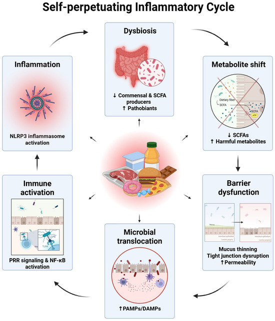
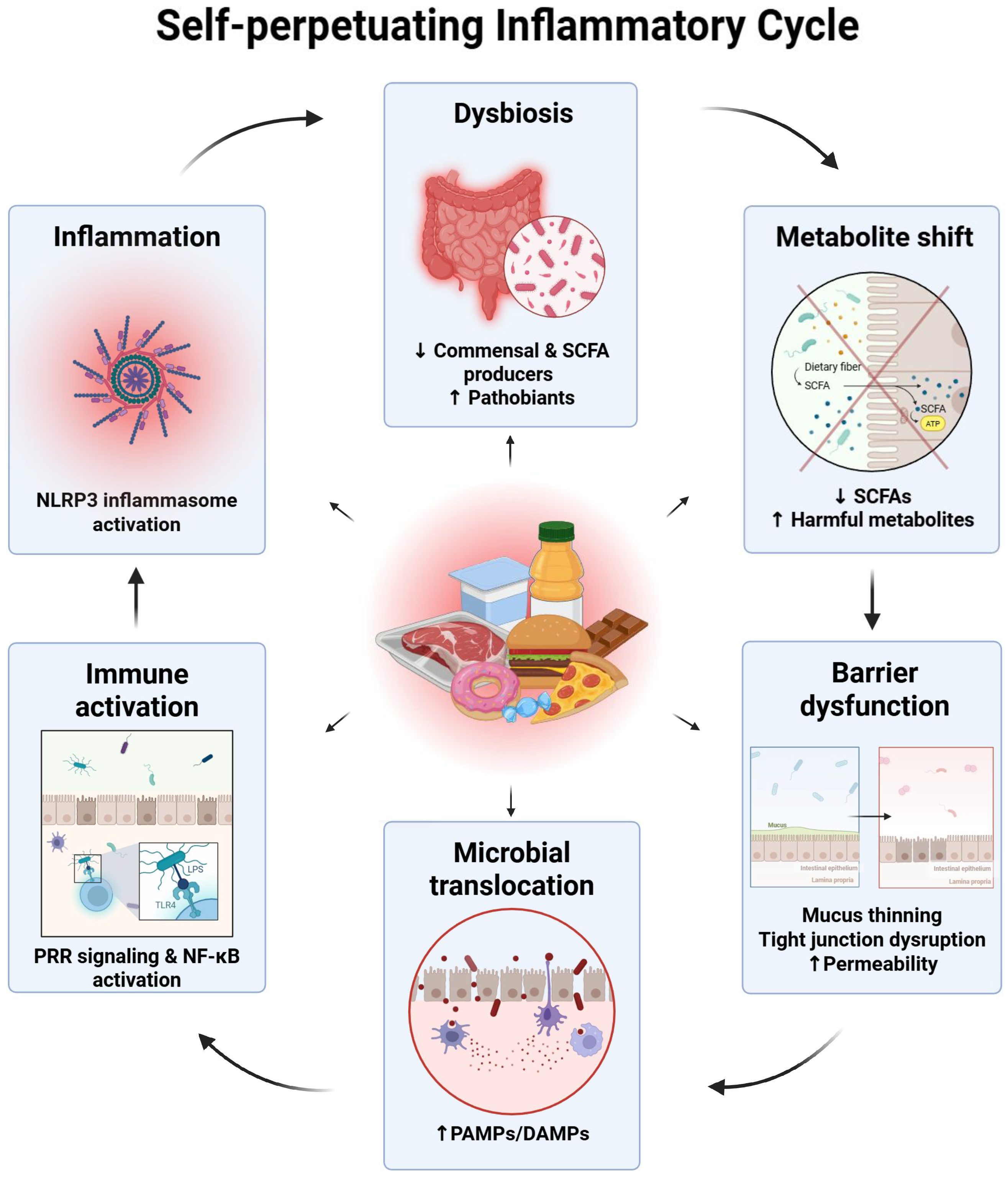

Disclaimer/Publisher’s Note: The statements, opinions and data contained in all publications are solely those of the individual author(s) and contributor(s) and not of MDPI and/or the editor(s). MDPI and/or the editor(s) disclaim responsibility for any injury to people or property resulting from any ideas, methods, instructions or products referred to in the content. |
© 2026 by the authors. Licensee MDPI, Basel, Switzerland. This article is an open access article distributed under the terms and conditions of the Creative Commons Attribution (CC BY) license.