


Abstract
Chagas disease, caused by Trypanosoma cruzi, affects a significant proportion of patients who develop digestive and cardiac complications, including megaviscera. This pathogenesis has been associated with autoimmune mechanisms mediated by molecular mimicry. In this study, an in silico evaluation of the potential structural basis of cross-reactivity of β-tubulin 1.9 of T. cruzi and the human β-4A tubulin isoform 3 was conducted. Using bioinformatics tools, homologous regions were identified and potentially immunogenic epitopes were predicted, considering their structural modeling and molecular docking. The proteins shared 87% sequence identity and 95% similarity, with an almost identical structural overlap, RMSD 0.291 Å. Three epitopes, VPFPRLHFF, NDLVSEYQQYQDATI, and GQSGAGNNWAKGHYTEGAELIDS, exhibited high predicted antigenicity, with the 9-mer and 16-mer peptides displaying structurally compatible docking poses within the binding grooves of MHC class I and class II molecules, respectively, while B-cell epitope potential was inferred from sequence-based property predictions. Normal mode analysis, used as an exploratory approach, suggested comparable flexibility profiles for the parasitic- and human-derived peptide–MHC complexes. These findings provide an exploratory structural framework supporting a potential role of β-tubulin epitopes in molecular mimicry processes implicated in the development of chagasic megaviscera.
1. Introduction
Chagas disease is a parasitic infection caused by the protozoan Trypanosoma cruzi, transmitted through the infected feces of triatomine vectors such as Triatoma infestans and Rhodnius prolixus. Transmission occurs when the feces come into contact with a bite wound or the mucous membranes of the host [1]. Chagas disease is endemic in Central and South America; however, due to migration, it has expanded to Europe, the United States, Canada, Japan, and Australia. It has been reported that 70% of infected individuals are asymptomatic, while the remaining 30% develop cardiac, digestive, neurological, or mixed disorders [2].
The immune response developed by the host against the parasite is critical for the outcome of the infection. Antibodies are central components of the host immune response, participating in the early control of the parasite, and in the pathophysiology of the disease [2,3], with special emphasis on the presence of anti-α-galactoside (anti-Gal) antibodies, which recognize glycans on the surface of T. cruzi, and mediate the lysis of the parasite in its trypomastigote form [4]. Studies have also identified another antibody in mice is β-tubulin, which is present in the parasite’s flagellum. β-tubulin has been identified as one of the main T. cruzi antigens recognized by IgG [4].
Disorders such as Chagas disease are commonly associated with the development of autoimmune diseases caused by molecular mimicry, a mechanism that occurs when similarities between the protein sequences of parasite and human epitopes trigger the activation of autoantibodies [5]. One example is the monoclonal autoantibody A2R1, which exhibits cross-reactivity between human β-tubulin and the β-tubulin of T. cruzi [2]. Furthermore, in the study conducted by Ternynck et al., a high presence of anti-tubulin IgG autoantibodies was observed in mice infected with T. cruzi [6]. This finding is supported by additional studies that have identified autoantibodies developed during infection with greater affinity and binding efficiency toward the tubulin protein, reinforcing the potential relationship between the autoimmune response and T. cruzi infection [7].
Autoantibodies identified as reactive with tubulin, myosin, and actin have also been associated with complications such as megaviscera (megacolon, megaesophagus, and splenomegaly) [2], arising from neurosensory system alterations in the muscle cells of affected organs [8], induced by infection of this protozoan [9]. Moreover, CD4+ T cells that recognize human myosin and the peptide of the T. cruzi B13 protein may drive Chagas myocardiopathy, characterized by T cell/macrophage infiltration, fibrosis and damage to cardiac fibers [10,11].
The severity of chagasic cardiomyopathy and megasyndromes is intrinsically linked to the massive neuronal destruction of the enteric nervous system (ENS) [12,13,14], which requires that any molecular mimicry model focus on key proteins within this tissue, since the clinical manifestation of megacolon, for example, requires the loss of more than half of colonic enteric neurons [14]. Accordingly, the human β-4A isoform (TUBB4A) is established as the ideal target, as this protein, a microtubule subunit, is essential for neuronal integrity and intracellular transport [15,16,17] and exhibits a highly specialized and enriched expression within the nervous system, including the brain [13,17] and the ENS. This specialization confers a critical vulnerability in disease, as evidenced by severe neurodegenerative disorders, such as H-ABC leukodystrophy, caused by mutations in its gene (TUBB4A), where the resulting damage disrupts microtubule stability and leads to neuronal dysfunction and loss [14,18,19]. In response to this target, T. cruzi β-tubulin 1.9 (AAL75956.1) emerges as the most relevant antigen: its immunodominance is remarkable, as its transcripts (together with the 2.3-kb transcript) are 3–6-fold more abundant in epimastigotes (the stage that elicits a strong immunological stimulus) than in trypomastigotes or amastigotes [20].
Currently, the role and origin of β-tubulin autoantibodies in the development of megaviscera after T. cruzi infection remain unclear. The aim of this study is to identify potential molecular mimicry between T. cruzi β-tubulin and human proteome through immunopeptidomics and immunoinformatics, as a possible cause of a pathological autoimmune reaction.
2. Materials and Methods
2.1. Antigen Selection—BLAST
An in silico immunoinformatic study was conducted to identify T. cruzi proteins with immunological relevance. A general sequence homology analysis was performed focusing on T. cruzi proteins of interest. Using the NCBI database, the antigen sequence was retrieved, and the BLAST tool 2.16.0 (National Center for Biotechnology Information, Bethesda, MD, USA) (available at: https://blast.ncbi.nlm.nih.gov/BlastAlign.cgi, accessed on 21 May 2025) [21] was applied for the comparative alignment between immunogenic protein of interest from T. cruzi (Taxid: 5693) β-tubulin 1.9 (NCBI Code: >AAL75956.1) and the Homo Sapiens proteome (Taxid: 9606), revealing proteins with high sequence similarity.
2.2. Sequence Alignment in MUSCLE and BLAST
Through the MUSCLE free server (CA, USA) (available at: https://www.ebi.ac.uk/jdispatcher/msa/muscle, accessed on 23 May 2025) [22], it offers high accuracy in alignments of a small number of homologous sequences due to its iterative refinement and the use of a log-expectation scoring function in its algorithm (Multiple Sequence Comparison by Log-Expectation), which enhances the detection of conserved residues. A comparative alignment was performed between the antigen sequence and the selected human protein, β-4A tubulin chain isoform 3 (NCBI code: NP_001276058.1), resulting in a global alignment that enabled the assessment of the degree of similarity between both sequences and the identification of regions potentially involved in cross-reactive recognition mechanisms.
Additionally, the BLAST tool was employed to complement the analysis through local alignments, allowing the identification of specific regions of high sequence similarity between the compared sequences, which may correspond to epitopes with potential immunological relevance.
2.3. Epitope Prediction—IEDB
Using the IEDB database (La Jolla Institute for Allergy and Immunology, CA, USA) (available at: https://nextgen-tools.iedb.org/pipeline/56a665e4-d5e7-418f-ba12-03b7fa253065, accessed on 26 May 2025) [23], the antigen sequence of T. cruzi was analyzed for T-cell epitope prediction (major histocompatibility complex class I and II) via NetMHCpan 4.1 EL algorithm, which predicts epitopes based on a large dataset of eluted ligands. The specific alleles selected for reporting were MHC I (HLA-B*15:01) and MHC II (HLA-DQA1*01:01/DQB1*05:01), chosen because they exhibited the highest affinity scores, representing the most favorable immunological interactions. Epitopes presenting a netMHCpan EL score higher than 0.5 were selected in order to prioritize peptides with a higher probability of immunological recognition. These epitopes were compared between the antigen protein and the corresponding human protein using sequence alignments performed with MUSCLE, aiming to evaluate sequence conservation and structurally compatible features inferred from the sequence. Preference was given to epitopes exhibiting complete conservation, as well as to cases in which the epitope core was fully conserved, defined as the presence of ≥6 identical central amino acids.
For B-cell epitope prediction, the BepiPred Linear B-Cell Epitope Prediction 2.0 (Technical University of Denmark, Denmark) (available at: https://services.healthtech.dtu.dk/services/BepiPred-2.0/, accessed on 29 May 2025) and Discotope 2.0 (Technical University of Denmark, Denmark) (available at: https://services.healthtech.dtu.dk/services/DiscoTope-2.0/, accessed on 29 May 2025) servers were employed. For B-cell epitope prediction, BepiPred Linear B-Cell Epitope Prediction 2.0 and DiscoTope 2.0 were employed. BepiPred 2.0 was selected for its improved accuracy in linear epitope prediction derived from training on experimentally validated conformational epitopes, while DiscoTope 2.0 was used to identify discontinuous, surface-exposed epitopes based on three-dimensional structural information. Epitopes predicted con cordantly by both tools were prioritized, taking into account their immunological relevance and surface accessibility.
Additionally, B cell epitopes were compared with the corresponding human sequences using sequence alignments. Epitopes fulfilling simultaneously the criteria of sequence similarity, accessibility, and structural conservation were considered candidates with potential involvement in molecular mimicry mechanisms and autoantibody recognition.
2.4. Properties of Analyzed Epitope
To analyze the properties of the selected epitopes, VaxiJen v1.0 (Institute of Microbiology, Bulgarian Academy of Sciences, Bulgaria) (available at: https://www.ddg-pharmfac.net/vaxijen/VaxiJen/VaxiJen.html, accessed on 30 May 2025) [24] was used to assess antigenicity; Bepipred v2.0 (available at: https://services.healthtech.dtu.dk/services/BepiPred-2.0/, accessed on 30 May 2025) [25] to evaluate antigenicity and epitope localization; Toxinpred v1.0 (Indian Institute of Technology Delhi, New Delhi, India) (available at: https://webs.iiitd.edu.in/raghava/toxinpred/design.php, accessed on 30 May 2025) [26] to exclude potential toxicity, and DNASTAR to calculate Jameson-Wolf index for each residue antigenicity, the hydrophobicity index for predicting exposed regions, and therefore its accessibility; and the Chou-Fasman and Garnier-Robson algorithms for secondary structure prediction of the epitope (WI, USA).
2.5. Three-Dimensional Protein Modeling—Alpha Fold
For 3D modeling of both proteins (>NP_001276058.1 and >AAL75956.1), the Alpha Fold 3 server (Boston University, MA, USA) (available at: https://alphafoldserver.com/about, accessed on 14 June 2025) [27] was used, along with the Pymol software (Version 3.1.6.1, Schrödinger, NY, USA), which employs its own rendering engine based on OpenGL (version 4.6). In both the T. cruzi β-tubulin 1.9 and human β-tubulin 4A isoform 3 models, regions with reduced confidence (50 < pLDDT < 70) were confined to the extreme C-terminal tail (residues 433–439 in T. cruzi and 433–442 in the human protein), while very low-confidence residues (pLDDT < 50) were limited to positions 438–439 in the human model. These regions did not coincide with the predicted epitopes or docking interfaces; therefore, no additional refinement or exclusion was performed prior to downstream docking and dynamic analyses.
2.6. Three-Dimensional Prediction of the Epitope Docking with MHC I and II
Epitope docking was carried out using ClusPro (DeepMind, UK) (available at: https://cluspro.org/home.php, accessed on 5 June 2025) [28], selecting models with the lowest energy scores (more negative values indicate more favorable interactions). Subsequently, an exploratory normal mode analysis (NMA) based on elastic network models was carried out using iMODS (Universitat Autònoma de Barcelona, Spain) (available at: https://imods.iqf.csic.es/, accessed on 5 June 2025) [29] calculating complex deformation under force field variations over time intervals, and estimating protein deformability based on eigenvalues. The 3D epitope models were retrieved from the RPBS database (France) (available at: https://bioserv.rpbs.univ-paris-diderot.fr/services/PEP-FOLD4/, accessed on 5 June 2025) [30]. The models of the Major Histocompatibility Complexes (MHC) I (Code: 4U6Y), II (Code: 3C5J), were downloaded from the RCSB PDB database (NJ, USA) (available at: https://www.rcsb.org/, accessed on 5 June 2025) [31].
3. Results
3.1. Selection of Candidate Antigens
The human protein tubulin beta-4A chain isoform 3 (NP_001276058.1) was selected based on its broad neuronal expression, accounting for approximately 30% of total tubulin in neuronal cells, with the exception of mature glial cells. Notably, this isoform is highly enriched in the peripheral nervous system, where it plays a crucial role in axonal regeneration [32]. Additionally, within the enteric nervous system, this isoform contributes significantly to the structural organization and functional maintenance of nerve fibers [33].
A pairwise sequence alignment between human β-tubulin (NP_001276058.1) and T. cruzi β-tubulin (AAL75956.1) revealed 87% identity and 95% similarity across 429 amino acid residues, with no insertions or deletions (0% gaps), indicating a high degree of conservation (Table 1). Although multiple T. cruzi β-tubulin isoforms were initially evaluated, only β-tubulin 1.9 fulfilled the combined criteria of high sequence identity and structural conservation with the neuronal human β-tubulin 4A isoform, particularly within functionally relevant regions involved in microtubule dynamics. Other isoforms exhibited reduced conservation in these regions and were therefore excluded, as they were considered less suitable for modeling molecular mimicry associated with neuronal damage. Analysis was performed using the BLOSUM62 substitution matrix with the low-complexity filter enabled. The E-value of 0.0 denotes a true evolutionary homology, suggesting that both proteins likely originated from a common ancestor and retain conserved structural and functional characteristics.
Table 1.
Proteins selected for alignment.
3.2. Sequence Alignment in MUSCLE
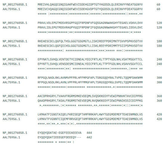
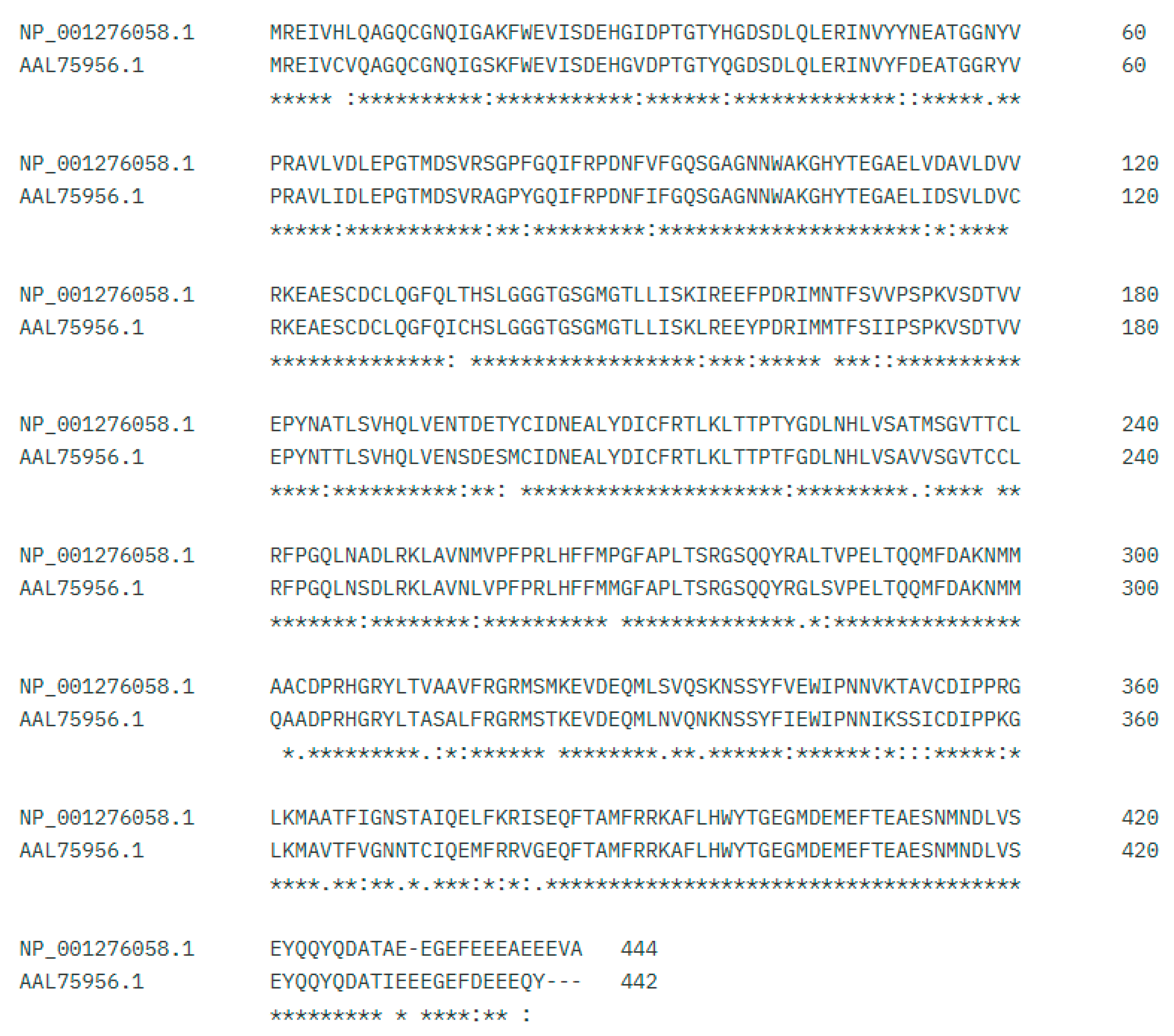
MUSCLE identified a high similarity (86.65%) between human tubulin β-4A isoform 3 (NP_001276058.1) and β-tubulin 1.9 protein (AAL75956.1), as shown in Figure 1.
Figure 1.
Sequence alignment, where (*) denotes identity, indicating an exact match between both sequences, while (:) and (.) represent similarity, corresponding to conservative substitutions indicating high and low similarity, respectively.
3.3. Epitope Prediction—IEDB
Epitope prediction was performed using IEDB for T cells (MHC class I and II) and B cells. These results are shown in Table 2.
Table 2.
Immunogenic epitopes with high affinity for HLA-I, HLA-II alleles, and B cells predicted in homologous regions between human tubulin β-4A isoform 3 and β-tubulin 1.9 protein of T. cruzi.
In Table 2, the epitopes with the best scores and the highest percentage of similarity among amino acids from both sequences were selected. The epitope GQSGAGNNWAKGHYTEGAELIDS does not include a NetMHCII pan score or allele information, as the B-cell antigenicity predictor in IEDB was used, in contrast to the other epitopes that were analyzed using MHC class I and class II prediction tools.
3.4. Predicted Properties of the Analyzed Epitope
Before verifying the epitope properties, those without full sequence similarity were discarded using MUSCLE. VaxiJen v2.0 was used to analyze antigenicity; ToxinPred v1.0 to assess toxicity; BepiPred v2.0, specifically for B cells (optimal cutoff point at a threshold of 0.5–0.55, where sensitivity and specificity are balanced at approximately 50–60%), and Netsurfp v3.0 to evaluate surface exposure; and DNASTAR. This data for the three selected epitopes is summarized in Table 3.
Table 3.
Summary of the properties from selected epitopes.
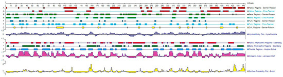
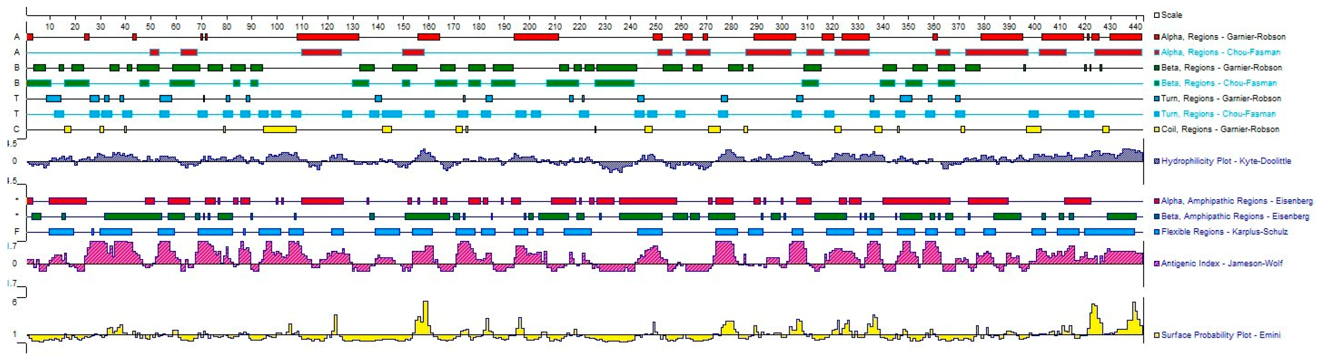
Figure 2 shows the GQSGAGNNWAKGHYTEGAELIDS (93–115) epitope exhibits abrupt changes in the chain, as well as disordered coils, and an alpha helix at the end of the epitope. The VPFPRLHFF (258–266) epitope shows a higher presence of alpha-helical regions along with abrupt changes in the chain; whereas the NDLVSEYQQYQDATI (416–430) epitope displays only alpha-helical regions in the structure and an abrupt change in the chain.
Figure 2.
Diagram of antigenic site prediction and three-dimensional structure analysis. Structural and antigenic analyses of the protein were performed using secondary structure prediction methods, including Chou–Fasman and Garnier–Robson. In addition, antigenicity was evaluated using the Jameson–Wolf index, together with analyses of surface accessibility, hydrophilicity (Kyte–Doolittle), and amphipathicity/hydrophobicity (Eisenberg), employing the DNASTAR Protean software (https://www.dnastar.com/software/lasergene/protean-3d/, accessed on 18 December 2025).
Antigenicity analysis using the Jameson–Wolf index for the B-cell epitope GQSGAGNNWAKGHYTEGAELIDS indicated a high probability of antigenic recognition. This result is associated with the combination of properties such as high hydrophilicity and surface accessibility, suggesting that this region may be solvent-exposed and accessible for antibody recognition.
Finally, the evaluation of hydrophilicity and hydrophobicity using the Kyte–Doolittle and Eisenberg indices, respectively, for the T-cell epitopes VPFPRLHFF and NDLVSEYQQYQDATI revealed the presence of β-amphipathic regions. This pattern indicates the coexistence of hydrophilic and hydrophobic residues, and the presence of the latter at specific positions has previously been associated with peptide interaction and anchoring within MHC class I and II molecules.
Overall, the three epitopes evaluated exhibit favorable structural and physicochemical properties that support their consideration as immunologically relevant epitope candidates.
3.5. Three-Dimensional Protein Modeling
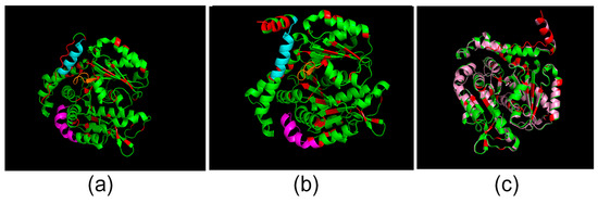
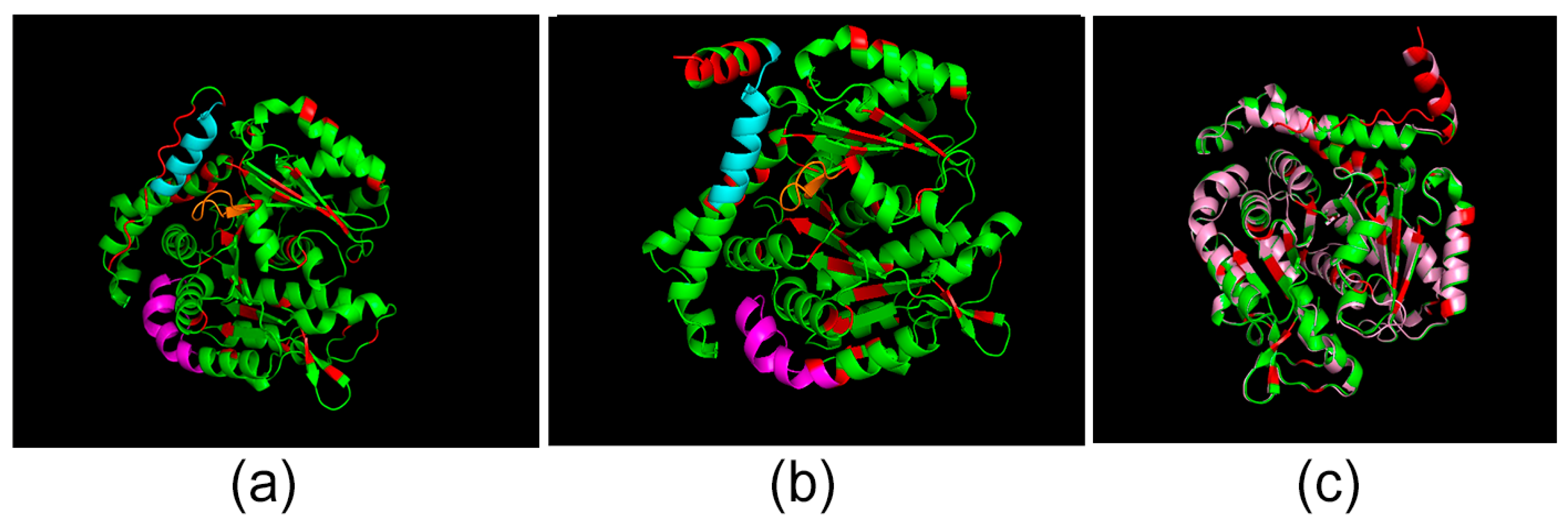
The human tubulin β-4A isoform 3 and the β-tubulin 1.9 protein from T. cruzi were modeled, with the epitopes highlighted (shown in Figure 3).
Figure 3.
Three-dimensional model prediction of the evaluated proteins. Predicted structures with highlighted epitopes: GQSGAGNNWAKGHYTEGAEL (magenta), NDLVSEYQQYQDATI (cyan) and VPFPRLHFF (orange), also regions that differ between the sequences are shown in red. (a) Human tubulin β-4A isoform 3 prediction. (b) T. cruzi β-tubulin 1.9 protein prediction. (c) Structural alignment of both proteins: human tubulin β-4A isoform 3 (yellow) and β-tubulin 1.9 protein from T. cruzi (pink).
High structural similarity was observed through alignment (Figure 3c), and the selected epitopes were found to be sequentially identical in both proteins, as shown in Figure 3a,b.
In Figure 3a–c, the models achieved pTM scores of 0.93, 0.93, and 0.83, respectively, and pLDDT values above 90 for most residues, indicating high prediction quality. All contact maps (provided by AlphaFold) showed good structural consistency, with low-confidence regions mainly corresponding to terminal or flexible loop areas. Given the high reliability of the models, no further modifications were applied. The alignment yielded a Root Mean Square Deviation (RMSD) of 0.291 Å (for 2749 paired atoms), as shown in Figure 3. This low deviation value confirms a superposition of high fidelity, establishing that the structural conformations are virtually identical.
3.6. Three-Dimensional Alignment Prediction
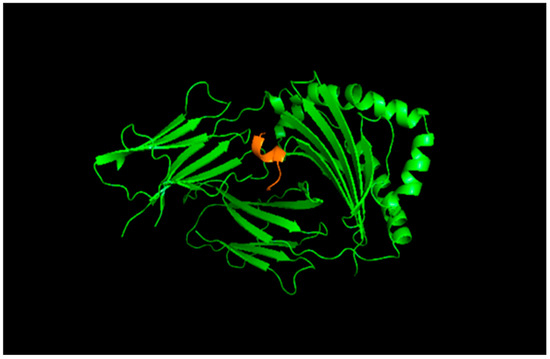
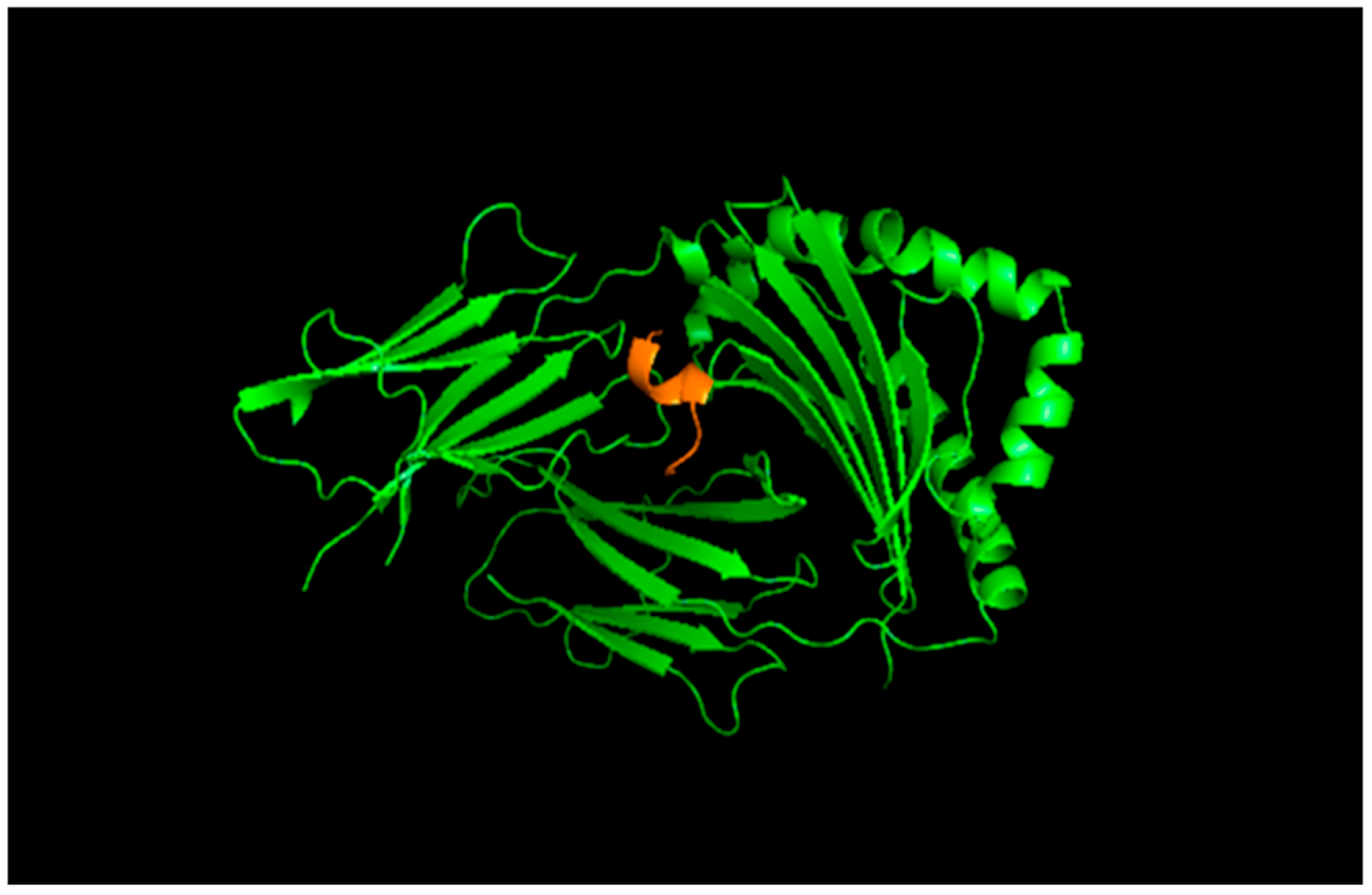
Using the ClusPro server, the binding affinity between the selected T-cell epitopes and MHC class I and class II molecules was evaluated.
The results are expressed as a Weighted Score, an empirical metric that integrates the repulsive and attractive van der Waals contributions, electrostatic energy, and statistical desolvation. More negative values of this score indicate a more stable interaction. The models corresponding to cluster 0 were selected, as this cluster contained the largest number of members, indicating a more recurrent binding conformation.
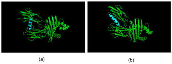
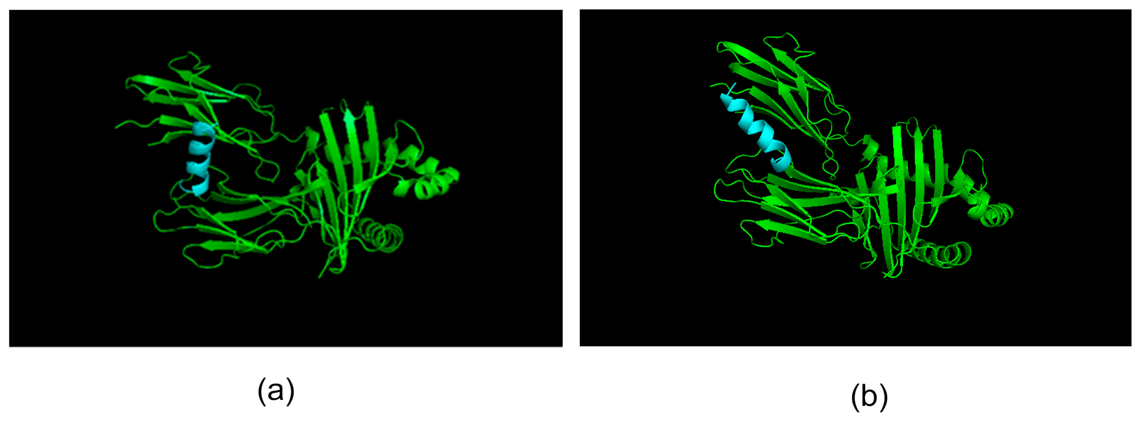
The molecular docking analysis presented in Figure 4 and Figure 5 revealed low (negative) minimum binding energy values, suggesting favorable interactions between the analyzed epitopes and MHC molecules under computational conditions. For MHC class I, the epitopes NDLVSEYQQYQDATI and VPFPRLHFF exhibited identical minimum binding energy values (−746.5 kcal/mol), indicating a stable interaction at the structural level. In the case of MHC class II, the epitope NDLVSEYQQYQDATI showed the lowest minimum binding energy (−768.2 kcal/mol). Although NDLVSEYQQYQDATI was originally predicted as a candidate epitope for MHC class II using the IEDB platform, additional docking with MHC class I was performed for exploratory purposes, aiming to assess potential cross-binding tendencies or structural similarities relevant in the context of molecular mimicry. Taken together, these results indicate that NDLVSEYQQYQDATI exhibits favorable interactions with MHC molecules.
Figure 4.
3D docking of the T-cell epitope with MHC class II (HLA-DQA101:01/DQB105:01). Epitopes are shown in cyan. (a) The T. cruzi-derived epitope NDLVSEYQQYQDATI exhibited a minimum binding energy of −709.2 kcal/mol and a central energy of −562.7 kcal/mol. (b) The Homo sapiens-derived epitope NDLVSEYQQYQDATA showed a minimum binding energy of −740.7 kcal/mol and a central energy of −595.2 kcal/mol.
Figure 5.
Three-dimensional docking of the VPFPRLHFF epitope, represented in orange, with MHC I (HLA B*15:01), showing a minimum binding energy of −727.1 kcal/mol and a central energy of −624.3 kcal/mol.
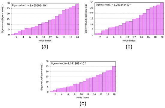
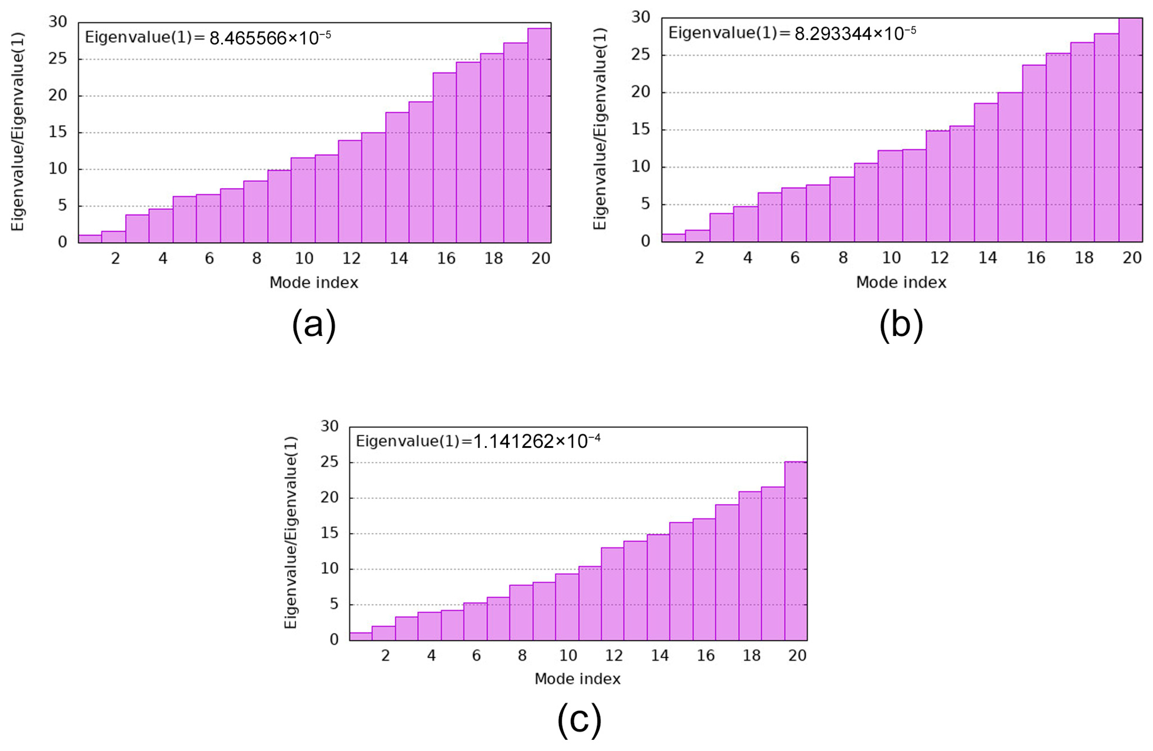
Continuing the analysis of the predicted epitopes, the epitope–MHC I and II complexes, both of human origin and derived from T. cruzi, were subjected to normal mode analysis (NMA) using iMODS to assess structural flexibility, including deformability profiles, mobility, and eigenvalue distributions, which reflect global structural rigidity.
Since iMODS does not allow the direct identification of residue–residue interactions, the amino acids involved in the epitope–MHC interface were previously identified using the PyMOL tool. This structural analysis enabled the localization of MHC residues directly involved in epitope binding, which was essential for the interpretation of the dynamic parameters obtained from iMODS.
Structural analysis of the NDLVSEYQQYQDATI epitope showed that most MHC class II amino acids involved in the interaction with the T. cruzi-derived epitope also participate in binding to the corresponding human epitope, indicating a high degree of similarity in the binding mode of both complexes. Accordingly, deformability and mobility profiles obtained using iMODS revealed that the MHC class II regions involved in the interaction exhibit highly similar structural flexibility patterns in both complexes, with only minor variations observed in a limited number of residues. These regions exhibited heterogeneous pattern structural flexibility, including rigid residues alongside areas of moderate to high flexibility.
Similarly, for the VPFPRLHFF epitope, which is identically conserved in Homo sapiens and T. cruzi, analysis of the interaction interface revealed a combination of rigid and highly deformable regions. While the more rigid residues may contribute to local mechanical support of the complex, the presence of highly deformable regions suggests a substantial degree of global flexibility, indicating a balance between mechanical rigidity and flexibility.
Additionally, the global stiffness of the epitope–MHC complexes was evaluated through eigenvalue analysis using iMODS.
The plots presented in Figure 6 were used to compare the intrinsic mechanical properties of complexes derived from T. cruzi and human origin. The first normal mode of the T. cruzi epitope (NDLVSEYQQYQDATI)–MHC class II complex (Figure 6a) exhibited an eigenvalue of 8.465566 × 10⁻⁵, whereas the human epitope (NDLVSEYQQYQDATA)–MHC class II complex (Figure 6b) showed an eigenvalue of 8.293344 × 10⁻⁵. The close similarity between these values indicates comparable global rigidity of both complexes, suggesting a similar intrinsic mechanical behavior of MHC class II upon binding to epitopes of different origin. In addition, the relatively low eigenvalues observed in both cases are consistent with a moderate degree of global structural flexibility.
Figure 6.
Comparison of eigenvalue plots between human- and T. cruzi–derived epitope–MHC complexes, employing the server iMODS (a) T. cruzi epitope (NDLVSEYQQYQDATI)—MHC II complex and (b) human epitope (NDLVSEYQQYQDATA)–MHC II complex. 8.293344 × 10⁻⁵ (c) T. cruzi and human epitope (VPFPRLHFF)—MHC I complex. 1.141262 × 10⁻⁴.
In the case of the VPFPRLHFF epitope, the analysis was performed only once, as this sequence is identical in both organisms. This complex exhibited an eigenvalue of 1.141263 × 10⁻⁴, indicating a relative global flexibility, although slightly lower compared to the previously described epitope.
By contrast, the epitope GQSGAGNNWAKGHYTEGAELIDS was not analyzed using iMODS, as it corresponds to a B-cell epitope, which does not require presentation by MHC molecules and therefore is not suitable for normal mode analysis in the context of epitope–MHC complexes.
4. Discussion
Chagas disease often presents digestive pathological manifestations such as megaviscera, associated with enteric neuronal cell death, reaching up to 85% in megaesophagus and 50% in megacolon [34]. Furthermore, a strong association has been reported between neuronal destruction and the presence of potentially cytotoxic cells such as eosinophils and mast cells [35]. These cells participate in inflammatory processes that may cause tissue damage by promoting cytokines secretion including IL-1, TNF-α, and IL-6 [36], as well as the production of nitric oxide and free radicals, which can lead to fibrosis [35]. Also, the proposed association between β-tubulin mimicry and the development of megasyndromes is rooted in the intrinsic structural vulnerability of the human β-4A isoform (TUBB4A), which is highly enriched in neuronal populations [37,38,39].This protein is indispensable for cytoskeletal organization and axonal transport, and its dysfunction parallels severe neurodegenerative disorders, such as hypomyelination with atrophy of the basal ganglia and cerebellum (H-ABC) and DYT4 dystonia, in which β-tubulin mutations, particularly within the H12 helix, profoundly impair kinesin (KIF)-mediated transport of organelles and vesicles [37,38,39,40]. Within a speculative yet mechanistically substantiated framework, anti-tubulin autoantibodies elicited against the immunodominant parasitic antigen (MAP) [41], whose cross-reactivity with nervous system tubulin has been experimentally demonstrated, may sterically hinder the processive motility of KIFs or destabilize microtubules, a mechanism that has been validated in vivo using isoform-specific antisera [40,41]. This sustained disruption of axonal transport culminates in distal axonal degeneration and subsequent neuronal soma loss, manifesting as aganglionosis [40]. However, this structural mechanism operates in concert with additional pathogenic pathways [41]. Disease progression is further driven by early functional impairment mediated by autoantibodies targeting G protein-coupled receptors (GPCRs), specifically anti–β2-adrenergic and anti–M2-muscarinic profiles, which induce dysautonomia, as well as by direct immunopathology maintained by persistent parasitic DNA and chronic inflammation (ganglionitis), resulting in neurotoxicity via oxidative stress and reactive nitrogen species (RNS) [42,43,44,45,46,47,48,49].
In this context, the present study provides a consistent computational structural rationale suggesting a potential molecular mimicry phenomenon between T. cruzi β-tubulin and human β-tubulin, with possible immunopathological implications. The high level of sequence conservation observed (87% identity and 95% similarity) confirms a true evolutionary homology derived from a common eukaryotic ancestor, supporting the possibility of cross-reactive immune recognition [50]. This hypothesis is further reinforced by the high structural overlap observed (RMSD = 0.291 Å), indicating that conserved epitopes adopt similar three-dimensional conformations. Epitope analysis identified conserved regions with predicted MHC class I and class II binding potential. The VPFPRLHFF epitope showed a favorable predicted interaction profile with HLA-B15:01, which has been associated with CD8+ T lymphocyte activation and inflammatory processes [51]. In turn, the NDLVSEYQQYQDATI epitope exhibited a favorable predicted interaction with the HLA-DQA101:01/DQB105:01 heterodimer, previously linked to the activation of pathogenic CD4+ T lymphocytes in Chagasic cardiomyopathy [52] and to susceptibility to autoimmune diseases of the central nervous system [53], although a modulatory role in antigen recognition has also been described [54]. The structurally compatible interaction of this epitope with both MHC class I and class II molecules could promote the simultaneous activation of CD4+ and CD8+ immune responses. Additionally, the B-cell epitope GQSGAGNNWAKGHYTEGAELIDS displayed favorable antigenic properties, characterized by high hydrophilicity and surface accessibility, supporting the potential for antibody-mediated cross-reactivity [55]. The presence of amphipathic features in the VPFPRLHFF and NDLVSEYQQYQDATI epitopes is consistent with properties commonly described for MHC-binding peptides [56]. However, the literature predominantly reports α-helical conformations for such peptides, whereas the epitopes identified in this study exhibited β-sheet structures, which may partially affect their MHC binding compatibility. Nevertheless, other properties reinforce their immunological relevance. Molecular docking combined with normal mode analysis performed for T-cell epitopes revealed comparable interaction geometries and similar flexibility profiles, as reflected by nearly identical eigenvalues for epitope–MHC complexes of parasitic and human origin, indicating similar binding modes and dynamic behavior. Collectively, these results suggest that the immune system may have limited capacity to discriminate between T. cruzi epitopes and their human counterparts during antigen presentation.
Based on the results obtained, it can be suggested that the epitopes identified in T. cruzi β-tubulin may participate in a molecular mimicry mechanism with the human β-tubulin 4A isoform 3, which potentially contributes to the enteric nervous system damage observed in Chagas disease.
The limited efficacy and contradictory outcomes of current antiparasitic treatments, benznidazole and nifurtimox (20–60% cure rate [55]), during the chronic phase of Chagas disease, in which neuronal damage is irreversible, require a therapeutic reorientation toward neuroimmunomodulation and structural protection [57,58]. The identification of specific epitopes from T. cruzi β-tubulin 1.9 that mimic the human TUBB4A isoform suggests potential therapeutic avenues. Given that the pathological hallmark, aganglionosis, is a consequence of axonal transport disruption caused by interference with tubulin, treatment strategies may be redirected toward structural neuroprotection [48,59]. This includes the use of microtubule-stabilizing agents to counteract the destabilizing effects of autoimmune-mediated cytoskeletal attack within the enteric nervous system, ENS, an approach analogous to therapeutic strategies explored for TUBB4A-related genetic tubulinopathies [44,60].
Within the context of adaptive immunomodulation, the epitopes VPFPRLHFF, MHC I, and NDLVSEYQQYQDATI, MHC II, suggest the feasibility of employing tolerogenic peptides to selectively desensitize or eliminate cytotoxic T-cell clones targeting nervous tissue [48]. In addition, the B-cell epitope GQSGAGNNWAKGHYTEGAELIDS is associated with the generation of cross-reactive autoantibodies, AABs, such as A2R1 [2], whose binding has been shown to depend on post-translational modifications, PTMs, of tubulin [16,61]. This specificity enables targeted immunoneutralization or immunoadsorption strategies for the removal of circulating pathogenic AABs, which may slow irreversible tissue damage [61]. Finally, to eliminate the antigenic source that perpetuates the autoimmune response, molecular characterization of parasitic β-tubulin 1.9 also supports the exploration of selective protein degradation technologies, such as TrypPROTACs [62].
It is important to note that neuronal damage could also result from an immune response directed against the parasite itself [63]. Thus, both direct immunopathology and autoimmunity may be involved in the pathogenesis. A critical alternative mechanism is the collapse of the biological pacemaker system due to depletion of interstitial cells of Cajal [64,65], ICCs, whose density is reduced significantly, thereby disrupting the propagation of contractile waves and neuromuscular coordination. This injury is further amplified by microvascular alterations, such as chronic vasospasm and focal ischemia, which induce myenteric necrosis and reparative fibrosis [66,67,68]. Likewise, persistent inflammation drives aberrant extracellular matrix remodeling, in which dense collagen deposition and the activity of matrix metalloproteinases, MMP-2 and MMP-9, increase visceral wall stiffness, rendering it unable to recover its original caliber. Finally, the clinical phenotype is further modulated by a neuroendocrine imbalance, characterized by increased inhibitory signaling, such as somatostatin and GLP-1, together with dysbiosis of the intestinal microbiota, the so-called tripobiome, which perpetuates a proinflammatory microenvironment and chronic stasis [69,70,71,72,73,74].
Importantly, the present study does not aim to quantify binding affinities, thermodynamic stability, or kinetic parameters of peptide–MHC interactions. Instead, it provides a structural and immunoinformatic analysis intended to prioritize candidate epitopes and to generate testable hypotheses regarding their potential involvement in autoimmune mechanisms associated with Chagas disease. While molecular mimicry may contribute to the pathogenesis of Chagas disease, further experimental and computational studies are required to elucidate its precise role.
This study has limitations, particularly the absence of experimental validation, since the approach employed was an in silico analysis. Consequently, the results obtained may not exactly represent the complexity of the immune system.
5. Conclusions
In conclusion, this study provides an exploratory in silico structural framework supporting the plausibility of molecular mimicry between T.cruzi β-tubulin 1.9 and human β-4A tubulin isoform 3, based on high sequence identity, structural convergence, and near-identical conformations. Shared predicted T-cell epitopes displayed structurally compatible peptide–MHC class I and class II interaction patterns, whereas B-cell epitope potential was inferred exclusively from sequence-based analyses, without MHC binding evaluation. Together, these findings support the feasibility of cross-reactive immune recognition involving the neuron-enriched TUBB4A protein. Although this work does not address binding affinities, thermodynamic stability, or specific pathogenic mechanisms, it proposes a focused and testable hypothesis linking parasite-derived tubulin epitopes to autoimmune processes that may contribute to enteric neuronal damage and megaviscera in Chagas disease. The results therefore provide a computational rationale for prioritizing selected epitopes for future experimental validation and advanced computational studies.
Author Contributions
Conceptualization, A.V.C.-I., C.A.Q.-J. and G.A.O.-P.; Methodology, A.V.C.-I. and C.A.Q.-J.; Software, A.V.C.-I., C.A.Q.-J., A.S.R.-Z. and P.G.-M.; Validation, G.A.O.-P. and L.A.P.-S.; Formal Analysis, A.V.C.-I., C.A.Q.-J. and G.A.O.-P.; Investigation, A.V.C.-I. and C.A.Q.-J.; Data Curation, A.V.C.-I. and C.A.Q.-J.; Writing—Original Draft Preparation, A.V.C.-I. and C.A.Q.-J.; Writing—Review & Editing, A.V.C.-I., C.A.Q.-J., A.S.R.-Z. and P.G.-M.; Visualization, G.A.O.-P. and L.A.P.-S.; Supervision, G.A.O.-P. and L.A.P.-S. All authors have read and agreed to the published version of the manuscript.
Funding
This research received no external funding.
Informed Consent Statement
Not applicable.
Data Availability Statement
Figure 1 was generated using the free server MUSCLE (available at: https://www.ebi.ac.uk/jdispatcher/msa/clustalo, accessed on 23 May 2025) [23], where a comparative alignment was performed between antigen sequences and the selected human tubulin β-4A isoform 3 protein (NCBI code: >NP_001276058.1), focusing on regions showing significant similarity to predict potential epitopes. Table 2 was generated from the epitope prediction results using the IEDB database (available at: https://nextgen-tools.iedb.org/pipeline/56a665e4-d5e7-418f-ba12-03b7fa253065, accessed on 26 May 2025) [24]. This table summarizes the immunogenic epitopes with high affinity for HLA-I, HLA-II alleles, and B cells, which were predicted in homologous regions between human tubulin β-4A isoform 3 and T. cruzi β-tubulin 1.9 protein. For T cells, MHC class I and II epitope prediction was performed using the NetMHCpan 4.1 EL algorithm. For B-cell epitope prediction, the BepiPred Linear B-Cell Epitope Prediction 2.0 and Discotopoe 2.0 servers were employed. Table 3 presents a summary of the properties of the selected epitopes. Antigenicity was analyzed with VaxiJen v2.0 (available at: https://www.ddg-pharmfac.net/vaxijen/VaxiJen/VaxiJen.html, accessed on 30 May 2025) [25]. Toxicity evaluation was performed with ToxinPred v1.0 (available at: https://webs.iiitd.edu.in/raghava/toxinpred/design.php accessed on 30 May 2025) [27], indicating that the three selected epitopes are non-toxic. Epitope antigenicity and localization (specifically for B cells) were evaluated with BepiPred v2.0 (available at: https://services.healthtech.dtu.dk/services/BepiPred-2.0/, accessed on 30 May 2025) [26] and surface exposure with Netsurfp v3.0. Finally, DNASTAR (WI, USA) was used to calculate the Jameson–Wolf index for residue antigenicity, the hydrophobicity index to predict exposed and, therefore, accessible regions, and the Chou–Fasman and Garnier–Robson algorithms for epitope secondary structure prediction. Figure 2 was generated using DNASTAR (WI, USA) to calculate the Jameson-Wolf index for residue antigenicity, the hydrophobicity index to predict exposed and, therefore, accessible regions, and the Chou-Fasman and Garnier-Robson algorithms for epitope secondary structure prediction. Figure 3 was generated with PyMOL software, which employs its own OpenGL-based rendering engine (version 4.6). Figure 4 and Figure 5 were generated using ClusPro (available at: https://cluspro.org/home.php, accessed on 5 June 2025) [29], selecting models with the lowest energy scores, as more negative values indicate more favorable interactions. Figure 6 was generated with the iMODS server (available at: https://imods.iqf.csic.es/, accessed on 5 June 2025) [30] through normal mode analysis, calculating complex deformation under force field variations over time intervals and estimating protein deformability based on eigenvalues. The results presented in the figures and tables mentioned above are published in the Figshare repository, as part of this manuscript: https://doi.org/10.6084/m9.figshare.30121390.v2.
Acknowledgments
This works is supported by Vicerrectorado de investigation of the Universidad Católica de Santa María.
Conflicts of Interest
The authors declare no conflicts of interest.
References
- Bern, C. Chagas’ Disease. N. Engl. J. Med. 2015, 373, 456–466. [Google Scholar] [CrossRef] [Scilit] [PubMed]
- Niborski, L.L.; Potenza, M.; Chirivi, R.G.S.; Simonetti, L.; Ossowski, M.S.; Grippo, V.; May, M.; Staquicini, D.I.; Parodi-Talice, A.; Robello, C.; et al. Recombinant antibody against Trypanosoma cruzi from patients with chronic Chagas heart disease recognizes mammalian nervous system. EBioMedicine 2021, 63, 103206. [Google Scholar] [CrossRef] [Scilit] [PubMed]
- Almeida, I.C.; Milani, S.R.; Gorin, P.A.; Travassos, L.R. Complement-mediated lysis of Trypanosoma cruzi trypomastigotes by human anti-alpha-galactosyl antibodies. J. Immunol. 1991, 146, 2394–2400. [Google Scholar] [CrossRef] [Scilit] [PubMed]
- Montalvão, F.; Nascimento, D.O.; Nunes, M.P.; Koeller, C.M.; Morrot, A.; Lery, L.M.S.; Bisch, P.M.; Teixeira, S.M.R.; Vasconcellos, R.; Freire-De-Lima, L.; et al. Antibody Repertoires Identify β-Tubulin as a Host Protective Parasite Antigen in Mice Infected with Trypanosoma cruzi. Front. Immunol. 2018, 13, 9. [Google Scholar] [CrossRef] [Scilit]
- Rojas, M.; Restrepo-Jiménez, P.; Monsalve, D.M.; Pacheco, Y.; Acosta-Ampudia, Y.; Ramírez-Santana, C.; Leung, P.S.C.; Ansari, A.A.; Gershwin, M.E.; Anaya, J.M. Molecular mimicry and autoimmunity. J. Autoimmun. 2018, 95, 100–123. [Google Scholar] [CrossRef] [Scilit]
- Ternynck, T.; Bleux, C.; Gregoire, J.; Avrameas, S.; Kanellopoulos-Langevin, C. Comparison between autoantibodies arising during Trypanosoma cruzi infection in mice and natural autoantibodies. J. Immunol. 1990, 144, 1504–1511. [Google Scholar] [CrossRef] [Scilit]
- Kahn, S.; Kahn, M.; Eisen, H. Polyreactive autoantibodies to negatively charged epitopes following Trypanosoma cruzi infection. Eur. J. Immunol. 1992, 22, 3051–3056. [Google Scholar] [CrossRef] [Scilit]
- Hugo García Orozco, V.; Enrique Villalvazo Navarro, J.; Solar Aguirre, C.; Manuel Ibarra Ocampo, C.; Iván Díaz Sandoval, C.; Alejandro Ortíz Gallegos, C.; Camacho, D.J.O.; Bucio, A.N. Digestive Disorders in Chagas Disease: Megaesophagus and Chagasic Megacolon. In Chagas Disease-From Cellular and Molecular Aspects of Trypanosoma cruzi-Host Interactions to the Clinical Intervention; IntechOpen: London, UK, 2022. [Google Scholar] [CrossRef] [Scilit]
- Leon, J.S.; Godsel, L.M.; Wang, K.; Engman, D.M. Cardiac Myosin Autoimmunity in Acute Chagas’ Heart Disease. Infect. Immun. 2001, 69, 5643–5649. [Google Scholar] [CrossRef] [Scilit]
- Iwai, L.K.; Juliano, M.A.; Juliano, L.; Kalil, J.; Cunha-Neto, E. T-cell molecular mimicry in Chagas disease: Identification and partial structural analysis of multiple cross-reactive epitopes between Trypanosoma cruzi B13 and cardiac myosin heavy chain. J. Autoimmun. 2005, 24, 111–117. [Google Scholar] [CrossRef] [Scilit]
- Enfermedad de Chagas-OPS/OMS|Organización Panamericana de la Salud-PAHO. Available online: https://www.paho.org/es/temas/enfermedad-chagas (accessed on 17 December 2025).
- Chagas Disease: An Important Cause of Megaesophagus in Latin America. Available online: https://archbronconeumol.org/en-chagas-disease-an-important-cause-articulo-S0300289617300340 (accessed on 18 December 2025).
- Chagasic Megacolon: Enteric Neurons and Related Structures-PMC-NIH. Available online: https://pmc.ncbi.nlm.nih.gov/articles/PMC4133073/ (accessed on 17 December 2025).
- 10382-Gene ResultTUBB4A Tubulin Beta 4A Class IVa [(human)]-NCBI. Available online: https://www.ncbi.nlm.nih.gov/gene/10382 (accessed on 16 December 2025).
- TUBB4A-Tubulin Beta-4A Chain-Homo Sapiens (Human)|UniProtKB|UniProt. Available online: https://www.uniprot.org/uniprotkb/P04350/entry (accessed on 17 December 2025).
- Sánchez-Huertas, C.; Freixo, F.; Viais, R.; Lacasa, C.; Soriano, E.; Lüders, J. Non-centrosomal nucleation mediated by augmin organizes microtubules in post-mitotic neurons and controls axonal microtubule polarity. Nat. Commun. 2016, 7, 12187. [Google Scholar] [CrossRef] [Scilit]
- Sase, S.; Almad, A.A.; Boecker, C.A.; Guedes-Dias, P.; Li, J.J.; Takanohashi, A.; Patel, A.; McCaffrey, T.; Patel, H.; Sirdeshpande, D.; et al. TUBB4A mutations result in both glial and neuronal degeneration in an H-ABC leukodystrophy mouse model. eLife 2020, 9, e52986. [Google Scholar] [CrossRef] [Scilit]
- Simons, C.; Wolf, N.I.; McNeil, N.; Caldovic, L.; Devaney, J.M.; Takanohashi, A.; Crawford, J.; Ru, K.; Grimmond, S.M.; Miller, D.; et al. A de novo mutation in the β-tubulin gene TUBB4A results in the leukoencephalopathy hypomyelination with atrophy of the basal ganglia and cerebellum. Am. J. Hum. Genet. 2013, 92, 767–773. [Google Scholar] [CrossRef] [Scilit]
- Bartholomeu, D.C.; Silva, R.A.; Galvão, L.M.; Sayed, N.M.E.; Donelson, J.E.; Teixeira, S.M. Trypanosoma cruzi: RNA structure and post-transcriptional control of tubulin gene expression. Exp. Parasitol. 2002, 102, 123–133. [Google Scholar] [CrossRef] [Scilit]
- Altschul, S.F.; Gish, W.; Miller, W.; Myers, E.W.; Lipman, D.J. Basic local alignment search tool. J. Mol. Biol. 1990, 215, 403–410. [Google Scholar] [CrossRef]
- Madeira, F.; Madhusoodanan, N.; Lee, J.; Eusebi, A.; Niewielska, A.; Tivey, A.R.N.; Lopez, R.; Butcher, S. The EMBL-EBI Job Dispatcher sequence analysis tools framework in 2024. Nucleic Acids Res. 2024, 52, W521–W525. [Google Scholar] [CrossRef] [Scilit]
- Yan, Z.; Kim, K.; Kim, H.; Ha, B.; Gambiez, A.; Bennett, J.; Mendes, M.F.d.A.; Trevizani, R.; Mahita, J.; Richardson, E.; et al. Next-generation IEDB tools: A platform for epitope prediction and analysis. Nucleic Acids Res. 2024, 52, W526–W532. [Google Scholar] [CrossRef] [Scilit]
- Doytchinova, I.A.; Flower, D.R. VaxiJen: A server for prediction of protective antigens, tumour antigens and subunit vaccines. BMC Bioinform. 2007, 8, 4. [Google Scholar] [CrossRef] [Scilit] [PubMed]
- Jespersen, M.C.; Peters, B.; Nielsen, M.; Marcatili, P. BepiPred-2.0: Improving sequence-based B-cell epitope prediction using conformational epitopes. Nucleic Acids Res. 2017, 45, W24–W29. [Google Scholar] [CrossRef] [Scilit] [PubMed]
- Gupta, S.; Kapoor, P.; Chaudhary, K.; Gautam, A.; Kumar, R.; Raghava, G.P.S. In Silico Approach for Predicting Toxicity of Peptides and Proteins. PLoS ONE 2013, 8, e73957. [Google Scholar] [CrossRef] [Scilit] [PubMed]
- Abramson, J.; Adler, J.; Dunger, J.; Evans, R.; Green, T.; Pritzel, A.; Ronneberger, O.; Willmore, L.; Ballard, A.J.; Bambrick, J.; et al. Accurate structure prediction of biomolecular interactions with AlphaFold 3. Nature 2024, 630, 493–500. [Google Scholar] [CrossRef] [Scilit]
- Jones, G.; Jindal, A.; Ghani, U.; Kotelnikov, S.; Egbert, M.; Hashemi, N.; Vajda, S.; Padhorny, D.; Kozakov, D. Elucidation of protein function using computational docking and hotspot analysis by ClusPro and FTMap. Acta Crystallogr. D Struct. Biol. 2022, 78, 690–697. [Google Scholar] [CrossRef] [Scilit]
- López-Blanco, J.R.; Aliaga, J.I.; Quintana-Ortí, E.S.; Chacón, P. iMODS: Internal coordinates normal mode analysis server. Nucleic Acids Res. 2014, 42, W271–W276. [Google Scholar] [CrossRef] [Scilit] [PubMed]
- Alland, C.; Moreews, F.; Boens, D.; Carpentier, M.; Chiusa, S.; Lonquety, M.; Renault, N.; Wong, Y.; Cantalloube, H.; Chomilier, J.; et al. RPBS: A web resource for structural bioinformatics. Nucleic Acids Res. 2005, 33, W44–W49. [Google Scholar] [CrossRef] [Scilit]
- Berman, H.M. The Protein Data Bank. Nucleic Acids Res. 2000, 28, 235–242. [Google Scholar] [CrossRef] [Scilit]
- Latremoliere, A.; Cheng, L.; DeLisle, M.; Wu, C.; Chew, S.; Hutchinson, E.B.; Sheridan, A.; Alexandre, C.; Latremoliere, F.; Sheu, S.-H.; et al. Neuronal-Specific TUBB3 Is Not Required for Normal Neuronal Function but Is Essential for Timely Axon Regeneration. Cell Rep. 2018, 24, 1865–1879.e9. [Google Scholar] [CrossRef] [Scilit]
- Hausrat, T.J.; Radwitz, J.; Lombino, F.L.; Breiden, P.; Kneussel, M. Alpha- and beta-tubulin isotypes are differentially expressed during brain development. Dev. Neurobiol. 2021, 81, 333–350. [Google Scholar] [CrossRef] [Scilit] [PubMed]
- Dutra, W.O.; Menezes, C.A.S.; Villani, F.N.A.; da Costa, G.C.; da Silveira, A.B.M.; Reis, D.D.; Gollob, K.J. Cellular and genetic mechanisms involved in the generation of protective and pathogenic immune responses in human Chagas disease. Mem. Inst. Oswaldo Cruz 2009, 104, 208–218. [Google Scholar] [CrossRef] [Scilit]
- Adad, S.J.; Andrade, D.C.; Lopes, E.R.; Chapadeiro, E. Pathological anatomy of chagasic megaesophagus. Rev. Inst. Med. Trop. Sao Paulo 1991, 33, 443–450. [Google Scholar] [CrossRef] [Scilit] [PubMed]
- Cardoso, G.M. Comparative analysis of cell phenotypes in different severe clinical forms of Chagas’ disease. Front. Biosci. 2006, 11, 1158. [Google Scholar] [CrossRef] [Scilit]
- Hersheson, J.; Mencacci, N.E.; Davis, M.; MacDonald, N.; Trabzuni, D.; Ryten, M.; Pittman, A.; Paudel, R.; Kara, E.; Fawcett, K.; et al. Mutations in the autoregulatory domain ofbeta-tubulin 4a cause hereditary dystonia. Ann Neurol. 2013, 73, 546–553. [Google Scholar] [CrossRef] [Scilit]
- Hamilton, E.M.; Polder, E.; Vanderver, A.; Naidu, S.; Schiffmann, R.; Fisher, K.; Raguz, A.B.; Blumkin, L.; H-ABC Research Group; van Berkel, C.G.; et al. Hypomyelination with atrophy of the basal ganglia and cerebellum: Further delineation of the phenotype and genotype-phenotype correlation. Brain 2014, 137, 1921–1930. [Google Scholar] [CrossRef] [Scilit]
- Kancheva, D.; Chamova, T.; Guergueltcheva, V.; Mitev, V.; Azmanov, D.N.; Kalaydjieva, L.; Tournev, I.; Jordanova, A. Mosaic dominant TUBB4A mutation in an inbred family with complicated hereditary spastic paraplegia. Mov. Disord. 2015, 30, 854–858. [Google Scholar] [CrossRef] [Scilit]
- β-Tubulin Mutations that Cause Severe Neuropathies Disrupt Axonal Transport-PMC. Available online: https://pmc.ncbi.nlm.nih.gov/articles/PMC3655465/ (accessed on 17 December 2025).
- Johnston, K.M.; Connolly, J.A.; van der Kooy, D. Inhibition of axonal transport ‘in vivo’ by a tubulin-specific antibody. Brain Res. 1986, 385, 38–45. [Google Scholar] [CrossRef] [Scilit] [PubMed]
- Distinct Patterns of Autoantibodies Against G-Protein-Coupled. Available online: https://www.researchgate.net/publication/45423500_Distinct_Patterns_of_Autoantibodies_Against_G-Protein-Coupled_Receptors_in_Chagas’_Cardiomyopathy_and_Megacolon_Their_Potential_Impact_for_Early_Risk_Assessment_in_Asymptomatic_Chagas’_Patients (accessed on 17 December 2025).
- Wallukat, G.; Muñoz Saravia, S.; Haberland, A.; Bartel, S.; Araujo, R.; Valda, G.; Duchen, D.; Ramirez, I.D.; Borges, A.C.; Schimke, I. Distinct Patterns of Autoantibodies Against G-Protein–Coupled Receptors in Chagas’ Cardiomyopathy and Megacolon: Their Potential Impact for Early Risk Assessment in Asymptomatic Chagas’ Patients. J. Am. Coll. Cardiol. 2010, 55, 463–468. [Google Scholar] [CrossRef] [Scilit]
- Beltrame, S.; Páez, L.; Auger, S.; Sabra, A.; Bilder, C.; Waldner, C.; Goin, J. Impairment of agonist-induced M2 muscarinic receptor activation by autoantibodies from chagasic patients with cardiovascular dysautonomia. Clin. Immunol. 2020, 212, 108346. [Google Scholar] [CrossRef] [Scilit]
- De Bona, E.; Lidani, K.; Bavia, L.; Omidian, Z.; Gremski, L.; Sandri, T.; De Messias Reason, I. Autoimmunity in Chronic Chagas Disease: A Road of Multiple Pathways to Cardiomyopathy? Front. Immunol. 2018, 9, 45–54. [Google Scholar] [CrossRef] [Scilit]
- Lopez, M.; Tanowitz, H.B.; Garg, N.J. Pathogenesis of Chronic Chagas Disease: Macrophages, Mitochondria, and Oxidative Stress. Curr. Clin. Microbiol. Rep. 2018, 5, 45–54. [Google Scholar] [CrossRef] [Scilit] [PubMed]
- Choudhuri, S.; Rios, L.; Vázquez-Chagoyán, J.C.; Garg, N.J. Oxidative stress implications for therapeutic vaccine development against Chagas disease. Expert Rev. Vaccines 2021, 20, 1395–1406. [Google Scholar] [CrossRef] [Scilit] [PubMed]
- Wan, X.; Garg, N. Sirtuin Control of Mitochondrial Dysfunction, Oxidative Stress, and Inflammation in Chagas Disease Models. Front. Cell. Infect. Microbiol. 2021, 11, 693051. [Google Scholar] [CrossRef] [Scilit] [PubMed]
- Ricci, M.F.; Béla, S.; Moraes, M.; Bahia, M.T.; Mazzeti, A.L.; Oliveira, A.C.S.; Andrade, L.O.; Radi, R.; Piacenza, L.; Arantes, R. Neuronal Parasitism, Early Myenteric Neurons Depopulation and Continuous Axonal Networking Damage as Underlying Mechanisms of the Experimental Intestinal Chagas’ Disease. Front. Cell. Infect. Microbiol. 2020, 10, 583899. [Google Scholar] [CrossRef] [Scilit]
- Wang, W.; Peng, D.; Baptista, R.P.; Li, Y.; Kissinger, J.C.; Tarleton, R.L. Evolución genómica específica de cepa en Trypanosoma cruzi, agente de la enfermedad de Chagas. PLoS Pathog. 2021, 17, e1009254. [Google Scholar] [CrossRef] [Scilit]
- Toor, J.; Rao, A.; McShan, A.; Yarmarkovich, M.; Nerli, S.; Yamaguchi, K.; Madejska, A.A.; Nguyen, S.; Tripathi, S.; Maris, J.M.; et al. Una mutación recurrente en la quinasa del linfoma anaplásico con distintas conformaciones de neoepítopos. Front. Immunol. 2018, 9, 99. [Google Scholar] [CrossRef] [Scilit]
- Souza-Silva, T.; Neves, E.; Teixeira-Carvalho, A.; Figueiredo, A.B.; Morais, K.L.P.; Apostólico, J.; Rodrigues, H.; Kalil, J.; Juliano, M.A.; Juliano, L.; et al. Los péptidos derivados de auto y parásitos seleccionados en anillos HLA-DRB1 que contienen DERAA activan las células T CD4+ de pacientes con miocardiopatía de Chagas y se asocian con disfunción ventricular. Front. Immunol. 2025, 16, 1527115. [Google Scholar] [CrossRef] [Scilit]
- Yogeshwar, S.; Muniz-Castrillo, S.; Sabater, L.; Peris-Sempere, V.; Mallajosyula, V.; Luo, G.; Yan, H.; Yu, E.; Zhang, J.; Lin, L.; et al. Los subtipos HLA-DQB105 y no DRB110:01 median el riesgo en la enfermedad anti-IgLON5. Brain 2024, 147, 2579–2592. [Google Scholar] [CrossRef] [Scilit]
- Deitiker, P.; Oshima, M.; Jankovic, J.; Atassi, M. Influences of HLA DRB1, DQA1 and DQB1 on T-cell recognition of epitopes and of larger regions of the botulinum neurotoxin molecule. Immunol. Lett. 2017, 190, 257–264. [Google Scholar] [CrossRef] [Scilit]
- DeLisi, C.; Berzofsky, J. T-cell antigenic sites tend to be amphipathic structures. Proc. Natl. Acad. Sci. USA 1985, 82, 7048–7052. [Google Scholar] [CrossRef] [Scilit] [PubMed]
- Advances in Preclinical Approaches to Chagas Disease Drug Discovery–PMC. Available online: https://pmc.ncbi.nlm.nih.gov/articles/PMC6779130/ (accessed on 18 December 2025).
- Zuma, A.; de Souza, W. Chagas Disease Chemotherapy: What Do We Know So Far? Curr. Pharm. Des. 2021, 27, 3963–3995. [Google Scholar] [CrossRef] [Scilit]
- Chronic Chagas’ Disease: Targeting the Interleukin-2 Axis and Regulatory T Cells in a Condition for Which There Is No Treatment–Frontiers. Available online: https://www.frontiersin.org/journals/microbiology/articles/10.3389/fmicb.2016.00675/full (accessed on 18 December 2025).
- Iantorno, G.; Bassotti, G.; Kogan, Z.; Lumi, C.M.; Cabanne, A.M.; Fisogni, S.; Varrica, L.M.; Bilder, C.R.; Munoz, J.P.; Liserre, B.; et al. The enteric nervous system in chagasic and idiopathic megacolon. Am. J. Surg. Pathol. 2007, 31, 460–468. [Google Scholar] [CrossRef] [Scilit] [PubMed]
- Tubulin Beta 4a in Central Nervous System Development and Disease–Grantome. Available online: https://grantome.com/grant/NIH/R01-NS097928-05 (accessed on 18 December 2025).
- Banerjee, A.; Jensen-Smith, H.; Lazzell, A.; Prasad, V.; Elguezabal, G.; Hallworth, R.; Ludueña, R.F. Localization of βv tubulin in the cochlea and cultured cells with a novel monoclonal antibody. Cell Motil. Cytoskeleton. 2008, 65, 505–514. [Google Scholar] [CrossRef] [Scilit]
- Rodriguez Gini, A.L.; Souza Tada da Cunha, P.; João, E.E.; Man Chin, C.; Dos Santos, J.L.; Serra, E.C.; Benito Scarim, C. TrypPROTACs Unlocking New Therapeutic Strategies for Chagas Disease. Pharmaceuticals 2025, 18, 919. [Google Scholar] [CrossRef] [Scilit] [PubMed] [PubMed Central]
- Kierszenbaum, F. Views on the autoimmunity hypothesis for Chagas disease pathogenesis. FEMS Immunol. Med. Microbiol. 2003, 37, 1–11. [Google Scholar] [CrossRef] [Scilit]
- Jabari, S.; da Silveira, A.B.M.; de Oliveira, E.C.; Quint, K.; Wirries, A.; Neuhuber, W.; Brehmer, A. Interstitial cells of Cajal: Crucial for the development of megacolon in human Chagas’ disease? Color. Dis. 2013, 15, e592–e598. [Google Scholar] [CrossRef] [Scilit]
- Adad, S.J.; Silva, G.B.; Jammal, A.A. The significantly reduced number of interstitial cells of Cajal in chagasic megacolon (CM) patients might contribute to the pathophysiology of CM. Virchows Arch. 2012, 461, 385–392. [Google Scholar] [CrossRef] [Scilit] [PubMed]
- Araujo, S.; Dumarco, R.; Rawet, V.; Seid, V.; Bocchini, S.F.; Nahas, S. Depopulation of interstitial cells of Cajal in chagasic megacolon: Towards tailored surgery? Abcd-Arq. Bras. De Cir. Dig. Arch. Dig. Surg. 2010, 23, 81–85. [Google Scholar] [CrossRef] [Scilit]
- Friedmacher, F.; Rolle, U. Interstitial cells of Cajal: Clinical relevance in pediatric gastrointestinal motility disorders. Pediatr. Surg. Int. 2023, 39, 188. [Google Scholar] [CrossRef] [Scilit]
- Kannen, V.; Sakita, J.; Carneiro, Z.; Bader, M.; Alenina, N.; Teixeira, R.R.; de Oliveira, E.C.; Brunaldi, M.O.; Gasparotto, B.; Sartori, D.C.; et al. Mast Cells and Serotonin Synthesis Modulate Chagas Disease in the Colon: Clinical and Experimental Evidence. Dig. Dis. Sci. 2018, 63, 1473–1484. [Google Scholar] [CrossRef] [Scilit]
- Rossi, M.A.; Tanowitz, H.B.; Malvestio, L.M.; Celes, M.R.; Campos, E.C.; Blefari, V.; Prado, C.M. Coronary microvascular disease in chronic Chagas cardiomyopathy. PLoS Negl. Trop. Dis. 2010, 4, e674. [Google Scholar] [CrossRef] [Scilit]
- Marin-Neto, J.A.; Simões, M.V.; Rassi, A., Jr. Pathogenesis of chronic Chagas cardiomyopathy: The role of coronary microvascular derangements. Rev. Soc. Bras. Med. Trop. 2013, 46, 536–541. [Google Scholar] [CrossRef] [Scilit] [PubMed]
- Karamanos, N.K.; Theocharis, A.D.; Neill, T.; Iozzo, R.V. Matrix modeling and remodeling: A biological interplay regulating tissue homeostasis and diseases. Matrix Biol. 2019, 75–76, 1–11. [Google Scholar] [CrossRef] [Scilit] [PubMed]
- da Silva, A.D.; Ramirez, L.E.; Vargas, M.; Chapadeiro, E.; Brener, Z. Evaluation of the rabbit as a model for Chagas disease-II. Mem. Inst. Oswaldo Cruz. 1996, 91, 199–206. [Google Scholar] [CrossRef] [Scilit]
- Frade, A.F.; Guérin, H.; Nunes, J.P.; Silva, L.F.S.; Roda, V.M.d.P.; Madeira, R.P.; Brochet, P.; Andrieux, P.; Kalil, J.; Chevillard, C.; et al. Cardiac and Digestive Forms of Chagas Disease: An Update on Pathogenesis, Genetics, and Therapeutic Targets. Mediat. Inflamm. 2025, 2025, 8862004. [Google Scholar] [CrossRef] [Scilit] [PubMed]
- Jayawardhana, S.; Olmo, F.; Francisco, A.F.; Khan, A.A.; Lewis, M.D.; Taylor, M.C.; Kelly, J.M. Intermittent suppressive posaconazole therapy is ineffective at mitigating cardiac and digestive tract pathologies in chronic Chagas disease. Antimicrob Agents Chemother. 2024, 69, e0178624. [Google Scholar] [CrossRef] [Scilit] [PubMed]
- Gironès, N.; Cuervo, H.; Fresno, M. Trypanosoma cruzi-Induced Molecular Mimicry and Chagas’ Disease. In Molecular Mimicry: Infection-Inducing Autoimmune Disease; Springer: Berlin/Heidelberg, Germany, 2005; pp. 89–123. [Google Scholar] [CrossRef] [Scilit]

Disclaimer/Publisher’s Note: The statements, opinions and data contained in all publications are solely those of the individual author(s) and contributor(s) and not of MDPI and/or the editor(s). MDPI and/or the editor(s) disclaim responsibility for any injury to people or property resulting from any ideas, methods, instructions or products referred to in the content. |
© 2026 by the authors. Licensee MDPI, Basel, Switzerland. This article is an open access article distributed under the terms and conditions of the Creative Commons Attribution (CC BY) license.