Article

Conditional Stat2 Knockout Mice as a Platform for Modeling Human Diseases

1
Department of General Surgery, Cooper University Hospital, Camden, NJ 08103, USA
2
Department of Medical Genetics and Molecular Biochemistry, Lewis Katz School of Medicine, Temple University, Philadelphia, PA 19140, USA
3
The Jackson Laboratory, Bar Harbor, ME 04609, USA
*
Author to whom correspondence should be addressed.
Primary author.
Submission received: 7 December 2025 / Revised: 3 January 2026 / Accepted: 9 January 2026 / Published: 12 January 2026

Abstract

Signal transducer and activator of transcription 2 (STAT2) is a key component of the type I interferon (IFN-I/III) signaling pathway, which is pivotal in host defense against cancer and viral infections and in shaping immune responses. Building on our previously reported conditional Stat2 knockout (KO) mouse, we expand its utility by validating additional tissue-specific models and exploring novel functional contexts. Mice carrying loxP-flanked Stat2 alleles were crossed with CMV-Cre, Cdx2-Cre or CD11c-Cre mice. Deletion of STAT2 was validated by PCR genotyping and western blotting in the relevant tissues. To confirm defective IFN-I signaling with STAT2 deletion, IFN-β stimulation of splenocytes from CMV-Cre Stat2 KO mice showed a lack of induction of canonical IFN-I target genes, confirming functional disruption of the pathway. In vivo, global Stat2 deletion significantly impaired the antitumor efficacy of IFN-β treatment. Similarly, lung fibroblasts isolated from globally deleted Stat2 KO mice showed defective antiviral responses to IFN-β. Tissue-specific Cre models demonstrated selective ablation of STAT2 in target compartments without affecting its expression in non-target tissues. Together, these studies expand our published conditional Stat2 KO findings and highlight the value of this model as a versatile platform for dissecting STAT2-dependent signaling pathways in a tissue- and disease-specific manner.

1. Introduction

Signal transducer and activator of transcription 2 (STAT2) is a central downstream effector of type I and type III interferon (IFN-I/III) signaling and a key regulator of antiviral and antitumor immunity and immune homeostasis [1,2,3]. IFN-I/III-induced activation of tyrosine kinases JAK1 and TYK2 results in the phosphorylation of STAT2 and STAT1, enabling their heterodimerization and association with IRF9 to form the interferon-stimulated gene factor 3 (ISGF3) transcriptional complex and transcription of interferon-stimulated genes (ISGs) [4]. Through this canonical pathway, STAT2 has long been regarded as operating strictly within the IFN-I/III signaling axis.
Recent findings, however, reveal that this canonical view does not fully capture STAT2 biology. STAT2 can participate in signaling programs independent of IFN stimulation or even in the absence of STAT1, influencing metabolic pathways, inflammatory regulation, and tissue-specific immune function [5]. Importantly, phenotypic differences between Stat1 and Stat2 knockout (KO) mice underscore that STAT2 performs unique, nonredundant roles that cannot be inferred from STAT1 loss alone [6,7]. These observations highlight the need to interrogate STAT2 directly within defined cellular and tissue contexts.
Despite increasing recognition of STAT2 functions beyond canonical IFN-I/III signaling, mechanistic dissection of STAT2 activity in vivo has been constrained by the absence of a conditional genetic model. Although global Stat2 KO mice have been instrumental in defining defects in IFN-I signaling, they do not allow disentangling between systemic and tissue-restricted effects, nor do they permit evaluation of cell-intrinsic STAT2 functions within distinct specialized immune or stromal compartments [8,9]. As STAT2 becomes increasingly implicated in tumor biology and context-dependent inflammatory processes, the availability of a flexible genetic system for targeted Stat2 deletion has become critical. Notably, we previously reported targeted deletion of Stat2 in conventional dendritic cells (cDCs), which revealed a critical cell-intrinsic role for STAT2 in mediating antitumor immunity in vivo [10]. Similarly, another group reported the deleterious cell-intrinsic effect of STAT2 in pancreatitis [11]. This study demonstrated that tissue-restricted Stat2 ablation can uncover biologically important functions that are masked in global knockout models, further highlighting the need for versatile conditional approaches.
Building on our previously reported conditional Stat2 KO model, here, we validate additional tissue-specific Cre lines and demonstrate its versatility for studying STAT2-dependent signaling in both canonical and non-canonical pathways in vivo. This model enables investigation of STAT2 functions in diverse disease contexts, including cancer, viral infection, and tissue-specific immune regulation.

2. Materials and Methods

2.1. Generation of Stat2 Floxed Mice

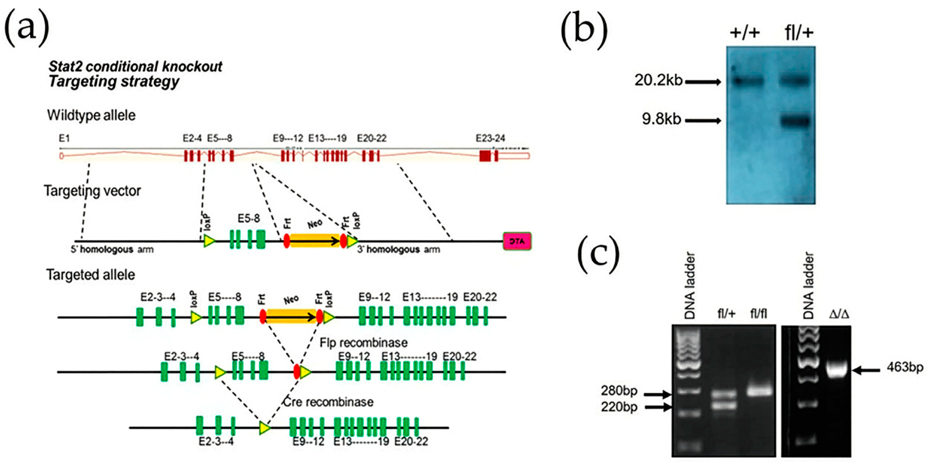
Stat2 conditional knockout mice (Stat2fl/fl) were generated with Biocytogen (Waltham, MA, USA) using a gene-targeting strategy in which exons 5–8 of the Stat2 locus were flanked by loxP sites. The targeting construct was introduced into C57BL/6 (B6) embryonic stem cells by electroporation with the distal loxP site linked to a neomycin-resistant cassette flanked by FRT sites. Correct genomic integration was verified by Southern blot analysis. The neomycin resistance cassette was subsequently excised by crossing with deleter mice carrying Flp recombinase. F1 heterozygous for the floxed Stat2 allele were intercrossed to obtain homozygous Stat2fl/fl mice. All animal experiments were approved by the Temple University Institutional Animal Care and Use Committee. Wild-type (WT) and Stat2 KO mice, previously backcrossed onto the B6 genetic background [12], were bred and maintained in our pathogen-free animal facility. The following mouse strains on the B6 background were purchased from The Jackson Laboratory: FLPe deleter (Strain#: 009086), CMV-Cre (Strain#: 006054), CD11c-Cre (Strain#: 008068) and Cdx2-Cre (Strain#: 009350). Global Stat2 deletion was achieved by crossing Stat2fl/fl mice with CMV-Cre mice. Targeted deletion of Stat2 in cDCs and colonic epithelial cells was accomplished by crossing Stat2fl/fl mice with CD11c-Cre+ or Cdx2-Cre mouse strains, respectively. Deletion of Stat2 was verified by genotyping using primers in Appendix A; Table A2.

2.2. qRT-PCR Analysis

Total RNA was isolated from individual mouse tissues stored in RNA-later stabilization solution (cat#AM7020; Invitrogen, Carlsbad, CA, USA) using Trizol® Reagent (Invitrogen, Carlsbad, CA, USA). Contaminating DNA in RNA samples was removed with a DNA-free removal kit (cat#AM1906; Invitrogen, Carlsbad, CA, USA). ISG expression in freshly isolated splenocytes treated with or without recombinant murine IFN-β (1000 U/mL) at 37 °C for 6 h was determined by qRT-PCR. RT-PCR was performed as a two-step process using High-Capacity cDNA Reverse Transcription (Applied Biosystems; Foster City, CA, USA) and SYBR Green (Bioland Scientific LLC, Los Angeles, CA, USA). Each cDNA sample was run in triplicate using the Step One Plus Real Time PCR system (Applied Biosystems). Primer sequences were obtained from Harvard PrimerBank [13] or from the published literature (Appendix A; Table A2). Results were analyzed using the comparative Δ CT method. Data were normalized to Gapdh. Relative Stat2 gene expression in Stat2Δ/Δ or Stat2 KO tissues was calculated relative to WT control cells. Relative ISG expression in IFN-β treated cells was calculated relative to its corresponding untreated cells.

2.3. Tumor Cell Lines, Antibodies and Cytokines

Murine B16-F1 melanoma cells were cultured in DMEM medium (Mediatech, Inc; Herndon, VA, USA) and supplemented with 5% heat-inactivated fetal bovine serum (FBS), 2 mM L-glutamine, 1 mM sodium pyruvate, penicillin (100 U/mL) and streptomycin (100 µg/mL) (Invitrogen Corp; Carlsbad, CA, USA) at 37 °C in 5% CO2. Murine EL-4 lymphoma cells were cultured in DMEM medium containing 10% heat-inactivated horse serum, L-glutamine and sodium pyruvate. Murine IFN-β was generously provided by Biogen, Idec. GM-CSF was purchased from BD Biosciences, San Jose, CA (cat# 554586). Antibodies against STAT1 (Cat#10144-2-AP), STAT2 (cat#51075-2-AP), β-Actin (cat#66009-1-Ig), HRP-conjugated anti-mouse IgG (cat#SA00001-1) and anti-rabbit-IgG (cat#SA00001-2) were purchased from Proteintech, Rosemont, IL, USA.

2.4. Tumor Transplantation

C57BL/6 mice (6–8 weeks old) received a single subcutaneous (s.c.) injection in the dorsal flank of either 1 × 10⁶ B16-F1 melanoma cells or 3 × 10⁵ EL4 lymphoma cells suspended in 200 µL of endotoxin-free 0.9% saline solution. Tumor measurements started at day 7 using a digital caliper. Tumor volume was determined with the formula V = a2b, where a is the shorter diameter and b is the longer diameter of the tumor. The study was terminated when the tumors reached a diameter of 20 mm. No unexpected deaths occurred during the study.

2.5. Western Blot Analysis

Cells and tissues were lysed as previously described [12]. Protein lysates were resolved on precast SurePAGE 4–12% gradient gels (GenScript, Piscataway, NJ, USA) and transferred to polyvinylidene difluoride membranes. Membranes were blocked with Casein Blocker in TBS (Bio-Rad, Hercules, CA, USA) and incubated with the appropriate primary antibodies followed by HRP-conjugated secondary antibodies in TBS-T + 3% BSA. Protein signals were detected using enhanced chemiluminescence reagent (Cat# 1705060; Bio-Rad) and visualized with a Bio-Rad ChemiDoc imaging system. β-actin served as an internal loading control.

2.6. Vesicular Stomatitis Virus (VSV) Infection

Lung fibroblasts of varying genotypes seeded in 12-well plates were left untreated or pretreated with 100 U/mL of murine IFN-β for 24 h. Cells were then infected with vesicular stomatitis virus (VSV) with a GFP-expressing gene [14]. VSV was added to cells at a multiplicity of infection of 0.01 for WT, Stat2 KO, Stat2fl/fl and Stat2Δ/Δ fibroblasts under serum-free medium conditions for 1 h at 37 °C. Cells were washed twice with PBS. Complete DMEM was then re-added. Cells were imaged using a Nikon inverted fluorescent microscope after 24 h.

2.7. Lung Fibroblasts Isolation

Lung tissue fragments were extracted from 3 to 5-week-old mice and transferred into a tissue culture dish according to an established protocol [15]. The fragments were cut into 1 mm pieces, washed with PBS and placed into a beaker containing Collagenase II (2 mg/mL) and DNase I (100 μg/mL). The cells were incubated for 1 h at 37 °C. The solution was pipetted to break down clumps and transferred to a 50 mL tube where FBS was added to stop digestion. Cells were spun down and resuspended in complete media (DMEM contained 10% FBS and 1% penicillin–streptomycin). Cells were transferred to a tissue dish and incubated at 37 °C overnight. The plates were monitored for changes in media color and washed to remove non-viable cells. The cells were incubated for 7–14 days before use.

2.8. Bone Marrow–Derived Conventional Dendritic Cells

Bone marrow–derived DCs were generated, as previously described [10], from different mouse genotypes. Briefly, bone marrow precursors were flushed from the femurs and tibias of mice and then seeded at 5 × 10⁵/well in complete IMDM (Mediatech, Manassas, VA, USA) (10% FBS, penicillin/streptomycin, gentamicin and 2-ME) (Life Technologies, Grand Island, NY, USA) and enriched with 3.3 ng/mL GM-CSF in 48-well plates or at 106/well in 24-well plates. Half medium was added on day 2, and half was replaced on day 5 and on each subsequent day until the culture was used for Western blot analysis.

2.9. Statistical Analysis

Prism software (Version 8, GraphPad, San Diego, CA, USA) was used for statistical analysis. In vitro results were analyzed using the Student’s t-test to assess significance. In comparing multiple parameters, two-tailed one-way ANOVA followed by Dunn’s multiple comparison test was applied. In vivo data were analyzed using the Mann–Whitney U test. Values of p ≤ 0.05 were considered statistically significant. Experiments were repeated 2 to 4 times. All data are presented as mean ± SEM.

3. Results

3.1. Targeting Strategy for Generating Conditional Stat2 KO Mice

We generated a conditional Stat2fl/fl mouse to investigate the specific contribution of cell-autonomous STAT2 function in IFN signaling and cancer. The mouse Stat2 gene contains 24 exons, spans approximately 22 kilobases and is located on chromosome 10 (forward strand). Exons 5–8 were selected because their removal eliminates a critical protein domain of STAT2, resulting in a frameshift mutation and a non-functional protein. Global Cre-mediated deletion of the floxed Stat2 locus (Stat2Δ/Δ) was achieved by crossing Stat2fl/fl mice with those expressing ubiquitous CMV-Cre-recombinase. This particular Cre strain was selected to ensure efficient global deletion of Stat2 in the Stat2fl/fl background. The resulting mice were fertile and viable, consistent with the previously reported phenotype of conventional Stat2 KO mice [8]. Targeted deletion removed exons 5–8, and Cre-mediated recombination introduced a frameshift predicted to generate a truncated protein of 153 amino acids (Figure 1a,b). Efficient Stat2 deletion was confirmed by genotyping, which produced a 436 bp band in contrast to the 220 bp and 280 bp bands observed in wild-type (WT) and Stat2fl/fl mice, respectively (Figure 1c).

3.2. Impaired IFN-I Signaling in Stat2-Deleted Mice

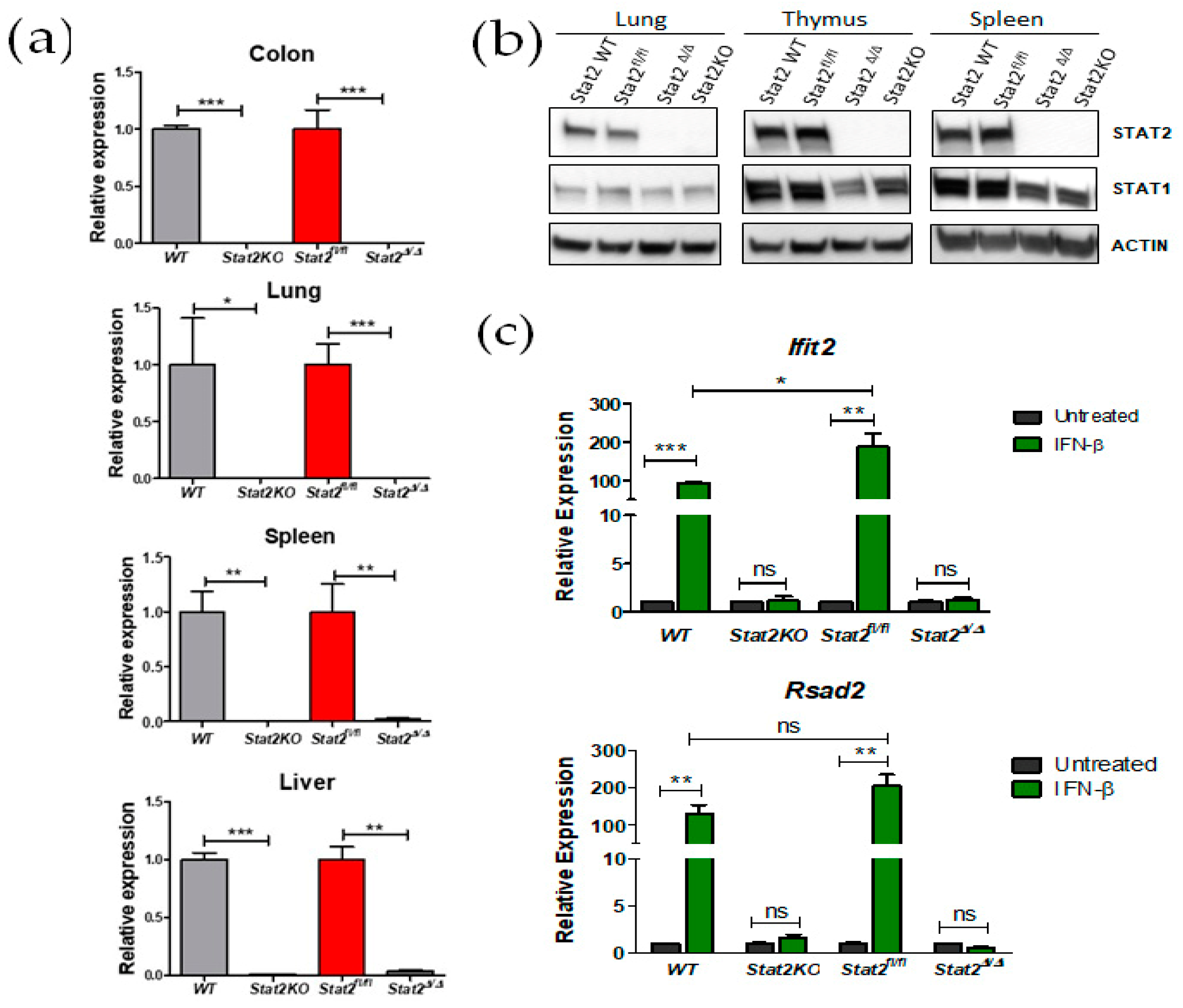
We confirmed that loss of Stat2 mRNA across multiple organs (colon, lung, spleen and liver) in Stat2Δ/Δ; CMV-Cre mice, and the extent of this loss was comparable to that observed in conventional Stat2 KO mice (Figure 2a). Similarly, Stat2 protein expression was also absent (Figure 2b; Figure S1). In addition, reduced levels of STAT1 protein were tissue-dependent in global Stat2Δ/Δ mice, faithfully recapitulating a known feature of Stat2 KO mice [8]. As expected, induction of IFN-I target genes (Rsad2 and Ifit2) was markedly impaired in both Stat2Δ/Δ and conventional Stat2 KO mice (Figure 2c). Altogether, these data confirm that the floxed Stat2 allele was efficiently deleted, resulting in defective IFN-I signaling.

3.3. Tumor Growth Is Accelerated in Stat2-Deleted Mice

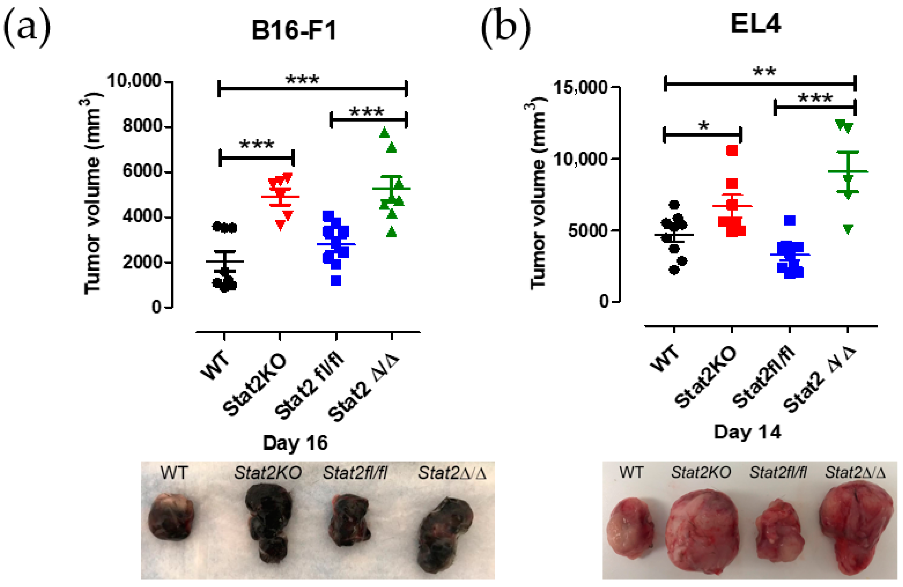
We previously reported that, in a syngeneic tumor transplantation model, Stat2 KO mice developed larger tumors than WT mice [12]. We selected the B16-F1 and EL4 cell lines because they are reliably tumorigenic in vivo and reproducibly form measurable tumors within 2–3 weeks. Consistent with these findings, Stat2Δ/Δ mice that received a subcutaneous injection of either B16-F1 or EL4 tumor cells formed progressively larger tumors compared with WT and Stat2fl/fl mice (Figure 3a,b). Together, these results confirm that ubiquitous Cre-mediated Stat2 deletion in Stat2fl/fl mice was effective and recapitulates the tumor-promoting phenotype observed in Stat2 KO mice, highlighting the importance of STAT2 in the hostile tumor microenvironment.

3.4. Stat2 Deletion Compromises IFN-I–Mediated Antiviral Protection in Lung Fibroblasts

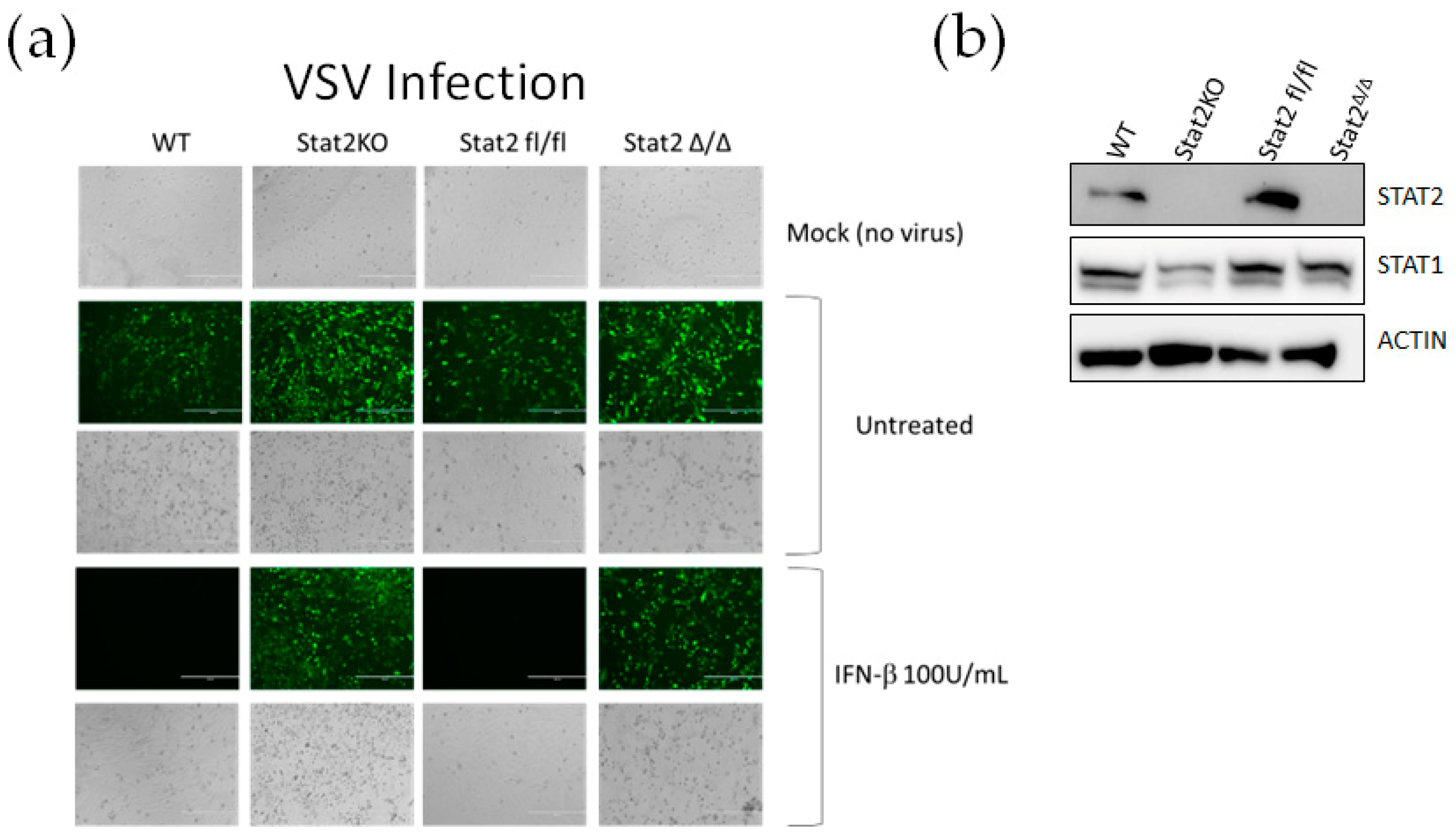
STAT2 plays a critical role in mediating the antiviral effects of IFN-I and IFN-III, as documented in individuals born with a STAT2 deficiency [16]. To evaluate the IFN-I induced antiviral response, we used vesicular stomatitis virus expressing GFP (VSV-GFP) as a reporter of infection. Lung fibroblasts isolated from WT, Stat2 KO, Stat2fl/fl and Stat2Δ/Δ mice were left untreated or pretreated overnight with 100 U/mL IFN-β and subsequently infected with VSV-GFP (Figure 4a; Figure S2). Loss of Stat2 protein in Stat2KO and Stat2Δ/Δ fibroblasts was confirmed by Western blot analysis prior to viral infection (Figure 4b). Compared with WT and Stat2fl/fl fibroblasts, both untreated Stat2 KO and Stat2Δ/Δ exhibited a markedly higher level of infection. As expected, pretreatment with IFN-β conferred antiviral protection only in WT and Stat2 fl/fl fibroblasts, whereas Stat2-deficient fibroblasts remained fully susceptible to VSV.

3.5. Conditional Stat2 Allele Enables Efficient Cre-Restricted Deletion

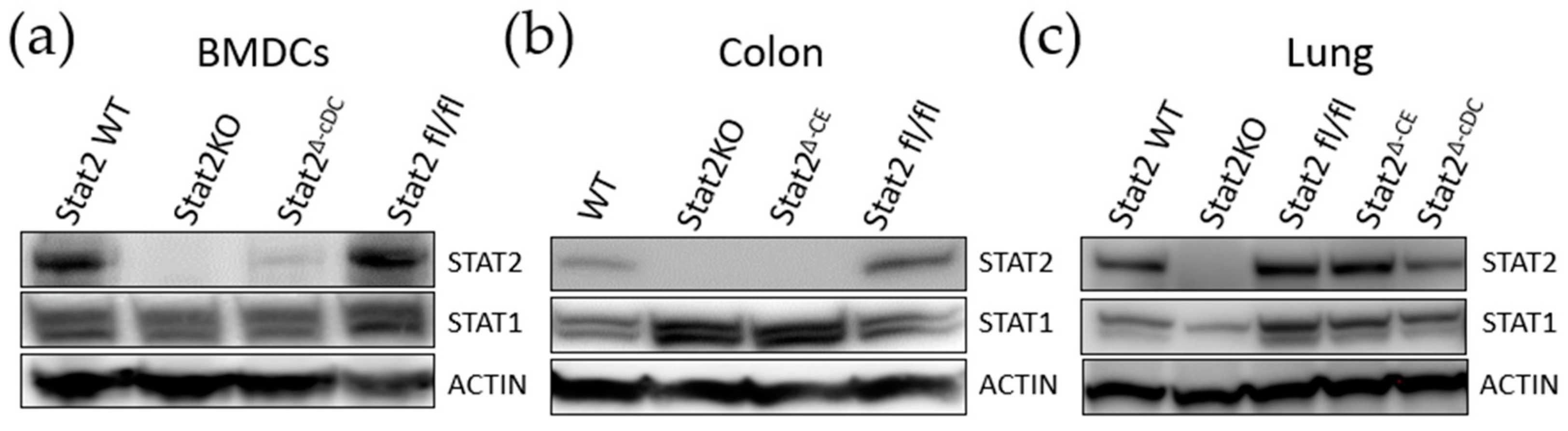
To further validate the specificity and efficiency of our conditional Stat2 KO mouse, we crossed Stat2fl/fl mice with CD11c-Cre mice to delete STAT2 in conventional dendritic cells (Stat2Δ-cDC) and with Cdx2-Cre mice to delete STAT2 in colonic epithelial cells (Stat2Δ-CE). We assessed STAT1 and STAT2 protein expression by Western blot across multiple tissues, including bone marrow-derived DCs (BM-DCs), the colon and the lung. Robust expression of both proteins was observed in WT and Stat2 fl/fl mice, whereas Stat2 KO tissues showed loss of STAT2 expression and marked reduction in STAT1 levels only in the lungs (Figure 5a–c and Figure S3). No marked differences in STAT1 levels were noted in BM-DCs. STAT2 expression was absent in cDCs of Stat2Δc-DC and in colons of Stat2Δ-CE mice, consistent with the specific activity of these Cre drivers. Unexpectedly, we found that colons from Stat2 KO and Stat2Δ-CE mice had increased STAT1 expression, whereas STAT2 levels in the lungs of Stat2Δ-cDC and Stat2Δ-CE mice remained unaffected. To our knowledge, this is the first time increased STAT1 levels in the absence of STAT2 have been observed in colons. Together, these findings demonstrate that targeted Stat2 deletion is efficient in the global knockout setting and specific to Cre-expressing lineages (Figure 5). More importantly, targeted STAT2 deletion can affect STAT1 levels in a tissue-dependent manner.

4. Discussion

Our study expands on the characterization of a conditional Stat2 KO mouse as a versatile platform to investigate STAT2 function in a tissue-specific and cell-intrinsic manner. Consistent with prior work, global deletion of Stat2 reproduced canonical phenotypes—including impaired IFN-I signaling, reduced STAT1 expression, enhanced tumor growth and defective antiviral responses—confirming that the floxed allele faithfully disrupts STAT2 function across tissues [8,10].
Building on earlier findings, we previously demonstrated that global and targeted loss of STAT2 in cDCs compromises the antitumor effects of IFN-I, and that global Stat2 deficiency further accelerates tumor growth [10]. Here, we show impaired IFN-I–mediated antiviral responses in Stat2-deleted fibroblasts; an established antiviral function of STAT2 [17]. These complementary data reinforce that the conditional allele produces biologically meaningful outcomes in distinct cellular contexts and support its utility for dissecting STAT2-dependent pathways.
A key strength of this model is its capacity for precise, Cre-restricted deletion, as demonstrated here in cDCs and colonic epithelial cells. This specificity preserves STAT2 expression in non-target tissues and overcomes a major limitation of conventional Stat2 KO models that cannot separate systemic from cell-intrinsic functions in vivo [5]. Importantly, conventional studies often rely on isolating tissues or cells from knockout animals for in vitro experiments. While informative, these approaches cannot fully capture tissue- and cell-specific interactions in vivo. The conditional Stat2 KO model addresses this limitation by allowing targeted deletion directly within the native physiological environment.
Beyond canonical IFN-I/III signaling, STAT2 also participates in non-canonical pathways regulating cellular metabolism, inflammation and tissue-specific immunity, some of which occur independently of STAT1 [18,19,20]. The conditional STAT2 KO model enables investigation of these roles in specific cell compartments, providing a framework to disentangle tissue- and cell-type-specific functions. For example, a wide range of tissue-specific Cre lines can be applied in gut inflammation or metabolic dysfunction models to clarify how STAT2 regulates context-dependent inflammatory responses within organs or specific cell populations. By facilitating such precise mechanistic studies, this platform offers a versatile tool to define both canonical and non-canonical STAT2 functions in tumor biology, cell metabolism, antiviral defense and immune regulation.
In summary, this conditional Stat2 KO model provides a powerful and flexible genetic platform to interrogate STAT2 function in vivo across tissues and disease settings, enabling mechanistic insight that is not achievable with conventional KO models.

5. Conclusions

Our conditional Stat2 KO mouse allows both global and cell–type–specific deletion of STAT2. Global deletion recapitulated known knockout phenotypes, including impaired IFN-I signaling, reduced STAT1 protein expression, enhanced tumor growth and impaired antiviral protection. Cell-type–specific deletion was precise and restricted to Cre-expressing lineages, demonstrating the fidelity of targeted ablation. These results establish the conditional Stat2 KO model as a powerful, versatile platform for dissecting STAT2’s tissue-specific roles in immunity, tumor biology, metabolism and antiviral defense.

Supplementary Materials

The following supporting information can be downloaded at https://www.mdpi.com/article/10.3390/immuno6010007/s1, Figure S1:Integration of Stat2 targeting vector and validation by genotyping. Figure S2: Levels of STAT1 and STAT2 after in vivo CMV-Cre recombination. Figure S3: Levels of STAT1 and STAT2 in isolated lung fibroblasts in different genotypes. Figure S4: Level of STAT1 and STAT2 after targeted deletion in cDCs and colonic epithelium.

Author Contributions

Conceptualization, A.M.G.; methodology, T.C., N.M., K.P.K. and A.A.; investigation, T.C., N.M. and A.A.; formal analyses, T.C., K.P.K. and N.M.; resources, L.Y. and A.M.G.; writing—original draft preparation, A.A. and A.M.G.; writing—review and editing, T.C., L.Y. and A.M.G.; supervision, A.M.G.; funding acquisition, A.M.G. All authors have read and agreed to the published version of the manuscript.

Funding

This research was funded in part by the National Cancer Institute (NCI; R03 CA273613-01A1 and R03 CA215929-01 and NIAMS, R21 AR078350-01A1).

Institutional Review Board Statement

The animal study protocol was approved by the Animal Care and Use Committee at Temple University (ACUP# 5029, with date of 6 December 2022) for studies involving animals.

Data Availability Statement

The data presented in this study are fully available.

Acknowledgments

The authors have reviewed and edited the output and take full responsibility for the content of this publication.

Conflicts of Interest

The authors declare that they have no known competing financial interests or personal relationships that could have appeared to influence the work reported in this paper.

Abbreviations

The following abbreviations are used in this manuscript:
STAT2Signal transducer and activator of transcription 2
CEColonic epithelium
cDCsConventional dendritic cells
FLFloxed
IFNInterferon
GFPGreen fluorescent protein
ISGF3Interferon-stimulated gene factor 3
HRPHorseradish peroxidase
KOKnockout
PCRPolymerase chain reaction
VSVVesicular stomatitis virus
WTWild type

Appendix A

Table A1. Primer sequences for genotyping.
Stat2Δ/Δ (463 bp)
Forward: 5′-TGTCTCAGACCAGGATCTCCTCCAC-3′
Reverse: 5′-ATGCGGCAAACCCCACTGTAAATAG-3′
Stat2WT (227 bp); Stat2fl/fl (285 bp)
Forward: 5′-TAATCCTAGCATTCTGGGCTGCA-3′
Reverse: 5′-GTTCCGAGTGTGTTTGAACTCTGA-3′
Table A2. qPCR primer sequences.
GeneForwardReverse
Rsad2TGCTGGCTGAGAATAGCATTAGGGCTGAGTGCTGTTCCCATCT
Ifit2AGTACAACGAGTAAGGAGTCACTAGGCCAGTATGTTGCACATGG
Stat2CTGAAGGACGAACAGGATGTCCAGGGTGGTTAATCGGCCAA
GapdhTGTAGACCATGTAGTTGAGGTCA AGGTCGGTGTGAACGGATTTG

References

  1. Lee, C.-J.; An, H.-J.; Cho, E.S.; Kang, H.C.; Lee, J.Y.; Lee, H.S.; Cho, Y.-Y. Stat2 stability regulation: An intersection between immunity and carcinogenesis. Exp. Mol. Med. 2020, 52, 1526–1536. [Google Scholar] [CrossRef] [Scilit] [PubMed]
  2. Canar, J.; Darling, K.; Dadey, R.; Gamero, A.M. The duality of STAT2 mediated type I interferon signaling in the tumor microenvironment and chemoresistance. Cytokine 2023, 161, 156081. [Google Scholar] [CrossRef] [Scilit] [PubMed]
  3. Xie, S.; Yan, J.; Jiang, B.; Liu, J.; Song, J. Immune evasion strategies of Seneca Valley virus: Mechanisms of host innate immune suppression. Agric. Commun. 2025, 3, 100100. [Google Scholar] [CrossRef] [Scilit]
  4. Lazear, H.M.; Schoggins, J.W.; Diamond, M.S. Shared and Distinct Functions of Type I and Type III Interferons. Immunity 2019, 50, 907–923. [Google Scholar] [CrossRef] [Scilit] [PubMed]
  5. Fortelny, N.; Farlik, M.; Fife, V.; Gorki, A.-D.; Lassnig, C.; Maurer, B.; Meissl, K.; Dolezal, M.; Boccuni, L.; Ravi Sundar Jose Geetha, A.; et al. JAK-STAT signaling maintains homeostasis in T cells and macrophages. Nat. Immunol. 2024, 25, 847–859. [Google Scholar] [CrossRef] [Scilit] [PubMed]
  6. Stolzer, I.; Dressel, A.; Chiriac, M.T.; Neurath, M.F.; Günther, C. An IFN-STAT Axis Augments Tissue Damage and Inflammation in a Mouse Model of Crohn’s Disease. Front. Med. 2021, 8, 644244. [Google Scholar] [CrossRef] [Scilit] [PubMed]
  7. Perry, S.T.; Buck, M.D.; Lada, S.M.; Schindler, C.; Shresta, S. STAT2 Mediates Innate Immunity to Dengue Virus in the Absence of STAT1 via the Type I Interferon Receptor. PLoS Pathog. 2011, 7, e1001297. [Google Scholar] [CrossRef] [Scilit] [PubMed]
  8. Park, C.; Li, S.; Cha, E.; Schindler, C. Immune response in Stat2 knockout mice. Immunity 2000, 13, 795–804. [Google Scholar] [CrossRef] [Scilit]
  9. Boudewijns, R.; Thibaut, H.J.; Kaptein, S.J.F.; Li, R.; Vergote, V.; Seldeslachts, L.; Van Weyenbergh, J.; De Keyzer, C.; Bervoets, L.; Sharma, S.; et al. STAT2 signaling restricts viral dissemination but drives severe pneumonia in SARS-CoV-2 infected hamsters. Nat. Commun. 2020, 11, 5838. [Google Scholar] [CrossRef] [Scilit]
  10. Qiu, C.C.; Kotredes, K.P.; Cremers, T.; Patel, S.; Afanassiev, A.; Slifker, M.; Gallucci, S.; Gamero, A.M. Targeted Stat2 deletion in conventional dendritic cells impairs CTL responses but does not affect antibody production. Oncoimmunology 2020, 10, 1860477. [Google Scholar] [CrossRef] [Scilit] [PubMed]
  11. Heath, H.; Britton, G.; Kudo, H.; Renney, G.; Ward, M.; Hutchins, R.; Foster, G.R.; Goldin, R.D.; Alazawi, W. Stat2 loss disrupts damage signalling and is protective in acute pancreatitis. J. Pathol. 2020, 252, 41–52. [Google Scholar] [CrossRef] [Scilit] [PubMed]
  12. Yue, C.; Xu, J.; Tan Estioko, M.D.; Kotredes, K.P.; Lopez-Otalora, Y.; Hilliard, B.A.; Baker, D.P.; Gallucci, S.; Gamero, A.M. Host STAT2/type I interferon axis controls tumor growth. Int. J. Cancer 2015, 136, 117–126. [Google Scholar] [CrossRef] [Scilit] [PubMed]
  13. Spandidos, A.; Wang, X.; Wang, H.; Seed, B. PrimerBank: A resource of human and mouse PCR primer pairs for gene expression detection and quantification. Nucleic Acids Res. 2010, 38, D792–D799. [Google Scholar] [CrossRef] [Scilit] [PubMed]
  14. Fernandez, M.; Porosnicu, M.; Markovic, D.; Barber, G.N. Genetically engineered vesicular stomatitis virus in gene therapy: Application for treatment of malignant disease. J. Virol. 2002, 76, 895–904. [Google Scholar] [CrossRef] [Scilit] [PubMed]
  15. Seluanov, A.; Vaidya, A.; Gorbunova, V. Establishing Primary Adult Fibroblast Cultures from Rodents. J. Vis. Exp. 2010, 44, e2033. [Google Scholar] [PubMed]
  16. Bucciol, G.; Meyts, I. Spotlight: “Human STAT2 deficiency: A severe defect of antiviral immunity”. Genes Immun. 2024, 25, 261–263. [Google Scholar] [CrossRef] [Scilit] [PubMed]
  17. Wang, Y.; Song, Q.; Huang, W.; Lin, Y.; Wang, X.; Wang, C.; Willard, B.; Zhao, C.; Nan, J.; Holvey-Bates, E.; et al. A virus-induced conformational switch of STAT1-STAT2 dimers boosts antiviral defenses. Cell Res. 2021, 31, 206–218. [Google Scholar] [CrossRef] [Scilit] [PubMed]
  18. Gopal, R.; Lee, B.; McHugh, K.J.; Rich, H.E.; Ramanan, K.; Mandalapu, S.; Clay, M.E.; Seger, P.J.; Enelow, R.I.; Manni, M.L.; et al. STAT2 Signaling Regulates Macrophage Phenotype During Influenza and Bacterial Super-Infection. Front. Immunol. 2018, 9, 2151. [Google Scholar] [CrossRef] [Scilit] [PubMed]
  19. Wang, Z.; Chen, W.; Zuo, L.; Xu, M.; Wu, Y.; Huang, J.; Zhang, X.; Li, Y.; Wang, J.; Chen, J.; et al. The Fibrillin-1/VEGFR2/STAT2 signaling axis promotes chemoresistance via modulating glycolysis and angiogenesis in ovarian cancer organoids and cells. Cancer Commun. 2022, 42, 245–265. [Google Scholar] [CrossRef] [Scilit] [PubMed]
  20. Xu, S.; Fang, Y.; Chang, L.; Bian, Y.; Wang, Y.; Ding, J.; Wang, Y.; Zhang, Y.; Pu, J.; Wang, K. STAT2-induced linc02231 promotes tumorigenesis and angiogenesis through modulation of hnRNPA1/ANGPTL4 in colorectal cancer. J. Gene Med. 2023, 25, e3506. [Google Scholar] [CrossRef] [Scilit] [PubMed]
Figure 1. Generation of conditional Stat2 KO mice. (a) Targeting strategy used to construct Stat2 fl/fl mice with exons 5 through 8 flanked with loxP sites. E represents exons with their corresponding number. Brown boxes depict exons in wild type allele and green boxes depict exons after successful integration of targeting vector. (b) Confirmation of loxP sites integration in the Stat2 gene by Southern blot analysis (9.8 kb DNA fragment). (c) Genotyping of mouse-tail DNA by PCR confirms deletion of Stat2 floxed allele after breeding with CMV-Cre mouse.
Immuno 06 00007 g001
Figure 2. Impaired IFN-I signaling after conditional deletion of Stat2 by CMV-Cre—mediated recombination. (a) Loss of Stat2 gene expression validated in four different organs by qRT-PCR analysis. (b) Western blot analyses performed on several tissues confirm global Stat2 deletion (Stat2Δ/Δ) by ubiquitous CMV-Cre recombinase. (c) Splenocytes of the indicated genotypes were left untreated or treated with IFN-β for 6 h, and ISG expression was determined by qRT-PCR. * p ≤ 0.05; ** p ≤ 0.01; *** p ≤ 0.001. Data are presented as SEM of three independent experiments. n.s; not significant.
Immuno 06 00007 g002
Figure 3. Stat2Δ/Δ mice show enhanced tumor growth. (a) B16-F1 or (b) EL4 tumor cells were injected subcutaneously in WT, Stat2KO, Stat2fl/fl and Stat2Δ/Δ. Values are shown as mean tumor volume determined over 20 days. Representative images of individual tumors are shown. *, p ≤ 0.05; **, p ≤ 0.01; and ***, p ≤ 0.001. n = 6–8 animals/group.
Immuno 06 00007 g003
Figure 4. Stat2Δ/Δ fibroblasts have impaired antiviral response to type I IFNs. (a) Lung fibroblasts derived from the indicated genotypes were pretreated with IFN-β for 24 h. Cells were then infected with VSV-GFP (green) at an MOI of 0.01 and imaged after 24 h. Representative bright field and fluorescent images are shown. (b) Western blot analysis shows STAT2 expression in fibroblasts of indicated genotypes. Representative images are shown of n = 2.
Immuno 06 00007 g004
Figure 5. Efficient targeted deletion of Stat2 in mouse tissues. Expression of STAT2 and STAT1 was analyzed in various tissues by Western blot analysis. (a) Bone marrow–derived cDCs generated from Stat2 fl/fl mice crossed with CD11c-Cre mice (Stat2∆-cDC). (b) Colons from Stat2 fl/fl mice crossed with Cdx2-Cre mice (Stat2∆-CE). (c) Lungs from Stat2∆-cDC and Stat2∆-CE mice were included for specificity of targeted deletion. Wild-type (WT) and Stat2KO mice served as positive and negative controls, respectively. ACTIN was used as an internal protein loading control.
Immuno 06 00007 g005
Disclaimer/Publisher’s Note: The statements, opinions and data contained in all publications are solely those of the individual author(s) and contributor(s) and not of MDPI and/or the editor(s). MDPI and/or the editor(s) disclaim responsibility for any injury to people or property resulting from any ideas, methods, instructions or products referred to in the content.